User login
Breast cancer: Cardiac risk increases with radiation dose to heart
The risk of major ischemic coronary events was significantly and proportionately associated with the estimated mean radiation dose to the heart in a study of women in Sweden and Denmark who received radiotherapy for breast cancer over a 43-year period.
"The risk of a major coronary event increased linearly with the mean dose to the heart," reported Sarah Darby, Ph.D., of the University of Oxford (England), and her associates. The risk began to increase within the first 5 years of treatment and continued to increase for at least 20 years.

The findings make it possible for a woman to estimate her absolute risk of radiation-related ischemic heart disease, the authors wrote. "This absolute risk can be weighed against the probable absolute reduction in her risk of recurrence or death from breast cancer that would be achieved without radiotherapy" (N. Engl. J. Med. 2013;368:987-98 [doi: 10.1056/NEJMoa1209825]).
The population-based study included 2,168 women who had been treated with external-beam radiation for invasive breast cancer between 1958 and 2001, and were enrolled in the Swedish National Cancer Register or the Danish Breast Cancer Cooperative Group. The 963 women who were subsequently diagnosed with a major coronary event (myocardial infarction, coronary revascularization, or death from ischemic heart disease, but not angina) were compared with 1,205 controls.
The major coronary events were diagnosed in the first decade after breast cancer diagnosis in 44% of patients; 33% of events were diagnosed 10-19 years after breast cancer diagnosis; and 23% occurred 20 or more years later. Of the cases, 54% died of ischemic heart disease.
The estimated mean radiation dose to the heart overall was 4.9 Gy (range, 0.03-27.72 Gy). For those with cancer in their left breast, the mean dose exposure to the heart was 6.6 Gy; for those with right-breast tumors, it was 2.9 Gy. Major coronary events were significantly higher among the women with radiation to the left breast.
The estimated dose to the heart of women who are currently treated with radiotherapy ranges from 1 to 5 Gy, the authors said.
For each 1-Gy increase in the mean dose of radiation to the heart, the rate of major coronary events increased by 7.4%, which was a highly statistically significant finding. Compared with controls who had no cardiac dose, the rate of major coronary events increased by 10% among those exposed to a mean radiation dose of less than 2 Gy, by 30% among those exposed to 2-4 Gy, by 40% among those exposed to 5-9 Gy, and by 116% in those exposed to 10 Gy or more.
Among women with a history of ischemic heart disease, the risk of major coronary events was almost sevenfold higher than it was in women with no history of ischemic heart disease. This risk was increased by about 13-fold during the first 10 years after treatment and was about twofold higher in later years.
"Absolute increases in risk for a given dose to the heart were larger for women with preexisting risk factors," they wrote, so "clinicians may wish to consider cardiac dose and cardiac risk factors as well as tumor control when making decisions about the use of radiotherapy for breast cancer."
Among the strengths of the study was that the analysis included all women who were documented as having received radiotherapy for breast cancer in the two countries during the time period studied. The authors cautioned against applying the results to breast cancer patients who are treated before age 30 because few women in this age group were included in the study.
The study was supported by the Oxford University Clinical Trial Service Unit from Cancer Research UK, the British Heart Foundation, and the UK Medical Research Council.
While the results of the study are interesting, they likely overestimate the risk of coronary events associated with contemporary radiation therapy. Of greater concern, the findings could be misinterpreted and could deter women from having potentially lifesaving treatment.
Increases in the rate of major coronary events – about 20% higher per 1 Gy – were far greater among the women diagnosed in the 1980s, who drive much of the increase in risk. There was barely an increase in the rate of events – 0.85% per 1 Gy – among those diagnosed in the 1990s, for example.
Most of the data are taken from a time when radiation was administered by techniques that differ from those used today, which are associated with a lot less scatter to the heart. Three-dimensional, CT-based planning was not used for the women in the study, which the authors acknowledged was a limitation. Also, the dose was estimated using radiotherapy charts, which are notoriously inaccurate.
Further, there is now a better understanding of which patients are likely to have a survival benefit from radiation. Correctly targeted radiation therapy plays an incredibly important role in the excellent results we see today, with the majority of breast cancer patients surviving their disease.
Dr. Hope Rugo is professor of medicine at the University of California, San Francisco, and director of breast oncology and clinical trials education at the UCSF Helen Diller Family Comprehensive Cancer Center. She had no relevant financial disclosures.
While the results of the study are interesting, they likely overestimate the risk of coronary events associated with contemporary radiation therapy. Of greater concern, the findings could be misinterpreted and could deter women from having potentially lifesaving treatment.
Increases in the rate of major coronary events – about 20% higher per 1 Gy – were far greater among the women diagnosed in the 1980s, who drive much of the increase in risk. There was barely an increase in the rate of events – 0.85% per 1 Gy – among those diagnosed in the 1990s, for example.
Most of the data are taken from a time when radiation was administered by techniques that differ from those used today, which are associated with a lot less scatter to the heart. Three-dimensional, CT-based planning was not used for the women in the study, which the authors acknowledged was a limitation. Also, the dose was estimated using radiotherapy charts, which are notoriously inaccurate.
Further, there is now a better understanding of which patients are likely to have a survival benefit from radiation. Correctly targeted radiation therapy plays an incredibly important role in the excellent results we see today, with the majority of breast cancer patients surviving their disease.
Dr. Hope Rugo is professor of medicine at the University of California, San Francisco, and director of breast oncology and clinical trials education at the UCSF Helen Diller Family Comprehensive Cancer Center. She had no relevant financial disclosures.
While the results of the study are interesting, they likely overestimate the risk of coronary events associated with contemporary radiation therapy. Of greater concern, the findings could be misinterpreted and could deter women from having potentially lifesaving treatment.
Increases in the rate of major coronary events – about 20% higher per 1 Gy – were far greater among the women diagnosed in the 1980s, who drive much of the increase in risk. There was barely an increase in the rate of events – 0.85% per 1 Gy – among those diagnosed in the 1990s, for example.
Most of the data are taken from a time when radiation was administered by techniques that differ from those used today, which are associated with a lot less scatter to the heart. Three-dimensional, CT-based planning was not used for the women in the study, which the authors acknowledged was a limitation. Also, the dose was estimated using radiotherapy charts, which are notoriously inaccurate.
Further, there is now a better understanding of which patients are likely to have a survival benefit from radiation. Correctly targeted radiation therapy plays an incredibly important role in the excellent results we see today, with the majority of breast cancer patients surviving their disease.
Dr. Hope Rugo is professor of medicine at the University of California, San Francisco, and director of breast oncology and clinical trials education at the UCSF Helen Diller Family Comprehensive Cancer Center. She had no relevant financial disclosures.
The risk of major ischemic coronary events was significantly and proportionately associated with the estimated mean radiation dose to the heart in a study of women in Sweden and Denmark who received radiotherapy for breast cancer over a 43-year period.
"The risk of a major coronary event increased linearly with the mean dose to the heart," reported Sarah Darby, Ph.D., of the University of Oxford (England), and her associates. The risk began to increase within the first 5 years of treatment and continued to increase for at least 20 years.
The findings make it possible for a woman to estimate her absolute risk of radiation-related ischemic heart disease, the authors wrote. "This absolute risk can be weighed against the probable absolute reduction in her risk of recurrence or death from breast cancer that would be achieved without radiotherapy" (N. Engl. J. Med. 2013;368:987-98 [doi: 10.1056/NEJMoa1209825]).
The population-based study included 2,168 women who had been treated with external-beam radiation for invasive breast cancer between 1958 and 2001, and were enrolled in the Swedish National Cancer Register or the Danish Breast Cancer Cooperative Group. The 963 women who were subsequently diagnosed with a major coronary event (myocardial infarction, coronary revascularization, or death from ischemic heart disease, but not angina) were compared with 1,205 controls.
The major coronary events were diagnosed in the first decade after breast cancer diagnosis in 44% of patients; 33% of events were diagnosed 10-19 years after breast cancer diagnosis; and 23% occurred 20 or more years later. Of the cases, 54% died of ischemic heart disease.
The estimated mean radiation dose to the heart overall was 4.9 Gy (range, 0.03-27.72 Gy). For those with cancer in their left breast, the mean dose exposure to the heart was 6.6 Gy; for those with right-breast tumors, it was 2.9 Gy. Major coronary events were significantly higher among the women with radiation to the left breast.
The estimated dose to the heart of women who are currently treated with radiotherapy ranges from 1 to 5 Gy, the authors said.
For each 1-Gy increase in the mean dose of radiation to the heart, the rate of major coronary events increased by 7.4%, which was a highly statistically significant finding. Compared with controls who had no cardiac dose, the rate of major coronary events increased by 10% among those exposed to a mean radiation dose of less than 2 Gy, by 30% among those exposed to 2-4 Gy, by 40% among those exposed to 5-9 Gy, and by 116% in those exposed to 10 Gy or more.
Among women with a history of ischemic heart disease, the risk of major coronary events was almost sevenfold higher than it was in women with no history of ischemic heart disease. This risk was increased by about 13-fold during the first 10 years after treatment and was about twofold higher in later years.
"Absolute increases in risk for a given dose to the heart were larger for women with preexisting risk factors," they wrote, so "clinicians may wish to consider cardiac dose and cardiac risk factors as well as tumor control when making decisions about the use of radiotherapy for breast cancer."
Among the strengths of the study was that the analysis included all women who were documented as having received radiotherapy for breast cancer in the two countries during the time period studied. The authors cautioned against applying the results to breast cancer patients who are treated before age 30 because few women in this age group were included in the study.
The study was supported by the Oxford University Clinical Trial Service Unit from Cancer Research UK, the British Heart Foundation, and the UK Medical Research Council.
The risk of major ischemic coronary events was significantly and proportionately associated with the estimated mean radiation dose to the heart in a study of women in Sweden and Denmark who received radiotherapy for breast cancer over a 43-year period.
"The risk of a major coronary event increased linearly with the mean dose to the heart," reported Sarah Darby, Ph.D., of the University of Oxford (England), and her associates. The risk began to increase within the first 5 years of treatment and continued to increase for at least 20 years.
The findings make it possible for a woman to estimate her absolute risk of radiation-related ischemic heart disease, the authors wrote. "This absolute risk can be weighed against the probable absolute reduction in her risk of recurrence or death from breast cancer that would be achieved without radiotherapy" (N. Engl. J. Med. 2013;368:987-98 [doi: 10.1056/NEJMoa1209825]).
The population-based study included 2,168 women who had been treated with external-beam radiation for invasive breast cancer between 1958 and 2001, and were enrolled in the Swedish National Cancer Register or the Danish Breast Cancer Cooperative Group. The 963 women who were subsequently diagnosed with a major coronary event (myocardial infarction, coronary revascularization, or death from ischemic heart disease, but not angina) were compared with 1,205 controls.
The major coronary events were diagnosed in the first decade after breast cancer diagnosis in 44% of patients; 33% of events were diagnosed 10-19 years after breast cancer diagnosis; and 23% occurred 20 or more years later. Of the cases, 54% died of ischemic heart disease.
The estimated mean radiation dose to the heart overall was 4.9 Gy (range, 0.03-27.72 Gy). For those with cancer in their left breast, the mean dose exposure to the heart was 6.6 Gy; for those with right-breast tumors, it was 2.9 Gy. Major coronary events were significantly higher among the women with radiation to the left breast.
The estimated dose to the heart of women who are currently treated with radiotherapy ranges from 1 to 5 Gy, the authors said.
For each 1-Gy increase in the mean dose of radiation to the heart, the rate of major coronary events increased by 7.4%, which was a highly statistically significant finding. Compared with controls who had no cardiac dose, the rate of major coronary events increased by 10% among those exposed to a mean radiation dose of less than 2 Gy, by 30% among those exposed to 2-4 Gy, by 40% among those exposed to 5-9 Gy, and by 116% in those exposed to 10 Gy or more.
Among women with a history of ischemic heart disease, the risk of major coronary events was almost sevenfold higher than it was in women with no history of ischemic heart disease. This risk was increased by about 13-fold during the first 10 years after treatment and was about twofold higher in later years.
"Absolute increases in risk for a given dose to the heart were larger for women with preexisting risk factors," they wrote, so "clinicians may wish to consider cardiac dose and cardiac risk factors as well as tumor control when making decisions about the use of radiotherapy for breast cancer."
Among the strengths of the study was that the analysis included all women who were documented as having received radiotherapy for breast cancer in the two countries during the time period studied. The authors cautioned against applying the results to breast cancer patients who are treated before age 30 because few women in this age group were included in the study.
The study was supported by the Oxford University Clinical Trial Service Unit from Cancer Research UK, the British Heart Foundation, and the UK Medical Research Council.
FROM THE NEW ENGLAND JOURNAL OF MEDICINE
Major finding: The risk of major coronary events increased by 7.4% per 1 Gy of radiation received as treatment for breast cancer.
Data source: A population-based, case-control study in 2,168 women treated with external-beam radiation therapy for invasive breast cancer in Sweden and Denmark between 1958 and 2001.
Disclosures: The study was supported by the Oxford University Clinical Trial Service Unit from Cancer Research UK, the British Heart Foundation, and the UK Medical Research Council.

Lipid metabolism genes linked to breast cancer subtype
Fine-needle aspirant samples taken from the healthy contralateral breast of patients undergoing surgery for breast cancer contained newly identified genetic markers that were expressed differently in estrogen receptor–negative and estrogen receptor–positive tumors.
The findings suggest that a metabolic derangement in lipid processing may precede the development of a breast tumor.
All of these genes are involved in lipid metabolism, an unexpected finding that speaks to the long-observed relationship between weight and breast cancer risk, Dr. Seema Khan and her colleagues wrote in the March issue of Cancer Prevention Research (2013 [doi:10.1158/1940-6207.CAPR-12-0304]).

"This was interesting because obesity is a breast cancer risk factor for postmenopausal women, but obese women are generally thought to be at increased risk for hormone-sensitive cancer," Khan said in a press statement. "We were surprised to see that some of these genes that are associated with lipid metabolism, or the metabolism of fats, are actually more highly expressed in the unaffected breasts of women with estrogen receptor–negative breast cancer."
The investigators, all from Northwestern University in Chicago, conducted their initial analysis on a set of 30 breast cancer patients – 15 with ER-negative tumors and 15 with ER-positive tumors. They then validated their results on 36-subjects, 12 with ER-negative cancers, 12 with ER-positive cancers, and 12 controls. All of the women in the study were matched for age, menopausal status, weight, and, in the patients, HER2 status. All subjects were followed for a minimum of 3 years.
Based on RNA extracted from fine-needle aspirations of the subjects’ contralateral breasts, eight unique genes were identified. There was significant differential expression of the genes between the groups. All of the genes were directly involved in lipid metabolism, and seven of them were significantly more common in ER-negative tumors.
Similar results were observed in the validation group, with all eight genes observed to be significantly more common in the ER-negative group than in the ER-positive group.
When the ER-negative cases were compared with the controls, four genes were significantly overexpressed and were observed to be up to six times more common in cases. The genetic markers were similarly expressed in ER-positive cases and in controls, however.
Two of the remaining four genes were significantly underexpressed in ER-positive cases, compared with controls (three and six times less likely to occur, respectively), indicating that both genes may protect against the development of ER-negative tumors. Of the remaining two genes, one was significantly under-expressed in both ER-negative and ER-positive groups, compared with controls (15 and 11 times less common, respectively). This, the authors said, indicates that the gene may protect against both types of cancer.
There were no significant associations between the final gene and either cancer subtype.
A clustering analysis of the eight genes separated the cases into low- mid- and high-expression groups, and also successfully separated the controls from the cases. In this analysis, 70% of the cases in the low-expression group were ER-positive; 67% of the cases in the mid-expression group were ER-positive; and 88% of the cases in the high-expression group were ER-negative.
The analysis also identified a high- and a low-expression group among the 12 controls. Four of these control cases had high-expression profiles, similar to those of the high-expression cases. The cytology of these four samples was atypical in two, borderline in one, and benign in one. The other eight samples had low gene expression and all had benign cytology.
"The potential involvement of lipid metabolism–related genes to ER-negative breast cancer is unexpected, though evidence pointing to a link of lipid/steroid metabolism with ER-negative breast cancer risk and outcomes exists," the authors said. "ER-negative/PR (progesterone receptor)-negative tumors are more common in obese premenopausal women, and large hip circumference has a particularly strong association with premenopausal ER-negative/PR-negative breast cancer. The functions of these genes in relation to lipid modification and elimination, and to transportation and detoxification of distinct lipid compounds, suggest that their expression results in a specific microenvironment of steroid hormone metabolites, which may determine whether initiated cells progress to ER-positive or ER-negative tumors."
None of the authors declared any financial relationships. The study was funded by the Lynn Sage Cancer Research Foundation, the Avon Foundation, and a private contribution.
Fine-needle aspirant samples taken from the healthy contralateral breast of patients undergoing surgery for breast cancer contained newly identified genetic markers that were expressed differently in estrogen receptor–negative and estrogen receptor–positive tumors.
The findings suggest that a metabolic derangement in lipid processing may precede the development of a breast tumor.
All of these genes are involved in lipid metabolism, an unexpected finding that speaks to the long-observed relationship between weight and breast cancer risk, Dr. Seema Khan and her colleagues wrote in the March issue of Cancer Prevention Research (2013 [doi:10.1158/1940-6207.CAPR-12-0304]).
"This was interesting because obesity is a breast cancer risk factor for postmenopausal women, but obese women are generally thought to be at increased risk for hormone-sensitive cancer," Khan said in a press statement. "We were surprised to see that some of these genes that are associated with lipid metabolism, or the metabolism of fats, are actually more highly expressed in the unaffected breasts of women with estrogen receptor–negative breast cancer."
The investigators, all from Northwestern University in Chicago, conducted their initial analysis on a set of 30 breast cancer patients – 15 with ER-negative tumors and 15 with ER-positive tumors. They then validated their results on 36-subjects, 12 with ER-negative cancers, 12 with ER-positive cancers, and 12 controls. All of the women in the study were matched for age, menopausal status, weight, and, in the patients, HER2 status. All subjects were followed for a minimum of 3 years.
Based on RNA extracted from fine-needle aspirations of the subjects’ contralateral breasts, eight unique genes were identified. There was significant differential expression of the genes between the groups. All of the genes were directly involved in lipid metabolism, and seven of them were significantly more common in ER-negative tumors.
Similar results were observed in the validation group, with all eight genes observed to be significantly more common in the ER-negative group than in the ER-positive group.
When the ER-negative cases were compared with the controls, four genes were significantly overexpressed and were observed to be up to six times more common in cases. The genetic markers were similarly expressed in ER-positive cases and in controls, however.
Two of the remaining four genes were significantly underexpressed in ER-positive cases, compared with controls (three and six times less likely to occur, respectively), indicating that both genes may protect against the development of ER-negative tumors. Of the remaining two genes, one was significantly under-expressed in both ER-negative and ER-positive groups, compared with controls (15 and 11 times less common, respectively). This, the authors said, indicates that the gene may protect against both types of cancer.
There were no significant associations between the final gene and either cancer subtype.
A clustering analysis of the eight genes separated the cases into low- mid- and high-expression groups, and also successfully separated the controls from the cases. In this analysis, 70% of the cases in the low-expression group were ER-positive; 67% of the cases in the mid-expression group were ER-positive; and 88% of the cases in the high-expression group were ER-negative.
The analysis also identified a high- and a low-expression group among the 12 controls. Four of these control cases had high-expression profiles, similar to those of the high-expression cases. The cytology of these four samples was atypical in two, borderline in one, and benign in one. The other eight samples had low gene expression and all had benign cytology.
"The potential involvement of lipid metabolism–related genes to ER-negative breast cancer is unexpected, though evidence pointing to a link of lipid/steroid metabolism with ER-negative breast cancer risk and outcomes exists," the authors said. "ER-negative/PR (progesterone receptor)-negative tumors are more common in obese premenopausal women, and large hip circumference has a particularly strong association with premenopausal ER-negative/PR-negative breast cancer. The functions of these genes in relation to lipid modification and elimination, and to transportation and detoxification of distinct lipid compounds, suggest that their expression results in a specific microenvironment of steroid hormone metabolites, which may determine whether initiated cells progress to ER-positive or ER-negative tumors."
None of the authors declared any financial relationships. The study was funded by the Lynn Sage Cancer Research Foundation, the Avon Foundation, and a private contribution.
Fine-needle aspirant samples taken from the healthy contralateral breast of patients undergoing surgery for breast cancer contained newly identified genetic markers that were expressed differently in estrogen receptor–negative and estrogen receptor–positive tumors.
The findings suggest that a metabolic derangement in lipid processing may precede the development of a breast tumor.
All of these genes are involved in lipid metabolism, an unexpected finding that speaks to the long-observed relationship between weight and breast cancer risk, Dr. Seema Khan and her colleagues wrote in the March issue of Cancer Prevention Research (2013 [doi:10.1158/1940-6207.CAPR-12-0304]).
"This was interesting because obesity is a breast cancer risk factor for postmenopausal women, but obese women are generally thought to be at increased risk for hormone-sensitive cancer," Khan said in a press statement. "We were surprised to see that some of these genes that are associated with lipid metabolism, or the metabolism of fats, are actually more highly expressed in the unaffected breasts of women with estrogen receptor–negative breast cancer."
The investigators, all from Northwestern University in Chicago, conducted their initial analysis on a set of 30 breast cancer patients – 15 with ER-negative tumors and 15 with ER-positive tumors. They then validated their results on 36-subjects, 12 with ER-negative cancers, 12 with ER-positive cancers, and 12 controls. All of the women in the study were matched for age, menopausal status, weight, and, in the patients, HER2 status. All subjects were followed for a minimum of 3 years.
Based on RNA extracted from fine-needle aspirations of the subjects’ contralateral breasts, eight unique genes were identified. There was significant differential expression of the genes between the groups. All of the genes were directly involved in lipid metabolism, and seven of them were significantly more common in ER-negative tumors.
Similar results were observed in the validation group, with all eight genes observed to be significantly more common in the ER-negative group than in the ER-positive group.
When the ER-negative cases were compared with the controls, four genes were significantly overexpressed and were observed to be up to six times more common in cases. The genetic markers were similarly expressed in ER-positive cases and in controls, however.
Two of the remaining four genes were significantly underexpressed in ER-positive cases, compared with controls (three and six times less likely to occur, respectively), indicating that both genes may protect against the development of ER-negative tumors. Of the remaining two genes, one was significantly under-expressed in both ER-negative and ER-positive groups, compared with controls (15 and 11 times less common, respectively). This, the authors said, indicates that the gene may protect against both types of cancer.
There were no significant associations between the final gene and either cancer subtype.
A clustering analysis of the eight genes separated the cases into low- mid- and high-expression groups, and also successfully separated the controls from the cases. In this analysis, 70% of the cases in the low-expression group were ER-positive; 67% of the cases in the mid-expression group were ER-positive; and 88% of the cases in the high-expression group were ER-negative.
The analysis also identified a high- and a low-expression group among the 12 controls. Four of these control cases had high-expression profiles, similar to those of the high-expression cases. The cytology of these four samples was atypical in two, borderline in one, and benign in one. The other eight samples had low gene expression and all had benign cytology.
"The potential involvement of lipid metabolism–related genes to ER-negative breast cancer is unexpected, though evidence pointing to a link of lipid/steroid metabolism with ER-negative breast cancer risk and outcomes exists," the authors said. "ER-negative/PR (progesterone receptor)-negative tumors are more common in obese premenopausal women, and large hip circumference has a particularly strong association with premenopausal ER-negative/PR-negative breast cancer. The functions of these genes in relation to lipid modification and elimination, and to transportation and detoxification of distinct lipid compounds, suggest that their expression results in a specific microenvironment of steroid hormone metabolites, which may determine whether initiated cells progress to ER-positive or ER-negative tumors."
None of the authors declared any financial relationships. The study was funded by the Lynn Sage Cancer Research Foundation, the Avon Foundation, and a private contribution.
FROM CANCER PREVENTION RESEARCH
Major finding: A clustering analysis of eight genes separated cases into expression groups; 70% of the low-expression cases had ER-positive tumors, 67% of the mid-expression cases had ER-positive tumors; and 88% of the high-expression group had ER-negative tumors.
Data source: The findings are from investigation and validation groups that comprised a total of 24 cases and 12 controls.
Disclosures: None of the authors declared any financial relationships. The study was funded by the Lynn Sage Cancer Research Foundation, the Avon Foundation, and a private contribution.

Imaging agent approved for locating lymph nodes
A radioactive diagnostic imaging agent called Lymphoseek (technetium Tc 99m tilmanocept) Injection has been approved for locating lymph nodes in patients who have breast cancer or melanoma and are undergoing surgery to remove tumor-draining lymph nodes, the Food and Drug Administration announced.
Lymphoseek is the first new drug for lymph-node mapping to be approved in more than 30 years. Other FDA-approved drugs used for lymph-node mapping include sulfur colloid and isosulfan blue.

"To use Lymphoseek, doctors inject the drug into the tumor area and later, using a handheld radiation detector, find lymph nodes that have taken up Lymphoseek’s radioactivity," said Dr. Shaw Chen, deputy director of the Office of Drug Evaluation IV in the FDA’s Center for Drug Evaluation and Research.
Lymphoseek is marketed by Navidea Biopharmaceuticals. The manufacturer’s website notes that the ability to rapidly locate and biopsy sentinel nodes enables surgical management to be tailored specifically to each patient’s burden of disease.
In two clinical trials, 332 patients with melanoma or breast cancer were injected with Lymphoseek and blue dye. Surgeons subsequently removed suspected lymph nodes for pathologic examination. Confirmed lymph nodes were examined for their content of blue dye and Lymphoseek. The combination of Lymphoseek and blue dye localized most lymph nodes, although a notable number of nodes were localized only by Lymphoseek.
The most common side effects identified in clinical trials were pain and irritation at the injection site.
According to the manufacturer, a clinical trial involving patients with head and neck cancer is completing enrollment and is expected to be the subject of a future New Drug Application amendment. An initial Marketing Authorization Application filing in the European Union is anticipated by the end of 2012.
A radioactive diagnostic imaging agent called Lymphoseek (technetium Tc 99m tilmanocept) Injection has been approved for locating lymph nodes in patients who have breast cancer or melanoma and are undergoing surgery to remove tumor-draining lymph nodes, the Food and Drug Administration announced.
Lymphoseek is the first new drug for lymph-node mapping to be approved in more than 30 years. Other FDA-approved drugs used for lymph-node mapping include sulfur colloid and isosulfan blue.
"To use Lymphoseek, doctors inject the drug into the tumor area and later, using a handheld radiation detector, find lymph nodes that have taken up Lymphoseek’s radioactivity," said Dr. Shaw Chen, deputy director of the Office of Drug Evaluation IV in the FDA’s Center for Drug Evaluation and Research.
Lymphoseek is marketed by Navidea Biopharmaceuticals. The manufacturer’s website notes that the ability to rapidly locate and biopsy sentinel nodes enables surgical management to be tailored specifically to each patient’s burden of disease.
In two clinical trials, 332 patients with melanoma or breast cancer were injected with Lymphoseek and blue dye. Surgeons subsequently removed suspected lymph nodes for pathologic examination. Confirmed lymph nodes were examined for their content of blue dye and Lymphoseek. The combination of Lymphoseek and blue dye localized most lymph nodes, although a notable number of nodes were localized only by Lymphoseek.
The most common side effects identified in clinical trials were pain and irritation at the injection site.
According to the manufacturer, a clinical trial involving patients with head and neck cancer is completing enrollment and is expected to be the subject of a future New Drug Application amendment. An initial Marketing Authorization Application filing in the European Union is anticipated by the end of 2012.
A radioactive diagnostic imaging agent called Lymphoseek (technetium Tc 99m tilmanocept) Injection has been approved for locating lymph nodes in patients who have breast cancer or melanoma and are undergoing surgery to remove tumor-draining lymph nodes, the Food and Drug Administration announced.
Lymphoseek is the first new drug for lymph-node mapping to be approved in more than 30 years. Other FDA-approved drugs used for lymph-node mapping include sulfur colloid and isosulfan blue.
"To use Lymphoseek, doctors inject the drug into the tumor area and later, using a handheld radiation detector, find lymph nodes that have taken up Lymphoseek’s radioactivity," said Dr. Shaw Chen, deputy director of the Office of Drug Evaluation IV in the FDA’s Center for Drug Evaluation and Research.
Lymphoseek is marketed by Navidea Biopharmaceuticals. The manufacturer’s website notes that the ability to rapidly locate and biopsy sentinel nodes enables surgical management to be tailored specifically to each patient’s burden of disease.
In two clinical trials, 332 patients with melanoma or breast cancer were injected with Lymphoseek and blue dye. Surgeons subsequently removed suspected lymph nodes for pathologic examination. Confirmed lymph nodes were examined for their content of blue dye and Lymphoseek. The combination of Lymphoseek and blue dye localized most lymph nodes, although a notable number of nodes were localized only by Lymphoseek.
The most common side effects identified in clinical trials were pain and irritation at the injection site.
According to the manufacturer, a clinical trial involving patients with head and neck cancer is completing enrollment and is expected to be the subject of a future New Drug Application amendment. An initial Marketing Authorization Application filing in the European Union is anticipated by the end of 2012.

Direct access to unexpected genetic test results
Direct-to-consumer genetic testing, including personal genome analysis, has been available for several years and raises challenging questions of risk and benefit.
Among the potential harms are that clients may not fully understand their results, that abnormal results may lead to undue anxiety and/or inappropriate responses, and that normal results may lead to false reassurance ("Direct-to-consumer genetic analysis," Internal Medicine News, Oct. 1, 2008).

This is particularly concerning in the context of single-gene disorders such as hereditary breast and ovarian cancer syndrome, in which a mutation in either the BRCA1 or BRCA2 gene confers up to 50%-60% lifetime risk of female breast cancer and up to 40% lifetime risk of ovarian cancer – as well as increasing the risk of female primary papillary serous peritoneal carcinoma, male breast cancer, prostate cancer, and pancreatic cancer ("BRCA1 and BRCA2 Hereditary Breast and Ovarian Cancer," GeneReviews 1998 [Updated 2011 Jan. 20]).
The three mutations in these two genes that are most common among Ashkenazi Jews are now included in commercial direct-to-consumer (DTC) personal genome analysis, making it possible for consumers to screen themselves for these mutations.
In February, a DTC genetic testing company published a report suggesting that the potential harms of such testing may be overstated (Peer J. 2013;1:e8). They invited all 136 adult clients who had one of the three common Ashkenazi BRCA gene mutations and had elected to view their BRCA results to participate in an interview about their experience. A total of 32 agreed: 16 men and 16 women.
For 25 of them, this was a newly discovered mutation, while 7 previously knew that they carried a mutation. Thirty-one mutation-negative clients were interviewed as controls. The groups were identical for most demographics, but differed as expected with respect to cancer history.
Four of the 16 women with mutations had a personal history of breast and/or ovarian cancer, but only 1 of 18 mutation-negative women had breast cancer. There was a positive family history of breast/ovarian cancer for 72% of those with mutations and 48% of those without mutations.
Some of those with mutations were surprised at the news, because they had no significant family history of breast/ovarian cancer. But most were not surprised, because they either knew their mutation status or suspected it, based on family history or Ashkenazi ancestry.
However, no one reported feeling extremely upset, and only four people (12.5%) reported moderate upset feelings. Nine (28%) had self-limited disappointment or anxiety, and 17 (53%) had neutral feelings about their result. Even among the 25 for whom this was a new finding, 11 (44%) felt neutral. Only 1 of the 32 participants expressed regret about learning his result, citing the emotional cost of knowing he has a mutation and that he might pass it on to his children.
Overall, these anxiety results are mostly reassuring and are in line with other published studies of patients’ emotional response to receiving adverse test results (both genetic and nongenetic).
Half of the mutation-negative clients felt neutral about their result, and the other half expressed relief. Almost all of those who felt relieved had a positive family history of breast/ovarian cancer.
Fears of false assurance
The question of false reassurance for these individuals remains a concern. The DTC company required all clients to read a brief written passage about the meaning and limitations of these results prior to viewing them, including a statement that this test does not identify all genetic causes of breast and ovarian cancer.
The interview responses are summarized to indicate that the majority of mutation-negative participants understood that their risk of developing breast cancer was unchanged as a result of their negative testing.
However, because the testing did not involve full sequencing of all known genes associated with breast and ovarian cancer, it is quite possible that some of those who were negative in this study might in fact truly have a genetic cancer predisposition syndrome and be at substantially increased (but unrecognized) risk of breast, ovarian, and possibly other cancers.
Actions taken by clients after learning of a BRCA mutation are generally reassuring: 60% discussed their results with a physician (28% with their primary care physician). Most of the women obtained appropriate medical counseling and took steps to reduce their risks (with prophylactic surgery and/or increased cancer screening). Many people shared the information with family members, resulting in identification of additional relatives with the same mutations, who then sought appropriate screening and prevention.
Several of the mutation carriers, both those in the original study and their relatives, do not meet current criteria for BRCA gene testing or intensified screening and prophylaxis. Assuming such identification reduces future morbidity and mortality, these results argue in favor of more widespread screening for such mutations. But many more data need to be considered before implementing such a program.
Interestingly, a few clients who initially felt neutral about their abnormal test result reported anxiety only after learning more about the implications and management options, including mastectomy and oophorectomy.
This difference in emotional response could be related to the method of receiving information: reading it online in a comfortable environment when the client chooses to, vs. receiving it in a medical setting when the clinician is able to. Or it might reflect the physical and temporal separation of receiving an abnormal test result from the counseling and discussion of its management. More research is needed to better understand these possibilities.
Only one of the mutation-positive men sought medical advice, but most reported intent to pursue regular breast and prostate screening. However, it is unclear how they would act on that intent without involving a physician – leaving open the possibility that they may not fully understand the clinical significance of their positive result.
Also of concern, only seven (23%) of the mutation-negative clients discussed their result with a health care provider. Among those seven, most discussed it with their primary care physician, who generally did not know what to do with the information.
This represents another missed opportunity to identify patients with a positive family history who might benefit from additional genetic counseling and/or testing.
There are multiple limitations to this study. It is based on a very small and highly selected sample that is biased toward the desire to participate in research, to pursue genetic testing, to specifically know one’s BRCA mutation status, and to share one’s experience learning that status.
There is also the risk of observer bias (the authors are all employees of the company that performed the testing) and participant bias (the subjects are all clients of the company and knew that they were being interviewed by a representative of the company).
Autonomy vs. beneficence
Overall, the results suggest that, at least for a segment of the population, direct access to genetic data of potentially great medical significance may not be associated with as high a risk of emotional distress and inappropriate action as previously thought. But some individuals likely are at risk for unnecessary anxiety and/or misinterpretation of the results.
Central to the discussion is tension over how best to balance autonomy, in the form of a patient’s right to access personal medical information however she or he desires, against beneficence, in the form of minimizing harm.
There is some basic information that, ideally, should be available to all consumers (patients) for every test, whether genetic or not. This includes the meaning of a positive, negative, or inconclusive result; the limitations of conclusions that can be drawn from the result; the possibility of strong emotional reactions to both negative and positive results; and the possible courses of action to consider in response to the result.
Physician-ordered testing is probably the best way to accomplish those goals, but direct-to-consumer testing is here to stay and may be appropriate for some individuals. Hopefully, the companies offering such testing are providing accurate and understandable information to help their clients make the best and safest use of their results.
As primary care physicians, we should encourage best practices among ourselves and DTC companies, and remain available to provide advice, interpretation, and specialty referral to patients who may have started down the path of DTC testing and later decide to seek our professional assistance.
Dr. Levy is with the division of general internal medicine and the McKusick-Nathans Institute of Genetic Medicine at Johns Hopkins University, Baltimore. He reports having no conflicts of interest.
Direct-to-consumer genetic testing, including personal genome analysis, has been available for several years and raises challenging questions of risk and benefit.
Among the potential harms are that clients may not fully understand their results, that abnormal results may lead to undue anxiety and/or inappropriate responses, and that normal results may lead to false reassurance ("Direct-to-consumer genetic analysis," Internal Medicine News, Oct. 1, 2008).
This is particularly concerning in the context of single-gene disorders such as hereditary breast and ovarian cancer syndrome, in which a mutation in either the BRCA1 or BRCA2 gene confers up to 50%-60% lifetime risk of female breast cancer and up to 40% lifetime risk of ovarian cancer – as well as increasing the risk of female primary papillary serous peritoneal carcinoma, male breast cancer, prostate cancer, and pancreatic cancer ("BRCA1 and BRCA2 Hereditary Breast and Ovarian Cancer," GeneReviews 1998 [Updated 2011 Jan. 20]).
The three mutations in these two genes that are most common among Ashkenazi Jews are now included in commercial direct-to-consumer (DTC) personal genome analysis, making it possible for consumers to screen themselves for these mutations.
In February, a DTC genetic testing company published a report suggesting that the potential harms of such testing may be overstated (Peer J. 2013;1:e8). They invited all 136 adult clients who had one of the three common Ashkenazi BRCA gene mutations and had elected to view their BRCA results to participate in an interview about their experience. A total of 32 agreed: 16 men and 16 women.
For 25 of them, this was a newly discovered mutation, while 7 previously knew that they carried a mutation. Thirty-one mutation-negative clients were interviewed as controls. The groups were identical for most demographics, but differed as expected with respect to cancer history.
Four of the 16 women with mutations had a personal history of breast and/or ovarian cancer, but only 1 of 18 mutation-negative women had breast cancer. There was a positive family history of breast/ovarian cancer for 72% of those with mutations and 48% of those without mutations.
Some of those with mutations were surprised at the news, because they had no significant family history of breast/ovarian cancer. But most were not surprised, because they either knew their mutation status or suspected it, based on family history or Ashkenazi ancestry.
However, no one reported feeling extremely upset, and only four people (12.5%) reported moderate upset feelings. Nine (28%) had self-limited disappointment or anxiety, and 17 (53%) had neutral feelings about their result. Even among the 25 for whom this was a new finding, 11 (44%) felt neutral. Only 1 of the 32 participants expressed regret about learning his result, citing the emotional cost of knowing he has a mutation and that he might pass it on to his children.
Overall, these anxiety results are mostly reassuring and are in line with other published studies of patients’ emotional response to receiving adverse test results (both genetic and nongenetic).
Half of the mutation-negative clients felt neutral about their result, and the other half expressed relief. Almost all of those who felt relieved had a positive family history of breast/ovarian cancer.
Fears of false assurance
The question of false reassurance for these individuals remains a concern. The DTC company required all clients to read a brief written passage about the meaning and limitations of these results prior to viewing them, including a statement that this test does not identify all genetic causes of breast and ovarian cancer.
The interview responses are summarized to indicate that the majority of mutation-negative participants understood that their risk of developing breast cancer was unchanged as a result of their negative testing.
However, because the testing did not involve full sequencing of all known genes associated with breast and ovarian cancer, it is quite possible that some of those who were negative in this study might in fact truly have a genetic cancer predisposition syndrome and be at substantially increased (but unrecognized) risk of breast, ovarian, and possibly other cancers.
Actions taken by clients after learning of a BRCA mutation are generally reassuring: 60% discussed their results with a physician (28% with their primary care physician). Most of the women obtained appropriate medical counseling and took steps to reduce their risks (with prophylactic surgery and/or increased cancer screening). Many people shared the information with family members, resulting in identification of additional relatives with the same mutations, who then sought appropriate screening and prevention.
Several of the mutation carriers, both those in the original study and their relatives, do not meet current criteria for BRCA gene testing or intensified screening and prophylaxis. Assuming such identification reduces future morbidity and mortality, these results argue in favor of more widespread screening for such mutations. But many more data need to be considered before implementing such a program.
Interestingly, a few clients who initially felt neutral about their abnormal test result reported anxiety only after learning more about the implications and management options, including mastectomy and oophorectomy.
This difference in emotional response could be related to the method of receiving information: reading it online in a comfortable environment when the client chooses to, vs. receiving it in a medical setting when the clinician is able to. Or it might reflect the physical and temporal separation of receiving an abnormal test result from the counseling and discussion of its management. More research is needed to better understand these possibilities.
Only one of the mutation-positive men sought medical advice, but most reported intent to pursue regular breast and prostate screening. However, it is unclear how they would act on that intent without involving a physician – leaving open the possibility that they may not fully understand the clinical significance of their positive result.
Also of concern, only seven (23%) of the mutation-negative clients discussed their result with a health care provider. Among those seven, most discussed it with their primary care physician, who generally did not know what to do with the information.
This represents another missed opportunity to identify patients with a positive family history who might benefit from additional genetic counseling and/or testing.
There are multiple limitations to this study. It is based on a very small and highly selected sample that is biased toward the desire to participate in research, to pursue genetic testing, to specifically know one’s BRCA mutation status, and to share one’s experience learning that status.
There is also the risk of observer bias (the authors are all employees of the company that performed the testing) and participant bias (the subjects are all clients of the company and knew that they were being interviewed by a representative of the company).
Autonomy vs. beneficence
Overall, the results suggest that, at least for a segment of the population, direct access to genetic data of potentially great medical significance may not be associated with as high a risk of emotional distress and inappropriate action as previously thought. But some individuals likely are at risk for unnecessary anxiety and/or misinterpretation of the results.
Central to the discussion is tension over how best to balance autonomy, in the form of a patient’s right to access personal medical information however she or he desires, against beneficence, in the form of minimizing harm.
There is some basic information that, ideally, should be available to all consumers (patients) for every test, whether genetic or not. This includes the meaning of a positive, negative, or inconclusive result; the limitations of conclusions that can be drawn from the result; the possibility of strong emotional reactions to both negative and positive results; and the possible courses of action to consider in response to the result.
Physician-ordered testing is probably the best way to accomplish those goals, but direct-to-consumer testing is here to stay and may be appropriate for some individuals. Hopefully, the companies offering such testing are providing accurate and understandable information to help their clients make the best and safest use of their results.
As primary care physicians, we should encourage best practices among ourselves and DTC companies, and remain available to provide advice, interpretation, and specialty referral to patients who may have started down the path of DTC testing and later decide to seek our professional assistance.
Dr. Levy is with the division of general internal medicine and the McKusick-Nathans Institute of Genetic Medicine at Johns Hopkins University, Baltimore. He reports having no conflicts of interest.
Direct-to-consumer genetic testing, including personal genome analysis, has been available for several years and raises challenging questions of risk and benefit.
Among the potential harms are that clients may not fully understand their results, that abnormal results may lead to undue anxiety and/or inappropriate responses, and that normal results may lead to false reassurance ("Direct-to-consumer genetic analysis," Internal Medicine News, Oct. 1, 2008).
This is particularly concerning in the context of single-gene disorders such as hereditary breast and ovarian cancer syndrome, in which a mutation in either the BRCA1 or BRCA2 gene confers up to 50%-60% lifetime risk of female breast cancer and up to 40% lifetime risk of ovarian cancer – as well as increasing the risk of female primary papillary serous peritoneal carcinoma, male breast cancer, prostate cancer, and pancreatic cancer ("BRCA1 and BRCA2 Hereditary Breast and Ovarian Cancer," GeneReviews 1998 [Updated 2011 Jan. 20]).
The three mutations in these two genes that are most common among Ashkenazi Jews are now included in commercial direct-to-consumer (DTC) personal genome analysis, making it possible for consumers to screen themselves for these mutations.
In February, a DTC genetic testing company published a report suggesting that the potential harms of such testing may be overstated (Peer J. 2013;1:e8). They invited all 136 adult clients who had one of the three common Ashkenazi BRCA gene mutations and had elected to view their BRCA results to participate in an interview about their experience. A total of 32 agreed: 16 men and 16 women.
For 25 of them, this was a newly discovered mutation, while 7 previously knew that they carried a mutation. Thirty-one mutation-negative clients were interviewed as controls. The groups were identical for most demographics, but differed as expected with respect to cancer history.
Four of the 16 women with mutations had a personal history of breast and/or ovarian cancer, but only 1 of 18 mutation-negative women had breast cancer. There was a positive family history of breast/ovarian cancer for 72% of those with mutations and 48% of those without mutations.
Some of those with mutations were surprised at the news, because they had no significant family history of breast/ovarian cancer. But most were not surprised, because they either knew their mutation status or suspected it, based on family history or Ashkenazi ancestry.
However, no one reported feeling extremely upset, and only four people (12.5%) reported moderate upset feelings. Nine (28%) had self-limited disappointment or anxiety, and 17 (53%) had neutral feelings about their result. Even among the 25 for whom this was a new finding, 11 (44%) felt neutral. Only 1 of the 32 participants expressed regret about learning his result, citing the emotional cost of knowing he has a mutation and that he might pass it on to his children.
Overall, these anxiety results are mostly reassuring and are in line with other published studies of patients’ emotional response to receiving adverse test results (both genetic and nongenetic).
Half of the mutation-negative clients felt neutral about their result, and the other half expressed relief. Almost all of those who felt relieved had a positive family history of breast/ovarian cancer.
Fears of false assurance
The question of false reassurance for these individuals remains a concern. The DTC company required all clients to read a brief written passage about the meaning and limitations of these results prior to viewing them, including a statement that this test does not identify all genetic causes of breast and ovarian cancer.
The interview responses are summarized to indicate that the majority of mutation-negative participants understood that their risk of developing breast cancer was unchanged as a result of their negative testing.
However, because the testing did not involve full sequencing of all known genes associated with breast and ovarian cancer, it is quite possible that some of those who were negative in this study might in fact truly have a genetic cancer predisposition syndrome and be at substantially increased (but unrecognized) risk of breast, ovarian, and possibly other cancers.
Actions taken by clients after learning of a BRCA mutation are generally reassuring: 60% discussed their results with a physician (28% with their primary care physician). Most of the women obtained appropriate medical counseling and took steps to reduce their risks (with prophylactic surgery and/or increased cancer screening). Many people shared the information with family members, resulting in identification of additional relatives with the same mutations, who then sought appropriate screening and prevention.
Several of the mutation carriers, both those in the original study and their relatives, do not meet current criteria for BRCA gene testing or intensified screening and prophylaxis. Assuming such identification reduces future morbidity and mortality, these results argue in favor of more widespread screening for such mutations. But many more data need to be considered before implementing such a program.
Interestingly, a few clients who initially felt neutral about their abnormal test result reported anxiety only after learning more about the implications and management options, including mastectomy and oophorectomy.
This difference in emotional response could be related to the method of receiving information: reading it online in a comfortable environment when the client chooses to, vs. receiving it in a medical setting when the clinician is able to. Or it might reflect the physical and temporal separation of receiving an abnormal test result from the counseling and discussion of its management. More research is needed to better understand these possibilities.
Only one of the mutation-positive men sought medical advice, but most reported intent to pursue regular breast and prostate screening. However, it is unclear how they would act on that intent without involving a physician – leaving open the possibility that they may not fully understand the clinical significance of their positive result.
Also of concern, only seven (23%) of the mutation-negative clients discussed their result with a health care provider. Among those seven, most discussed it with their primary care physician, who generally did not know what to do with the information.
This represents another missed opportunity to identify patients with a positive family history who might benefit from additional genetic counseling and/or testing.
There are multiple limitations to this study. It is based on a very small and highly selected sample that is biased toward the desire to participate in research, to pursue genetic testing, to specifically know one’s BRCA mutation status, and to share one’s experience learning that status.
There is also the risk of observer bias (the authors are all employees of the company that performed the testing) and participant bias (the subjects are all clients of the company and knew that they were being interviewed by a representative of the company).
Autonomy vs. beneficence
Overall, the results suggest that, at least for a segment of the population, direct access to genetic data of potentially great medical significance may not be associated with as high a risk of emotional distress and inappropriate action as previously thought. But some individuals likely are at risk for unnecessary anxiety and/or misinterpretation of the results.
Central to the discussion is tension over how best to balance autonomy, in the form of a patient’s right to access personal medical information however she or he desires, against beneficence, in the form of minimizing harm.
There is some basic information that, ideally, should be available to all consumers (patients) for every test, whether genetic or not. This includes the meaning of a positive, negative, or inconclusive result; the limitations of conclusions that can be drawn from the result; the possibility of strong emotional reactions to both negative and positive results; and the possible courses of action to consider in response to the result.
Physician-ordered testing is probably the best way to accomplish those goals, but direct-to-consumer testing is here to stay and may be appropriate for some individuals. Hopefully, the companies offering such testing are providing accurate and understandable information to help their clients make the best and safest use of their results.
As primary care physicians, we should encourage best practices among ourselves and DTC companies, and remain available to provide advice, interpretation, and specialty referral to patients who may have started down the path of DTC testing and later decide to seek our professional assistance.
Dr. Levy is with the division of general internal medicine and the McKusick-Nathans Institute of Genetic Medicine at Johns Hopkins University, Baltimore. He reports having no conflicts of interest.

Treatment patterns in HER2-/HR-positive postmenopausal women with metastatic breast cancer initiating first-line treatment in a community oncology setting in the US
Background Within community oncology practices, the regimens used for treatment of postmenopausal women with human epidermal growth factor receptor 2- and hormone receptor-positive metastatic breast cancer (MBC) may vary.
Objective A retrospective observational study was conducted to examine treatment patterns in HER2-/HR-positive patients initiating first-line treatment in a community oncology setting.
Methods Using US Oncology’s iKnowMed electronic health records (EHRs), postmenopausal HER2-/HR-positive patients who had been newly diagnosed with MBC between January 1, 2007 and June 30, 2010 were identified and stratified by visceral crisis.
Results We identified 347 postmenopausal HER2-/HR-positive patients, of whom 258 (74%) did not have evidence of visceral crisis. Chemotherapy plus targeted plus hormone therapy was the most frequently used treatment strategy (33%). Trastuzumab was the most frequently used HER2-targeted therapy (77% and 66% with and without visceral crisis, respectively); followed by lapatinib. Paclitaxel (24%, nonvisceral; 39% visceral) and letrozole (26%, nonvisceral; 28% visceral) were the most frequently used chemotherapy and endocrine therapies, respectively. Over time, trastuzumab use decreased whereas lapatinib use increased.
Limitation The heterogeneity in the regimens prescribed precluded large sample sizes for robust statistical analyses to link specific therapeutic combinations with outcomes.
Conclusion Community oncologists use a variety of treatments in postmenopausal women with HER2-/HR-positive MBC. Although a combination of chemotherapy, targeted HER2 therapy, and hormone therapy were the most common first-line therapies used, contrary to treatment guidelines, a large proportion of patients received no chemotherapy in the first-line setting.
*Click on the link to the left for a PDF of the full article.
Background Within community oncology practices, the regimens used for treatment of postmenopausal women with human epidermal growth factor receptor 2- and hormone receptor-positive metastatic breast cancer (MBC) may vary.
Objective A retrospective observational study was conducted to examine treatment patterns in HER2-/HR-positive patients initiating first-line treatment in a community oncology setting.
Methods Using US Oncology’s iKnowMed electronic health records (EHRs), postmenopausal HER2-/HR-positive patients who had been newly diagnosed with MBC between January 1, 2007 and June 30, 2010 were identified and stratified by visceral crisis.
Results We identified 347 postmenopausal HER2-/HR-positive patients, of whom 258 (74%) did not have evidence of visceral crisis. Chemotherapy plus targeted plus hormone therapy was the most frequently used treatment strategy (33%). Trastuzumab was the most frequently used HER2-targeted therapy (77% and 66% with and without visceral crisis, respectively); followed by lapatinib. Paclitaxel (24%, nonvisceral; 39% visceral) and letrozole (26%, nonvisceral; 28% visceral) were the most frequently used chemotherapy and endocrine therapies, respectively. Over time, trastuzumab use decreased whereas lapatinib use increased.
Limitation The heterogeneity in the regimens prescribed precluded large sample sizes for robust statistical analyses to link specific therapeutic combinations with outcomes.
Conclusion Community oncologists use a variety of treatments in postmenopausal women with HER2-/HR-positive MBC. Although a combination of chemotherapy, targeted HER2 therapy, and hormone therapy were the most common first-line therapies used, contrary to treatment guidelines, a large proportion of patients received no chemotherapy in the first-line setting.
*Click on the link to the left for a PDF of the full article.
Background Within community oncology practices, the regimens used for treatment of postmenopausal women with human epidermal growth factor receptor 2- and hormone receptor-positive metastatic breast cancer (MBC) may vary.
Objective A retrospective observational study was conducted to examine treatment patterns in HER2-/HR-positive patients initiating first-line treatment in a community oncology setting.
Methods Using US Oncology’s iKnowMed electronic health records (EHRs), postmenopausal HER2-/HR-positive patients who had been newly diagnosed with MBC between January 1, 2007 and June 30, 2010 were identified and stratified by visceral crisis.
Results We identified 347 postmenopausal HER2-/HR-positive patients, of whom 258 (74%) did not have evidence of visceral crisis. Chemotherapy plus targeted plus hormone therapy was the most frequently used treatment strategy (33%). Trastuzumab was the most frequently used HER2-targeted therapy (77% and 66% with and without visceral crisis, respectively); followed by lapatinib. Paclitaxel (24%, nonvisceral; 39% visceral) and letrozole (26%, nonvisceral; 28% visceral) were the most frequently used chemotherapy and endocrine therapies, respectively. Over time, trastuzumab use decreased whereas lapatinib use increased.
Limitation The heterogeneity in the regimens prescribed precluded large sample sizes for robust statistical analyses to link specific therapeutic combinations with outcomes.
Conclusion Community oncologists use a variety of treatments in postmenopausal women with HER2-/HR-positive MBC. Although a combination of chemotherapy, targeted HER2 therapy, and hormone therapy were the most common first-line therapies used, contrary to treatment guidelines, a large proportion of patients received no chemotherapy in the first-line setting.
*Click on the link to the left for a PDF of the full article.
Trastuzumab emtansine in advanced HER2-positive breast cancer
Trastuzumab emtansine is an antibody drug conjugate composed of trastuzumab (T) linked to a highly potent cytotoxic derivative of maytansine (DM1) by a stable linker (a nonreducible thioether, SMCC).1 DM1 binds to intracellular tubulin and prevents the assembly of microtubules, resulting in cell death. Trastuzumab targets the conjugate to the human epidermal growth factor receptor 2 (HER2) protein and the stable linker releases the cytotoxic agent only when the compound is internalized through receptor endocytosis. Trastuzumab emtansine (T-DM1) has been in found to be active in trastuzumab- and lapatinib-resistant disease, as well as in trastuzumab-naïve tumors...
*Click on the links to the left for PDFs of the full article and accompanying Commentary.
Trastuzumab emtansine is an antibody drug conjugate composed of trastuzumab (T) linked to a highly potent cytotoxic derivative of maytansine (DM1) by a stable linker (a nonreducible thioether, SMCC).1 DM1 binds to intracellular tubulin and prevents the assembly of microtubules, resulting in cell death. Trastuzumab targets the conjugate to the human epidermal growth factor receptor 2 (HER2) protein and the stable linker releases the cytotoxic agent only when the compound is internalized through receptor endocytosis. Trastuzumab emtansine (T-DM1) has been in found to be active in trastuzumab- and lapatinib-resistant disease, as well as in trastuzumab-naïve tumors...
*Click on the links to the left for PDFs of the full article and accompanying Commentary.
Trastuzumab emtansine is an antibody drug conjugate composed of trastuzumab (T) linked to a highly potent cytotoxic derivative of maytansine (DM1) by a stable linker (a nonreducible thioether, SMCC).1 DM1 binds to intracellular tubulin and prevents the assembly of microtubules, resulting in cell death. Trastuzumab targets the conjugate to the human epidermal growth factor receptor 2 (HER2) protein and the stable linker releases the cytotoxic agent only when the compound is internalized through receptor endocytosis. Trastuzumab emtansine (T-DM1) has been in found to be active in trastuzumab- and lapatinib-resistant disease, as well as in trastuzumab-naïve tumors...
*Click on the links to the left for PDFs of the full article and accompanying Commentary.
A randomized, double-blind, placebo-controlled study of oral coenzyme Q 10 to relieve self-reported treatment-related fatigue in newly diagnosed patients with breast cancer
Glenn J. Lesser, MD, Doug Case, PhD, Nancy Stark, RN, PhD, et al
Background Coenzyme Q10 (CoQ10) is a common antioxidant supplement with known cardioprotective effects and potential anticancer benefits.
Objectives We performed a randomized, double-blind, placebo-controlled study of oral CoQ10 in female breast cancer patients with the primary objective of determining CoQ10 ’s effects on self-reported fatigue, depression, and quality of life (QOL).
Methods Eligible women with newly diagnosed breast cancer and planned adjuvant chemotherapy were randomized to oralsupplements of 300 mg CoQ10 or placebo, each combined with 300 IU vitamin E, divided into 3 daily doses. Treatment wascontinued for 24 weeks. Blood tests, QOL measures, and levels of plasma CoQ10 and vitamin E were obtained at baseline and at 8,16, and 24 weeks. Mixed-effects models were used to assess treatment differences in outcomes over time.
Results Between September 2004 and March 2009, 236 women were enrolled. Treatment arms were well balanced with respect to age(range, 28-85 years), pathologic stage (stage 0, 91%; stage I, 8%; stage II, 1%), ethnicity (white, 87%; black, 11%; Hispanic, 2%), and planned therapy. Baseline CoQ10 levels in the CoQ10 and placebo arms were 0.70 and 0.73 g/mL, respectively; the 24-week CoQ10 levels were 1.83 and 0.79g/mL, respectively. There were no significant differences between the CoQ10 and placebo arms at 24 weeks for scores on the Profile of Mood States–Fatigue questionnaire (least squares means, 7.08 vs 8.24, P = .257), the Functional Assessment of Chronic Illness Therapy–Fatigue tool (37.6 vs 37.6, P = .965), the Functional Assessment of Cancer Therapy–Breast Cancer instrument (111.9 vs 110.4, P = .577), or the Center for Epidemiologic Studies–Depression scale (11.6 vs 12.3, P = .632).
Conclusions Supplementation with conventional doses of CoQ10 led to sustained increases in plasma CoQ10 levels but did not result in improved self-reported fatigue or QOL after 24 weeks of treatment.
*For a PDF of the full article, click on the link to the left of this introduction.
Glenn J. Lesser, MD, Doug Case, PhD, Nancy Stark, RN, PhD, et al
Background Coenzyme Q10 (CoQ10) is a common antioxidant supplement with known cardioprotective effects and potential anticancer benefits.
Objectives We performed a randomized, double-blind, placebo-controlled study of oral CoQ10 in female breast cancer patients with the primary objective of determining CoQ10 ’s effects on self-reported fatigue, depression, and quality of life (QOL).
Methods Eligible women with newly diagnosed breast cancer and planned adjuvant chemotherapy were randomized to oralsupplements of 300 mg CoQ10 or placebo, each combined with 300 IU vitamin E, divided into 3 daily doses. Treatment wascontinued for 24 weeks. Blood tests, QOL measures, and levels of plasma CoQ10 and vitamin E were obtained at baseline and at 8,16, and 24 weeks. Mixed-effects models were used to assess treatment differences in outcomes over time.
Results Between September 2004 and March 2009, 236 women were enrolled. Treatment arms were well balanced with respect to age(range, 28-85 years), pathologic stage (stage 0, 91%; stage I, 8%; stage II, 1%), ethnicity (white, 87%; black, 11%; Hispanic, 2%), and planned therapy. Baseline CoQ10 levels in the CoQ10 and placebo arms were 0.70 and 0.73 g/mL, respectively; the 24-week CoQ10 levels were 1.83 and 0.79g/mL, respectively. There were no significant differences between the CoQ10 and placebo arms at 24 weeks for scores on the Profile of Mood States–Fatigue questionnaire (least squares means, 7.08 vs 8.24, P = .257), the Functional Assessment of Chronic Illness Therapy–Fatigue tool (37.6 vs 37.6, P = .965), the Functional Assessment of Cancer Therapy–Breast Cancer instrument (111.9 vs 110.4, P = .577), or the Center for Epidemiologic Studies–Depression scale (11.6 vs 12.3, P = .632).
Conclusions Supplementation with conventional doses of CoQ10 led to sustained increases in plasma CoQ10 levels but did not result in improved self-reported fatigue or QOL after 24 weeks of treatment.
*For a PDF of the full article, click on the link to the left of this introduction.
Glenn J. Lesser, MD, Doug Case, PhD, Nancy Stark, RN, PhD, et al
Background Coenzyme Q10 (CoQ10) is a common antioxidant supplement with known cardioprotective effects and potential anticancer benefits.
Objectives We performed a randomized, double-blind, placebo-controlled study of oral CoQ10 in female breast cancer patients with the primary objective of determining CoQ10 ’s effects on self-reported fatigue, depression, and quality of life (QOL).
Methods Eligible women with newly diagnosed breast cancer and planned adjuvant chemotherapy were randomized to oralsupplements of 300 mg CoQ10 or placebo, each combined with 300 IU vitamin E, divided into 3 daily doses. Treatment wascontinued for 24 weeks. Blood tests, QOL measures, and levels of plasma CoQ10 and vitamin E were obtained at baseline and at 8,16, and 24 weeks. Mixed-effects models were used to assess treatment differences in outcomes over time.
Results Between September 2004 and March 2009, 236 women were enrolled. Treatment arms were well balanced with respect to age(range, 28-85 years), pathologic stage (stage 0, 91%; stage I, 8%; stage II, 1%), ethnicity (white, 87%; black, 11%; Hispanic, 2%), and planned therapy. Baseline CoQ10 levels in the CoQ10 and placebo arms were 0.70 and 0.73 g/mL, respectively; the 24-week CoQ10 levels were 1.83 and 0.79g/mL, respectively. There were no significant differences between the CoQ10 and placebo arms at 24 weeks for scores on the Profile of Mood States–Fatigue questionnaire (least squares means, 7.08 vs 8.24, P = .257), the Functional Assessment of Chronic Illness Therapy–Fatigue tool (37.6 vs 37.6, P = .965), the Functional Assessment of Cancer Therapy–Breast Cancer instrument (111.9 vs 110.4, P = .577), or the Center for Epidemiologic Studies–Depression scale (11.6 vs 12.3, P = .632).
Conclusions Supplementation with conventional doses of CoQ10 led to sustained increases in plasma CoQ10 levels but did not result in improved self-reported fatigue or QOL after 24 weeks of treatment.
*For a PDF of the full article, click on the link to the left of this introduction.
Relationship between arm morbidity and patient-reported outcomes following surgery in women with node-negative breast cancer: NSABP protocol B-32
Background The impact of arm morbidity following breast cancer surgery on patient-observed changes in daily functioningand health-related quality of life (HRQoL) has not been well-studied.
Objective To examine the association of objective measures such as range of motion (ROM) and lymphedema, with patient-reported outcomes (PROs) in the arm and breast, upper extremity function, activities, and HRQoL.
Methods The National Surgical Adjuvant Breast and Bowel Project Protocol B-32 was a randomized trial comparingsentinel node resection (SNR) with axillary dissection (AD) in women with node-negative breast cancer. ROM and armvolume were measured objectively. PROs included symptoms; arm function; limitations in social, recreational, occupational,and other regular activities; and a global index of HRQoL. Statistical methods included cross-tabulations and multivariablelinear regression models.
Results In all, 744 women provided at least 1 postsurgery assessment. About one-third of the patients experienced arm mobility restrictions. A similar number of patients avoided the use of the arm 6 months after surgery. Limitations in work and other regular activities were reported by about a quarter of the patients. In this multivariable analysis, arm mobility and sensory neuropathy were predictors of patient-reported arm function and overall HRQoL. Predictors for activity limitations also included side of surgery (dominant vs nondominant). Edema was not significant after adjustment for sensory neuropathy and ROM.
Limitations Arm mobility and edema were measured simultaneously only once during the follow-up (6 months).
Conclusion Clinical measures of sensory neuropathy and restrictions in arm mobility following breast cancer surgery are associated with self-reported limitations in activity and reductions in overall HRQoL.
Click on the PDF icon at the top of this introduction to read the full article.
Background The impact of arm morbidity following breast cancer surgery on patient-observed changes in daily functioningand health-related quality of life (HRQoL) has not been well-studied.
Objective To examine the association of objective measures such as range of motion (ROM) and lymphedema, with patient-reported outcomes (PROs) in the arm and breast, upper extremity function, activities, and HRQoL.
Methods The National Surgical Adjuvant Breast and Bowel Project Protocol B-32 was a randomized trial comparingsentinel node resection (SNR) with axillary dissection (AD) in women with node-negative breast cancer. ROM and armvolume were measured objectively. PROs included symptoms; arm function; limitations in social, recreational, occupational,and other regular activities; and a global index of HRQoL. Statistical methods included cross-tabulations and multivariablelinear regression models.
Results In all, 744 women provided at least 1 postsurgery assessment. About one-third of the patients experienced arm mobility restrictions. A similar number of patients avoided the use of the arm 6 months after surgery. Limitations in work and other regular activities were reported by about a quarter of the patients. In this multivariable analysis, arm mobility and sensory neuropathy were predictors of patient-reported arm function and overall HRQoL. Predictors for activity limitations also included side of surgery (dominant vs nondominant). Edema was not significant after adjustment for sensory neuropathy and ROM.
Limitations Arm mobility and edema were measured simultaneously only once during the follow-up (6 months).
Conclusion Clinical measures of sensory neuropathy and restrictions in arm mobility following breast cancer surgery are associated with self-reported limitations in activity and reductions in overall HRQoL.
Click on the PDF icon at the top of this introduction to read the full article.
Background The impact of arm morbidity following breast cancer surgery on patient-observed changes in daily functioningand health-related quality of life (HRQoL) has not been well-studied.
Objective To examine the association of objective measures such as range of motion (ROM) and lymphedema, with patient-reported outcomes (PROs) in the arm and breast, upper extremity function, activities, and HRQoL.
Methods The National Surgical Adjuvant Breast and Bowel Project Protocol B-32 was a randomized trial comparingsentinel node resection (SNR) with axillary dissection (AD) in women with node-negative breast cancer. ROM and armvolume were measured objectively. PROs included symptoms; arm function; limitations in social, recreational, occupational,and other regular activities; and a global index of HRQoL. Statistical methods included cross-tabulations and multivariablelinear regression models.
Results In all, 744 women provided at least 1 postsurgery assessment. About one-third of the patients experienced arm mobility restrictions. A similar number of patients avoided the use of the arm 6 months after surgery. Limitations in work and other regular activities were reported by about a quarter of the patients. In this multivariable analysis, arm mobility and sensory neuropathy were predictors of patient-reported arm function and overall HRQoL. Predictors for activity limitations also included side of surgery (dominant vs nondominant). Edema was not significant after adjustment for sensory neuropathy and ROM.
Limitations Arm mobility and edema were measured simultaneously only once during the follow-up (6 months).
Conclusion Clinical measures of sensory neuropathy and restrictions in arm mobility following breast cancer surgery are associated with self-reported limitations in activity and reductions in overall HRQoL.
Click on the PDF icon at the top of this introduction to read the full article.
UPDATE ON BREAST HEALTH
Women with ER-positive breast cancer may soon extend tamoxifen therapy to 10 years
Janelle Yates (February 2013)
Is overdiagnosis of breast cancer common among women screened
by mammography?
Andrew M. Kaunitz, MD (Examining the Evidence; January 2013)
Breast cancer genome analysis highlights 4 subtypes, link to
ovarian cancer
Janelle Yates (News for Your Practice; November 2012)
The effects of breast cancer on obstetric and gynecologic practices are pervasive. In this article, we touch on three aspects of breast cancer that are particularly relevant to the practicing ObGyn:
- the need to identify women at high risk for breast cancer and select those who would benefit from a discussion of the advantages and risks of chemoprophylaxis, which can reduce the likelihood of breast cancer by 50% or more
- the need for strategies to manage menopausal symptoms in the general population without increasing the risk of breast cancer. The traditional approach to this problem changed dramatically with the Women’s Health Initiative (WHI), which demonstrated an increased risk of breast cancer in women taking conjugated equine estrogen and progestin. The widely publicized initial findings of the estrogen-progestin arm of the WHI sharply contrast the equally relevant, somewhat unexpected, and less publicized results of the estrogen-alone arm, which demonstrated a substantial and statistically significant decrease in the incidence of breast cancer, even after estrogen was discontinued.
- the potential effects of breast cancer treatment on ovarian function in young women. This year, of the approximately 250,000 women who will be diagnosed with invasive breast cancer, more than 50,000 women will be of reproductive age. Most of these young women will require adjuvant chemotherapy; as a result, many will experience the premature onset of menopause. Along with the attendant loss of fertility these women will face, many will also develop distressing and life-altering menopausal symptoms. Management of these women before and after initiation of chemotherapy requires an understanding of both the expected effects of the chemotherapy and knowledge of how to actively manage these women with strategies to either prevent these events or to manage menopausal symptoms.
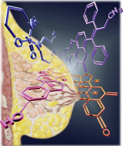
In women at normal risk for breast cancer, unopposed estrogen lowers the rate of the malignancy and the likelihood of mortality if the cancer occurs—but is not recommended as a prophylactic agent. Tamoxifen and other chemoprophylactic drugs can halve the rate of breast cancer in high-risk women but are not without drawbacks.
A look at the lower rate of breast cancer in the estrogen-alone arm of the WHI
Anderson GL, Chlebowski RT, Aragaki AK, et al. Conjugated equine oestrogen and breast cancer incidence and mortality in postmenopausal women with hysterectomy: extended follow-up of the Women’s Health Initiative randomised placebo-controlled trial. Lancet Oncol. 2012;13(5):476–486.
From 1993 through 1998, the WHI enrolled 10,739 postmenopausal women in the largest prospective trial evaluating the effect of hormone therapy (HT) on various clinical outcomes. The women were randomly allocated to three groups:
- conjugated estrogen with medroxyprogesterone acetate
- conjugated estrogen alone (in women with a prior hysterectomy)
- placebo.
The negative effects of estrogen plus progestin on the risk of breast cancer were the most widely discussed oucomes.1 Shortly after the findings from this arm of the study were published, the use of HT in the United States declined dramatically and unequivocally.2
In 2012, WHI published the results of the estrogen-alone arm in the British cancer specialty journal Lancet Oncology. As shown in the TABLE below, the incidence of breast cancer was statistically significantly lower (23%) in the estrogen group than in the placebo group. Women who were treated with estrogen alone were also 63% less likely to die of breast cancer, and all-cause mortality was 38% lower; both of these findings were statistically significant. Not only was there a significant reduction in the incidence of invasive breast cancer while the subjects were taking estrogen, but that reduction continued for a median of 4.7 years of follow-up after discontinuation of estrogen.
Breast cancer incidence and mortality in the estrogen-only arm of the WHI, compared with placebo*
| Event | Estrogen only (n = 5,310) | Placebo (n = 5,429) | Hazard ratio (95% confidence interval) |
|---|---|---|---|
| Invasive breast cancer | 151 (0.27%) | 199 (0.35%) | 0.77 (0.62–0.95) |
| Node-negative breast cancer | 88 (0.16%) | 134 (0.24%) | 0.67 (0.51–0.88) |
| Breast cancer mortality | 6 (0.009%) | 16 (0.024%) | 0.37 (0.13–0.91) |
| All-cause mortality | 30 (0.046%) | 50 (0.076%) | 0.62 (0.39–0.97) |
| * Median follow-up of 11.8 years | |||
The incidence figure is somewhat remarkable (199 in the placebo group versus 151 in the estrogen-alone group) in that it was nearly the exact reverse of the estrogen-progestin arm of the WHI trial (199 in the estrogen/progestin group vs 150 in the placebo group).3
Estrogen alone reduced both breast cancer incidence and breast cancer mortality while women were on therapy and for 5 years after discontinuing therapy. This finding should reassure women who have undergone hysterectomy, as well as their clinicians, that estrogen alone reduces the future likelihood of breast cancer. It should be noted that the effect of estrogen alone in women in higher-risk categories did not show a reduction in breast cancer, and for this reason, the authors cautioned against considering the use of estrogen alone in menopausal women as a breast cancer chemoprophylaxis agent.
All breast cancer chemoprophylactic agents carry risks as well as benefits
Goss P, Ingle J, Ales-Martinez J, et al. Exemestane for breast-cancer prevention in postmenopausal women. N Engl J Med. 2011;364(25):2381–2391.
Cheung A, Tile L, Cardew S, et al. Bone density and structure in healthy postmenopausal women treated with exemestane for the primary prevention of breast cancer: a nested substudy of the MAP.3 randomized controlled trial. Lancet Oncol. 2012;13(3):275–284.
Vogel V, Costantino J, Wickerham L, et al. Update of the National Surgical Adjuvant Breast and Bowel Project Study of Tamoxifen and Raloxifene (STAR) P-2 Trial: preventing breast cancer. Cancer Prev Res. 2010;3(6):696–706.
The number of new cases of breast cancer in the United States last year reached nearly a quarter-million. Clearly, reducing this number remains an important goal.4 Chemoprevention—the use of medication to reduce cancer risk—may be offered to women who are at high risk of developing breast cancer.
In the National Surgical Adjuvant Breast and Bowel Project (NSABP) P-1 trial, tamoxifen (a selective estrogen-receptor modulator) was shown to reduce the risk of invasive breast cancer by 49% in a high-risk population, resulting in the FDA approving tamoxifen as the first drug for breast cancer prevention.5 The P-1 trial was followed by the NSABP Study of Tamoxifen and Raloxifene (STAR) P-2 trial, which demonstrated relative equivalence between the two medications as cancer prevention agents in menopausal women.6 Serious side effects of these medications limit their use among eligible women, although raloxifene seems to be associated with fewer adverse events. In the update of the STAR trial with an average of 81 months of follow-up, the risk ratio for adverse events (raloxifene:tamoxifen) was 0.75 for thromboembolic events, 0.55 for endometrial cancer, and 0.19 for uterine hyperplasia.
Another drug used for cancer treatment has now entered the prevention scene. In 2011, the NCIC Clinical Trials Group Mammary Prevention.3 trial (NCIC CTG MAP.3) compared exemestane (an aromatase inhibitor) with placebo for menopausal women at high risk for breast cancer, demonstrating a 65% relative reduction in the incidence of invasive breast cancer. This study validated another option for cancer prevention in high-risk women, although its adoption is likely also to be limited by side effects, including vasomotor symptoms, a high rate of arthralgias, and vaginal dryness/dyspareunia. The greatest concern may be the potential effect on bone density. Though the rates of serious adverse events including fracture did not differ in the MAP.3 trial at 35 months of follow-up, women on exemestane had significantly larger losses of bone mineral density, compared with controls.
Chemoprophylaxis reduces the risk of breast cancer in high-risk women by about 50%. Who are good candidates for these medications? Based on these trials, menopausal women considered at high risk might include those with a Gail risk score of at least 1.66% (ie, risk of developing breast cancer in 5 years), age 60 years or older, and women with biopsy results demonstrating atypical hyperplasia or lobular carcinoma in situ (LCIS). (The Gail model is available at www.cancer.gov/bcrisktool.) Tamoxifen is the only option for premenopausal women age 35 and older. Those who have histologic markers of risk (atypical hyperplasia, LCIS) likely stand to derive the greatest benefit.4
Managing the reproductive health concerns of young women with breast cancer
Azim H, Kroman N, Paesmans M, et al. Prognostic impact of pregnancy after breast cancer according to estrogen receptor status: a multicenter retrospective study. J Clin Oncol. 2013;31(1):73–79.
Howard-Anderson J, Ganz P, Bower J, Stanton A. Quality of life, fertility concerns, and behavioral health outcomes in younger breast cancer survivors: a systematic review. J Natl Cancer Inst. 2012;104(5):1386–1405.
Of the approximately 230,000 new cases of invasive breast cancer identified in 2011, 50,430 cases involved women less than 50 years of age.4 For these women, the diagnosis of cancer raises multifaceted concerns, including the physical changes that accompany breast cancer treatment, concerns about recurrence and mortality, and significant sexual and reproductive consequences of treatment that alters ovarian function. Pregnancy-associated breast cancers (breast cancers diagnosed during pregnancy, lactation, and for 12 months postpartum) represent a small subset of these cancers and occur in about 1 in 3,000 pregnancies. One might anticipate that this rate will increase as women continue to delay childbearing, because pregnancy-associated breast cancers are more common in older women.
In the review article by Howard-Anderson and colleagues, the importance of these reproductive health consequences in young women diagnosed with breast cancer is highlighted. The women who transition to menopause as a result of chemotherapy (reported to range from 33%–73%) experience more symptoms, including hot flashes, night sweats, breast pain, vaginal dryness, and lack of sexual desire. Sixty-one percent of women younger than 40 years at diagnosis reported that they were concerned about menopause, and 30% reported that this concern influenced their treatment decisions. Thirty-nine percent of women in this group had major concerns about treatment-associated infertility, and only half of the women studied felt that their fertility concerns were adequately addressed.
On a positive note, for women who successfully achieve pregnancy after breast cancer, pregnancy outcomes appear to be similar to those of their nonpregnant peers. In the study by Azim and colleagues, women who became pregnant after a breast cancer diagnosis had disease-free survival that was statistically similar to that of matched women who did not have subsequent pregnancies. In addition, this outcome did not differ based on estrogen/progesterone receptor status (ER/PR positive or negative).
Both alkylating chemotherapeutic agents (eg, cyclophosphamide) and selective estrogen receptor modulating agents (for women with estrogen-receptor–positive tumors) are routine parts of adjuvant treatment for premenopausal women with invasive breast cancers.
These agents can have profound effects on both ovarian hormonal function and fertility. ObGyns and reproductive endocrinology/infertility specialists have a great opportunity to partner with our oncology colleagues to enhance the counseling that young women receive before, during, and after breast cancer treatment.
Women who are considering future childbearing should receive information about the impact of breast cancer treatment on fertility and options for fertility preservation prior to initiating treatment. For women who have completed childbearing, information on what to expect if menopause occurs and available options for symptom relief can be empowering as they make treatment decisions.
We want to hear from you! Tell us what you think.
1. Grady D. Study finds new risks in hormone therapy. New York Times. http://www.nytimes.com/2003/06/25/us/study-finds-new-risks-in-hormone-therapy.html?pagewanted=all&src=pm. Published June 25 2003. Accessed February 11, 2013.
2. Hersh AL, Stefanick ML, Stafford RS. National use of menopausal hormone therapy: annual trends and response to recent evidence. JAMA. 2004;291(1):47-53.
3. Chlebowski RT, Kuller LH, Prentice RL, et al. Women’s Health Initiative Investigators. Breast cancer after use of estrogen plus progestin in postmenopausal women. N Engl J Med. 2009;360(6):573-587.
4. American Cancer Society. Breast Cancer Facts and Figures 2011-2012. Atlanta, GA: American Cancer Society. http://www.cancer.org/research/cancerfactsfigures/breastcancerfactsfigures/breast-cancer-facts-and-figures-2011-2012. Accessed February 11, 2013.
5. Fisher B, Constantino J, Wickerham L, et al. Tamoxifen for prevention of breast cancer: report of the National Surgical Adjuvant Breast and Bowel Project P-1 Study. J Natl Cancer Inst. 1998;90(18):1371-1388.
6. Vogel V, Costantino J, Wickerham DL, et al. Effects of tamoxifen vs raloxifene on the risk of developing invasive breast cancer and other disease outcomes. The NSABP Study of Tamoxifen and Raloxifene (STAR) P-2 Trial. JAMA. 2006;295(23):2727-2741.

Mark D. Pearlman, MD
Dr. Pearlman is S. Jan Behrman Professor, Vice Chair, and Service Chief, Department of Obstetrics and Gynecology, and Professor, Department of Surgery, University of Michigan Medical School, Ann Arbor, Michigan. He is also Associate Chief of Clinical Affairs and Medical Director of Pharmacy, University of Michigan Hospital and Health Systems.

Jennifer L. Griffin Miller, MD, MPH
Dr. Griffin is Assistant Professor, Department of Obstetrics and Gynecology, University of Nebraska College of Medicine, Omaha, Nebraska.
The authors report no financial relationships relevant to this article.
Mark D. Pearlman, MD
Dr. Pearlman is S. Jan Behrman Professor, Vice Chair, and Service Chief, Department of Obstetrics and Gynecology, and Professor, Department of Surgery, University of Michigan Medical School, Ann Arbor, Michigan. He is also Associate Chief of Clinical Affairs and Medical Director of Pharmacy, University of Michigan Hospital and Health Systems.
Jennifer L. Griffin Miller, MD, MPH
Dr. Griffin is Assistant Professor, Department of Obstetrics and Gynecology, University of Nebraska College of Medicine, Omaha, Nebraska.
The authors report no financial relationships relevant to this article.
Mark D. Pearlman, MD
Dr. Pearlman is S. Jan Behrman Professor, Vice Chair, and Service Chief, Department of Obstetrics and Gynecology, and Professor, Department of Surgery, University of Michigan Medical School, Ann Arbor, Michigan. He is also Associate Chief of Clinical Affairs and Medical Director of Pharmacy, University of Michigan Hospital and Health Systems.
Jennifer L. Griffin Miller, MD, MPH
Dr. Griffin is Assistant Professor, Department of Obstetrics and Gynecology, University of Nebraska College of Medicine, Omaha, Nebraska.
The authors report no financial relationships relevant to this article.
Women with ER-positive breast cancer may soon extend tamoxifen therapy to 10 years
Janelle Yates (February 2013)
Is overdiagnosis of breast cancer common among women screened
by mammography?
Andrew M. Kaunitz, MD (Examining the Evidence; January 2013)
Breast cancer genome analysis highlights 4 subtypes, link to
ovarian cancer
Janelle Yates (News for Your Practice; November 2012)
The effects of breast cancer on obstetric and gynecologic practices are pervasive. In this article, we touch on three aspects of breast cancer that are particularly relevant to the practicing ObGyn:
- the need to identify women at high risk for breast cancer and select those who would benefit from a discussion of the advantages and risks of chemoprophylaxis, which can reduce the likelihood of breast cancer by 50% or more
- the need for strategies to manage menopausal symptoms in the general population without increasing the risk of breast cancer. The traditional approach to this problem changed dramatically with the Women’s Health Initiative (WHI), which demonstrated an increased risk of breast cancer in women taking conjugated equine estrogen and progestin. The widely publicized initial findings of the estrogen-progestin arm of the WHI sharply contrast the equally relevant, somewhat unexpected, and less publicized results of the estrogen-alone arm, which demonstrated a substantial and statistically significant decrease in the incidence of breast cancer, even after estrogen was discontinued.
- the potential effects of breast cancer treatment on ovarian function in young women. This year, of the approximately 250,000 women who will be diagnosed with invasive breast cancer, more than 50,000 women will be of reproductive age. Most of these young women will require adjuvant chemotherapy; as a result, many will experience the premature onset of menopause. Along with the attendant loss of fertility these women will face, many will also develop distressing and life-altering menopausal symptoms. Management of these women before and after initiation of chemotherapy requires an understanding of both the expected effects of the chemotherapy and knowledge of how to actively manage these women with strategies to either prevent these events or to manage menopausal symptoms.
In women at normal risk for breast cancer, unopposed estrogen lowers the rate of the malignancy and the likelihood of mortality if the cancer occurs—but is not recommended as a prophylactic agent. Tamoxifen and other chemoprophylactic drugs can halve the rate of breast cancer in high-risk women but are not without drawbacks.
A look at the lower rate of breast cancer in the estrogen-alone arm of the WHI
Anderson GL, Chlebowski RT, Aragaki AK, et al. Conjugated equine oestrogen and breast cancer incidence and mortality in postmenopausal women with hysterectomy: extended follow-up of the Women’s Health Initiative randomised placebo-controlled trial. Lancet Oncol. 2012;13(5):476–486.
From 1993 through 1998, the WHI enrolled 10,739 postmenopausal women in the largest prospective trial evaluating the effect of hormone therapy (HT) on various clinical outcomes. The women were randomly allocated to three groups:
- conjugated estrogen with medroxyprogesterone acetate
- conjugated estrogen alone (in women with a prior hysterectomy)
- placebo.
The negative effects of estrogen plus progestin on the risk of breast cancer were the most widely discussed oucomes.1 Shortly after the findings from this arm of the study were published, the use of HT in the United States declined dramatically and unequivocally.2
In 2012, WHI published the results of the estrogen-alone arm in the British cancer specialty journal Lancet Oncology. As shown in the TABLE below, the incidence of breast cancer was statistically significantly lower (23%) in the estrogen group than in the placebo group. Women who were treated with estrogen alone were also 63% less likely to die of breast cancer, and all-cause mortality was 38% lower; both of these findings were statistically significant. Not only was there a significant reduction in the incidence of invasive breast cancer while the subjects were taking estrogen, but that reduction continued for a median of 4.7 years of follow-up after discontinuation of estrogen.
Breast cancer incidence and mortality in the estrogen-only arm of the WHI, compared with placebo*
| Event | Estrogen only (n = 5,310) | Placebo (n = 5,429) | Hazard ratio (95% confidence interval) |
|---|---|---|---|
| Invasive breast cancer | 151 (0.27%) | 199 (0.35%) | 0.77 (0.62–0.95) |
| Node-negative breast cancer | 88 (0.16%) | 134 (0.24%) | 0.67 (0.51–0.88) |
| Breast cancer mortality | 6 (0.009%) | 16 (0.024%) | 0.37 (0.13–0.91) |
| All-cause mortality | 30 (0.046%) | 50 (0.076%) | 0.62 (0.39–0.97) |
| * Median follow-up of 11.8 years | |||
The incidence figure is somewhat remarkable (199 in the placebo group versus 151 in the estrogen-alone group) in that it was nearly the exact reverse of the estrogen-progestin arm of the WHI trial (199 in the estrogen/progestin group vs 150 in the placebo group).3
Estrogen alone reduced both breast cancer incidence and breast cancer mortality while women were on therapy and for 5 years after discontinuing therapy. This finding should reassure women who have undergone hysterectomy, as well as their clinicians, that estrogen alone reduces the future likelihood of breast cancer. It should be noted that the effect of estrogen alone in women in higher-risk categories did not show a reduction in breast cancer, and for this reason, the authors cautioned against considering the use of estrogen alone in menopausal women as a breast cancer chemoprophylaxis agent.
All breast cancer chemoprophylactic agents carry risks as well as benefits
Goss P, Ingle J, Ales-Martinez J, et al. Exemestane for breast-cancer prevention in postmenopausal women. N Engl J Med. 2011;364(25):2381–2391.
Cheung A, Tile L, Cardew S, et al. Bone density and structure in healthy postmenopausal women treated with exemestane for the primary prevention of breast cancer: a nested substudy of the MAP.3 randomized controlled trial. Lancet Oncol. 2012;13(3):275–284.
Vogel V, Costantino J, Wickerham L, et al. Update of the National Surgical Adjuvant Breast and Bowel Project Study of Tamoxifen and Raloxifene (STAR) P-2 Trial: preventing breast cancer. Cancer Prev Res. 2010;3(6):696–706.
The number of new cases of breast cancer in the United States last year reached nearly a quarter-million. Clearly, reducing this number remains an important goal.4 Chemoprevention—the use of medication to reduce cancer risk—may be offered to women who are at high risk of developing breast cancer.
In the National Surgical Adjuvant Breast and Bowel Project (NSABP) P-1 trial, tamoxifen (a selective estrogen-receptor modulator) was shown to reduce the risk of invasive breast cancer by 49% in a high-risk population, resulting in the FDA approving tamoxifen as the first drug for breast cancer prevention.5 The P-1 trial was followed by the NSABP Study of Tamoxifen and Raloxifene (STAR) P-2 trial, which demonstrated relative equivalence between the two medications as cancer prevention agents in menopausal women.6 Serious side effects of these medications limit their use among eligible women, although raloxifene seems to be associated with fewer adverse events. In the update of the STAR trial with an average of 81 months of follow-up, the risk ratio for adverse events (raloxifene:tamoxifen) was 0.75 for thromboembolic events, 0.55 for endometrial cancer, and 0.19 for uterine hyperplasia.
Another drug used for cancer treatment has now entered the prevention scene. In 2011, the NCIC Clinical Trials Group Mammary Prevention.3 trial (NCIC CTG MAP.3) compared exemestane (an aromatase inhibitor) with placebo for menopausal women at high risk for breast cancer, demonstrating a 65% relative reduction in the incidence of invasive breast cancer. This study validated another option for cancer prevention in high-risk women, although its adoption is likely also to be limited by side effects, including vasomotor symptoms, a high rate of arthralgias, and vaginal dryness/dyspareunia. The greatest concern may be the potential effect on bone density. Though the rates of serious adverse events including fracture did not differ in the MAP.3 trial at 35 months of follow-up, women on exemestane had significantly larger losses of bone mineral density, compared with controls.
Chemoprophylaxis reduces the risk of breast cancer in high-risk women by about 50%. Who are good candidates for these medications? Based on these trials, menopausal women considered at high risk might include those with a Gail risk score of at least 1.66% (ie, risk of developing breast cancer in 5 years), age 60 years or older, and women with biopsy results demonstrating atypical hyperplasia or lobular carcinoma in situ (LCIS). (The Gail model is available at www.cancer.gov/bcrisktool.) Tamoxifen is the only option for premenopausal women age 35 and older. Those who have histologic markers of risk (atypical hyperplasia, LCIS) likely stand to derive the greatest benefit.4
Managing the reproductive health concerns of young women with breast cancer
Azim H, Kroman N, Paesmans M, et al. Prognostic impact of pregnancy after breast cancer according to estrogen receptor status: a multicenter retrospective study. J Clin Oncol. 2013;31(1):73–79.
Howard-Anderson J, Ganz P, Bower J, Stanton A. Quality of life, fertility concerns, and behavioral health outcomes in younger breast cancer survivors: a systematic review. J Natl Cancer Inst. 2012;104(5):1386–1405.
Of the approximately 230,000 new cases of invasive breast cancer identified in 2011, 50,430 cases involved women less than 50 years of age.4 For these women, the diagnosis of cancer raises multifaceted concerns, including the physical changes that accompany breast cancer treatment, concerns about recurrence and mortality, and significant sexual and reproductive consequences of treatment that alters ovarian function. Pregnancy-associated breast cancers (breast cancers diagnosed during pregnancy, lactation, and for 12 months postpartum) represent a small subset of these cancers and occur in about 1 in 3,000 pregnancies. One might anticipate that this rate will increase as women continue to delay childbearing, because pregnancy-associated breast cancers are more common in older women.
In the review article by Howard-Anderson and colleagues, the importance of these reproductive health consequences in young women diagnosed with breast cancer is highlighted. The women who transition to menopause as a result of chemotherapy (reported to range from 33%–73%) experience more symptoms, including hot flashes, night sweats, breast pain, vaginal dryness, and lack of sexual desire. Sixty-one percent of women younger than 40 years at diagnosis reported that they were concerned about menopause, and 30% reported that this concern influenced their treatment decisions. Thirty-nine percent of women in this group had major concerns about treatment-associated infertility, and only half of the women studied felt that their fertility concerns were adequately addressed.
On a positive note, for women who successfully achieve pregnancy after breast cancer, pregnancy outcomes appear to be similar to those of their nonpregnant peers. In the study by Azim and colleagues, women who became pregnant after a breast cancer diagnosis had disease-free survival that was statistically similar to that of matched women who did not have subsequent pregnancies. In addition, this outcome did not differ based on estrogen/progesterone receptor status (ER/PR positive or negative).
Both alkylating chemotherapeutic agents (eg, cyclophosphamide) and selective estrogen receptor modulating agents (for women with estrogen-receptor–positive tumors) are routine parts of adjuvant treatment for premenopausal women with invasive breast cancers.
These agents can have profound effects on both ovarian hormonal function and fertility. ObGyns and reproductive endocrinology/infertility specialists have a great opportunity to partner with our oncology colleagues to enhance the counseling that young women receive before, during, and after breast cancer treatment.
Women who are considering future childbearing should receive information about the impact of breast cancer treatment on fertility and options for fertility preservation prior to initiating treatment. For women who have completed childbearing, information on what to expect if menopause occurs and available options for symptom relief can be empowering as they make treatment decisions.
We want to hear from you! Tell us what you think.
Women with ER-positive breast cancer may soon extend tamoxifen therapy to 10 years
Janelle Yates (February 2013)
Is overdiagnosis of breast cancer common among women screened
by mammography?
Andrew M. Kaunitz, MD (Examining the Evidence; January 2013)
Breast cancer genome analysis highlights 4 subtypes, link to
ovarian cancer
Janelle Yates (News for Your Practice; November 2012)
The effects of breast cancer on obstetric and gynecologic practices are pervasive. In this article, we touch on three aspects of breast cancer that are particularly relevant to the practicing ObGyn:
- the need to identify women at high risk for breast cancer and select those who would benefit from a discussion of the advantages and risks of chemoprophylaxis, which can reduce the likelihood of breast cancer by 50% or more
- the need for strategies to manage menopausal symptoms in the general population without increasing the risk of breast cancer. The traditional approach to this problem changed dramatically with the Women’s Health Initiative (WHI), which demonstrated an increased risk of breast cancer in women taking conjugated equine estrogen and progestin. The widely publicized initial findings of the estrogen-progestin arm of the WHI sharply contrast the equally relevant, somewhat unexpected, and less publicized results of the estrogen-alone arm, which demonstrated a substantial and statistically significant decrease in the incidence of breast cancer, even after estrogen was discontinued.
- the potential effects of breast cancer treatment on ovarian function in young women. This year, of the approximately 250,000 women who will be diagnosed with invasive breast cancer, more than 50,000 women will be of reproductive age. Most of these young women will require adjuvant chemotherapy; as a result, many will experience the premature onset of menopause. Along with the attendant loss of fertility these women will face, many will also develop distressing and life-altering menopausal symptoms. Management of these women before and after initiation of chemotherapy requires an understanding of both the expected effects of the chemotherapy and knowledge of how to actively manage these women with strategies to either prevent these events or to manage menopausal symptoms.
In women at normal risk for breast cancer, unopposed estrogen lowers the rate of the malignancy and the likelihood of mortality if the cancer occurs—but is not recommended as a prophylactic agent. Tamoxifen and other chemoprophylactic drugs can halve the rate of breast cancer in high-risk women but are not without drawbacks.
A look at the lower rate of breast cancer in the estrogen-alone arm of the WHI
Anderson GL, Chlebowski RT, Aragaki AK, et al. Conjugated equine oestrogen and breast cancer incidence and mortality in postmenopausal women with hysterectomy: extended follow-up of the Women’s Health Initiative randomised placebo-controlled trial. Lancet Oncol. 2012;13(5):476–486.
From 1993 through 1998, the WHI enrolled 10,739 postmenopausal women in the largest prospective trial evaluating the effect of hormone therapy (HT) on various clinical outcomes. The women were randomly allocated to three groups:
- conjugated estrogen with medroxyprogesterone acetate
- conjugated estrogen alone (in women with a prior hysterectomy)
- placebo.
The negative effects of estrogen plus progestin on the risk of breast cancer were the most widely discussed oucomes.1 Shortly after the findings from this arm of the study were published, the use of HT in the United States declined dramatically and unequivocally.2
In 2012, WHI published the results of the estrogen-alone arm in the British cancer specialty journal Lancet Oncology. As shown in the TABLE below, the incidence of breast cancer was statistically significantly lower (23%) in the estrogen group than in the placebo group. Women who were treated with estrogen alone were also 63% less likely to die of breast cancer, and all-cause mortality was 38% lower; both of these findings were statistically significant. Not only was there a significant reduction in the incidence of invasive breast cancer while the subjects were taking estrogen, but that reduction continued for a median of 4.7 years of follow-up after discontinuation of estrogen.
Breast cancer incidence and mortality in the estrogen-only arm of the WHI, compared with placebo*
| Event | Estrogen only (n = 5,310) | Placebo (n = 5,429) | Hazard ratio (95% confidence interval) |
|---|---|---|---|
| Invasive breast cancer | 151 (0.27%) | 199 (0.35%) | 0.77 (0.62–0.95) |
| Node-negative breast cancer | 88 (0.16%) | 134 (0.24%) | 0.67 (0.51–0.88) |
| Breast cancer mortality | 6 (0.009%) | 16 (0.024%) | 0.37 (0.13–0.91) |
| All-cause mortality | 30 (0.046%) | 50 (0.076%) | 0.62 (0.39–0.97) |
| * Median follow-up of 11.8 years | |||
The incidence figure is somewhat remarkable (199 in the placebo group versus 151 in the estrogen-alone group) in that it was nearly the exact reverse of the estrogen-progestin arm of the WHI trial (199 in the estrogen/progestin group vs 150 in the placebo group).3
Estrogen alone reduced both breast cancer incidence and breast cancer mortality while women were on therapy and for 5 years after discontinuing therapy. This finding should reassure women who have undergone hysterectomy, as well as their clinicians, that estrogen alone reduces the future likelihood of breast cancer. It should be noted that the effect of estrogen alone in women in higher-risk categories did not show a reduction in breast cancer, and for this reason, the authors cautioned against considering the use of estrogen alone in menopausal women as a breast cancer chemoprophylaxis agent.
All breast cancer chemoprophylactic agents carry risks as well as benefits
Goss P, Ingle J, Ales-Martinez J, et al. Exemestane for breast-cancer prevention in postmenopausal women. N Engl J Med. 2011;364(25):2381–2391.
Cheung A, Tile L, Cardew S, et al. Bone density and structure in healthy postmenopausal women treated with exemestane for the primary prevention of breast cancer: a nested substudy of the MAP.3 randomized controlled trial. Lancet Oncol. 2012;13(3):275–284.
Vogel V, Costantino J, Wickerham L, et al. Update of the National Surgical Adjuvant Breast and Bowel Project Study of Tamoxifen and Raloxifene (STAR) P-2 Trial: preventing breast cancer. Cancer Prev Res. 2010;3(6):696–706.
The number of new cases of breast cancer in the United States last year reached nearly a quarter-million. Clearly, reducing this number remains an important goal.4 Chemoprevention—the use of medication to reduce cancer risk—may be offered to women who are at high risk of developing breast cancer.
In the National Surgical Adjuvant Breast and Bowel Project (NSABP) P-1 trial, tamoxifen (a selective estrogen-receptor modulator) was shown to reduce the risk of invasive breast cancer by 49% in a high-risk population, resulting in the FDA approving tamoxifen as the first drug for breast cancer prevention.5 The P-1 trial was followed by the NSABP Study of Tamoxifen and Raloxifene (STAR) P-2 trial, which demonstrated relative equivalence between the two medications as cancer prevention agents in menopausal women.6 Serious side effects of these medications limit their use among eligible women, although raloxifene seems to be associated with fewer adverse events. In the update of the STAR trial with an average of 81 months of follow-up, the risk ratio for adverse events (raloxifene:tamoxifen) was 0.75 for thromboembolic events, 0.55 for endometrial cancer, and 0.19 for uterine hyperplasia.
Another drug used for cancer treatment has now entered the prevention scene. In 2011, the NCIC Clinical Trials Group Mammary Prevention.3 trial (NCIC CTG MAP.3) compared exemestane (an aromatase inhibitor) with placebo for menopausal women at high risk for breast cancer, demonstrating a 65% relative reduction in the incidence of invasive breast cancer. This study validated another option for cancer prevention in high-risk women, although its adoption is likely also to be limited by side effects, including vasomotor symptoms, a high rate of arthralgias, and vaginal dryness/dyspareunia. The greatest concern may be the potential effect on bone density. Though the rates of serious adverse events including fracture did not differ in the MAP.3 trial at 35 months of follow-up, women on exemestane had significantly larger losses of bone mineral density, compared with controls.
Chemoprophylaxis reduces the risk of breast cancer in high-risk women by about 50%. Who are good candidates for these medications? Based on these trials, menopausal women considered at high risk might include those with a Gail risk score of at least 1.66% (ie, risk of developing breast cancer in 5 years), age 60 years or older, and women with biopsy results demonstrating atypical hyperplasia or lobular carcinoma in situ (LCIS). (The Gail model is available at www.cancer.gov/bcrisktool.) Tamoxifen is the only option for premenopausal women age 35 and older. Those who have histologic markers of risk (atypical hyperplasia, LCIS) likely stand to derive the greatest benefit.4
Managing the reproductive health concerns of young women with breast cancer
Azim H, Kroman N, Paesmans M, et al. Prognostic impact of pregnancy after breast cancer according to estrogen receptor status: a multicenter retrospective study. J Clin Oncol. 2013;31(1):73–79.
Howard-Anderson J, Ganz P, Bower J, Stanton A. Quality of life, fertility concerns, and behavioral health outcomes in younger breast cancer survivors: a systematic review. J Natl Cancer Inst. 2012;104(5):1386–1405.
Of the approximately 230,000 new cases of invasive breast cancer identified in 2011, 50,430 cases involved women less than 50 years of age.4 For these women, the diagnosis of cancer raises multifaceted concerns, including the physical changes that accompany breast cancer treatment, concerns about recurrence and mortality, and significant sexual and reproductive consequences of treatment that alters ovarian function. Pregnancy-associated breast cancers (breast cancers diagnosed during pregnancy, lactation, and for 12 months postpartum) represent a small subset of these cancers and occur in about 1 in 3,000 pregnancies. One might anticipate that this rate will increase as women continue to delay childbearing, because pregnancy-associated breast cancers are more common in older women.
In the review article by Howard-Anderson and colleagues, the importance of these reproductive health consequences in young women diagnosed with breast cancer is highlighted. The women who transition to menopause as a result of chemotherapy (reported to range from 33%–73%) experience more symptoms, including hot flashes, night sweats, breast pain, vaginal dryness, and lack of sexual desire. Sixty-one percent of women younger than 40 years at diagnosis reported that they were concerned about menopause, and 30% reported that this concern influenced their treatment decisions. Thirty-nine percent of women in this group had major concerns about treatment-associated infertility, and only half of the women studied felt that their fertility concerns were adequately addressed.
On a positive note, for women who successfully achieve pregnancy after breast cancer, pregnancy outcomes appear to be similar to those of their nonpregnant peers. In the study by Azim and colleagues, women who became pregnant after a breast cancer diagnosis had disease-free survival that was statistically similar to that of matched women who did not have subsequent pregnancies. In addition, this outcome did not differ based on estrogen/progesterone receptor status (ER/PR positive or negative).
Both alkylating chemotherapeutic agents (eg, cyclophosphamide) and selective estrogen receptor modulating agents (for women with estrogen-receptor–positive tumors) are routine parts of adjuvant treatment for premenopausal women with invasive breast cancers.
These agents can have profound effects on both ovarian hormonal function and fertility. ObGyns and reproductive endocrinology/infertility specialists have a great opportunity to partner with our oncology colleagues to enhance the counseling that young women receive before, during, and after breast cancer treatment.
Women who are considering future childbearing should receive information about the impact of breast cancer treatment on fertility and options for fertility preservation prior to initiating treatment. For women who have completed childbearing, information on what to expect if menopause occurs and available options for symptom relief can be empowering as they make treatment decisions.
We want to hear from you! Tell us what you think.
1. Grady D. Study finds new risks in hormone therapy. New York Times. http://www.nytimes.com/2003/06/25/us/study-finds-new-risks-in-hormone-therapy.html?pagewanted=all&src=pm. Published June 25 2003. Accessed February 11, 2013.
2. Hersh AL, Stefanick ML, Stafford RS. National use of menopausal hormone therapy: annual trends and response to recent evidence. JAMA. 2004;291(1):47-53.
3. Chlebowski RT, Kuller LH, Prentice RL, et al. Women’s Health Initiative Investigators. Breast cancer after use of estrogen plus progestin in postmenopausal women. N Engl J Med. 2009;360(6):573-587.
4. American Cancer Society. Breast Cancer Facts and Figures 2011-2012. Atlanta, GA: American Cancer Society. http://www.cancer.org/research/cancerfactsfigures/breastcancerfactsfigures/breast-cancer-facts-and-figures-2011-2012. Accessed February 11, 2013.
5. Fisher B, Constantino J, Wickerham L, et al. Tamoxifen for prevention of breast cancer: report of the National Surgical Adjuvant Breast and Bowel Project P-1 Study. J Natl Cancer Inst. 1998;90(18):1371-1388.
6. Vogel V, Costantino J, Wickerham DL, et al. Effects of tamoxifen vs raloxifene on the risk of developing invasive breast cancer and other disease outcomes. The NSABP Study of Tamoxifen and Raloxifene (STAR) P-2 Trial. JAMA. 2006;295(23):2727-2741.
1. Grady D. Study finds new risks in hormone therapy. New York Times. http://www.nytimes.com/2003/06/25/us/study-finds-new-risks-in-hormone-therapy.html?pagewanted=all&src=pm. Published June 25 2003. Accessed February 11, 2013.
2. Hersh AL, Stefanick ML, Stafford RS. National use of menopausal hormone therapy: annual trends and response to recent evidence. JAMA. 2004;291(1):47-53.
3. Chlebowski RT, Kuller LH, Prentice RL, et al. Women’s Health Initiative Investigators. Breast cancer after use of estrogen plus progestin in postmenopausal women. N Engl J Med. 2009;360(6):573-587.
4. American Cancer Society. Breast Cancer Facts and Figures 2011-2012. Atlanta, GA: American Cancer Society. http://www.cancer.org/research/cancerfactsfigures/breastcancerfactsfigures/breast-cancer-facts-and-figures-2011-2012. Accessed February 11, 2013.
5. Fisher B, Constantino J, Wickerham L, et al. Tamoxifen for prevention of breast cancer: report of the National Surgical Adjuvant Breast and Bowel Project P-1 Study. J Natl Cancer Inst. 1998;90(18):1371-1388.
6. Vogel V, Costantino J, Wickerham DL, et al. Effects of tamoxifen vs raloxifene on the risk of developing invasive breast cancer and other disease outcomes. The NSABP Study of Tamoxifen and Raloxifene (STAR) P-2 Trial. JAMA. 2006;295(23):2727-2741.
Metastatic breast cancer incidence is rising among young women
The incidence of breast cancer presenting with distant involvement has risen significantly over the past 30 years among young women, and this trend shows no signs of abating, according to a report in the Feb. 27 issue of JAMA.
In contrast, the rate of locoregional breast cancer has not increased in this age group, and the incidence of all stages of the disease have not shown any increasing trends among older women, said Dr. Rebecca H. Johnson of Seattle Children’s Hospital and the University of Washington, Seattle, and her associates.

This trajectory "predicts that an increasing number of young women in the United States will present with metastatic breast cancer in an age group that already has the worst prognosis, no recommended routine screening practice, the least health insurance, and the most potential years of life" lost, the investigators noted.
They used data from three of the National Cancer Institute’s Surveillance, Epidemiology, and End Results (SEER) registries to examine time trends in breast cancer between 1976 and 2009. They found a steady, and possibly accelerating, rise in the rate of women aged 25-39 years presenting with metastatic breast cancer, from 1.53/100,000 in 1976 to 2.90/100,000 in 2009.
"No other age group had statistically significant increases, either for distant, regional, or localized disease at diagnosis," Dr. Johnson and her colleagues said (JAMA 2013;309:800-5).
In a different analysis of the data, the category of metastatic breast cancer as a proportion of all invasive breast cancer in this age group rose from 4.4% in the 1970s to 4.8% in the 1980s, 5.5% in the 1990s, and 7.2% in the early 2000s.
This trend was evident in women of all races/ethnicities, in women residing in both urban and nonurban regions, and in women with estrogen receptor–positive and estrogen receptor–negative tumors. "Non-Hispanic white and African-American individuals appear to have been more affected by the increase, as have women with the ER-positive subtype of the disease," they said.
"The absolute increase of 1.37/100,000 over 34 years is relatively small, but the trend shows no evidence of abatement and may indicate increasing epidemiologic and clinical significance," the researchers said.
These findings must be corroborated in other studies. If they are confirmed, they will be particularly concerning "because young age itself is an independent adverse prognostic factor for breast cancer.
"The most recent national 5-year survival data for distant disease for 25- to 39-year-old women is only 31% ... compared with a 5-year survival of 87% for women with locoregional breast cancer," they added.
Dr. Johnson reported having served on a board for Critical Mass Young Adult Cancer Alliance and having served as a speaker at the Leukemia and Lymphoma Society AYA Survivorship Conference. One of her colleagues reported being a consultant and speaker for Sigma-Tau Pharmaceuticals; another researcher’s salary was funded by the Seattle Children’s Guild Association Teen Cancer Grant. No other disclosures were reported.
The incidence of breast cancer presenting with distant involvement has risen significantly over the past 30 years among young women, and this trend shows no signs of abating, according to a report in the Feb. 27 issue of JAMA.
In contrast, the rate of locoregional breast cancer has not increased in this age group, and the incidence of all stages of the disease have not shown any increasing trends among older women, said Dr. Rebecca H. Johnson of Seattle Children’s Hospital and the University of Washington, Seattle, and her associates.
This trajectory "predicts that an increasing number of young women in the United States will present with metastatic breast cancer in an age group that already has the worst prognosis, no recommended routine screening practice, the least health insurance, and the most potential years of life" lost, the investigators noted.
They used data from three of the National Cancer Institute’s Surveillance, Epidemiology, and End Results (SEER) registries to examine time trends in breast cancer between 1976 and 2009. They found a steady, and possibly accelerating, rise in the rate of women aged 25-39 years presenting with metastatic breast cancer, from 1.53/100,000 in 1976 to 2.90/100,000 in 2009.
"No other age group had statistically significant increases, either for distant, regional, or localized disease at diagnosis," Dr. Johnson and her colleagues said (JAMA 2013;309:800-5).
In a different analysis of the data, the category of metastatic breast cancer as a proportion of all invasive breast cancer in this age group rose from 4.4% in the 1970s to 4.8% in the 1980s, 5.5% in the 1990s, and 7.2% in the early 2000s.
This trend was evident in women of all races/ethnicities, in women residing in both urban and nonurban regions, and in women with estrogen receptor–positive and estrogen receptor–negative tumors. "Non-Hispanic white and African-American individuals appear to have been more affected by the increase, as have women with the ER-positive subtype of the disease," they said.
"The absolute increase of 1.37/100,000 over 34 years is relatively small, but the trend shows no evidence of abatement and may indicate increasing epidemiologic and clinical significance," the researchers said.
These findings must be corroborated in other studies. If they are confirmed, they will be particularly concerning "because young age itself is an independent adverse prognostic factor for breast cancer.
"The most recent national 5-year survival data for distant disease for 25- to 39-year-old women is only 31% ... compared with a 5-year survival of 87% for women with locoregional breast cancer," they added.
Dr. Johnson reported having served on a board for Critical Mass Young Adult Cancer Alliance and having served as a speaker at the Leukemia and Lymphoma Society AYA Survivorship Conference. One of her colleagues reported being a consultant and speaker for Sigma-Tau Pharmaceuticals; another researcher’s salary was funded by the Seattle Children’s Guild Association Teen Cancer Grant. No other disclosures were reported.
The incidence of breast cancer presenting with distant involvement has risen significantly over the past 30 years among young women, and this trend shows no signs of abating, according to a report in the Feb. 27 issue of JAMA.
In contrast, the rate of locoregional breast cancer has not increased in this age group, and the incidence of all stages of the disease have not shown any increasing trends among older women, said Dr. Rebecca H. Johnson of Seattle Children’s Hospital and the University of Washington, Seattle, and her associates.
This trajectory "predicts that an increasing number of young women in the United States will present with metastatic breast cancer in an age group that already has the worst prognosis, no recommended routine screening practice, the least health insurance, and the most potential years of life" lost, the investigators noted.
They used data from three of the National Cancer Institute’s Surveillance, Epidemiology, and End Results (SEER) registries to examine time trends in breast cancer between 1976 and 2009. They found a steady, and possibly accelerating, rise in the rate of women aged 25-39 years presenting with metastatic breast cancer, from 1.53/100,000 in 1976 to 2.90/100,000 in 2009.
"No other age group had statistically significant increases, either for distant, regional, or localized disease at diagnosis," Dr. Johnson and her colleagues said (JAMA 2013;309:800-5).
In a different analysis of the data, the category of metastatic breast cancer as a proportion of all invasive breast cancer in this age group rose from 4.4% in the 1970s to 4.8% in the 1980s, 5.5% in the 1990s, and 7.2% in the early 2000s.
This trend was evident in women of all races/ethnicities, in women residing in both urban and nonurban regions, and in women with estrogen receptor–positive and estrogen receptor–negative tumors. "Non-Hispanic white and African-American individuals appear to have been more affected by the increase, as have women with the ER-positive subtype of the disease," they said.
"The absolute increase of 1.37/100,000 over 34 years is relatively small, but the trend shows no evidence of abatement and may indicate increasing epidemiologic and clinical significance," the researchers said.
These findings must be corroborated in other studies. If they are confirmed, they will be particularly concerning "because young age itself is an independent adverse prognostic factor for breast cancer.
"The most recent national 5-year survival data for distant disease for 25- to 39-year-old women is only 31% ... compared with a 5-year survival of 87% for women with locoregional breast cancer," they added.
Dr. Johnson reported having served on a board for Critical Mass Young Adult Cancer Alliance and having served as a speaker at the Leukemia and Lymphoma Society AYA Survivorship Conference. One of her colleagues reported being a consultant and speaker for Sigma-Tau Pharmaceuticals; another researcher’s salary was funded by the Seattle Children’s Guild Association Teen Cancer Grant. No other disclosures were reported.
FROM JAMA
Major Finding: The incidence of women aged 25-39 years presenting with metastatic breast cancer rose steadily from 1.53/100,000 in 1976 to 2.90/100,000 in 2009.
Data Source: An analysis of data from the National Cancer Institute’s SEER registries concerning breast cancer incidence in the United States between 1976 and 2009.
Disclosures: Dr. Johnson reported having served on a board for Critical Mass Young Adult Cancer Alliance and having served as a speaker at the Leukemia and Lymphoma Society AYA Survivorship Conference. One of her colleagues reported being a consultant and speaker for Sigma-Tau Pharmaceuticals; another researcher’s salary was funded by the Seattle Children’s Guild Association Teen Cancer Grant. No other disclosures were reported.