User login
MDedge conference coverage features onsite reporting of the latest study results and expert perspectives from leading researchers.
Studies gauge toll of pausing fertility treatment during pandemic
More than 60% of patients at a center for reproductive medicine in Utah who had fertility treatments canceled because of the COVID-19 pandemic opted to resume treatment once the suspension was lifted about 7 weeks later.
At another fertility center in New York, a survey found that 96% of respondents who had a cycle canceled because of the pandemic found it upsetting, and 22% found it extremely upsetting, with extremely upsetting defined as equivalent to the loss of a child.
The indefinite time frame for resuming treatment when the New York survey was conducted may have been a major source of distress for patients, one of the researchers said at the American Society for Reproductive Medicine’s 2020 annual meeting, held virtually this year.
“They don’t know when they might have that chance again,” said Jenna M. Turocy, MD, of Columbia University Fertility Center, New York.
COVID-19 guidelines published by ASRM on March 17 recommended the suspension of new treatment cycles, including ovulation induction, intrauterine inseminations, and in vitro fertilization (IVF).
An ASRM COVID-19 task force has since supported “the measured resumption of fertility care following the easing of restrictions,” said Paul C. Lin, MD, president of the Society for Assisted Reproductive Technology and a member of the task force.
“Over the past several months, significant knowledge has been gained regarding the COVID-19 virus and its impact on patients and the medical system,” he said in a news release about the two studies that assessed the pandemic’s effects.
Certain precautions remain. “It has become clear that we will need to be practicing COVID-19 protocols at least until an effective and safe vaccine or broadly effective treatment becomes widely available,” Dr. Lin said.
Desire to proceed during a pandemic
The center continued to offer IVF cycles for oncofertility patients on an urgent basis.
In early May, patients whose cycles had been suspended had the option to receive treatment.
“Upon reopening, every patient received standardized counseling from their primary IVF physician,” Lauren Verrilli, MD, a reproductive endocrinology and infertility fellow at the University of Utah, Salt Lake City, said at the virtual meeting.
Doctors explained that much remained unknown about COVID-19 in pregnancy, and that it was unclear whether the clinic would need to shut down again. In addition, patients had to undergo COVID-19 testing.
To identify factors associated with proceeding with treatment after the suspension, the researchers compared patients who resumed treatment with patients who did not.
Their analysis included 278 patients who had planned an IVF cycle or frozen embryo transfer (FET) prior to the shutdown. The researchers examined factors such as age, parity, anti-Müllerian hormone, antral follicle count, history of prior IVF cycles or FET, number of frozen blastocysts, gamete source, and use of a gestational carrier.
In all, 62% of patients opted to receive treatment once restrictions were lifted, including 69 of the 133 (52%) patients with planned fresh cycles and 104 of the 145 (72%) patients with planned FET cycles.
Among those with planned fresh cycles, those who opted to resume treatment tended to be older than those who did not resume treatment, with a median age of 37 years versus 35 years, but the difference was not statistically significant.
Among patients with planned FET cycles, those who did not resume treatment were more likely to have a gestational carrier, compared with those who resumed treatment (7% vs. 1%). In some cases, gestational carriers lived in another state and the pandemic complicated travel arrangements, which contributed to delays, Dr. Verrilli said.
The analysis did not include information about income or socioeconomic status, which may play a role in patients’ decisions, Dr. Verrilli said.
Emotional impact of indefinite delay
Fertility treatment is often time sensitive, particularly for patients with advanced reproductive age or diminished ovarian reserve, and indefinite postponement of fertility treatment potentially could lead some patients to lose the ability to conceive with their own gametes, Dr. Turocy said.
In early April, Dr. Turocy and colleagues surveyed patients at their academic fertility center in New York City to assess patients’ reactions to the ASRM recommendations. They decided to conduct the study after they realized that treatment cancellations were having a significant emotional effect.
Investigators emailed an 18-item survey to more than 3,000 patients.
In all, 518 patients completed the survey, a response rate of 17%. Patients had an average age of 37 years (range, 23-52 years), and 92% were female. About 24% had children, and 66% had received at least one fertility treatment.
Half had a cycle canceled because of the COVID-19 pandemic, including timed intercourse cycles (5%), intrauterine insemination cycles (23%), IVF cycles with a planned fresh embryo transfer (10%), IVF with all frozen embryos (27%), egg freeze cycles (3%), and FET cycles (30%).
In response to survey questions about whether they agreed with ASRM recommendations, “the reactions were mixed,” Dr. Turocy said.
About 36% of patients agreed all fertility cycles should be canceled, 22% were unsure, and 43% disagreed with the recommendation. Patients who had a cycle canceled were slightly more likely to agree with the cancellations (40% agreed) than those who did not have a cycle canceled (30% agreed), Dr. Turocy said.
Most respondents would have preferred an option to start a treatment cycle in consultation with their doctor. Half “would have chosen to start a new cycle during the height of the pandemic in New York City,” Dr. Turocy said.
Patient opinions may vary by region and depend on the severity of COVID-19 outbreaks there, and they also might change over time, Dr. Turocy suggested. In addition, the opinions and characteristics of patients who responded to the anonymous survey may differ from those of patients who did not respond.
Dr. Verrilli, Dr. Turocy, and Dr. Lin had no relevant financial disclosures.
More than 60% of patients at a center for reproductive medicine in Utah who had fertility treatments canceled because of the COVID-19 pandemic opted to resume treatment once the suspension was lifted about 7 weeks later.
At another fertility center in New York, a survey found that 96% of respondents who had a cycle canceled because of the pandemic found it upsetting, and 22% found it extremely upsetting, with extremely upsetting defined as equivalent to the loss of a child.
The indefinite time frame for resuming treatment when the New York survey was conducted may have been a major source of distress for patients, one of the researchers said at the American Society for Reproductive Medicine’s 2020 annual meeting, held virtually this year.
“They don’t know when they might have that chance again,” said Jenna M. Turocy, MD, of Columbia University Fertility Center, New York.
COVID-19 guidelines published by ASRM on March 17 recommended the suspension of new treatment cycles, including ovulation induction, intrauterine inseminations, and in vitro fertilization (IVF).
An ASRM COVID-19 task force has since supported “the measured resumption of fertility care following the easing of restrictions,” said Paul C. Lin, MD, president of the Society for Assisted Reproductive Technology and a member of the task force.
“Over the past several months, significant knowledge has been gained regarding the COVID-19 virus and its impact on patients and the medical system,” he said in a news release about the two studies that assessed the pandemic’s effects.
Certain precautions remain. “It has become clear that we will need to be practicing COVID-19 protocols at least until an effective and safe vaccine or broadly effective treatment becomes widely available,” Dr. Lin said.
Desire to proceed during a pandemic
The center continued to offer IVF cycles for oncofertility patients on an urgent basis.
In early May, patients whose cycles had been suspended had the option to receive treatment.
“Upon reopening, every patient received standardized counseling from their primary IVF physician,” Lauren Verrilli, MD, a reproductive endocrinology and infertility fellow at the University of Utah, Salt Lake City, said at the virtual meeting.
Doctors explained that much remained unknown about COVID-19 in pregnancy, and that it was unclear whether the clinic would need to shut down again. In addition, patients had to undergo COVID-19 testing.
To identify factors associated with proceeding with treatment after the suspension, the researchers compared patients who resumed treatment with patients who did not.
Their analysis included 278 patients who had planned an IVF cycle or frozen embryo transfer (FET) prior to the shutdown. The researchers examined factors such as age, parity, anti-Müllerian hormone, antral follicle count, history of prior IVF cycles or FET, number of frozen blastocysts, gamete source, and use of a gestational carrier.
In all, 62% of patients opted to receive treatment once restrictions were lifted, including 69 of the 133 (52%) patients with planned fresh cycles and 104 of the 145 (72%) patients with planned FET cycles.
Among those with planned fresh cycles, those who opted to resume treatment tended to be older than those who did not resume treatment, with a median age of 37 years versus 35 years, but the difference was not statistically significant.
Among patients with planned FET cycles, those who did not resume treatment were more likely to have a gestational carrier, compared with those who resumed treatment (7% vs. 1%). In some cases, gestational carriers lived in another state and the pandemic complicated travel arrangements, which contributed to delays, Dr. Verrilli said.
The analysis did not include information about income or socioeconomic status, which may play a role in patients’ decisions, Dr. Verrilli said.
Emotional impact of indefinite delay
Fertility treatment is often time sensitive, particularly for patients with advanced reproductive age or diminished ovarian reserve, and indefinite postponement of fertility treatment potentially could lead some patients to lose the ability to conceive with their own gametes, Dr. Turocy said.
In early April, Dr. Turocy and colleagues surveyed patients at their academic fertility center in New York City to assess patients’ reactions to the ASRM recommendations. They decided to conduct the study after they realized that treatment cancellations were having a significant emotional effect.
Investigators emailed an 18-item survey to more than 3,000 patients.
In all, 518 patients completed the survey, a response rate of 17%. Patients had an average age of 37 years (range, 23-52 years), and 92% were female. About 24% had children, and 66% had received at least one fertility treatment.
Half had a cycle canceled because of the COVID-19 pandemic, including timed intercourse cycles (5%), intrauterine insemination cycles (23%), IVF cycles with a planned fresh embryo transfer (10%), IVF with all frozen embryos (27%), egg freeze cycles (3%), and FET cycles (30%).
In response to survey questions about whether they agreed with ASRM recommendations, “the reactions were mixed,” Dr. Turocy said.
About 36% of patients agreed all fertility cycles should be canceled, 22% were unsure, and 43% disagreed with the recommendation. Patients who had a cycle canceled were slightly more likely to agree with the cancellations (40% agreed) than those who did not have a cycle canceled (30% agreed), Dr. Turocy said.
Most respondents would have preferred an option to start a treatment cycle in consultation with their doctor. Half “would have chosen to start a new cycle during the height of the pandemic in New York City,” Dr. Turocy said.
Patient opinions may vary by region and depend on the severity of COVID-19 outbreaks there, and they also might change over time, Dr. Turocy suggested. In addition, the opinions and characteristics of patients who responded to the anonymous survey may differ from those of patients who did not respond.
Dr. Verrilli, Dr. Turocy, and Dr. Lin had no relevant financial disclosures.
More than 60% of patients at a center for reproductive medicine in Utah who had fertility treatments canceled because of the COVID-19 pandemic opted to resume treatment once the suspension was lifted about 7 weeks later.
At another fertility center in New York, a survey found that 96% of respondents who had a cycle canceled because of the pandemic found it upsetting, and 22% found it extremely upsetting, with extremely upsetting defined as equivalent to the loss of a child.
The indefinite time frame for resuming treatment when the New York survey was conducted may have been a major source of distress for patients, one of the researchers said at the American Society for Reproductive Medicine’s 2020 annual meeting, held virtually this year.
“They don’t know when they might have that chance again,” said Jenna M. Turocy, MD, of Columbia University Fertility Center, New York.
COVID-19 guidelines published by ASRM on March 17 recommended the suspension of new treatment cycles, including ovulation induction, intrauterine inseminations, and in vitro fertilization (IVF).
An ASRM COVID-19 task force has since supported “the measured resumption of fertility care following the easing of restrictions,” said Paul C. Lin, MD, president of the Society for Assisted Reproductive Technology and a member of the task force.
“Over the past several months, significant knowledge has been gained regarding the COVID-19 virus and its impact on patients and the medical system,” he said in a news release about the two studies that assessed the pandemic’s effects.
Certain precautions remain. “It has become clear that we will need to be practicing COVID-19 protocols at least until an effective and safe vaccine or broadly effective treatment becomes widely available,” Dr. Lin said.
Desire to proceed during a pandemic
The center continued to offer IVF cycles for oncofertility patients on an urgent basis.
In early May, patients whose cycles had been suspended had the option to receive treatment.
“Upon reopening, every patient received standardized counseling from their primary IVF physician,” Lauren Verrilli, MD, a reproductive endocrinology and infertility fellow at the University of Utah, Salt Lake City, said at the virtual meeting.
Doctors explained that much remained unknown about COVID-19 in pregnancy, and that it was unclear whether the clinic would need to shut down again. In addition, patients had to undergo COVID-19 testing.
To identify factors associated with proceeding with treatment after the suspension, the researchers compared patients who resumed treatment with patients who did not.
Their analysis included 278 patients who had planned an IVF cycle or frozen embryo transfer (FET) prior to the shutdown. The researchers examined factors such as age, parity, anti-Müllerian hormone, antral follicle count, history of prior IVF cycles or FET, number of frozen blastocysts, gamete source, and use of a gestational carrier.
In all, 62% of patients opted to receive treatment once restrictions were lifted, including 69 of the 133 (52%) patients with planned fresh cycles and 104 of the 145 (72%) patients with planned FET cycles.
Among those with planned fresh cycles, those who opted to resume treatment tended to be older than those who did not resume treatment, with a median age of 37 years versus 35 years, but the difference was not statistically significant.
Among patients with planned FET cycles, those who did not resume treatment were more likely to have a gestational carrier, compared with those who resumed treatment (7% vs. 1%). In some cases, gestational carriers lived in another state and the pandemic complicated travel arrangements, which contributed to delays, Dr. Verrilli said.
The analysis did not include information about income or socioeconomic status, which may play a role in patients’ decisions, Dr. Verrilli said.
Emotional impact of indefinite delay
Fertility treatment is often time sensitive, particularly for patients with advanced reproductive age or diminished ovarian reserve, and indefinite postponement of fertility treatment potentially could lead some patients to lose the ability to conceive with their own gametes, Dr. Turocy said.
In early April, Dr. Turocy and colleagues surveyed patients at their academic fertility center in New York City to assess patients’ reactions to the ASRM recommendations. They decided to conduct the study after they realized that treatment cancellations were having a significant emotional effect.
Investigators emailed an 18-item survey to more than 3,000 patients.
In all, 518 patients completed the survey, a response rate of 17%. Patients had an average age of 37 years (range, 23-52 years), and 92% were female. About 24% had children, and 66% had received at least one fertility treatment.
Half had a cycle canceled because of the COVID-19 pandemic, including timed intercourse cycles (5%), intrauterine insemination cycles (23%), IVF cycles with a planned fresh embryo transfer (10%), IVF with all frozen embryos (27%), egg freeze cycles (3%), and FET cycles (30%).
In response to survey questions about whether they agreed with ASRM recommendations, “the reactions were mixed,” Dr. Turocy said.
About 36% of patients agreed all fertility cycles should be canceled, 22% were unsure, and 43% disagreed with the recommendation. Patients who had a cycle canceled were slightly more likely to agree with the cancellations (40% agreed) than those who did not have a cycle canceled (30% agreed), Dr. Turocy said.
Most respondents would have preferred an option to start a treatment cycle in consultation with their doctor. Half “would have chosen to start a new cycle during the height of the pandemic in New York City,” Dr. Turocy said.
Patient opinions may vary by region and depend on the severity of COVID-19 outbreaks there, and they also might change over time, Dr. Turocy suggested. In addition, the opinions and characteristics of patients who responded to the anonymous survey may differ from those of patients who did not respond.
Dr. Verrilli, Dr. Turocy, and Dr. Lin had no relevant financial disclosures.
FROM ASRM 2020
Mixed outcomes in tenofovir trial for chronic hepatitis B
About one-third of patients with chronic hepatitis B maintained a profile consistent with inactive disease 1 year after withdrawal from treatment in the randomized HBRN trial, which compared tenofovir with and without pegylated interferon (PEG-IFN). The two treatment groups, however, had similarly low rates of hepatitis B surface antigen (HBsAg) loss, the trial’s primary end point.
The successful withdrawals could inform discussions with patients who are “very motivated to have a finite treatment course,” said investigator Norah Terrault, MD, from the University of Southern California, Los Angeles. The results might “help patients in talking about expectations,” she said, because “there’s a one in three chance they won’t go back on treatment” if they meet specific metrics.
In HBRN, the metrics for withdrawal from treatment after 192 weeks included low levels of viral DNA (<1,000 IU/mL) for at least 24 weeks, no cirrhosis, negative week 144 test results for the hepatitis B envelope antigen (HBeAg), and week 180 conversion to anti-HBe positivity.
Of 102 patients who received tenofovir monotherapy for 192 weeks and who completed the trial, 51 met these criteria. After withdrawal from treatment, 30% still had DNA levels below 1,000 IU/mL and normal ALT at week 240, which is consistent with inactive chronic hepatitis B.
Of the 99 participants in the combination group – who received PEG-IFN for the first 24 of 192 weeks in addition to tenofovir – 60 met the withdrawal criteria at 192 weeks. At week 240, 39% of this withdrawal group still had DNA and ALT values consistent with inactive disease.
Rates of HBsAg loss, which signals functional cure, were low in the two groups, however. At week 240, fewer patients in the tenofovir monotherapy group tested negative for HBsAg than in the tenofovir plus PEG-IFN combination group, but the difference was not significant (4.5% vs. 5.7%).
The timing of HBsAg loss differed between the groups. In the combination group, the loss largely occurred before treatment withdrawal, likely because of the antiviral effects of interferon, Dr. Terrault said in an interview. In the monotherapy group, the loss occurred after 192 weeks, possibly reflecting the immunologic consequences of treatment withdrawal.
The timing of ALT flares also differed between groups. In the combination group, 58% of flares occurred during the 24-week PEG-IFN period. In the monotherapy group, 70% of flares occurred after tenofovir was stopped at 192 weeks.
The flare picture is a tricky one, said Dr. Terrault. The episodes might be a positive factor in HBsAg loss, but severe flares carry a risk for decompensation. Good predictors of the severity of flares are lacking, and “that is the hurdle” to finding a balance with these trade-offs.
‘Partially a failure and partially a success’
The findings are “partially a failure and partially a success,” said Robert Gish, MD, from Loma Linda (Calif.) University of Health, who was not involved in the study.
The low rates of HBsAg loss and the similarity between the two treatment groups represent the failure, he explained. The success is for the patients who were HBeAg-positive when the study began because they had high HBeAg loss rates in both the monotherapy and combination groups (41% vs. 61%; P = .06).
Loss of HBeAg was numerically higher in the combination group because of the interferon effect. That could be viewed as a “subjective benefit” of PEG-IFN, even though the difference wasn’t statistically significant, said Dr. Gish.
The low rates of HBsAg loss could relate to two features of the patient profile, he explained. At study entry, the participants had moderately high levels of quantitative HBsAg and were predominately of Asian ancestry, which are predisposing factors for limited HBsAg loss.
Previous studies have suggested that peak HBsAg loss could take 2-3 years to develop after treatment withdrawal in a trial population. In the HBRN trial, rates almost 1 year after withdrawal are similar to 1-year rates from other studies, Dr. Terrault said. How these results for HBsAg loss in the two treatment groups will look at the 3-year mark is not known.
The trial design standardized withdrawal protocol and the length of time patients were on treatment before withdrawal was attempted, which are strengths of this study, said Dr. Terrault. And “a triumph of this study is execution of a standard for nucleic acid treatment in a protocolized way, followed by withdrawal. That is something we are happy about.”
Dr. Terrault reported receiving institutional grant support from Roche/Genentech and Gilead Sciences. Dr. Gish reported receiving research support from Gilead Sciences and serving as a consultant and on advisory boards for several pharmaceutical companies.
This article first appeared on Medscape.com.
About one-third of patients with chronic hepatitis B maintained a profile consistent with inactive disease 1 year after withdrawal from treatment in the randomized HBRN trial, which compared tenofovir with and without pegylated interferon (PEG-IFN). The two treatment groups, however, had similarly low rates of hepatitis B surface antigen (HBsAg) loss, the trial’s primary end point.
The successful withdrawals could inform discussions with patients who are “very motivated to have a finite treatment course,” said investigator Norah Terrault, MD, from the University of Southern California, Los Angeles. The results might “help patients in talking about expectations,” she said, because “there’s a one in three chance they won’t go back on treatment” if they meet specific metrics.
In HBRN, the metrics for withdrawal from treatment after 192 weeks included low levels of viral DNA (<1,000 IU/mL) for at least 24 weeks, no cirrhosis, negative week 144 test results for the hepatitis B envelope antigen (HBeAg), and week 180 conversion to anti-HBe positivity.
Of 102 patients who received tenofovir monotherapy for 192 weeks and who completed the trial, 51 met these criteria. After withdrawal from treatment, 30% still had DNA levels below 1,000 IU/mL and normal ALT at week 240, which is consistent with inactive chronic hepatitis B.
Of the 99 participants in the combination group – who received PEG-IFN for the first 24 of 192 weeks in addition to tenofovir – 60 met the withdrawal criteria at 192 weeks. At week 240, 39% of this withdrawal group still had DNA and ALT values consistent with inactive disease.
Rates of HBsAg loss, which signals functional cure, were low in the two groups, however. At week 240, fewer patients in the tenofovir monotherapy group tested negative for HBsAg than in the tenofovir plus PEG-IFN combination group, but the difference was not significant (4.5% vs. 5.7%).
The timing of HBsAg loss differed between the groups. In the combination group, the loss largely occurred before treatment withdrawal, likely because of the antiviral effects of interferon, Dr. Terrault said in an interview. In the monotherapy group, the loss occurred after 192 weeks, possibly reflecting the immunologic consequences of treatment withdrawal.
The timing of ALT flares also differed between groups. In the combination group, 58% of flares occurred during the 24-week PEG-IFN period. In the monotherapy group, 70% of flares occurred after tenofovir was stopped at 192 weeks.
The flare picture is a tricky one, said Dr. Terrault. The episodes might be a positive factor in HBsAg loss, but severe flares carry a risk for decompensation. Good predictors of the severity of flares are lacking, and “that is the hurdle” to finding a balance with these trade-offs.
‘Partially a failure and partially a success’
The findings are “partially a failure and partially a success,” said Robert Gish, MD, from Loma Linda (Calif.) University of Health, who was not involved in the study.
The low rates of HBsAg loss and the similarity between the two treatment groups represent the failure, he explained. The success is for the patients who were HBeAg-positive when the study began because they had high HBeAg loss rates in both the monotherapy and combination groups (41% vs. 61%; P = .06).
Loss of HBeAg was numerically higher in the combination group because of the interferon effect. That could be viewed as a “subjective benefit” of PEG-IFN, even though the difference wasn’t statistically significant, said Dr. Gish.
The low rates of HBsAg loss could relate to two features of the patient profile, he explained. At study entry, the participants had moderately high levels of quantitative HBsAg and were predominately of Asian ancestry, which are predisposing factors for limited HBsAg loss.
Previous studies have suggested that peak HBsAg loss could take 2-3 years to develop after treatment withdrawal in a trial population. In the HBRN trial, rates almost 1 year after withdrawal are similar to 1-year rates from other studies, Dr. Terrault said. How these results for HBsAg loss in the two treatment groups will look at the 3-year mark is not known.
The trial design standardized withdrawal protocol and the length of time patients were on treatment before withdrawal was attempted, which are strengths of this study, said Dr. Terrault. And “a triumph of this study is execution of a standard for nucleic acid treatment in a protocolized way, followed by withdrawal. That is something we are happy about.”
Dr. Terrault reported receiving institutional grant support from Roche/Genentech and Gilead Sciences. Dr. Gish reported receiving research support from Gilead Sciences and serving as a consultant and on advisory boards for several pharmaceutical companies.
This article first appeared on Medscape.com.
About one-third of patients with chronic hepatitis B maintained a profile consistent with inactive disease 1 year after withdrawal from treatment in the randomized HBRN trial, which compared tenofovir with and without pegylated interferon (PEG-IFN). The two treatment groups, however, had similarly low rates of hepatitis B surface antigen (HBsAg) loss, the trial’s primary end point.
The successful withdrawals could inform discussions with patients who are “very motivated to have a finite treatment course,” said investigator Norah Terrault, MD, from the University of Southern California, Los Angeles. The results might “help patients in talking about expectations,” she said, because “there’s a one in three chance they won’t go back on treatment” if they meet specific metrics.
In HBRN, the metrics for withdrawal from treatment after 192 weeks included low levels of viral DNA (<1,000 IU/mL) for at least 24 weeks, no cirrhosis, negative week 144 test results for the hepatitis B envelope antigen (HBeAg), and week 180 conversion to anti-HBe positivity.
Of 102 patients who received tenofovir monotherapy for 192 weeks and who completed the trial, 51 met these criteria. After withdrawal from treatment, 30% still had DNA levels below 1,000 IU/mL and normal ALT at week 240, which is consistent with inactive chronic hepatitis B.
Of the 99 participants in the combination group – who received PEG-IFN for the first 24 of 192 weeks in addition to tenofovir – 60 met the withdrawal criteria at 192 weeks. At week 240, 39% of this withdrawal group still had DNA and ALT values consistent with inactive disease.
Rates of HBsAg loss, which signals functional cure, were low in the two groups, however. At week 240, fewer patients in the tenofovir monotherapy group tested negative for HBsAg than in the tenofovir plus PEG-IFN combination group, but the difference was not significant (4.5% vs. 5.7%).
The timing of HBsAg loss differed between the groups. In the combination group, the loss largely occurred before treatment withdrawal, likely because of the antiviral effects of interferon, Dr. Terrault said in an interview. In the monotherapy group, the loss occurred after 192 weeks, possibly reflecting the immunologic consequences of treatment withdrawal.
The timing of ALT flares also differed between groups. In the combination group, 58% of flares occurred during the 24-week PEG-IFN period. In the monotherapy group, 70% of flares occurred after tenofovir was stopped at 192 weeks.
The flare picture is a tricky one, said Dr. Terrault. The episodes might be a positive factor in HBsAg loss, but severe flares carry a risk for decompensation. Good predictors of the severity of flares are lacking, and “that is the hurdle” to finding a balance with these trade-offs.
‘Partially a failure and partially a success’
The findings are “partially a failure and partially a success,” said Robert Gish, MD, from Loma Linda (Calif.) University of Health, who was not involved in the study.
The low rates of HBsAg loss and the similarity between the two treatment groups represent the failure, he explained. The success is for the patients who were HBeAg-positive when the study began because they had high HBeAg loss rates in both the monotherapy and combination groups (41% vs. 61%; P = .06).
Loss of HBeAg was numerically higher in the combination group because of the interferon effect. That could be viewed as a “subjective benefit” of PEG-IFN, even though the difference wasn’t statistically significant, said Dr. Gish.
The low rates of HBsAg loss could relate to two features of the patient profile, he explained. At study entry, the participants had moderately high levels of quantitative HBsAg and were predominately of Asian ancestry, which are predisposing factors for limited HBsAg loss.
Previous studies have suggested that peak HBsAg loss could take 2-3 years to develop after treatment withdrawal in a trial population. In the HBRN trial, rates almost 1 year after withdrawal are similar to 1-year rates from other studies, Dr. Terrault said. How these results for HBsAg loss in the two treatment groups will look at the 3-year mark is not known.
The trial design standardized withdrawal protocol and the length of time patients were on treatment before withdrawal was attempted, which are strengths of this study, said Dr. Terrault. And “a triumph of this study is execution of a standard for nucleic acid treatment in a protocolized way, followed by withdrawal. That is something we are happy about.”
Dr. Terrault reported receiving institutional grant support from Roche/Genentech and Gilead Sciences. Dr. Gish reported receiving research support from Gilead Sciences and serving as a consultant and on advisory boards for several pharmaceutical companies.
This article first appeared on Medscape.com.
Liver-related deaths decline after Medicaid expansion under ACA
Liver-related deaths declined and liver transplant waitlist inequities decreased in states that implemented Medicaid expansion under the Affordable Care Act (ACA), results of an innovative study showed.
About 1 year after Medicaid expansion began on Jan. 1, 2014, the rate of liver-related mortality in 18 states that took advantage of expanded coverage began to decline, whereas the rate of liver-related deaths in 14 states that did not expand Medicaid continued to climb, reported Nabeel Wahid, MD, a resident at Weill Cornell Medicine, New York.
The differences in liver-related mortality between Medicaid expansion and nonexpansion states was particularly pronounced in people of Asian background, in Whites, and in African Americans, he said in an oral abstract presentation during the virtual annual meeting of the American Association for the Study of Liver Diseases.
“The expansion of government health care programs such as Medicaid may improve liver-related mortality and liver transplant waitlist placement,” he said.
The implementation of the ACA, also known as Obamacare, resulted in “a pretty dramatic increase in the number of Americans with health insurance today, and there’s a lot of literature out there looking at a variety of domains throughout health care that have found that Medicaid expansion increased access and decreased disparities in care,” he added.
For example, a report published in 2019 by the nonpartisan National Bureau of Economic Research Priorities showed a significant reduction in disease-related deaths in Americans aged 55-64 in Medicaid expansion states compared with nonexpansion states.
In addition as previously reported, before Medicaid expansion African Americans were 4.8% less likely than were Whites to receive timely cancer treatment, defined as treatment starting within 30 days of diagnosis of an advanced or metastatic solid tumor. After Medicaid expansion, however, the difference between the racial groups had dwindled to just 0.8% and was no longer statistically significant.
“However, specifically in the realm of liver transplantation and liver disease, there’s very limited literature showing any sort of significant impact on care resulting from Medicaid expansion,” Dr. Wahid said.
Listing-to-death ratio
To test their hypothesis that Medicaid expansion decreased racial disparities and improved liver-related deaths and transplant waitlist placement, Dr. Wahid and colleagues compared liver-related deaths and liver transplants listings between Medicaid expansion and nonexpansion states in the 5 years before expansion (2009-2013) and in the 5 years after expansion (2014-2018). They excluded all states without transplant centers as well as patients younger than 25 or older than 64, who were likely to be covered by other types of insurance.
They obtained data for listing from the United Network for Organ Sharing (UNOS) and on end-stage liver disease from the Centers for Disease Control and Prevention’s WONDER database.
They also used a novel measure called the listing-to-death ratio (LDR), a surrogate endpoint for waitlist placement calculated as the ratio of listings for liver transplantation relative to the number of deaths from liver disease, with a higher LDR score corresponding to improved waitlist placement.
They found that throughout the entire study period, Medicaid expansion states had lower liver-related deaths, higher liver transplant listings, and higher LDR.
Using joinpoint regression to examine changes at a specific time point, the investigators determined that the annual percentage change in liver-related deaths increased in both nonexpansion states (mean 4.3%) and expansion states (3.0%) before 2014. However, beginning around 2015, liver-related deaths began to decline in expansion states by a mean of –0.6%, while they continued on an upward trajectory in the nonexpansion states, albeit at a somewhat slower pace (mean APC 2% from 2014 through 2018).
Among all racial and ethnic groups (Whites, African Americans, Hispanics, and Asians) liver-related deaths increased from 2014 to 2018 in nonexpansion states, with the highest annual percentage change in Asians, at slightly more than 8%.
In contrast, among Asians in the expansion states, liver-related deaths over the same period increased by less than 1%, and in both Whites and African Americans liver-related deaths declined.
In addition, starting in 2015, the annual percent change in LDR increased only in expansion states primarily because of fewer end-stage liver disease deaths (the denominator in the LDR equation) rather than increased listings (the numerator).
‘No-brainer’
A gastroenterologist who was not involved in the study said that there are two key points in favor of the study.
“The obvious message is that transplant is costly, and patients need to be insured to get a liver transplant, because nobody is paying for this out of pocket. So if more candidates are insured that will reduce disparities and improve access to liver transplant for those who need it,” said Elliot Benjamin Tapper, MD, of the University of Michigan, Ann Arbor.
“It’s a no-brainer that the lack of insurance accessibility for the most vulnerable people in the United States meant that they were dying of cirrhosis instead of being transplanted,” he said in an interview.
He also commended the authors on their use of population-based data to identify outcomes.
“We know how many people are listed for liver transplant, but we don’t know how many people could have been listed. We know how many people are transplanted, but we don’t know how many people with decompensated cirrhosis should have been given that chance. We lacked that denominator,” he said.
“The innovation that makes this particular paper worthwhile is that in the absence of that denominator, they were able to construct it, so we can know from other data sources distinct from the waitlist rolls how many people are dying of cirrhosis,” he added.
No study funding source was reported. Dr. Wahid and Dr. Tapper reported no conflicts of interest.
SOURCE: Wahid N et al. AASLD 2020. Abstract 153.
Liver-related deaths declined and liver transplant waitlist inequities decreased in states that implemented Medicaid expansion under the Affordable Care Act (ACA), results of an innovative study showed.
About 1 year after Medicaid expansion began on Jan. 1, 2014, the rate of liver-related mortality in 18 states that took advantage of expanded coverage began to decline, whereas the rate of liver-related deaths in 14 states that did not expand Medicaid continued to climb, reported Nabeel Wahid, MD, a resident at Weill Cornell Medicine, New York.
The differences in liver-related mortality between Medicaid expansion and nonexpansion states was particularly pronounced in people of Asian background, in Whites, and in African Americans, he said in an oral abstract presentation during the virtual annual meeting of the American Association for the Study of Liver Diseases.
“The expansion of government health care programs such as Medicaid may improve liver-related mortality and liver transplant waitlist placement,” he said.
The implementation of the ACA, also known as Obamacare, resulted in “a pretty dramatic increase in the number of Americans with health insurance today, and there’s a lot of literature out there looking at a variety of domains throughout health care that have found that Medicaid expansion increased access and decreased disparities in care,” he added.
For example, a report published in 2019 by the nonpartisan National Bureau of Economic Research Priorities showed a significant reduction in disease-related deaths in Americans aged 55-64 in Medicaid expansion states compared with nonexpansion states.
In addition as previously reported, before Medicaid expansion African Americans were 4.8% less likely than were Whites to receive timely cancer treatment, defined as treatment starting within 30 days of diagnosis of an advanced or metastatic solid tumor. After Medicaid expansion, however, the difference between the racial groups had dwindled to just 0.8% and was no longer statistically significant.
“However, specifically in the realm of liver transplantation and liver disease, there’s very limited literature showing any sort of significant impact on care resulting from Medicaid expansion,” Dr. Wahid said.
Listing-to-death ratio
To test their hypothesis that Medicaid expansion decreased racial disparities and improved liver-related deaths and transplant waitlist placement, Dr. Wahid and colleagues compared liver-related deaths and liver transplants listings between Medicaid expansion and nonexpansion states in the 5 years before expansion (2009-2013) and in the 5 years after expansion (2014-2018). They excluded all states without transplant centers as well as patients younger than 25 or older than 64, who were likely to be covered by other types of insurance.
They obtained data for listing from the United Network for Organ Sharing (UNOS) and on end-stage liver disease from the Centers for Disease Control and Prevention’s WONDER database.
They also used a novel measure called the listing-to-death ratio (LDR), a surrogate endpoint for waitlist placement calculated as the ratio of listings for liver transplantation relative to the number of deaths from liver disease, with a higher LDR score corresponding to improved waitlist placement.
They found that throughout the entire study period, Medicaid expansion states had lower liver-related deaths, higher liver transplant listings, and higher LDR.
Using joinpoint regression to examine changes at a specific time point, the investigators determined that the annual percentage change in liver-related deaths increased in both nonexpansion states (mean 4.3%) and expansion states (3.0%) before 2014. However, beginning around 2015, liver-related deaths began to decline in expansion states by a mean of –0.6%, while they continued on an upward trajectory in the nonexpansion states, albeit at a somewhat slower pace (mean APC 2% from 2014 through 2018).
Among all racial and ethnic groups (Whites, African Americans, Hispanics, and Asians) liver-related deaths increased from 2014 to 2018 in nonexpansion states, with the highest annual percentage change in Asians, at slightly more than 8%.
In contrast, among Asians in the expansion states, liver-related deaths over the same period increased by less than 1%, and in both Whites and African Americans liver-related deaths declined.
In addition, starting in 2015, the annual percent change in LDR increased only in expansion states primarily because of fewer end-stage liver disease deaths (the denominator in the LDR equation) rather than increased listings (the numerator).
‘No-brainer’
A gastroenterologist who was not involved in the study said that there are two key points in favor of the study.
“The obvious message is that transplant is costly, and patients need to be insured to get a liver transplant, because nobody is paying for this out of pocket. So if more candidates are insured that will reduce disparities and improve access to liver transplant for those who need it,” said Elliot Benjamin Tapper, MD, of the University of Michigan, Ann Arbor.
“It’s a no-brainer that the lack of insurance accessibility for the most vulnerable people in the United States meant that they were dying of cirrhosis instead of being transplanted,” he said in an interview.
He also commended the authors on their use of population-based data to identify outcomes.
“We know how many people are listed for liver transplant, but we don’t know how many people could have been listed. We know how many people are transplanted, but we don’t know how many people with decompensated cirrhosis should have been given that chance. We lacked that denominator,” he said.
“The innovation that makes this particular paper worthwhile is that in the absence of that denominator, they were able to construct it, so we can know from other data sources distinct from the waitlist rolls how many people are dying of cirrhosis,” he added.
No study funding source was reported. Dr. Wahid and Dr. Tapper reported no conflicts of interest.
SOURCE: Wahid N et al. AASLD 2020. Abstract 153.
Liver-related deaths declined and liver transplant waitlist inequities decreased in states that implemented Medicaid expansion under the Affordable Care Act (ACA), results of an innovative study showed.
About 1 year after Medicaid expansion began on Jan. 1, 2014, the rate of liver-related mortality in 18 states that took advantage of expanded coverage began to decline, whereas the rate of liver-related deaths in 14 states that did not expand Medicaid continued to climb, reported Nabeel Wahid, MD, a resident at Weill Cornell Medicine, New York.
The differences in liver-related mortality between Medicaid expansion and nonexpansion states was particularly pronounced in people of Asian background, in Whites, and in African Americans, he said in an oral abstract presentation during the virtual annual meeting of the American Association for the Study of Liver Diseases.
“The expansion of government health care programs such as Medicaid may improve liver-related mortality and liver transplant waitlist placement,” he said.
The implementation of the ACA, also known as Obamacare, resulted in “a pretty dramatic increase in the number of Americans with health insurance today, and there’s a lot of literature out there looking at a variety of domains throughout health care that have found that Medicaid expansion increased access and decreased disparities in care,” he added.
For example, a report published in 2019 by the nonpartisan National Bureau of Economic Research Priorities showed a significant reduction in disease-related deaths in Americans aged 55-64 in Medicaid expansion states compared with nonexpansion states.
In addition as previously reported, before Medicaid expansion African Americans were 4.8% less likely than were Whites to receive timely cancer treatment, defined as treatment starting within 30 days of diagnosis of an advanced or metastatic solid tumor. After Medicaid expansion, however, the difference between the racial groups had dwindled to just 0.8% and was no longer statistically significant.
“However, specifically in the realm of liver transplantation and liver disease, there’s very limited literature showing any sort of significant impact on care resulting from Medicaid expansion,” Dr. Wahid said.
Listing-to-death ratio
To test their hypothesis that Medicaid expansion decreased racial disparities and improved liver-related deaths and transplant waitlist placement, Dr. Wahid and colleagues compared liver-related deaths and liver transplants listings between Medicaid expansion and nonexpansion states in the 5 years before expansion (2009-2013) and in the 5 years after expansion (2014-2018). They excluded all states without transplant centers as well as patients younger than 25 or older than 64, who were likely to be covered by other types of insurance.
They obtained data for listing from the United Network for Organ Sharing (UNOS) and on end-stage liver disease from the Centers for Disease Control and Prevention’s WONDER database.
They also used a novel measure called the listing-to-death ratio (LDR), a surrogate endpoint for waitlist placement calculated as the ratio of listings for liver transplantation relative to the number of deaths from liver disease, with a higher LDR score corresponding to improved waitlist placement.
They found that throughout the entire study period, Medicaid expansion states had lower liver-related deaths, higher liver transplant listings, and higher LDR.
Using joinpoint regression to examine changes at a specific time point, the investigators determined that the annual percentage change in liver-related deaths increased in both nonexpansion states (mean 4.3%) and expansion states (3.0%) before 2014. However, beginning around 2015, liver-related deaths began to decline in expansion states by a mean of –0.6%, while they continued on an upward trajectory in the nonexpansion states, albeit at a somewhat slower pace (mean APC 2% from 2014 through 2018).
Among all racial and ethnic groups (Whites, African Americans, Hispanics, and Asians) liver-related deaths increased from 2014 to 2018 in nonexpansion states, with the highest annual percentage change in Asians, at slightly more than 8%.
In contrast, among Asians in the expansion states, liver-related deaths over the same period increased by less than 1%, and in both Whites and African Americans liver-related deaths declined.
In addition, starting in 2015, the annual percent change in LDR increased only in expansion states primarily because of fewer end-stage liver disease deaths (the denominator in the LDR equation) rather than increased listings (the numerator).
‘No-brainer’
A gastroenterologist who was not involved in the study said that there are two key points in favor of the study.
“The obvious message is that transplant is costly, and patients need to be insured to get a liver transplant, because nobody is paying for this out of pocket. So if more candidates are insured that will reduce disparities and improve access to liver transplant for those who need it,” said Elliot Benjamin Tapper, MD, of the University of Michigan, Ann Arbor.
“It’s a no-brainer that the lack of insurance accessibility for the most vulnerable people in the United States meant that they were dying of cirrhosis instead of being transplanted,” he said in an interview.
He also commended the authors on their use of population-based data to identify outcomes.
“We know how many people are listed for liver transplant, but we don’t know how many people could have been listed. We know how many people are transplanted, but we don’t know how many people with decompensated cirrhosis should have been given that chance. We lacked that denominator,” he said.
“The innovation that makes this particular paper worthwhile is that in the absence of that denominator, they were able to construct it, so we can know from other data sources distinct from the waitlist rolls how many people are dying of cirrhosis,” he added.
No study funding source was reported. Dr. Wahid and Dr. Tapper reported no conflicts of interest.
SOURCE: Wahid N et al. AASLD 2020. Abstract 153.
FROM THE LIVER MEETING DIGITAL EXPERIENCE
Liver injury linked to COVID-19–related coagulopathy
There is a link between liver injury and a tendency toward excessive clotting in patients with COVID-19, and the organ’s own blood vessels could be responsible, new research shows.
Cells that line the liver’s blood vessels produce high levels of factor VIII, a coagulation factor, when they are exposed to interleukin-6, an inflammatory molecule associated with COVID-19.
These findings “center the liver in global coagulopathy of COVID-19 and define a mechanism for increased coagulation factor levels that may be treatment targets,” said investigator Matthew McConnell, MD, from the Yale University, New Haven, Conn.
The effect of IL-6 on the liver sinusoidal endothelial cells lining the liver blood vessels creates a prothrombotic environment that includes the release of factor VIII, said Dr. McConnell, who presented the results at the virtual annual meeting of the American Association for the Study of Liver Diseases.
These associations offer insights into why COVID-19 patients with underlying liver disease can experience “devastating complications” related to improper blood vessel function in the organ, he added.
For their study, Dr. McConnell and colleagues analyzed data on ALT and hypercoagulability from 68 adults treated at the Yale–New Haven Hospital. The liver and coagulation tests were administered within 5 days of each other.
The team set the ALT cutoff for liver injury at three times the upper limit of normal. Patients with two or more parameters indicating excessive clotting were considered to have a hypercoagulable profile, which Dr. McConnell called “a signature clinical finding of COVID-19 infection.”
Patients with high levels of ALT also experienced elevations in clotting-related factors, such as fibrinogen levels and the activity of factor VIII and factor II. Furthermore, liver injury was significantly associated with hypercoagulability (P < .05).
Because COVID-19 is linked to the proinflammatory IL-6, the investigators examined how this cytokine and its receptor affect human liver sinusoidal cells. Cells exposed to IL-6 and its receptor pumped out factor VIII at levels that were significantly higher than in unexposed cells (P < .01). Exposed cells also produced significantly more von Willebrand factor (P < .05), another prothrombotic molecule, and showed increased expression of genes that induce the expression of factor VIII.
“As we learn more about COVID-19, we find that it is as much a coagulatory as a respiratory disease,” said Tien Dong, MD, PhD, from the University of California, Los Angeles, who was not involved in the study.
These findings are in line with a lot of other COVID-19-related research that suggests a link between hepatocyte injury and clotting disorders, he added.
One important factor is existing liver disease, said Dr. Dong. “If you have COVID-19 on top of that, you’re probably at risk of developing acute liver injury from the infection itself.”
That said, it’s still a good idea to check liver function in patients with COVID-19 and no known liver disease, he advised. Staying on top of these measures will keep the odds of long-term problems “a lot lower.”
There is utility in the findings beyond COVID-19, said Dr. McConnell. They provide “insights into complications of critical illness, in general, in the liver blood vessels” of patients with underlying liver disease.
Dr. McConnell and Dr. Dong have disclosed no relevant financial relationships.
A version of this article originally appeared on Medscape.com.
There is a link between liver injury and a tendency toward excessive clotting in patients with COVID-19, and the organ’s own blood vessels could be responsible, new research shows.
Cells that line the liver’s blood vessels produce high levels of factor VIII, a coagulation factor, when they are exposed to interleukin-6, an inflammatory molecule associated with COVID-19.
These findings “center the liver in global coagulopathy of COVID-19 and define a mechanism for increased coagulation factor levels that may be treatment targets,” said investigator Matthew McConnell, MD, from the Yale University, New Haven, Conn.
The effect of IL-6 on the liver sinusoidal endothelial cells lining the liver blood vessels creates a prothrombotic environment that includes the release of factor VIII, said Dr. McConnell, who presented the results at the virtual annual meeting of the American Association for the Study of Liver Diseases.
These associations offer insights into why COVID-19 patients with underlying liver disease can experience “devastating complications” related to improper blood vessel function in the organ, he added.
For their study, Dr. McConnell and colleagues analyzed data on ALT and hypercoagulability from 68 adults treated at the Yale–New Haven Hospital. The liver and coagulation tests were administered within 5 days of each other.
The team set the ALT cutoff for liver injury at three times the upper limit of normal. Patients with two or more parameters indicating excessive clotting were considered to have a hypercoagulable profile, which Dr. McConnell called “a signature clinical finding of COVID-19 infection.”
Patients with high levels of ALT also experienced elevations in clotting-related factors, such as fibrinogen levels and the activity of factor VIII and factor II. Furthermore, liver injury was significantly associated with hypercoagulability (P < .05).
Because COVID-19 is linked to the proinflammatory IL-6, the investigators examined how this cytokine and its receptor affect human liver sinusoidal cells. Cells exposed to IL-6 and its receptor pumped out factor VIII at levels that were significantly higher than in unexposed cells (P < .01). Exposed cells also produced significantly more von Willebrand factor (P < .05), another prothrombotic molecule, and showed increased expression of genes that induce the expression of factor VIII.
“As we learn more about COVID-19, we find that it is as much a coagulatory as a respiratory disease,” said Tien Dong, MD, PhD, from the University of California, Los Angeles, who was not involved in the study.
These findings are in line with a lot of other COVID-19-related research that suggests a link between hepatocyte injury and clotting disorders, he added.
One important factor is existing liver disease, said Dr. Dong. “If you have COVID-19 on top of that, you’re probably at risk of developing acute liver injury from the infection itself.”
That said, it’s still a good idea to check liver function in patients with COVID-19 and no known liver disease, he advised. Staying on top of these measures will keep the odds of long-term problems “a lot lower.”
There is utility in the findings beyond COVID-19, said Dr. McConnell. They provide “insights into complications of critical illness, in general, in the liver blood vessels” of patients with underlying liver disease.
Dr. McConnell and Dr. Dong have disclosed no relevant financial relationships.
A version of this article originally appeared on Medscape.com.
There is a link between liver injury and a tendency toward excessive clotting in patients with COVID-19, and the organ’s own blood vessels could be responsible, new research shows.
Cells that line the liver’s blood vessels produce high levels of factor VIII, a coagulation factor, when they are exposed to interleukin-6, an inflammatory molecule associated with COVID-19.
These findings “center the liver in global coagulopathy of COVID-19 and define a mechanism for increased coagulation factor levels that may be treatment targets,” said investigator Matthew McConnell, MD, from the Yale University, New Haven, Conn.
The effect of IL-6 on the liver sinusoidal endothelial cells lining the liver blood vessels creates a prothrombotic environment that includes the release of factor VIII, said Dr. McConnell, who presented the results at the virtual annual meeting of the American Association for the Study of Liver Diseases.
These associations offer insights into why COVID-19 patients with underlying liver disease can experience “devastating complications” related to improper blood vessel function in the organ, he added.
For their study, Dr. McConnell and colleagues analyzed data on ALT and hypercoagulability from 68 adults treated at the Yale–New Haven Hospital. The liver and coagulation tests were administered within 5 days of each other.
The team set the ALT cutoff for liver injury at three times the upper limit of normal. Patients with two or more parameters indicating excessive clotting were considered to have a hypercoagulable profile, which Dr. McConnell called “a signature clinical finding of COVID-19 infection.”
Patients with high levels of ALT also experienced elevations in clotting-related factors, such as fibrinogen levels and the activity of factor VIII and factor II. Furthermore, liver injury was significantly associated with hypercoagulability (P < .05).
Because COVID-19 is linked to the proinflammatory IL-6, the investigators examined how this cytokine and its receptor affect human liver sinusoidal cells. Cells exposed to IL-6 and its receptor pumped out factor VIII at levels that were significantly higher than in unexposed cells (P < .01). Exposed cells also produced significantly more von Willebrand factor (P < .05), another prothrombotic molecule, and showed increased expression of genes that induce the expression of factor VIII.
“As we learn more about COVID-19, we find that it is as much a coagulatory as a respiratory disease,” said Tien Dong, MD, PhD, from the University of California, Los Angeles, who was not involved in the study.
These findings are in line with a lot of other COVID-19-related research that suggests a link between hepatocyte injury and clotting disorders, he added.
One important factor is existing liver disease, said Dr. Dong. “If you have COVID-19 on top of that, you’re probably at risk of developing acute liver injury from the infection itself.”
That said, it’s still a good idea to check liver function in patients with COVID-19 and no known liver disease, he advised. Staying on top of these measures will keep the odds of long-term problems “a lot lower.”
There is utility in the findings beyond COVID-19, said Dr. McConnell. They provide “insights into complications of critical illness, in general, in the liver blood vessels” of patients with underlying liver disease.
Dr. McConnell and Dr. Dong have disclosed no relevant financial relationships.
A version of this article originally appeared on Medscape.com.
First SGLT1/2 inhibitor shows ‘spectacular’ phase 3 safety and efficacy in T2D
Sotagliflozin, a novel type of sodium-glucose cotransporter inhibitor, showed the diverse benefits this drug class provides along some new twists in a pair of international pivotal trials that together enrolled nearly 12,000 patients with type 2 diabetes.

Unprecedented benefits were seen for the first time with a drug, sotagliflozin (Zynquista) that produces both sodium-glucose cotransporter 2 inhibition as well as SGLT1 inhibition.
They included a big reduction in both MIs and strokes; an ability to meaningfully reduce hyperglycemia in patients with severe renal dysfunction with an estimated glomerular filtration rate (eGFR) of 25-29 mL/min per 1.73 m2; an ability to safely and effectively start in patients still hospitalized (but stable) for an acute heart failure episode; and a striking 37% relative risk reduction in cardiovascular death, heart failure hospitalizations, or an urgent outpatient visit for heart failure in 739 of the patients enrolled in both trials who had heart failure with preserved ejection fraction (HFpEF).
These studies produced for the first time evidence from controlled, prospective, randomized trials that a drug could improve the outcome of HFpEF patients.
All these novel outcomes came on top of the usual benefits clinicians have generally seen across the SGLT2 inhibitors already on the U.S. market: reductions in cardiovascular death and heart failure hospitalizations among all patients with type 2 diabetes, preservation of renal function, and hemoglobin A1c lowering among T2D patients with eGFR levels of at least 30 mL/min per 1.73 m2.
“The data look spectacular,” summed up Deepak L. Bhatt, MD, who presented the results from the two trials, SOLOIST-WHF and SCORED, in talks at the virtual scientific sessions of the American Heart Association.
“I think sotagliflozin has the potential to be the best in class” based on the several added attributes shown in the two trials, he said in an interview. “We’ve shown that it is very safe, well tolerated, and effective.”
The primary results were a significant 33% relative risk reduction with sotagliflozin treatment, compared with placebo in the rate of total cardiovascular deaths, hospitalizations for heart failure, or urgent outpatient visits for heart failure during just over 9 months of median follow-up among patients with T2D recently hospitalized for heart failure in SOLOIST-WFH. And a significant 26% relative risk reduction with sotagliflozin for the same endpoint after a median follow-up of just over 14 months in SCORED, which enrolled patients with T2D and chronic kidney disease.
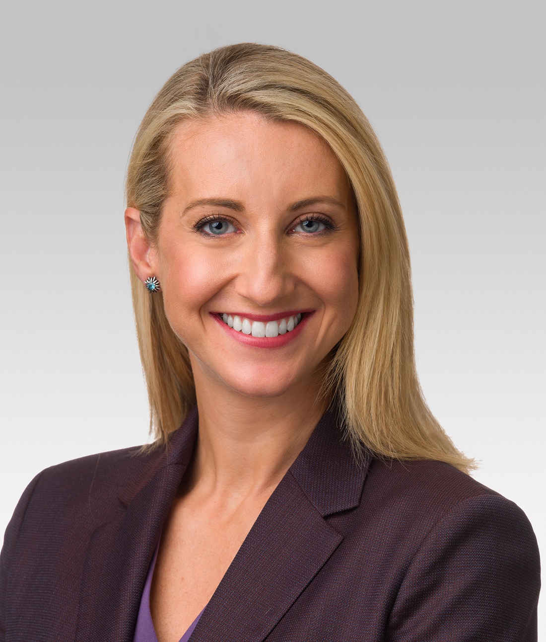
“Sotagliflozin adds to the SGLT2 inhibitor story,” and the SOLOIST-WHF results “may shift our focus to vulnerable, acute heart failure patients with an opportunity to treat during the transition phase,” when these patients leave the hospital, commented Jane E. Wilcox, MD, the study’s designated discussant and a heart failure cardiologist at Northwestern Medicine in Chicago.
A dual SGLT inhibitor
What sets sotagliflozin apart from the SGLT2 inhibitors is that it not only inhibits that protein but also SGTL1, which primarily resides in the gastrointestinal tract and is the main route for gut absorption of glucose. Dr. Bhatt said that he was unaware of any other SGLT1/2 inhibitors currently in advanced clinical testing.
The activity of sotagliflozin against the SGLT1 protein likely explains its ability to cut A1c levels in patients with severe renal dysfunction, a condition that stymies glucose lowering by SGLT2 inhibitors. In SCORED, which randomized 10,584 patients with T2D at 750 study sites in 44 countries, 813 patients (8%) had an eGFR of 25-29 mL/min per 1.73 m2 at enrollment. Sotagliflozin treatment led to an average 0.6% cut in A1c in this subgroup, and by the same average amount among the patients with GFRs of 30-60 mL/min per 1.73 m2.
“This is a huge finding for endocrinologists and primary care physicians” who treat patients with T2D who have severe renal dysfunction, said Dr. Bhatt, a professor of medicine at Harvard Medical School in Boston. “It’s a good enough reason by itself to approve this drug.”
The same mechanism may also be behind another unexpected finding in SCORED. Treatment with sotagliflozin cut the rate of total episodes of cardiovascular death, nonfatal MI, or nonfatal stroke by an absolute 1.6%, compared with placebo, and by a relative 23%. This benefit was largely driven by a 32% relative risk reduction total in MIs, and a 34% relative risk reduction in total stroke, both significant differences.
“No SGLT2 inhibitor has shown a reduction in stroke, and the MI signals have been mixed. The sizable MI and stroke effects are unique to sotagliflozin,” compared with the SGLT2 inhibitors, and likely reflect one or more mechanisms that result from blocked gut SGLT1 and a cut in GI glucose uptake, said Dr. Bhatt. “Probably some novel mechanism we don’t fully understand.”
First-ever HFpEF benefit
In contrast to these two benefits that are probably unique to drugs that inhibit the SGLT1 protein, sotagliflozin showed two other notable and unprecedented benefits that are likely generalizable to the SGLT2 inhibitors.
First is the striking benefit for HFpEF. Neither SOLOIST, which enrolled 1,222 patients with T2D and just hospitalized for worsening heart failure, nor SCORED, which enrolled patients with T2D and chronic kidney disease based exclusively on an eGFR of 25-60 mL/min per 1.73 m2, excluded patients with HFpEF, defined as heart failure patients with a left ventricular ejection fraction of at least 50%. The two studies together included a total of 739 of these patients, and they split fairly evenly between treatment with sotagliflozin or placebo.
The combined analysis showed that the incidence rate for the primary endpoint in both SOLOIST and SCORED was 59% with placebo and 39% with sotagliflozin, an absolute event reduction of 11.6 events/100 patient-years, and a significant 37% relative risk reduction, with a number needed to treat to prevent 1 event per year event of 9.
Although this observation comes from a nonprespecified combined analysis, “to me this result seems real, and I think it’s a class effect that I’m willing to extrapolate to the SGLT2 inhibitors,” Dr. Bhatt said. “It will change my practice,” he added, by spurring him to more aggressively prescribe an SGLT2 inhibitor to a patient with T2D and HFpEF.
“I think there has been some hesitation to use SGLT2 inhibitors in T2D patients with HFpEF” because of the paucity of data in this population, even though labeling and society recommendations do not rule it out. “I hope this finding will move that needle, and also generally improve SGLT2 inhibitor uptake, which has been low,” he said.
Also safe soon after acute heart failure decompensation
The other finding likely generalizable to SGLT2 inhibitors stems from the design of SOLOIST-WHF, which tested the efficacy and safety of starting sotagliflozin in patients with T2D as soon as they were stable after hospitalization for acute heart failure decompensation.
“Showing safety and efficacy when started in the hospital is pretty meaningful, because its tells patients that this drug is important and they should stay on it,” which should improve adherence, predicted Dr. Bhatt, who is also executive director of Interventional Cardiovascular Programs at Brigham and Women’s Hospital in Boston. “That’s the ultimate treatment path to prevent patients from falling through the cracks” and failing to receive an SGLT2 inhibitor.
SOLOIST-WHF enrolled patients hospitalized for worsening heart failure who also required intravenous diuretic treatment but had become stable enough to transition to an oral diuretic and come off oxygen. During a median follow-up of just over 9 months (both SOLOIST-WHF and SCORED ended sooner than planned because of a change in drug company sponsorship), treatment with sotagliflozin cut the primary endpoint by a relative 33%, compared with placebo, and with an absolute reduction of 25 events per 100 patient-years for a number needed to treat of 4. Sotagliflozin produced a strikingly high level of treatment efficiency driven by the high event rate in these recently decompensated patients. The benefit also appeared quickly, with a significant cut in events discernible within 28 days.
Extrapolating this finding to the SGLT2 inhibitors is “not a huge leap of faith,” Dr. Bhatt said.
“There is a role for sotagliflozin in acute heart failure. It showed benefit in these high-risk, transition-phase patients,” said Dr. Wilcox.
Simultaneously with Dr. Bhatt’s presentation, results of SOLOIST-WHF and SCORED were published online in the New England Journal of Medicine.
The trials were sponsored initially by Sanofi, and more recently by Lexicon. Dr. Bhatt has received research funding from both companies, and also from several other companies. He also is an adviser to several companies. Dr. Wilcox has been a consultant to Boehringer Ingelheim and Medtronic.
Sotagliflozin, a novel type of sodium-glucose cotransporter inhibitor, showed the diverse benefits this drug class provides along some new twists in a pair of international pivotal trials that together enrolled nearly 12,000 patients with type 2 diabetes.
Unprecedented benefits were seen for the first time with a drug, sotagliflozin (Zynquista) that produces both sodium-glucose cotransporter 2 inhibition as well as SGLT1 inhibition.
They included a big reduction in both MIs and strokes; an ability to meaningfully reduce hyperglycemia in patients with severe renal dysfunction with an estimated glomerular filtration rate (eGFR) of 25-29 mL/min per 1.73 m2; an ability to safely and effectively start in patients still hospitalized (but stable) for an acute heart failure episode; and a striking 37% relative risk reduction in cardiovascular death, heart failure hospitalizations, or an urgent outpatient visit for heart failure in 739 of the patients enrolled in both trials who had heart failure with preserved ejection fraction (HFpEF).
These studies produced for the first time evidence from controlled, prospective, randomized trials that a drug could improve the outcome of HFpEF patients.
All these novel outcomes came on top of the usual benefits clinicians have generally seen across the SGLT2 inhibitors already on the U.S. market: reductions in cardiovascular death and heart failure hospitalizations among all patients with type 2 diabetes, preservation of renal function, and hemoglobin A1c lowering among T2D patients with eGFR levels of at least 30 mL/min per 1.73 m2.
“The data look spectacular,” summed up Deepak L. Bhatt, MD, who presented the results from the two trials, SOLOIST-WHF and SCORED, in talks at the virtual scientific sessions of the American Heart Association.
“I think sotagliflozin has the potential to be the best in class” based on the several added attributes shown in the two trials, he said in an interview. “We’ve shown that it is very safe, well tolerated, and effective.”
The primary results were a significant 33% relative risk reduction with sotagliflozin treatment, compared with placebo in the rate of total cardiovascular deaths, hospitalizations for heart failure, or urgent outpatient visits for heart failure during just over 9 months of median follow-up among patients with T2D recently hospitalized for heart failure in SOLOIST-WFH. And a significant 26% relative risk reduction with sotagliflozin for the same endpoint after a median follow-up of just over 14 months in SCORED, which enrolled patients with T2D and chronic kidney disease.
“Sotagliflozin adds to the SGLT2 inhibitor story,” and the SOLOIST-WHF results “may shift our focus to vulnerable, acute heart failure patients with an opportunity to treat during the transition phase,” when these patients leave the hospital, commented Jane E. Wilcox, MD, the study’s designated discussant and a heart failure cardiologist at Northwestern Medicine in Chicago.
A dual SGLT inhibitor
What sets sotagliflozin apart from the SGLT2 inhibitors is that it not only inhibits that protein but also SGTL1, which primarily resides in the gastrointestinal tract and is the main route for gut absorption of glucose. Dr. Bhatt said that he was unaware of any other SGLT1/2 inhibitors currently in advanced clinical testing.
The activity of sotagliflozin against the SGLT1 protein likely explains its ability to cut A1c levels in patients with severe renal dysfunction, a condition that stymies glucose lowering by SGLT2 inhibitors. In SCORED, which randomized 10,584 patients with T2D at 750 study sites in 44 countries, 813 patients (8%) had an eGFR of 25-29 mL/min per 1.73 m2 at enrollment. Sotagliflozin treatment led to an average 0.6% cut in A1c in this subgroup, and by the same average amount among the patients with GFRs of 30-60 mL/min per 1.73 m2.
“This is a huge finding for endocrinologists and primary care physicians” who treat patients with T2D who have severe renal dysfunction, said Dr. Bhatt, a professor of medicine at Harvard Medical School in Boston. “It’s a good enough reason by itself to approve this drug.”
The same mechanism may also be behind another unexpected finding in SCORED. Treatment with sotagliflozin cut the rate of total episodes of cardiovascular death, nonfatal MI, or nonfatal stroke by an absolute 1.6%, compared with placebo, and by a relative 23%. This benefit was largely driven by a 32% relative risk reduction total in MIs, and a 34% relative risk reduction in total stroke, both significant differences.
“No SGLT2 inhibitor has shown a reduction in stroke, and the MI signals have been mixed. The sizable MI and stroke effects are unique to sotagliflozin,” compared with the SGLT2 inhibitors, and likely reflect one or more mechanisms that result from blocked gut SGLT1 and a cut in GI glucose uptake, said Dr. Bhatt. “Probably some novel mechanism we don’t fully understand.”
First-ever HFpEF benefit
In contrast to these two benefits that are probably unique to drugs that inhibit the SGLT1 protein, sotagliflozin showed two other notable and unprecedented benefits that are likely generalizable to the SGLT2 inhibitors.
First is the striking benefit for HFpEF. Neither SOLOIST, which enrolled 1,222 patients with T2D and just hospitalized for worsening heart failure, nor SCORED, which enrolled patients with T2D and chronic kidney disease based exclusively on an eGFR of 25-60 mL/min per 1.73 m2, excluded patients with HFpEF, defined as heart failure patients with a left ventricular ejection fraction of at least 50%. The two studies together included a total of 739 of these patients, and they split fairly evenly between treatment with sotagliflozin or placebo.
The combined analysis showed that the incidence rate for the primary endpoint in both SOLOIST and SCORED was 59% with placebo and 39% with sotagliflozin, an absolute event reduction of 11.6 events/100 patient-years, and a significant 37% relative risk reduction, with a number needed to treat to prevent 1 event per year event of 9.
Although this observation comes from a nonprespecified combined analysis, “to me this result seems real, and I think it’s a class effect that I’m willing to extrapolate to the SGLT2 inhibitors,” Dr. Bhatt said. “It will change my practice,” he added, by spurring him to more aggressively prescribe an SGLT2 inhibitor to a patient with T2D and HFpEF.
“I think there has been some hesitation to use SGLT2 inhibitors in T2D patients with HFpEF” because of the paucity of data in this population, even though labeling and society recommendations do not rule it out. “I hope this finding will move that needle, and also generally improve SGLT2 inhibitor uptake, which has been low,” he said.
Also safe soon after acute heart failure decompensation
The other finding likely generalizable to SGLT2 inhibitors stems from the design of SOLOIST-WHF, which tested the efficacy and safety of starting sotagliflozin in patients with T2D as soon as they were stable after hospitalization for acute heart failure decompensation.
“Showing safety and efficacy when started in the hospital is pretty meaningful, because its tells patients that this drug is important and they should stay on it,” which should improve adherence, predicted Dr. Bhatt, who is also executive director of Interventional Cardiovascular Programs at Brigham and Women’s Hospital in Boston. “That’s the ultimate treatment path to prevent patients from falling through the cracks” and failing to receive an SGLT2 inhibitor.
SOLOIST-WHF enrolled patients hospitalized for worsening heart failure who also required intravenous diuretic treatment but had become stable enough to transition to an oral diuretic and come off oxygen. During a median follow-up of just over 9 months (both SOLOIST-WHF and SCORED ended sooner than planned because of a change in drug company sponsorship), treatment with sotagliflozin cut the primary endpoint by a relative 33%, compared with placebo, and with an absolute reduction of 25 events per 100 patient-years for a number needed to treat of 4. Sotagliflozin produced a strikingly high level of treatment efficiency driven by the high event rate in these recently decompensated patients. The benefit also appeared quickly, with a significant cut in events discernible within 28 days.
Extrapolating this finding to the SGLT2 inhibitors is “not a huge leap of faith,” Dr. Bhatt said.
“There is a role for sotagliflozin in acute heart failure. It showed benefit in these high-risk, transition-phase patients,” said Dr. Wilcox.
Simultaneously with Dr. Bhatt’s presentation, results of SOLOIST-WHF and SCORED were published online in the New England Journal of Medicine.
The trials were sponsored initially by Sanofi, and more recently by Lexicon. Dr. Bhatt has received research funding from both companies, and also from several other companies. He also is an adviser to several companies. Dr. Wilcox has been a consultant to Boehringer Ingelheim and Medtronic.
Sotagliflozin, a novel type of sodium-glucose cotransporter inhibitor, showed the diverse benefits this drug class provides along some new twists in a pair of international pivotal trials that together enrolled nearly 12,000 patients with type 2 diabetes.
Unprecedented benefits were seen for the first time with a drug, sotagliflozin (Zynquista) that produces both sodium-glucose cotransporter 2 inhibition as well as SGLT1 inhibition.
They included a big reduction in both MIs and strokes; an ability to meaningfully reduce hyperglycemia in patients with severe renal dysfunction with an estimated glomerular filtration rate (eGFR) of 25-29 mL/min per 1.73 m2; an ability to safely and effectively start in patients still hospitalized (but stable) for an acute heart failure episode; and a striking 37% relative risk reduction in cardiovascular death, heart failure hospitalizations, or an urgent outpatient visit for heart failure in 739 of the patients enrolled in both trials who had heart failure with preserved ejection fraction (HFpEF).
These studies produced for the first time evidence from controlled, prospective, randomized trials that a drug could improve the outcome of HFpEF patients.
All these novel outcomes came on top of the usual benefits clinicians have generally seen across the SGLT2 inhibitors already on the U.S. market: reductions in cardiovascular death and heart failure hospitalizations among all patients with type 2 diabetes, preservation of renal function, and hemoglobin A1c lowering among T2D patients with eGFR levels of at least 30 mL/min per 1.73 m2.
“The data look spectacular,” summed up Deepak L. Bhatt, MD, who presented the results from the two trials, SOLOIST-WHF and SCORED, in talks at the virtual scientific sessions of the American Heart Association.
“I think sotagliflozin has the potential to be the best in class” based on the several added attributes shown in the two trials, he said in an interview. “We’ve shown that it is very safe, well tolerated, and effective.”
The primary results were a significant 33% relative risk reduction with sotagliflozin treatment, compared with placebo in the rate of total cardiovascular deaths, hospitalizations for heart failure, or urgent outpatient visits for heart failure during just over 9 months of median follow-up among patients with T2D recently hospitalized for heart failure in SOLOIST-WFH. And a significant 26% relative risk reduction with sotagliflozin for the same endpoint after a median follow-up of just over 14 months in SCORED, which enrolled patients with T2D and chronic kidney disease.
“Sotagliflozin adds to the SGLT2 inhibitor story,” and the SOLOIST-WHF results “may shift our focus to vulnerable, acute heart failure patients with an opportunity to treat during the transition phase,” when these patients leave the hospital, commented Jane E. Wilcox, MD, the study’s designated discussant and a heart failure cardiologist at Northwestern Medicine in Chicago.
A dual SGLT inhibitor
What sets sotagliflozin apart from the SGLT2 inhibitors is that it not only inhibits that protein but also SGTL1, which primarily resides in the gastrointestinal tract and is the main route for gut absorption of glucose. Dr. Bhatt said that he was unaware of any other SGLT1/2 inhibitors currently in advanced clinical testing.
The activity of sotagliflozin against the SGLT1 protein likely explains its ability to cut A1c levels in patients with severe renal dysfunction, a condition that stymies glucose lowering by SGLT2 inhibitors. In SCORED, which randomized 10,584 patients with T2D at 750 study sites in 44 countries, 813 patients (8%) had an eGFR of 25-29 mL/min per 1.73 m2 at enrollment. Sotagliflozin treatment led to an average 0.6% cut in A1c in this subgroup, and by the same average amount among the patients with GFRs of 30-60 mL/min per 1.73 m2.
“This is a huge finding for endocrinologists and primary care physicians” who treat patients with T2D who have severe renal dysfunction, said Dr. Bhatt, a professor of medicine at Harvard Medical School in Boston. “It’s a good enough reason by itself to approve this drug.”
The same mechanism may also be behind another unexpected finding in SCORED. Treatment with sotagliflozin cut the rate of total episodes of cardiovascular death, nonfatal MI, or nonfatal stroke by an absolute 1.6%, compared with placebo, and by a relative 23%. This benefit was largely driven by a 32% relative risk reduction total in MIs, and a 34% relative risk reduction in total stroke, both significant differences.
“No SGLT2 inhibitor has shown a reduction in stroke, and the MI signals have been mixed. The sizable MI and stroke effects are unique to sotagliflozin,” compared with the SGLT2 inhibitors, and likely reflect one or more mechanisms that result from blocked gut SGLT1 and a cut in GI glucose uptake, said Dr. Bhatt. “Probably some novel mechanism we don’t fully understand.”
First-ever HFpEF benefit
In contrast to these two benefits that are probably unique to drugs that inhibit the SGLT1 protein, sotagliflozin showed two other notable and unprecedented benefits that are likely generalizable to the SGLT2 inhibitors.
First is the striking benefit for HFpEF. Neither SOLOIST, which enrolled 1,222 patients with T2D and just hospitalized for worsening heart failure, nor SCORED, which enrolled patients with T2D and chronic kidney disease based exclusively on an eGFR of 25-60 mL/min per 1.73 m2, excluded patients with HFpEF, defined as heart failure patients with a left ventricular ejection fraction of at least 50%. The two studies together included a total of 739 of these patients, and they split fairly evenly between treatment with sotagliflozin or placebo.
The combined analysis showed that the incidence rate for the primary endpoint in both SOLOIST and SCORED was 59% with placebo and 39% with sotagliflozin, an absolute event reduction of 11.6 events/100 patient-years, and a significant 37% relative risk reduction, with a number needed to treat to prevent 1 event per year event of 9.
Although this observation comes from a nonprespecified combined analysis, “to me this result seems real, and I think it’s a class effect that I’m willing to extrapolate to the SGLT2 inhibitors,” Dr. Bhatt said. “It will change my practice,” he added, by spurring him to more aggressively prescribe an SGLT2 inhibitor to a patient with T2D and HFpEF.
“I think there has been some hesitation to use SGLT2 inhibitors in T2D patients with HFpEF” because of the paucity of data in this population, even though labeling and society recommendations do not rule it out. “I hope this finding will move that needle, and also generally improve SGLT2 inhibitor uptake, which has been low,” he said.
Also safe soon after acute heart failure decompensation
The other finding likely generalizable to SGLT2 inhibitors stems from the design of SOLOIST-WHF, which tested the efficacy and safety of starting sotagliflozin in patients with T2D as soon as they were stable after hospitalization for acute heart failure decompensation.
“Showing safety and efficacy when started in the hospital is pretty meaningful, because its tells patients that this drug is important and they should stay on it,” which should improve adherence, predicted Dr. Bhatt, who is also executive director of Interventional Cardiovascular Programs at Brigham and Women’s Hospital in Boston. “That’s the ultimate treatment path to prevent patients from falling through the cracks” and failing to receive an SGLT2 inhibitor.
SOLOIST-WHF enrolled patients hospitalized for worsening heart failure who also required intravenous diuretic treatment but had become stable enough to transition to an oral diuretic and come off oxygen. During a median follow-up of just over 9 months (both SOLOIST-WHF and SCORED ended sooner than planned because of a change in drug company sponsorship), treatment with sotagliflozin cut the primary endpoint by a relative 33%, compared with placebo, and with an absolute reduction of 25 events per 100 patient-years for a number needed to treat of 4. Sotagliflozin produced a strikingly high level of treatment efficiency driven by the high event rate in these recently decompensated patients. The benefit also appeared quickly, with a significant cut in events discernible within 28 days.
Extrapolating this finding to the SGLT2 inhibitors is “not a huge leap of faith,” Dr. Bhatt said.
“There is a role for sotagliflozin in acute heart failure. It showed benefit in these high-risk, transition-phase patients,” said Dr. Wilcox.
Simultaneously with Dr. Bhatt’s presentation, results of SOLOIST-WHF and SCORED were published online in the New England Journal of Medicine.
The trials were sponsored initially by Sanofi, and more recently by Lexicon. Dr. Bhatt has received research funding from both companies, and also from several other companies. He also is an adviser to several companies. Dr. Wilcox has been a consultant to Boehringer Ingelheim and Medtronic.
FROM AHA 2020
Factor XI inhibitor–based anticoagulation strategies gain ground
according to Jeffrey I. Weitz, MD.
These strategies could pick up where direct-acting oral anticoagulants leave off, he suggested during a presentation at the biennial summit of the Thrombosis & Hemostasis Societies of North America.
“We all know that the direct oral anticoagulants – the DOACs – are an advance over vitamin K antagonists,” said Dr. Weitz, professor of medicine and biochemistry at McMaster University, Hamilton, Ontario.
Not only are DOACs at least as effective as vitamin K antagonists such as warfarin for stroke prevention in atrial fibrillation or for treatment of venous thromboembolism (VTE), but they also reduce intracranial bleeding and major bleeding risk in those settings, respectively, and they are more convenient to administer because they can be delivered using fixed doses without the need for coagulation monitoring, he added.
Still, new targets are needed, he said, explaining that, although DOACs moved closer to the goal of attenuating thrombosis without increasing the risk of bleeding, annual rates of major bleeding remain at 2%-3% in the atrial fibrillation population, and rates of major and clinically relevant nonmajor bleeding are about 10%.
“The fear of bleeding leads to underuse of anticoagulants for eligible patients with atrial fibrillation and inappropriate use of low-dose [non–vitamin K antagonist oral anticoagulant] regimens, which can leave patients unprotected from thrombotic complications,” he said.
Factor XI
That’s where Factor XI (FXI) may come in, Dr. Weitz said.
Current anticoagulants target enzymes, including FXa or thrombin, in the common pathway of coagulation, but the intrinsic pathway at the level of FXI and FXII has attracted attention in recent years.
The intrinsic pathway is activated when blood comes into contact with medical devices like stents, mechanical heart valves, or central venous catheters, but evidence also suggests that it plays a role in clot stabilization and growth, he explained, noting additional evidence of attenuation of thrombosis in mice deficient in FXI or FXII and in animals with FXI or FXII inhibitors.
“There is no bleeding with congenital FXII deficiency, and patients with FXI deficiency rarely have spontaneous bleeding, although they can bleed with surgery or trauma,” he noted. “Therefore, the promise of contact pathway inhibition is that we can attenuate thrombosis with little or no disruption of hemostasis.”
The initiators of the intrinsic pathway are naturally occurring polyphosphates that can activate FXI and FXII, promote platelet activation, and lead to thrombosis. A number of agents are being investigated to target these enzymes – particularly FXI, for which the strongest epidemiological and other evidence of its link with thrombosis exists. He noted that FXI deficiency appears protective against deep-vein thrombosis (DVT) and ischemic stroke, whereas high levels are linked with an increased risk of venous and arterial thrombosis.
Investigative strategies include the use of antisense oligonucleotides to reduce hepatic synthesis of FXI, aptamers to bind FXI and block its activity, antibodies to bind FXI and block its activation or activity, and small molecules to bind reversibly to the active site of FXI and block its activity “much like the DOACs block the activity of FXa or thrombin.”
“We have to remember that the DOACs have taken over from vitamin K antagonists, like warfarin, for many indications, and as they go generic their uptake will increase even further,” Dr. Weitz said. “When we compare the FXI inhibitors with existing anticoagulants, we don’t necessarily want to go up against the DOACs – we’re looking for indications where [DOACs] have yet to be tested or may be unsafe.”
Potential indications include the following:
Prevention of major adverse cardiovascular events in patients with end-stage renal disease with or without atrial fibrillation.
Provision of a safer platform for antiplatelet therapy in patients with acute coronary syndrome.
Secondary stroke prevention.
Prevention or treatment of cancer-associated VTE.
Prevention of thrombosis associated with central venous catheters, left ventricular assist devices, or mechanical heart valves.
Agents in development
Of the FXI inhibitors in development, ISIS-FXIRx, an antisense oligonucleotide against FXI, is furthest along. In a study published in Blood, ISIS-FXIRx produced a dose-dependent and sustained reduction in FXI levels in healthy volunteers, and in a later randomized study published in The New England Journal of Medicine, it significantly reduced the incidence of DVT in patients undergoing voluntary total knee arthroplasty (30.4% with enoxaparin vs. 4.2% with ISIS-FXIRx at a dose of 300 mg). Bleeding rates were 8.3% and 2.6%, respectively.
The findings showed the potential for reducing thrombosis without increasing bleeding by targeting FXI, Dr. Weitz said, adding that ISIS-FXIRx was also evaluated in a small study of patients with end-stage renal disease undergoing hemodialysis and was shown to produce a dose-dependent reduction in FXI levels and to reduce the incidence of category 3 and 4 clotting in the air trap and dialyzer, compared with placebo, when given in addition to heparin.
This suggests that FXI knockdown can attenuate device-associated clotting to a greater extent than heparin alone, Dr. Weitz said.
The FXIa-directed inhibitory antibody osocimab has also been evaluated in both healthy volunteers and in patients undergoing total knee arthroplasty. In a 2019 study of healthy volunteers, a single IV injection showed a dose-dependent pharmacokinetic profile and produced FXI inhibition for about 1 month, and in the FOXTROT trial published in January in JAMA by Dr. Weitz and colleagues, osocimab was shown to reduce the incidence of symptomatic VTE, asymptomatic DVT, and VTE-related death up to day 10-13 after total knee arthroplasty.
Osocimab at doses ranging from 0.3-1.8 mg/kg given postoperatively or preoperatively were noninferior to enoxaparin (rates of 15.7%-23.7% vs. 26.3%), and osocimab at a preoperative dose of 1.8 mg/kg was superior to both enoxaparin and apixaban (11.5% vs. 26.3% and 14.5%, respectively), he said.
Bleeding rates ranged from 0%-5% with osocimab, compared with 6% with enoxaparin and 2% with apixaban
Ongoing studies
Currently ongoing studies of FXI-directed anticoagulation strategies include a study comparing ISIS-FXIRx with placebo in 200 patients with end-stage renal disease, a study comparing osocimab with placebo in 600 patients with end-stage renal disease, and a study comparing abelacimab – an antibody that binds to FXI and prevents its activation by either FXIIa or thrombin, with enoxaparin in 700 patients undergoing total knee arthroplasty, Dr. Weitz said.
Additionally, there is “considerable activity” with small molecule inhibitors of FXIa, including a phase 2, placebo-controlled, dose-ranging study looking at the novel JNG-7003/BMS-986177 agent for secondary stroke/transient ischemic attack prevention in 2,500 patients and a phase 2 study comparing it with enoxaparin for postoperative thromboprophylaxis in 1,200 patients undergoing total knee arthroplasty.
Parallel phase 2 studies are also underway to compare the novel BAY-2433334 small molecule inhibitor with placebo for stroke/transient ischemic attack prevention, with apixaban for atrial fibrillation, and for prevention of major adverse cardiovascular events in patients with acute MI.
These ongoing trials will help determine the risk-benefit profile of FXI inhibitors he said.
Session comoderator Anne Rose, PharmD, pharmacy coordinator at the University of Wisconsin, Madison, noted that these types of agents have been discussed “for quite some time” and asked whether they will be available for use in clinical practice in the near future.
Dr. Weitz predicted it will be at least a few years. The studies are just now moving to phase 2b and will still need to be evaluated in phase 3 trials and for appropriate new indications, he said.
Dr. Weitz reported research support from Canadian Institutes of Health research, Heart and Stroke Foundation, and Canadian Fund for Innovation, and he is a consultant and/or scientific advisory board member for Anthos, Bayer, Boehringer-Ingelheim, Bristol-Myers Squibb, Daiichi-Sankyo, Ionis Pharmaceuticals, Janssen, Merck, Novartis, Pfizer, Portola, Servier , and Thetherex.
according to Jeffrey I. Weitz, MD.
These strategies could pick up where direct-acting oral anticoagulants leave off, he suggested during a presentation at the biennial summit of the Thrombosis & Hemostasis Societies of North America.
“We all know that the direct oral anticoagulants – the DOACs – are an advance over vitamin K antagonists,” said Dr. Weitz, professor of medicine and biochemistry at McMaster University, Hamilton, Ontario.
Not only are DOACs at least as effective as vitamin K antagonists such as warfarin for stroke prevention in atrial fibrillation or for treatment of venous thromboembolism (VTE), but they also reduce intracranial bleeding and major bleeding risk in those settings, respectively, and they are more convenient to administer because they can be delivered using fixed doses without the need for coagulation monitoring, he added.
Still, new targets are needed, he said, explaining that, although DOACs moved closer to the goal of attenuating thrombosis without increasing the risk of bleeding, annual rates of major bleeding remain at 2%-3% in the atrial fibrillation population, and rates of major and clinically relevant nonmajor bleeding are about 10%.
“The fear of bleeding leads to underuse of anticoagulants for eligible patients with atrial fibrillation and inappropriate use of low-dose [non–vitamin K antagonist oral anticoagulant] regimens, which can leave patients unprotected from thrombotic complications,” he said.
Factor XI
That’s where Factor XI (FXI) may come in, Dr. Weitz said.
Current anticoagulants target enzymes, including FXa or thrombin, in the common pathway of coagulation, but the intrinsic pathway at the level of FXI and FXII has attracted attention in recent years.
The intrinsic pathway is activated when blood comes into contact with medical devices like stents, mechanical heart valves, or central venous catheters, but evidence also suggests that it plays a role in clot stabilization and growth, he explained, noting additional evidence of attenuation of thrombosis in mice deficient in FXI or FXII and in animals with FXI or FXII inhibitors.
“There is no bleeding with congenital FXII deficiency, and patients with FXI deficiency rarely have spontaneous bleeding, although they can bleed with surgery or trauma,” he noted. “Therefore, the promise of contact pathway inhibition is that we can attenuate thrombosis with little or no disruption of hemostasis.”
The initiators of the intrinsic pathway are naturally occurring polyphosphates that can activate FXI and FXII, promote platelet activation, and lead to thrombosis. A number of agents are being investigated to target these enzymes – particularly FXI, for which the strongest epidemiological and other evidence of its link with thrombosis exists. He noted that FXI deficiency appears protective against deep-vein thrombosis (DVT) and ischemic stroke, whereas high levels are linked with an increased risk of venous and arterial thrombosis.
Investigative strategies include the use of antisense oligonucleotides to reduce hepatic synthesis of FXI, aptamers to bind FXI and block its activity, antibodies to bind FXI and block its activation or activity, and small molecules to bind reversibly to the active site of FXI and block its activity “much like the DOACs block the activity of FXa or thrombin.”
“We have to remember that the DOACs have taken over from vitamin K antagonists, like warfarin, for many indications, and as they go generic their uptake will increase even further,” Dr. Weitz said. “When we compare the FXI inhibitors with existing anticoagulants, we don’t necessarily want to go up against the DOACs – we’re looking for indications where [DOACs] have yet to be tested or may be unsafe.”
Potential indications include the following:
Prevention of major adverse cardiovascular events in patients with end-stage renal disease with or without atrial fibrillation.
Provision of a safer platform for antiplatelet therapy in patients with acute coronary syndrome.
Secondary stroke prevention.
Prevention or treatment of cancer-associated VTE.
Prevention of thrombosis associated with central venous catheters, left ventricular assist devices, or mechanical heart valves.
Agents in development
Of the FXI inhibitors in development, ISIS-FXIRx, an antisense oligonucleotide against FXI, is furthest along. In a study published in Blood, ISIS-FXIRx produced a dose-dependent and sustained reduction in FXI levels in healthy volunteers, and in a later randomized study published in The New England Journal of Medicine, it significantly reduced the incidence of DVT in patients undergoing voluntary total knee arthroplasty (30.4% with enoxaparin vs. 4.2% with ISIS-FXIRx at a dose of 300 mg). Bleeding rates were 8.3% and 2.6%, respectively.
The findings showed the potential for reducing thrombosis without increasing bleeding by targeting FXI, Dr. Weitz said, adding that ISIS-FXIRx was also evaluated in a small study of patients with end-stage renal disease undergoing hemodialysis and was shown to produce a dose-dependent reduction in FXI levels and to reduce the incidence of category 3 and 4 clotting in the air trap and dialyzer, compared with placebo, when given in addition to heparin.
This suggests that FXI knockdown can attenuate device-associated clotting to a greater extent than heparin alone, Dr. Weitz said.
The FXIa-directed inhibitory antibody osocimab has also been evaluated in both healthy volunteers and in patients undergoing total knee arthroplasty. In a 2019 study of healthy volunteers, a single IV injection showed a dose-dependent pharmacokinetic profile and produced FXI inhibition for about 1 month, and in the FOXTROT trial published in January in JAMA by Dr. Weitz and colleagues, osocimab was shown to reduce the incidence of symptomatic VTE, asymptomatic DVT, and VTE-related death up to day 10-13 after total knee arthroplasty.
Osocimab at doses ranging from 0.3-1.8 mg/kg given postoperatively or preoperatively were noninferior to enoxaparin (rates of 15.7%-23.7% vs. 26.3%), and osocimab at a preoperative dose of 1.8 mg/kg was superior to both enoxaparin and apixaban (11.5% vs. 26.3% and 14.5%, respectively), he said.
Bleeding rates ranged from 0%-5% with osocimab, compared with 6% with enoxaparin and 2% with apixaban
Ongoing studies
Currently ongoing studies of FXI-directed anticoagulation strategies include a study comparing ISIS-FXIRx with placebo in 200 patients with end-stage renal disease, a study comparing osocimab with placebo in 600 patients with end-stage renal disease, and a study comparing abelacimab – an antibody that binds to FXI and prevents its activation by either FXIIa or thrombin, with enoxaparin in 700 patients undergoing total knee arthroplasty, Dr. Weitz said.
Additionally, there is “considerable activity” with small molecule inhibitors of FXIa, including a phase 2, placebo-controlled, dose-ranging study looking at the novel JNG-7003/BMS-986177 agent for secondary stroke/transient ischemic attack prevention in 2,500 patients and a phase 2 study comparing it with enoxaparin for postoperative thromboprophylaxis in 1,200 patients undergoing total knee arthroplasty.
Parallel phase 2 studies are also underway to compare the novel BAY-2433334 small molecule inhibitor with placebo for stroke/transient ischemic attack prevention, with apixaban for atrial fibrillation, and for prevention of major adverse cardiovascular events in patients with acute MI.
These ongoing trials will help determine the risk-benefit profile of FXI inhibitors he said.
Session comoderator Anne Rose, PharmD, pharmacy coordinator at the University of Wisconsin, Madison, noted that these types of agents have been discussed “for quite some time” and asked whether they will be available for use in clinical practice in the near future.
Dr. Weitz predicted it will be at least a few years. The studies are just now moving to phase 2b and will still need to be evaluated in phase 3 trials and for appropriate new indications, he said.
Dr. Weitz reported research support from Canadian Institutes of Health research, Heart and Stroke Foundation, and Canadian Fund for Innovation, and he is a consultant and/or scientific advisory board member for Anthos, Bayer, Boehringer-Ingelheim, Bristol-Myers Squibb, Daiichi-Sankyo, Ionis Pharmaceuticals, Janssen, Merck, Novartis, Pfizer, Portola, Servier , and Thetherex.
according to Jeffrey I. Weitz, MD.
These strategies could pick up where direct-acting oral anticoagulants leave off, he suggested during a presentation at the biennial summit of the Thrombosis & Hemostasis Societies of North America.
“We all know that the direct oral anticoagulants – the DOACs – are an advance over vitamin K antagonists,” said Dr. Weitz, professor of medicine and biochemistry at McMaster University, Hamilton, Ontario.
Not only are DOACs at least as effective as vitamin K antagonists such as warfarin for stroke prevention in atrial fibrillation or for treatment of venous thromboembolism (VTE), but they also reduce intracranial bleeding and major bleeding risk in those settings, respectively, and they are more convenient to administer because they can be delivered using fixed doses without the need for coagulation monitoring, he added.
Still, new targets are needed, he said, explaining that, although DOACs moved closer to the goal of attenuating thrombosis without increasing the risk of bleeding, annual rates of major bleeding remain at 2%-3% in the atrial fibrillation population, and rates of major and clinically relevant nonmajor bleeding are about 10%.
“The fear of bleeding leads to underuse of anticoagulants for eligible patients with atrial fibrillation and inappropriate use of low-dose [non–vitamin K antagonist oral anticoagulant] regimens, which can leave patients unprotected from thrombotic complications,” he said.
Factor XI
That’s where Factor XI (FXI) may come in, Dr. Weitz said.
Current anticoagulants target enzymes, including FXa or thrombin, in the common pathway of coagulation, but the intrinsic pathway at the level of FXI and FXII has attracted attention in recent years.
The intrinsic pathway is activated when blood comes into contact with medical devices like stents, mechanical heart valves, or central venous catheters, but evidence also suggests that it plays a role in clot stabilization and growth, he explained, noting additional evidence of attenuation of thrombosis in mice deficient in FXI or FXII and in animals with FXI or FXII inhibitors.
“There is no bleeding with congenital FXII deficiency, and patients with FXI deficiency rarely have spontaneous bleeding, although they can bleed with surgery or trauma,” he noted. “Therefore, the promise of contact pathway inhibition is that we can attenuate thrombosis with little or no disruption of hemostasis.”
The initiators of the intrinsic pathway are naturally occurring polyphosphates that can activate FXI and FXII, promote platelet activation, and lead to thrombosis. A number of agents are being investigated to target these enzymes – particularly FXI, for which the strongest epidemiological and other evidence of its link with thrombosis exists. He noted that FXI deficiency appears protective against deep-vein thrombosis (DVT) and ischemic stroke, whereas high levels are linked with an increased risk of venous and arterial thrombosis.
Investigative strategies include the use of antisense oligonucleotides to reduce hepatic synthesis of FXI, aptamers to bind FXI and block its activity, antibodies to bind FXI and block its activation or activity, and small molecules to bind reversibly to the active site of FXI and block its activity “much like the DOACs block the activity of FXa or thrombin.”
“We have to remember that the DOACs have taken over from vitamin K antagonists, like warfarin, for many indications, and as they go generic their uptake will increase even further,” Dr. Weitz said. “When we compare the FXI inhibitors with existing anticoagulants, we don’t necessarily want to go up against the DOACs – we’re looking for indications where [DOACs] have yet to be tested or may be unsafe.”
Potential indications include the following:
Prevention of major adverse cardiovascular events in patients with end-stage renal disease with or without atrial fibrillation.
Provision of a safer platform for antiplatelet therapy in patients with acute coronary syndrome.
Secondary stroke prevention.
Prevention or treatment of cancer-associated VTE.
Prevention of thrombosis associated with central venous catheters, left ventricular assist devices, or mechanical heart valves.
Agents in development
Of the FXI inhibitors in development, ISIS-FXIRx, an antisense oligonucleotide against FXI, is furthest along. In a study published in Blood, ISIS-FXIRx produced a dose-dependent and sustained reduction in FXI levels in healthy volunteers, and in a later randomized study published in The New England Journal of Medicine, it significantly reduced the incidence of DVT in patients undergoing voluntary total knee arthroplasty (30.4% with enoxaparin vs. 4.2% with ISIS-FXIRx at a dose of 300 mg). Bleeding rates were 8.3% and 2.6%, respectively.
The findings showed the potential for reducing thrombosis without increasing bleeding by targeting FXI, Dr. Weitz said, adding that ISIS-FXIRx was also evaluated in a small study of patients with end-stage renal disease undergoing hemodialysis and was shown to produce a dose-dependent reduction in FXI levels and to reduce the incidence of category 3 and 4 clotting in the air trap and dialyzer, compared with placebo, when given in addition to heparin.
This suggests that FXI knockdown can attenuate device-associated clotting to a greater extent than heparin alone, Dr. Weitz said.
The FXIa-directed inhibitory antibody osocimab has also been evaluated in both healthy volunteers and in patients undergoing total knee arthroplasty. In a 2019 study of healthy volunteers, a single IV injection showed a dose-dependent pharmacokinetic profile and produced FXI inhibition for about 1 month, and in the FOXTROT trial published in January in JAMA by Dr. Weitz and colleagues, osocimab was shown to reduce the incidence of symptomatic VTE, asymptomatic DVT, and VTE-related death up to day 10-13 after total knee arthroplasty.
Osocimab at doses ranging from 0.3-1.8 mg/kg given postoperatively or preoperatively were noninferior to enoxaparin (rates of 15.7%-23.7% vs. 26.3%), and osocimab at a preoperative dose of 1.8 mg/kg was superior to both enoxaparin and apixaban (11.5% vs. 26.3% and 14.5%, respectively), he said.
Bleeding rates ranged from 0%-5% with osocimab, compared with 6% with enoxaparin and 2% with apixaban
Ongoing studies
Currently ongoing studies of FXI-directed anticoagulation strategies include a study comparing ISIS-FXIRx with placebo in 200 patients with end-stage renal disease, a study comparing osocimab with placebo in 600 patients with end-stage renal disease, and a study comparing abelacimab – an antibody that binds to FXI and prevents its activation by either FXIIa or thrombin, with enoxaparin in 700 patients undergoing total knee arthroplasty, Dr. Weitz said.
Additionally, there is “considerable activity” with small molecule inhibitors of FXIa, including a phase 2, placebo-controlled, dose-ranging study looking at the novel JNG-7003/BMS-986177 agent for secondary stroke/transient ischemic attack prevention in 2,500 patients and a phase 2 study comparing it with enoxaparin for postoperative thromboprophylaxis in 1,200 patients undergoing total knee arthroplasty.
Parallel phase 2 studies are also underway to compare the novel BAY-2433334 small molecule inhibitor with placebo for stroke/transient ischemic attack prevention, with apixaban for atrial fibrillation, and for prevention of major adverse cardiovascular events in patients with acute MI.
These ongoing trials will help determine the risk-benefit profile of FXI inhibitors he said.
Session comoderator Anne Rose, PharmD, pharmacy coordinator at the University of Wisconsin, Madison, noted that these types of agents have been discussed “for quite some time” and asked whether they will be available for use in clinical practice in the near future.
Dr. Weitz predicted it will be at least a few years. The studies are just now moving to phase 2b and will still need to be evaluated in phase 3 trials and for appropriate new indications, he said.
Dr. Weitz reported research support from Canadian Institutes of Health research, Heart and Stroke Foundation, and Canadian Fund for Innovation, and he is a consultant and/or scientific advisory board member for Anthos, Bayer, Boehringer-Ingelheim, Bristol-Myers Squibb, Daiichi-Sankyo, Ionis Pharmaceuticals, Janssen, Merck, Novartis, Pfizer, Portola, Servier , and Thetherex.
FROM THE THSNA BIENNIAL SUMMIT
Metapneumovirus infections clinically indistinguishable from flu, RSV
The all-consuming news about SARS-CoV-2 and COVID-19 has overshadowed other viral pathogens that are the cause of severe or fatal lower respiratory infections (LRI) including human metapneumovirus (HMPV).
“MPV is really a leading cause of LRI not just in children but in adults, with high mortality rates in the frail elderly, long-term care facilities, and cancer patients with pneumonia, “ said John Williams, MD, from the department of pediatric infectious diseases at the University of Pittsburgh Medical Center.
“Right now we have no effective antivirals. There are monoclonal antibodies in development that my group and others have discovered. In fact, some of these treat MPV and RSV [respiratory syncytial virus], so we may have good options,” he said in an online presentation during an annual scientific meeting on infectious diseases.
The virus preys, wolf-like, on the most vulnerable patients, including children and frail elderly adults, as well as other adults with predisposing conditions, he said.
HMPV causes acute respiratory illnesses in approximately 2%-11% of hospitalized adults, 3%-25% of organ transplant recipients or cancer patients, 4%-12% of chronic obstructive pulmonary disease exacerbations, 5%-20% of asthma exacerbations, and it has been identified in multiple outbreaks at long-term care facilities.
Relative newcomer
Metapneumovirus was isolated and discovered from children with respiratory tract disease in the early 2000s. Once included in the family of paramyxoviruses (including measles, mumps, Nipah virus, and parainfluenza virus 1-4), HMPV and RSV are now classified as pneumoviruses, based on gene order and other characteristics, Dr. Williams explained.
Various studies have consistently placed the prevalence of HMPV ranging from 5%-14% in young children with LRI, children hospitalized for wheezing, adults with cancer and LRI, adults with asthma admissions, children with upper respiratory infections, and children hospitalized in the United States and Jordan for LRI, as well as children hospitalized in the United States and Peru with acute respiratory infections.
A study tracking respiratory infections in a Rochester, N.Y., cohort from 1999 through 2003 showed that healthy elderly patients had and annual incidence of HMPV infections of 5.9%, compared with 9.1% for high-risk patients, 13.1% for young patients, and 8.5% among hospitalized adult patients.
“These percentages are virtually identical to what has been seen in the same cohort for respiratory syncytial virus, so in this multiyear prospective cohort, metapneumovirus was as common as RSV,” Dr. Williams said.
Although the incidences of both HMPV and RSV were lower among hospitalized adults “clinically, we can’t tell these respiratory viruses apart. If we know it’s circulating we can make a guess, but we really can’t discriminate them,” he added.
In the Rochester cohort the frequency of clinical symptoms – including congestion, sore throat, cough, sputum production, dyspnea, and fever – were similar among patients infected with HMPV, RSV, or influenza A, with the exception of a slightly higher incidence of wheezing (80%) with HMPV, compared with influenza.
“I can tell you as a pediatrician, this is absolutely true in children, that metapneumovirus is indistinguishable from other respiratory viruses in kids,” he said.
Fatalities among older adults
As noted before, HMPV can cause severe and fatal illness in adults. For example, during an outbreak in North Dakota in 2016, 3 of 27 hospitalized adults with HMPV (median age, 69 years) died, and 10 required mechanical or noninvasive ventilation.
In a study from Korea comparing outcomes of severe HMPV-associated community-acquired pneumonia (CAP) with those of severe influenza-associated CAP, the investigators found that 30- and 60-day mortality rates were similar between the groups, at 24% of patients with HMPV-associated CAP and 32.1% for influenza-associated CAP, and 32% versus 38.5%, respectively.
Patients at high risk for severe disease or death from HMPV infection include those over 65 years, especially frail elderly, patients with chronic obstructive pulmonary disease, immunocompromised patients, and those with cardiopulmonary diseases such as congestive heart failure.
Supportive care only
“Do we have anything for treatment? The short answer is, No,” Dr. Williams.
Supportive care is currently the only effective approach for patients with severe HMPV infection.
Ribavirin, used to treat patients with acute RSV infection, has poor in vitro activity against HMPV and poor oral bioavailability and hemolysis, and there are no randomized controlled trials to support its use in this situation.
“It really can’t be recommended, and I don’t recommend it,” he said.
Virology may still help
Mark J. Siedner, MD, an infectious diseases physician at Mass General and associate professor of medicine at Harvard Medical School, both in Boston, who was not involved in the study, said that, despite the inability to clinically distinguish HMPV from RSV or influenza A, there is still clinical value to identifying HMPV infections.
“We spend millions of dollar each year treating people for upper respiratory tract infections, often with antibacterials, sometimes with antivirals, but those have costs to the health care system, and they also have costs in terms of drug resistance,” he said in an interview seeking objective commentary.
“Diagnostic tests that determine the actual source or the cause of these upper respiratory tract infections and encourage both patients and physicians not to be using antibiotics have value,” he said.
Identifying the pathogen can also help clinicians take appropriate infection-control precautions to prevent patient-to-clinician or patient-to-patient transmission of viral infections, he added.
Dr. Williams’ research is supported by the National Institutes of Health, Henry L. Hillman Foundation, and Asher Krop Memorial Fund of Children’s Hospital of Pittsburgh. Dr. Williams and Dr. Siedner reported no relevant conflict of interest disclosures.
The all-consuming news about SARS-CoV-2 and COVID-19 has overshadowed other viral pathogens that are the cause of severe or fatal lower respiratory infections (LRI) including human metapneumovirus (HMPV).
“MPV is really a leading cause of LRI not just in children but in adults, with high mortality rates in the frail elderly, long-term care facilities, and cancer patients with pneumonia, “ said John Williams, MD, from the department of pediatric infectious diseases at the University of Pittsburgh Medical Center.
“Right now we have no effective antivirals. There are monoclonal antibodies in development that my group and others have discovered. In fact, some of these treat MPV and RSV [respiratory syncytial virus], so we may have good options,” he said in an online presentation during an annual scientific meeting on infectious diseases.
The virus preys, wolf-like, on the most vulnerable patients, including children and frail elderly adults, as well as other adults with predisposing conditions, he said.
HMPV causes acute respiratory illnesses in approximately 2%-11% of hospitalized adults, 3%-25% of organ transplant recipients or cancer patients, 4%-12% of chronic obstructive pulmonary disease exacerbations, 5%-20% of asthma exacerbations, and it has been identified in multiple outbreaks at long-term care facilities.
Relative newcomer
Metapneumovirus was isolated and discovered from children with respiratory tract disease in the early 2000s. Once included in the family of paramyxoviruses (including measles, mumps, Nipah virus, and parainfluenza virus 1-4), HMPV and RSV are now classified as pneumoviruses, based on gene order and other characteristics, Dr. Williams explained.
Various studies have consistently placed the prevalence of HMPV ranging from 5%-14% in young children with LRI, children hospitalized for wheezing, adults with cancer and LRI, adults with asthma admissions, children with upper respiratory infections, and children hospitalized in the United States and Jordan for LRI, as well as children hospitalized in the United States and Peru with acute respiratory infections.
A study tracking respiratory infections in a Rochester, N.Y., cohort from 1999 through 2003 showed that healthy elderly patients had and annual incidence of HMPV infections of 5.9%, compared with 9.1% for high-risk patients, 13.1% for young patients, and 8.5% among hospitalized adult patients.
“These percentages are virtually identical to what has been seen in the same cohort for respiratory syncytial virus, so in this multiyear prospective cohort, metapneumovirus was as common as RSV,” Dr. Williams said.
Although the incidences of both HMPV and RSV were lower among hospitalized adults “clinically, we can’t tell these respiratory viruses apart. If we know it’s circulating we can make a guess, but we really can’t discriminate them,” he added.
In the Rochester cohort the frequency of clinical symptoms – including congestion, sore throat, cough, sputum production, dyspnea, and fever – were similar among patients infected with HMPV, RSV, or influenza A, with the exception of a slightly higher incidence of wheezing (80%) with HMPV, compared with influenza.
“I can tell you as a pediatrician, this is absolutely true in children, that metapneumovirus is indistinguishable from other respiratory viruses in kids,” he said.
Fatalities among older adults
As noted before, HMPV can cause severe and fatal illness in adults. For example, during an outbreak in North Dakota in 2016, 3 of 27 hospitalized adults with HMPV (median age, 69 years) died, and 10 required mechanical or noninvasive ventilation.
In a study from Korea comparing outcomes of severe HMPV-associated community-acquired pneumonia (CAP) with those of severe influenza-associated CAP, the investigators found that 30- and 60-day mortality rates were similar between the groups, at 24% of patients with HMPV-associated CAP and 32.1% for influenza-associated CAP, and 32% versus 38.5%, respectively.
Patients at high risk for severe disease or death from HMPV infection include those over 65 years, especially frail elderly, patients with chronic obstructive pulmonary disease, immunocompromised patients, and those with cardiopulmonary diseases such as congestive heart failure.
Supportive care only
“Do we have anything for treatment? The short answer is, No,” Dr. Williams.
Supportive care is currently the only effective approach for patients with severe HMPV infection.
Ribavirin, used to treat patients with acute RSV infection, has poor in vitro activity against HMPV and poor oral bioavailability and hemolysis, and there are no randomized controlled trials to support its use in this situation.
“It really can’t be recommended, and I don’t recommend it,” he said.
Virology may still help
Mark J. Siedner, MD, an infectious diseases physician at Mass General and associate professor of medicine at Harvard Medical School, both in Boston, who was not involved in the study, said that, despite the inability to clinically distinguish HMPV from RSV or influenza A, there is still clinical value to identifying HMPV infections.
“We spend millions of dollar each year treating people for upper respiratory tract infections, often with antibacterials, sometimes with antivirals, but those have costs to the health care system, and they also have costs in terms of drug resistance,” he said in an interview seeking objective commentary.
“Diagnostic tests that determine the actual source or the cause of these upper respiratory tract infections and encourage both patients and physicians not to be using antibiotics have value,” he said.
Identifying the pathogen can also help clinicians take appropriate infection-control precautions to prevent patient-to-clinician or patient-to-patient transmission of viral infections, he added.
Dr. Williams’ research is supported by the National Institutes of Health, Henry L. Hillman Foundation, and Asher Krop Memorial Fund of Children’s Hospital of Pittsburgh. Dr. Williams and Dr. Siedner reported no relevant conflict of interest disclosures.
The all-consuming news about SARS-CoV-2 and COVID-19 has overshadowed other viral pathogens that are the cause of severe or fatal lower respiratory infections (LRI) including human metapneumovirus (HMPV).
“MPV is really a leading cause of LRI not just in children but in adults, with high mortality rates in the frail elderly, long-term care facilities, and cancer patients with pneumonia, “ said John Williams, MD, from the department of pediatric infectious diseases at the University of Pittsburgh Medical Center.
“Right now we have no effective antivirals. There are monoclonal antibodies in development that my group and others have discovered. In fact, some of these treat MPV and RSV [respiratory syncytial virus], so we may have good options,” he said in an online presentation during an annual scientific meeting on infectious diseases.
The virus preys, wolf-like, on the most vulnerable patients, including children and frail elderly adults, as well as other adults with predisposing conditions, he said.
HMPV causes acute respiratory illnesses in approximately 2%-11% of hospitalized adults, 3%-25% of organ transplant recipients or cancer patients, 4%-12% of chronic obstructive pulmonary disease exacerbations, 5%-20% of asthma exacerbations, and it has been identified in multiple outbreaks at long-term care facilities.
Relative newcomer
Metapneumovirus was isolated and discovered from children with respiratory tract disease in the early 2000s. Once included in the family of paramyxoviruses (including measles, mumps, Nipah virus, and parainfluenza virus 1-4), HMPV and RSV are now classified as pneumoviruses, based on gene order and other characteristics, Dr. Williams explained.
Various studies have consistently placed the prevalence of HMPV ranging from 5%-14% in young children with LRI, children hospitalized for wheezing, adults with cancer and LRI, adults with asthma admissions, children with upper respiratory infections, and children hospitalized in the United States and Jordan for LRI, as well as children hospitalized in the United States and Peru with acute respiratory infections.
A study tracking respiratory infections in a Rochester, N.Y., cohort from 1999 through 2003 showed that healthy elderly patients had and annual incidence of HMPV infections of 5.9%, compared with 9.1% for high-risk patients, 13.1% for young patients, and 8.5% among hospitalized adult patients.
“These percentages are virtually identical to what has been seen in the same cohort for respiratory syncytial virus, so in this multiyear prospective cohort, metapneumovirus was as common as RSV,” Dr. Williams said.
Although the incidences of both HMPV and RSV were lower among hospitalized adults “clinically, we can’t tell these respiratory viruses apart. If we know it’s circulating we can make a guess, but we really can’t discriminate them,” he added.
In the Rochester cohort the frequency of clinical symptoms – including congestion, sore throat, cough, sputum production, dyspnea, and fever – were similar among patients infected with HMPV, RSV, or influenza A, with the exception of a slightly higher incidence of wheezing (80%) with HMPV, compared with influenza.
“I can tell you as a pediatrician, this is absolutely true in children, that metapneumovirus is indistinguishable from other respiratory viruses in kids,” he said.
Fatalities among older adults
As noted before, HMPV can cause severe and fatal illness in adults. For example, during an outbreak in North Dakota in 2016, 3 of 27 hospitalized adults with HMPV (median age, 69 years) died, and 10 required mechanical or noninvasive ventilation.
In a study from Korea comparing outcomes of severe HMPV-associated community-acquired pneumonia (CAP) with those of severe influenza-associated CAP, the investigators found that 30- and 60-day mortality rates were similar between the groups, at 24% of patients with HMPV-associated CAP and 32.1% for influenza-associated CAP, and 32% versus 38.5%, respectively.
Patients at high risk for severe disease or death from HMPV infection include those over 65 years, especially frail elderly, patients with chronic obstructive pulmonary disease, immunocompromised patients, and those with cardiopulmonary diseases such as congestive heart failure.
Supportive care only
“Do we have anything for treatment? The short answer is, No,” Dr. Williams.
Supportive care is currently the only effective approach for patients with severe HMPV infection.
Ribavirin, used to treat patients with acute RSV infection, has poor in vitro activity against HMPV and poor oral bioavailability and hemolysis, and there are no randomized controlled trials to support its use in this situation.
“It really can’t be recommended, and I don’t recommend it,” he said.
Virology may still help
Mark J. Siedner, MD, an infectious diseases physician at Mass General and associate professor of medicine at Harvard Medical School, both in Boston, who was not involved in the study, said that, despite the inability to clinically distinguish HMPV from RSV or influenza A, there is still clinical value to identifying HMPV infections.
“We spend millions of dollar each year treating people for upper respiratory tract infections, often with antibacterials, sometimes with antivirals, but those have costs to the health care system, and they also have costs in terms of drug resistance,” he said in an interview seeking objective commentary.
“Diagnostic tests that determine the actual source or the cause of these upper respiratory tract infections and encourage both patients and physicians not to be using antibiotics have value,” he said.
Identifying the pathogen can also help clinicians take appropriate infection-control precautions to prevent patient-to-clinician or patient-to-patient transmission of viral infections, he added.
Dr. Williams’ research is supported by the National Institutes of Health, Henry L. Hillman Foundation, and Asher Krop Memorial Fund of Children’s Hospital of Pittsburgh. Dr. Williams and Dr. Siedner reported no relevant conflict of interest disclosures.
FROM IDWEEK 2020
Osteoporosis drugs don’t worsen COVID-19 risk, may help
New observational data are the first to support recommendations to continue osteoporosis medications during the COVID-19 pandemic, and even suggest that some agents may protect against the virus.
Findings from the cross-sectional study of 2,102 patients with osteoporosis, osteoarthritis, and/or fibromyalgia – so-called noninflammatory rheumatic conditions – during March 1 to May 3, 2020, were recently published in Aging by Josep Blanch-Rubió, MD, scientific clinical director of the Rheumatology Service, Hospital del Mar, Barcelona, and colleagues.
Patients taking denosumab, zoledronate, and calcium showed trends toward lower incidence of developing symptomatic presumed COVID-19 (polymerase chain reaction tests weren’t widely available at the time), as did those taking the antidepressant serotonin/norepinephrine inhibitor duloxetine.
Some analgesics, particularly pregabalin and most other antidepressants, were associated with higher incidences of COVID-19, while oral bisphosphonates, vitamin D, thiazide diuretics, antihypertensive drugs, and chronic nonsteroidal anti-inflammatory drugs had no effect on COVID-19 incidence.
These data are the first to support guidance issued in May 2020 by the American Society for Bone and Mineral Research and four other professional societies advising continuation of osteoporosis medications during the pandemic. That statement’s authors acknowledged that, lacking data, their recommendations were based primarily on expert opinion.
“There were guidelines without any scientific base. ... This is the first scientific evidence showing that indeed you should continue your osteoporosis treatment if you have COVID-19. This is the first study to provide scientific support for the guidelines,” study coauthor Rafael Maldonado, MD, PhD, of the Laboratory of Neuropharmacology, Universitat Pompeu Fabra, Barcelona, said in an interview.
And while the data don’t offer proof of benefit for any drug – all of the 95% confidence intervals crossed 1.0 – they do show trends that deserve further study, Dr. Maldonado said.
“What we observed is that there is no harm. Treatments should be continued.”
“But we obtained very interesting results with denosumab, zoledronate, calcium, and duloxetine. ... There is a clear tendency, and the message is we should promote studies to see if these four treatments provide benefit.”
Different mechanisms for each?
Asked to comment on the findings, Matthew T. Drake, MD, PhD, said in an interview, “I would agree that there’s no reason any of these medications should be stopped or discontinued since there’s no evidence that they make the risk for infection worse.”
“But how [some of them may] improve or reduce the infection risk in my mind is somewhat unclear. ... It’s hard to come up with a unifying explanation” because those mentioned as potentially beneficial “are fairly different,” he noted.
Dr. Drake, associate professor of medicine in the department of endocrinology at the Mayo Clinic, Rochester, Minn., said he agreed with the study authors that denosumab’s targeting of the RANK/RANKL system is a possible anti-COVID-19 mechanism for that drug because that system is involved in immune response.
Regarding zoledronate/zoledronic acid, both the Spanish authors and Dr. Drake pointed to a landmark study linking the intravenous drug to longer survival in patients with hip fracture. The study authors note that there could be several mechanisms for an overall survival benefit, but additionally, “zoledronate may make dendritic cells and their precursors less susceptible to SARS-CoV-2 infection, which could explain the beneficial effects here ... on COVID-19 incidence.”
And, the authors hypothesized, the reason for the lack of benefit with oral bisphosphonates might relate to the higher potency of the intravenous zoledronate. Dr. Drake added that its higher bioavailability may also play a role.
As for calcium, the authors suggest that the beneficial effect against COVID-19 could relate to its action in generating two immune cell types – T follicular helper cells and T follicular regulatory cells – which promote an appropriate immune response against infectious agents, including viruses.
Data supporting the guidelines
Of the 2,102 patients in the study by Blanch-Rubió and colleagues, 80.5% were women, and their mean age was 66.4 years. Overall, 63.7% had osteoarthritis, 43.5% had osteoporosis, and 27.2% had fibromyalgia. Treatments included vitamin D in 62%, calcium in 23.3%, denosumab in 12.6%, and intravenous zoledronate in 8.5%. Over half were taking analgesics and nearly a third antidepressants, with 9.9% taking duloxetine.
During the study period, 5.2%, or 109 individuals, were diagnosed with COVID-19 based on presenting for medical care with hallmark symptoms.
After adjustments for sex, age, diabetes, pulmonary disease, cardiovascular disease, chronic kidney disease, and active cancer or treatment, the relative risks for COVID-19 were 0.58 for denosumab, 0.62 for intravenous zoledronate, and 0.64 for calcium, all nonsignificant trends. No associations were found between COVID-19 and oral bisphosphonates, vitamin D, or thiazide diuretics. Increased but nonsignificant relative risks for COVID-19 were seen with analgesics, particularly pregabalin (1.55), gabapentin (1.39), and opioids (1.25).
Among antidepressants, there was a relative risk of 1.54 for selective serotonin reuptake inhibitors, 1.38 for amitriptyline, and 1.22 for all dual-action antidepressants together. In contrast, there was a negative association with the dual-action antidepressant duloxetine, with an adjusted relative risk of 0.68.
“The good news,” Dr. Drake said, “is that none of it appears bad.”
Dr. Blanch-Rubió has received grants or consulting fees from Amgen, Laboratorio Stada, Gedeon-Rhicter Ibérica, Lilly España, Pfizer, Gebro Pharma, and UCB Pharma. Dr. Maldonado has received research grants or consulting fees from Aelis, Almirall, Boehringer Ingelheim, BrainCo, Esteve, Ferrer, GlaxoSmithKline, Grünenthal, GW Pharmaceuticals, Janus, Lundbeck, Pharmaleads, Phytoplant, Rhodes, Sanofi, Spherium, Union de Pharmacologie Scientifique Appliquée, Upjohn, and Uriach. Dr. Drake has reported no relevant financial relationships.
A version of this article originally appeared on Medscape.com.
New observational data are the first to support recommendations to continue osteoporosis medications during the COVID-19 pandemic, and even suggest that some agents may protect against the virus.
Findings from the cross-sectional study of 2,102 patients with osteoporosis, osteoarthritis, and/or fibromyalgia – so-called noninflammatory rheumatic conditions – during March 1 to May 3, 2020, were recently published in Aging by Josep Blanch-Rubió, MD, scientific clinical director of the Rheumatology Service, Hospital del Mar, Barcelona, and colleagues.
Patients taking denosumab, zoledronate, and calcium showed trends toward lower incidence of developing symptomatic presumed COVID-19 (polymerase chain reaction tests weren’t widely available at the time), as did those taking the antidepressant serotonin/norepinephrine inhibitor duloxetine.
Some analgesics, particularly pregabalin and most other antidepressants, were associated with higher incidences of COVID-19, while oral bisphosphonates, vitamin D, thiazide diuretics, antihypertensive drugs, and chronic nonsteroidal anti-inflammatory drugs had no effect on COVID-19 incidence.
These data are the first to support guidance issued in May 2020 by the American Society for Bone and Mineral Research and four other professional societies advising continuation of osteoporosis medications during the pandemic. That statement’s authors acknowledged that, lacking data, their recommendations were based primarily on expert opinion.
“There were guidelines without any scientific base. ... This is the first scientific evidence showing that indeed you should continue your osteoporosis treatment if you have COVID-19. This is the first study to provide scientific support for the guidelines,” study coauthor Rafael Maldonado, MD, PhD, of the Laboratory of Neuropharmacology, Universitat Pompeu Fabra, Barcelona, said in an interview.
And while the data don’t offer proof of benefit for any drug – all of the 95% confidence intervals crossed 1.0 – they do show trends that deserve further study, Dr. Maldonado said.
“What we observed is that there is no harm. Treatments should be continued.”
“But we obtained very interesting results with denosumab, zoledronate, calcium, and duloxetine. ... There is a clear tendency, and the message is we should promote studies to see if these four treatments provide benefit.”
Different mechanisms for each?
Asked to comment on the findings, Matthew T. Drake, MD, PhD, said in an interview, “I would agree that there’s no reason any of these medications should be stopped or discontinued since there’s no evidence that they make the risk for infection worse.”
“But how [some of them may] improve or reduce the infection risk in my mind is somewhat unclear. ... It’s hard to come up with a unifying explanation” because those mentioned as potentially beneficial “are fairly different,” he noted.
Dr. Drake, associate professor of medicine in the department of endocrinology at the Mayo Clinic, Rochester, Minn., said he agreed with the study authors that denosumab’s targeting of the RANK/RANKL system is a possible anti-COVID-19 mechanism for that drug because that system is involved in immune response.
Regarding zoledronate/zoledronic acid, both the Spanish authors and Dr. Drake pointed to a landmark study linking the intravenous drug to longer survival in patients with hip fracture. The study authors note that there could be several mechanisms for an overall survival benefit, but additionally, “zoledronate may make dendritic cells and their precursors less susceptible to SARS-CoV-2 infection, which could explain the beneficial effects here ... on COVID-19 incidence.”
And, the authors hypothesized, the reason for the lack of benefit with oral bisphosphonates might relate to the higher potency of the intravenous zoledronate. Dr. Drake added that its higher bioavailability may also play a role.
As for calcium, the authors suggest that the beneficial effect against COVID-19 could relate to its action in generating two immune cell types – T follicular helper cells and T follicular regulatory cells – which promote an appropriate immune response against infectious agents, including viruses.
Data supporting the guidelines
Of the 2,102 patients in the study by Blanch-Rubió and colleagues, 80.5% were women, and their mean age was 66.4 years. Overall, 63.7% had osteoarthritis, 43.5% had osteoporosis, and 27.2% had fibromyalgia. Treatments included vitamin D in 62%, calcium in 23.3%, denosumab in 12.6%, and intravenous zoledronate in 8.5%. Over half were taking analgesics and nearly a third antidepressants, with 9.9% taking duloxetine.
During the study period, 5.2%, or 109 individuals, were diagnosed with COVID-19 based on presenting for medical care with hallmark symptoms.
After adjustments for sex, age, diabetes, pulmonary disease, cardiovascular disease, chronic kidney disease, and active cancer or treatment, the relative risks for COVID-19 were 0.58 for denosumab, 0.62 for intravenous zoledronate, and 0.64 for calcium, all nonsignificant trends. No associations were found between COVID-19 and oral bisphosphonates, vitamin D, or thiazide diuretics. Increased but nonsignificant relative risks for COVID-19 were seen with analgesics, particularly pregabalin (1.55), gabapentin (1.39), and opioids (1.25).
Among antidepressants, there was a relative risk of 1.54 for selective serotonin reuptake inhibitors, 1.38 for amitriptyline, and 1.22 for all dual-action antidepressants together. In contrast, there was a negative association with the dual-action antidepressant duloxetine, with an adjusted relative risk of 0.68.
“The good news,” Dr. Drake said, “is that none of it appears bad.”
Dr. Blanch-Rubió has received grants or consulting fees from Amgen, Laboratorio Stada, Gedeon-Rhicter Ibérica, Lilly España, Pfizer, Gebro Pharma, and UCB Pharma. Dr. Maldonado has received research grants or consulting fees from Aelis, Almirall, Boehringer Ingelheim, BrainCo, Esteve, Ferrer, GlaxoSmithKline, Grünenthal, GW Pharmaceuticals, Janus, Lundbeck, Pharmaleads, Phytoplant, Rhodes, Sanofi, Spherium, Union de Pharmacologie Scientifique Appliquée, Upjohn, and Uriach. Dr. Drake has reported no relevant financial relationships.
A version of this article originally appeared on Medscape.com.
New observational data are the first to support recommendations to continue osteoporosis medications during the COVID-19 pandemic, and even suggest that some agents may protect against the virus.
Findings from the cross-sectional study of 2,102 patients with osteoporosis, osteoarthritis, and/or fibromyalgia – so-called noninflammatory rheumatic conditions – during March 1 to May 3, 2020, were recently published in Aging by Josep Blanch-Rubió, MD, scientific clinical director of the Rheumatology Service, Hospital del Mar, Barcelona, and colleagues.
Patients taking denosumab, zoledronate, and calcium showed trends toward lower incidence of developing symptomatic presumed COVID-19 (polymerase chain reaction tests weren’t widely available at the time), as did those taking the antidepressant serotonin/norepinephrine inhibitor duloxetine.
Some analgesics, particularly pregabalin and most other antidepressants, were associated with higher incidences of COVID-19, while oral bisphosphonates, vitamin D, thiazide diuretics, antihypertensive drugs, and chronic nonsteroidal anti-inflammatory drugs had no effect on COVID-19 incidence.
These data are the first to support guidance issued in May 2020 by the American Society for Bone and Mineral Research and four other professional societies advising continuation of osteoporosis medications during the pandemic. That statement’s authors acknowledged that, lacking data, their recommendations were based primarily on expert opinion.
“There were guidelines without any scientific base. ... This is the first scientific evidence showing that indeed you should continue your osteoporosis treatment if you have COVID-19. This is the first study to provide scientific support for the guidelines,” study coauthor Rafael Maldonado, MD, PhD, of the Laboratory of Neuropharmacology, Universitat Pompeu Fabra, Barcelona, said in an interview.
And while the data don’t offer proof of benefit for any drug – all of the 95% confidence intervals crossed 1.0 – they do show trends that deserve further study, Dr. Maldonado said.
“What we observed is that there is no harm. Treatments should be continued.”
“But we obtained very interesting results with denosumab, zoledronate, calcium, and duloxetine. ... There is a clear tendency, and the message is we should promote studies to see if these four treatments provide benefit.”
Different mechanisms for each?
Asked to comment on the findings, Matthew T. Drake, MD, PhD, said in an interview, “I would agree that there’s no reason any of these medications should be stopped or discontinued since there’s no evidence that they make the risk for infection worse.”
“But how [some of them may] improve or reduce the infection risk in my mind is somewhat unclear. ... It’s hard to come up with a unifying explanation” because those mentioned as potentially beneficial “are fairly different,” he noted.
Dr. Drake, associate professor of medicine in the department of endocrinology at the Mayo Clinic, Rochester, Minn., said he agreed with the study authors that denosumab’s targeting of the RANK/RANKL system is a possible anti-COVID-19 mechanism for that drug because that system is involved in immune response.
Regarding zoledronate/zoledronic acid, both the Spanish authors and Dr. Drake pointed to a landmark study linking the intravenous drug to longer survival in patients with hip fracture. The study authors note that there could be several mechanisms for an overall survival benefit, but additionally, “zoledronate may make dendritic cells and their precursors less susceptible to SARS-CoV-2 infection, which could explain the beneficial effects here ... on COVID-19 incidence.”
And, the authors hypothesized, the reason for the lack of benefit with oral bisphosphonates might relate to the higher potency of the intravenous zoledronate. Dr. Drake added that its higher bioavailability may also play a role.
As for calcium, the authors suggest that the beneficial effect against COVID-19 could relate to its action in generating two immune cell types – T follicular helper cells and T follicular regulatory cells – which promote an appropriate immune response against infectious agents, including viruses.
Data supporting the guidelines
Of the 2,102 patients in the study by Blanch-Rubió and colleagues, 80.5% were women, and their mean age was 66.4 years. Overall, 63.7% had osteoarthritis, 43.5% had osteoporosis, and 27.2% had fibromyalgia. Treatments included vitamin D in 62%, calcium in 23.3%, denosumab in 12.6%, and intravenous zoledronate in 8.5%. Over half were taking analgesics and nearly a third antidepressants, with 9.9% taking duloxetine.
During the study period, 5.2%, or 109 individuals, were diagnosed with COVID-19 based on presenting for medical care with hallmark symptoms.
After adjustments for sex, age, diabetes, pulmonary disease, cardiovascular disease, chronic kidney disease, and active cancer or treatment, the relative risks for COVID-19 were 0.58 for denosumab, 0.62 for intravenous zoledronate, and 0.64 for calcium, all nonsignificant trends. No associations were found between COVID-19 and oral bisphosphonates, vitamin D, or thiazide diuretics. Increased but nonsignificant relative risks for COVID-19 were seen with analgesics, particularly pregabalin (1.55), gabapentin (1.39), and opioids (1.25).
Among antidepressants, there was a relative risk of 1.54 for selective serotonin reuptake inhibitors, 1.38 for amitriptyline, and 1.22 for all dual-action antidepressants together. In contrast, there was a negative association with the dual-action antidepressant duloxetine, with an adjusted relative risk of 0.68.
“The good news,” Dr. Drake said, “is that none of it appears bad.”
Dr. Blanch-Rubió has received grants or consulting fees from Amgen, Laboratorio Stada, Gedeon-Rhicter Ibérica, Lilly España, Pfizer, Gebro Pharma, and UCB Pharma. Dr. Maldonado has received research grants or consulting fees from Aelis, Almirall, Boehringer Ingelheim, BrainCo, Esteve, Ferrer, GlaxoSmithKline, Grünenthal, GW Pharmaceuticals, Janus, Lundbeck, Pharmaleads, Phytoplant, Rhodes, Sanofi, Spherium, Union de Pharmacologie Scientifique Appliquée, Upjohn, and Uriach. Dr. Drake has reported no relevant financial relationships.
A version of this article originally appeared on Medscape.com.
Cardiac arrest in COVID-19 pandemic: ‘Survival is possible’
In the early weeks of the COVID-19 pandemic in the United States, rates of sustained return of spontaneous circulation after out-of-hospital cardiac arrest were lower throughout the country, compared with a year earlier, in one study.
A second study of that period showed that patients with COVID-19 had rates that were better than previously reported of surviving in-hospital cardiac arrest.
Paul S. Chan, MD, presented the out-of-hospital cardiac arrest research, and Oscar J. Mitchell, MD, presented the in-hospital cardiac arrest findings in a late-breaking resuscitation science session at the American Heart Association scientific sessions. The former study was also simultaneously published online Nov. 14 in JAMA Cardiology.
Importantly, “the survival rates were not zero in either setting,” said Dr. Chan, commenting on the implications of both studies taken together.
“The survival rates – either return of circulation or survival to discharge – were not futile,” Dr. Chan, from Saint Luke’s Mid America Heart Institute, Kansas City, Missouri, said in an interview.
“And I think that’s an overall important message – that we can’t write off patients who have a cardiac arrest at this point,” he stressed. “They deserve a response. Although the outcomes might not be as good as we had seen in years prior, we are seeing patients making it out of the hospital and surviving.”
Dr. Mitchell, from the University of Pennsylvania in Philadelphia, echoed this message in an interview.
“I think that the key finding here is that survival is possible after patients with COVID-19 suffer an in-hospital cardiac arrest,” Dr. Mitchell said. “We hope that the information from our study will be of use to frontline providers who are treating patients with COVID-19.”
“In coming weeks, there will likely be increased hospital strain and enormous challenges to providing COVID-19 care,” added Benjamin S. Abella, MD, the senior author of the in-hospital study. Dr. Abella is also from the University of Pennsylvania and was cochair of the Resuscitation Science symposium during the AHA meeting.
“It is crucial that hospital leaders prepare now for how they will manage COVID-19 resuscitation efforts,” Dr. Abella said. “Emergency medicine and critical care leaders must be mindful that many COVID-19 patients with arrest could survive to return to their families.”
“It is important to note both studies demonstrated variations in outcome and that those differences were associated with the differential COVID prevalence and mortality,” session comoderator Cindy H. Hsu, MD, PhD, University of Michigan, said in an interview.
“Future studies,” she said, “should address knowledge gaps including associated comorbidities and affected resuscitation process variables during the COVID-19 pandemic.”
Out-of-hospital cardiac arrest, March 2019 vs. March 2020
Compared with 2019, in 2020, the reported rates of return of spontaneous circulation after out-of-hospital cardiac arrest fell from 25% to 10.6% in New York and from 13.5% to 5.0% in northern Italy – two areas that were severely affected, Dr. Chan noted.
In this study, the researchers aimed to examine whether out-of-hospital cardiac arrest outcomes would be similar throughout the United States, including areas that were less severely affected, in the first weeks of the pandemic.
They linked data from the Cardiac Arrest Registry to Enhance Survival (CARES), which covers an area with about 152 million U.S. residents, with COVID-19 disease mortality data.
There were 9,863 out-of-hospital arrests from March 16 to April 30, 2020, compared with 9,440 cases during this time in 2019.
The patients in both years had a similar age (mean, 62 years) and sex (62% male), but there were more Black patients in 2020 (28% vs. 23%).
Overall, in communities with low to high rates of death from COVID-19, the rate of return of spontaneous circulation was 18% lower in that early pandemic period than in the same time in the previous year (23% vs. 29.8%; adjusted rate ratio, 0.82).
The rates of return of spontaneous circulation were also lower in communities with a low rate of COVID-19 mortality, but to a lesser extent (11%-15% lower in 2020 vs. 2019).
In the subset of emergency medical agencies with complete data on hospital survival, overall rates of survival to discharge were 17% lower during the studied pandemic period versus the same time a year earlier (6.6% vs. 9.8%; adjusted RR, 0.83).
This drop in survival was greater in communities with moderate to high COVID-19 mortality.
These outcomes were not explained by differences in emergency medical services arrival or treatment times, rates of bystander CPR, or initial out-of-hospital cardiac arrest rhythm.
Dr. Chan was a coauthor of an interim guidance issued April 9, 2020, by the AHA and several other medical societies for ways to protect frontline workers from contracting COVID-19 while they were performing CPR.
Communities that were not heavily affected by COVID-19 could have also been following the recommendations, which might have affected outcomes, he speculated.
For example, “when we pause chest compressions it can potentially worsen survival even if it’s for a short period of time. That might explain the lower rates of return of circulation.”
“That guidance was really meant for heavily affected communities,” Dr. Chan added. “Of course, as we speak, the pandemic is pretty much everywhere in the United States. It’s not just in the northeast; it’s not just in Arizona, Florida, California, Texas like it was in the summer. You are seeing surges in 46 of the 50 states.
“If your community is heavily affected by COVID-19 in terms of deaths at this time, paramedics will need to take caution to also help protect themselves, and the guidance may apply at that point,” he said.
In-hospital cardiac arrest, March Through May 2020
The early studies of in-hospital cardiac arrest in patients with COVID-19 showed “concerningly low rates” of return of spontaneous circulation and survival, said Dr. Mitchell.
“The first was a study from Wuhan, which demonstrated a 2.9% 30-day survival and the second was a small cohort from NYC with 0% survival to hospital discharge,” he said. “This raised concerns that offering CPR to patients who had a cardiac arrest from COVID-19 might only hold a low probability of success.”
To investigate this, the researchers formed a COVID study group comprising two hospitals in New York and nine hospitals in the Northeast and West Coast.
They identified 260 hospitalized adult patients with COVID-19 who had in-hospital cardiac arrest between March 1 and May 31, 2020. The patients had a median age of 69 years, and 72% were male. Most had preexisting comorbidities. Most of the cardiac arrests were in the ICU (64%), and almost all were witnessed (91%).
Return of spontaneous circulation occurred in 22% of the patients, and 12% had survived 30 days later. Of the 260 cardiac arrests, most (204) occurred in the New York hospitals.
There was a huge variation in outcomes. The rate of sustained return of spontaneous circulation was much lower in the two hospitals in New York compared with elsewhere (11% vs. 64%), as was 30-day survival (6% vs. 36%).
“Variation in outcomes from [in-hospital cardiac arrest] has been well described prior to the COVID-19 pandemic,” said Dr. Mitchell, “and is felt to be due to a range of factors, including variation in detection and prevention of cardiac arrest, management of patients during the cardiac arrest, and differences in postarrest care – including targeted temperature management and neuroprognostication.”
“We hypothesize that the strains of the COVID-19 pandemic may have amplified these variations (although we were unable to compare hospital performance before and after the pandemic),” he said.
Nevertheless, “in contrast to [earlier] studies, we have found that survival with a good neurological status is possible after in-hospital cardiac arrest in patients with COVID-19, which is certainly reassuring for those of us on the front line.”
Dr. Chan has received research support from the American Heart Association (which helps fund CARES); the National Heart, Lung, and Blood Institute; and Optum Rx. Dr. Abella has received honoraria from NeuroproteXeon, Becton Dickinson, and Physio-Control, and research grants from Medtronic, PCORI, Physio-Control, Stryker, and TerSera. Dr. Mitchell has disclosed no relevant financial relationships.
A version of this article originally appeared on Medscape.com.
In the early weeks of the COVID-19 pandemic in the United States, rates of sustained return of spontaneous circulation after out-of-hospital cardiac arrest were lower throughout the country, compared with a year earlier, in one study.
A second study of that period showed that patients with COVID-19 had rates that were better than previously reported of surviving in-hospital cardiac arrest.
Paul S. Chan, MD, presented the out-of-hospital cardiac arrest research, and Oscar J. Mitchell, MD, presented the in-hospital cardiac arrest findings in a late-breaking resuscitation science session at the American Heart Association scientific sessions. The former study was also simultaneously published online Nov. 14 in JAMA Cardiology.
Importantly, “the survival rates were not zero in either setting,” said Dr. Chan, commenting on the implications of both studies taken together.
“The survival rates – either return of circulation or survival to discharge – were not futile,” Dr. Chan, from Saint Luke’s Mid America Heart Institute, Kansas City, Missouri, said in an interview.
“And I think that’s an overall important message – that we can’t write off patients who have a cardiac arrest at this point,” he stressed. “They deserve a response. Although the outcomes might not be as good as we had seen in years prior, we are seeing patients making it out of the hospital and surviving.”
Dr. Mitchell, from the University of Pennsylvania in Philadelphia, echoed this message in an interview.
“I think that the key finding here is that survival is possible after patients with COVID-19 suffer an in-hospital cardiac arrest,” Dr. Mitchell said. “We hope that the information from our study will be of use to frontline providers who are treating patients with COVID-19.”
“In coming weeks, there will likely be increased hospital strain and enormous challenges to providing COVID-19 care,” added Benjamin S. Abella, MD, the senior author of the in-hospital study. Dr. Abella is also from the University of Pennsylvania and was cochair of the Resuscitation Science symposium during the AHA meeting.
“It is crucial that hospital leaders prepare now for how they will manage COVID-19 resuscitation efforts,” Dr. Abella said. “Emergency medicine and critical care leaders must be mindful that many COVID-19 patients with arrest could survive to return to their families.”
“It is important to note both studies demonstrated variations in outcome and that those differences were associated with the differential COVID prevalence and mortality,” session comoderator Cindy H. Hsu, MD, PhD, University of Michigan, said in an interview.
“Future studies,” she said, “should address knowledge gaps including associated comorbidities and affected resuscitation process variables during the COVID-19 pandemic.”
Out-of-hospital cardiac arrest, March 2019 vs. March 2020
Compared with 2019, in 2020, the reported rates of return of spontaneous circulation after out-of-hospital cardiac arrest fell from 25% to 10.6% in New York and from 13.5% to 5.0% in northern Italy – two areas that were severely affected, Dr. Chan noted.
In this study, the researchers aimed to examine whether out-of-hospital cardiac arrest outcomes would be similar throughout the United States, including areas that were less severely affected, in the first weeks of the pandemic.
They linked data from the Cardiac Arrest Registry to Enhance Survival (CARES), which covers an area with about 152 million U.S. residents, with COVID-19 disease mortality data.
There were 9,863 out-of-hospital arrests from March 16 to April 30, 2020, compared with 9,440 cases during this time in 2019.
The patients in both years had a similar age (mean, 62 years) and sex (62% male), but there were more Black patients in 2020 (28% vs. 23%).
Overall, in communities with low to high rates of death from COVID-19, the rate of return of spontaneous circulation was 18% lower in that early pandemic period than in the same time in the previous year (23% vs. 29.8%; adjusted rate ratio, 0.82).
The rates of return of spontaneous circulation were also lower in communities with a low rate of COVID-19 mortality, but to a lesser extent (11%-15% lower in 2020 vs. 2019).
In the subset of emergency medical agencies with complete data on hospital survival, overall rates of survival to discharge were 17% lower during the studied pandemic period versus the same time a year earlier (6.6% vs. 9.8%; adjusted RR, 0.83).
This drop in survival was greater in communities with moderate to high COVID-19 mortality.
These outcomes were not explained by differences in emergency medical services arrival or treatment times, rates of bystander CPR, or initial out-of-hospital cardiac arrest rhythm.
Dr. Chan was a coauthor of an interim guidance issued April 9, 2020, by the AHA and several other medical societies for ways to protect frontline workers from contracting COVID-19 while they were performing CPR.
Communities that were not heavily affected by COVID-19 could have also been following the recommendations, which might have affected outcomes, he speculated.
For example, “when we pause chest compressions it can potentially worsen survival even if it’s for a short period of time. That might explain the lower rates of return of circulation.”
“That guidance was really meant for heavily affected communities,” Dr. Chan added. “Of course, as we speak, the pandemic is pretty much everywhere in the United States. It’s not just in the northeast; it’s not just in Arizona, Florida, California, Texas like it was in the summer. You are seeing surges in 46 of the 50 states.
“If your community is heavily affected by COVID-19 in terms of deaths at this time, paramedics will need to take caution to also help protect themselves, and the guidance may apply at that point,” he said.
In-hospital cardiac arrest, March Through May 2020
The early studies of in-hospital cardiac arrest in patients with COVID-19 showed “concerningly low rates” of return of spontaneous circulation and survival, said Dr. Mitchell.
“The first was a study from Wuhan, which demonstrated a 2.9% 30-day survival and the second was a small cohort from NYC with 0% survival to hospital discharge,” he said. “This raised concerns that offering CPR to patients who had a cardiac arrest from COVID-19 might only hold a low probability of success.”
To investigate this, the researchers formed a COVID study group comprising two hospitals in New York and nine hospitals in the Northeast and West Coast.
They identified 260 hospitalized adult patients with COVID-19 who had in-hospital cardiac arrest between March 1 and May 31, 2020. The patients had a median age of 69 years, and 72% were male. Most had preexisting comorbidities. Most of the cardiac arrests were in the ICU (64%), and almost all were witnessed (91%).
Return of spontaneous circulation occurred in 22% of the patients, and 12% had survived 30 days later. Of the 260 cardiac arrests, most (204) occurred in the New York hospitals.
There was a huge variation in outcomes. The rate of sustained return of spontaneous circulation was much lower in the two hospitals in New York compared with elsewhere (11% vs. 64%), as was 30-day survival (6% vs. 36%).
“Variation in outcomes from [in-hospital cardiac arrest] has been well described prior to the COVID-19 pandemic,” said Dr. Mitchell, “and is felt to be due to a range of factors, including variation in detection and prevention of cardiac arrest, management of patients during the cardiac arrest, and differences in postarrest care – including targeted temperature management and neuroprognostication.”
“We hypothesize that the strains of the COVID-19 pandemic may have amplified these variations (although we were unable to compare hospital performance before and after the pandemic),” he said.
Nevertheless, “in contrast to [earlier] studies, we have found that survival with a good neurological status is possible after in-hospital cardiac arrest in patients with COVID-19, which is certainly reassuring for those of us on the front line.”
Dr. Chan has received research support from the American Heart Association (which helps fund CARES); the National Heart, Lung, and Blood Institute; and Optum Rx. Dr. Abella has received honoraria from NeuroproteXeon, Becton Dickinson, and Physio-Control, and research grants from Medtronic, PCORI, Physio-Control, Stryker, and TerSera. Dr. Mitchell has disclosed no relevant financial relationships.
A version of this article originally appeared on Medscape.com.
In the early weeks of the COVID-19 pandemic in the United States, rates of sustained return of spontaneous circulation after out-of-hospital cardiac arrest were lower throughout the country, compared with a year earlier, in one study.
A second study of that period showed that patients with COVID-19 had rates that were better than previously reported of surviving in-hospital cardiac arrest.
Paul S. Chan, MD, presented the out-of-hospital cardiac arrest research, and Oscar J. Mitchell, MD, presented the in-hospital cardiac arrest findings in a late-breaking resuscitation science session at the American Heart Association scientific sessions. The former study was also simultaneously published online Nov. 14 in JAMA Cardiology.
Importantly, “the survival rates were not zero in either setting,” said Dr. Chan, commenting on the implications of both studies taken together.
“The survival rates – either return of circulation or survival to discharge – were not futile,” Dr. Chan, from Saint Luke’s Mid America Heart Institute, Kansas City, Missouri, said in an interview.
“And I think that’s an overall important message – that we can’t write off patients who have a cardiac arrest at this point,” he stressed. “They deserve a response. Although the outcomes might not be as good as we had seen in years prior, we are seeing patients making it out of the hospital and surviving.”
Dr. Mitchell, from the University of Pennsylvania in Philadelphia, echoed this message in an interview.
“I think that the key finding here is that survival is possible after patients with COVID-19 suffer an in-hospital cardiac arrest,” Dr. Mitchell said. “We hope that the information from our study will be of use to frontline providers who are treating patients with COVID-19.”
“In coming weeks, there will likely be increased hospital strain and enormous challenges to providing COVID-19 care,” added Benjamin S. Abella, MD, the senior author of the in-hospital study. Dr. Abella is also from the University of Pennsylvania and was cochair of the Resuscitation Science symposium during the AHA meeting.
“It is crucial that hospital leaders prepare now for how they will manage COVID-19 resuscitation efforts,” Dr. Abella said. “Emergency medicine and critical care leaders must be mindful that many COVID-19 patients with arrest could survive to return to their families.”
“It is important to note both studies demonstrated variations in outcome and that those differences were associated with the differential COVID prevalence and mortality,” session comoderator Cindy H. Hsu, MD, PhD, University of Michigan, said in an interview.
“Future studies,” she said, “should address knowledge gaps including associated comorbidities and affected resuscitation process variables during the COVID-19 pandemic.”
Out-of-hospital cardiac arrest, March 2019 vs. March 2020
Compared with 2019, in 2020, the reported rates of return of spontaneous circulation after out-of-hospital cardiac arrest fell from 25% to 10.6% in New York and from 13.5% to 5.0% in northern Italy – two areas that were severely affected, Dr. Chan noted.
In this study, the researchers aimed to examine whether out-of-hospital cardiac arrest outcomes would be similar throughout the United States, including areas that were less severely affected, in the first weeks of the pandemic.
They linked data from the Cardiac Arrest Registry to Enhance Survival (CARES), which covers an area with about 152 million U.S. residents, with COVID-19 disease mortality data.
There were 9,863 out-of-hospital arrests from March 16 to April 30, 2020, compared with 9,440 cases during this time in 2019.
The patients in both years had a similar age (mean, 62 years) and sex (62% male), but there were more Black patients in 2020 (28% vs. 23%).
Overall, in communities with low to high rates of death from COVID-19, the rate of return of spontaneous circulation was 18% lower in that early pandemic period than in the same time in the previous year (23% vs. 29.8%; adjusted rate ratio, 0.82).
The rates of return of spontaneous circulation were also lower in communities with a low rate of COVID-19 mortality, but to a lesser extent (11%-15% lower in 2020 vs. 2019).
In the subset of emergency medical agencies with complete data on hospital survival, overall rates of survival to discharge were 17% lower during the studied pandemic period versus the same time a year earlier (6.6% vs. 9.8%; adjusted RR, 0.83).
This drop in survival was greater in communities with moderate to high COVID-19 mortality.
These outcomes were not explained by differences in emergency medical services arrival or treatment times, rates of bystander CPR, or initial out-of-hospital cardiac arrest rhythm.
Dr. Chan was a coauthor of an interim guidance issued April 9, 2020, by the AHA and several other medical societies for ways to protect frontline workers from contracting COVID-19 while they were performing CPR.
Communities that were not heavily affected by COVID-19 could have also been following the recommendations, which might have affected outcomes, he speculated.
For example, “when we pause chest compressions it can potentially worsen survival even if it’s for a short period of time. That might explain the lower rates of return of circulation.”
“That guidance was really meant for heavily affected communities,” Dr. Chan added. “Of course, as we speak, the pandemic is pretty much everywhere in the United States. It’s not just in the northeast; it’s not just in Arizona, Florida, California, Texas like it was in the summer. You are seeing surges in 46 of the 50 states.
“If your community is heavily affected by COVID-19 in terms of deaths at this time, paramedics will need to take caution to also help protect themselves, and the guidance may apply at that point,” he said.
In-hospital cardiac arrest, March Through May 2020
The early studies of in-hospital cardiac arrest in patients with COVID-19 showed “concerningly low rates” of return of spontaneous circulation and survival, said Dr. Mitchell.
“The first was a study from Wuhan, which demonstrated a 2.9% 30-day survival and the second was a small cohort from NYC with 0% survival to hospital discharge,” he said. “This raised concerns that offering CPR to patients who had a cardiac arrest from COVID-19 might only hold a low probability of success.”
To investigate this, the researchers formed a COVID study group comprising two hospitals in New York and nine hospitals in the Northeast and West Coast.
They identified 260 hospitalized adult patients with COVID-19 who had in-hospital cardiac arrest between March 1 and May 31, 2020. The patients had a median age of 69 years, and 72% were male. Most had preexisting comorbidities. Most of the cardiac arrests were in the ICU (64%), and almost all were witnessed (91%).
Return of spontaneous circulation occurred in 22% of the patients, and 12% had survived 30 days later. Of the 260 cardiac arrests, most (204) occurred in the New York hospitals.
There was a huge variation in outcomes. The rate of sustained return of spontaneous circulation was much lower in the two hospitals in New York compared with elsewhere (11% vs. 64%), as was 30-day survival (6% vs. 36%).
“Variation in outcomes from [in-hospital cardiac arrest] has been well described prior to the COVID-19 pandemic,” said Dr. Mitchell, “and is felt to be due to a range of factors, including variation in detection and prevention of cardiac arrest, management of patients during the cardiac arrest, and differences in postarrest care – including targeted temperature management and neuroprognostication.”
“We hypothesize that the strains of the COVID-19 pandemic may have amplified these variations (although we were unable to compare hospital performance before and after the pandemic),” he said.
Nevertheless, “in contrast to [earlier] studies, we have found that survival with a good neurological status is possible after in-hospital cardiac arrest in patients with COVID-19, which is certainly reassuring for those of us on the front line.”
Dr. Chan has received research support from the American Heart Association (which helps fund CARES); the National Heart, Lung, and Blood Institute; and Optum Rx. Dr. Abella has received honoraria from NeuroproteXeon, Becton Dickinson, and Physio-Control, and research grants from Medtronic, PCORI, Physio-Control, Stryker, and TerSera. Dr. Mitchell has disclosed no relevant financial relationships.
A version of this article originally appeared on Medscape.com.
FROM AHA 2020
Immunotherapy for ALL: Roles emerge in R/R disease, MRD+ disease
“Most of the emerging therapies in ALL are immunotherapies that have really made an impact in the relapsed and refractory setting,” he said during a presentation at the National Comprehensive Cancer Network Hematologic Malignancies Annual Congress. “Another very exciting development is that these immunotherapies are now demonstrating efficacy and increased tolerability over chemotherapy in the minimal residual disease (MRD)-positive setting up front.”
Dr. Brown, director of the pediatric leukemia program at the Sidney Kimmel Comprehensive Cancer Center at Johns Hopkins University, Baltimore, focused on blinatumomab, inotuzumab, and chimeric antigen receptor (CAR) T-cell therapy for ALL, and explained the rationale for their use.
Why immunotherapy?
“It turns out that in normal B cell development there are a number of proteins that are expressed on the surface of B cells and these same proteins are expressed on the surface of many B-cell malignancies,” he said, noting that ALL is “probably the least differentiated of the B-lineage malignancies,” but the vast majority of ALL cases will express CD19 and CD22, and – in adults more often than pediatric patients – CD20.
These antigens make good targets for ALL therapy because they aren’t expressed on bone marrow stem cells or other tissues in the body.
“They really are specific for B cells,” he said, explaining that inotuzumab, a CD22 antibody drug conjugate (ADC), and blinatumomab, a bi-specific T cell-engaging antibody (BiTE) that targets CD19, are antibody-based immunotherapies, whereas CAR T-cell therapies are a separate category that can be single- or multi-antigen targeted.
Inotuzumab, blinatumomab, and CAR T cells
Inotuzumab targets the CD22 immunotoxin antigen via a T-cell independent process and is delivered as a once-weekly 1-hour infusion. It is approved for adult relapsed/refractory B-ALL. Blinatumomab binds CD19 on the surface of the tumor cells and CD3 on the surface of any T cell in the vicinity of the tumor cell.
“The recognition that blinatumomab allows between the tumor cell and the T cell is independent of the specificity of the T-cell receptor. It also does not require [major histocompatibility complex] class 1 or peptide antigens on the surface of the T cell,” he said, adding that it does, however, rely on a functional endogenous cytotoxic T-cell response, unlike inotuzumab. “It’s also very difficult technically to give because it’s given as a 28-day continuous IV infusion with bag changes required every 4-7 days.”
Blinatumomab is approved for adult and pediatric Philadelphia chromosome-negative relapsed/refractory B-cell precursor ALL and MRD-positive B-cell precursor ALL.
CAR T-cell therapy, an autologous immunotherapy, is “really kind of the pinnacle of technological advances in immunotherapy in that it combine three different modalities into one: cellular therapy, gene therapy, and immunotherapy,” he said, noting that the process of genetically engineering T cells to express a CAR is complex and costly and access is limited, but expanding with about 90 centers in the U.S. now providing CAR T-cell therapy.
Response rates with each of these therapies represent a paradigm shift in the relapsed/refractory ALL setting, Dr. Brown said.
Studies have shown complete remission (CR) and minimum residual disease (MRD)-negative CR rates of 81% and 78%, respectively, with inotuzumab, and 43% and 33%, respectively, with blinatumomab.
“This depth of remission was really not seen with prior salvage therapies,” he noted, but added that neither has shown significant durable improvement in overall survival (OS) rates.
CAR T-cell therapy, however, has the highest response rates, with tisagenlecleucel – which targets CD19 and was the first CAR T-cell therapy approved for refractory or second or greater relapse in patients up to age 26 years – showing 81% CR and MRD-negative CR rates and providing a durable survival advantage without subsequent therapy in 40-50% of patients.
“So CAR T cells can represent definitive therapy in a subset of patients,” Dr. Brown said. “One thing we’re struggling with is to be able to predict which patients those are, and there are some emerging biomarkers that may help us with that, but as of now it’s very difficult to predict which patients, when you’re treating them, are going to be in [that group].”
Toxicities and limitations
Cytokine release syndrome and neurotoxicity are the primary toxicities associated with both blinatumomab and tisagenlecleucel. Hepatotoxicity is a major concern with inotuzumab.
“This is particularly important because that hepatotoxicity appears to be primarily a problem in patients who receive inotuzumab either after or prior to hematopoietic stem cell transplant, and since this therapy does not represent definitive therapy and often is really a bridge to transplant, this ... can be a significant limitation to this product,” Dr. Brown said.
A limitation of CAR T cells is failure to manufacture the product, which occurs most often in very young and heavily pretreated patients in whom it can be difficult to obtain enough functional T cells to create the product. Failure to engraft or lack of persistence of the CAR T cells can also occur.
Endogenous or CAR T-cell exhaustion is another potential limitation with blinatumomab and CAR T-cell therapy, and antigen escape can occur with both therapies, as well.
Strategies are being investigated to overcome treatment challenges, Dr. Brown noted.
Examples include efforts to develop universal “off-the-shelf” allogeneic CAR T-cell products to address failure to manufacture, working on more co-stimulatory domains that may be more effective to promote engraftment and persistence, adding immune checkpoint inhibitors to therapy to combat endogenous or CAR T-cell exhaustion, and developing multi-antigen targeted approaches to overcome antigen escape, he said.
NCCN Treatment Guidelines
Based on the currently available data, the NCCN has included these immunotherapies in guidelines for both adolescent and young adult (AYA)/adult ALL and for pediatric ALL.
Each of the treatments is listed as an option to consider in both Philadelphia chromosome-positive and -negative AYA and adult patients under age 65 years. Additionally, blinatumomab is listed as an option for up-front treatment of MRD-positive Philadelphia chromosome-negative AYA patients and older patients.
Pediatric guidelines include blinatumomab and tisagenlecleucel as options for patients with MRD-positive disease after induction and for first relapse, and they include all three therapies as options in patients with multiple relapses or refractory disease, said Dr. Brown who chairs the NCCN Clinical Practice Guidelines panel for adult and pediatric ALL.
Treatment decision making
Asked by session moderator Ranjana H. Advani, MD, how to decide between the available immunotherapies, Dr. Brown said there is no one-size-fits-all answer.
“Is it availability, insurance coverage, the patient fits better with one therapy,” asked Dr. Advani, the Saul Rosenberg Professor of Lymphoma and the Physician Leader of the Lymphoma Clinical Care Program of Stanford Cancer Institute, Palo Alto, Calif.
“All of the above,” Dr. Brown said. “In 2020 with all these options available, we are a little bit spoiled for choice ... but every patient is an individual case and the risk -benefit ratios of all these therapies differ.”
An exception is that CAR T-cell therapy is a clear stand-out for the patient who isn’t transplant eligible, he noted, adding that CAR T cells “probably give that patient the best chance of survival.”
In a patient who could potentially go to transplant, selection is a bit more challenging, but given the risks associated with inotuzumab, blinatumomab is generally the preferred non-CAR T option, he said.
“It’s a complicated question, and the answer ... is [that it is] an individualized patient-by-patient decision,” he added.
Dr. Brown reported consulting, advisory board, or expert witness activity for Novartis Pharmaceuticals Corporation and Takeda Pharmaceuticals North America Inc.
“Most of the emerging therapies in ALL are immunotherapies that have really made an impact in the relapsed and refractory setting,” he said during a presentation at the National Comprehensive Cancer Network Hematologic Malignancies Annual Congress. “Another very exciting development is that these immunotherapies are now demonstrating efficacy and increased tolerability over chemotherapy in the minimal residual disease (MRD)-positive setting up front.”
Dr. Brown, director of the pediatric leukemia program at the Sidney Kimmel Comprehensive Cancer Center at Johns Hopkins University, Baltimore, focused on blinatumomab, inotuzumab, and chimeric antigen receptor (CAR) T-cell therapy for ALL, and explained the rationale for their use.
Why immunotherapy?
“It turns out that in normal B cell development there are a number of proteins that are expressed on the surface of B cells and these same proteins are expressed on the surface of many B-cell malignancies,” he said, noting that ALL is “probably the least differentiated of the B-lineage malignancies,” but the vast majority of ALL cases will express CD19 and CD22, and – in adults more often than pediatric patients – CD20.
These antigens make good targets for ALL therapy because they aren’t expressed on bone marrow stem cells or other tissues in the body.
“They really are specific for B cells,” he said, explaining that inotuzumab, a CD22 antibody drug conjugate (ADC), and blinatumomab, a bi-specific T cell-engaging antibody (BiTE) that targets CD19, are antibody-based immunotherapies, whereas CAR T-cell therapies are a separate category that can be single- or multi-antigen targeted.
Inotuzumab, blinatumomab, and CAR T cells
Inotuzumab targets the CD22 immunotoxin antigen via a T-cell independent process and is delivered as a once-weekly 1-hour infusion. It is approved for adult relapsed/refractory B-ALL. Blinatumomab binds CD19 on the surface of the tumor cells and CD3 on the surface of any T cell in the vicinity of the tumor cell.
“The recognition that blinatumomab allows between the tumor cell and the T cell is independent of the specificity of the T-cell receptor. It also does not require [major histocompatibility complex] class 1 or peptide antigens on the surface of the T cell,” he said, adding that it does, however, rely on a functional endogenous cytotoxic T-cell response, unlike inotuzumab. “It’s also very difficult technically to give because it’s given as a 28-day continuous IV infusion with bag changes required every 4-7 days.”
Blinatumomab is approved for adult and pediatric Philadelphia chromosome-negative relapsed/refractory B-cell precursor ALL and MRD-positive B-cell precursor ALL.
CAR T-cell therapy, an autologous immunotherapy, is “really kind of the pinnacle of technological advances in immunotherapy in that it combine three different modalities into one: cellular therapy, gene therapy, and immunotherapy,” he said, noting that the process of genetically engineering T cells to express a CAR is complex and costly and access is limited, but expanding with about 90 centers in the U.S. now providing CAR T-cell therapy.
Response rates with each of these therapies represent a paradigm shift in the relapsed/refractory ALL setting, Dr. Brown said.
Studies have shown complete remission (CR) and minimum residual disease (MRD)-negative CR rates of 81% and 78%, respectively, with inotuzumab, and 43% and 33%, respectively, with blinatumomab.
“This depth of remission was really not seen with prior salvage therapies,” he noted, but added that neither has shown significant durable improvement in overall survival (OS) rates.
CAR T-cell therapy, however, has the highest response rates, with tisagenlecleucel – which targets CD19 and was the first CAR T-cell therapy approved for refractory or second or greater relapse in patients up to age 26 years – showing 81% CR and MRD-negative CR rates and providing a durable survival advantage without subsequent therapy in 40-50% of patients.
“So CAR T cells can represent definitive therapy in a subset of patients,” Dr. Brown said. “One thing we’re struggling with is to be able to predict which patients those are, and there are some emerging biomarkers that may help us with that, but as of now it’s very difficult to predict which patients, when you’re treating them, are going to be in [that group].”
Toxicities and limitations
Cytokine release syndrome and neurotoxicity are the primary toxicities associated with both blinatumomab and tisagenlecleucel. Hepatotoxicity is a major concern with inotuzumab.
“This is particularly important because that hepatotoxicity appears to be primarily a problem in patients who receive inotuzumab either after or prior to hematopoietic stem cell transplant, and since this therapy does not represent definitive therapy and often is really a bridge to transplant, this ... can be a significant limitation to this product,” Dr. Brown said.
A limitation of CAR T cells is failure to manufacture the product, which occurs most often in very young and heavily pretreated patients in whom it can be difficult to obtain enough functional T cells to create the product. Failure to engraft or lack of persistence of the CAR T cells can also occur.
Endogenous or CAR T-cell exhaustion is another potential limitation with blinatumomab and CAR T-cell therapy, and antigen escape can occur with both therapies, as well.
Strategies are being investigated to overcome treatment challenges, Dr. Brown noted.
Examples include efforts to develop universal “off-the-shelf” allogeneic CAR T-cell products to address failure to manufacture, working on more co-stimulatory domains that may be more effective to promote engraftment and persistence, adding immune checkpoint inhibitors to therapy to combat endogenous or CAR T-cell exhaustion, and developing multi-antigen targeted approaches to overcome antigen escape, he said.
NCCN Treatment Guidelines
Based on the currently available data, the NCCN has included these immunotherapies in guidelines for both adolescent and young adult (AYA)/adult ALL and for pediatric ALL.
Each of the treatments is listed as an option to consider in both Philadelphia chromosome-positive and -negative AYA and adult patients under age 65 years. Additionally, blinatumomab is listed as an option for up-front treatment of MRD-positive Philadelphia chromosome-negative AYA patients and older patients.
Pediatric guidelines include blinatumomab and tisagenlecleucel as options for patients with MRD-positive disease after induction and for first relapse, and they include all three therapies as options in patients with multiple relapses or refractory disease, said Dr. Brown who chairs the NCCN Clinical Practice Guidelines panel for adult and pediatric ALL.
Treatment decision making
Asked by session moderator Ranjana H. Advani, MD, how to decide between the available immunotherapies, Dr. Brown said there is no one-size-fits-all answer.
“Is it availability, insurance coverage, the patient fits better with one therapy,” asked Dr. Advani, the Saul Rosenberg Professor of Lymphoma and the Physician Leader of the Lymphoma Clinical Care Program of Stanford Cancer Institute, Palo Alto, Calif.
“All of the above,” Dr. Brown said. “In 2020 with all these options available, we are a little bit spoiled for choice ... but every patient is an individual case and the risk -benefit ratios of all these therapies differ.”
An exception is that CAR T-cell therapy is a clear stand-out for the patient who isn’t transplant eligible, he noted, adding that CAR T cells “probably give that patient the best chance of survival.”
In a patient who could potentially go to transplant, selection is a bit more challenging, but given the risks associated with inotuzumab, blinatumomab is generally the preferred non-CAR T option, he said.
“It’s a complicated question, and the answer ... is [that it is] an individualized patient-by-patient decision,” he added.
Dr. Brown reported consulting, advisory board, or expert witness activity for Novartis Pharmaceuticals Corporation and Takeda Pharmaceuticals North America Inc.
“Most of the emerging therapies in ALL are immunotherapies that have really made an impact in the relapsed and refractory setting,” he said during a presentation at the National Comprehensive Cancer Network Hematologic Malignancies Annual Congress. “Another very exciting development is that these immunotherapies are now demonstrating efficacy and increased tolerability over chemotherapy in the minimal residual disease (MRD)-positive setting up front.”
Dr. Brown, director of the pediatric leukemia program at the Sidney Kimmel Comprehensive Cancer Center at Johns Hopkins University, Baltimore, focused on blinatumomab, inotuzumab, and chimeric antigen receptor (CAR) T-cell therapy for ALL, and explained the rationale for their use.
Why immunotherapy?
“It turns out that in normal B cell development there are a number of proteins that are expressed on the surface of B cells and these same proteins are expressed on the surface of many B-cell malignancies,” he said, noting that ALL is “probably the least differentiated of the B-lineage malignancies,” but the vast majority of ALL cases will express CD19 and CD22, and – in adults more often than pediatric patients – CD20.
These antigens make good targets for ALL therapy because they aren’t expressed on bone marrow stem cells or other tissues in the body.
“They really are specific for B cells,” he said, explaining that inotuzumab, a CD22 antibody drug conjugate (ADC), and blinatumomab, a bi-specific T cell-engaging antibody (BiTE) that targets CD19, are antibody-based immunotherapies, whereas CAR T-cell therapies are a separate category that can be single- or multi-antigen targeted.
Inotuzumab, blinatumomab, and CAR T cells
Inotuzumab targets the CD22 immunotoxin antigen via a T-cell independent process and is delivered as a once-weekly 1-hour infusion. It is approved for adult relapsed/refractory B-ALL. Blinatumomab binds CD19 on the surface of the tumor cells and CD3 on the surface of any T cell in the vicinity of the tumor cell.
“The recognition that blinatumomab allows between the tumor cell and the T cell is independent of the specificity of the T-cell receptor. It also does not require [major histocompatibility complex] class 1 or peptide antigens on the surface of the T cell,” he said, adding that it does, however, rely on a functional endogenous cytotoxic T-cell response, unlike inotuzumab. “It’s also very difficult technically to give because it’s given as a 28-day continuous IV infusion with bag changes required every 4-7 days.”
Blinatumomab is approved for adult and pediatric Philadelphia chromosome-negative relapsed/refractory B-cell precursor ALL and MRD-positive B-cell precursor ALL.
CAR T-cell therapy, an autologous immunotherapy, is “really kind of the pinnacle of technological advances in immunotherapy in that it combine three different modalities into one: cellular therapy, gene therapy, and immunotherapy,” he said, noting that the process of genetically engineering T cells to express a CAR is complex and costly and access is limited, but expanding with about 90 centers in the U.S. now providing CAR T-cell therapy.
Response rates with each of these therapies represent a paradigm shift in the relapsed/refractory ALL setting, Dr. Brown said.
Studies have shown complete remission (CR) and minimum residual disease (MRD)-negative CR rates of 81% and 78%, respectively, with inotuzumab, and 43% and 33%, respectively, with blinatumomab.
“This depth of remission was really not seen with prior salvage therapies,” he noted, but added that neither has shown significant durable improvement in overall survival (OS) rates.
CAR T-cell therapy, however, has the highest response rates, with tisagenlecleucel – which targets CD19 and was the first CAR T-cell therapy approved for refractory or second or greater relapse in patients up to age 26 years – showing 81% CR and MRD-negative CR rates and providing a durable survival advantage without subsequent therapy in 40-50% of patients.
“So CAR T cells can represent definitive therapy in a subset of patients,” Dr. Brown said. “One thing we’re struggling with is to be able to predict which patients those are, and there are some emerging biomarkers that may help us with that, but as of now it’s very difficult to predict which patients, when you’re treating them, are going to be in [that group].”
Toxicities and limitations
Cytokine release syndrome and neurotoxicity are the primary toxicities associated with both blinatumomab and tisagenlecleucel. Hepatotoxicity is a major concern with inotuzumab.
“This is particularly important because that hepatotoxicity appears to be primarily a problem in patients who receive inotuzumab either after or prior to hematopoietic stem cell transplant, and since this therapy does not represent definitive therapy and often is really a bridge to transplant, this ... can be a significant limitation to this product,” Dr. Brown said.
A limitation of CAR T cells is failure to manufacture the product, which occurs most often in very young and heavily pretreated patients in whom it can be difficult to obtain enough functional T cells to create the product. Failure to engraft or lack of persistence of the CAR T cells can also occur.
Endogenous or CAR T-cell exhaustion is another potential limitation with blinatumomab and CAR T-cell therapy, and antigen escape can occur with both therapies, as well.
Strategies are being investigated to overcome treatment challenges, Dr. Brown noted.
Examples include efforts to develop universal “off-the-shelf” allogeneic CAR T-cell products to address failure to manufacture, working on more co-stimulatory domains that may be more effective to promote engraftment and persistence, adding immune checkpoint inhibitors to therapy to combat endogenous or CAR T-cell exhaustion, and developing multi-antigen targeted approaches to overcome antigen escape, he said.
NCCN Treatment Guidelines
Based on the currently available data, the NCCN has included these immunotherapies in guidelines for both adolescent and young adult (AYA)/adult ALL and for pediatric ALL.
Each of the treatments is listed as an option to consider in both Philadelphia chromosome-positive and -negative AYA and adult patients under age 65 years. Additionally, blinatumomab is listed as an option for up-front treatment of MRD-positive Philadelphia chromosome-negative AYA patients and older patients.
Pediatric guidelines include blinatumomab and tisagenlecleucel as options for patients with MRD-positive disease after induction and for first relapse, and they include all three therapies as options in patients with multiple relapses or refractory disease, said Dr. Brown who chairs the NCCN Clinical Practice Guidelines panel for adult and pediatric ALL.
Treatment decision making
Asked by session moderator Ranjana H. Advani, MD, how to decide between the available immunotherapies, Dr. Brown said there is no one-size-fits-all answer.
“Is it availability, insurance coverage, the patient fits better with one therapy,” asked Dr. Advani, the Saul Rosenberg Professor of Lymphoma and the Physician Leader of the Lymphoma Clinical Care Program of Stanford Cancer Institute, Palo Alto, Calif.
“All of the above,” Dr. Brown said. “In 2020 with all these options available, we are a little bit spoiled for choice ... but every patient is an individual case and the risk -benefit ratios of all these therapies differ.”
An exception is that CAR T-cell therapy is a clear stand-out for the patient who isn’t transplant eligible, he noted, adding that CAR T cells “probably give that patient the best chance of survival.”
In a patient who could potentially go to transplant, selection is a bit more challenging, but given the risks associated with inotuzumab, blinatumomab is generally the preferred non-CAR T option, he said.
“It’s a complicated question, and the answer ... is [that it is] an individualized patient-by-patient decision,” he added.
Dr. Brown reported consulting, advisory board, or expert witness activity for Novartis Pharmaceuticals Corporation and Takeda Pharmaceuticals North America Inc.
FROM NCCN HEMATOLOGIC MALIGNANCIES