User login
MDedge conference coverage features onsite reporting of the latest study results and expert perspectives from leading researchers.
COVID-19 continues to complicate children’s mental health care
The COVID-19 pandemic continues to impact child and adolescent mental health, and clinicians are learning as they go to develop strategies that address the challenges of providing both medical and mental health care to young patients, including those who test positive for COVID-19, according to Hani Talebi, PhD, director of pediatric psychology, and Jorge Ganem, MD, FAAP, director of pediatric hospital medicine, both of the University of Texas at Austin and Dell Children’s Medical Center.
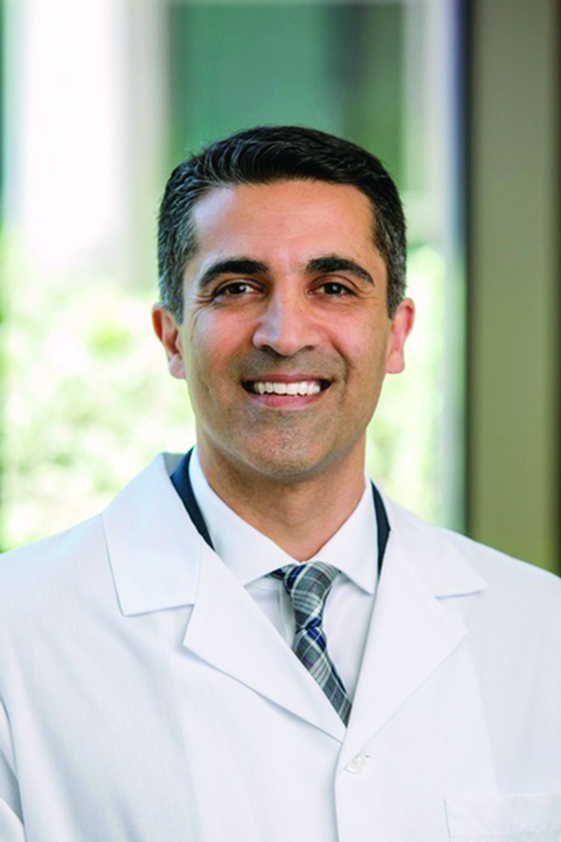
In a presentation at the 2021 virtual Pediatric Hospital Medicine conference, Dr. Talebi and Dr. Ganem shared their experiences in identifying the impact of the pandemic on mental health services in a freestanding hospital, and synthesizing inpatient mental health care and medical care outside of a dedicated mental health unit.

Mental health is a significant pediatric issue; approximately one in five children have a diagnosable mental or behavioral health problem, but nearly two-thirds get little or no help, Dr. Talebi said. “COVID-19 has only exacerbated these mental health challenges,” he said.
He noted that beginning in April 2020, the proportion of children’s mental health-related emergency department visits increased and remained elevated through the spring, summer, and fall of 2020, as families fearful of COVID-19 avoided regular hospital visits.
Data suggest that up to 50% of all adolescent psychiatric crises that led to inpatient admissions were related in some way to COVID-19, Dr. Talebi said. In addition, “individuals with a recent diagnosis of a mental health disorder are at increased risk for COVID-19 infection,” and the risk is even higher among women and African Americans, he said.
The past year significantly impacted the mental wellbeing of parents and children, Dr. Talebi said. He cited a June 2020 study in Pediatrics in which 27% of parents reported worsening mental health for themselves, and 14% reported worsening behavioral health for their children. Ongoing issues including food insecurity, loss of regular child care, and an overall “very disorienting experience in the day-to-day” compromised the mental health of families, Dr. Talebi emphasized. Children isolated at home were not meeting developmental milestones that organically occur when socializing with peers, parents didn’t know how to handle some of their children’s issues without support from schools, and many people were struggling with other preexisting health conditions, he said.
This confluence of factors helped drive a surge in emergency department visits, meaning longer wait times and concerns about meeting urgent medical and mental health needs while maintaining safety, he added.
Parents and children waited longer to seek care, and community hospitals such as Dell Children’s Medical Center were faced with children in the emergency department with crisis-level mental health issues, along with children already waiting in the ED to address medical emergencies. All these patients had to be tested for COVID-19 and managed accordingly, Dr. Talebi noted.
Dr. Talebi emphasized the need for clinically robust care of the children who were in isolation for 10 days on the medical unit, waiting to test negative. New protocols were created for social workers to conduct daily safety checks, and to develop regular schedules for screening, “so they are having an experience on the medical floors similar to what they would have in a mental health unit,” he said.
Dr. Ganem reflected on the logistical challenges of managing mental health care while observing COVID-19 safety protocols. “COVID-19 added a new wrinkle of isolation,” he said. As institutional guidelines on testing and isolation evolved, negative COVID-19 tests were required for admission to the mental health units both in the hospital and throughout the region. Patients who tested positive had to be quarantined for 10 days, at which time they could be admitted to a mental health unit if necessary, he said.
Dr. Ganem shared details of some strategies adopted by Dell Children’s. He explained that the COVID-19 psychiatry patient workflow started with an ED evaluation, followed by medical clearance and consideration for admission.
“There was significant coordination between the social worker in the emergency department and the psychiatry social worker,” he said.
Key elements of the treatment plan for children with positive COVID-19 tests included an “interprofessional huddle” to coordinate the plan of care, goals for admission, and goals for safety, Dr. Ganem said.
Patients who required admission were expected to have an initial length of stay of 72 hours, and those who tested positive for COVID-19 were admitted to a medical unit with COVID-19 isolation, he said.
Once a patient is admitted, an RN activates a suicide prevention pathway, and an interprofessional team meets to determine what patients need for safe and effective discharge, said Dr. Ganem. He cited the SAFE-T protocol (Suicide Assessment Five-step Evaluation and Triage) as one of the tools used to determine safe discharge criteria. Considerations on the SAFE-T list include family support, an established outpatient therapist and psychiatrist, no suicide attempts prior to the current admission, or a low lethality attempt, and access to partial hospitalization or intensive outpatient programs.
Patients who could not be discharged because of suicidality or inadequate support or concerns about safety at home were considered for inpatient admission. Patients with COVID-19–positive tests who had continued need for inpatient mental health services could be transferred to an inpatient mental health unit after a 10-day quarantine.
Overall, “this has been a continuum of lessons learned, with some things we know now that we didn’t know in April or May of 2020,” Dr. Ganem said. Early in the pandemic, the focus was on minimizing risk, securing personal protective equipment, and determining who provided services in a patient’s room. “We developed new paradigms on the fly,” he said, including the use of virtual visits, which included securing and cleaning devices, as well as learning how to use them in this setting,” he said.
More recently, the emphasis has been on providing services to patients before they need to visit the hospital, rather than automatically admitting any patients with suicidal ideation and a positive COVID-19 test, Dr. Ganem said.
Dr. Talebi and Dr. Ganem had no financial conflicts to disclose. The conference was sponsored by the Society of Hospital Medicine, the American Academy of Pediatrics, and the Academic Pediatric Association.
The COVID-19 pandemic continues to impact child and adolescent mental health, and clinicians are learning as they go to develop strategies that address the challenges of providing both medical and mental health care to young patients, including those who test positive for COVID-19, according to Hani Talebi, PhD, director of pediatric psychology, and Jorge Ganem, MD, FAAP, director of pediatric hospital medicine, both of the University of Texas at Austin and Dell Children’s Medical Center.
In a presentation at the 2021 virtual Pediatric Hospital Medicine conference, Dr. Talebi and Dr. Ganem shared their experiences in identifying the impact of the pandemic on mental health services in a freestanding hospital, and synthesizing inpatient mental health care and medical care outside of a dedicated mental health unit.
Mental health is a significant pediatric issue; approximately one in five children have a diagnosable mental or behavioral health problem, but nearly two-thirds get little or no help, Dr. Talebi said. “COVID-19 has only exacerbated these mental health challenges,” he said.
He noted that beginning in April 2020, the proportion of children’s mental health-related emergency department visits increased and remained elevated through the spring, summer, and fall of 2020, as families fearful of COVID-19 avoided regular hospital visits.
Data suggest that up to 50% of all adolescent psychiatric crises that led to inpatient admissions were related in some way to COVID-19, Dr. Talebi said. In addition, “individuals with a recent diagnosis of a mental health disorder are at increased risk for COVID-19 infection,” and the risk is even higher among women and African Americans, he said.
The past year significantly impacted the mental wellbeing of parents and children, Dr. Talebi said. He cited a June 2020 study in Pediatrics in which 27% of parents reported worsening mental health for themselves, and 14% reported worsening behavioral health for their children. Ongoing issues including food insecurity, loss of regular child care, and an overall “very disorienting experience in the day-to-day” compromised the mental health of families, Dr. Talebi emphasized. Children isolated at home were not meeting developmental milestones that organically occur when socializing with peers, parents didn’t know how to handle some of their children’s issues without support from schools, and many people were struggling with other preexisting health conditions, he said.
This confluence of factors helped drive a surge in emergency department visits, meaning longer wait times and concerns about meeting urgent medical and mental health needs while maintaining safety, he added.
Parents and children waited longer to seek care, and community hospitals such as Dell Children’s Medical Center were faced with children in the emergency department with crisis-level mental health issues, along with children already waiting in the ED to address medical emergencies. All these patients had to be tested for COVID-19 and managed accordingly, Dr. Talebi noted.
Dr. Talebi emphasized the need for clinically robust care of the children who were in isolation for 10 days on the medical unit, waiting to test negative. New protocols were created for social workers to conduct daily safety checks, and to develop regular schedules for screening, “so they are having an experience on the medical floors similar to what they would have in a mental health unit,” he said.
Dr. Ganem reflected on the logistical challenges of managing mental health care while observing COVID-19 safety protocols. “COVID-19 added a new wrinkle of isolation,” he said. As institutional guidelines on testing and isolation evolved, negative COVID-19 tests were required for admission to the mental health units both in the hospital and throughout the region. Patients who tested positive had to be quarantined for 10 days, at which time they could be admitted to a mental health unit if necessary, he said.
Dr. Ganem shared details of some strategies adopted by Dell Children’s. He explained that the COVID-19 psychiatry patient workflow started with an ED evaluation, followed by medical clearance and consideration for admission.
“There was significant coordination between the social worker in the emergency department and the psychiatry social worker,” he said.
Key elements of the treatment plan for children with positive COVID-19 tests included an “interprofessional huddle” to coordinate the plan of care, goals for admission, and goals for safety, Dr. Ganem said.
Patients who required admission were expected to have an initial length of stay of 72 hours, and those who tested positive for COVID-19 were admitted to a medical unit with COVID-19 isolation, he said.
Once a patient is admitted, an RN activates a suicide prevention pathway, and an interprofessional team meets to determine what patients need for safe and effective discharge, said Dr. Ganem. He cited the SAFE-T protocol (Suicide Assessment Five-step Evaluation and Triage) as one of the tools used to determine safe discharge criteria. Considerations on the SAFE-T list include family support, an established outpatient therapist and psychiatrist, no suicide attempts prior to the current admission, or a low lethality attempt, and access to partial hospitalization or intensive outpatient programs.
Patients who could not be discharged because of suicidality or inadequate support or concerns about safety at home were considered for inpatient admission. Patients with COVID-19–positive tests who had continued need for inpatient mental health services could be transferred to an inpatient mental health unit after a 10-day quarantine.
Overall, “this has been a continuum of lessons learned, with some things we know now that we didn’t know in April or May of 2020,” Dr. Ganem said. Early in the pandemic, the focus was on minimizing risk, securing personal protective equipment, and determining who provided services in a patient’s room. “We developed new paradigms on the fly,” he said, including the use of virtual visits, which included securing and cleaning devices, as well as learning how to use them in this setting,” he said.
More recently, the emphasis has been on providing services to patients before they need to visit the hospital, rather than automatically admitting any patients with suicidal ideation and a positive COVID-19 test, Dr. Ganem said.
Dr. Talebi and Dr. Ganem had no financial conflicts to disclose. The conference was sponsored by the Society of Hospital Medicine, the American Academy of Pediatrics, and the Academic Pediatric Association.
The COVID-19 pandemic continues to impact child and adolescent mental health, and clinicians are learning as they go to develop strategies that address the challenges of providing both medical and mental health care to young patients, including those who test positive for COVID-19, according to Hani Talebi, PhD, director of pediatric psychology, and Jorge Ganem, MD, FAAP, director of pediatric hospital medicine, both of the University of Texas at Austin and Dell Children’s Medical Center.
In a presentation at the 2021 virtual Pediatric Hospital Medicine conference, Dr. Talebi and Dr. Ganem shared their experiences in identifying the impact of the pandemic on mental health services in a freestanding hospital, and synthesizing inpatient mental health care and medical care outside of a dedicated mental health unit.
Mental health is a significant pediatric issue; approximately one in five children have a diagnosable mental or behavioral health problem, but nearly two-thirds get little or no help, Dr. Talebi said. “COVID-19 has only exacerbated these mental health challenges,” he said.
He noted that beginning in April 2020, the proportion of children’s mental health-related emergency department visits increased and remained elevated through the spring, summer, and fall of 2020, as families fearful of COVID-19 avoided regular hospital visits.
Data suggest that up to 50% of all adolescent psychiatric crises that led to inpatient admissions were related in some way to COVID-19, Dr. Talebi said. In addition, “individuals with a recent diagnosis of a mental health disorder are at increased risk for COVID-19 infection,” and the risk is even higher among women and African Americans, he said.
The past year significantly impacted the mental wellbeing of parents and children, Dr. Talebi said. He cited a June 2020 study in Pediatrics in which 27% of parents reported worsening mental health for themselves, and 14% reported worsening behavioral health for their children. Ongoing issues including food insecurity, loss of regular child care, and an overall “very disorienting experience in the day-to-day” compromised the mental health of families, Dr. Talebi emphasized. Children isolated at home were not meeting developmental milestones that organically occur when socializing with peers, parents didn’t know how to handle some of their children’s issues without support from schools, and many people were struggling with other preexisting health conditions, he said.
This confluence of factors helped drive a surge in emergency department visits, meaning longer wait times and concerns about meeting urgent medical and mental health needs while maintaining safety, he added.
Parents and children waited longer to seek care, and community hospitals such as Dell Children’s Medical Center were faced with children in the emergency department with crisis-level mental health issues, along with children already waiting in the ED to address medical emergencies. All these patients had to be tested for COVID-19 and managed accordingly, Dr. Talebi noted.
Dr. Talebi emphasized the need for clinically robust care of the children who were in isolation for 10 days on the medical unit, waiting to test negative. New protocols were created for social workers to conduct daily safety checks, and to develop regular schedules for screening, “so they are having an experience on the medical floors similar to what they would have in a mental health unit,” he said.
Dr. Ganem reflected on the logistical challenges of managing mental health care while observing COVID-19 safety protocols. “COVID-19 added a new wrinkle of isolation,” he said. As institutional guidelines on testing and isolation evolved, negative COVID-19 tests were required for admission to the mental health units both in the hospital and throughout the region. Patients who tested positive had to be quarantined for 10 days, at which time they could be admitted to a mental health unit if necessary, he said.
Dr. Ganem shared details of some strategies adopted by Dell Children’s. He explained that the COVID-19 psychiatry patient workflow started with an ED evaluation, followed by medical clearance and consideration for admission.
“There was significant coordination between the social worker in the emergency department and the psychiatry social worker,” he said.
Key elements of the treatment plan for children with positive COVID-19 tests included an “interprofessional huddle” to coordinate the plan of care, goals for admission, and goals for safety, Dr. Ganem said.
Patients who required admission were expected to have an initial length of stay of 72 hours, and those who tested positive for COVID-19 were admitted to a medical unit with COVID-19 isolation, he said.
Once a patient is admitted, an RN activates a suicide prevention pathway, and an interprofessional team meets to determine what patients need for safe and effective discharge, said Dr. Ganem. He cited the SAFE-T protocol (Suicide Assessment Five-step Evaluation and Triage) as one of the tools used to determine safe discharge criteria. Considerations on the SAFE-T list include family support, an established outpatient therapist and psychiatrist, no suicide attempts prior to the current admission, or a low lethality attempt, and access to partial hospitalization or intensive outpatient programs.
Patients who could not be discharged because of suicidality or inadequate support or concerns about safety at home were considered for inpatient admission. Patients with COVID-19–positive tests who had continued need for inpatient mental health services could be transferred to an inpatient mental health unit after a 10-day quarantine.
Overall, “this has been a continuum of lessons learned, with some things we know now that we didn’t know in April or May of 2020,” Dr. Ganem said. Early in the pandemic, the focus was on minimizing risk, securing personal protective equipment, and determining who provided services in a patient’s room. “We developed new paradigms on the fly,” he said, including the use of virtual visits, which included securing and cleaning devices, as well as learning how to use them in this setting,” he said.
More recently, the emphasis has been on providing services to patients before they need to visit the hospital, rather than automatically admitting any patients with suicidal ideation and a positive COVID-19 test, Dr. Ganem said.
Dr. Talebi and Dr. Ganem had no financial conflicts to disclose. The conference was sponsored by the Society of Hospital Medicine, the American Academy of Pediatrics, and the Academic Pediatric Association.
FROM PHM 2021
Clinical genetic testing for skin disorders continues to advance
and families of pediatric patients to navigate the landscape.

“Testing options range from targeted variant testing and single-gene testing to exome and genome sequencing,” Gabriele Richard, MD, said at the annual meeting of the Society for Pediatric Dermatology. “It is not always easy to determine which testing is right.”
Increasingly, clinical genomic tests, including exome and genome sequencing, are used for patients with complex phenotypes, and possibly multiple disorders, who might have no diagnosis despite extensive prior testing, said Dr. Richard, medical director at Gaithersburg, Md.–based GeneDx., a molecular diagnostic laboratory that performs comprehensive testing for rare genetic disorders. These tests are also being used more for first-line testing in critically ill patients in the neonatal and pediatric intensive care units, and “have heralded a whole new era of gene and disease discovery,” she added.
Targeted variant testing is used for known familial variants, to test family members for carrier status and segregation, and to make a prenatal diagnosis, she said. Single-gene testing is available for most genes and has its place for conditions that can be clinically well-recognized, such as ichthyosis vulgaris, Darier disease, or Papillon-Lefèvre syndrome.
Specific tests for identifying gene deletions or duplications are exon-level microarrays, multiplex ligation-dependent probe amplification (MLPA), and chromosomal microarray analysis. “The latter has been successful in identifying diseases causing chromosomal abnormalities in over 10% of cases overall,” Dr. Richard said. An example of a skin disorder is X-linked ichthyosis caused by a deletion of the steroid sulfatase locus in more than 95% of affected males, she said.
“However, the current staple of molecular diagnostic testing is multigene next-generation sequencing (NGS) panels, which allow you to interrogate two to hundreds of genes concurrently, including sequencing and deletion duplication testing.” These tests are the most cost effective, she said, and are available for almost any genodermatosis or group of disorders with overlapping phenotypes, such as albinism or ichthyosis, epidermolysis bullosa and skin fragility, ectodermal dysplasia, or porphyria. According to Dr. Richard, the diagnostic outcomes of NGS panels mainly depend on test indication, panel size and gene curation, age of onset, and prevailing inheritance pattern of disorders.
Her recommended criteria for distinguishing the myriad of available NGS panels include checking gene content, technical sensitivity of sequencing and deletion/duplication analysis, quality of variant interpretation and reporting, turn-around time, and available familial follow-up testing. “If a family might consider future prenatal diagnosis, choose the lab that performs prenatal and diagnostic testing,” Dr. Richard said. “Equally important are client services such as ease of ordering, insurance coverage, and the ability to determine out-of-pocket cost to patients.”
Resources that enable consumers to compare panel content, methodology, turnaround time, and other parameters include the Genetic Testing Registry (GTR) operated by the National Center for Biotechnology Information, and Concert Genetics, a genetic testing company. The National Society of Genetic Counselors also offers a searchable database for finding a genetic counselor.
Exome sequencing includes the coding sequences of about 20,000 genes and has an average depth of 50 to about 150 reads. “It is a phenotype-driven test where only select variants are being reported fitting the phenotype,” Dr. Richard said. “The outcome of exome and genome sequencing much depends on optimization of bioinformatic pipelines and tools.” Besides small sequence variants, exome sequencing is able to identify a variety of different types of disease-causing variants, such as gene copy number variants seen in about 6% of positive cases, mosaicism, regions of homozygosity, uniparental disomy, and other unusual events and is cost effective.
Whole-genome sequencing, meanwhile, includes the entire genome, particularly noncoding regions, and has an average depth of more than 30 reads. “It’s based on single-molecule sequencing, has longer reads and more uniform coverage, compared to exome sequencing,” she said. “Higher cost, variant interpretation, and lack of coverage by payers are still presenting challenges for genome sequencing.” Genome sequencing can be done in a day or less.
According to diagnostic outcomes based on 280,000 individuals including 125,000 probands from GeneDx data, a definitive diagnosis was made in 26% of probands, of which 2.8% had more than one diagnostic finding and 1.8% had actionable secondary findings. In addition, 7% of the variants were found in candidate genes; 31% of probands had variants of uncertain significance, while 36% tested negative. “Nevertheless, the diagnostic yield of exome sequencing depends on the phenotype and cohort studied,” Dr. Richard continued.
At her company, she said, the highest positive rate is for multiple congenital anomalies (34%), skeletal system abnormalities (30%), and nervous system abnormalities (29%). Trio testing – the concurrent analysis of both biological parents and proband for all genes – “is a critical factor for success,” she added. “It not only improves the variant calling because we have three times the data and increases test sensitivity, it also provides more certain results, determines inheritance and allows for detection of parental mosaicism.”
According to Dr. Richard, trio testing has a one-third higher diagnostic rate than sequencing of the proband alone. Citing a published prospective study that compiled data from eight different exome- and genome-sequencing studies in critically ill neonates and children, trio testing made it possible to make a genetic diagnosis in up to 58% of children.
Whole-genome sequencing is estimated to have a 5%-10% higher diagnostic rate than exome sequencing. “However, we are still a ways away from using it as a routine diagnostic test for all test indications,” Dr. Richard said. “Automation, special bioinformatics algorithms and databases, and combination of genome sequencing with mRNA sequencing are being explored and built to further improve the diagnostic yield.”
Dr. Richard had no disclosures other than being an employee of GeneDx.
and families of pediatric patients to navigate the landscape.
“Testing options range from targeted variant testing and single-gene testing to exome and genome sequencing,” Gabriele Richard, MD, said at the annual meeting of the Society for Pediatric Dermatology. “It is not always easy to determine which testing is right.”
Increasingly, clinical genomic tests, including exome and genome sequencing, are used for patients with complex phenotypes, and possibly multiple disorders, who might have no diagnosis despite extensive prior testing, said Dr. Richard, medical director at Gaithersburg, Md.–based GeneDx., a molecular diagnostic laboratory that performs comprehensive testing for rare genetic disorders. These tests are also being used more for first-line testing in critically ill patients in the neonatal and pediatric intensive care units, and “have heralded a whole new era of gene and disease discovery,” she added.
Targeted variant testing is used for known familial variants, to test family members for carrier status and segregation, and to make a prenatal diagnosis, she said. Single-gene testing is available for most genes and has its place for conditions that can be clinically well-recognized, such as ichthyosis vulgaris, Darier disease, or Papillon-Lefèvre syndrome.
Specific tests for identifying gene deletions or duplications are exon-level microarrays, multiplex ligation-dependent probe amplification (MLPA), and chromosomal microarray analysis. “The latter has been successful in identifying diseases causing chromosomal abnormalities in over 10% of cases overall,” Dr. Richard said. An example of a skin disorder is X-linked ichthyosis caused by a deletion of the steroid sulfatase locus in more than 95% of affected males, she said.
“However, the current staple of molecular diagnostic testing is multigene next-generation sequencing (NGS) panels, which allow you to interrogate two to hundreds of genes concurrently, including sequencing and deletion duplication testing.” These tests are the most cost effective, she said, and are available for almost any genodermatosis or group of disorders with overlapping phenotypes, such as albinism or ichthyosis, epidermolysis bullosa and skin fragility, ectodermal dysplasia, or porphyria. According to Dr. Richard, the diagnostic outcomes of NGS panels mainly depend on test indication, panel size and gene curation, age of onset, and prevailing inheritance pattern of disorders.
Her recommended criteria for distinguishing the myriad of available NGS panels include checking gene content, technical sensitivity of sequencing and deletion/duplication analysis, quality of variant interpretation and reporting, turn-around time, and available familial follow-up testing. “If a family might consider future prenatal diagnosis, choose the lab that performs prenatal and diagnostic testing,” Dr. Richard said. “Equally important are client services such as ease of ordering, insurance coverage, and the ability to determine out-of-pocket cost to patients.”
Resources that enable consumers to compare panel content, methodology, turnaround time, and other parameters include the Genetic Testing Registry (GTR) operated by the National Center for Biotechnology Information, and Concert Genetics, a genetic testing company. The National Society of Genetic Counselors also offers a searchable database for finding a genetic counselor.
Exome sequencing includes the coding sequences of about 20,000 genes and has an average depth of 50 to about 150 reads. “It is a phenotype-driven test where only select variants are being reported fitting the phenotype,” Dr. Richard said. “The outcome of exome and genome sequencing much depends on optimization of bioinformatic pipelines and tools.” Besides small sequence variants, exome sequencing is able to identify a variety of different types of disease-causing variants, such as gene copy number variants seen in about 6% of positive cases, mosaicism, regions of homozygosity, uniparental disomy, and other unusual events and is cost effective.
Whole-genome sequencing, meanwhile, includes the entire genome, particularly noncoding regions, and has an average depth of more than 30 reads. “It’s based on single-molecule sequencing, has longer reads and more uniform coverage, compared to exome sequencing,” she said. “Higher cost, variant interpretation, and lack of coverage by payers are still presenting challenges for genome sequencing.” Genome sequencing can be done in a day or less.
According to diagnostic outcomes based on 280,000 individuals including 125,000 probands from GeneDx data, a definitive diagnosis was made in 26% of probands, of which 2.8% had more than one diagnostic finding and 1.8% had actionable secondary findings. In addition, 7% of the variants were found in candidate genes; 31% of probands had variants of uncertain significance, while 36% tested negative. “Nevertheless, the diagnostic yield of exome sequencing depends on the phenotype and cohort studied,” Dr. Richard continued.
At her company, she said, the highest positive rate is for multiple congenital anomalies (34%), skeletal system abnormalities (30%), and nervous system abnormalities (29%). Trio testing – the concurrent analysis of both biological parents and proband for all genes – “is a critical factor for success,” she added. “It not only improves the variant calling because we have three times the data and increases test sensitivity, it also provides more certain results, determines inheritance and allows for detection of parental mosaicism.”
According to Dr. Richard, trio testing has a one-third higher diagnostic rate than sequencing of the proband alone. Citing a published prospective study that compiled data from eight different exome- and genome-sequencing studies in critically ill neonates and children, trio testing made it possible to make a genetic diagnosis in up to 58% of children.
Whole-genome sequencing is estimated to have a 5%-10% higher diagnostic rate than exome sequencing. “However, we are still a ways away from using it as a routine diagnostic test for all test indications,” Dr. Richard said. “Automation, special bioinformatics algorithms and databases, and combination of genome sequencing with mRNA sequencing are being explored and built to further improve the diagnostic yield.”
Dr. Richard had no disclosures other than being an employee of GeneDx.
and families of pediatric patients to navigate the landscape.
“Testing options range from targeted variant testing and single-gene testing to exome and genome sequencing,” Gabriele Richard, MD, said at the annual meeting of the Society for Pediatric Dermatology. “It is not always easy to determine which testing is right.”
Increasingly, clinical genomic tests, including exome and genome sequencing, are used for patients with complex phenotypes, and possibly multiple disorders, who might have no diagnosis despite extensive prior testing, said Dr. Richard, medical director at Gaithersburg, Md.–based GeneDx., a molecular diagnostic laboratory that performs comprehensive testing for rare genetic disorders. These tests are also being used more for first-line testing in critically ill patients in the neonatal and pediatric intensive care units, and “have heralded a whole new era of gene and disease discovery,” she added.
Targeted variant testing is used for known familial variants, to test family members for carrier status and segregation, and to make a prenatal diagnosis, she said. Single-gene testing is available for most genes and has its place for conditions that can be clinically well-recognized, such as ichthyosis vulgaris, Darier disease, or Papillon-Lefèvre syndrome.
Specific tests for identifying gene deletions or duplications are exon-level microarrays, multiplex ligation-dependent probe amplification (MLPA), and chromosomal microarray analysis. “The latter has been successful in identifying diseases causing chromosomal abnormalities in over 10% of cases overall,” Dr. Richard said. An example of a skin disorder is X-linked ichthyosis caused by a deletion of the steroid sulfatase locus in more than 95% of affected males, she said.
“However, the current staple of molecular diagnostic testing is multigene next-generation sequencing (NGS) panels, which allow you to interrogate two to hundreds of genes concurrently, including sequencing and deletion duplication testing.” These tests are the most cost effective, she said, and are available for almost any genodermatosis or group of disorders with overlapping phenotypes, such as albinism or ichthyosis, epidermolysis bullosa and skin fragility, ectodermal dysplasia, or porphyria. According to Dr. Richard, the diagnostic outcomes of NGS panels mainly depend on test indication, panel size and gene curation, age of onset, and prevailing inheritance pattern of disorders.
Her recommended criteria for distinguishing the myriad of available NGS panels include checking gene content, technical sensitivity of sequencing and deletion/duplication analysis, quality of variant interpretation and reporting, turn-around time, and available familial follow-up testing. “If a family might consider future prenatal diagnosis, choose the lab that performs prenatal and diagnostic testing,” Dr. Richard said. “Equally important are client services such as ease of ordering, insurance coverage, and the ability to determine out-of-pocket cost to patients.”
Resources that enable consumers to compare panel content, methodology, turnaround time, and other parameters include the Genetic Testing Registry (GTR) operated by the National Center for Biotechnology Information, and Concert Genetics, a genetic testing company. The National Society of Genetic Counselors also offers a searchable database for finding a genetic counselor.
Exome sequencing includes the coding sequences of about 20,000 genes and has an average depth of 50 to about 150 reads. “It is a phenotype-driven test where only select variants are being reported fitting the phenotype,” Dr. Richard said. “The outcome of exome and genome sequencing much depends on optimization of bioinformatic pipelines and tools.” Besides small sequence variants, exome sequencing is able to identify a variety of different types of disease-causing variants, such as gene copy number variants seen in about 6% of positive cases, mosaicism, regions of homozygosity, uniparental disomy, and other unusual events and is cost effective.
Whole-genome sequencing, meanwhile, includes the entire genome, particularly noncoding regions, and has an average depth of more than 30 reads. “It’s based on single-molecule sequencing, has longer reads and more uniform coverage, compared to exome sequencing,” she said. “Higher cost, variant interpretation, and lack of coverage by payers are still presenting challenges for genome sequencing.” Genome sequencing can be done in a day or less.
According to diagnostic outcomes based on 280,000 individuals including 125,000 probands from GeneDx data, a definitive diagnosis was made in 26% of probands, of which 2.8% had more than one diagnostic finding and 1.8% had actionable secondary findings. In addition, 7% of the variants were found in candidate genes; 31% of probands had variants of uncertain significance, while 36% tested negative. “Nevertheless, the diagnostic yield of exome sequencing depends on the phenotype and cohort studied,” Dr. Richard continued.
At her company, she said, the highest positive rate is for multiple congenital anomalies (34%), skeletal system abnormalities (30%), and nervous system abnormalities (29%). Trio testing – the concurrent analysis of both biological parents and proband for all genes – “is a critical factor for success,” she added. “It not only improves the variant calling because we have three times the data and increases test sensitivity, it also provides more certain results, determines inheritance and allows for detection of parental mosaicism.”
According to Dr. Richard, trio testing has a one-third higher diagnostic rate than sequencing of the proband alone. Citing a published prospective study that compiled data from eight different exome- and genome-sequencing studies in critically ill neonates and children, trio testing made it possible to make a genetic diagnosis in up to 58% of children.
Whole-genome sequencing is estimated to have a 5%-10% higher diagnostic rate than exome sequencing. “However, we are still a ways away from using it as a routine diagnostic test for all test indications,” Dr. Richard said. “Automation, special bioinformatics algorithms and databases, and combination of genome sequencing with mRNA sequencing are being explored and built to further improve the diagnostic yield.”
Dr. Richard had no disclosures other than being an employee of GeneDx.
FROM SPD 2021
Six shifts driving the future of medicine, strategist says
Contact lenses that detect glucose in tears. Capsules embedded in clothes that can be used to counteract the risk of sensitive skin conditions.

These are just .
At the annual meeting of the Society for Pediatric Dermatology, Zayna Khayat, PhD, said that the future of medicine is driven by six shifts pulling society from a past oriented around the health care system – the buildings, clinicians, and payers – to a patient-oriented perspective. “That doesn’t just happen on its own,” said Dr. Khayat, a future strategist at Toronto-based SE Health. “There are big forces that are pulling us to the future whether we want it to or not. One is that patients have woken up. They have grown to have power in many other complex decisions in their life, and they’re expecting no less from our health care system.”
During her presentation, she discussed the six shifts:
1. The timing of service placement. The traditional model of medicine is “an intermittent and interventional science that waits for the symptoms and goes in and either fixes or manages them,” she said. “So, it’s not really health care; it’s sick care. That’s been fine in the industrial era when we needed to get medicine to stop catastrophic events. Not only is it shifting to be proactive and preventative but it’s shifting to a new science of medicine called predictive medicine.”
As for proactive and preventative care, she continued, each patient’s choice of behaviors related to diet, exercise, and stress “mingles with DNA to produce health, yet we spend about 90% of our resources on sick care. Now, health systems are moving their resources to things like education, housing, transportation, food security, equity, and racial divides. ... This is trickling down to how we train health care professionals. We know that patients live very little of their time in formal care settings, so all of their health is created – or destroyed – well outside of the clinical setting. We train our health professionals mostly in a clinical setting. Health systems are now starting to reimagine how training happens so we can train people to understand the fully loaded context of their patients’ lives.”
2. A shift in precision. For all its advances and science breakthroughs, medicine “is still quite crude,” said Dr. Khayat, who is also an adjunct professor in the Rotman School of Management at the University of Toronto. “It’s very analog, based on a one-size-fits-all approach. In the business world, we call this a segment of one: the idea that in some clinical trial, a result was produced that was based on the average of everybody, and therefore we just give everybody what worked for the average. ... We don’t need to have that trade-off anymore, because it won’t be a trade-off of higher cost to tailor down to an N of 1. It will be highly personalized, intelligent medicine, very precise.”
3. A shift from institution-centered to person-centered care. “The artifacts that health care was built on are very analog and are going to get decentralized out of buildings, dephysicalized, disintermediated,” she predicted. “We’ll have a seamless digital physical experience, expanded channels through which patients can access their services. Pick a channel that makes sense for the patient and don’t let care follow the place but rather let care follow the person.”
4. A shift in care duration, from episodic and intermittent care to more continuous care. “With very little input you should know what’s going on at any point in time instead of time-sharing access to diagnostics and to clinicians,” Dr. Khayat said. Wrist-worn devices that gather personal omics “are now really democratized, with every aspect of a diagnostic clinic available within or connected to a smartphone. This allows for data to be gathered and shared with clinicians, including tools under the skin that can get some of the biochemical data in real time instead of poking and prodding and waiting for a diagnostic lab.” These devices, she said, will become easier to use, cheaper, and will work faster, and provide much better data “at almost zero cost.”
Technologies being developed include tattoos that can read biomarkers, innovations in clothing that can detect biochemical reactions in the skin, underwear that can read vital signs, and contact lenses that can measure glucose levels. “The skin will become a major noninvasive way to obtain information,” she said.
5. A shift in power from providers to patients. “It’s estimated that about 80% of health care decisions could be self-managed by people in their communities,” Dr. Khayat said.
6. A shift from volume-based to value-based care. “Because we’ve been obsessed with the system, we’ve paid for stuff like visits, pills, MRI scans, et cetera,” she said. “We don’t need to do that anymore. Health systems don’t want to keep paying for stuff if they don’t see the results. Because of all the other shifts, we can pay for results. Some call this value-based care. I call it fee-for-health.”
She noted that the future of medicine is underpinned by innovations in AI/predictalytics, voice recognition, virtual reality, blockchain, IoT sensors, 3D printing, omics, robotics, autonomous transport, neurotechnology, nanobiology, and cellular therapy. “They’re moving at a very fast pace because they don’t need the kind of cost, capital, and expertise that the previous tools did,” she said. “This is the promise that technology can bring.”
Dr. Khayat disclosed that she has been a workshop participant for Roche Canada.
Contact lenses that detect glucose in tears. Capsules embedded in clothes that can be used to counteract the risk of sensitive skin conditions.
These are just .
At the annual meeting of the Society for Pediatric Dermatology, Zayna Khayat, PhD, said that the future of medicine is driven by six shifts pulling society from a past oriented around the health care system – the buildings, clinicians, and payers – to a patient-oriented perspective. “That doesn’t just happen on its own,” said Dr. Khayat, a future strategist at Toronto-based SE Health. “There are big forces that are pulling us to the future whether we want it to or not. One is that patients have woken up. They have grown to have power in many other complex decisions in their life, and they’re expecting no less from our health care system.”
During her presentation, she discussed the six shifts:
1. The timing of service placement. The traditional model of medicine is “an intermittent and interventional science that waits for the symptoms and goes in and either fixes or manages them,” she said. “So, it’s not really health care; it’s sick care. That’s been fine in the industrial era when we needed to get medicine to stop catastrophic events. Not only is it shifting to be proactive and preventative but it’s shifting to a new science of medicine called predictive medicine.”
As for proactive and preventative care, she continued, each patient’s choice of behaviors related to diet, exercise, and stress “mingles with DNA to produce health, yet we spend about 90% of our resources on sick care. Now, health systems are moving their resources to things like education, housing, transportation, food security, equity, and racial divides. ... This is trickling down to how we train health care professionals. We know that patients live very little of their time in formal care settings, so all of their health is created – or destroyed – well outside of the clinical setting. We train our health professionals mostly in a clinical setting. Health systems are now starting to reimagine how training happens so we can train people to understand the fully loaded context of their patients’ lives.”
2. A shift in precision. For all its advances and science breakthroughs, medicine “is still quite crude,” said Dr. Khayat, who is also an adjunct professor in the Rotman School of Management at the University of Toronto. “It’s very analog, based on a one-size-fits-all approach. In the business world, we call this a segment of one: the idea that in some clinical trial, a result was produced that was based on the average of everybody, and therefore we just give everybody what worked for the average. ... We don’t need to have that trade-off anymore, because it won’t be a trade-off of higher cost to tailor down to an N of 1. It will be highly personalized, intelligent medicine, very precise.”
3. A shift from institution-centered to person-centered care. “The artifacts that health care was built on are very analog and are going to get decentralized out of buildings, dephysicalized, disintermediated,” she predicted. “We’ll have a seamless digital physical experience, expanded channels through which patients can access their services. Pick a channel that makes sense for the patient and don’t let care follow the place but rather let care follow the person.”
4. A shift in care duration, from episodic and intermittent care to more continuous care. “With very little input you should know what’s going on at any point in time instead of time-sharing access to diagnostics and to clinicians,” Dr. Khayat said. Wrist-worn devices that gather personal omics “are now really democratized, with every aspect of a diagnostic clinic available within or connected to a smartphone. This allows for data to be gathered and shared with clinicians, including tools under the skin that can get some of the biochemical data in real time instead of poking and prodding and waiting for a diagnostic lab.” These devices, she said, will become easier to use, cheaper, and will work faster, and provide much better data “at almost zero cost.”
Technologies being developed include tattoos that can read biomarkers, innovations in clothing that can detect biochemical reactions in the skin, underwear that can read vital signs, and contact lenses that can measure glucose levels. “The skin will become a major noninvasive way to obtain information,” she said.
5. A shift in power from providers to patients. “It’s estimated that about 80% of health care decisions could be self-managed by people in their communities,” Dr. Khayat said.
6. A shift from volume-based to value-based care. “Because we’ve been obsessed with the system, we’ve paid for stuff like visits, pills, MRI scans, et cetera,” she said. “We don’t need to do that anymore. Health systems don’t want to keep paying for stuff if they don’t see the results. Because of all the other shifts, we can pay for results. Some call this value-based care. I call it fee-for-health.”
She noted that the future of medicine is underpinned by innovations in AI/predictalytics, voice recognition, virtual reality, blockchain, IoT sensors, 3D printing, omics, robotics, autonomous transport, neurotechnology, nanobiology, and cellular therapy. “They’re moving at a very fast pace because they don’t need the kind of cost, capital, and expertise that the previous tools did,” she said. “This is the promise that technology can bring.”
Dr. Khayat disclosed that she has been a workshop participant for Roche Canada.
Contact lenses that detect glucose in tears. Capsules embedded in clothes that can be used to counteract the risk of sensitive skin conditions.
These are just .
At the annual meeting of the Society for Pediatric Dermatology, Zayna Khayat, PhD, said that the future of medicine is driven by six shifts pulling society from a past oriented around the health care system – the buildings, clinicians, and payers – to a patient-oriented perspective. “That doesn’t just happen on its own,” said Dr. Khayat, a future strategist at Toronto-based SE Health. “There are big forces that are pulling us to the future whether we want it to or not. One is that patients have woken up. They have grown to have power in many other complex decisions in their life, and they’re expecting no less from our health care system.”
During her presentation, she discussed the six shifts:
1. The timing of service placement. The traditional model of medicine is “an intermittent and interventional science that waits for the symptoms and goes in and either fixes or manages them,” she said. “So, it’s not really health care; it’s sick care. That’s been fine in the industrial era when we needed to get medicine to stop catastrophic events. Not only is it shifting to be proactive and preventative but it’s shifting to a new science of medicine called predictive medicine.”
As for proactive and preventative care, she continued, each patient’s choice of behaviors related to diet, exercise, and stress “mingles with DNA to produce health, yet we spend about 90% of our resources on sick care. Now, health systems are moving their resources to things like education, housing, transportation, food security, equity, and racial divides. ... This is trickling down to how we train health care professionals. We know that patients live very little of their time in formal care settings, so all of their health is created – or destroyed – well outside of the clinical setting. We train our health professionals mostly in a clinical setting. Health systems are now starting to reimagine how training happens so we can train people to understand the fully loaded context of their patients’ lives.”
2. A shift in precision. For all its advances and science breakthroughs, medicine “is still quite crude,” said Dr. Khayat, who is also an adjunct professor in the Rotman School of Management at the University of Toronto. “It’s very analog, based on a one-size-fits-all approach. In the business world, we call this a segment of one: the idea that in some clinical trial, a result was produced that was based on the average of everybody, and therefore we just give everybody what worked for the average. ... We don’t need to have that trade-off anymore, because it won’t be a trade-off of higher cost to tailor down to an N of 1. It will be highly personalized, intelligent medicine, very precise.”
3. A shift from institution-centered to person-centered care. “The artifacts that health care was built on are very analog and are going to get decentralized out of buildings, dephysicalized, disintermediated,” she predicted. “We’ll have a seamless digital physical experience, expanded channels through which patients can access their services. Pick a channel that makes sense for the patient and don’t let care follow the place but rather let care follow the person.”
4. A shift in care duration, from episodic and intermittent care to more continuous care. “With very little input you should know what’s going on at any point in time instead of time-sharing access to diagnostics and to clinicians,” Dr. Khayat said. Wrist-worn devices that gather personal omics “are now really democratized, with every aspect of a diagnostic clinic available within or connected to a smartphone. This allows for data to be gathered and shared with clinicians, including tools under the skin that can get some of the biochemical data in real time instead of poking and prodding and waiting for a diagnostic lab.” These devices, she said, will become easier to use, cheaper, and will work faster, and provide much better data “at almost zero cost.”
Technologies being developed include tattoos that can read biomarkers, innovations in clothing that can detect biochemical reactions in the skin, underwear that can read vital signs, and contact lenses that can measure glucose levels. “The skin will become a major noninvasive way to obtain information,” she said.
5. A shift in power from providers to patients. “It’s estimated that about 80% of health care decisions could be self-managed by people in their communities,” Dr. Khayat said.
6. A shift from volume-based to value-based care. “Because we’ve been obsessed with the system, we’ve paid for stuff like visits, pills, MRI scans, et cetera,” she said. “We don’t need to do that anymore. Health systems don’t want to keep paying for stuff if they don’t see the results. Because of all the other shifts, we can pay for results. Some call this value-based care. I call it fee-for-health.”
She noted that the future of medicine is underpinned by innovations in AI/predictalytics, voice recognition, virtual reality, blockchain, IoT sensors, 3D printing, omics, robotics, autonomous transport, neurotechnology, nanobiology, and cellular therapy. “They’re moving at a very fast pace because they don’t need the kind of cost, capital, and expertise that the previous tools did,” she said. “This is the promise that technology can bring.”
Dr. Khayat disclosed that she has been a workshop participant for Roche Canada.
FROM SPD 2021
Is Nissen fundoplication the best we can do?
As an esophagologist that does not perform fundoplication, LINX, or TIF, I find it difficult to debate the merits of one procedure over another based on my experience. In fact, I have always stated that it is difficult to assess a procedure or test that one has not used. That being said, maybe the fact that I have not performed these procedures makes me more objective and I can only use my experience with patients and the data to make the case that we need options beyond Nissen fundoplication.
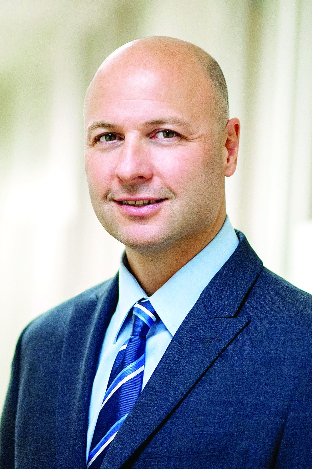
The recent VA Randomized trial in refractory GERD published by Spechler and colleagues once again highlighted the fact that there are some patients that require a mechanical solution to reflux disease.1 In this study, the authors carefully defined a patient population with refractory GERD and showed that Nissen fundoplication was superior to medical management in patients who did not respond to proton pump inhibitors. However, of the 27 patients who underwent fundoplication, one patient had major complications which required a repeat operation and prolonged hospital stay. These findings highlight the main problem with Nissen fundoplication. Dr. Watson elegantly argued in his assertion during our debate that Nissen and fundoplication are not the same. In this position, he was noting the side effects associated with Nissen fundoplication,2 and he focused his argument on the comparison between a partial wrap versus LINX and TIF to level the playing field. On that note, I agree with Dr. Watson that a well-done partial fundoplication is a great option for patients with a mechanical problem.
Nonetheless, Redo operations have an escalating risk of severe debilitating consequences and we should do everything possible to reduce that risk.3 The LINX and the TIF procedure have data to support their effectiveness, and the initial studies a more favorable side effect profile.4,5 The ability to perform these procedures in patients with hiatal hernia and the fact that these approaches do not exclude the possibility of fundoplication in the future make them an attractive alternative.
In the end, more rigorous comparative studies should be performed to truly determine which approach is better. Although we have good surgical and medical options, we all recognize that they are not perfect and we should not settle on the current state of GERD management.
John E. Pandolfino, MD, MSCI, is the Hans Popper Professor of Medicine and Division Chief, Gastroenterology and Hepatology at Northwestern University, Chicago. He disclosed relationships with Ethicon/Johnson & Johnson, Endogastric Solutions, and Medtronic. These remarks were made during one of the AGA Postgraduate Course sessions held at DDW 2021.
References
1. Spechler SJ et al. N Engl J Med. 2019 Oct 17;381[16]:1513-23.
2. Yadlapati R et al. Am J Gastroenterol. 2018 Aug;113[8]:1137-47.
3. Singhal S et al. J Gastrointest Surg. 2018 Feb;22[2]:177-86.
4. Ganz RA et al. Clin Gastroenterol Hepatol. 2016 May;14(5):671-7.
Endosc Int Open. 2019 May;7(5):E647-E654
As an esophagologist that does not perform fundoplication, LINX, or TIF, I find it difficult to debate the merits of one procedure over another based on my experience. In fact, I have always stated that it is difficult to assess a procedure or test that one has not used. That being said, maybe the fact that I have not performed these procedures makes me more objective and I can only use my experience with patients and the data to make the case that we need options beyond Nissen fundoplication.
The recent VA Randomized trial in refractory GERD published by Spechler and colleagues once again highlighted the fact that there are some patients that require a mechanical solution to reflux disease.1 In this study, the authors carefully defined a patient population with refractory GERD and showed that Nissen fundoplication was superior to medical management in patients who did not respond to proton pump inhibitors. However, of the 27 patients who underwent fundoplication, one patient had major complications which required a repeat operation and prolonged hospital stay. These findings highlight the main problem with Nissen fundoplication. Dr. Watson elegantly argued in his assertion during our debate that Nissen and fundoplication are not the same. In this position, he was noting the side effects associated with Nissen fundoplication,2 and he focused his argument on the comparison between a partial wrap versus LINX and TIF to level the playing field. On that note, I agree with Dr. Watson that a well-done partial fundoplication is a great option for patients with a mechanical problem.
Nonetheless, Redo operations have an escalating risk of severe debilitating consequences and we should do everything possible to reduce that risk.3 The LINX and the TIF procedure have data to support their effectiveness, and the initial studies a more favorable side effect profile.4,5 The ability to perform these procedures in patients with hiatal hernia and the fact that these approaches do not exclude the possibility of fundoplication in the future make them an attractive alternative.
In the end, more rigorous comparative studies should be performed to truly determine which approach is better. Although we have good surgical and medical options, we all recognize that they are not perfect and we should not settle on the current state of GERD management.
John E. Pandolfino, MD, MSCI, is the Hans Popper Professor of Medicine and Division Chief, Gastroenterology and Hepatology at Northwestern University, Chicago. He disclosed relationships with Ethicon/Johnson & Johnson, Endogastric Solutions, and Medtronic. These remarks were made during one of the AGA Postgraduate Course sessions held at DDW 2021.
References
1. Spechler SJ et al. N Engl J Med. 2019 Oct 17;381[16]:1513-23.
2. Yadlapati R et al. Am J Gastroenterol. 2018 Aug;113[8]:1137-47.
3. Singhal S et al. J Gastrointest Surg. 2018 Feb;22[2]:177-86.
4. Ganz RA et al. Clin Gastroenterol Hepatol. 2016 May;14(5):671-7.
Endosc Int Open. 2019 May;7(5):E647-E654
As an esophagologist that does not perform fundoplication, LINX, or TIF, I find it difficult to debate the merits of one procedure over another based on my experience. In fact, I have always stated that it is difficult to assess a procedure or test that one has not used. That being said, maybe the fact that I have not performed these procedures makes me more objective and I can only use my experience with patients and the data to make the case that we need options beyond Nissen fundoplication.
The recent VA Randomized trial in refractory GERD published by Spechler and colleagues once again highlighted the fact that there are some patients that require a mechanical solution to reflux disease.1 In this study, the authors carefully defined a patient population with refractory GERD and showed that Nissen fundoplication was superior to medical management in patients who did not respond to proton pump inhibitors. However, of the 27 patients who underwent fundoplication, one patient had major complications which required a repeat operation and prolonged hospital stay. These findings highlight the main problem with Nissen fundoplication. Dr. Watson elegantly argued in his assertion during our debate that Nissen and fundoplication are not the same. In this position, he was noting the side effects associated with Nissen fundoplication,2 and he focused his argument on the comparison between a partial wrap versus LINX and TIF to level the playing field. On that note, I agree with Dr. Watson that a well-done partial fundoplication is a great option for patients with a mechanical problem.
Nonetheless, Redo operations have an escalating risk of severe debilitating consequences and we should do everything possible to reduce that risk.3 The LINX and the TIF procedure have data to support their effectiveness, and the initial studies a more favorable side effect profile.4,5 The ability to perform these procedures in patients with hiatal hernia and the fact that these approaches do not exclude the possibility of fundoplication in the future make them an attractive alternative.
In the end, more rigorous comparative studies should be performed to truly determine which approach is better. Although we have good surgical and medical options, we all recognize that they are not perfect and we should not settle on the current state of GERD management.
John E. Pandolfino, MD, MSCI, is the Hans Popper Professor of Medicine and Division Chief, Gastroenterology and Hepatology at Northwestern University, Chicago. He disclosed relationships with Ethicon/Johnson & Johnson, Endogastric Solutions, and Medtronic. These remarks were made during one of the AGA Postgraduate Course sessions held at DDW 2021.
References
1. Spechler SJ et al. N Engl J Med. 2019 Oct 17;381[16]:1513-23.
2. Yadlapati R et al. Am J Gastroenterol. 2018 Aug;113[8]:1137-47.
3. Singhal S et al. J Gastrointest Surg. 2018 Feb;22[2]:177-86.
4. Ganz RA et al. Clin Gastroenterol Hepatol. 2016 May;14(5):671-7.
Endosc Int Open. 2019 May;7(5):E647-E654
Targeted therapies for vascular anomalies continue to be refined
“The medicines we had were believed to be antiangiogenic and they were used not only for tumors but for all sorts of malformations,” Dr. Adams, a pediatric hematologist-oncologist at Children’s Hospital of Philadelphia, recalled during the annual meeting of the Society for Pediatric Dermatology. “I didn’t understand how so many different phenotypes could respond to the same medicine. Not all of them did, but some did have some response.”
She also grew frustrated by the lack of clinical trials and collaborative research groups involving patients with vascular anomalies. “I called this the chicken soup of medical management,” she said. “As we got more involved in vascular anomalies, the power of one patient or that power of a few patients led us in a direction for improved medical management. Or knowledge was gained by one patient who failed all noted medical management and led us into a direction repurposing a drug that actually wound up working.”
Propranolol, for example, became a key medicine for the treatment of vascular anomalies when it was found to improve hemangiomas in children who were given the drug for other reasons. “From this observation a key prospective study was performed and this beta-blocker became FDA approved for the treatment of complicated hemangiomas,” said Dr. Adams, who directs the hospital’s Comprehensive Vascular Anomalies Program. “That was how a bedside observation let to bench intervention, and how presently we are investigating bench interventions related to the mechanism of propranolol therapy.”
Then there is the story of the mammalian target of rapamycin (mTOR) inhibitor sirolimus. In her previous role as medical director of the Hemangioma and Vascular Malformation Center at Cincinnati Children’s Hospital, Dr. Adams and colleagues cared for an infant who presented with a Kaposiform hemangioendothelioma (KHE). “At that time, she was given our standard of practice for the treatment, but our standard of practice was not good enough,” she said.
While other options were being discussed for this patient, “we had been doing some collaborative work with pathology and nephrology on the PIKC3A pathway, because we knew that germline mutations of TEK were involved in this pathway, and we knew that 50% of patients with PTEN mutations had vascular anomalies. So, we hypothesized that this pathway was involved in vascular anomalies.”
They also had earlier success using mTOR inhibition for tuberous sclerosis patients with angiomyolipomas and patients with neurofibromatosis. “We needed a medicine that could be given orally because we did not think this patient was going to do well, so we started her on sirolimus,” Dr. Adams said. “She had a great response. This was followed by a phase 2 study, which proved efficacy and led to discovery of biomarkers.” This is where the angiopoietin-2 story started, she said, noting that this biomarker is now used “to differentiate KLA [Kaposiform lymphangiomatosis] from KHE and KLAs and KHE from other disorders.”
This bedside work helped researchers to better understand the mechanism of action in other disorders, such as observing somatic mutations in PIK3CA in patients with CLOVES syndrome. “This meant that we could now correlate the phenotype to the genotype, and it opened up targeted therapy with developmental therapeutics that were already in use for oncology,” Dr. Adams said. “We know we had mTOR inhibition with sirolimus and everolimus. We now have an AKT inhibitor, a PIK3CA inhibitor, and we now have another side of the pathway which deals with RASopathies, and some other medicines that we can use.”
Miransertib, a potent PAN-AKT inhibitor initially used for breast cancer, is currently being evaluated in open-label, phase 1 and 2 trials in patients with PIK3CA-related overgrowth spectrum (PROS) and Proteus syndrome. The dose used in a pilot study is about one-sixth of the dose used for oncology patients, Dr. Adams said.
She and her colleagues used miransertib to treat a 3-year-old with CLOVES syndrome who had lipomatous infiltration of the abdomen and retroperitoneum with failure to thrive. “He was not eating and was G-tube dependent,” she recalled. “After a month of therapy, he started eating and had improvement in his quality of life,” although despite this improvement volumetric MRI remained unchanged.
Advances in bench to bedside approaches are also under way. Hakon Hakonarson, MD, PhD, the founding director of the Center for Applied Genomics at CHOP, has discovered several genes with in vitro testing and zebra fish modeling, which has been followed by testing medicines on patients.
One such patient, according to Dr. Adams, had a severe central conducting lymphatic anomaly, with a pericardial effusion and significant dysfunction of the central conducting system. The patient was found to have an ARAF mutation, which induces ERK activation. “ERK is downstream of MEK, so the question was whether a MEK inhibitor, trametinib, could be used to treat this patient,” she said. “Trametinib was first used in tissue culture, then used in a zebra fish model and it showed some positive results. Then it was taken to the patient, who had improvement of pulmonary function, remodeling of the lymphatic system, and decrease in the size of his legs.”
Other antiangiogenic agents being used for the treatment of vascular anomalies include bevacizumab, which is being used in hereditary hemorrhagic telangiectasia, and thalidomide for HHT and arteriovenous malformations. For more information, Dr. Adams recommended a comprehensive review of vascular anomalies, related genes, and treatments that was published in Circulation Research.
The goal of future drug therapies is to support normal growth, “so we don’t need a maximum tolerated dose,” Dr. Adams said. “We need to be very careful of short-term and long-term side effects.”
Going forward, she said that she would like to see more natural history studies of vascular anomalies, improved outcome measures for clinical trials, adaptive study design, preclinical testing, animal model studies, universal availability of genomic testing, improvement of NIH funding, research collaboration nationally and internationally, and industry support.
Dr. Adams disclosed that she is a consultant to Venthera and Novartis.
“The medicines we had were believed to be antiangiogenic and they were used not only for tumors but for all sorts of malformations,” Dr. Adams, a pediatric hematologist-oncologist at Children’s Hospital of Philadelphia, recalled during the annual meeting of the Society for Pediatric Dermatology. “I didn’t understand how so many different phenotypes could respond to the same medicine. Not all of them did, but some did have some response.”
She also grew frustrated by the lack of clinical trials and collaborative research groups involving patients with vascular anomalies. “I called this the chicken soup of medical management,” she said. “As we got more involved in vascular anomalies, the power of one patient or that power of a few patients led us in a direction for improved medical management. Or knowledge was gained by one patient who failed all noted medical management and led us into a direction repurposing a drug that actually wound up working.”
Propranolol, for example, became a key medicine for the treatment of vascular anomalies when it was found to improve hemangiomas in children who were given the drug for other reasons. “From this observation a key prospective study was performed and this beta-blocker became FDA approved for the treatment of complicated hemangiomas,” said Dr. Adams, who directs the hospital’s Comprehensive Vascular Anomalies Program. “That was how a bedside observation let to bench intervention, and how presently we are investigating bench interventions related to the mechanism of propranolol therapy.”
Then there is the story of the mammalian target of rapamycin (mTOR) inhibitor sirolimus. In her previous role as medical director of the Hemangioma and Vascular Malformation Center at Cincinnati Children’s Hospital, Dr. Adams and colleagues cared for an infant who presented with a Kaposiform hemangioendothelioma (KHE). “At that time, she was given our standard of practice for the treatment, but our standard of practice was not good enough,” she said.
While other options were being discussed for this patient, “we had been doing some collaborative work with pathology and nephrology on the PIKC3A pathway, because we knew that germline mutations of TEK were involved in this pathway, and we knew that 50% of patients with PTEN mutations had vascular anomalies. So, we hypothesized that this pathway was involved in vascular anomalies.”
They also had earlier success using mTOR inhibition for tuberous sclerosis patients with angiomyolipomas and patients with neurofibromatosis. “We needed a medicine that could be given orally because we did not think this patient was going to do well, so we started her on sirolimus,” Dr. Adams said. “She had a great response. This was followed by a phase 2 study, which proved efficacy and led to discovery of biomarkers.” This is where the angiopoietin-2 story started, she said, noting that this biomarker is now used “to differentiate KLA [Kaposiform lymphangiomatosis] from KHE and KLAs and KHE from other disorders.”
This bedside work helped researchers to better understand the mechanism of action in other disorders, such as observing somatic mutations in PIK3CA in patients with CLOVES syndrome. “This meant that we could now correlate the phenotype to the genotype, and it opened up targeted therapy with developmental therapeutics that were already in use for oncology,” Dr. Adams said. “We know we had mTOR inhibition with sirolimus and everolimus. We now have an AKT inhibitor, a PIK3CA inhibitor, and we now have another side of the pathway which deals with RASopathies, and some other medicines that we can use.”
Miransertib, a potent PAN-AKT inhibitor initially used for breast cancer, is currently being evaluated in open-label, phase 1 and 2 trials in patients with PIK3CA-related overgrowth spectrum (PROS) and Proteus syndrome. The dose used in a pilot study is about one-sixth of the dose used for oncology patients, Dr. Adams said.
She and her colleagues used miransertib to treat a 3-year-old with CLOVES syndrome who had lipomatous infiltration of the abdomen and retroperitoneum with failure to thrive. “He was not eating and was G-tube dependent,” she recalled. “After a month of therapy, he started eating and had improvement in his quality of life,” although despite this improvement volumetric MRI remained unchanged.
Advances in bench to bedside approaches are also under way. Hakon Hakonarson, MD, PhD, the founding director of the Center for Applied Genomics at CHOP, has discovered several genes with in vitro testing and zebra fish modeling, which has been followed by testing medicines on patients.
One such patient, according to Dr. Adams, had a severe central conducting lymphatic anomaly, with a pericardial effusion and significant dysfunction of the central conducting system. The patient was found to have an ARAF mutation, which induces ERK activation. “ERK is downstream of MEK, so the question was whether a MEK inhibitor, trametinib, could be used to treat this patient,” she said. “Trametinib was first used in tissue culture, then used in a zebra fish model and it showed some positive results. Then it was taken to the patient, who had improvement of pulmonary function, remodeling of the lymphatic system, and decrease in the size of his legs.”
Other antiangiogenic agents being used for the treatment of vascular anomalies include bevacizumab, which is being used in hereditary hemorrhagic telangiectasia, and thalidomide for HHT and arteriovenous malformations. For more information, Dr. Adams recommended a comprehensive review of vascular anomalies, related genes, and treatments that was published in Circulation Research.
The goal of future drug therapies is to support normal growth, “so we don’t need a maximum tolerated dose,” Dr. Adams said. “We need to be very careful of short-term and long-term side effects.”
Going forward, she said that she would like to see more natural history studies of vascular anomalies, improved outcome measures for clinical trials, adaptive study design, preclinical testing, animal model studies, universal availability of genomic testing, improvement of NIH funding, research collaboration nationally and internationally, and industry support.
Dr. Adams disclosed that she is a consultant to Venthera and Novartis.
“The medicines we had were believed to be antiangiogenic and they were used not only for tumors but for all sorts of malformations,” Dr. Adams, a pediatric hematologist-oncologist at Children’s Hospital of Philadelphia, recalled during the annual meeting of the Society for Pediatric Dermatology. “I didn’t understand how so many different phenotypes could respond to the same medicine. Not all of them did, but some did have some response.”
She also grew frustrated by the lack of clinical trials and collaborative research groups involving patients with vascular anomalies. “I called this the chicken soup of medical management,” she said. “As we got more involved in vascular anomalies, the power of one patient or that power of a few patients led us in a direction for improved medical management. Or knowledge was gained by one patient who failed all noted medical management and led us into a direction repurposing a drug that actually wound up working.”
Propranolol, for example, became a key medicine for the treatment of vascular anomalies when it was found to improve hemangiomas in children who were given the drug for other reasons. “From this observation a key prospective study was performed and this beta-blocker became FDA approved for the treatment of complicated hemangiomas,” said Dr. Adams, who directs the hospital’s Comprehensive Vascular Anomalies Program. “That was how a bedside observation let to bench intervention, and how presently we are investigating bench interventions related to the mechanism of propranolol therapy.”
Then there is the story of the mammalian target of rapamycin (mTOR) inhibitor sirolimus. In her previous role as medical director of the Hemangioma and Vascular Malformation Center at Cincinnati Children’s Hospital, Dr. Adams and colleagues cared for an infant who presented with a Kaposiform hemangioendothelioma (KHE). “At that time, she was given our standard of practice for the treatment, but our standard of practice was not good enough,” she said.
While other options were being discussed for this patient, “we had been doing some collaborative work with pathology and nephrology on the PIKC3A pathway, because we knew that germline mutations of TEK were involved in this pathway, and we knew that 50% of patients with PTEN mutations had vascular anomalies. So, we hypothesized that this pathway was involved in vascular anomalies.”
They also had earlier success using mTOR inhibition for tuberous sclerosis patients with angiomyolipomas and patients with neurofibromatosis. “We needed a medicine that could be given orally because we did not think this patient was going to do well, so we started her on sirolimus,” Dr. Adams said. “She had a great response. This was followed by a phase 2 study, which proved efficacy and led to discovery of biomarkers.” This is where the angiopoietin-2 story started, she said, noting that this biomarker is now used “to differentiate KLA [Kaposiform lymphangiomatosis] from KHE and KLAs and KHE from other disorders.”
This bedside work helped researchers to better understand the mechanism of action in other disorders, such as observing somatic mutations in PIK3CA in patients with CLOVES syndrome. “This meant that we could now correlate the phenotype to the genotype, and it opened up targeted therapy with developmental therapeutics that were already in use for oncology,” Dr. Adams said. “We know we had mTOR inhibition with sirolimus and everolimus. We now have an AKT inhibitor, a PIK3CA inhibitor, and we now have another side of the pathway which deals with RASopathies, and some other medicines that we can use.”
Miransertib, a potent PAN-AKT inhibitor initially used for breast cancer, is currently being evaluated in open-label, phase 1 and 2 trials in patients with PIK3CA-related overgrowth spectrum (PROS) and Proteus syndrome. The dose used in a pilot study is about one-sixth of the dose used for oncology patients, Dr. Adams said.
She and her colleagues used miransertib to treat a 3-year-old with CLOVES syndrome who had lipomatous infiltration of the abdomen and retroperitoneum with failure to thrive. “He was not eating and was G-tube dependent,” she recalled. “After a month of therapy, he started eating and had improvement in his quality of life,” although despite this improvement volumetric MRI remained unchanged.
Advances in bench to bedside approaches are also under way. Hakon Hakonarson, MD, PhD, the founding director of the Center for Applied Genomics at CHOP, has discovered several genes with in vitro testing and zebra fish modeling, which has been followed by testing medicines on patients.
One such patient, according to Dr. Adams, had a severe central conducting lymphatic anomaly, with a pericardial effusion and significant dysfunction of the central conducting system. The patient was found to have an ARAF mutation, which induces ERK activation. “ERK is downstream of MEK, so the question was whether a MEK inhibitor, trametinib, could be used to treat this patient,” she said. “Trametinib was first used in tissue culture, then used in a zebra fish model and it showed some positive results. Then it was taken to the patient, who had improvement of pulmonary function, remodeling of the lymphatic system, and decrease in the size of his legs.”
Other antiangiogenic agents being used for the treatment of vascular anomalies include bevacizumab, which is being used in hereditary hemorrhagic telangiectasia, and thalidomide for HHT and arteriovenous malformations. For more information, Dr. Adams recommended a comprehensive review of vascular anomalies, related genes, and treatments that was published in Circulation Research.
The goal of future drug therapies is to support normal growth, “so we don’t need a maximum tolerated dose,” Dr. Adams said. “We need to be very careful of short-term and long-term side effects.”
Going forward, she said that she would like to see more natural history studies of vascular anomalies, improved outcome measures for clinical trials, adaptive study design, preclinical testing, animal model studies, universal availability of genomic testing, improvement of NIH funding, research collaboration nationally and internationally, and industry support.
Dr. Adams disclosed that she is a consultant to Venthera and Novartis.
FROM SPD 2021
‘Lopioid protocol’ – low-dose opioids – proposed for fracture surgery
In a paper presented at the annual meeting of the American Academy of Orthopaedic Surgeons, researchers from NYU reported on the implementation of their multimodal strategy, dubbed the “lopioid protocol.”
According to the 2019 National Survey on Drug Use and Health, orthopedic surgeons are the third-highest opioid prescribers in the United States.
Kennneth A. Egol, MD, vice chair of the department of orthopedic surgery at NYU, who is the first author of the study, was motivated to help create the protocol following misconceptions that orthopedic surgeons were helping to fuel the opioid epidemic.
Dr. Egol pointed to the year 1995, when pain became the fifth vital sign after body temperature, pulse rate, respiratory rate, and blood pressure.
Since then, in light of the opioid epidemic, the focus of physicians has shifted away from prescribing strong pain medication and reducing pain scores to zero to instead reducing pain to a manageable level.
Reducing opioid prescriptions can be challenging when patients are prescribed an anti-inflammatory and they subsequently ask their physician for a “pain pill.” Patients sometimes don’t understand that inflammation is what causes pain.
It can also be difficult to convince patients that medications that they can buy over the counter can adequately control their pain, as confirmed in numerous studies.
Multimodal pain therapy aims to reduce the need for opioids by supplementing their use with other oral medications and, at times, long-lasting regional nerve blocks.
Anti-inflammatories act at the site of injury or surgery where inflammation is occurring. Nerves then carry the pain signal to the brain. These signals can be dampened by medications such as gabapentin that act on the nerves themselves. The pain signal is received in the brain, where opioids act by binding to receptors in the brain.
The so-called lopioid protocol does not eliminate opioids completely but rather uses “safer” opioids, such as tramadol, in lieu of stronger narcotics.
The protocol began at NYU on Jan. 1, 2019. It consists in the prescribing of tramadol, meloxicam, gabapentin, and acetaminophen.
The study presented at the AAOS meeting demonstrated statistically significant reductions in visual analogue pain scores at discharge and subsequent medication refills for the 931 patients in the lopioid group, compared with a group of 848 patients who received narcotic prescriptions containing oxycodone from the year prior to the protocol initiation.
Educating patients on the rationale for the prescription combination can help to allay their fears. Dr. Egol thinks it’s important for physicians to explain the dangers of opioids to patients. He said in an interview that he also believes surgeons need to “give [patients] an understanding of why we are pursuing these protocols. They also need to know we will not ignore their pain and concerns.”
Brannon Orton, MD, is an orthopedic surgeon at Confluence Health, in Moses Lake, Wash. He sees a large number of trauma patients and thinks NYU is doing a good job of addressing a difficult problem in orthopedics – especially in the field of trauma.
He said in an interview: “Managing narcotics postoperatively can be challenging due to the fact that many people come into these fractures with a history of narcotic use.” Not only are they used to turning to opioids for pain relief, but they also may have built up a tolerance to them.
Although he hasn’t been using the lopioid protocol specifically, he has been following a multimodal approach regarding the postoperative use of narcotics. Of the study by Dr. Egol and colleagues, he said, “I think their paper presents an effective way of decreasing use of oral narcotics and still adequately managing patients’ pain postoperatively.” Dr. Orton’s own practice utilizes tramadol, acetaminophen, and ibuprofen after fracture surgery.
From Dr. Orton’s perspective, a significant challenge in implementing the lopioid protocol in practice is simply sticking to the plan. “It can become difficult when patients are pressuring staff or physicians for more narcotics. However, I feel that if everybody is on the same page with the plan, then it can be very doable.”
Dr. Egol and NYU try to limit narcotic prescriptions beginning with the patient’s initial visit to the ED. The ED physicians at his institution only “prescribe small amounts of narcotics. Our ED people really limit the amount of opioids prescribed.”
Dr. Egol recommends that all practitioners begin with nonnarcotic medication, even if treating a fracture nonoperatively. “Start low and go higher. I always try to start with NSAIDs and Tylenol,” he said.
Dr. Egol and Dr. Orton reported no relevant financial disclosures.
A version of this article first appeared on Medscape.com.
In a paper presented at the annual meeting of the American Academy of Orthopaedic Surgeons, researchers from NYU reported on the implementation of their multimodal strategy, dubbed the “lopioid protocol.”
According to the 2019 National Survey on Drug Use and Health, orthopedic surgeons are the third-highest opioid prescribers in the United States.
Kennneth A. Egol, MD, vice chair of the department of orthopedic surgery at NYU, who is the first author of the study, was motivated to help create the protocol following misconceptions that orthopedic surgeons were helping to fuel the opioid epidemic.
Dr. Egol pointed to the year 1995, when pain became the fifth vital sign after body temperature, pulse rate, respiratory rate, and blood pressure.
Since then, in light of the opioid epidemic, the focus of physicians has shifted away from prescribing strong pain medication and reducing pain scores to zero to instead reducing pain to a manageable level.
Reducing opioid prescriptions can be challenging when patients are prescribed an anti-inflammatory and they subsequently ask their physician for a “pain pill.” Patients sometimes don’t understand that inflammation is what causes pain.
It can also be difficult to convince patients that medications that they can buy over the counter can adequately control their pain, as confirmed in numerous studies.
Multimodal pain therapy aims to reduce the need for opioids by supplementing their use with other oral medications and, at times, long-lasting regional nerve blocks.
Anti-inflammatories act at the site of injury or surgery where inflammation is occurring. Nerves then carry the pain signal to the brain. These signals can be dampened by medications such as gabapentin that act on the nerves themselves. The pain signal is received in the brain, where opioids act by binding to receptors in the brain.
The so-called lopioid protocol does not eliminate opioids completely but rather uses “safer” opioids, such as tramadol, in lieu of stronger narcotics.
The protocol began at NYU on Jan. 1, 2019. It consists in the prescribing of tramadol, meloxicam, gabapentin, and acetaminophen.
The study presented at the AAOS meeting demonstrated statistically significant reductions in visual analogue pain scores at discharge and subsequent medication refills for the 931 patients in the lopioid group, compared with a group of 848 patients who received narcotic prescriptions containing oxycodone from the year prior to the protocol initiation.
Educating patients on the rationale for the prescription combination can help to allay their fears. Dr. Egol thinks it’s important for physicians to explain the dangers of opioids to patients. He said in an interview that he also believes surgeons need to “give [patients] an understanding of why we are pursuing these protocols. They also need to know we will not ignore their pain and concerns.”
Brannon Orton, MD, is an orthopedic surgeon at Confluence Health, in Moses Lake, Wash. He sees a large number of trauma patients and thinks NYU is doing a good job of addressing a difficult problem in orthopedics – especially in the field of trauma.
He said in an interview: “Managing narcotics postoperatively can be challenging due to the fact that many people come into these fractures with a history of narcotic use.” Not only are they used to turning to opioids for pain relief, but they also may have built up a tolerance to them.
Although he hasn’t been using the lopioid protocol specifically, he has been following a multimodal approach regarding the postoperative use of narcotics. Of the study by Dr. Egol and colleagues, he said, “I think their paper presents an effective way of decreasing use of oral narcotics and still adequately managing patients’ pain postoperatively.” Dr. Orton’s own practice utilizes tramadol, acetaminophen, and ibuprofen after fracture surgery.
From Dr. Orton’s perspective, a significant challenge in implementing the lopioid protocol in practice is simply sticking to the plan. “It can become difficult when patients are pressuring staff or physicians for more narcotics. However, I feel that if everybody is on the same page with the plan, then it can be very doable.”
Dr. Egol and NYU try to limit narcotic prescriptions beginning with the patient’s initial visit to the ED. The ED physicians at his institution only “prescribe small amounts of narcotics. Our ED people really limit the amount of opioids prescribed.”
Dr. Egol recommends that all practitioners begin with nonnarcotic medication, even if treating a fracture nonoperatively. “Start low and go higher. I always try to start with NSAIDs and Tylenol,” he said.
Dr. Egol and Dr. Orton reported no relevant financial disclosures.
A version of this article first appeared on Medscape.com.
In a paper presented at the annual meeting of the American Academy of Orthopaedic Surgeons, researchers from NYU reported on the implementation of their multimodal strategy, dubbed the “lopioid protocol.”
According to the 2019 National Survey on Drug Use and Health, orthopedic surgeons are the third-highest opioid prescribers in the United States.
Kennneth A. Egol, MD, vice chair of the department of orthopedic surgery at NYU, who is the first author of the study, was motivated to help create the protocol following misconceptions that orthopedic surgeons were helping to fuel the opioid epidemic.
Dr. Egol pointed to the year 1995, when pain became the fifth vital sign after body temperature, pulse rate, respiratory rate, and blood pressure.
Since then, in light of the opioid epidemic, the focus of physicians has shifted away from prescribing strong pain medication and reducing pain scores to zero to instead reducing pain to a manageable level.
Reducing opioid prescriptions can be challenging when patients are prescribed an anti-inflammatory and they subsequently ask their physician for a “pain pill.” Patients sometimes don’t understand that inflammation is what causes pain.
It can also be difficult to convince patients that medications that they can buy over the counter can adequately control their pain, as confirmed in numerous studies.
Multimodal pain therapy aims to reduce the need for opioids by supplementing their use with other oral medications and, at times, long-lasting regional nerve blocks.
Anti-inflammatories act at the site of injury or surgery where inflammation is occurring. Nerves then carry the pain signal to the brain. These signals can be dampened by medications such as gabapentin that act on the nerves themselves. The pain signal is received in the brain, where opioids act by binding to receptors in the brain.
The so-called lopioid protocol does not eliminate opioids completely but rather uses “safer” opioids, such as tramadol, in lieu of stronger narcotics.
The protocol began at NYU on Jan. 1, 2019. It consists in the prescribing of tramadol, meloxicam, gabapentin, and acetaminophen.
The study presented at the AAOS meeting demonstrated statistically significant reductions in visual analogue pain scores at discharge and subsequent medication refills for the 931 patients in the lopioid group, compared with a group of 848 patients who received narcotic prescriptions containing oxycodone from the year prior to the protocol initiation.
Educating patients on the rationale for the prescription combination can help to allay their fears. Dr. Egol thinks it’s important for physicians to explain the dangers of opioids to patients. He said in an interview that he also believes surgeons need to “give [patients] an understanding of why we are pursuing these protocols. They also need to know we will not ignore their pain and concerns.”
Brannon Orton, MD, is an orthopedic surgeon at Confluence Health, in Moses Lake, Wash. He sees a large number of trauma patients and thinks NYU is doing a good job of addressing a difficult problem in orthopedics – especially in the field of trauma.
He said in an interview: “Managing narcotics postoperatively can be challenging due to the fact that many people come into these fractures with a history of narcotic use.” Not only are they used to turning to opioids for pain relief, but they also may have built up a tolerance to them.
Although he hasn’t been using the lopioid protocol specifically, he has been following a multimodal approach regarding the postoperative use of narcotics. Of the study by Dr. Egol and colleagues, he said, “I think their paper presents an effective way of decreasing use of oral narcotics and still adequately managing patients’ pain postoperatively.” Dr. Orton’s own practice utilizes tramadol, acetaminophen, and ibuprofen after fracture surgery.
From Dr. Orton’s perspective, a significant challenge in implementing the lopioid protocol in practice is simply sticking to the plan. “It can become difficult when patients are pressuring staff or physicians for more narcotics. However, I feel that if everybody is on the same page with the plan, then it can be very doable.”
Dr. Egol and NYU try to limit narcotic prescriptions beginning with the patient’s initial visit to the ED. The ED physicians at his institution only “prescribe small amounts of narcotics. Our ED people really limit the amount of opioids prescribed.”
Dr. Egol recommends that all practitioners begin with nonnarcotic medication, even if treating a fracture nonoperatively. “Start low and go higher. I always try to start with NSAIDs and Tylenol,” he said.
Dr. Egol and Dr. Orton reported no relevant financial disclosures.
A version of this article first appeared on Medscape.com.
Tranexamic acid fails to prevent ICH growth: TRAIGE trial results
(ICH), a new study shows.
In the randomized controlled trial, the rate of hematoma expansion was 40.4% among patients who received tranexamic acid and 41.5% among those who received placebo. The degree of disability at 90 days also did not differ between treatment arms.
“Our work has once again shown that tranexamic acid is safe in spontaneous ICH,” said Jingyi Liu, MD, a physician in the neurocritical care unit at Beijing Tiantan Hospital, Capital Medical University, Beijing. “Larger studies with more specified population are needed to further assess safety and efficacy of tranexamic acid in patients with ICH.”
The findings of the TRAIGE study were presented at the virtual European Stroke Organization Conference (ESOC) 2021. They were also published online June 28 in Stroke and Vascular Neurology.
Imaging-based patient selection
ICH is often fatal and entails a high risk for disability, the researchers wrote. Approximately 40% of patients with ICH die within a month of onset, and about two-thirds of patients do not achieve long-term functional independence.
Intracerebral hematoma expansion is predictive of poor clinical outcome in ICH. Data indicate that tranexamic acid, an antifibrinolytic agent, reduces hematoma expansion. But evidence of a clinical benefit of tranexamic acid has been elusive, they noted.
This lack of observed benefit may result from the inappropriate selection of research participants. The emergence of imaging biomarkers may help address this potential problem. In recent years, the blend sign and the black hole sign on noncontrast CT, as well as the spot sign on CT angiography, have been associated with higher risk for hematoma expansion and worse clinical outcome, the researchers wrote.
Between January 2015 and March 2020, the investigators enrolled consecutive patients with acute primary spontaneous ICH into their prospective study. Eligible patients presented at any of 10 stroke centers in China. They had the spot sign, blend sign, or black hole sign at admission and were able to receive treatment within 8 hours of onset.
The investigators randomly assigned patients in equal groups to receive placebo (0.9% NaCl) or tranexamic acid. Patients and study investigators were blinded to treatment assignment. Treatment was administered as an intravenous infusion over 8 hours.
The study’s primary endpoint was intracerebral hematoma expansion by 24 hours after start of treatment. Expansion was defined as an increase of > 6 mL or a growth of > 33% from baseline. Secondary endpoints included poor clinical outcome, defined as a Modified Rankin Scale (mRS) score of 4-6, and all-cause mortality, both at 90 days.
No differences in disability
The investigators enrolled 171 patients in their study; 24-hour CT images were available for 169 of them. Follow-up data at 90 days were available for 164 patients. The mean age of the patients was 55.9 years, and 72.5% of participants were men. At baseline, the mean ICH volume was 23.7 mL, and the median hematoma volume was 19.8 mL.
All patients received treatment within 8 hours. Hematoma expansion occurred in 40.9% of patients overall; 34.9% had a poor clinical outcome.
The investigators found no significant difference between treatment arms in the rate of hematoma expansion. This outcome occurred in 40.4% of the tranexamic acid group and 41.5% of the placebo group (odds ratio, 0.96; P = .89).
In addition, the researchers found no significant difference in the distribution of mRS scores at day 90 (P = .70). The rate of all-cause mortality at 90 days was lower in the tranexamic acid group (8.1%) than in the control group (10.0%), but this difference was not statistically significant (P = .71).
Potential clotting risk
One reason for the lack of observed benefit with tranexamic acid may be an inappropriate sample size, said Dr. Liu. Patient recruitment was difficult, especially in centers that used the spot sign as an inclusion criterion.
“We think a positive result could be seen in a substantially larger sample size,” said Dr. Liu. “Furthermore, we infer from our subgroup analysis that a more specified patient selection and shorter treatment window may be required for better effect.”
In some of their subgroup analyses, the researchers found a trend toward an increased effect in patients with moderate-size hematoma who received treatment in an earlier window. “That could be the targeted population for future studies,” said Dr. Liu. “We are working on further analysis of the population and possibly international collaboration.”
But tranexamic acid also entails risks, said Louis R. Caplan, MD, professor of neurology at Harvard Medical School, Boston, who provided independent commentary on the findings. “Tranexamic acid works on the thrombolytic system, so it increases clotting, and it does have a risk in people who are older and have risk factors for coronary disease and pulmonary embolism.”
As in ischemic stroke, time to treatment is a crucial consideration. Patients with ICH may receive treatment within 5 or 6 hours of onset, but most hemorrhages have reached their maximum size at that point. “The number of people that you can actually help by reducing the size is small,” said Dr. Caplan. “And then reducing the size in most hemorrhages doesn’t make any clinical difference.”
Stereotactic drainage, in which fluid is physically removed, is more likely to lead to long-term improvement for some patients with hemorrhage than limiting expansion, said Dr. Caplan. “That seems to be a more promising therapy,” he added.
The study was supported by the National Key R&D program of China, the National Natural Science Foundation of China, and the Beijing Science and Technology Commission. Dr. Liu and Dr. Caplan have disclosed no relevant financial relationshps.
A version of this article first appeared on Medscape.com.
(ICH), a new study shows.
In the randomized controlled trial, the rate of hematoma expansion was 40.4% among patients who received tranexamic acid and 41.5% among those who received placebo. The degree of disability at 90 days also did not differ between treatment arms.
“Our work has once again shown that tranexamic acid is safe in spontaneous ICH,” said Jingyi Liu, MD, a physician in the neurocritical care unit at Beijing Tiantan Hospital, Capital Medical University, Beijing. “Larger studies with more specified population are needed to further assess safety and efficacy of tranexamic acid in patients with ICH.”
The findings of the TRAIGE study were presented at the virtual European Stroke Organization Conference (ESOC) 2021. They were also published online June 28 in Stroke and Vascular Neurology.
Imaging-based patient selection
ICH is often fatal and entails a high risk for disability, the researchers wrote. Approximately 40% of patients with ICH die within a month of onset, and about two-thirds of patients do not achieve long-term functional independence.
Intracerebral hematoma expansion is predictive of poor clinical outcome in ICH. Data indicate that tranexamic acid, an antifibrinolytic agent, reduces hematoma expansion. But evidence of a clinical benefit of tranexamic acid has been elusive, they noted.
This lack of observed benefit may result from the inappropriate selection of research participants. The emergence of imaging biomarkers may help address this potential problem. In recent years, the blend sign and the black hole sign on noncontrast CT, as well as the spot sign on CT angiography, have been associated with higher risk for hematoma expansion and worse clinical outcome, the researchers wrote.
Between January 2015 and March 2020, the investigators enrolled consecutive patients with acute primary spontaneous ICH into their prospective study. Eligible patients presented at any of 10 stroke centers in China. They had the spot sign, blend sign, or black hole sign at admission and were able to receive treatment within 8 hours of onset.
The investigators randomly assigned patients in equal groups to receive placebo (0.9% NaCl) or tranexamic acid. Patients and study investigators were blinded to treatment assignment. Treatment was administered as an intravenous infusion over 8 hours.
The study’s primary endpoint was intracerebral hematoma expansion by 24 hours after start of treatment. Expansion was defined as an increase of > 6 mL or a growth of > 33% from baseline. Secondary endpoints included poor clinical outcome, defined as a Modified Rankin Scale (mRS) score of 4-6, and all-cause mortality, both at 90 days.
No differences in disability
The investigators enrolled 171 patients in their study; 24-hour CT images were available for 169 of them. Follow-up data at 90 days were available for 164 patients. The mean age of the patients was 55.9 years, and 72.5% of participants were men. At baseline, the mean ICH volume was 23.7 mL, and the median hematoma volume was 19.8 mL.
All patients received treatment within 8 hours. Hematoma expansion occurred in 40.9% of patients overall; 34.9% had a poor clinical outcome.
The investigators found no significant difference between treatment arms in the rate of hematoma expansion. This outcome occurred in 40.4% of the tranexamic acid group and 41.5% of the placebo group (odds ratio, 0.96; P = .89).
In addition, the researchers found no significant difference in the distribution of mRS scores at day 90 (P = .70). The rate of all-cause mortality at 90 days was lower in the tranexamic acid group (8.1%) than in the control group (10.0%), but this difference was not statistically significant (P = .71).
Potential clotting risk
One reason for the lack of observed benefit with tranexamic acid may be an inappropriate sample size, said Dr. Liu. Patient recruitment was difficult, especially in centers that used the spot sign as an inclusion criterion.
“We think a positive result could be seen in a substantially larger sample size,” said Dr. Liu. “Furthermore, we infer from our subgroup analysis that a more specified patient selection and shorter treatment window may be required for better effect.”
In some of their subgroup analyses, the researchers found a trend toward an increased effect in patients with moderate-size hematoma who received treatment in an earlier window. “That could be the targeted population for future studies,” said Dr. Liu. “We are working on further analysis of the population and possibly international collaboration.”
But tranexamic acid also entails risks, said Louis R. Caplan, MD, professor of neurology at Harvard Medical School, Boston, who provided independent commentary on the findings. “Tranexamic acid works on the thrombolytic system, so it increases clotting, and it does have a risk in people who are older and have risk factors for coronary disease and pulmonary embolism.”
As in ischemic stroke, time to treatment is a crucial consideration. Patients with ICH may receive treatment within 5 or 6 hours of onset, but most hemorrhages have reached their maximum size at that point. “The number of people that you can actually help by reducing the size is small,” said Dr. Caplan. “And then reducing the size in most hemorrhages doesn’t make any clinical difference.”
Stereotactic drainage, in which fluid is physically removed, is more likely to lead to long-term improvement for some patients with hemorrhage than limiting expansion, said Dr. Caplan. “That seems to be a more promising therapy,” he added.
The study was supported by the National Key R&D program of China, the National Natural Science Foundation of China, and the Beijing Science and Technology Commission. Dr. Liu and Dr. Caplan have disclosed no relevant financial relationshps.
A version of this article first appeared on Medscape.com.
(ICH), a new study shows.
In the randomized controlled trial, the rate of hematoma expansion was 40.4% among patients who received tranexamic acid and 41.5% among those who received placebo. The degree of disability at 90 days also did not differ between treatment arms.
“Our work has once again shown that tranexamic acid is safe in spontaneous ICH,” said Jingyi Liu, MD, a physician in the neurocritical care unit at Beijing Tiantan Hospital, Capital Medical University, Beijing. “Larger studies with more specified population are needed to further assess safety and efficacy of tranexamic acid in patients with ICH.”
The findings of the TRAIGE study were presented at the virtual European Stroke Organization Conference (ESOC) 2021. They were also published online June 28 in Stroke and Vascular Neurology.
Imaging-based patient selection
ICH is often fatal and entails a high risk for disability, the researchers wrote. Approximately 40% of patients with ICH die within a month of onset, and about two-thirds of patients do not achieve long-term functional independence.
Intracerebral hematoma expansion is predictive of poor clinical outcome in ICH. Data indicate that tranexamic acid, an antifibrinolytic agent, reduces hematoma expansion. But evidence of a clinical benefit of tranexamic acid has been elusive, they noted.
This lack of observed benefit may result from the inappropriate selection of research participants. The emergence of imaging biomarkers may help address this potential problem. In recent years, the blend sign and the black hole sign on noncontrast CT, as well as the spot sign on CT angiography, have been associated with higher risk for hematoma expansion and worse clinical outcome, the researchers wrote.
Between January 2015 and March 2020, the investigators enrolled consecutive patients with acute primary spontaneous ICH into their prospective study. Eligible patients presented at any of 10 stroke centers in China. They had the spot sign, blend sign, or black hole sign at admission and were able to receive treatment within 8 hours of onset.
The investigators randomly assigned patients in equal groups to receive placebo (0.9% NaCl) or tranexamic acid. Patients and study investigators were blinded to treatment assignment. Treatment was administered as an intravenous infusion over 8 hours.
The study’s primary endpoint was intracerebral hematoma expansion by 24 hours after start of treatment. Expansion was defined as an increase of > 6 mL or a growth of > 33% from baseline. Secondary endpoints included poor clinical outcome, defined as a Modified Rankin Scale (mRS) score of 4-6, and all-cause mortality, both at 90 days.
No differences in disability
The investigators enrolled 171 patients in their study; 24-hour CT images were available for 169 of them. Follow-up data at 90 days were available for 164 patients. The mean age of the patients was 55.9 years, and 72.5% of participants were men. At baseline, the mean ICH volume was 23.7 mL, and the median hematoma volume was 19.8 mL.
All patients received treatment within 8 hours. Hematoma expansion occurred in 40.9% of patients overall; 34.9% had a poor clinical outcome.
The investigators found no significant difference between treatment arms in the rate of hematoma expansion. This outcome occurred in 40.4% of the tranexamic acid group and 41.5% of the placebo group (odds ratio, 0.96; P = .89).
In addition, the researchers found no significant difference in the distribution of mRS scores at day 90 (P = .70). The rate of all-cause mortality at 90 days was lower in the tranexamic acid group (8.1%) than in the control group (10.0%), but this difference was not statistically significant (P = .71).
Potential clotting risk
One reason for the lack of observed benefit with tranexamic acid may be an inappropriate sample size, said Dr. Liu. Patient recruitment was difficult, especially in centers that used the spot sign as an inclusion criterion.
“We think a positive result could be seen in a substantially larger sample size,” said Dr. Liu. “Furthermore, we infer from our subgroup analysis that a more specified patient selection and shorter treatment window may be required for better effect.”
In some of their subgroup analyses, the researchers found a trend toward an increased effect in patients with moderate-size hematoma who received treatment in an earlier window. “That could be the targeted population for future studies,” said Dr. Liu. “We are working on further analysis of the population and possibly international collaboration.”
But tranexamic acid also entails risks, said Louis R. Caplan, MD, professor of neurology at Harvard Medical School, Boston, who provided independent commentary on the findings. “Tranexamic acid works on the thrombolytic system, so it increases clotting, and it does have a risk in people who are older and have risk factors for coronary disease and pulmonary embolism.”
As in ischemic stroke, time to treatment is a crucial consideration. Patients with ICH may receive treatment within 5 or 6 hours of onset, but most hemorrhages have reached their maximum size at that point. “The number of people that you can actually help by reducing the size is small,” said Dr. Caplan. “And then reducing the size in most hemorrhages doesn’t make any clinical difference.”
Stereotactic drainage, in which fluid is physically removed, is more likely to lead to long-term improvement for some patients with hemorrhage than limiting expansion, said Dr. Caplan. “That seems to be a more promising therapy,” he added.
The study was supported by the National Key R&D program of China, the National Natural Science Foundation of China, and the Beijing Science and Technology Commission. Dr. Liu and Dr. Caplan have disclosed no relevant financial relationshps.
A version of this article first appeared on Medscape.com.
FROM ESOC 2021
New European guidelines on CVD prevention
The new guidelines were published online Aug. 30 in the European Heart Journal to coincide with presentation at the European Stroke Congress (ESOC) 2021.
They were developed by an ESOC task force in collaboration with 12 medical societies and with special contribution of the European Association of Preventive Cardiology.
“A chief goal of the task force was to create a single CVD prevention guideline for everyone – for primary care, for hospital care, for guiding clinical practice – so one guideline for all,” said cochair of the guideline committee Frank Visseren, MD, PhD, University Medical Center Utrecht, Netherlands. “We also wanted to make a more personalized CVD prevention guideline, instead of a one-size-fits-all. In clinical practice, people are very, very different, and we really want to have a more individualized prevention guideline,” said Dr. Visseren, as well as provide “more room for shared decision-making.”
Prevention at the individual and population levels
The new guidelines also give more attention to CVD prevention in older persons. “Many of our patients are over 70 years old and we really want to have more detail, more guidance on older persons,” said Dr. Visseren.
The guideline is divided into two sections. One section covers CVD prevention at the individual level in apparently healthy people, in patients with established CVD, and in those with diabetes, familial hypercholesterolemia, or chronic kidney disease.
The other section covers CVD prevention at the population level, including public health policy, interventions, and the environment, including putting in place measures to reduce air pollution, use of fossil fuels, and limiting carbon dioxide emissions.
Targets for blood lipids, blood pressure, and glycemic control in diabetes remain in line with recent ESC guidelines on dyslipidemias, hypertension, or diabetes.
However, the guidelines introduce a new stepwise treatment-intensification approach to achieve these targets, with consideration of CVD risk, treatment benefit of risk factors, risk modifiers, comorbidities, and patient preferences.
The 2021 CVD prevention guidelines also embrace the recently published Systemic Coronary Risk Estimation 2 (SCORE2) and Systemic Coronary Risk Estimation 2-Older Persons (SCORE2-OP) algorithms. “The algorithms we are using are a bit old and we want to have more updated risk prediction, because that’s the starting point of CVD prevention,” Dr. Visseren said.
The guidelines also introduce age-specific risk thresholds for risk factor treatments in apparently healthy people and provide estimation of lifetime CVD risk and treatment benefit. This will allow clinicians to have “an informed discussion with patients on lifetime risk and potential treatment benefits,” Dr. Visseren said.
For the first time, the guidelines recommend smoking cessation regardless of whether it leads to weight gain, as weight gain does not lessen the benefits of cessation.
Regarding exercise, adults of all ages should aim for at least 150-300 minutes a week of moderate, or 75-150 minutes a week of vigorous, aerobic physical activity. The guidelines recommend reducing sedentary time and engaging in at least light activity throughout the day.
Regarding nutrition, the guidelines advise adopting a Mediterranean or similar diet; restricting alcohol intake to a maximum of 100 g per week (a standard drink is 8-14 g); eating fish, preferably fatty fish, at least once a week; and restricting consumption of meat, particularly processed meat.
Also for the first time, the guidelines state that bariatric surgery should be considered for obese individuals at elevated risk of CVD when a healthy diet and exercise fail to lead to weight loss that is maintained.
They note that individuals with mental disorders need additional attention and support to improve adherence to lifestyle changes and drug treatment.
They advise consideration of referring patients with heart disease and significant stress and anxiety to psychotherapeutic stress management to reduce stress symptoms and improve CV outcomes.
Potential cost issues that could be considered when implementing the guidelines are also reviewed.
Dr. Visseren acknowledged and thanked the task force members for continuing their work on the guidelines over the 2 “challenging” years.
Setting the bar lower?
Discussant for the guideline presentation, Diederick Grobbee, MD, University Medical Center Utrecht, who was not involved in drafting the guidelines, said he does have one conflict of interest, which is a “passion for prevention.” From that perspective, he said the guideline panel “should be applauded; the once-every-5-year issuing of the prevention guidelines is a major event.”
Dr. Grobbee noted that the working group “really tried to follow their ambitions and goals, in a way making the guidelines simpler, or perhaps setting the bar not initially as high as we used to do, which may, in fact, sometimes scare off physicians and patients alike.”
“We’ve had prevention guidelines for quite some time now, yet looking at what is accomplished in practice is sobering,” said Dr. Grobbee. Introducing a stepwise approach is “really appealing,” he added.
A version of this article first appeared on Medscape.com.
The new guidelines were published online Aug. 30 in the European Heart Journal to coincide with presentation at the European Stroke Congress (ESOC) 2021.
They were developed by an ESOC task force in collaboration with 12 medical societies and with special contribution of the European Association of Preventive Cardiology.
“A chief goal of the task force was to create a single CVD prevention guideline for everyone – for primary care, for hospital care, for guiding clinical practice – so one guideline for all,” said cochair of the guideline committee Frank Visseren, MD, PhD, University Medical Center Utrecht, Netherlands. “We also wanted to make a more personalized CVD prevention guideline, instead of a one-size-fits-all. In clinical practice, people are very, very different, and we really want to have a more individualized prevention guideline,” said Dr. Visseren, as well as provide “more room for shared decision-making.”
Prevention at the individual and population levels
The new guidelines also give more attention to CVD prevention in older persons. “Many of our patients are over 70 years old and we really want to have more detail, more guidance on older persons,” said Dr. Visseren.
The guideline is divided into two sections. One section covers CVD prevention at the individual level in apparently healthy people, in patients with established CVD, and in those with diabetes, familial hypercholesterolemia, or chronic kidney disease.
The other section covers CVD prevention at the population level, including public health policy, interventions, and the environment, including putting in place measures to reduce air pollution, use of fossil fuels, and limiting carbon dioxide emissions.
Targets for blood lipids, blood pressure, and glycemic control in diabetes remain in line with recent ESC guidelines on dyslipidemias, hypertension, or diabetes.
However, the guidelines introduce a new stepwise treatment-intensification approach to achieve these targets, with consideration of CVD risk, treatment benefit of risk factors, risk modifiers, comorbidities, and patient preferences.
The 2021 CVD prevention guidelines also embrace the recently published Systemic Coronary Risk Estimation 2 (SCORE2) and Systemic Coronary Risk Estimation 2-Older Persons (SCORE2-OP) algorithms. “The algorithms we are using are a bit old and we want to have more updated risk prediction, because that’s the starting point of CVD prevention,” Dr. Visseren said.
The guidelines also introduce age-specific risk thresholds for risk factor treatments in apparently healthy people and provide estimation of lifetime CVD risk and treatment benefit. This will allow clinicians to have “an informed discussion with patients on lifetime risk and potential treatment benefits,” Dr. Visseren said.
For the first time, the guidelines recommend smoking cessation regardless of whether it leads to weight gain, as weight gain does not lessen the benefits of cessation.
Regarding exercise, adults of all ages should aim for at least 150-300 minutes a week of moderate, or 75-150 minutes a week of vigorous, aerobic physical activity. The guidelines recommend reducing sedentary time and engaging in at least light activity throughout the day.
Regarding nutrition, the guidelines advise adopting a Mediterranean or similar diet; restricting alcohol intake to a maximum of 100 g per week (a standard drink is 8-14 g); eating fish, preferably fatty fish, at least once a week; and restricting consumption of meat, particularly processed meat.
Also for the first time, the guidelines state that bariatric surgery should be considered for obese individuals at elevated risk of CVD when a healthy diet and exercise fail to lead to weight loss that is maintained.
They note that individuals with mental disorders need additional attention and support to improve adherence to lifestyle changes and drug treatment.
They advise consideration of referring patients with heart disease and significant stress and anxiety to psychotherapeutic stress management to reduce stress symptoms and improve CV outcomes.
Potential cost issues that could be considered when implementing the guidelines are also reviewed.
Dr. Visseren acknowledged and thanked the task force members for continuing their work on the guidelines over the 2 “challenging” years.
Setting the bar lower?
Discussant for the guideline presentation, Diederick Grobbee, MD, University Medical Center Utrecht, who was not involved in drafting the guidelines, said he does have one conflict of interest, which is a “passion for prevention.” From that perspective, he said the guideline panel “should be applauded; the once-every-5-year issuing of the prevention guidelines is a major event.”
Dr. Grobbee noted that the working group “really tried to follow their ambitions and goals, in a way making the guidelines simpler, or perhaps setting the bar not initially as high as we used to do, which may, in fact, sometimes scare off physicians and patients alike.”
“We’ve had prevention guidelines for quite some time now, yet looking at what is accomplished in practice is sobering,” said Dr. Grobbee. Introducing a stepwise approach is “really appealing,” he added.
A version of this article first appeared on Medscape.com.
The new guidelines were published online Aug. 30 in the European Heart Journal to coincide with presentation at the European Stroke Congress (ESOC) 2021.
They were developed by an ESOC task force in collaboration with 12 medical societies and with special contribution of the European Association of Preventive Cardiology.
“A chief goal of the task force was to create a single CVD prevention guideline for everyone – for primary care, for hospital care, for guiding clinical practice – so one guideline for all,” said cochair of the guideline committee Frank Visseren, MD, PhD, University Medical Center Utrecht, Netherlands. “We also wanted to make a more personalized CVD prevention guideline, instead of a one-size-fits-all. In clinical practice, people are very, very different, and we really want to have a more individualized prevention guideline,” said Dr. Visseren, as well as provide “more room for shared decision-making.”
Prevention at the individual and population levels
The new guidelines also give more attention to CVD prevention in older persons. “Many of our patients are over 70 years old and we really want to have more detail, more guidance on older persons,” said Dr. Visseren.
The guideline is divided into two sections. One section covers CVD prevention at the individual level in apparently healthy people, in patients with established CVD, and in those with diabetes, familial hypercholesterolemia, or chronic kidney disease.
The other section covers CVD prevention at the population level, including public health policy, interventions, and the environment, including putting in place measures to reduce air pollution, use of fossil fuels, and limiting carbon dioxide emissions.
Targets for blood lipids, blood pressure, and glycemic control in diabetes remain in line with recent ESC guidelines on dyslipidemias, hypertension, or diabetes.
However, the guidelines introduce a new stepwise treatment-intensification approach to achieve these targets, with consideration of CVD risk, treatment benefit of risk factors, risk modifiers, comorbidities, and patient preferences.
The 2021 CVD prevention guidelines also embrace the recently published Systemic Coronary Risk Estimation 2 (SCORE2) and Systemic Coronary Risk Estimation 2-Older Persons (SCORE2-OP) algorithms. “The algorithms we are using are a bit old and we want to have more updated risk prediction, because that’s the starting point of CVD prevention,” Dr. Visseren said.
The guidelines also introduce age-specific risk thresholds for risk factor treatments in apparently healthy people and provide estimation of lifetime CVD risk and treatment benefit. This will allow clinicians to have “an informed discussion with patients on lifetime risk and potential treatment benefits,” Dr. Visseren said.
For the first time, the guidelines recommend smoking cessation regardless of whether it leads to weight gain, as weight gain does not lessen the benefits of cessation.
Regarding exercise, adults of all ages should aim for at least 150-300 minutes a week of moderate, or 75-150 minutes a week of vigorous, aerobic physical activity. The guidelines recommend reducing sedentary time and engaging in at least light activity throughout the day.
Regarding nutrition, the guidelines advise adopting a Mediterranean or similar diet; restricting alcohol intake to a maximum of 100 g per week (a standard drink is 8-14 g); eating fish, preferably fatty fish, at least once a week; and restricting consumption of meat, particularly processed meat.
Also for the first time, the guidelines state that bariatric surgery should be considered for obese individuals at elevated risk of CVD when a healthy diet and exercise fail to lead to weight loss that is maintained.
They note that individuals with mental disorders need additional attention and support to improve adherence to lifestyle changes and drug treatment.
They advise consideration of referring patients with heart disease and significant stress and anxiety to psychotherapeutic stress management to reduce stress symptoms and improve CV outcomes.
Potential cost issues that could be considered when implementing the guidelines are also reviewed.
Dr. Visseren acknowledged and thanked the task force members for continuing their work on the guidelines over the 2 “challenging” years.
Setting the bar lower?
Discussant for the guideline presentation, Diederick Grobbee, MD, University Medical Center Utrecht, who was not involved in drafting the guidelines, said he does have one conflict of interest, which is a “passion for prevention.” From that perspective, he said the guideline panel “should be applauded; the once-every-5-year issuing of the prevention guidelines is a major event.”
Dr. Grobbee noted that the working group “really tried to follow their ambitions and goals, in a way making the guidelines simpler, or perhaps setting the bar not initially as high as we used to do, which may, in fact, sometimes scare off physicians and patients alike.”
“We’ve had prevention guidelines for quite some time now, yet looking at what is accomplished in practice is sobering,” said Dr. Grobbee. Introducing a stepwise approach is “really appealing,” he added.
A version of this article first appeared on Medscape.com.
FROM ESC 2021
The postpandemic path forward for GI research
The National Institute of Diabetes and Digestive and Kidney Diseases (NIDDK) recognizes the alarming impact of COVID-19 on the biomedical research community. The Institute has taken steps to address the pandemic’s immediate challenges, such as supporting COVID-19 research within its mission and implementing policies that ease grantees’ concerns about funding and lost time. The NIDDK has also sought to balance the needs brought about by the pandemic with its responsibility to continue research on the many diseases and conditions in the NIDDK’s purview.

The NIDDK continues to support most research through unsolicited R01 awards. It also continues to support organized consortia that aim to improve our understanding and treatment of digestive diseases; research centers that provide valuable sources of collaboration among researchers investigating digestive diseases and/or nutrition and obesity; and programs that encourage transitions to different career levels.
The pandemic has shown in stark relief the devastating impact of health disparities. Because many NIDDK mission diseases place disparate burdens on minority groups and people with limited resources, the NIDDK remains committed to combating health disparities, whether pandemic related or not. The Institute recruits diverse study cohorts inclusive of those most affected. It seeks to open doors for young people from underrepresented groups through training, support, and inspiration to pursue research careers, such as through partnerships with organizations like the American Gastroenterological Association. The NIDDK is also implementing strategies to promote participant engagement, not only as study volunteers, but also in study design, recruitment, and consent. And, importantly, the Institute is supporting research to identify the causes of health disparities, including research on social determinants of health.
This year, the NIDDK embarked on the development of a 5-year Strategic Plan to develop a broad vision for accelerating research on diseases and conditions across its mission. This plan is meant to be overarching and will complement the NIDDK’s disease-specific planning efforts. The first draft of the plan is currently being developed based on the input received from a Strategic Plan Working Group (which includes several AGA members), a public Request for Information, and NIDDK’s Advisory Council. The draft will be available through the NIDDK website (niddk.nih.gov) for public comment.
By taking these actions, the NIDDK aims to continue reducing the burden of digestive diseases and improving health for all people.
Dr. Rodgers is director of the National Institute of Diabetes and Digestive and Kidney Diseases at the National Institutes of Health. He has no conflicts. Dr. Rodgers made these comments during the AGA Institute Presidential Plenary at the annual Digestive Disease Week®.
The National Institute of Diabetes and Digestive and Kidney Diseases (NIDDK) recognizes the alarming impact of COVID-19 on the biomedical research community. The Institute has taken steps to address the pandemic’s immediate challenges, such as supporting COVID-19 research within its mission and implementing policies that ease grantees’ concerns about funding and lost time. The NIDDK has also sought to balance the needs brought about by the pandemic with its responsibility to continue research on the many diseases and conditions in the NIDDK’s purview.
The NIDDK continues to support most research through unsolicited R01 awards. It also continues to support organized consortia that aim to improve our understanding and treatment of digestive diseases; research centers that provide valuable sources of collaboration among researchers investigating digestive diseases and/or nutrition and obesity; and programs that encourage transitions to different career levels.
The pandemic has shown in stark relief the devastating impact of health disparities. Because many NIDDK mission diseases place disparate burdens on minority groups and people with limited resources, the NIDDK remains committed to combating health disparities, whether pandemic related or not. The Institute recruits diverse study cohorts inclusive of those most affected. It seeks to open doors for young people from underrepresented groups through training, support, and inspiration to pursue research careers, such as through partnerships with organizations like the American Gastroenterological Association. The NIDDK is also implementing strategies to promote participant engagement, not only as study volunteers, but also in study design, recruitment, and consent. And, importantly, the Institute is supporting research to identify the causes of health disparities, including research on social determinants of health.
This year, the NIDDK embarked on the development of a 5-year Strategic Plan to develop a broad vision for accelerating research on diseases and conditions across its mission. This plan is meant to be overarching and will complement the NIDDK’s disease-specific planning efforts. The first draft of the plan is currently being developed based on the input received from a Strategic Plan Working Group (which includes several AGA members), a public Request for Information, and NIDDK’s Advisory Council. The draft will be available through the NIDDK website (niddk.nih.gov) for public comment.
By taking these actions, the NIDDK aims to continue reducing the burden of digestive diseases and improving health for all people.
Dr. Rodgers is director of the National Institute of Diabetes and Digestive and Kidney Diseases at the National Institutes of Health. He has no conflicts. Dr. Rodgers made these comments during the AGA Institute Presidential Plenary at the annual Digestive Disease Week®.
The National Institute of Diabetes and Digestive and Kidney Diseases (NIDDK) recognizes the alarming impact of COVID-19 on the biomedical research community. The Institute has taken steps to address the pandemic’s immediate challenges, such as supporting COVID-19 research within its mission and implementing policies that ease grantees’ concerns about funding and lost time. The NIDDK has also sought to balance the needs brought about by the pandemic with its responsibility to continue research on the many diseases and conditions in the NIDDK’s purview.
The NIDDK continues to support most research through unsolicited R01 awards. It also continues to support organized consortia that aim to improve our understanding and treatment of digestive diseases; research centers that provide valuable sources of collaboration among researchers investigating digestive diseases and/or nutrition and obesity; and programs that encourage transitions to different career levels.
The pandemic has shown in stark relief the devastating impact of health disparities. Because many NIDDK mission diseases place disparate burdens on minority groups and people with limited resources, the NIDDK remains committed to combating health disparities, whether pandemic related or not. The Institute recruits diverse study cohorts inclusive of those most affected. It seeks to open doors for young people from underrepresented groups through training, support, and inspiration to pursue research careers, such as through partnerships with organizations like the American Gastroenterological Association. The NIDDK is also implementing strategies to promote participant engagement, not only as study volunteers, but also in study design, recruitment, and consent. And, importantly, the Institute is supporting research to identify the causes of health disparities, including research on social determinants of health.
This year, the NIDDK embarked on the development of a 5-year Strategic Plan to develop a broad vision for accelerating research on diseases and conditions across its mission. This plan is meant to be overarching and will complement the NIDDK’s disease-specific planning efforts. The first draft of the plan is currently being developed based on the input received from a Strategic Plan Working Group (which includes several AGA members), a public Request for Information, and NIDDK’s Advisory Council. The draft will be available through the NIDDK website (niddk.nih.gov) for public comment.
By taking these actions, the NIDDK aims to continue reducing the burden of digestive diseases and improving health for all people.
Dr. Rodgers is director of the National Institute of Diabetes and Digestive and Kidney Diseases at the National Institutes of Health. He has no conflicts. Dr. Rodgers made these comments during the AGA Institute Presidential Plenary at the annual Digestive Disease Week®.
The making of the Pfizer-BioNTech COVID-19 vaccine
Days after the World Health Organization declared the COVID-19 outbreak a global pandemic, Pfizer and BioNTech announced plans to codevelop a potential mRNA-based vaccine to help prevent COVID-19. The mRNA platform was selected given its potential for high potency and capacity for rapid development. A bold decision was made to invest in R&D and manufacturing at risk.
Two candidates, BNT162b1 and BNT162b2, quickly emerged as most promising. After extensive review of preclinical and early clinical data and in consultation with regulators, we advanced BNT162b2 into a global, Phase 2/3 efficacy trial in July 2020.
Enrollment was later expanded to increase diversity, and also to include adolescents 12 and older and people with chronic, stable HIV, Hepatitis C, or Hepatitis B.
In November 2020, we announced the results of our ongoing Phase 3 study with BNT162b2 demonstrating a vaccine efficacy rate of 95% against COVID-19 beginning 28 days after dose one. This result showed our ability to leverage decades of scientific expertise to execute a rigorous Phase 3 clinical program to make a potential vaccine available as quickly and safely as possible. The emergency use authorization that followed was a big step, but our research did not stop there.
Pfizer and BioNTech continue to evaluate data from the landmark trial, which ultimately enrolled 46,331 participants. We are also conducting trials in special populations, such as pregnant women and children under 12. To date, real-world evidence has demonstrated lower COVID-19 incidence in vaccinated individuals and has not shown escape of variant viruses from BNT162b2-mediated protection. Studies are ongoing to explore the effect of a third dose on immunity and to prepare in case a variant emerges that escapes protection.
We continue to identify improvements to increase production and are on track to deliver approximately 2.5 billion doses in 2021. Next generation ready-to-use and freeze-dried formulations are in development.
This pandemic sparked an unparalleled period of innovation, investment, and partnership with lessons learned that will help us prepare for future pandemics and accelerate R&D of therapeutic candidates for other debilitating and life-threatening conditions.
The Pfizer-BioNTech COVID-19 vaccine has not been approved or licensed by the U.S. Food and Drug Administration but has been authorized for emergency use to prevent COVID-19 in individuals 12+. See conditions of use: http://cvdvaccine.com
Dr. Dolsten is the Chief Scientific Officer and President of Worldwide Research, Development and Medical at Pfizer. He has no other conflicts. Dr. Dolsten made these comments during the AGA Institute Presidential Plenary at the annual Digestive Disease Week®.
Days after the World Health Organization declared the COVID-19 outbreak a global pandemic, Pfizer and BioNTech announced plans to codevelop a potential mRNA-based vaccine to help prevent COVID-19. The mRNA platform was selected given its potential for high potency and capacity for rapid development. A bold decision was made to invest in R&D and manufacturing at risk.
Two candidates, BNT162b1 and BNT162b2, quickly emerged as most promising. After extensive review of preclinical and early clinical data and in consultation with regulators, we advanced BNT162b2 into a global, Phase 2/3 efficacy trial in July 2020.
Enrollment was later expanded to increase diversity, and also to include adolescents 12 and older and people with chronic, stable HIV, Hepatitis C, or Hepatitis B.
In November 2020, we announced the results of our ongoing Phase 3 study with BNT162b2 demonstrating a vaccine efficacy rate of 95% against COVID-19 beginning 28 days after dose one. This result showed our ability to leverage decades of scientific expertise to execute a rigorous Phase 3 clinical program to make a potential vaccine available as quickly and safely as possible. The emergency use authorization that followed was a big step, but our research did not stop there.
Pfizer and BioNTech continue to evaluate data from the landmark trial, which ultimately enrolled 46,331 participants. We are also conducting trials in special populations, such as pregnant women and children under 12. To date, real-world evidence has demonstrated lower COVID-19 incidence in vaccinated individuals and has not shown escape of variant viruses from BNT162b2-mediated protection. Studies are ongoing to explore the effect of a third dose on immunity and to prepare in case a variant emerges that escapes protection.
We continue to identify improvements to increase production and are on track to deliver approximately 2.5 billion doses in 2021. Next generation ready-to-use and freeze-dried formulations are in development.
This pandemic sparked an unparalleled period of innovation, investment, and partnership with lessons learned that will help us prepare for future pandemics and accelerate R&D of therapeutic candidates for other debilitating and life-threatening conditions.
The Pfizer-BioNTech COVID-19 vaccine has not been approved or licensed by the U.S. Food and Drug Administration but has been authorized for emergency use to prevent COVID-19 in individuals 12+. See conditions of use: http://cvdvaccine.com
Dr. Dolsten is the Chief Scientific Officer and President of Worldwide Research, Development and Medical at Pfizer. He has no other conflicts. Dr. Dolsten made these comments during the AGA Institute Presidential Plenary at the annual Digestive Disease Week®.
Days after the World Health Organization declared the COVID-19 outbreak a global pandemic, Pfizer and BioNTech announced plans to codevelop a potential mRNA-based vaccine to help prevent COVID-19. The mRNA platform was selected given its potential for high potency and capacity for rapid development. A bold decision was made to invest in R&D and manufacturing at risk.
Two candidates, BNT162b1 and BNT162b2, quickly emerged as most promising. After extensive review of preclinical and early clinical data and in consultation with regulators, we advanced BNT162b2 into a global, Phase 2/3 efficacy trial in July 2020.
Enrollment was later expanded to increase diversity, and also to include adolescents 12 and older and people with chronic, stable HIV, Hepatitis C, or Hepatitis B.
In November 2020, we announced the results of our ongoing Phase 3 study with BNT162b2 demonstrating a vaccine efficacy rate of 95% against COVID-19 beginning 28 days after dose one. This result showed our ability to leverage decades of scientific expertise to execute a rigorous Phase 3 clinical program to make a potential vaccine available as quickly and safely as possible. The emergency use authorization that followed was a big step, but our research did not stop there.
Pfizer and BioNTech continue to evaluate data from the landmark trial, which ultimately enrolled 46,331 participants. We are also conducting trials in special populations, such as pregnant women and children under 12. To date, real-world evidence has demonstrated lower COVID-19 incidence in vaccinated individuals and has not shown escape of variant viruses from BNT162b2-mediated protection. Studies are ongoing to explore the effect of a third dose on immunity and to prepare in case a variant emerges that escapes protection.
We continue to identify improvements to increase production and are on track to deliver approximately 2.5 billion doses in 2021. Next generation ready-to-use and freeze-dried formulations are in development.
This pandemic sparked an unparalleled period of innovation, investment, and partnership with lessons learned that will help us prepare for future pandemics and accelerate R&D of therapeutic candidates for other debilitating and life-threatening conditions.
The Pfizer-BioNTech COVID-19 vaccine has not been approved or licensed by the U.S. Food and Drug Administration but has been authorized for emergency use to prevent COVID-19 in individuals 12+. See conditions of use: http://cvdvaccine.com
Dr. Dolsten is the Chief Scientific Officer and President of Worldwide Research, Development and Medical at Pfizer. He has no other conflicts. Dr. Dolsten made these comments during the AGA Institute Presidential Plenary at the annual Digestive Disease Week®.