User login
Stay tuned
Two events that will impact our practices occurred in November: 1) an election and 2) the Centers for Medicare & Medicaid Services final rule. The election returned us to a split government with Democrats controlling the U.S. House and Republicans controlling the Senate (without a filibuster-proof majority). This means that ACA repeal and dramatic alterations to Medicaid will be off the table. Pressures on ACA’s margins will remain in both the legislative and judicial arms of government. Federal and state governments will continue to try to stabilize the individual markets by using reinsurance and premium support. The number of states expanding Medicaid eligibility will continue to grow (now at 37). There will be further pressure on drug pricing, likely targeted to Part B and 340b drugs. This will affect academic centers and hospital margins substantially.

CMS issued its final rule for the Physician Fee Schedule. AGA and the other GI societies have published a detailed member alert that can be found here. Key points involve simplified documentation for evaluation and management visits, site-neutrality reimbursement for clinic visits, identification of colonoscopy and EGD codes for CMS review, and changes in calculating practice expense, among others. MACRA rules are evolving with further pressure on practices and health systems to evolve into alternative payment models. Commercial insurers are finally near a tipping point in pressing for two-sided risk contracts. Practices should be alert for local and regional pressures around price transparency and narrow networks. Health systems (including academic centers) must plan for margin reductions due to changes in pharmacy reimbursement, network price tiering, a continued shift toward government payers, and other pressures that could drive large systems into the red.
For the first time since 1996, discretionary programs including NIH, CDC, AHRQ, and VA research all have been included in a budget (as opposed to a Continuing Resolution) that was passed by Congress and signed into law. This gives us some stability and predictability; however, the looming (and increasing) budget deficit will prompt Congress to increase fiscal pressure on domestic programs such as Social Security, Medicare, and Medicaid. Stay tuned and stay involved.
John I. Allen, MD, MBA, AGAF
Editor in Chief
Two events that will impact our practices occurred in November: 1) an election and 2) the Centers for Medicare & Medicaid Services final rule. The election returned us to a split government with Democrats controlling the U.S. House and Republicans controlling the Senate (without a filibuster-proof majority). This means that ACA repeal and dramatic alterations to Medicaid will be off the table. Pressures on ACA’s margins will remain in both the legislative and judicial arms of government. Federal and state governments will continue to try to stabilize the individual markets by using reinsurance and premium support. The number of states expanding Medicaid eligibility will continue to grow (now at 37). There will be further pressure on drug pricing, likely targeted to Part B and 340b drugs. This will affect academic centers and hospital margins substantially.
CMS issued its final rule for the Physician Fee Schedule. AGA and the other GI societies have published a detailed member alert that can be found here. Key points involve simplified documentation for evaluation and management visits, site-neutrality reimbursement for clinic visits, identification of colonoscopy and EGD codes for CMS review, and changes in calculating practice expense, among others. MACRA rules are evolving with further pressure on practices and health systems to evolve into alternative payment models. Commercial insurers are finally near a tipping point in pressing for two-sided risk contracts. Practices should be alert for local and regional pressures around price transparency and narrow networks. Health systems (including academic centers) must plan for margin reductions due to changes in pharmacy reimbursement, network price tiering, a continued shift toward government payers, and other pressures that could drive large systems into the red.
For the first time since 1996, discretionary programs including NIH, CDC, AHRQ, and VA research all have been included in a budget (as opposed to a Continuing Resolution) that was passed by Congress and signed into law. This gives us some stability and predictability; however, the looming (and increasing) budget deficit will prompt Congress to increase fiscal pressure on domestic programs such as Social Security, Medicare, and Medicaid. Stay tuned and stay involved.
John I. Allen, MD, MBA, AGAF
Editor in Chief

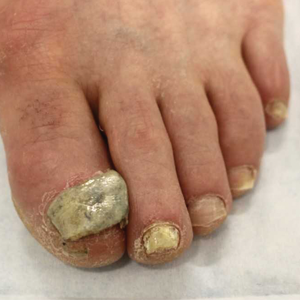
Should toe amputation be delayed in diabetic patients with osteomyelitis?
Amputation: Resistance is not futile!
What’s in a toe you may ask? Why worry about saving it? Just amputate and move on ...

Not so! I implore you to resist the desire. We vascular surgeons are accustomed to cutting off toes, even feet and legs. But when it comes to diabetic feet please reconsider. Just because there is osteomyelitis, I argue that does not necessitate amputation.
We all agree that ischemic gangrene and black mummified digits are beyond salvage. That’s not what my concern is. My focus is nonhealing ulcers with underlying osteomyelitis. Whether ischemic in etiology or neuropathic (or both), give salvage a try.
Why is this so important? My opponent will try to convince you that it’s not. He’ll try to sell you on how well people walk after amputation and that functional outcomes are great. But think beyond that for a second.
Amputation changes the foot architecture and weight distribution. In a person with neuropathy, this only predisposes them to more ulcers. More ulcers will mean more infection, which will lead to more amputations. This finally culminates in a major amputation.
In one reported study,1 researchers followed more than 200,000 diabetics from 2010 until 2013. While the risk of amputation overall was relatively small (0.36% for major and 0.56% for minor amputations), prior minor amputation increased the risk of major amputation 10-fold and increased the risk of another minor (below-ankle) amputation 20-fold. Of those who had a major amputation, 57% died over the 3 years. This is not insignificant.
This does not also consider the morbidity and impact on lifestyle and quality of life for these patients. Many may not walk. Some will be relegated to nursing homes. Some will suffer from phantom limb pain. Many may never return to work. Even more will have difficulty with their daily lives, not to mention the psychological recovery also required.
The foot seems to be the only place where amputation as first-line therapy for osteomyelitis is accepted. We don’t do a hip disarticulation for ischial pressure sores with osteomyelitis. Calvarial osteomyelitis is also treated with antibiotics. I implore you: Don’t treat toes like vestigial organs.
Granted, there are subsets of patients who would benefit from amputations. A patient with painful Charcot foot may elect to have a below-knee amputation and move on with life. Another who has lost jobs or significant time due to recurrence of osteomyelitis may progress. A patient with severe sepsis and infection into a joint may need amputation.
But what other treatment options are there? I’m glad you inquired.
I primarily treat diabetic feet by treating the soft tissue envelope. Even if a patient presents with midfoot infection or necrotizing soft tissue infection, I treat it like a good old-fashioned abscess or necrotizing fasciitis:
1) Drain pus
2) Resect the dead stuff
3) Supportive care (antibiotics, fluids, aggressive wound care, etc.)
I try to leave the bones intact. When bone is exposed I take biopsies for culture and pathology. Any bone destroyed by the infection is focally debrided. I also take a specimen of the “bone margin” that I’m leaving behind and I send this to pathology looking for residual acute osteomyelitis. These steps are important as they dictate duration and choice of antibiotic therapy. This is in keeping with the consensus recommendations published in 2016.2
Even chronic wounds get a similar approach. If there is granulation, let it granulate and see if it will fill the wound. “Just because osteomyelitis is there, it doesn’t mean that for the toe we won’t care!”
There are exceptions of course. If the soft tissue is severely affected so the phalynx protrudes like something from the movie “Coco,” probably that should be amputated. Repeat offenders also may progress to amputation. But otherwise, hold off and give it a chance.
For the inpatient, aggressive irrigation of the wounds using the Veraflo system promotes granulation, even for short hospital stays of 1 week or less. Any ischemic component is worked up and addressed with percutaneous or open revascularization. We treat with prolonged antibiotics, and in questionable cases err on the side of giving long-term courses. These wounds need to be offloaded for tasks of daily living (going to the bathroom, making a sandwich, etc.) but otherwise we instruct patients to be effectively non–weight-bearing on that limb.
We also refer patients for hyperbaric therapy frequently. Now if you’re done groaning, I assure you this is not phony medicine. There is growing evidence to support not only improved rates of healing, but also significant cost savings and improved quality of life.3
In young patients or those with large defects, we also involve plastic and reconstructive surgery for secondary closure approaches (free flaps, adjacent tissue transfers, local autogenous or prosthetic grafting [Integra, Stravix, Dermacell, etc.] or other advanced techniques). This is particularly important in plantar wounds that will need to bear weight in the future, or in young patients for improved functional and cosmetic outcomes. For smaller wounds, we often use dermal/subdermal graft substitutes ourselves.
Even still, in nonambulatory or chronically debilitated and medically high-risk patients, maybe a different option is palliative wound care with or without antibiotics. A nonoperative approach to allow individuals to live the rest of their remaining days without undergoing a morbid and disfiguring amputation is not unreasonable. Many families are thankful for this option when given it. In the absence of refractory pain or overwhelming sepsis, we just let the wound do what it will do, understanding that someday the plan may change. This allows patients to continue to treat the wound without escalation to surgery or resorting to amputation.
In the end, just like we vascular surgeons tailor our “holistic” approach to the needs and desires of a single particular patient, we should approach wounds with a similar attitude. The presence of osteomyelitis in and of itself should not prompt one to bypass an entire algorithm, go straight to amputation, do not pass “Go” or “collect $200” (although the professional fee for a toe amputation is probably around $200). With a multidisciplinary and multimodal approach, and vested patients, salvage is possible in the majority of cases.
References
1. Diabetologia. 2018 Mar;61(3):626-35.
2. Diabet Foot Ankle. 2016 Jul 12. doi: 10.3402/dfa.v7.30079.
3. Int J Technol Assess Health Care. 2008 Spring;24(2):178-83.
Dr. Issam Koleilat is assistant professor and associate program director, Vascular Surgery Residency and Fellowship, Division of Vascular Surgery, Albert Einstein College of Medicine/Montefiore Medical Center, New York. He had no relevant disclosures.
Amputation: Often the best option
For many years there has been debate about the best management strategy for diabetic foot infection including osteomyelitis. The principles of appropriate antibiotics, surgical debridement, good wound care, and proper offloading will always remain. There are no randomized controlled trials of medical vs. surgical management of diabetic foot ulceration with osteomyelitis.

We now have a number of widely accepted ways to define wounds including Wagner and the SVS-adopted WIFI score. Historical papers are somewhat plagued by heterogeneity in the wounds included. This is even more apparent with any attempted meta-analyses. I think everyone would agree that the superficial toe wound with minimal cellulitis is best managed medically. The issue at hand is the profoundly neuropathic diabetic often with underlying anatomic abnormality and osteomyelitis. My esteemed colleague would suggest that we are too quick to pull a trigger and amputate a toe with underlying osteomyelitis.
I think the initial item for debate is the technique of diagnosis of osteomyelitis. We have multiple ways this is reported. Plain x-ray, bone scan, MRI, and “clinical osteomyelitis” are among the alternative ways osteomyelitis is diagnosed. The reliability of the last is the most variable because clinical osteomyelitis ranges from “probes close to bone” to exposed bone visible protruding from the wound bed. Given the variability of diagnostic techniques, the literature is an amalgam of clinical scenarios and difficult to navigate in a way to affect treatment decisions.
In addition, the medical treatment for osteomyelitis is highly variable. This commonly involves tunneled catheter insertion and 6 weeks to 3 months of IV antibiotics. In some institutions antibiotics are tailored to “wound culture.” Several of our infectious disease specialists prefer bone culture and pathology of bone demonstrating an acute destructive process. Obviously, this often requires surgical debridement to obtain a specimen. Antibiotic duration recommendations may vary from 1 week (if all infected bone is resected) to 90 days if a standalone antibiotic management is selected. Chronic osteomyelitis has a reinfection rate of up to 30%.1
Medical management is not without risk. These risks include recurring infection with resistant organisms, wound deterioration, gastrointestinal complications (Clostridium difficile), catheter-related complications, and acute kidney injury. A recent paper found over 30% of patients treated medically for osteomyelitis developed acute kidney injury. These patients had more frequent hospitalization, recurring ulceration, and infection.2 We have all experienced the patient with multiple hospitalizations and episodic AKI that culminates in ESRD requiring hemodialysis.
If the argument is with good follow-up these patients will ultimately experience preservation of the toe, I would take the stance that in our patient population of diabetics presenting with foot ulcer and osteomyelitis the average hemoglobin A1c is over 9. Although this is not only related to patient compliance, in many instances this is a large piece of the puzzle. It is hard to infer that suddenly with biopsy-proven osteomyelitis the patient will become compliant with medical management of the disease process. Certainly, in some circumstances, this is the case. There are a number of studies with a wide range of findings on HbA1c as it relates to predictive value of wound healing.
There are various studies comparing surgical to medical management for osteomyelitis. Limb salvage is contingent upon location (forefoot, midfoot, hindfoot), the extent of infection, and patient comorbidities. The conclusion of the majority of these studies is that a standalone antibiotic treatment algorithm results in greater limb loss. Patients with peripheral occlusive disease and preadmission antibiotic use have been shown to have decreased wound healing. Minor amputation has been shown to be protective from mortality, risk of major amputation, and unfavorable discharge in patients admitted with a diagnosis of osteomyelitis.3 The major limb amputation rate for antibiotics alone is 20%-30% according to two trials with duration of antibiotics of 3 months.4,5 The available randomized trials tend to exclude patients with severe infection (poorly defined), those with PAD, or those with severe comorbid conditions.
Cost of treatment is even more poorly delineated. Obviously, surgical treatment is not without cost to the health care system. Toe amputation especially when including the metatarsal head shifts pressure points and in the neuropathic patient may lead to recurrent ulceration. The average outpatient cost per patient per ulcer is often over $30,000. The goal of surgical treatment can be defined as trying to maintain the greatest degree of function with the least risk. Removing infected bone (i.e., minor amputation) limits exposure to prolonged antibiotic treatment and hopefully lessens recurring ulceration and hospitalization. This is only one piece of the puzzle, however. A multidisciplinary approach with endocrinology, infectious disease, and orthotics for offloading are keys to decrease future ulceration.
Although I do not advocate for widespread toe carnage as suggested by Dr. Koleilat, I do think liberal application of minor amputation to limit hospital stay, limit antibiotic duration and its inherent risk, and possibly affect readmission is often in the best interest of the patient and the system as a whole. Obviously, based on the variable reports in the literature there cannot be a single approach to these patients and the treatment must be individualized based on extent of infection, compliance of the patient, access to multidisciplinary care, and comorbid conditions.
References
1. World J Diabetes. 2017 Apr 15;8(4):135-42.
2. Diabetes Res Clin Pract. 2018 Jan;135:58-64.
3. Ann Surg. 2005;241(6):885-94.
4. Am J Med. 1987 Oct;83(4):653-60.
5. Am J Med.1989 Jun;86(6 Pt 2):801-8.
Dr. Mark P. Androes is division chief, vascular surgery, Greenville (S.C.) Health System. He had no relevant disclosures.
Amputation: Resistance is not futile!
What’s in a toe you may ask? Why worry about saving it? Just amputate and move on ...
Not so! I implore you to resist the desire. We vascular surgeons are accustomed to cutting off toes, even feet and legs. But when it comes to diabetic feet please reconsider. Just because there is osteomyelitis, I argue that does not necessitate amputation.
We all agree that ischemic gangrene and black mummified digits are beyond salvage. That’s not what my concern is. My focus is nonhealing ulcers with underlying osteomyelitis. Whether ischemic in etiology or neuropathic (or both), give salvage a try.
Why is this so important? My opponent will try to convince you that it’s not. He’ll try to sell you on how well people walk after amputation and that functional outcomes are great. But think beyond that for a second.
Amputation changes the foot architecture and weight distribution. In a person with neuropathy, this only predisposes them to more ulcers. More ulcers will mean more infection, which will lead to more amputations. This finally culminates in a major amputation.
In one reported study,1 researchers followed more than 200,000 diabetics from 2010 until 2013. While the risk of amputation overall was relatively small (0.36% for major and 0.56% for minor amputations), prior minor amputation increased the risk of major amputation 10-fold and increased the risk of another minor (below-ankle) amputation 20-fold. Of those who had a major amputation, 57% died over the 3 years. This is not insignificant.
This does not also consider the morbidity and impact on lifestyle and quality of life for these patients. Many may not walk. Some will be relegated to nursing homes. Some will suffer from phantom limb pain. Many may never return to work. Even more will have difficulty with their daily lives, not to mention the psychological recovery also required.
The foot seems to be the only place where amputation as first-line therapy for osteomyelitis is accepted. We don’t do a hip disarticulation for ischial pressure sores with osteomyelitis. Calvarial osteomyelitis is also treated with antibiotics. I implore you: Don’t treat toes like vestigial organs.
Granted, there are subsets of patients who would benefit from amputations. A patient with painful Charcot foot may elect to have a below-knee amputation and move on with life. Another who has lost jobs or significant time due to recurrence of osteomyelitis may progress. A patient with severe sepsis and infection into a joint may need amputation.
But what other treatment options are there? I’m glad you inquired.
I primarily treat diabetic feet by treating the soft tissue envelope. Even if a patient presents with midfoot infection or necrotizing soft tissue infection, I treat it like a good old-fashioned abscess or necrotizing fasciitis:
1) Drain pus
2) Resect the dead stuff
3) Supportive care (antibiotics, fluids, aggressive wound care, etc.)
I try to leave the bones intact. When bone is exposed I take biopsies for culture and pathology. Any bone destroyed by the infection is focally debrided. I also take a specimen of the “bone margin” that I’m leaving behind and I send this to pathology looking for residual acute osteomyelitis. These steps are important as they dictate duration and choice of antibiotic therapy. This is in keeping with the consensus recommendations published in 2016.2
Even chronic wounds get a similar approach. If there is granulation, let it granulate and see if it will fill the wound. “Just because osteomyelitis is there, it doesn’t mean that for the toe we won’t care!”
There are exceptions of course. If the soft tissue is severely affected so the phalynx protrudes like something from the movie “Coco,” probably that should be amputated. Repeat offenders also may progress to amputation. But otherwise, hold off and give it a chance.
For the inpatient, aggressive irrigation of the wounds using the Veraflo system promotes granulation, even for short hospital stays of 1 week or less. Any ischemic component is worked up and addressed with percutaneous or open revascularization. We treat with prolonged antibiotics, and in questionable cases err on the side of giving long-term courses. These wounds need to be offloaded for tasks of daily living (going to the bathroom, making a sandwich, etc.) but otherwise we instruct patients to be effectively non–weight-bearing on that limb.
We also refer patients for hyperbaric therapy frequently. Now if you’re done groaning, I assure you this is not phony medicine. There is growing evidence to support not only improved rates of healing, but also significant cost savings and improved quality of life.3
In young patients or those with large defects, we also involve plastic and reconstructive surgery for secondary closure approaches (free flaps, adjacent tissue transfers, local autogenous or prosthetic grafting [Integra, Stravix, Dermacell, etc.] or other advanced techniques). This is particularly important in plantar wounds that will need to bear weight in the future, or in young patients for improved functional and cosmetic outcomes. For smaller wounds, we often use dermal/subdermal graft substitutes ourselves.
Even still, in nonambulatory or chronically debilitated and medically high-risk patients, maybe a different option is palliative wound care with or without antibiotics. A nonoperative approach to allow individuals to live the rest of their remaining days without undergoing a morbid and disfiguring amputation is not unreasonable. Many families are thankful for this option when given it. In the absence of refractory pain or overwhelming sepsis, we just let the wound do what it will do, understanding that someday the plan may change. This allows patients to continue to treat the wound without escalation to surgery or resorting to amputation.
In the end, just like we vascular surgeons tailor our “holistic” approach to the needs and desires of a single particular patient, we should approach wounds with a similar attitude. The presence of osteomyelitis in and of itself should not prompt one to bypass an entire algorithm, go straight to amputation, do not pass “Go” or “collect $200” (although the professional fee for a toe amputation is probably around $200). With a multidisciplinary and multimodal approach, and vested patients, salvage is possible in the majority of cases.
References
1. Diabetologia. 2018 Mar;61(3):626-35.
2. Diabet Foot Ankle. 2016 Jul 12. doi: 10.3402/dfa.v7.30079.
3. Int J Technol Assess Health Care. 2008 Spring;24(2):178-83.
Dr. Issam Koleilat is assistant professor and associate program director, Vascular Surgery Residency and Fellowship, Division of Vascular Surgery, Albert Einstein College of Medicine/Montefiore Medical Center, New York. He had no relevant disclosures.
Amputation: Often the best option
For many years there has been debate about the best management strategy for diabetic foot infection including osteomyelitis. The principles of appropriate antibiotics, surgical debridement, good wound care, and proper offloading will always remain. There are no randomized controlled trials of medical vs. surgical management of diabetic foot ulceration with osteomyelitis.
We now have a number of widely accepted ways to define wounds including Wagner and the SVS-adopted WIFI score. Historical papers are somewhat plagued by heterogeneity in the wounds included. This is even more apparent with any attempted meta-analyses. I think everyone would agree that the superficial toe wound with minimal cellulitis is best managed medically. The issue at hand is the profoundly neuropathic diabetic often with underlying anatomic abnormality and osteomyelitis. My esteemed colleague would suggest that we are too quick to pull a trigger and amputate a toe with underlying osteomyelitis.
I think the initial item for debate is the technique of diagnosis of osteomyelitis. We have multiple ways this is reported. Plain x-ray, bone scan, MRI, and “clinical osteomyelitis” are among the alternative ways osteomyelitis is diagnosed. The reliability of the last is the most variable because clinical osteomyelitis ranges from “probes close to bone” to exposed bone visible protruding from the wound bed. Given the variability of diagnostic techniques, the literature is an amalgam of clinical scenarios and difficult to navigate in a way to affect treatment decisions.
In addition, the medical treatment for osteomyelitis is highly variable. This commonly involves tunneled catheter insertion and 6 weeks to 3 months of IV antibiotics. In some institutions antibiotics are tailored to “wound culture.” Several of our infectious disease specialists prefer bone culture and pathology of bone demonstrating an acute destructive process. Obviously, this often requires surgical debridement to obtain a specimen. Antibiotic duration recommendations may vary from 1 week (if all infected bone is resected) to 90 days if a standalone antibiotic management is selected. Chronic osteomyelitis has a reinfection rate of up to 30%.1
Medical management is not without risk. These risks include recurring infection with resistant organisms, wound deterioration, gastrointestinal complications (Clostridium difficile), catheter-related complications, and acute kidney injury. A recent paper found over 30% of patients treated medically for osteomyelitis developed acute kidney injury. These patients had more frequent hospitalization, recurring ulceration, and infection.2 We have all experienced the patient with multiple hospitalizations and episodic AKI that culminates in ESRD requiring hemodialysis.
If the argument is with good follow-up these patients will ultimately experience preservation of the toe, I would take the stance that in our patient population of diabetics presenting with foot ulcer and osteomyelitis the average hemoglobin A1c is over 9. Although this is not only related to patient compliance, in many instances this is a large piece of the puzzle. It is hard to infer that suddenly with biopsy-proven osteomyelitis the patient will become compliant with medical management of the disease process. Certainly, in some circumstances, this is the case. There are a number of studies with a wide range of findings on HbA1c as it relates to predictive value of wound healing.
There are various studies comparing surgical to medical management for osteomyelitis. Limb salvage is contingent upon location (forefoot, midfoot, hindfoot), the extent of infection, and patient comorbidities. The conclusion of the majority of these studies is that a standalone antibiotic treatment algorithm results in greater limb loss. Patients with peripheral occlusive disease and preadmission antibiotic use have been shown to have decreased wound healing. Minor amputation has been shown to be protective from mortality, risk of major amputation, and unfavorable discharge in patients admitted with a diagnosis of osteomyelitis.3 The major limb amputation rate for antibiotics alone is 20%-30% according to two trials with duration of antibiotics of 3 months.4,5 The available randomized trials tend to exclude patients with severe infection (poorly defined), those with PAD, or those with severe comorbid conditions.
Cost of treatment is even more poorly delineated. Obviously, surgical treatment is not without cost to the health care system. Toe amputation especially when including the metatarsal head shifts pressure points and in the neuropathic patient may lead to recurrent ulceration. The average outpatient cost per patient per ulcer is often over $30,000. The goal of surgical treatment can be defined as trying to maintain the greatest degree of function with the least risk. Removing infected bone (i.e., minor amputation) limits exposure to prolonged antibiotic treatment and hopefully lessens recurring ulceration and hospitalization. This is only one piece of the puzzle, however. A multidisciplinary approach with endocrinology, infectious disease, and orthotics for offloading are keys to decrease future ulceration.
Although I do not advocate for widespread toe carnage as suggested by Dr. Koleilat, I do think liberal application of minor amputation to limit hospital stay, limit antibiotic duration and its inherent risk, and possibly affect readmission is often in the best interest of the patient and the system as a whole. Obviously, based on the variable reports in the literature there cannot be a single approach to these patients and the treatment must be individualized based on extent of infection, compliance of the patient, access to multidisciplinary care, and comorbid conditions.
References
1. World J Diabetes. 2017 Apr 15;8(4):135-42.
2. Diabetes Res Clin Pract. 2018 Jan;135:58-64.
3. Ann Surg. 2005;241(6):885-94.
4. Am J Med. 1987 Oct;83(4):653-60.
5. Am J Med.1989 Jun;86(6 Pt 2):801-8.
Dr. Mark P. Androes is division chief, vascular surgery, Greenville (S.C.) Health System. He had no relevant disclosures.

Testicular cancer on the rise
Testicular cancer is noted to be the most common malignant cancer among men aged 15-44 years.1 Despite its being the most common, studies show testicular cancer accounts for only 1% of cancers among men over all. Although several risk factors have been identified, there are no clear direct causes of testicular cancer, and its incidence is on the rise.

In 2004 and again in 2010, the United States Preventive Services Task Force (USPSTF) determined that routine screening in asymptomatic males for testicular cancer either by self-exam or ultrasound did not yield better health outcomes.1 A 2015 study found that testicular cancer incidence in males over 15 years in the United States rose from 5.7/100,000 in 1992 to 6.8/100,000 in 2009, with a significant annual percentage change of 1.1% (P less than .001).2 Thankfully, mortality rates have declined since the 1970s because of the improvements made in chemotherapy regimens.3
Although several studies have investigated this issue, few have been able to determine an exact cause and effect, but several theories have been put forth as likely causes in the rising rates. Most of the established risk factors – such as cryptorchidism, age, race, and exposure to estrogen-mimicking chemicals in utero – have been well described in the literature, but what other factors are affecting our youth?
Increase in sedentary lifestyle is believed to play a role, mostly in relationship to the increase in heat exposure.4 As with cryptorchidism, the elevated temperatures associated with internal body temperatures and testicular exposure to it for extended time periods makes this association reasonable.
Dietary factors have shown a strong relationship to development of testicular cancer, as well. High-fat diets and large intake of dairy products were particularly implicated and correlated regionally with the highest incidences.5 Highest rates of testicular cancer are noted to be in Denmark and other European countries in which there is a high intake of dairy products.
Physical activity such as horseback riding, bicycle riding, and motorcycle riding had varied results when studied, but repeated low-level trauma has been associated with an increased risk of testicular cancer.6
Occupations that have repeated exposure to high-heat environments, such as fireman and factory workers, also showed elevated incidences. Aircraft maintenance and handling of heavy metal and pesticides also have showed a correlation. A substantial amount of evidence indicates that environmental pollutants with estrogenic or antiandrogenic activity are associated with increasing incidence of testicular cancer.4

Genetics have the strongest correlation as a risk factor. It has been well documented that there is an eight- to tenfold increase in risk if a brother has been diagnosed with testicular cancer and a four- to sixfold in risk for the son if a father had testicular cancer. Down syndrome also showed increased genetic risk for testicular cancer.3
Although mortality rates are declining and screening for testicular cancer is not indicated, it is important to remember the risk factors and consider it in the differential diagnosis of a symptomatic male given 26%-56% of newly diagnosed testicular cancer patients were wrongly diagnosed initially.1 Dietary guidance also can be helpful for patients who do have increased risk factors; have them avoid high fat diets and excessive dairy intake. Make patients aware that testicular cancer is most common in younger men, and if they note any changes in their testicle, they should seek medical attention.
Dr. Pearce is a pediatrician in Frankfort, Ill. She said she had no relevant financial disclosures. Email her at pdnews@mdedge.com.
References
1. Ann Intern Med. 2010;153:396-9.
2. World J Urol. 2015 May;33(5):623-31.
3. N Engl J Med. 2014;371:2005-16.
4. CMAJ. 1999 Jan 26;160(2):213-4.
5. Nat Rev Urol. 2012;9(6):339-49.
6. Int J Cancer. 2005 Sep 1;116(3):331-9.
Testicular cancer is noted to be the most common malignant cancer among men aged 15-44 years.1 Despite its being the most common, studies show testicular cancer accounts for only 1% of cancers among men over all. Although several risk factors have been identified, there are no clear direct causes of testicular cancer, and its incidence is on the rise.
In 2004 and again in 2010, the United States Preventive Services Task Force (USPSTF) determined that routine screening in asymptomatic males for testicular cancer either by self-exam or ultrasound did not yield better health outcomes.1 A 2015 study found that testicular cancer incidence in males over 15 years in the United States rose from 5.7/100,000 in 1992 to 6.8/100,000 in 2009, with a significant annual percentage change of 1.1% (P less than .001).2 Thankfully, mortality rates have declined since the 1970s because of the improvements made in chemotherapy regimens.3
Although several studies have investigated this issue, few have been able to determine an exact cause and effect, but several theories have been put forth as likely causes in the rising rates. Most of the established risk factors – such as cryptorchidism, age, race, and exposure to estrogen-mimicking chemicals in utero – have been well described in the literature, but what other factors are affecting our youth?
Increase in sedentary lifestyle is believed to play a role, mostly in relationship to the increase in heat exposure.4 As with cryptorchidism, the elevated temperatures associated with internal body temperatures and testicular exposure to it for extended time periods makes this association reasonable.
Dietary factors have shown a strong relationship to development of testicular cancer, as well. High-fat diets and large intake of dairy products were particularly implicated and correlated regionally with the highest incidences.5 Highest rates of testicular cancer are noted to be in Denmark and other European countries in which there is a high intake of dairy products.
Physical activity such as horseback riding, bicycle riding, and motorcycle riding had varied results when studied, but repeated low-level trauma has been associated with an increased risk of testicular cancer.6
Occupations that have repeated exposure to high-heat environments, such as fireman and factory workers, also showed elevated incidences. Aircraft maintenance and handling of heavy metal and pesticides also have showed a correlation. A substantial amount of evidence indicates that environmental pollutants with estrogenic or antiandrogenic activity are associated with increasing incidence of testicular cancer.4
Genetics have the strongest correlation as a risk factor. It has been well documented that there is an eight- to tenfold increase in risk if a brother has been diagnosed with testicular cancer and a four- to sixfold in risk for the son if a father had testicular cancer. Down syndrome also showed increased genetic risk for testicular cancer.3
Although mortality rates are declining and screening for testicular cancer is not indicated, it is important to remember the risk factors and consider it in the differential diagnosis of a symptomatic male given 26%-56% of newly diagnosed testicular cancer patients were wrongly diagnosed initially.1 Dietary guidance also can be helpful for patients who do have increased risk factors; have them avoid high fat diets and excessive dairy intake. Make patients aware that testicular cancer is most common in younger men, and if they note any changes in their testicle, they should seek medical attention.
Dr. Pearce is a pediatrician in Frankfort, Ill. She said she had no relevant financial disclosures. Email her at pdnews@mdedge.com.
References
1. Ann Intern Med. 2010;153:396-9.
2. World J Urol. 2015 May;33(5):623-31.
3. N Engl J Med. 2014;371:2005-16.
4. CMAJ. 1999 Jan 26;160(2):213-4.
5. Nat Rev Urol. 2012;9(6):339-49.
6. Int J Cancer. 2005 Sep 1;116(3):331-9.
Data on perinatal choline, neurodevelopment sparking practice changes
Pregnant women at University of Illinois at Chicago will be offered choline supplements
Finally, the evidence is in: Three evidence-based studies show that perinatal choline supports proper neurodevelopment in fetuses.1,2,3

As anyone who has been following my prevention efforts knows, 4 out of 10 patients at Jackson Park Hospital on Chicago’s Southside who presented to their family medicine clinic for psychiatric care have clinical profiles that are consistent with neurobehavioral disorder associated with prenatal alcohol exposure (ND-PAE).4 Furthermore, since only a little can be done to ameliorate these patients’ psychopathology, I have sought out prevention interventions to stem the tide of what I have thought was a silent epidemic (“occult prenatal alcohol exposure”) for decades.
So I have been heartened that there is some sound science to suggest that perinatal choline supplementation could help. That reality, along with the American Medical Association’s resolution to support evidence-based amounts of choline in all prenatal vitamins, spurred the University of Illinois at Chicago to do something.
Thanks to the support of Enrico Benedetti, MD, professor and head of the department of surgery at the University of Illinois at Chicago, pregnant women will be offered choline supplements to support their fetuses’ neurodevelopment. In addition,
Other efforts are afoot aimed at getting this prevention intervention up and running. For example, Yavar Moghimi, MD, who is the behavioral health director for a Medicaid managed care organization in Washington, recently informed me that its clinical policy committee approved a policy highlighting the evidence behind choline supplements during pregnancy.
I am hoping the University of Illinois at Chicago initiative, entitled the “Healthy Prenatal Brain Program” will help all women by preventing the unrecognized problem I have seen among African American women who engage in social drinking before they realize that they are pregnant.5 After all, the problem of choline deficiency is not tied simply to prenatal alcohol exposure but also to dietary habits. For example, a study by Helen H. Jensen, PhD, and her associates found that 90% of pregnant women do not get enough choline.6 It is just that low-income people are the “canaries in the coal mine” when it comes to being alerted to major public health problems in America.
Another positive development is a website set up by Robert R. Freedman, MD, former chairman of the psychiatry department at the University of Colorado Denver. The site, called prenataldoctoradvice.com, provides guidance to patients about steps they can take, such as taking choline supplements during pregnancy, to improve their children's brain development and mental health.
The public health fix we are suggesting in not difficult; after all, choline is an over-the-counter nutrient, and it does not have to be prescribed by a physician. Ideally, the public health initiatives being advocated are so affordable and easy to implement that this practice will become ubiquitous, and our children will be healthier as a result. It is just a matter of taking action. Now that the evidence is finally in that perinatal choline supplements support proper neurodevelopment in fetuses, we all should move forward – and do something.
Dr. Bell is staff psychiatrist at Jackson Park Hospital’s surgical-medical/psychiatric inpatient unit in Chicago and chairman of the department of psychiatry at Windsor University, St. Kitts, USVI. He also is clinical professor emeritus in the department of psychiatry at the University of Illinois at Chicago; former president/CEO of Community Mental Health Council; and former director of the Institute for Juvenile Research (the birthplace of child psychiatry), all in Chicago.
References
1. Alcohol Clin Exp Res. 2018 Jul;42(7):1327-41.
2. Am J Psychiatry. 2016 May 1;173(5):509-16.
3. Alcohol. 2015 Nov;49(7):647-56.
4. Psychiatr Serv. 2015 May 1;66(5):539-42.
5. MDedge Psychcast. 2018 Oct 17. Fetal alcohol spectrum disorder, part II.
6. The FASEB Journal. 2007;21(6):1b21.
*This column was updated 11/30/2018.
Pregnant women at University of Illinois at Chicago will be offered choline supplements
Pregnant women at University of Illinois at Chicago will be offered choline supplements
Finally, the evidence is in: Three evidence-based studies show that perinatal choline supports proper neurodevelopment in fetuses.1,2,3
As anyone who has been following my prevention efforts knows, 4 out of 10 patients at Jackson Park Hospital on Chicago’s Southside who presented to their family medicine clinic for psychiatric care have clinical profiles that are consistent with neurobehavioral disorder associated with prenatal alcohol exposure (ND-PAE).4 Furthermore, since only a little can be done to ameliorate these patients’ psychopathology, I have sought out prevention interventions to stem the tide of what I have thought was a silent epidemic (“occult prenatal alcohol exposure”) for decades.
So I have been heartened that there is some sound science to suggest that perinatal choline supplementation could help. That reality, along with the American Medical Association’s resolution to support evidence-based amounts of choline in all prenatal vitamins, spurred the University of Illinois at Chicago to do something.
Thanks to the support of Enrico Benedetti, MD, professor and head of the department of surgery at the University of Illinois at Chicago, pregnant women will be offered choline supplements to support their fetuses’ neurodevelopment. In addition,
Other efforts are afoot aimed at getting this prevention intervention up and running. For example, Yavar Moghimi, MD, who is the behavioral health director for a Medicaid managed care organization in Washington, recently informed me that its clinical policy committee approved a policy highlighting the evidence behind choline supplements during pregnancy.
I am hoping the University of Illinois at Chicago initiative, entitled the “Healthy Prenatal Brain Program” will help all women by preventing the unrecognized problem I have seen among African American women who engage in social drinking before they realize that they are pregnant.5 After all, the problem of choline deficiency is not tied simply to prenatal alcohol exposure but also to dietary habits. For example, a study by Helen H. Jensen, PhD, and her associates found that 90% of pregnant women do not get enough choline.6 It is just that low-income people are the “canaries in the coal mine” when it comes to being alerted to major public health problems in America.
Another positive development is a website set up by Robert R. Freedman, MD, former chairman of the psychiatry department at the University of Colorado Denver. The site, called prenataldoctoradvice.com, provides guidance to patients about steps they can take, such as taking choline supplements during pregnancy, to improve their children's brain development and mental health.
The public health fix we are suggesting in not difficult; after all, choline is an over-the-counter nutrient, and it does not have to be prescribed by a physician. Ideally, the public health initiatives being advocated are so affordable and easy to implement that this practice will become ubiquitous, and our children will be healthier as a result. It is just a matter of taking action. Now that the evidence is finally in that perinatal choline supplements support proper neurodevelopment in fetuses, we all should move forward – and do something.
Dr. Bell is staff psychiatrist at Jackson Park Hospital’s surgical-medical/psychiatric inpatient unit in Chicago and chairman of the department of psychiatry at Windsor University, St. Kitts, USVI. He also is clinical professor emeritus in the department of psychiatry at the University of Illinois at Chicago; former president/CEO of Community Mental Health Council; and former director of the Institute for Juvenile Research (the birthplace of child psychiatry), all in Chicago.
References
1. Alcohol Clin Exp Res. 2018 Jul;42(7):1327-41.
2. Am J Psychiatry. 2016 May 1;173(5):509-16.
3. Alcohol. 2015 Nov;49(7):647-56.
4. Psychiatr Serv. 2015 May 1;66(5):539-42.
5. MDedge Psychcast. 2018 Oct 17. Fetal alcohol spectrum disorder, part II.
6. The FASEB Journal. 2007;21(6):1b21.
*This column was updated 11/30/2018.
NIH director expresses concern over CRISPR-cas9 baby claim
The National Institutes of Health is deeply concerned about the work just presented at the Second International Summit on Human Genome Editing in Hong Kong by Dr. He Jiankui, who described his effort using CRISPR-Cas9 on human embryos to disable the CCR5 gene. He claims that the two embryos were subsequently implanted, and infant twins have been born.

This work represents a deeply disturbing willingness by Dr. He and his team to flout international ethical norms. The project was largely carried out in secret, the medical necessity for inactivation of CCR5 in these infants is utterly unconvincing, the informed consent process appears highly questionable, and the possibility of damaging off-target effects has not been satisfactorily explored. It is profoundly unfortunate that the first apparent application of this powerful technique to the human germline has been carried out so irresponsibly.
The need for development of binding international consensus on setting limits for this kind of research, now being debated in Hong Kong, has never been more apparent. Without such limits, the world will face the serious risk of a deluge of similarly ill-considered and unethical projects.
Should such epic scientific misadventures proceed, a technology with enormous promise for prevention and treatment of disease will be overshadowed by justifiable public outrage, fear, and disgust.
Lest there be any doubt, and as we have stated previously, NIH does not support the use of gene-editing technologies in human embryos.
Francis S. Collins, M.D., Ph.D. is director of the National Institutes of Health. His comments were made in a statement Nov. 28.
The National Institutes of Health is deeply concerned about the work just presented at the Second International Summit on Human Genome Editing in Hong Kong by Dr. He Jiankui, who described his effort using CRISPR-Cas9 on human embryos to disable the CCR5 gene. He claims that the two embryos were subsequently implanted, and infant twins have been born.
This work represents a deeply disturbing willingness by Dr. He and his team to flout international ethical norms. The project was largely carried out in secret, the medical necessity for inactivation of CCR5 in these infants is utterly unconvincing, the informed consent process appears highly questionable, and the possibility of damaging off-target effects has not been satisfactorily explored. It is profoundly unfortunate that the first apparent application of this powerful technique to the human germline has been carried out so irresponsibly.
The need for development of binding international consensus on setting limits for this kind of research, now being debated in Hong Kong, has never been more apparent. Without such limits, the world will face the serious risk of a deluge of similarly ill-considered and unethical projects.
Should such epic scientific misadventures proceed, a technology with enormous promise for prevention and treatment of disease will be overshadowed by justifiable public outrage, fear, and disgust.
Lest there be any doubt, and as we have stated previously, NIH does not support the use of gene-editing technologies in human embryos.
Francis S. Collins, M.D., Ph.D. is director of the National Institutes of Health. His comments were made in a statement Nov. 28.
Help parents manage screen time thoughtfully
It has been 2 years since we last wrote about the potential risks to children and adolescents of spending too much time on screens. While there have been studies in the interval that offer us more information about the effects of heavy screen use and the developing brain, there is little certainty about what is optimal for children and adolescents, and less still on how parents might effectively equip their children to make good use of screens without suffering ill effects.

You might recall that back in October of 2016, the American Academy of Pediatrics published screen time guidelines: recommending no screen time for infants and children up to 18 months old, limiting all screen time to 1 hour per day for children up to 5 years old, and 2 hours daily for older children (up to 11 years old), so that it would not interfere with homework, social time, exercise, and sleep. At the time, data suggested that children from 2 to 11 years old were spending an average of 4.5 hours per day on screens (TV, computer, tablets, or smartphones, not counting homework).
The Adolescent Brain Cognitive Development Study began in September 2016 to evaluate the effects of Canadian recommendations for 8- to 11-year-olds (9-11 hours sleep nightly, 1 hour of exercise daily, and 2 hours or less of screen time daily; the study subjects are in the United States). This fall they published their initial results, demonstrating that only 51% get the recommended amount of sleep, only 37% kept their daily screen time to under 2 hours, and only 18% were getting the recommended amount of exercise. Only 5% of children consistently met all three recommendations while 29% of children didn’t meet any of the recommendations.
The researchers assessed the children’s cognitive development and found that after 1 year, those children who met the screen time recommendations, both screen time and sleep, or all three recommendations demonstrated “superior global cognition.” Children were spending an average of 3.7 hours daily on screens, and those children who were spending 2 hours or less on screens performed 4% better on tests of cognitive function than did children spending the average amount of time. Sleep and exercise differences alone did not contribute to significant differences in cognitive function. This study will continue for another 10 years.1
In a much smaller study out of Cincinnati Children’s Hospital, researchers asked parents to describe the amount of time a child spent on reading and in screen-based media activities, then completed MRI scans of the children’s brains.2 They found a strong association between reading time and higher functional connectivity between the parts of the brain responsible for visual word formation and those responsible for language and cognitive control, with a negative correlation between functional connectivity and time spent in screen-based media activities.
While these studies are important pieces of data as we build a deeper understanding about the effects of screen-based media use on children’s cognitive and behavioral development, they do not offer certainty about causality. These studies do not yet clarify whether certain children are especially vulnerable to the untoward effects of heavy screen-based media use. Perhaps the research will someday offer guidelines with certainty, but families need guidance now. Without doubt, digital devices are here to stay, are important to homework, and can facilitate independence, long-distance connections, important technical work-skills, and even senseless fun and relaxation. So we will focus on offering some principles to help you guide young people (or their parents) in approaching screen time thoughtfully.
While recommending no more than 2 hours of daily screen time seems reasonable, it may be more useful to focus on what young people are doing with the rest of their time. Are they getting adequate, restful sleep? Are they able to exercise most days? Do they have enough time for homework? Do they have time for friends (time actually together, not just texting)? What about time for hobbies? When parents focus on the precious resource of time and all of the activities their children both need and want to do, it sets the frame for them to say that their children are allowed to have time to relax with screen-based media as long as it does not take away from these other priorities. Ensuring that the child has at least 8 hours of sleep, after homework and sports, also will set natural limits on screen time.
Parents also can use the frame of development to guide their rules about screen time. If use of an electronic device serves a developmental task, then it is reasonable. If it interferes with a developmental task, then it should be limited. Adolescents (ages 12-20) should be exploring their own identities, establishing independence, deepening social relationships, and learning to manage their impulses. Some interests can be most easily explored with the aid of a computer (such as with programming, art history, or astronomy). Use of cellphones can facilitate teenagers’ being more independent with plans or transportation. Social connections can be supported by texting or FaceTime. Some close friends may be in a different sport or live far away, and it is possible to stay connected only virtually. However, when use of electronic devices keeps the child from engaging with new friends and new interests or from getting into the world to establish real independence (i.e., a job), then there should be limits. In all of these cases, it is critical that adults explain to teenagers what is guiding their thinking about limits on screen time. Open discussions about the great utility and fun that screens can provide, as well as the challenge of keeping those activities in balance with other important activities, helps adolescents set the frame for that rapidly approaching time when they will be making those choices without adult supervision.
Younger children (ages 8-11) should be sampling a wide array of activities and interests and experiencing challenges and eventual mastery across domains. Video games can be very compelling for this age group because they appeal to exactly this drive to master a challenge. Parents want to ensure that their children can have senseless fun, and still have enough time to explore actual activities: social, athletic, creative, and academic. They can be ready to explain the why of rules, but consistent rules, enforced for everyone at home, are most helpful for this age group.

You also can help parents to consider the child’s temperament when thinking about which rules will be appropriate. Anxious children and teenagers may be especially prone to immersive virtual activities that allow them to avoid the stress of real undertakings or interactions. But anxious children may be able to prepare for something anxiety provoking by exploring it virtually first. Youth with ADHD are going to struggle with shifting away from video games or other electronic activities they enjoy that don’t have a natural ending, and will need strict rules and patient support around balanced screen time use. Screen time may play to a child’s strengths, enabling creative children to take in a wide range of art or music and even create their own when other resources are limited.

Finally, all parents should consider what their own screen use is teaching their children. Adolescents are unlikely to listen to their parents’ recommendations if the parents spend hours online after work. Younger children need their parents’ engaged attention: being coaches and cheerleaders for all of their efforts at mastery. You can help parents to imagine rules that the whole family can follow. They can consider how screen time helps them connect with their children, such as watching a favorite program or sport together. They can explore shared interests online together. They can even relax with ridiculous cat videos together! Screen time together is valuable if it supports parents’ connections with their children, while their rules ensure adequate time for sleep, physical activity, and developmental priorities.
Dr. Swick is physician in chief at Ohana, Center for Child and Adolescent Behavioral Health, Community Hospital of the Monterey (Calif.) Peninsula. Dr. Jellinek is professor emeritus of psychiatry and pediatrics, Harvard Medical School, Boston. Email them at pdnews@mdedge.com.
References
1. Lancet Child Adolesc Health. 2018 Nov 1;2(11):783-91.
2. Acta Paediatra. 2018 Apr;107(4):685-93
It has been 2 years since we last wrote about the potential risks to children and adolescents of spending too much time on screens. While there have been studies in the interval that offer us more information about the effects of heavy screen use and the developing brain, there is little certainty about what is optimal for children and adolescents, and less still on how parents might effectively equip their children to make good use of screens without suffering ill effects.
You might recall that back in October of 2016, the American Academy of Pediatrics published screen time guidelines: recommending no screen time for infants and children up to 18 months old, limiting all screen time to 1 hour per day for children up to 5 years old, and 2 hours daily for older children (up to 11 years old), so that it would not interfere with homework, social time, exercise, and sleep. At the time, data suggested that children from 2 to 11 years old were spending an average of 4.5 hours per day on screens (TV, computer, tablets, or smartphones, not counting homework).
The Adolescent Brain Cognitive Development Study began in September 2016 to evaluate the effects of Canadian recommendations for 8- to 11-year-olds (9-11 hours sleep nightly, 1 hour of exercise daily, and 2 hours or less of screen time daily; the study subjects are in the United States). This fall they published their initial results, demonstrating that only 51% get the recommended amount of sleep, only 37% kept their daily screen time to under 2 hours, and only 18% were getting the recommended amount of exercise. Only 5% of children consistently met all three recommendations while 29% of children didn’t meet any of the recommendations.
The researchers assessed the children’s cognitive development and found that after 1 year, those children who met the screen time recommendations, both screen time and sleep, or all three recommendations demonstrated “superior global cognition.” Children were spending an average of 3.7 hours daily on screens, and those children who were spending 2 hours or less on screens performed 4% better on tests of cognitive function than did children spending the average amount of time. Sleep and exercise differences alone did not contribute to significant differences in cognitive function. This study will continue for another 10 years.1
In a much smaller study out of Cincinnati Children’s Hospital, researchers asked parents to describe the amount of time a child spent on reading and in screen-based media activities, then completed MRI scans of the children’s brains.2 They found a strong association between reading time and higher functional connectivity between the parts of the brain responsible for visual word formation and those responsible for language and cognitive control, with a negative correlation between functional connectivity and time spent in screen-based media activities.
While these studies are important pieces of data as we build a deeper understanding about the effects of screen-based media use on children’s cognitive and behavioral development, they do not offer certainty about causality. These studies do not yet clarify whether certain children are especially vulnerable to the untoward effects of heavy screen-based media use. Perhaps the research will someday offer guidelines with certainty, but families need guidance now. Without doubt, digital devices are here to stay, are important to homework, and can facilitate independence, long-distance connections, important technical work-skills, and even senseless fun and relaxation. So we will focus on offering some principles to help you guide young people (or their parents) in approaching screen time thoughtfully.
While recommending no more than 2 hours of daily screen time seems reasonable, it may be more useful to focus on what young people are doing with the rest of their time. Are they getting adequate, restful sleep? Are they able to exercise most days? Do they have enough time for homework? Do they have time for friends (time actually together, not just texting)? What about time for hobbies? When parents focus on the precious resource of time and all of the activities their children both need and want to do, it sets the frame for them to say that their children are allowed to have time to relax with screen-based media as long as it does not take away from these other priorities. Ensuring that the child has at least 8 hours of sleep, after homework and sports, also will set natural limits on screen time.
Parents also can use the frame of development to guide their rules about screen time. If use of an electronic device serves a developmental task, then it is reasonable. If it interferes with a developmental task, then it should be limited. Adolescents (ages 12-20) should be exploring their own identities, establishing independence, deepening social relationships, and learning to manage their impulses. Some interests can be most easily explored with the aid of a computer (such as with programming, art history, or astronomy). Use of cellphones can facilitate teenagers’ being more independent with plans or transportation. Social connections can be supported by texting or FaceTime. Some close friends may be in a different sport or live far away, and it is possible to stay connected only virtually. However, when use of electronic devices keeps the child from engaging with new friends and new interests or from getting into the world to establish real independence (i.e., a job), then there should be limits. In all of these cases, it is critical that adults explain to teenagers what is guiding their thinking about limits on screen time. Open discussions about the great utility and fun that screens can provide, as well as the challenge of keeping those activities in balance with other important activities, helps adolescents set the frame for that rapidly approaching time when they will be making those choices without adult supervision.
Younger children (ages 8-11) should be sampling a wide array of activities and interests and experiencing challenges and eventual mastery across domains. Video games can be very compelling for this age group because they appeal to exactly this drive to master a challenge. Parents want to ensure that their children can have senseless fun, and still have enough time to explore actual activities: social, athletic, creative, and academic. They can be ready to explain the why of rules, but consistent rules, enforced for everyone at home, are most helpful for this age group.
You also can help parents to consider the child’s temperament when thinking about which rules will be appropriate. Anxious children and teenagers may be especially prone to immersive virtual activities that allow them to avoid the stress of real undertakings or interactions. But anxious children may be able to prepare for something anxiety provoking by exploring it virtually first. Youth with ADHD are going to struggle with shifting away from video games or other electronic activities they enjoy that don’t have a natural ending, and will need strict rules and patient support around balanced screen time use. Screen time may play to a child’s strengths, enabling creative children to take in a wide range of art or music and even create their own when other resources are limited.
Finally, all parents should consider what their own screen use is teaching their children. Adolescents are unlikely to listen to their parents’ recommendations if the parents spend hours online after work. Younger children need their parents’ engaged attention: being coaches and cheerleaders for all of their efforts at mastery. You can help parents to imagine rules that the whole family can follow. They can consider how screen time helps them connect with their children, such as watching a favorite program or sport together. They can explore shared interests online together. They can even relax with ridiculous cat videos together! Screen time together is valuable if it supports parents’ connections with their children, while their rules ensure adequate time for sleep, physical activity, and developmental priorities.
Dr. Swick is physician in chief at Ohana, Center for Child and Adolescent Behavioral Health, Community Hospital of the Monterey (Calif.) Peninsula. Dr. Jellinek is professor emeritus of psychiatry and pediatrics, Harvard Medical School, Boston. Email them at pdnews@mdedge.com.
References
1. Lancet Child Adolesc Health. 2018 Nov 1;2(11):783-91.
2. Acta Paediatra. 2018 Apr;107(4):685-93
Recognize gender expression in youth
It has been known for decades that sex and gender cannot be determined solely by birth anatomy and chromosomes.1 Over the past decade, the medical community has been able to better understand the biologic underpinnings of gender identity, and we are gaining a better appreciation for the diversity of gender identities and gender expressions that exist.

Gender expression can be defined as the manner in which an individual chooses to present their gender to others through physical appearance and behaviors, such as style of hair or dress, voice or movement.2 Gender nonconformity (GNC) is when an individual’s gender expression does not fully conform with societal expectations often based on an individual’s sex assigned at birth. It is important to note that gender expression is independent of gender identity and may or may not align with gender identity. For example, a person whose sex assigned at birth is female may adopt hairstyles and clothing that are considered more masculine and enjoy activities that are typically associated with masculinity (for example, sports) yet identify as female. The majority of research to date focuses most on transgender individuals, broadly defined as those whose gender identity does not fully align with the sex assigned at birth.3,4 As our understanding of gender expression and GNC expands, more research is emerging on the prevalence of gender nonconformity in youth and potential associations with various health outcomes.
Stigma, discrimination, and harassment are known to have documented effects on health. GNC youth have been shown to experience discrimination and harassment at rates higher than their gender conforming peers.5,6 A recent study by Lowry et al. sought to examine the association between GNC and indicators of mental distress and substance use in adolescents.7 The authors analyzed a subset of cross-sectional data from more than 6,000 youth who had participated in the Youth Risk Behavior Surveillance–United States, 2015 (YRBS) in three large urban school districts (two in California and one in Florida). In addition to the standard YRBS questions, students at these three school districts were asked about their gender expression using the following question: “A person’s appearance style, dress, or the way they walk or talk may affect how people describe them. How do you think people at your school would describe you?” Based on responses, youth were categorized on a 7-point GNC scale with 1 being most gender conforming (a very feminine female student or very masculine male student) to 7 being most GNC (a very masculine female student or a very feminine male student). The study sample was ethnically diverse with 16% of students identifying as white non-Hispanic, 19% identifying as black non-Hispanic, and 55% identifying as Hispanic of any race.
In the study population, approximately one in five students reported either moderate (students who described themselves as equally feminine and masculine) or high (female students who described themselves as very/mostly/somewhat masculine or male students who described themselves as very/mostly/somewhat feminine) levels of GNC. Among female students, moderate GNC was significantly associated with feeling sad and hopeless, seriously considering attempting suicide, and making a suicide plan. However, in female students substance use was not associated with GNC. Among male students, suicidal thoughts, plans, and attempts all demonstrated a linear increase with GNC, with the greatest prevalence occurring in male students expressing high levels of GNC. Prevalence of substance use, specifically nonmedical use of prescription drugs, cocaine use, methamphetamine use, heroin use, and intravenous drug also was associated with high GNC in male students. Study authors hypothesize that these differences occur because GNC male youth experience more overt harassment, compared with GNC female youth, but further study is needed.
Our understanding of the diversity of gender expressions present in youth populations continues to evolve. Findings from this study add to a growing body of evidence demonstrating a relatively high prevalence of GNC in youth populations, and potential health disparities these youth may face. This study underscores the need for continued study in this area. Family support and acceptance have been demonstrated to be strong protective factors for transgender-, lesbian-, and gay-identified youth. Studies identifying protective factors for GNC youth are needed.4
As health care providers, we need to continue to ask patients and families about gender identity and be aware of gender expression. When youth present as GNC, we should recognize that they may be at increased risk and, in addition to assessing overall mental health and risk for substance use, also assess for degree of social/familial support and potential stressors.4 We also should continue to advocate for support systems within schools sensitive to the needs of GNC students, as these may be a potential avenue to improve overall mental health for students. It is important to continue to expand our understanding of the diverse gender identities and expressions of the youth we serve. This hopefully will allow us to identify not only potential risk factors and health disparities, but also protective factors that can help better inform the development of effective interventions so all youth can reach their full potential.
Dr. Chelvakumar is an attending physician in the division of adolescent medicine at Nationwide Children’s Hospital and an assistant professor of clinical pediatrics at the Ohio State University, both in Columbus. Email her at pdnews@mdedge.com.
References
1. “WPATH (World Professional Association of Transgender Health) Board Responds to Federal Effort to Redefine Gender,” press release, Oct. 23, 2018.
2. “LGBTQ+ Definitions” at Trans Student Educational Resources.3. J Sex Res. 2013;50(3-4):299-317.
4. JAMA Pediatr. 2018 Nov 1;172(11):1010-1.
5. Psychol Sex Orientat Gend Divers. 2016 Dec;3(4):489-98.
6. J Adolesc Health. 2016; 58(2)(supple):S1-2.
7. JAMA Pediatr. 2018 Nov;172(11):1020-8.
It has been known for decades that sex and gender cannot be determined solely by birth anatomy and chromosomes.1 Over the past decade, the medical community has been able to better understand the biologic underpinnings of gender identity, and we are gaining a better appreciation for the diversity of gender identities and gender expressions that exist.
Gender expression can be defined as the manner in which an individual chooses to present their gender to others through physical appearance and behaviors, such as style of hair or dress, voice or movement.2 Gender nonconformity (GNC) is when an individual’s gender expression does not fully conform with societal expectations often based on an individual’s sex assigned at birth. It is important to note that gender expression is independent of gender identity and may or may not align with gender identity. For example, a person whose sex assigned at birth is female may adopt hairstyles and clothing that are considered more masculine and enjoy activities that are typically associated with masculinity (for example, sports) yet identify as female. The majority of research to date focuses most on transgender individuals, broadly defined as those whose gender identity does not fully align with the sex assigned at birth.3,4 As our understanding of gender expression and GNC expands, more research is emerging on the prevalence of gender nonconformity in youth and potential associations with various health outcomes.
Stigma, discrimination, and harassment are known to have documented effects on health. GNC youth have been shown to experience discrimination and harassment at rates higher than their gender conforming peers.5,6 A recent study by Lowry et al. sought to examine the association between GNC and indicators of mental distress and substance use in adolescents.7 The authors analyzed a subset of cross-sectional data from more than 6,000 youth who had participated in the Youth Risk Behavior Surveillance–United States, 2015 (YRBS) in three large urban school districts (two in California and one in Florida). In addition to the standard YRBS questions, students at these three school districts were asked about their gender expression using the following question: “A person’s appearance style, dress, or the way they walk or talk may affect how people describe them. How do you think people at your school would describe you?” Based on responses, youth were categorized on a 7-point GNC scale with 1 being most gender conforming (a very feminine female student or very masculine male student) to 7 being most GNC (a very masculine female student or a very feminine male student). The study sample was ethnically diverse with 16% of students identifying as white non-Hispanic, 19% identifying as black non-Hispanic, and 55% identifying as Hispanic of any race.
In the study population, approximately one in five students reported either moderate (students who described themselves as equally feminine and masculine) or high (female students who described themselves as very/mostly/somewhat masculine or male students who described themselves as very/mostly/somewhat feminine) levels of GNC. Among female students, moderate GNC was significantly associated with feeling sad and hopeless, seriously considering attempting suicide, and making a suicide plan. However, in female students substance use was not associated with GNC. Among male students, suicidal thoughts, plans, and attempts all demonstrated a linear increase with GNC, with the greatest prevalence occurring in male students expressing high levels of GNC. Prevalence of substance use, specifically nonmedical use of prescription drugs, cocaine use, methamphetamine use, heroin use, and intravenous drug also was associated with high GNC in male students. Study authors hypothesize that these differences occur because GNC male youth experience more overt harassment, compared with GNC female youth, but further study is needed.
Our understanding of the diversity of gender expressions present in youth populations continues to evolve. Findings from this study add to a growing body of evidence demonstrating a relatively high prevalence of GNC in youth populations, and potential health disparities these youth may face. This study underscores the need for continued study in this area. Family support and acceptance have been demonstrated to be strong protective factors for transgender-, lesbian-, and gay-identified youth. Studies identifying protective factors for GNC youth are needed.4
As health care providers, we need to continue to ask patients and families about gender identity and be aware of gender expression. When youth present as GNC, we should recognize that they may be at increased risk and, in addition to assessing overall mental health and risk for substance use, also assess for degree of social/familial support and potential stressors.4 We also should continue to advocate for support systems within schools sensitive to the needs of GNC students, as these may be a potential avenue to improve overall mental health for students. It is important to continue to expand our understanding of the diverse gender identities and expressions of the youth we serve. This hopefully will allow us to identify not only potential risk factors and health disparities, but also protective factors that can help better inform the development of effective interventions so all youth can reach their full potential.
Dr. Chelvakumar is an attending physician in the division of adolescent medicine at Nationwide Children’s Hospital and an assistant professor of clinical pediatrics at the Ohio State University, both in Columbus. Email her at pdnews@mdedge.com.
References
1. “WPATH (World Professional Association of Transgender Health) Board Responds to Federal Effort to Redefine Gender,” press release, Oct. 23, 2018.
2. “LGBTQ+ Definitions” at Trans Student Educational Resources.3. J Sex Res. 2013;50(3-4):299-317.
4. JAMA Pediatr. 2018 Nov 1;172(11):1010-1.
5. Psychol Sex Orientat Gend Divers. 2016 Dec;3(4):489-98.
6. J Adolesc Health. 2016; 58(2)(supple):S1-2.
7. JAMA Pediatr. 2018 Nov;172(11):1020-8.

Optimizing Topical Therapy for Onychomycosis: The Importance of Patient Education
Onychomycosis is a fungal infection of the nail unit due to dermatophytes, yeasts, and nondermatophyte molds (NDMs). It accounts for approximately 50% of all nail disorders seen in clinical practice and is estimated to affect 10% to 12% of the US population.1,2 Oral medications approved by the US Food and Drug Administration (FDA) include terbinafine and itraconazole, which have demonstrated good efficacy in treating onychomycosis but are associated with potential drug-drug interactions and systemic side effects.3,4 Although liver failure associated with these drugs is rare,5 many patients are anxious about systemic adverse events and therefore prefer to use topical therapies for onychomycosis.
Many patients desire topical therapy but not every patient is an appropriate candidate. Patients who will likely respond well to topical therapy include those with superficial onychomycosis, distal lateral subungual onychomycosis that involves less than 50% of the nail plate surface area (without matrix involvement and a nail plate thickness less than 2 mm), and only up to 3 or 4 nails affected.6 In patients who have contraindications to oral therapy, topical therapy may be the only treatment option. To maximize efficacy of FDA-approved topical agents for onychomycosis therapy, patient education is of utmost importance. Failure to properly counsel the patient on proper medication application may result in decreased antifungal efficacy; poor patient compliance due to lack of improvement; and progression of disease, leading to increased onychodystrophy and pain.
Before initiating therapy, patients should be counseled that treatment with topical drugs is long, requiring daily application of the medication for 6 months for fingernails and 12 months for toenails, based on average nail growth rates (2–3 mm per month for fingernails; 1 mm per month for toenails).7 Patients also are advised to avoid nail polish application during the course of therapy, as clinical trials were performed without nail polish and the true efficacy with nail polish is unknown.8-10 Because patients who have had onychomycosis for shorter durations generally have better cure rates than those who have disease for longer durations, it is prudent to initiate topical therapy as early as possible.11,12 Treating the feet with an antifungal while treating the nails for onychomycosis further enhances efficacy.13,14 There are 3 FDA-approved topical therapies for onychomycosis: ciclopirox nail lacquer 8%, efinaconazole solution 10%, and tavaborole solution 5%.15-17
Ciclopirox is a hydroxypyridone with broad-spectrum antimicrobial activity against dermatophytes, NDMs, yeasts, and bacteria. Its mechanism of action is to chelate polyvalent cations, such as Fe3+, and inhibit fungal metal-dependent enzymes responsible for the degradation of toxic metabolites.15 Ciclopirox nail lacquer 8% was FDA approved for the treatment of onychomycosis in 1999, making it the first topical approved for this purpose. Its indication is for immunocompetent patients with mild to moderate onychomycosis (Trichophyton rubrum) without lunula involvement, with mycologic cure rates of 29% to 36% and complete cure rates of 5.5% to 8.5% (toenails).15 It is the only FDA-approved topical treatment for both fingernails and toenails. Using a brush applicator, it is applied daily to the nail plate and its undersurface, hyponychium, and 5 mm of the surrounding skin. It is important to counsel the patient to remove the lacquer from the nail plate weekly because failure to do so will result in accumulation of numerous layers of medication, such that the active drug cannot reach the site of infection (Figures 1 and 2). The nail plate also should be trimmed and filed weekly by the patient, with monthly clipping/debridement by a physician recommended.6,15
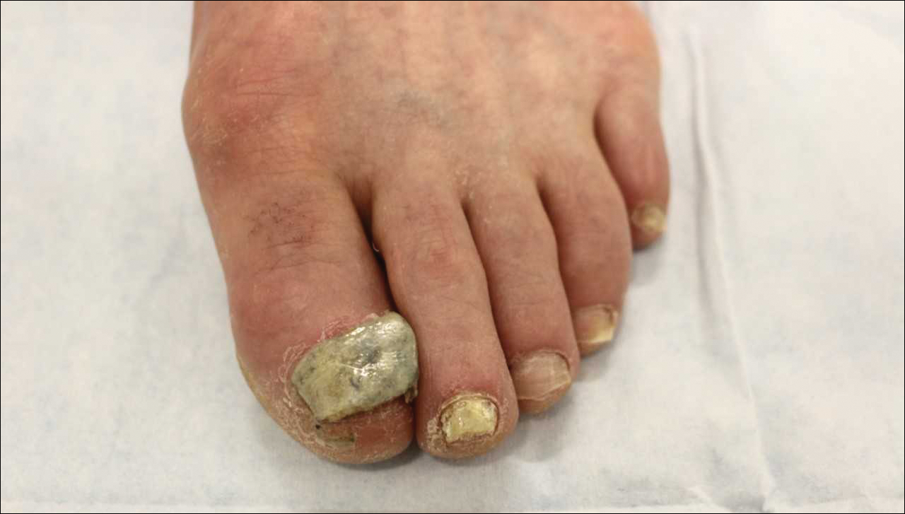
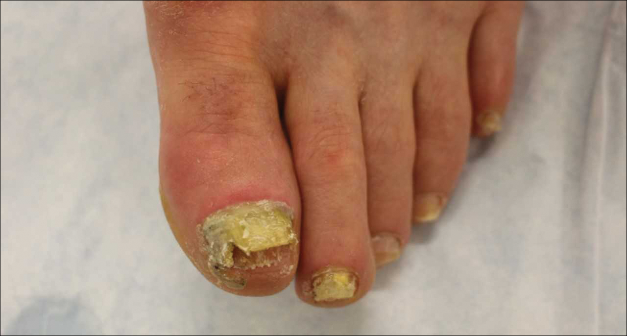
Efinaconazole is a triazole with antifungal activity against dermatophytes, NDMs, and Candida species. Its mechanism of action is inhibition of lanosterol 14α-demethylase, an enzyme involved in the biosynthesis of ergosterol, which is a component of the fungal cell membrane. Efinaconazole solution 10% was FDA approved in June 2014 for the treatment of toenail onychomycosis due to T rubrum and Trichophyton mentagrophytes, with package insert mycologic cure rates of 53.4% to 55.2% and complete cure rates of 15.2% to 17.8%.9,16 It is applied with a brush applicator to the nail plate, as well as its undersurface, nail folds, and hyponychium. Two drops are recommended for the great toenail and one drop for all other toenails, and no removal of the solution or debridement is required.6,16
Tavaborole is a benzoxaborole with antifungal activity against dermatophytes, NDMs, and yeasts. Its mechanism of action is inhibition of fungal aminoacyl transfer RNA synthetase, thus impeding protein synthesis.18 Tavaborole solution 5% was FDA approved in July 2014 for the treatment of toenail onychomycosis due to T rubrum and T mentagrophytes, with mycologic cure rates of 31.1% and 35.9% and complete cure rates of 6.5% and 9.1%, respectively.11,17 It is applied with a glass pointed-tip dropper to the nail plate, such that the entire nail is covered as well as under the nail tip. No removal of the solution or debridement is required.17
Topical therapies for onychomycosis require long treatment durations, thus excellent compliance and adherence to the treatment protocol are vital to maximize efficacy. Dermatologists who prescribe ciclopirox nail lacquer 8% should counsel patients to remove the lacquer with alcohol weekly, such that the antifungal penetrates the nail plate to reach the site of infection. Monthly debridement also must be clarified before initiating therapy. With all topical therapy for onychomycosis, it is important to treat early, treat concurrently for tinea pedis, and avoid use of nail polish so that patients have the best possible cure rates.
- Lipner SR, Scher RK. Onychomycosis: diagnosis and therapy. In: Razzaghi-Abyaneh M, Shams-Ghahfarokhi M, Rai M, eds. Medical Mycology: Current Trends and Future Prospects. Boca Raton, FL: CRC Press; 2015:28.
- Scher RK, Rich P, Pariser D, et al. The epidemiology, etiology, and pathophysiology of onychomycosis. Semin Cutan Med Surg. 2013;32(2 suppl 1):S2-S4.
- Lamisil [package insert]. East Hanover, NJ: Novartis Pharmaceuticals Corporation; 1997.
- Sporanox [package insert]. Titusville, NJ: Janssen Pharmaceuticals, Inc; 2001.
- National Institutes of Health. Terbinafine. LiverTox website. https://livertox.nlm.nih.gov/Terbinafine.htm. Accessed November 7, 2018.
- Lipner SR, Scher RK. Onychomycosis: topical therapy and devices. In: Rubin AI, Jellinek NJ, Daniel CR III, et al, eds. Scher and Daniel’s Nails: Diagnosis, Surgery, Therapy. 4th ed. Cham, Switzerland: Springer International Publishing; 2018:173-184.
- Lipner SR, Scher RK. Nail growth evaluation and factors affecting nail growth. In: Humbert P, Fanian F, Maibach HI, et al, eds. Agache’s Measuring the Skin. 2nd ed. Berlin, Germany: Springer; 2017:867-881.
- Gupta AK, Elewski BE, Sugarman JL, et al. The efficacy and safety of efinaconazole 10% solution for treatment of mild to moderate onychomycosis: a pooled analysis of two phase 3 randomized trials. J Drugs Dermatol. 2014;13:815-820.
- Elewski BE, Rich P, Pollak R, et al. Efinaconazole 10% solution in the treatment of toenail onychomycosis: two phase III multicenter, randomized, double-blind studies. J Am Acad Dermatol. 2013;68:600-608.
- Elewski BE, Aly R, Baldwin SL, et al. Efficacy and safety of tavaborole topical solution, 5%, a novel boron-based antifungal agent, for the treatment of toenail onychomycosis: results from 2 randomized phase-III studies. J Am Acad Dermatol. 2015;73:62-69.
- Rich P. Efinaconazole topical solution, 10%: the benefits of treating onychomycosis early. J Drugs Dermatol. 2015;14:58-62.
- Lipner SR, Scher RK. Efinaconazole 10% topical solution for the topical treatment of onychomycosis of the toenail. Expert Rev Clin Pharmacol. 2015;8:719-731.
- Del Rosso JQ. Onychomycosis of toenails and post-hoc analyses with efinaconazole 10% solution once-daily treatment: impact of disease severity and other concomitant associated factors on selection of therapy and therapeutic outcomes. J Clin Aesthet Dermatol. 2016;9:42.
- Lipner SR, Scher RK. Management of onychomycosis and co-existing tinea pedis. J Drugs Dermatol. 2015;14:492-494.
- Penlac [package insert]. Berwyn, PA: Dermik Laboratories; 2004.
- Jublia [package insert]. Bridgewater, NJ: Valeant Pharmaceuticals, LLC; 2014.
- Kerydin [package insert]. Palo Alto, CA: Anacor Pharmaceuticals, Inc; 2014.
- Rock FL, Mao W, Yaremchuk A, et al. An antifungal agent inhibits an aminoacyl-tRNA synthetase by trapping tRNA in the editing site. Science. 2007;316:1759-1761.
Onychomycosis is a fungal infection of the nail unit due to dermatophytes, yeasts, and nondermatophyte molds (NDMs). It accounts for approximately 50% of all nail disorders seen in clinical practice and is estimated to affect 10% to 12% of the US population.1,2 Oral medications approved by the US Food and Drug Administration (FDA) include terbinafine and itraconazole, which have demonstrated good efficacy in treating onychomycosis but are associated with potential drug-drug interactions and systemic side effects.3,4 Although liver failure associated with these drugs is rare,5 many patients are anxious about systemic adverse events and therefore prefer to use topical therapies for onychomycosis.
Many patients desire topical therapy but not every patient is an appropriate candidate. Patients who will likely respond well to topical therapy include those with superficial onychomycosis, distal lateral subungual onychomycosis that involves less than 50% of the nail plate surface area (without matrix involvement and a nail plate thickness less than 2 mm), and only up to 3 or 4 nails affected.6 In patients who have contraindications to oral therapy, topical therapy may be the only treatment option. To maximize efficacy of FDA-approved topical agents for onychomycosis therapy, patient education is of utmost importance. Failure to properly counsel the patient on proper medication application may result in decreased antifungal efficacy; poor patient compliance due to lack of improvement; and progression of disease, leading to increased onychodystrophy and pain.
Before initiating therapy, patients should be counseled that treatment with topical drugs is long, requiring daily application of the medication for 6 months for fingernails and 12 months for toenails, based on average nail growth rates (2–3 mm per month for fingernails; 1 mm per month for toenails).7 Patients also are advised to avoid nail polish application during the course of therapy, as clinical trials were performed without nail polish and the true efficacy with nail polish is unknown.8-10 Because patients who have had onychomycosis for shorter durations generally have better cure rates than those who have disease for longer durations, it is prudent to initiate topical therapy as early as possible.11,12 Treating the feet with an antifungal while treating the nails for onychomycosis further enhances efficacy.13,14 There are 3 FDA-approved topical therapies for onychomycosis: ciclopirox nail lacquer 8%, efinaconazole solution 10%, and tavaborole solution 5%.15-17
Ciclopirox is a hydroxypyridone with broad-spectrum antimicrobial activity against dermatophytes, NDMs, yeasts, and bacteria. Its mechanism of action is to chelate polyvalent cations, such as Fe3+, and inhibit fungal metal-dependent enzymes responsible for the degradation of toxic metabolites.15 Ciclopirox nail lacquer 8% was FDA approved for the treatment of onychomycosis in 1999, making it the first topical approved for this purpose. Its indication is for immunocompetent patients with mild to moderate onychomycosis (Trichophyton rubrum) without lunula involvement, with mycologic cure rates of 29% to 36% and complete cure rates of 5.5% to 8.5% (toenails).15 It is the only FDA-approved topical treatment for both fingernails and toenails. Using a brush applicator, it is applied daily to the nail plate and its undersurface, hyponychium, and 5 mm of the surrounding skin. It is important to counsel the patient to remove the lacquer from the nail plate weekly because failure to do so will result in accumulation of numerous layers of medication, such that the active drug cannot reach the site of infection (Figures 1 and 2). The nail plate also should be trimmed and filed weekly by the patient, with monthly clipping/debridement by a physician recommended.6,15
Efinaconazole is a triazole with antifungal activity against dermatophytes, NDMs, and Candida species. Its mechanism of action is inhibition of lanosterol 14α-demethylase, an enzyme involved in the biosynthesis of ergosterol, which is a component of the fungal cell membrane. Efinaconazole solution 10% was FDA approved in June 2014 for the treatment of toenail onychomycosis due to T rubrum and Trichophyton mentagrophytes, with package insert mycologic cure rates of 53.4% to 55.2% and complete cure rates of 15.2% to 17.8%.9,16 It is applied with a brush applicator to the nail plate, as well as its undersurface, nail folds, and hyponychium. Two drops are recommended for the great toenail and one drop for all other toenails, and no removal of the solution or debridement is required.6,16
Tavaborole is a benzoxaborole with antifungal activity against dermatophytes, NDMs, and yeasts. Its mechanism of action is inhibition of fungal aminoacyl transfer RNA synthetase, thus impeding protein synthesis.18 Tavaborole solution 5% was FDA approved in July 2014 for the treatment of toenail onychomycosis due to T rubrum and T mentagrophytes, with mycologic cure rates of 31.1% and 35.9% and complete cure rates of 6.5% and 9.1%, respectively.11,17 It is applied with a glass pointed-tip dropper to the nail plate, such that the entire nail is covered as well as under the nail tip. No removal of the solution or debridement is required.17
Topical therapies for onychomycosis require long treatment durations, thus excellent compliance and adherence to the treatment protocol are vital to maximize efficacy. Dermatologists who prescribe ciclopirox nail lacquer 8% should counsel patients to remove the lacquer with alcohol weekly, such that the antifungal penetrates the nail plate to reach the site of infection. Monthly debridement also must be clarified before initiating therapy. With all topical therapy for onychomycosis, it is important to treat early, treat concurrently for tinea pedis, and avoid use of nail polish so that patients have the best possible cure rates.
- Lipner SR, Scher RK. Onychomycosis: diagnosis and therapy. In: Razzaghi-Abyaneh M, Shams-Ghahfarokhi M, Rai M, eds. Medical Mycology: Current Trends and Future Prospects. Boca Raton, FL: CRC Press; 2015:28.
- Scher RK, Rich P, Pariser D, et al. The epidemiology, etiology, and pathophysiology of onychomycosis. Semin Cutan Med Surg. 2013;32(2 suppl 1):S2-S4.
- Lamisil [package insert]. East Hanover, NJ: Novartis Pharmaceuticals Corporation; 1997.
- Sporanox [package insert]. Titusville, NJ: Janssen Pharmaceuticals, Inc; 2001.
- National Institutes of Health. Terbinafine. LiverTox website. https://livertox.nlm.nih.gov/Terbinafine.htm. Accessed November 7, 2018.
- Lipner SR, Scher RK. Onychomycosis: topical therapy and devices. In: Rubin AI, Jellinek NJ, Daniel CR III, et al, eds. Scher and Daniel’s Nails: Diagnosis, Surgery, Therapy. 4th ed. Cham, Switzerland: Springer International Publishing; 2018:173-184.
- Lipner SR, Scher RK. Nail growth evaluation and factors affecting nail growth. In: Humbert P, Fanian F, Maibach HI, et al, eds. Agache’s Measuring the Skin. 2nd ed. Berlin, Germany: Springer; 2017:867-881.
- Gupta AK, Elewski BE, Sugarman JL, et al. The efficacy and safety of efinaconazole 10% solution for treatment of mild to moderate onychomycosis: a pooled analysis of two phase 3 randomized trials. J Drugs Dermatol. 2014;13:815-820.
- Elewski BE, Rich P, Pollak R, et al. Efinaconazole 10% solution in the treatment of toenail onychomycosis: two phase III multicenter, randomized, double-blind studies. J Am Acad Dermatol. 2013;68:600-608.
- Elewski BE, Aly R, Baldwin SL, et al. Efficacy and safety of tavaborole topical solution, 5%, a novel boron-based antifungal agent, for the treatment of toenail onychomycosis: results from 2 randomized phase-III studies. J Am Acad Dermatol. 2015;73:62-69.
- Rich P. Efinaconazole topical solution, 10%: the benefits of treating onychomycosis early. J Drugs Dermatol. 2015;14:58-62.
- Lipner SR, Scher RK. Efinaconazole 10% topical solution for the topical treatment of onychomycosis of the toenail. Expert Rev Clin Pharmacol. 2015;8:719-731.
- Del Rosso JQ. Onychomycosis of toenails and post-hoc analyses with efinaconazole 10% solution once-daily treatment: impact of disease severity and other concomitant associated factors on selection of therapy and therapeutic outcomes. J Clin Aesthet Dermatol. 2016;9:42.
- Lipner SR, Scher RK. Management of onychomycosis and co-existing tinea pedis. J Drugs Dermatol. 2015;14:492-494.
- Penlac [package insert]. Berwyn, PA: Dermik Laboratories; 2004.
- Jublia [package insert]. Bridgewater, NJ: Valeant Pharmaceuticals, LLC; 2014.
- Kerydin [package insert]. Palo Alto, CA: Anacor Pharmaceuticals, Inc; 2014.
- Rock FL, Mao W, Yaremchuk A, et al. An antifungal agent inhibits an aminoacyl-tRNA synthetase by trapping tRNA in the editing site. Science. 2007;316:1759-1761.