User login
Resective Epilepsy Surgery May Be Beneficial in Patients 70 and Older
HOUSTON—Elderly patients with refractory epilepsy may achieve a positive surgical outcome from resective epilepsy surgery, according to research presented at the 70th Annual Meeting of the American Epilepsy Society.

The findings “were a surprise to us,” said Ahmed Abdelkader, MD, a research fellow at University Hospitals Case Medical Center in Cleveland. “We expected that [the rate of] complications would be higher, because this is a vulnerable age group with multiple comorbidities.”
Cases of new-onset epilepsy have increased nearly fivefold during the past 40 years in patients ages 65 and older. “With a rapidly growing, healthier, and longer living population, it is a matter of time before we see more and more elderly patients with medically refractory epilepsy who may be potential candidates for resective epilepsy surgery,” said Dr. Abdelkader.
Dr. Abdelkader and colleagues used the Cleveland Clinic Epilepsy Center database to identify patients ages 70 and older who underwent resective epilepsy surgery between January 1, 2000, and September 30, 2015. They limited the analysis to seven patients who had at least one year of postsurgical follow-up. The mean age of the patients at surgery was 73, and the age at epilepsy onset ranged from 24 to 71, with a monthly frequency of 4.2 seizures. The mean Charlson Combined Comorbidity Index score was 4, which translated into a 10-year mean survival probability of 53%.
Four patients (57%) had a history of significant injuries due to seizures. All but one patient, however, had a positive MRI. Three patients had hippocampal sclerosis, “which is unique, because most cases of hippocampal sclerosis are in younger age groups,” said Dr. Abdelkader.
All patients underwent anterior temporal lobectomy, four on the left side. No surgical complications were reported. Six of the seven patients had a good surgical outcome, defined as a Class I or II on the Engel Epilepsy Surgery Outcome Scale. Four patients were completely free of seizures at one year of follow-up.
One of the patients underwent two resective epilepsy surgeries: the first at age 72 and the second at age 75. He died of natural causes 11 years after the first surgery; he was the only patient to pass away during the follow-up period.
Future multicenter collaborative studies should “prospectively study factors influencing resective epilepsy surgery recommendation and its outcome in this rapidly growing population,” said Dr. Abdelkader.
—Doug Brunk
Suggested Reading
Dewar S, Eliashiv D, Walshaw PD, et al. Safety, efficacy, and life satisfaction following epilepsy surgery in patients aged 60 years and older. J Neurosurg. 2016;124(4):945-951.
HOUSTON—Elderly patients with refractory epilepsy may achieve a positive surgical outcome from resective epilepsy surgery, according to research presented at the 70th Annual Meeting of the American Epilepsy Society.
The findings “were a surprise to us,” said Ahmed Abdelkader, MD, a research fellow at University Hospitals Case Medical Center in Cleveland. “We expected that [the rate of] complications would be higher, because this is a vulnerable age group with multiple comorbidities.”
Cases of new-onset epilepsy have increased nearly fivefold during the past 40 years in patients ages 65 and older. “With a rapidly growing, healthier, and longer living population, it is a matter of time before we see more and more elderly patients with medically refractory epilepsy who may be potential candidates for resective epilepsy surgery,” said Dr. Abdelkader.
Dr. Abdelkader and colleagues used the Cleveland Clinic Epilepsy Center database to identify patients ages 70 and older who underwent resective epilepsy surgery between January 1, 2000, and September 30, 2015. They limited the analysis to seven patients who had at least one year of postsurgical follow-up. The mean age of the patients at surgery was 73, and the age at epilepsy onset ranged from 24 to 71, with a monthly frequency of 4.2 seizures. The mean Charlson Combined Comorbidity Index score was 4, which translated into a 10-year mean survival probability of 53%.
Four patients (57%) had a history of significant injuries due to seizures. All but one patient, however, had a positive MRI. Three patients had hippocampal sclerosis, “which is unique, because most cases of hippocampal sclerosis are in younger age groups,” said Dr. Abdelkader.
All patients underwent anterior temporal lobectomy, four on the left side. No surgical complications were reported. Six of the seven patients had a good surgical outcome, defined as a Class I or II on the Engel Epilepsy Surgery Outcome Scale. Four patients were completely free of seizures at one year of follow-up.
One of the patients underwent two resective epilepsy surgeries: the first at age 72 and the second at age 75. He died of natural causes 11 years after the first surgery; he was the only patient to pass away during the follow-up period.
Future multicenter collaborative studies should “prospectively study factors influencing resective epilepsy surgery recommendation and its outcome in this rapidly growing population,” said Dr. Abdelkader.
—Doug Brunk
Suggested Reading
Dewar S, Eliashiv D, Walshaw PD, et al. Safety, efficacy, and life satisfaction following epilepsy surgery in patients aged 60 years and older. J Neurosurg. 2016;124(4):945-951.
HOUSTON—Elderly patients with refractory epilepsy may achieve a positive surgical outcome from resective epilepsy surgery, according to research presented at the 70th Annual Meeting of the American Epilepsy Society.
The findings “were a surprise to us,” said Ahmed Abdelkader, MD, a research fellow at University Hospitals Case Medical Center in Cleveland. “We expected that [the rate of] complications would be higher, because this is a vulnerable age group with multiple comorbidities.”
Cases of new-onset epilepsy have increased nearly fivefold during the past 40 years in patients ages 65 and older. “With a rapidly growing, healthier, and longer living population, it is a matter of time before we see more and more elderly patients with medically refractory epilepsy who may be potential candidates for resective epilepsy surgery,” said Dr. Abdelkader.
Dr. Abdelkader and colleagues used the Cleveland Clinic Epilepsy Center database to identify patients ages 70 and older who underwent resective epilepsy surgery between January 1, 2000, and September 30, 2015. They limited the analysis to seven patients who had at least one year of postsurgical follow-up. The mean age of the patients at surgery was 73, and the age at epilepsy onset ranged from 24 to 71, with a monthly frequency of 4.2 seizures. The mean Charlson Combined Comorbidity Index score was 4, which translated into a 10-year mean survival probability of 53%.
Four patients (57%) had a history of significant injuries due to seizures. All but one patient, however, had a positive MRI. Three patients had hippocampal sclerosis, “which is unique, because most cases of hippocampal sclerosis are in younger age groups,” said Dr. Abdelkader.
All patients underwent anterior temporal lobectomy, four on the left side. No surgical complications were reported. Six of the seven patients had a good surgical outcome, defined as a Class I or II on the Engel Epilepsy Surgery Outcome Scale. Four patients were completely free of seizures at one year of follow-up.
One of the patients underwent two resective epilepsy surgeries: the first at age 72 and the second at age 75. He died of natural causes 11 years after the first surgery; he was the only patient to pass away during the follow-up period.
Future multicenter collaborative studies should “prospectively study factors influencing resective epilepsy surgery recommendation and its outcome in this rapidly growing population,” said Dr. Abdelkader.
—Doug Brunk
Suggested Reading
Dewar S, Eliashiv D, Walshaw PD, et al. Safety, efficacy, and life satisfaction following epilepsy surgery in patients aged 60 years and older. J Neurosurg. 2016;124(4):945-951.

As-Needed Anticoagulation for Intermittent Atrial Fibrillation Raises Concerns
ORLANDO—As-needed anticoagulation could be effective in preventing stroke in at least some patients after successful ablation of atrial fibrillation, according to a pilot study presented at the 22nd Annual International Atrial Fibrillation Symposium. Neurologists are interpreting the results with caution, however.

The positive findings, originally reported at the 2016 Annual Meeting of the Heart Rhythm Society (HRS), were updated at the International Atrial Fibrillation Symposium by Francis Marchlinski, MD, Director of Cardiac Electrophysiology at the University of Pennsylvania in Philadelphia. When delivering the data, he provided several caveats before other atrial fibrillation experts added their own.
Guidelines Recommend Anticoagulation
The study was conducted in response to the substantial number of patients who request discontinuation of their anticoagulation therapy after a successful ablation for atrial fibrillation, according to Dr. Marchlinski. Current guidelines recommend anticoagulation in patients with atrial fibrillation following ablation if they have risk factors for stroke, even if their atrial fibrillation is controlled. The risk of stroke in patients with a negative ECG after ablation, however, appears to be “in the neighborhood of 0.1%,” according to Dr. Marchlinski, who cited five observational studies.
“There are no randomized prospective trials that have assessed the safety of stopping anticoagulants, but the fact is that this is a pretty low event rate if the observational studies are accurate, and even if they are off by severalfold, it is likely that we would be unable to show the benefit of continuing anticoagulants in these patients,” Dr. Marchlinski observed.
Researchers Observed One Cerebrovascular Accident
A strategy of as-needed anticoagulation has been made practical by the introduction of novel oral anticoagulants (NOACs), which have a rapid onset of action, relative to warfarin, and would therefore be expected to provide rapid protection against atrial-fibrillation-related stroke risk if initiated upon atrial fibrillation onset, according to Dr. Marchlinski. To test this approach, 105 “highly motivated” patients with atrial fibrillation were selected for the pilot study.
In addition to three weeks of ECG monitoring to confirm the absence of atrial fibrillation, patients participating in the trial were required to demonstrate skill in pulse assessment, which they agreed to perform on a twice-daily basis. Use of a smartphone app that can detect atrial fibrillation was encouraged, but not required. All patients were required to fill a prescription for a NOAC and told to initiate therapy for any atrial fibrillation episode of more than one hour.
Of the 105 patients, four were noncompliant with atrial fibrillation monitoring and were removed from the study. Another two patients voluntarily requested to return to daily NOAC treatment. The remaining 99 were followed for 30 months. Of these participants, 18 had multiple episodes of atrial fibrillation and were transitioned back to daily NOAC therapy. In all, 15 patients used NOAC on an as-needed basis at least once, but remained off daily therapy, and the remaining 66 did not have an episode of atrial fibrillation that triggered a course of NOAC therapy.
In 263 patient years of follow-up, there was a single cerebrovascular accident (CVA). This event occurred in an 81-year-old patient with a history of hypertrophic cardiomyopathy and an atherosclerotic aortic arch on imaging. The patient presented with neurologic symptoms, but had a negative ECG. The CVA symptoms resolved with treatment.
In presenting these data, Dr. Marchlinski said, “Pro re nata use of NOACs may be safe and effective to maintain a low risk of stroke when patients are adherent to diligent pulse monitoring.” However, he reiterated that the study group consisted of “a select group of motivated patients,” and he emphasized that the patients must be followed closely.
Are Risk Factors Well Understood?
In a discussion that followed this presentation, several experts expressed the usual caution about drawing conclusions from a single uncontrolled study, but Elaine M. Hylek, MD, Professor of Medicine at Boston University, expressed additional reservations about the “pill in a pocket” strategy. In particular, she noted an imperfect correlation between onset of atrial fibrillation and stroke risk. “I think this makes us [reluctant] to stop oral anticoagulation,” she said.
The available data suggest that “once the atrial fibrillation is gone, the risk of stroke recedes,” according to Daniel Singer, MD, Chief of Epidemiology at Harvard School of Public Health in Boston. He indicated, however, that all the variables of risk may not be fully understood. More “hard data” are needed to endorse a wider application of on-demand anticoagulation in patients like those entered into this study, he said.
The fact that patients without atrial fibrillation following ablation remain at substantial risk of atrial fibrillation recurrences, including asymptomatic episodes, is a liability of as-needed anticoagulation, conceded Dr. Marchlinski. However, these initial results provide promise for the substantial proportion of patients without atrial fibrillation after ablation who wish to avoid anticoagulants and are willing to consider risks and benefits.
Dr. Marchlinski reported financial relationships with Abbott, Biosense Webster, Biotronik, Boston Scientific, St. Jude Medical, and Medtronic.
—Ted Bosworth
Suggested Reading
Karasoy D, Gislason GH, Hansen J, et al. Oral anticoagulation therapy after radiofrequency ablation of atrial fibrillation and the risk of thromboembolism and serious bleeding: long-term follow-up in nationwide cohort of Denmark. Eur Heart J. 2015;36(5):307-314a.
Link MS, Haïssaguerre M, Natale A. Ablation of atrial fibrillation: patient selection, periprocedural anticoagulation, techniques, and preventive measures after ablation. Circulation. 2016;134(4):339-352.
Noseworthy PA, Yao X, Deshmukh AJ, et al. Patterns of anticoagulation use and cardioembolic risk after catheter ablation for atrial fibrillation. J Am Heart Assoc. 2015;4(11). pii: e002597.
ORLANDO—As-needed anticoagulation could be effective in preventing stroke in at least some patients after successful ablation of atrial fibrillation, according to a pilot study presented at the 22nd Annual International Atrial Fibrillation Symposium. Neurologists are interpreting the results with caution, however.
The positive findings, originally reported at the 2016 Annual Meeting of the Heart Rhythm Society (HRS), were updated at the International Atrial Fibrillation Symposium by Francis Marchlinski, MD, Director of Cardiac Electrophysiology at the University of Pennsylvania in Philadelphia. When delivering the data, he provided several caveats before other atrial fibrillation experts added their own.
Guidelines Recommend Anticoagulation
The study was conducted in response to the substantial number of patients who request discontinuation of their anticoagulation therapy after a successful ablation for atrial fibrillation, according to Dr. Marchlinski. Current guidelines recommend anticoagulation in patients with atrial fibrillation following ablation if they have risk factors for stroke, even if their atrial fibrillation is controlled. The risk of stroke in patients with a negative ECG after ablation, however, appears to be “in the neighborhood of 0.1%,” according to Dr. Marchlinski, who cited five observational studies.
“There are no randomized prospective trials that have assessed the safety of stopping anticoagulants, but the fact is that this is a pretty low event rate if the observational studies are accurate, and even if they are off by severalfold, it is likely that we would be unable to show the benefit of continuing anticoagulants in these patients,” Dr. Marchlinski observed.
Researchers Observed One Cerebrovascular Accident
A strategy of as-needed anticoagulation has been made practical by the introduction of novel oral anticoagulants (NOACs), which have a rapid onset of action, relative to warfarin, and would therefore be expected to provide rapid protection against atrial-fibrillation-related stroke risk if initiated upon atrial fibrillation onset, according to Dr. Marchlinski. To test this approach, 105 “highly motivated” patients with atrial fibrillation were selected for the pilot study.
In addition to three weeks of ECG monitoring to confirm the absence of atrial fibrillation, patients participating in the trial were required to demonstrate skill in pulse assessment, which they agreed to perform on a twice-daily basis. Use of a smartphone app that can detect atrial fibrillation was encouraged, but not required. All patients were required to fill a prescription for a NOAC and told to initiate therapy for any atrial fibrillation episode of more than one hour.
Of the 105 patients, four were noncompliant with atrial fibrillation monitoring and were removed from the study. Another two patients voluntarily requested to return to daily NOAC treatment. The remaining 99 were followed for 30 months. Of these participants, 18 had multiple episodes of atrial fibrillation and were transitioned back to daily NOAC therapy. In all, 15 patients used NOAC on an as-needed basis at least once, but remained off daily therapy, and the remaining 66 did not have an episode of atrial fibrillation that triggered a course of NOAC therapy.
In 263 patient years of follow-up, there was a single cerebrovascular accident (CVA). This event occurred in an 81-year-old patient with a history of hypertrophic cardiomyopathy and an atherosclerotic aortic arch on imaging. The patient presented with neurologic symptoms, but had a negative ECG. The CVA symptoms resolved with treatment.
In presenting these data, Dr. Marchlinski said, “Pro re nata use of NOACs may be safe and effective to maintain a low risk of stroke when patients are adherent to diligent pulse monitoring.” However, he reiterated that the study group consisted of “a select group of motivated patients,” and he emphasized that the patients must be followed closely.
Are Risk Factors Well Understood?
In a discussion that followed this presentation, several experts expressed the usual caution about drawing conclusions from a single uncontrolled study, but Elaine M. Hylek, MD, Professor of Medicine at Boston University, expressed additional reservations about the “pill in a pocket” strategy. In particular, she noted an imperfect correlation between onset of atrial fibrillation and stroke risk. “I think this makes us [reluctant] to stop oral anticoagulation,” she said.
The available data suggest that “once the atrial fibrillation is gone, the risk of stroke recedes,” according to Daniel Singer, MD, Chief of Epidemiology at Harvard School of Public Health in Boston. He indicated, however, that all the variables of risk may not be fully understood. More “hard data” are needed to endorse a wider application of on-demand anticoagulation in patients like those entered into this study, he said.
The fact that patients without atrial fibrillation following ablation remain at substantial risk of atrial fibrillation recurrences, including asymptomatic episodes, is a liability of as-needed anticoagulation, conceded Dr. Marchlinski. However, these initial results provide promise for the substantial proportion of patients without atrial fibrillation after ablation who wish to avoid anticoagulants and are willing to consider risks and benefits.
Dr. Marchlinski reported financial relationships with Abbott, Biosense Webster, Biotronik, Boston Scientific, St. Jude Medical, and Medtronic.
—Ted Bosworth
Suggested Reading
Karasoy D, Gislason GH, Hansen J, et al. Oral anticoagulation therapy after radiofrequency ablation of atrial fibrillation and the risk of thromboembolism and serious bleeding: long-term follow-up in nationwide cohort of Denmark. Eur Heart J. 2015;36(5):307-314a.
Link MS, Haïssaguerre M, Natale A. Ablation of atrial fibrillation: patient selection, periprocedural anticoagulation, techniques, and preventive measures after ablation. Circulation. 2016;134(4):339-352.
Noseworthy PA, Yao X, Deshmukh AJ, et al. Patterns of anticoagulation use and cardioembolic risk after catheter ablation for atrial fibrillation. J Am Heart Assoc. 2015;4(11). pii: e002597.
ORLANDO—As-needed anticoagulation could be effective in preventing stroke in at least some patients after successful ablation of atrial fibrillation, according to a pilot study presented at the 22nd Annual International Atrial Fibrillation Symposium. Neurologists are interpreting the results with caution, however.
The positive findings, originally reported at the 2016 Annual Meeting of the Heart Rhythm Society (HRS), were updated at the International Atrial Fibrillation Symposium by Francis Marchlinski, MD, Director of Cardiac Electrophysiology at the University of Pennsylvania in Philadelphia. When delivering the data, he provided several caveats before other atrial fibrillation experts added their own.
Guidelines Recommend Anticoagulation
The study was conducted in response to the substantial number of patients who request discontinuation of their anticoagulation therapy after a successful ablation for atrial fibrillation, according to Dr. Marchlinski. Current guidelines recommend anticoagulation in patients with atrial fibrillation following ablation if they have risk factors for stroke, even if their atrial fibrillation is controlled. The risk of stroke in patients with a negative ECG after ablation, however, appears to be “in the neighborhood of 0.1%,” according to Dr. Marchlinski, who cited five observational studies.
“There are no randomized prospective trials that have assessed the safety of stopping anticoagulants, but the fact is that this is a pretty low event rate if the observational studies are accurate, and even if they are off by severalfold, it is likely that we would be unable to show the benefit of continuing anticoagulants in these patients,” Dr. Marchlinski observed.
Researchers Observed One Cerebrovascular Accident
A strategy of as-needed anticoagulation has been made practical by the introduction of novel oral anticoagulants (NOACs), which have a rapid onset of action, relative to warfarin, and would therefore be expected to provide rapid protection against atrial-fibrillation-related stroke risk if initiated upon atrial fibrillation onset, according to Dr. Marchlinski. To test this approach, 105 “highly motivated” patients with atrial fibrillation were selected for the pilot study.
In addition to three weeks of ECG monitoring to confirm the absence of atrial fibrillation, patients participating in the trial were required to demonstrate skill in pulse assessment, which they agreed to perform on a twice-daily basis. Use of a smartphone app that can detect atrial fibrillation was encouraged, but not required. All patients were required to fill a prescription for a NOAC and told to initiate therapy for any atrial fibrillation episode of more than one hour.
Of the 105 patients, four were noncompliant with atrial fibrillation monitoring and were removed from the study. Another two patients voluntarily requested to return to daily NOAC treatment. The remaining 99 were followed for 30 months. Of these participants, 18 had multiple episodes of atrial fibrillation and were transitioned back to daily NOAC therapy. In all, 15 patients used NOAC on an as-needed basis at least once, but remained off daily therapy, and the remaining 66 did not have an episode of atrial fibrillation that triggered a course of NOAC therapy.
In 263 patient years of follow-up, there was a single cerebrovascular accident (CVA). This event occurred in an 81-year-old patient with a history of hypertrophic cardiomyopathy and an atherosclerotic aortic arch on imaging. The patient presented with neurologic symptoms, but had a negative ECG. The CVA symptoms resolved with treatment.
In presenting these data, Dr. Marchlinski said, “Pro re nata use of NOACs may be safe and effective to maintain a low risk of stroke when patients are adherent to diligent pulse monitoring.” However, he reiterated that the study group consisted of “a select group of motivated patients,” and he emphasized that the patients must be followed closely.
Are Risk Factors Well Understood?
In a discussion that followed this presentation, several experts expressed the usual caution about drawing conclusions from a single uncontrolled study, but Elaine M. Hylek, MD, Professor of Medicine at Boston University, expressed additional reservations about the “pill in a pocket” strategy. In particular, she noted an imperfect correlation between onset of atrial fibrillation and stroke risk. “I think this makes us [reluctant] to stop oral anticoagulation,” she said.
The available data suggest that “once the atrial fibrillation is gone, the risk of stroke recedes,” according to Daniel Singer, MD, Chief of Epidemiology at Harvard School of Public Health in Boston. He indicated, however, that all the variables of risk may not be fully understood. More “hard data” are needed to endorse a wider application of on-demand anticoagulation in patients like those entered into this study, he said.
The fact that patients without atrial fibrillation following ablation remain at substantial risk of atrial fibrillation recurrences, including asymptomatic episodes, is a liability of as-needed anticoagulation, conceded Dr. Marchlinski. However, these initial results provide promise for the substantial proportion of patients without atrial fibrillation after ablation who wish to avoid anticoagulants and are willing to consider risks and benefits.
Dr. Marchlinski reported financial relationships with Abbott, Biosense Webster, Biotronik, Boston Scientific, St. Jude Medical, and Medtronic.
—Ted Bosworth
Suggested Reading
Karasoy D, Gislason GH, Hansen J, et al. Oral anticoagulation therapy after radiofrequency ablation of atrial fibrillation and the risk of thromboembolism and serious bleeding: long-term follow-up in nationwide cohort of Denmark. Eur Heart J. 2015;36(5):307-314a.
Link MS, Haïssaguerre M, Natale A. Ablation of atrial fibrillation: patient selection, periprocedural anticoagulation, techniques, and preventive measures after ablation. Circulation. 2016;134(4):339-352.
Noseworthy PA, Yao X, Deshmukh AJ, et al. Patterns of anticoagulation use and cardioembolic risk after catheter ablation for atrial fibrillation. J Am Heart Assoc. 2015;4(11). pii: e002597.

Ebola research update: January-February 2017
The struggle to defeat Ebola virus disease continues globally, although it may not always make the headlines. To catch up on what you may have missed, here are some notable news items and journal articles published over the past few weeks that are worth a second look.
Italian researchers observed the presence of total Ebola virus RNA and replication markers in specimens of the lower respiratory tract, even after viral clearance from plasma, suggesting possible local replication.
Researchers have identified a mechanism that appears to represent one way that host cells have evolved to outsmart infection by Ebola and other viruses, according to a study in PLOS Pathogens.
Post–Ebola virus disease symptoms can remain long after recovery and long-term viral persistence in semen is confirmed, according to a study in Lancet Infectious Diseases. The authors say the results justify calls for regular check-ups of survivors at least 18 months after recovery.
A new mouse model of early Ebola virus infection may show how early immune responses can affect the development of Ebola virus disease, according to a study in Cell Reports, and identify protective immune responses as targets for developing human Ebola virus therapeutics.
The addition of disease surveillance officers in Kambia, Sierra Leone, enabled public health officials to provide a more timely response to Ebola virus alerts as well as conduct active case searching throughout the district, according to a report in MMWR, which investigators said was associated with earlier detection and a decline in number of new Ebola virus disease cases recorded.
A study published in PLOS Neglected Tropical Diseases proposed highly predictive and easy-to-use prognostic tools that stratify the risk of Ebola virus disease mortality at or after Ebola virus disease triage.
According to a study published in the Journal of Human Lactation, donor human milk processed at nonprofit milk banks is safe from Ebola and Marburg viruses because the viruses are safely inactivated in human milk by standard North American pasteurization techniques.
A recent study found that the 2014-2015 West Africa Ebola epidemic was largely driven and sustained by “superspreadings” that were ubiquitous throughout the outbreak, and that age is an important demographic predictor for superspreading. The authors said their results highlight the importance of control measures targeted at potential superspreaders and enhance understanding of causes and consequences of superspreading for Ebola virus.
Ebola, and more recently Zika and yellow fever, have demonstrated that the world does not yet have a reliable or robust global system for preventing, detecting, and responding to disease outbreaks, according to an analysis in BMJ.
Ebola virus strains can be generated that replicate and cause disease within new host rodent species, according to a bioinformatics study, raising concerns that few mutations may result in novel human pathogenic Ebola viruses.
A study in Sierra Leone using a new highly specific and sensitive assay found that asymptomatic infection with Ebola virus was uncommon despite high exposure. The authors said “low prevalence suggests asymptomatic infection contributes little to herd immunity in Ebola, and even if infectious, would account for few transmissions.”
Malaria parasite coinfection was common in patients presenting to Ebola Treatment Units in Sierra Leone, according to a study in The Lancet Infectious Diseases, and conferred an increased mortality risk in patients infected with Ebola virus, supporting empirical malaria treatment in Ebola Treatment Units. The authors said high mortality among patients without Ebola virus disease or malaria suggests expanded testing and treatment might improve care in future Ebola epidemics.
rpizzi@frontlinemedcom.com
On Twitter @richpizzi
The struggle to defeat Ebola virus disease continues globally, although it may not always make the headlines. To catch up on what you may have missed, here are some notable news items and journal articles published over the past few weeks that are worth a second look.
Italian researchers observed the presence of total Ebola virus RNA and replication markers in specimens of the lower respiratory tract, even after viral clearance from plasma, suggesting possible local replication.
Researchers have identified a mechanism that appears to represent one way that host cells have evolved to outsmart infection by Ebola and other viruses, according to a study in PLOS Pathogens.
Post–Ebola virus disease symptoms can remain long after recovery and long-term viral persistence in semen is confirmed, according to a study in Lancet Infectious Diseases. The authors say the results justify calls for regular check-ups of survivors at least 18 months after recovery.
A new mouse model of early Ebola virus infection may show how early immune responses can affect the development of Ebola virus disease, according to a study in Cell Reports, and identify protective immune responses as targets for developing human Ebola virus therapeutics.
The addition of disease surveillance officers in Kambia, Sierra Leone, enabled public health officials to provide a more timely response to Ebola virus alerts as well as conduct active case searching throughout the district, according to a report in MMWR, which investigators said was associated with earlier detection and a decline in number of new Ebola virus disease cases recorded.
A study published in PLOS Neglected Tropical Diseases proposed highly predictive and easy-to-use prognostic tools that stratify the risk of Ebola virus disease mortality at or after Ebola virus disease triage.
According to a study published in the Journal of Human Lactation, donor human milk processed at nonprofit milk banks is safe from Ebola and Marburg viruses because the viruses are safely inactivated in human milk by standard North American pasteurization techniques.
A recent study found that the 2014-2015 West Africa Ebola epidemic was largely driven and sustained by “superspreadings” that were ubiquitous throughout the outbreak, and that age is an important demographic predictor for superspreading. The authors said their results highlight the importance of control measures targeted at potential superspreaders and enhance understanding of causes and consequences of superspreading for Ebola virus.
Ebola, and more recently Zika and yellow fever, have demonstrated that the world does not yet have a reliable or robust global system for preventing, detecting, and responding to disease outbreaks, according to an analysis in BMJ.
Ebola virus strains can be generated that replicate and cause disease within new host rodent species, according to a bioinformatics study, raising concerns that few mutations may result in novel human pathogenic Ebola viruses.
A study in Sierra Leone using a new highly specific and sensitive assay found that asymptomatic infection with Ebola virus was uncommon despite high exposure. The authors said “low prevalence suggests asymptomatic infection contributes little to herd immunity in Ebola, and even if infectious, would account for few transmissions.”
Malaria parasite coinfection was common in patients presenting to Ebola Treatment Units in Sierra Leone, according to a study in The Lancet Infectious Diseases, and conferred an increased mortality risk in patients infected with Ebola virus, supporting empirical malaria treatment in Ebola Treatment Units. The authors said high mortality among patients without Ebola virus disease or malaria suggests expanded testing and treatment might improve care in future Ebola epidemics.
rpizzi@frontlinemedcom.com
On Twitter @richpizzi
The struggle to defeat Ebola virus disease continues globally, although it may not always make the headlines. To catch up on what you may have missed, here are some notable news items and journal articles published over the past few weeks that are worth a second look.
Italian researchers observed the presence of total Ebola virus RNA and replication markers in specimens of the lower respiratory tract, even after viral clearance from plasma, suggesting possible local replication.
Researchers have identified a mechanism that appears to represent one way that host cells have evolved to outsmart infection by Ebola and other viruses, according to a study in PLOS Pathogens.
Post–Ebola virus disease symptoms can remain long after recovery and long-term viral persistence in semen is confirmed, according to a study in Lancet Infectious Diseases. The authors say the results justify calls for regular check-ups of survivors at least 18 months after recovery.
A new mouse model of early Ebola virus infection may show how early immune responses can affect the development of Ebola virus disease, according to a study in Cell Reports, and identify protective immune responses as targets for developing human Ebola virus therapeutics.
The addition of disease surveillance officers in Kambia, Sierra Leone, enabled public health officials to provide a more timely response to Ebola virus alerts as well as conduct active case searching throughout the district, according to a report in MMWR, which investigators said was associated with earlier detection and a decline in number of new Ebola virus disease cases recorded.
A study published in PLOS Neglected Tropical Diseases proposed highly predictive and easy-to-use prognostic tools that stratify the risk of Ebola virus disease mortality at or after Ebola virus disease triage.
According to a study published in the Journal of Human Lactation, donor human milk processed at nonprofit milk banks is safe from Ebola and Marburg viruses because the viruses are safely inactivated in human milk by standard North American pasteurization techniques.
A recent study found that the 2014-2015 West Africa Ebola epidemic was largely driven and sustained by “superspreadings” that were ubiquitous throughout the outbreak, and that age is an important demographic predictor for superspreading. The authors said their results highlight the importance of control measures targeted at potential superspreaders and enhance understanding of causes and consequences of superspreading for Ebola virus.
Ebola, and more recently Zika and yellow fever, have demonstrated that the world does not yet have a reliable or robust global system for preventing, detecting, and responding to disease outbreaks, according to an analysis in BMJ.
Ebola virus strains can be generated that replicate and cause disease within new host rodent species, according to a bioinformatics study, raising concerns that few mutations may result in novel human pathogenic Ebola viruses.
A study in Sierra Leone using a new highly specific and sensitive assay found that asymptomatic infection with Ebola virus was uncommon despite high exposure. The authors said “low prevalence suggests asymptomatic infection contributes little to herd immunity in Ebola, and even if infectious, would account for few transmissions.”
Malaria parasite coinfection was common in patients presenting to Ebola Treatment Units in Sierra Leone, according to a study in The Lancet Infectious Diseases, and conferred an increased mortality risk in patients infected with Ebola virus, supporting empirical malaria treatment in Ebola Treatment Units. The authors said high mortality among patients without Ebola virus disease or malaria suggests expanded testing and treatment might improve care in future Ebola epidemics.
rpizzi@frontlinemedcom.com
On Twitter @richpizzi

Epilepsy Research Requires Better Selection Algorithms
Epilepsy-related research studies that rely on medical claims data have significant limitations that can be improved by choosing better selection algorithms. When Moura et al performed a medical chart review of 1377 patients, they found that the best algorithms to identify people with epilepsy included diagnostic and prescription drug data, as well as current antiepileptic drug usage, the location of the site that patients received medical care, and the specialty of the physician providing that care.
Moura LM, Price M, Cole AJ, et al. Accuracy of claims-based algorithms for epilepsy research: Revealing the unseen performance of claims-based studies. Epilepsia. 2017; Feb 15. doi: 10.1111/epi.13691. [Epub ahead of print]
Epilepsy-related research studies that rely on medical claims data have significant limitations that can be improved by choosing better selection algorithms. When Moura et al performed a medical chart review of 1377 patients, they found that the best algorithms to identify people with epilepsy included diagnostic and prescription drug data, as well as current antiepileptic drug usage, the location of the site that patients received medical care, and the specialty of the physician providing that care.
Moura LM, Price M, Cole AJ, et al. Accuracy of claims-based algorithms for epilepsy research: Revealing the unseen performance of claims-based studies. Epilepsia. 2017; Feb 15. doi: 10.1111/epi.13691. [Epub ahead of print]
Epilepsy-related research studies that rely on medical claims data have significant limitations that can be improved by choosing better selection algorithms. When Moura et al performed a medical chart review of 1377 patients, they found that the best algorithms to identify people with epilepsy included diagnostic and prescription drug data, as well as current antiepileptic drug usage, the location of the site that patients received medical care, and the specialty of the physician providing that care.
Moura LM, Price M, Cole AJ, et al. Accuracy of claims-based algorithms for epilepsy research: Revealing the unseen performance of claims-based studies. Epilepsia. 2017; Feb 15. doi: 10.1111/epi.13691. [Epub ahead of print]
Self-Management Skills Vary Among People with Epilepsy
People with epilepsy vary widely in their self-management skills according to a survey of 172 patients with the disease. Using the Epilepsy Self-Management Scale, investigators found that respondents scored better on medication, seizure, and safety management when compared to lifestyle and information management (P<.01). The differences have implications for how patients are counseled and educated, according to Ramon Edmundo D. Bautista of the University of Florida Health Sciences Center.
Bautista RE. Understanding the self-management skills of persons with epilepsy. Epilepsy Behav. 2017;69:7-11.
People with epilepsy vary widely in their self-management skills according to a survey of 172 patients with the disease. Using the Epilepsy Self-Management Scale, investigators found that respondents scored better on medication, seizure, and safety management when compared to lifestyle and information management (P<.01). The differences have implications for how patients are counseled and educated, according to Ramon Edmundo D. Bautista of the University of Florida Health Sciences Center.
Bautista RE. Understanding the self-management skills of persons with epilepsy. Epilepsy Behav. 2017;69:7-11.
People with epilepsy vary widely in their self-management skills according to a survey of 172 patients with the disease. Using the Epilepsy Self-Management Scale, investigators found that respondents scored better on medication, seizure, and safety management when compared to lifestyle and information management (P<.01). The differences have implications for how patients are counseled and educated, according to Ramon Edmundo D. Bautista of the University of Florida Health Sciences Center.
Bautista RE. Understanding the self-management skills of persons with epilepsy. Epilepsy Behav. 2017;69:7-11.
Epilepsy-Related Deaths Have Increased Over the Years
Epilepsy-related deaths increased by 47% from 2005 to 2014, according to a new analysis of data from the Centers for Disease Control and Prevention (CDC). Greenlund et al report that age-adjusted epilepsy mortality rates per 100,000 jumped from 0.58 to 0.85 in this time span. The increase warrants more attention be paid to preventive measures and the need for heightened attention and monitoring of epilepsy-related deaths, according to the researchers. Among non-Hispanic Blacks, epilepsy-related deaths were higher than in non-Hispanic Whites in 2014 (1.42/100,000 compared to 0.86/100,000). The report also found that males were more likely to die (1.01/100,000 vs 0.74/100,000).
Greenlund SF, Croft JB, Kibau R. Epilepsy by the Numbers. Epilepsy Behav. 2017; 69:28-30.
Epilepsy-related deaths increased by 47% from 2005 to 2014, according to a new analysis of data from the Centers for Disease Control and Prevention (CDC). Greenlund et al report that age-adjusted epilepsy mortality rates per 100,000 jumped from 0.58 to 0.85 in this time span. The increase warrants more attention be paid to preventive measures and the need for heightened attention and monitoring of epilepsy-related deaths, according to the researchers. Among non-Hispanic Blacks, epilepsy-related deaths were higher than in non-Hispanic Whites in 2014 (1.42/100,000 compared to 0.86/100,000). The report also found that males were more likely to die (1.01/100,000 vs 0.74/100,000).
Greenlund SF, Croft JB, Kibau R. Epilepsy by the Numbers. Epilepsy Behav. 2017; 69:28-30.
Epilepsy-related deaths increased by 47% from 2005 to 2014, according to a new analysis of data from the Centers for Disease Control and Prevention (CDC). Greenlund et al report that age-adjusted epilepsy mortality rates per 100,000 jumped from 0.58 to 0.85 in this time span. The increase warrants more attention be paid to preventive measures and the need for heightened attention and monitoring of epilepsy-related deaths, according to the researchers. Among non-Hispanic Blacks, epilepsy-related deaths were higher than in non-Hispanic Whites in 2014 (1.42/100,000 compared to 0.86/100,000). The report also found that males were more likely to die (1.01/100,000 vs 0.74/100,000).
Greenlund SF, Croft JB, Kibau R. Epilepsy by the Numbers. Epilepsy Behav. 2017; 69:28-30.
Open-capsule PPIs linked to faster ulcer healing after Roux-en-Y
The use of proton pump inhibitors in opened instead of closed capsules was associated with a nearly fourfold shorter median healing time among patients who developed marginal ulcers after Roux-en-Y gastric bypass, in a single-center retrospective cohort study.
In contrast, the specific class of proton pump inhibitor (PPI) did not affect healing times, wrote Allison R. Schulman, MD, and her associates at Brigham and Women’s Hospital, Boston. The report is in the April issue of Clinical Gastroenterology and Hepatology (doi: 10.1016/j.cgh.2016.10.015). “Given these results and the high prevalence of marginal ulceration in this patient population, further study in a randomized controlled setting is warranted, and use of open-capsule PPIs should be considered as a low-risk, low-cost alternative,” they added.
Roux-en-Y gastric bypass is one of the most common types of gastric bypass surgeries in the world, and up to 16% of patients develop postsurgical ulcers at the gastrojejunal anastomosis, the investigators noted. Acidity is a prime suspect in these “marginal ulcerations” because bypassing the acid-buffering duodenum exposes the jejunum to acid from the stomach, they added. High-dose PPIs are the main treatment, but there is no consensus on the formulation or dose of therapy. Because Roux-en-Y creates a small gastric pouch and hastens small-bowel transit, closed capsules designed to break down in the stomach “even may make their way to the colon before breakdown occurs,” they wrote.
They reviewed medical charts from patients who developed marginal ulcerations after undergoing Roux-en-Y gastric bypass at their hospital from 2000 through 2015. A total of 115 patients received open-capsule PPIs and 49 received intact capsules. All were followed until their ulcers healed.
For the open-capsule group, median time to healing was 91 days, compared with 342 days for the closed-capsule group (P less than .001). Importantly, capsule type was the only independent predictor of healing time (hazard ratio, 6.0; 95% confidence interval, 3.7 to 9.8; P less than .001) in a Cox regression model that included other known correlates of ulcer healing, including age, smoking status, the use of nonsteroidal anti-inflammatory drugs, Helicobacter pylori infection, the length of the gastric pouch, and the presence of fistulae or foreign bodies such as sutures or staples.
The use of sucralfate also did not affect time to ulcer healing, reflecting “many previous studies showing a lack of definitive benefit to this medication,” the researchers said. The findings have “tremendous implications” for health care utilization, they added. Indeed, patients who received open-capsule PPIs needed significantly fewer endoscopic procedures (median, 1.2 versus 1.8; P = .02) and used fewer health care resources overall ($7,206 versus $11,009; P = .05) compared with those prescribed intact PPI capsules.
This study was limited to patients who developed ulcer symptoms and underwent repeated surveillance endoscopies after surgery, the researchers noted. Selection bias is always a concern with retrospective studies, but insurers always covered both types of therapy and the choice of capsule type was entirely up to providers, all of whom consistently prescribed either open- or closed-capsule PPI therapy, they added.
The investigators did not acknowledge external funding sources. Dr. Schulman and four coinvestigators reported having no competing interests. One coinvestigator disclosed ties to Olympus, Boston Scientific, and Covidien.
Proton pump inhibitors (PPIs) are frequently employed to treat marginal ulcers after Roux-en-Y gastric bypass (RYGB). In a retrospective study, Schulman et al. compared intact vs. “open” PPI capsules.
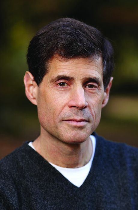
They state that “this may be overcome by use of a soluble form of PPI,” but don’t state what is meant by “soluble PPI” or how the open-capsule PPI was delivered. Among the PPIs they reported using to compare intact vs. open capsules was Protonix [pantoprazole] which is not produced as a capsule, and soluble Prevacid [lansoprazole], which is an orally disintegrating tablet that should provide characteristics similar to an “open capsule.”
PPI capsules provide PPI in enteric-coated granules, which are designed to protect the PPI from acid degradation in the stomach of individuals with intact gastrointestinal tracts and allow more of the PPI dose to reach the small intestine where it is absorbed. If capsules really fail to release their enteric-coated granules until very distally in RYGB patients, bypassing this step to allow earlier release of PPI makes intuitive sense; formulations such as suspensions and rapidly disintegrating tablets that deliver enteric-coated granules without capsules are currently available.
However, if this is an issue, administering a suspension of uncoated PPI with bicarbonate potentially might be the most attractive option, given more rapid absorption than PPI delivered as enteric-coated granules.
Loren Laine, MD, AGAF, professor of medicine, digestive diseases, Yale University, New Haven, Conn. He has no conflicts of interest.
Proton pump inhibitors (PPIs) are frequently employed to treat marginal ulcers after Roux-en-Y gastric bypass (RYGB). In a retrospective study, Schulman et al. compared intact vs. “open” PPI capsules.
They state that “this may be overcome by use of a soluble form of PPI,” but don’t state what is meant by “soluble PPI” or how the open-capsule PPI was delivered. Among the PPIs they reported using to compare intact vs. open capsules was Protonix [pantoprazole] which is not produced as a capsule, and soluble Prevacid [lansoprazole], which is an orally disintegrating tablet that should provide characteristics similar to an “open capsule.”
PPI capsules provide PPI in enteric-coated granules, which are designed to protect the PPI from acid degradation in the stomach of individuals with intact gastrointestinal tracts and allow more of the PPI dose to reach the small intestine where it is absorbed. If capsules really fail to release their enteric-coated granules until very distally in RYGB patients, bypassing this step to allow earlier release of PPI makes intuitive sense; formulations such as suspensions and rapidly disintegrating tablets that deliver enteric-coated granules without capsules are currently available.
However, if this is an issue, administering a suspension of uncoated PPI with bicarbonate potentially might be the most attractive option, given more rapid absorption than PPI delivered as enteric-coated granules.
Loren Laine, MD, AGAF, professor of medicine, digestive diseases, Yale University, New Haven, Conn. He has no conflicts of interest.
Proton pump inhibitors (PPIs) are frequently employed to treat marginal ulcers after Roux-en-Y gastric bypass (RYGB). In a retrospective study, Schulman et al. compared intact vs. “open” PPI capsules.
They state that “this may be overcome by use of a soluble form of PPI,” but don’t state what is meant by “soluble PPI” or how the open-capsule PPI was delivered. Among the PPIs they reported using to compare intact vs. open capsules was Protonix [pantoprazole] which is not produced as a capsule, and soluble Prevacid [lansoprazole], which is an orally disintegrating tablet that should provide characteristics similar to an “open capsule.”
PPI capsules provide PPI in enteric-coated granules, which are designed to protect the PPI from acid degradation in the stomach of individuals with intact gastrointestinal tracts and allow more of the PPI dose to reach the small intestine where it is absorbed. If capsules really fail to release their enteric-coated granules until very distally in RYGB patients, bypassing this step to allow earlier release of PPI makes intuitive sense; formulations such as suspensions and rapidly disintegrating tablets that deliver enteric-coated granules without capsules are currently available.
However, if this is an issue, administering a suspension of uncoated PPI with bicarbonate potentially might be the most attractive option, given more rapid absorption than PPI delivered as enteric-coated granules.
Loren Laine, MD, AGAF, professor of medicine, digestive diseases, Yale University, New Haven, Conn. He has no conflicts of interest.
The use of proton pump inhibitors in opened instead of closed capsules was associated with a nearly fourfold shorter median healing time among patients who developed marginal ulcers after Roux-en-Y gastric bypass, in a single-center retrospective cohort study.
In contrast, the specific class of proton pump inhibitor (PPI) did not affect healing times, wrote Allison R. Schulman, MD, and her associates at Brigham and Women’s Hospital, Boston. The report is in the April issue of Clinical Gastroenterology and Hepatology (doi: 10.1016/j.cgh.2016.10.015). “Given these results and the high prevalence of marginal ulceration in this patient population, further study in a randomized controlled setting is warranted, and use of open-capsule PPIs should be considered as a low-risk, low-cost alternative,” they added.
Roux-en-Y gastric bypass is one of the most common types of gastric bypass surgeries in the world, and up to 16% of patients develop postsurgical ulcers at the gastrojejunal anastomosis, the investigators noted. Acidity is a prime suspect in these “marginal ulcerations” because bypassing the acid-buffering duodenum exposes the jejunum to acid from the stomach, they added. High-dose PPIs are the main treatment, but there is no consensus on the formulation or dose of therapy. Because Roux-en-Y creates a small gastric pouch and hastens small-bowel transit, closed capsules designed to break down in the stomach “even may make their way to the colon before breakdown occurs,” they wrote.
They reviewed medical charts from patients who developed marginal ulcerations after undergoing Roux-en-Y gastric bypass at their hospital from 2000 through 2015. A total of 115 patients received open-capsule PPIs and 49 received intact capsules. All were followed until their ulcers healed.
For the open-capsule group, median time to healing was 91 days, compared with 342 days for the closed-capsule group (P less than .001). Importantly, capsule type was the only independent predictor of healing time (hazard ratio, 6.0; 95% confidence interval, 3.7 to 9.8; P less than .001) in a Cox regression model that included other known correlates of ulcer healing, including age, smoking status, the use of nonsteroidal anti-inflammatory drugs, Helicobacter pylori infection, the length of the gastric pouch, and the presence of fistulae or foreign bodies such as sutures or staples.
The use of sucralfate also did not affect time to ulcer healing, reflecting “many previous studies showing a lack of definitive benefit to this medication,” the researchers said. The findings have “tremendous implications” for health care utilization, they added. Indeed, patients who received open-capsule PPIs needed significantly fewer endoscopic procedures (median, 1.2 versus 1.8; P = .02) and used fewer health care resources overall ($7,206 versus $11,009; P = .05) compared with those prescribed intact PPI capsules.
This study was limited to patients who developed ulcer symptoms and underwent repeated surveillance endoscopies after surgery, the researchers noted. Selection bias is always a concern with retrospective studies, but insurers always covered both types of therapy and the choice of capsule type was entirely up to providers, all of whom consistently prescribed either open- or closed-capsule PPI therapy, they added.
The investigators did not acknowledge external funding sources. Dr. Schulman and four coinvestigators reported having no competing interests. One coinvestigator disclosed ties to Olympus, Boston Scientific, and Covidien.
The use of proton pump inhibitors in opened instead of closed capsules was associated with a nearly fourfold shorter median healing time among patients who developed marginal ulcers after Roux-en-Y gastric bypass, in a single-center retrospective cohort study.
In contrast, the specific class of proton pump inhibitor (PPI) did not affect healing times, wrote Allison R. Schulman, MD, and her associates at Brigham and Women’s Hospital, Boston. The report is in the April issue of Clinical Gastroenterology and Hepatology (doi: 10.1016/j.cgh.2016.10.015). “Given these results and the high prevalence of marginal ulceration in this patient population, further study in a randomized controlled setting is warranted, and use of open-capsule PPIs should be considered as a low-risk, low-cost alternative,” they added.
Roux-en-Y gastric bypass is one of the most common types of gastric bypass surgeries in the world, and up to 16% of patients develop postsurgical ulcers at the gastrojejunal anastomosis, the investigators noted. Acidity is a prime suspect in these “marginal ulcerations” because bypassing the acid-buffering duodenum exposes the jejunum to acid from the stomach, they added. High-dose PPIs are the main treatment, but there is no consensus on the formulation or dose of therapy. Because Roux-en-Y creates a small gastric pouch and hastens small-bowel transit, closed capsules designed to break down in the stomach “even may make their way to the colon before breakdown occurs,” they wrote.
They reviewed medical charts from patients who developed marginal ulcerations after undergoing Roux-en-Y gastric bypass at their hospital from 2000 through 2015. A total of 115 patients received open-capsule PPIs and 49 received intact capsules. All were followed until their ulcers healed.
For the open-capsule group, median time to healing was 91 days, compared with 342 days for the closed-capsule group (P less than .001). Importantly, capsule type was the only independent predictor of healing time (hazard ratio, 6.0; 95% confidence interval, 3.7 to 9.8; P less than .001) in a Cox regression model that included other known correlates of ulcer healing, including age, smoking status, the use of nonsteroidal anti-inflammatory drugs, Helicobacter pylori infection, the length of the gastric pouch, and the presence of fistulae or foreign bodies such as sutures or staples.
The use of sucralfate also did not affect time to ulcer healing, reflecting “many previous studies showing a lack of definitive benefit to this medication,” the researchers said. The findings have “tremendous implications” for health care utilization, they added. Indeed, patients who received open-capsule PPIs needed significantly fewer endoscopic procedures (median, 1.2 versus 1.8; P = .02) and used fewer health care resources overall ($7,206 versus $11,009; P = .05) compared with those prescribed intact PPI capsules.
This study was limited to patients who developed ulcer symptoms and underwent repeated surveillance endoscopies after surgery, the researchers noted. Selection bias is always a concern with retrospective studies, but insurers always covered both types of therapy and the choice of capsule type was entirely up to providers, all of whom consistently prescribed either open- or closed-capsule PPI therapy, they added.
The investigators did not acknowledge external funding sources. Dr. Schulman and four coinvestigators reported having no competing interests. One coinvestigator disclosed ties to Olympus, Boston Scientific, and Covidien.
FROM CLINICAL GASTROENTEROLOGY AND HEPATOLOGY
Key clinical point: The use of proton pump inhibitors in opened instead of closed capsules was associated with a nearly fourfold shorter median healing time among patients who developed ulcers at the gastrojejunal anastomosis after Roux-en-Y gastric bypass.
Major finding: The median time to ulcer healing was 91.0 versus 342.0 days for the open- and closed-capsule groups, respectively (P less than .001).
Data source: A single-center retrospective study of 162 patients.
Disclosures: The investigators did not acknowledge external funding sources. Dr. Schulman and four coinvestigators reported having no competing interests. One coinvestigator disclosed ties to Olympus, Boston Scientific, and Covidien.
Expect tricky journey with BDD patients
LAS VEGAS – When working with patients referred for suspected body dysmorphic disorder, expect the initial groundwork to take more time than for other disorders.
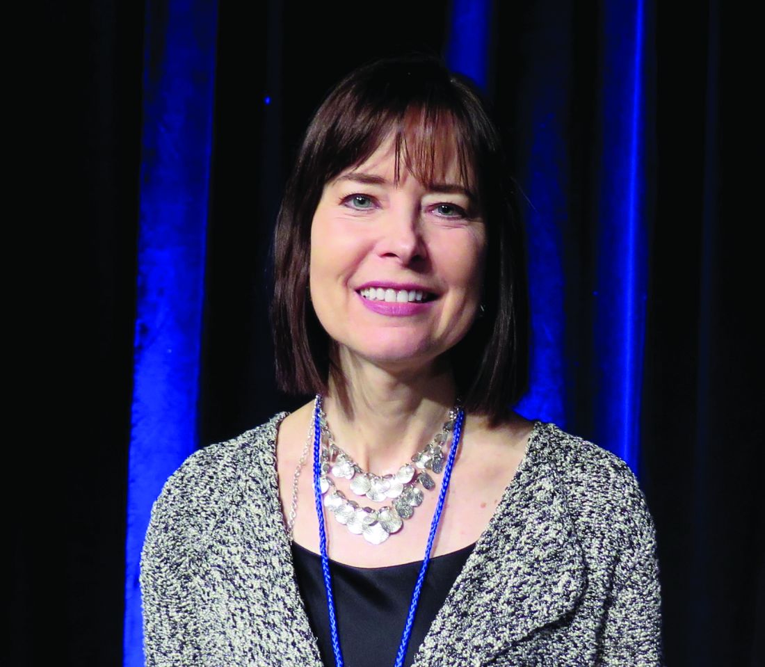
“I think that these patients are among the most severely ill that we see in clinical practice – although body dysmorphic disorder often goes unrecognized, because patients are often very ashamed of their concerns, and they find it hard to talk about them,” Katharine A. Phillips, MD, said at the annual psychopharmacology update held by the Nevada Psychiatric Association.
Defined in the DSM-5 as preoccupation with one or more perceived defects or flaws in physical appearance that are not observable or that appear slight to others, body dysmorphic disorder (BDD) often causes substantial distress and impairment of day-to-day functioning and is associated with a high rate of suicidality. “When these patients walk into your office, you cannot tell by looking at them what their appearance concern is going to be,” said Dr. Phillips, professor of psychiatry and human behavior at Brown University, Providence, R.I. “Sometimes they point it out to you and you can see that their nostrils are a tiny bit asymmetrical or that they have a little scar on their chin, but it’s not very noticeable. It’s not noticeable until the patient points it out, and even then it’s a slight flaw. But in most cases, the body areas that the patient is preoccupied with look entirely normal.”
BDD affects an estimated 1.7%-2.9% of the general population; about 60% are female. In two-thirds of cases, it onsets during childhood or adolescence. The preoccupations about physical appearance that are associated with the disorder “are very obsessional and distressing,” Dr. Phillips said. “They may think ‘I look ugly. People are laughing at me. I look like a freak.’ They can be focused on any part of their appearance, but most often it’s the face or head. Skin is No. 1, followed by hair and nose.” Complaints may include perceptions of scarring, perceptions of skin color, too much facial hair, or hair that’s too curly or straight. On average, BDD patients report thinking about their perceived flaws for 3-8 hours a day. “Some say it’s all they think about all day long,” said Dr. Phillips, who also directs the Body Dysmorphic Disorder Program at Rhode Island Hospital, in Providence.
“Insight is usually absent or poor, and BDD-related ideas or delusions of reference are common. A majority mistakenly think that other people are taking special notice of them in a negative way because of how they look. If they walk down the street, for example, they may misperceive people as staring at them. If they hear people talking to one another they may think, ‘They must be talking about how ugly my nose is.’ I have patients who have physically assaulted strangers on the street because they’re so certain that they’re being made fun of because of their appearance flaws, which exist in their mind.”
Functional MRI studies of BDD patients demonstrate aberrant visual processing. “They overfocus on tiny details, so the brain is trying to extract detail where there isn’t any,” she explained. “A complementary finding is that they have reduced visual processing of holistic visual stimuli (“seeing the big picture”), compared with healthy controls. One of my patients said to me, ‘When I look at myself I’m just one big pimple without any feet or even any toes.’ They focus in on the body areas they hate and have trouble perceiving the rest of themselves.”
Compulsive repetitive behaviors that are done in response to the appearance preoccupations may include camouflaging (for example, covering perceived hair thinning with a hat), comparing their appearance with that of other people, mirror checking, excessive grooming, questioning others about their appearance or seeking reassurance about the perceived flaws, skin picking, and tanning (often to darken “pale” skin). Functional impairment varies but is usually substantial. For example, in several studies, Dr. Phillips and her associates found that 39% of BDD patients were currently not working because of psychopathology (for most, BDD was their primary diagnosis), about 20% had dropped out of school primarily because of BDD symptoms, 29% had been housebound for at least a week because of their BDD symptoms, 38% had been psychiatrically hospitalized, and the rates of lifetime suicidal ideation ranged from 71% to 81%. “More than one-quarter have attempted suicide,” she said.
About three-quarters seek and two-thirds receive some kind of cosmetic treatment for BDD, most commonly dermatologic treatment and plastic surgery (most often rhinoplasty). “General recommendations are that cosmetic treatment should not be done on these patients,” Dr. Phillips said. A recent practice guideline from the American Academy of Otolaryngology–Head and Neck Surgery recommends that surgeons not operate on a rhinoplasty candidate who screens positive for BDD (Otolaryngol Head Neck Surg. 2017 Feb;156 [2 _suppl]:S1-30).
Serotonin reuptake inhibitors (SRIs) at a high enough dose and for a trial duration of 12-16 weeks are the first-line medications for both nondelusional BDD and delusional BDD. “Most patients with BDD don’t receive adequate first-line pharmacotherapy,” Dr. Phillips said. “Often, high doses of SRIs are needed, sometimes above the [Food and Drug Administration]-approved limits, but I don’t exceed these limits for clomipramine or citalopram.” Recommended SRI doses for BDD are similar to those in the American Psychiatric Association’s practice guideline for obsessive-compulsive disorder. She recommends checking an EKG when patients take a high dose of escitalopram.
In cases of partial or no response to an SRI, consider whether the dose was high enough. “In the vast majority of patients I see for consultation, it wasn’t,” Dr. Phillips said. “Check adherence, and extend the trial if necessary, with 3-4 weeks at the maximum recommended tolerated dose.” In her clinical experience, atypical antipsychotics can sometimes help when added to an SRI, especially in patients who are agitated, aggressive, impulsive, or severely anxious, but antipsychotics are not currently recommended as monotherapy. “In my view, the most pressing need in the BDD field is for research on the efficacy of antipsychotics, especially as SRI augmentation agents,” she said. “We have so little data.” SRI augmentation of buspirone also can prove helpful.
Cognitive-behavioral therapy is the psychosocial treatment of choice for BDD. However, Dr. Phillips cautioned that if BDD patients receive treatment comparable to that of patients with OCD or depression, they probably won’t get better. “It needs to be tailored to BDD symptoms, which are unique in many ways,” she said. “As you would with any patient, express empathy, instill hope, and attend to the therapeutic alliance.”
She recommended using one of the two published evidence-based CBT manuals for BDD: “Cognitive-Behavioral Therapy for Body Dysmorphic Disorder: A Treatment Manual,” by Sabine Wilhelm, PhD, Dr. Phillips, and Gail Steketee, PhD (New York: Guilford Press, 2013) and “Body Dysmorphic Disorder” by David Veale, MD and Fugen Neziroglu, PhD (West Sussex, U.K.: 2010).
Key CBT principles include helping patients cut down on BDD rituals – for example, by spending less time in front of mirrors or discarding their pocket mirrors. Gradual exposure to social situations, combined with behavioral experiments, also is recommended. Other core CBT components include cognitive therapy, perceptual retraining, motivational interviewing, and providing psychoeducation about BDD.
“I explain to patients that people with BDD see themselves differently than others do, which is supported by visual processing studies,” Dr. Phillips said. “They don’t necessarily buy it, but I think it’s worth putting it out there. Instead of trying to convince them that they look fine, focus on their suffering, preoccupation, and the effect of symptoms on their life. They will usually agree that they are suffering a lot, and that may motivate them for treatment.”
These questions that can help you better understand and diagnose the concerns of BDD patients: “Are you very worried about your appearance in any way?” If yes, “Can you tell me about your concern?”
Suggested questions regarding preoccupations about appearance (DSM-5 criteria A) are: “Does this concern preoccupy you? Do you think about it a lot and wish you could think about it less?” To elicit discussion on the topic of repetitive behaviors (DSM-5 criteria B), consider asking, “Is there anything you feel an urge to do over and over again in response to your appearance concerns?” To determine whether the preoccupations cause clinically significant distress or impairment in functioning (DSM-5 criteria C), ask, “How much does this concern upset you? Does it cause you any problems – socially, in relationships, or with school or work?”
Dr. Phillips disclosed that during the past year, she has received honoraria from the Merck Manual and from Royal Pharma as well as royalties from Oxford University Press, International Creative Management, American Psychiatric Publishing, Guilford Press, and UpToDate.
LAS VEGAS – When working with patients referred for suspected body dysmorphic disorder, expect the initial groundwork to take more time than for other disorders.
“I think that these patients are among the most severely ill that we see in clinical practice – although body dysmorphic disorder often goes unrecognized, because patients are often very ashamed of their concerns, and they find it hard to talk about them,” Katharine A. Phillips, MD, said at the annual psychopharmacology update held by the Nevada Psychiatric Association.
Defined in the DSM-5 as preoccupation with one or more perceived defects or flaws in physical appearance that are not observable or that appear slight to others, body dysmorphic disorder (BDD) often causes substantial distress and impairment of day-to-day functioning and is associated with a high rate of suicidality. “When these patients walk into your office, you cannot tell by looking at them what their appearance concern is going to be,” said Dr. Phillips, professor of psychiatry and human behavior at Brown University, Providence, R.I. “Sometimes they point it out to you and you can see that their nostrils are a tiny bit asymmetrical or that they have a little scar on their chin, but it’s not very noticeable. It’s not noticeable until the patient points it out, and even then it’s a slight flaw. But in most cases, the body areas that the patient is preoccupied with look entirely normal.”
BDD affects an estimated 1.7%-2.9% of the general population; about 60% are female. In two-thirds of cases, it onsets during childhood or adolescence. The preoccupations about physical appearance that are associated with the disorder “are very obsessional and distressing,” Dr. Phillips said. “They may think ‘I look ugly. People are laughing at me. I look like a freak.’ They can be focused on any part of their appearance, but most often it’s the face or head. Skin is No. 1, followed by hair and nose.” Complaints may include perceptions of scarring, perceptions of skin color, too much facial hair, or hair that’s too curly or straight. On average, BDD patients report thinking about their perceived flaws for 3-8 hours a day. “Some say it’s all they think about all day long,” said Dr. Phillips, who also directs the Body Dysmorphic Disorder Program at Rhode Island Hospital, in Providence.
“Insight is usually absent or poor, and BDD-related ideas or delusions of reference are common. A majority mistakenly think that other people are taking special notice of them in a negative way because of how they look. If they walk down the street, for example, they may misperceive people as staring at them. If they hear people talking to one another they may think, ‘They must be talking about how ugly my nose is.’ I have patients who have physically assaulted strangers on the street because they’re so certain that they’re being made fun of because of their appearance flaws, which exist in their mind.”
Functional MRI studies of BDD patients demonstrate aberrant visual processing. “They overfocus on tiny details, so the brain is trying to extract detail where there isn’t any,” she explained. “A complementary finding is that they have reduced visual processing of holistic visual stimuli (“seeing the big picture”), compared with healthy controls. One of my patients said to me, ‘When I look at myself I’m just one big pimple without any feet or even any toes.’ They focus in on the body areas they hate and have trouble perceiving the rest of themselves.”
Compulsive repetitive behaviors that are done in response to the appearance preoccupations may include camouflaging (for example, covering perceived hair thinning with a hat), comparing their appearance with that of other people, mirror checking, excessive grooming, questioning others about their appearance or seeking reassurance about the perceived flaws, skin picking, and tanning (often to darken “pale” skin). Functional impairment varies but is usually substantial. For example, in several studies, Dr. Phillips and her associates found that 39% of BDD patients were currently not working because of psychopathology (for most, BDD was their primary diagnosis), about 20% had dropped out of school primarily because of BDD symptoms, 29% had been housebound for at least a week because of their BDD symptoms, 38% had been psychiatrically hospitalized, and the rates of lifetime suicidal ideation ranged from 71% to 81%. “More than one-quarter have attempted suicide,” she said.
About three-quarters seek and two-thirds receive some kind of cosmetic treatment for BDD, most commonly dermatologic treatment and plastic surgery (most often rhinoplasty). “General recommendations are that cosmetic treatment should not be done on these patients,” Dr. Phillips said. A recent practice guideline from the American Academy of Otolaryngology–Head and Neck Surgery recommends that surgeons not operate on a rhinoplasty candidate who screens positive for BDD (Otolaryngol Head Neck Surg. 2017 Feb;156 [2 _suppl]:S1-30).
Serotonin reuptake inhibitors (SRIs) at a high enough dose and for a trial duration of 12-16 weeks are the first-line medications for both nondelusional BDD and delusional BDD. “Most patients with BDD don’t receive adequate first-line pharmacotherapy,” Dr. Phillips said. “Often, high doses of SRIs are needed, sometimes above the [Food and Drug Administration]-approved limits, but I don’t exceed these limits for clomipramine or citalopram.” Recommended SRI doses for BDD are similar to those in the American Psychiatric Association’s practice guideline for obsessive-compulsive disorder. She recommends checking an EKG when patients take a high dose of escitalopram.
In cases of partial or no response to an SRI, consider whether the dose was high enough. “In the vast majority of patients I see for consultation, it wasn’t,” Dr. Phillips said. “Check adherence, and extend the trial if necessary, with 3-4 weeks at the maximum recommended tolerated dose.” In her clinical experience, atypical antipsychotics can sometimes help when added to an SRI, especially in patients who are agitated, aggressive, impulsive, or severely anxious, but antipsychotics are not currently recommended as monotherapy. “In my view, the most pressing need in the BDD field is for research on the efficacy of antipsychotics, especially as SRI augmentation agents,” she said. “We have so little data.” SRI augmentation of buspirone also can prove helpful.
Cognitive-behavioral therapy is the psychosocial treatment of choice for BDD. However, Dr. Phillips cautioned that if BDD patients receive treatment comparable to that of patients with OCD or depression, they probably won’t get better. “It needs to be tailored to BDD symptoms, which are unique in many ways,” she said. “As you would with any patient, express empathy, instill hope, and attend to the therapeutic alliance.”
She recommended using one of the two published evidence-based CBT manuals for BDD: “Cognitive-Behavioral Therapy for Body Dysmorphic Disorder: A Treatment Manual,” by Sabine Wilhelm, PhD, Dr. Phillips, and Gail Steketee, PhD (New York: Guilford Press, 2013) and “Body Dysmorphic Disorder” by David Veale, MD and Fugen Neziroglu, PhD (West Sussex, U.K.: 2010).
Key CBT principles include helping patients cut down on BDD rituals – for example, by spending less time in front of mirrors or discarding their pocket mirrors. Gradual exposure to social situations, combined with behavioral experiments, also is recommended. Other core CBT components include cognitive therapy, perceptual retraining, motivational interviewing, and providing psychoeducation about BDD.
“I explain to patients that people with BDD see themselves differently than others do, which is supported by visual processing studies,” Dr. Phillips said. “They don’t necessarily buy it, but I think it’s worth putting it out there. Instead of trying to convince them that they look fine, focus on their suffering, preoccupation, and the effect of symptoms on their life. They will usually agree that they are suffering a lot, and that may motivate them for treatment.”
These questions that can help you better understand and diagnose the concerns of BDD patients: “Are you very worried about your appearance in any way?” If yes, “Can you tell me about your concern?”
Suggested questions regarding preoccupations about appearance (DSM-5 criteria A) are: “Does this concern preoccupy you? Do you think about it a lot and wish you could think about it less?” To elicit discussion on the topic of repetitive behaviors (DSM-5 criteria B), consider asking, “Is there anything you feel an urge to do over and over again in response to your appearance concerns?” To determine whether the preoccupations cause clinically significant distress or impairment in functioning (DSM-5 criteria C), ask, “How much does this concern upset you? Does it cause you any problems – socially, in relationships, or with school or work?”
Dr. Phillips disclosed that during the past year, she has received honoraria from the Merck Manual and from Royal Pharma as well as royalties from Oxford University Press, International Creative Management, American Psychiatric Publishing, Guilford Press, and UpToDate.
LAS VEGAS – When working with patients referred for suspected body dysmorphic disorder, expect the initial groundwork to take more time than for other disorders.
“I think that these patients are among the most severely ill that we see in clinical practice – although body dysmorphic disorder often goes unrecognized, because patients are often very ashamed of their concerns, and they find it hard to talk about them,” Katharine A. Phillips, MD, said at the annual psychopharmacology update held by the Nevada Psychiatric Association.
Defined in the DSM-5 as preoccupation with one or more perceived defects or flaws in physical appearance that are not observable or that appear slight to others, body dysmorphic disorder (BDD) often causes substantial distress and impairment of day-to-day functioning and is associated with a high rate of suicidality. “When these patients walk into your office, you cannot tell by looking at them what their appearance concern is going to be,” said Dr. Phillips, professor of psychiatry and human behavior at Brown University, Providence, R.I. “Sometimes they point it out to you and you can see that their nostrils are a tiny bit asymmetrical or that they have a little scar on their chin, but it’s not very noticeable. It’s not noticeable until the patient points it out, and even then it’s a slight flaw. But in most cases, the body areas that the patient is preoccupied with look entirely normal.”
BDD affects an estimated 1.7%-2.9% of the general population; about 60% are female. In two-thirds of cases, it onsets during childhood or adolescence. The preoccupations about physical appearance that are associated with the disorder “are very obsessional and distressing,” Dr. Phillips said. “They may think ‘I look ugly. People are laughing at me. I look like a freak.’ They can be focused on any part of their appearance, but most often it’s the face or head. Skin is No. 1, followed by hair and nose.” Complaints may include perceptions of scarring, perceptions of skin color, too much facial hair, or hair that’s too curly or straight. On average, BDD patients report thinking about their perceived flaws for 3-8 hours a day. “Some say it’s all they think about all day long,” said Dr. Phillips, who also directs the Body Dysmorphic Disorder Program at Rhode Island Hospital, in Providence.
“Insight is usually absent or poor, and BDD-related ideas or delusions of reference are common. A majority mistakenly think that other people are taking special notice of them in a negative way because of how they look. If they walk down the street, for example, they may misperceive people as staring at them. If they hear people talking to one another they may think, ‘They must be talking about how ugly my nose is.’ I have patients who have physically assaulted strangers on the street because they’re so certain that they’re being made fun of because of their appearance flaws, which exist in their mind.”
Functional MRI studies of BDD patients demonstrate aberrant visual processing. “They overfocus on tiny details, so the brain is trying to extract detail where there isn’t any,” she explained. “A complementary finding is that they have reduced visual processing of holistic visual stimuli (“seeing the big picture”), compared with healthy controls. One of my patients said to me, ‘When I look at myself I’m just one big pimple without any feet or even any toes.’ They focus in on the body areas they hate and have trouble perceiving the rest of themselves.”
Compulsive repetitive behaviors that are done in response to the appearance preoccupations may include camouflaging (for example, covering perceived hair thinning with a hat), comparing their appearance with that of other people, mirror checking, excessive grooming, questioning others about their appearance or seeking reassurance about the perceived flaws, skin picking, and tanning (often to darken “pale” skin). Functional impairment varies but is usually substantial. For example, in several studies, Dr. Phillips and her associates found that 39% of BDD patients were currently not working because of psychopathology (for most, BDD was their primary diagnosis), about 20% had dropped out of school primarily because of BDD symptoms, 29% had been housebound for at least a week because of their BDD symptoms, 38% had been psychiatrically hospitalized, and the rates of lifetime suicidal ideation ranged from 71% to 81%. “More than one-quarter have attempted suicide,” she said.
About three-quarters seek and two-thirds receive some kind of cosmetic treatment for BDD, most commonly dermatologic treatment and plastic surgery (most often rhinoplasty). “General recommendations are that cosmetic treatment should not be done on these patients,” Dr. Phillips said. A recent practice guideline from the American Academy of Otolaryngology–Head and Neck Surgery recommends that surgeons not operate on a rhinoplasty candidate who screens positive for BDD (Otolaryngol Head Neck Surg. 2017 Feb;156 [2 _suppl]:S1-30).
Serotonin reuptake inhibitors (SRIs) at a high enough dose and for a trial duration of 12-16 weeks are the first-line medications for both nondelusional BDD and delusional BDD. “Most patients with BDD don’t receive adequate first-line pharmacotherapy,” Dr. Phillips said. “Often, high doses of SRIs are needed, sometimes above the [Food and Drug Administration]-approved limits, but I don’t exceed these limits for clomipramine or citalopram.” Recommended SRI doses for BDD are similar to those in the American Psychiatric Association’s practice guideline for obsessive-compulsive disorder. She recommends checking an EKG when patients take a high dose of escitalopram.
In cases of partial or no response to an SRI, consider whether the dose was high enough. “In the vast majority of patients I see for consultation, it wasn’t,” Dr. Phillips said. “Check adherence, and extend the trial if necessary, with 3-4 weeks at the maximum recommended tolerated dose.” In her clinical experience, atypical antipsychotics can sometimes help when added to an SRI, especially in patients who are agitated, aggressive, impulsive, or severely anxious, but antipsychotics are not currently recommended as monotherapy. “In my view, the most pressing need in the BDD field is for research on the efficacy of antipsychotics, especially as SRI augmentation agents,” she said. “We have so little data.” SRI augmentation of buspirone also can prove helpful.
Cognitive-behavioral therapy is the psychosocial treatment of choice for BDD. However, Dr. Phillips cautioned that if BDD patients receive treatment comparable to that of patients with OCD or depression, they probably won’t get better. “It needs to be tailored to BDD symptoms, which are unique in many ways,” she said. “As you would with any patient, express empathy, instill hope, and attend to the therapeutic alliance.”
She recommended using one of the two published evidence-based CBT manuals for BDD: “Cognitive-Behavioral Therapy for Body Dysmorphic Disorder: A Treatment Manual,” by Sabine Wilhelm, PhD, Dr. Phillips, and Gail Steketee, PhD (New York: Guilford Press, 2013) and “Body Dysmorphic Disorder” by David Veale, MD and Fugen Neziroglu, PhD (West Sussex, U.K.: 2010).
Key CBT principles include helping patients cut down on BDD rituals – for example, by spending less time in front of mirrors or discarding their pocket mirrors. Gradual exposure to social situations, combined with behavioral experiments, also is recommended. Other core CBT components include cognitive therapy, perceptual retraining, motivational interviewing, and providing psychoeducation about BDD.
“I explain to patients that people with BDD see themselves differently than others do, which is supported by visual processing studies,” Dr. Phillips said. “They don’t necessarily buy it, but I think it’s worth putting it out there. Instead of trying to convince them that they look fine, focus on their suffering, preoccupation, and the effect of symptoms on their life. They will usually agree that they are suffering a lot, and that may motivate them for treatment.”
These questions that can help you better understand and diagnose the concerns of BDD patients: “Are you very worried about your appearance in any way?” If yes, “Can you tell me about your concern?”
Suggested questions regarding preoccupations about appearance (DSM-5 criteria A) are: “Does this concern preoccupy you? Do you think about it a lot and wish you could think about it less?” To elicit discussion on the topic of repetitive behaviors (DSM-5 criteria B), consider asking, “Is there anything you feel an urge to do over and over again in response to your appearance concerns?” To determine whether the preoccupations cause clinically significant distress or impairment in functioning (DSM-5 criteria C), ask, “How much does this concern upset you? Does it cause you any problems – socially, in relationships, or with school or work?”
Dr. Phillips disclosed that during the past year, she has received honoraria from the Merck Manual and from Royal Pharma as well as royalties from Oxford University Press, International Creative Management, American Psychiatric Publishing, Guilford Press, and UpToDate.
EXPERT ANALYSIS AT THE NPA PSYCHOPHARMACOLOGY UPDATE

Point/Counterpoint: Is endograft PAA repair durable?
Endovascular repair is durable
Endovascular repair of popliteal artery aneurysms is vastly superior to all other previous techniques of popliteal aneurysm repair. Half of all popliteal artery aneurysms are bilateral, and 40% are associated with abdominal aortic aneurysm; 1%-2% of patients with abdominal aortic aneurysm have a popliteal aneurysm (ANZ J Surg. 2006 Oct;76[10]:912-5). Less than 0.01% of hospitalized patients have popliteal artery aneurysms, and men are 20 times more prone to them than women are.
Traditional treatment involves either bypass with interval ligation or a direct posterior approach with an interposition graft, but surgery is not without its problems. I think of the retired anesthesiologist who came to me with a popliteal artery aneurysm (PAA) that his primary care doctor diagnosed. “I’m not having any damn femoral popliteal bypass operation,” he told me. “Every single one of those patients dies.”
Endograft repair is a technique that is reaching its prime, as a growing number of reports have shown – although none of these studies has large numbers because the volume just isn’t available. One recent paper compared 52 open and 23 endovascular PAA repairs (Ann Vasc Surg. 2016 Jan;30:253-7) and found both had similarly high rates of reintervention – 50% at 4 years. But it is noteworthy that the endovascular results improved with time.
A University of Pittsburgh study of 186 open and endovascular repairs found that patients with acute presentations of embolization or aneurysm thrombosis did better with open surgery. In addition, while open repair had superior patency initially after surgery, midterm secondary patency and amputation rates of open and endovascular repair were similar (J Vasc Surg. 2016 Jan;63[1]:70-6).
A Netherlands study of 72 PAA treated with endografting showed that 84% had primary patency at 1 year, and 74% had assisted primary patency at 3 years (Eur J Vasc Endovasc Surg. 2016 Jul;52[1]:99-104). Among these patients, 13 had late occlusions, 7 were converted to bypass, and 2 required thrombolysis; but none required limb amputation.
A meta-analysis of 540 patients found no statistically significant difference in outcomes between endovascular and open repair for PAA (Eur J Vasc Endovasc Surg. 2015 Sep;50(3):351-9). Another systematic review and meta-analysis of 14 studies and 514 patients also found no difference in pooled primary and secondary patency at 5 years (J Endovasc Ther. 2015 Jun;22[3]:330-7).
There certainly are contradictory studies, such as one by Dr. Alik Farber’s group in Boston that showed open repair is superior to endovascular surgery (J Vasc Surg. 2015 Mar;61[3]:663-9); but retrospective database mining certainly has its limitations. Their retrospective study queried the Vascular Quality Initiative database and found that 95% of patients who had open elective popliteal aneurysm repair were free from major adverse limb events, vs. 80% for endovascular treatments.
The best outcomes of open repair happen with autologous vein, but there is precious little of that around now. Emergency patients would probably do better with open surgery, but in elective repair there is no clear differential data.
So, if that’s the case, I’m going to take the small incision.
Peter Rossi, MD, FACS, is an associate professor of surgery and radiology, and the clinical director of vascular surgery, at the Medical College of Wisconsin, Milwaukee. He is also on staff at Clement J. Zablocki Veterans Affairs Medical Center in Milwaukee. Dr. Rossi had no financial relationships to disclose.
Endovascular repair may not be durable
Debating the durability of elective endovascular repair of popliteal artery aneurysm raises a question: Who determines durability anyway?
Is it the patients who only want the Band-Aid and no incision? I don’t think so. Is it the interventionalist who only does endovascular repairs? I don’t think so. I’m sure it’s not the insurance companies, who only worry about cost containment, either.
So, who should determine durability of endovascular popliteal artery aneurysm (PAA) repair?

So, the question is, do we have such data?
There are multiple reports looking at how well open repair works. It has been done for decades. In 2008, a Veterans Affairs study of 583 open PAA repairs reported low death rates and excellent rates of limb salvage at 2 years, even in high-risk patients (J Vasc Surg. 2008 Oct;48[4]:845-51). Open surgical repair has excellent documented durability, and that is not the question at hand.
Endovascular repair has some presumed advantages. It’s less invasive and involves less postoperative pain and a quicker recovery. But it is not without problems – graft thrombosis and occlusion, endoleaks, distal limb ischemia, and stent fractures among them.
Surgery, to be clear, is not perfect, either. One of my patients who years ago presented with an occluded PAA underwent open bypass repair – but then went on later to have a pseudoaneurysm of the proximal anastomosis. I repaired this with an endograft, and he has done quite well. So, we all do endograft repairs, walk out, chest bump the Gore rep, and send the patient home that day.
Is it durable, though?
Most of the data on endovascular repair are from single-center studies dating back to 2003. There’s only one prospective trial comparing endovascular vs. open repair (J Vasc Surg. 2005 Aug;42[2]:185-93), but it was a single-center trial with a severe power limitation, because it involved only 30 patients. It found endovascular repair was comparable to open surgery. Also, I suspect a great deal of selection bias is involved in studies of endovascular repair.
A number of studies have found endovascular repair is not inferior to surgical repair. For example, a study by Dr. Audra Duncan, at Mayo Clinic, and her colleagues found that primary and secondary patency rates of elective and emergent stenting were excellent – but the study results only extended out to 2 years (J Vasc Surg. 2013 May;57[5]:1299-305). I don’t think we could hang our hat on that.
A Swedish study that compared open and endovascular surgery in 592 patients reported that endovascular repair has “significantly inferior results compared with open repair,” particularly in those who present with acute ischemia (Eur J Vasc Endovasc Surg. 2015 Sep;50[3]:342-50). A close look at the data shows that primary patency rates were 89% for open repair and 67.4% for stent graft.
Referencing the systematic review and meta-analysis that Dr. Rossi cited, the primary patency of endovascular repair was only 69% and the secondary patency rate was 77% at 5 years (J Endovasc Ther. 2015 Jun;22[3]:330-7). As physicians, I submit that we can do better.
A Netherlands study investigated stent fractures, finding that 17% (13 out of 78 cases) had circumferential fractures (J Vasc Surg. 2010 Jun;51[6]:1413-8). This study only included circumferential stent fractures and excluded localized strut fractures. I think these studies show that endovascular repair is not always durable.
I want to remind you that we are vascular surgeons, so it is appropriate for us to embrace surgical bypass and its known durability, especially when the durability of endovascular repair is still not known.
Patrick Muck, MD, is chief of vascular surgery and director of vascular residency and fellowship at Good Samaritan Hospital, Cincinnati. He is also on staff at Bethesda North Hospital, Cincinnati, and is affiliated with TriHealth Heart Institute in southwestern Ohio. Dr. Muck had no financial relationships to disclose.
Endovascular repair is durable
Endovascular repair of popliteal artery aneurysms is vastly superior to all other previous techniques of popliteal aneurysm repair. Half of all popliteal artery aneurysms are bilateral, and 40% are associated with abdominal aortic aneurysm; 1%-2% of patients with abdominal aortic aneurysm have a popliteal aneurysm (ANZ J Surg. 2006 Oct;76[10]:912-5). Less than 0.01% of hospitalized patients have popliteal artery aneurysms, and men are 20 times more prone to them than women are.
Traditional treatment involves either bypass with interval ligation or a direct posterior approach with an interposition graft, but surgery is not without its problems. I think of the retired anesthesiologist who came to me with a popliteal artery aneurysm (PAA) that his primary care doctor diagnosed. “I’m not having any damn femoral popliteal bypass operation,” he told me. “Every single one of those patients dies.”
Endograft repair is a technique that is reaching its prime, as a growing number of reports have shown – although none of these studies has large numbers because the volume just isn’t available. One recent paper compared 52 open and 23 endovascular PAA repairs (Ann Vasc Surg. 2016 Jan;30:253-7) and found both had similarly high rates of reintervention – 50% at 4 years. But it is noteworthy that the endovascular results improved with time.
A University of Pittsburgh study of 186 open and endovascular repairs found that patients with acute presentations of embolization or aneurysm thrombosis did better with open surgery. In addition, while open repair had superior patency initially after surgery, midterm secondary patency and amputation rates of open and endovascular repair were similar (J Vasc Surg. 2016 Jan;63[1]:70-6).
A Netherlands study of 72 PAA treated with endografting showed that 84% had primary patency at 1 year, and 74% had assisted primary patency at 3 years (Eur J Vasc Endovasc Surg. 2016 Jul;52[1]:99-104). Among these patients, 13 had late occlusions, 7 were converted to bypass, and 2 required thrombolysis; but none required limb amputation.
A meta-analysis of 540 patients found no statistically significant difference in outcomes between endovascular and open repair for PAA (Eur J Vasc Endovasc Surg. 2015 Sep;50(3):351-9). Another systematic review and meta-analysis of 14 studies and 514 patients also found no difference in pooled primary and secondary patency at 5 years (J Endovasc Ther. 2015 Jun;22[3]:330-7).
There certainly are contradictory studies, such as one by Dr. Alik Farber’s group in Boston that showed open repair is superior to endovascular surgery (J Vasc Surg. 2015 Mar;61[3]:663-9); but retrospective database mining certainly has its limitations. Their retrospective study queried the Vascular Quality Initiative database and found that 95% of patients who had open elective popliteal aneurysm repair were free from major adverse limb events, vs. 80% for endovascular treatments.
The best outcomes of open repair happen with autologous vein, but there is precious little of that around now. Emergency patients would probably do better with open surgery, but in elective repair there is no clear differential data.
So, if that’s the case, I’m going to take the small incision.
Peter Rossi, MD, FACS, is an associate professor of surgery and radiology, and the clinical director of vascular surgery, at the Medical College of Wisconsin, Milwaukee. He is also on staff at Clement J. Zablocki Veterans Affairs Medical Center in Milwaukee. Dr. Rossi had no financial relationships to disclose.
Endovascular repair may not be durable
Debating the durability of elective endovascular repair of popliteal artery aneurysm raises a question: Who determines durability anyway?
Is it the patients who only want the Band-Aid and no incision? I don’t think so. Is it the interventionalist who only does endovascular repairs? I don’t think so. I’m sure it’s not the insurance companies, who only worry about cost containment, either.
So, who should determine durability of endovascular popliteal artery aneurysm (PAA) repair?
So, the question is, do we have such data?
There are multiple reports looking at how well open repair works. It has been done for decades. In 2008, a Veterans Affairs study of 583 open PAA repairs reported low death rates and excellent rates of limb salvage at 2 years, even in high-risk patients (J Vasc Surg. 2008 Oct;48[4]:845-51). Open surgical repair has excellent documented durability, and that is not the question at hand.
Endovascular repair has some presumed advantages. It’s less invasive and involves less postoperative pain and a quicker recovery. But it is not without problems – graft thrombosis and occlusion, endoleaks, distal limb ischemia, and stent fractures among them.
Surgery, to be clear, is not perfect, either. One of my patients who years ago presented with an occluded PAA underwent open bypass repair – but then went on later to have a pseudoaneurysm of the proximal anastomosis. I repaired this with an endograft, and he has done quite well. So, we all do endograft repairs, walk out, chest bump the Gore rep, and send the patient home that day.
Is it durable, though?
Most of the data on endovascular repair are from single-center studies dating back to 2003. There’s only one prospective trial comparing endovascular vs. open repair (J Vasc Surg. 2005 Aug;42[2]:185-93), but it was a single-center trial with a severe power limitation, because it involved only 30 patients. It found endovascular repair was comparable to open surgery. Also, I suspect a great deal of selection bias is involved in studies of endovascular repair.
A number of studies have found endovascular repair is not inferior to surgical repair. For example, a study by Dr. Audra Duncan, at Mayo Clinic, and her colleagues found that primary and secondary patency rates of elective and emergent stenting were excellent – but the study results only extended out to 2 years (J Vasc Surg. 2013 May;57[5]:1299-305). I don’t think we could hang our hat on that.
A Swedish study that compared open and endovascular surgery in 592 patients reported that endovascular repair has “significantly inferior results compared with open repair,” particularly in those who present with acute ischemia (Eur J Vasc Endovasc Surg. 2015 Sep;50[3]:342-50). A close look at the data shows that primary patency rates were 89% for open repair and 67.4% for stent graft.
Referencing the systematic review and meta-analysis that Dr. Rossi cited, the primary patency of endovascular repair was only 69% and the secondary patency rate was 77% at 5 years (J Endovasc Ther. 2015 Jun;22[3]:330-7). As physicians, I submit that we can do better.
A Netherlands study investigated stent fractures, finding that 17% (13 out of 78 cases) had circumferential fractures (J Vasc Surg. 2010 Jun;51[6]:1413-8). This study only included circumferential stent fractures and excluded localized strut fractures. I think these studies show that endovascular repair is not always durable.
I want to remind you that we are vascular surgeons, so it is appropriate for us to embrace surgical bypass and its known durability, especially when the durability of endovascular repair is still not known.
Patrick Muck, MD, is chief of vascular surgery and director of vascular residency and fellowship at Good Samaritan Hospital, Cincinnati. He is also on staff at Bethesda North Hospital, Cincinnati, and is affiliated with TriHealth Heart Institute in southwestern Ohio. Dr. Muck had no financial relationships to disclose.
Endovascular repair is durable
Endovascular repair of popliteal artery aneurysms is vastly superior to all other previous techniques of popliteal aneurysm repair. Half of all popliteal artery aneurysms are bilateral, and 40% are associated with abdominal aortic aneurysm; 1%-2% of patients with abdominal aortic aneurysm have a popliteal aneurysm (ANZ J Surg. 2006 Oct;76[10]:912-5). Less than 0.01% of hospitalized patients have popliteal artery aneurysms, and men are 20 times more prone to them than women are.
Traditional treatment involves either bypass with interval ligation or a direct posterior approach with an interposition graft, but surgery is not without its problems. I think of the retired anesthesiologist who came to me with a popliteal artery aneurysm (PAA) that his primary care doctor diagnosed. “I’m not having any damn femoral popliteal bypass operation,” he told me. “Every single one of those patients dies.”
Endograft repair is a technique that is reaching its prime, as a growing number of reports have shown – although none of these studies has large numbers because the volume just isn’t available. One recent paper compared 52 open and 23 endovascular PAA repairs (Ann Vasc Surg. 2016 Jan;30:253-7) and found both had similarly high rates of reintervention – 50% at 4 years. But it is noteworthy that the endovascular results improved with time.
A University of Pittsburgh study of 186 open and endovascular repairs found that patients with acute presentations of embolization or aneurysm thrombosis did better with open surgery. In addition, while open repair had superior patency initially after surgery, midterm secondary patency and amputation rates of open and endovascular repair were similar (J Vasc Surg. 2016 Jan;63[1]:70-6).
A Netherlands study of 72 PAA treated with endografting showed that 84% had primary patency at 1 year, and 74% had assisted primary patency at 3 years (Eur J Vasc Endovasc Surg. 2016 Jul;52[1]:99-104). Among these patients, 13 had late occlusions, 7 were converted to bypass, and 2 required thrombolysis; but none required limb amputation.
A meta-analysis of 540 patients found no statistically significant difference in outcomes between endovascular and open repair for PAA (Eur J Vasc Endovasc Surg. 2015 Sep;50(3):351-9). Another systematic review and meta-analysis of 14 studies and 514 patients also found no difference in pooled primary and secondary patency at 5 years (J Endovasc Ther. 2015 Jun;22[3]:330-7).
There certainly are contradictory studies, such as one by Dr. Alik Farber’s group in Boston that showed open repair is superior to endovascular surgery (J Vasc Surg. 2015 Mar;61[3]:663-9); but retrospective database mining certainly has its limitations. Their retrospective study queried the Vascular Quality Initiative database and found that 95% of patients who had open elective popliteal aneurysm repair were free from major adverse limb events, vs. 80% for endovascular treatments.
The best outcomes of open repair happen with autologous vein, but there is precious little of that around now. Emergency patients would probably do better with open surgery, but in elective repair there is no clear differential data.
So, if that’s the case, I’m going to take the small incision.
Peter Rossi, MD, FACS, is an associate professor of surgery and radiology, and the clinical director of vascular surgery, at the Medical College of Wisconsin, Milwaukee. He is also on staff at Clement J. Zablocki Veterans Affairs Medical Center in Milwaukee. Dr. Rossi had no financial relationships to disclose.
Endovascular repair may not be durable
Debating the durability of elective endovascular repair of popliteal artery aneurysm raises a question: Who determines durability anyway?
Is it the patients who only want the Band-Aid and no incision? I don’t think so. Is it the interventionalist who only does endovascular repairs? I don’t think so. I’m sure it’s not the insurance companies, who only worry about cost containment, either.
So, who should determine durability of endovascular popliteal artery aneurysm (PAA) repair?
So, the question is, do we have such data?
There are multiple reports looking at how well open repair works. It has been done for decades. In 2008, a Veterans Affairs study of 583 open PAA repairs reported low death rates and excellent rates of limb salvage at 2 years, even in high-risk patients (J Vasc Surg. 2008 Oct;48[4]:845-51). Open surgical repair has excellent documented durability, and that is not the question at hand.
Endovascular repair has some presumed advantages. It’s less invasive and involves less postoperative pain and a quicker recovery. But it is not without problems – graft thrombosis and occlusion, endoleaks, distal limb ischemia, and stent fractures among them.
Surgery, to be clear, is not perfect, either. One of my patients who years ago presented with an occluded PAA underwent open bypass repair – but then went on later to have a pseudoaneurysm of the proximal anastomosis. I repaired this with an endograft, and he has done quite well. So, we all do endograft repairs, walk out, chest bump the Gore rep, and send the patient home that day.
Is it durable, though?
Most of the data on endovascular repair are from single-center studies dating back to 2003. There’s only one prospective trial comparing endovascular vs. open repair (J Vasc Surg. 2005 Aug;42[2]:185-93), but it was a single-center trial with a severe power limitation, because it involved only 30 patients. It found endovascular repair was comparable to open surgery. Also, I suspect a great deal of selection bias is involved in studies of endovascular repair.
A number of studies have found endovascular repair is not inferior to surgical repair. For example, a study by Dr. Audra Duncan, at Mayo Clinic, and her colleagues found that primary and secondary patency rates of elective and emergent stenting were excellent – but the study results only extended out to 2 years (J Vasc Surg. 2013 May;57[5]:1299-305). I don’t think we could hang our hat on that.
A Swedish study that compared open and endovascular surgery in 592 patients reported that endovascular repair has “significantly inferior results compared with open repair,” particularly in those who present with acute ischemia (Eur J Vasc Endovasc Surg. 2015 Sep;50[3]:342-50). A close look at the data shows that primary patency rates were 89% for open repair and 67.4% for stent graft.
Referencing the systematic review and meta-analysis that Dr. Rossi cited, the primary patency of endovascular repair was only 69% and the secondary patency rate was 77% at 5 years (J Endovasc Ther. 2015 Jun;22[3]:330-7). As physicians, I submit that we can do better.
A Netherlands study investigated stent fractures, finding that 17% (13 out of 78 cases) had circumferential fractures (J Vasc Surg. 2010 Jun;51[6]:1413-8). This study only included circumferential stent fractures and excluded localized strut fractures. I think these studies show that endovascular repair is not always durable.
I want to remind you that we are vascular surgeons, so it is appropriate for us to embrace surgical bypass and its known durability, especially when the durability of endovascular repair is still not known.
Patrick Muck, MD, is chief of vascular surgery and director of vascular residency and fellowship at Good Samaritan Hospital, Cincinnati. He is also on staff at Bethesda North Hospital, Cincinnati, and is affiliated with TriHealth Heart Institute in southwestern Ohio. Dr. Muck had no financial relationships to disclose.
AT MIDWESTERN VASCULAR 2016