User login
Privacy, propaganda, and polarization
Because I rarely perform surgery, my primary product is providing relevant information delivered with competence, compassion, and commitment. I must make the correct diagnosis and prescribe the correct treatment. I deliver that information with compassion to meet the emotional and spiritual needs of my patients and their parents. Parents can trust that I am committed to providing the best possible care for their child, rather than primarily seeking to enrich myself. After all, I chose pediatrics.

Three years ago I wrote a column about the use of Google as an alternative to physicians. The public can access a massive amount of medical information through the Internet. That information has been growing exponentially. But let’s look at what else has happened in the past 3 years that reflects the difference between my professionalism and the merchandising of the Internet.
I am a medical professional committed to my patients. The purveyors of information via the Internet are primarily dedicated to increased advertising revenue through click baiting and profiling. Apple’s CEO Tim Cook put it this way: “A few years ago, users of Internet services began to realize that when an online service is free, you’re not the customer. You’re the product.”
Facebook and Google learn from the content of people’s messages and search terms to build a personal profile that is valuable to advertisers. Soon that profile could include health information. Recently, 300,000 users were tempted to download a survey app via Facebook. The app developer used Facebook tools to scrape profile information not just on those 300,000 users, but on 87,000,000 contacts who did not give explicit consent. This massive leak of privacy was used to target people’s votes. Similar profiles could be used in focused advertising of health care products and services.

I have a professional and legal responsibility to provide accurate information to my patients. Years ago, Internet service providers lobbied for and obtained legal protections saying that they were not responsible for content transmitted over their networks. That idea made some sense when Facebook was primarily sharing information within families and friends. But then Facebook began a news feed without reporters vetting information and without the ethics of journalism and the fourth estate. A generation ago, three television broadcasting companies competed to provide daily evening news programs consisting of four to six stories carefully chosen to be important and relevant. Now a myriad of polarized blogs on unaccountable social media are designed to solicit clicks, spread advertising, and influence shoppers. The result has been a massive, toxic spill of false information into the noosphere. Given the already poor state of health literacy, this fake news contributes to ongoing problems with vaccine hesitancy, worthless cures, and distrust of the medical profession.
It makes the BP/Deep Horizon oil spill into the Gulf look small by comparison. The cleanup of this social media mess is going to be costly and require new technology. Chemical companies used to dump vast quantities of toxic waste and byproducts into rivers and landfills. Superfund sites involve billion dollar cleanups. Efforts are made to trace where the chemicals came from and to bill the original companies. Under a “cradle to grave” concept, a chemical company cannot avoid liability by giving toxic waste to a fly-by-night waste disposal company. Two years ago, Volkswagen stock lost $15 billion overnight when fraud was exposed in diesel emissions testing. Fines and compensation exceeded $25 billion. It has gained it all back. Facebook stock is worth five times more that Volkswagen. So even billion dollar fines would be a small cost of doing business within social media.
One information technology that has resisted pollution is Wikipedia. Google has been featuring Wikipedia websites in its search engine results for many years. Now even Facebook is contemplating using Wikipedia to combat fake news. I would not treat a patient solely based on information I found on Wikipedia. But I do find it convenient to remind me of information I had learned in the past and to reassure me that my memory is neither faulty nor outdated.
One senator said Facebook had problems with privacy and propaganda.He missed a third issue – polarization. Internet apps are designed to affirm people’s biases.By targeting Facebook users with news feeds and advertisements tailored to their prior search terms, likes, sites visited, and friends, Facebook provides news feeds that support people’s current beliefs. My own use of Google to search for health information is similarly tainted. Social media also has contaminated the ability of government to solicit public comments on legislative proposals.Similar issues make product reviews unreliable.
Overall, it is clear that the public’s ability to use the Internet to improve their health has been markedly compromised over the past 3 years. Professionalism is important. Three years ago I asked who you were going to believe – me or billionaire Elizabeth Holmes, CEO of Theranos? Since then, one of us has not signed an agreement with the Securities and Exchange Commission involving massive fraud.
Dr. Powell is a pediatric hospitalist and clinical ethics consultant living in St. Louis. He said he had no relevant financial disclosures. Email him at pdnews@mdedge.com.
Because I rarely perform surgery, my primary product is providing relevant information delivered with competence, compassion, and commitment. I must make the correct diagnosis and prescribe the correct treatment. I deliver that information with compassion to meet the emotional and spiritual needs of my patients and their parents. Parents can trust that I am committed to providing the best possible care for their child, rather than primarily seeking to enrich myself. After all, I chose pediatrics.
Three years ago I wrote a column about the use of Google as an alternative to physicians. The public can access a massive amount of medical information through the Internet. That information has been growing exponentially. But let’s look at what else has happened in the past 3 years that reflects the difference between my professionalism and the merchandising of the Internet.
I am a medical professional committed to my patients. The purveyors of information via the Internet are primarily dedicated to increased advertising revenue through click baiting and profiling. Apple’s CEO Tim Cook put it this way: “A few years ago, users of Internet services began to realize that when an online service is free, you’re not the customer. You’re the product.”
Facebook and Google learn from the content of people’s messages and search terms to build a personal profile that is valuable to advertisers. Soon that profile could include health information. Recently, 300,000 users were tempted to download a survey app via Facebook. The app developer used Facebook tools to scrape profile information not just on those 300,000 users, but on 87,000,000 contacts who did not give explicit consent. This massive leak of privacy was used to target people’s votes. Similar profiles could be used in focused advertising of health care products and services.
I have a professional and legal responsibility to provide accurate information to my patients. Years ago, Internet service providers lobbied for and obtained legal protections saying that they were not responsible for content transmitted over their networks. That idea made some sense when Facebook was primarily sharing information within families and friends. But then Facebook began a news feed without reporters vetting information and without the ethics of journalism and the fourth estate. A generation ago, three television broadcasting companies competed to provide daily evening news programs consisting of four to six stories carefully chosen to be important and relevant. Now a myriad of polarized blogs on unaccountable social media are designed to solicit clicks, spread advertising, and influence shoppers. The result has been a massive, toxic spill of false information into the noosphere. Given the already poor state of health literacy, this fake news contributes to ongoing problems with vaccine hesitancy, worthless cures, and distrust of the medical profession.
It makes the BP/Deep Horizon oil spill into the Gulf look small by comparison. The cleanup of this social media mess is going to be costly and require new technology. Chemical companies used to dump vast quantities of toxic waste and byproducts into rivers and landfills. Superfund sites involve billion dollar cleanups. Efforts are made to trace where the chemicals came from and to bill the original companies. Under a “cradle to grave” concept, a chemical company cannot avoid liability by giving toxic waste to a fly-by-night waste disposal company. Two years ago, Volkswagen stock lost $15 billion overnight when fraud was exposed in diesel emissions testing. Fines and compensation exceeded $25 billion. It has gained it all back. Facebook stock is worth five times more that Volkswagen. So even billion dollar fines would be a small cost of doing business within social media.
One information technology that has resisted pollution is Wikipedia. Google has been featuring Wikipedia websites in its search engine results for many years. Now even Facebook is contemplating using Wikipedia to combat fake news. I would not treat a patient solely based on information I found on Wikipedia. But I do find it convenient to remind me of information I had learned in the past and to reassure me that my memory is neither faulty nor outdated.
One senator said Facebook had problems with privacy and propaganda.He missed a third issue – polarization. Internet apps are designed to affirm people’s biases.By targeting Facebook users with news feeds and advertisements tailored to their prior search terms, likes, sites visited, and friends, Facebook provides news feeds that support people’s current beliefs. My own use of Google to search for health information is similarly tainted. Social media also has contaminated the ability of government to solicit public comments on legislative proposals.Similar issues make product reviews unreliable.
Overall, it is clear that the public’s ability to use the Internet to improve their health has been markedly compromised over the past 3 years. Professionalism is important. Three years ago I asked who you were going to believe – me or billionaire Elizabeth Holmes, CEO of Theranos? Since then, one of us has not signed an agreement with the Securities and Exchange Commission involving massive fraud.
Dr. Powell is a pediatric hospitalist and clinical ethics consultant living in St. Louis. He said he had no relevant financial disclosures. Email him at pdnews@mdedge.com.
Because I rarely perform surgery, my primary product is providing relevant information delivered with competence, compassion, and commitment. I must make the correct diagnosis and prescribe the correct treatment. I deliver that information with compassion to meet the emotional and spiritual needs of my patients and their parents. Parents can trust that I am committed to providing the best possible care for their child, rather than primarily seeking to enrich myself. After all, I chose pediatrics.
Three years ago I wrote a column about the use of Google as an alternative to physicians. The public can access a massive amount of medical information through the Internet. That information has been growing exponentially. But let’s look at what else has happened in the past 3 years that reflects the difference between my professionalism and the merchandising of the Internet.
I am a medical professional committed to my patients. The purveyors of information via the Internet are primarily dedicated to increased advertising revenue through click baiting and profiling. Apple’s CEO Tim Cook put it this way: “A few years ago, users of Internet services began to realize that when an online service is free, you’re not the customer. You’re the product.”
Facebook and Google learn from the content of people’s messages and search terms to build a personal profile that is valuable to advertisers. Soon that profile could include health information. Recently, 300,000 users were tempted to download a survey app via Facebook. The app developer used Facebook tools to scrape profile information not just on those 300,000 users, but on 87,000,000 contacts who did not give explicit consent. This massive leak of privacy was used to target people’s votes. Similar profiles could be used in focused advertising of health care products and services.
I have a professional and legal responsibility to provide accurate information to my patients. Years ago, Internet service providers lobbied for and obtained legal protections saying that they were not responsible for content transmitted over their networks. That idea made some sense when Facebook was primarily sharing information within families and friends. But then Facebook began a news feed without reporters vetting information and without the ethics of journalism and the fourth estate. A generation ago, three television broadcasting companies competed to provide daily evening news programs consisting of four to six stories carefully chosen to be important and relevant. Now a myriad of polarized blogs on unaccountable social media are designed to solicit clicks, spread advertising, and influence shoppers. The result has been a massive, toxic spill of false information into the noosphere. Given the already poor state of health literacy, this fake news contributes to ongoing problems with vaccine hesitancy, worthless cures, and distrust of the medical profession.
It makes the BP/Deep Horizon oil spill into the Gulf look small by comparison. The cleanup of this social media mess is going to be costly and require new technology. Chemical companies used to dump vast quantities of toxic waste and byproducts into rivers and landfills. Superfund sites involve billion dollar cleanups. Efforts are made to trace where the chemicals came from and to bill the original companies. Under a “cradle to grave” concept, a chemical company cannot avoid liability by giving toxic waste to a fly-by-night waste disposal company. Two years ago, Volkswagen stock lost $15 billion overnight when fraud was exposed in diesel emissions testing. Fines and compensation exceeded $25 billion. It has gained it all back. Facebook stock is worth five times more that Volkswagen. So even billion dollar fines would be a small cost of doing business within social media.
One information technology that has resisted pollution is Wikipedia. Google has been featuring Wikipedia websites in its search engine results for many years. Now even Facebook is contemplating using Wikipedia to combat fake news. I would not treat a patient solely based on information I found on Wikipedia. But I do find it convenient to remind me of information I had learned in the past and to reassure me that my memory is neither faulty nor outdated.
One senator said Facebook had problems with privacy and propaganda.He missed a third issue – polarization. Internet apps are designed to affirm people’s biases.By targeting Facebook users with news feeds and advertisements tailored to their prior search terms, likes, sites visited, and friends, Facebook provides news feeds that support people’s current beliefs. My own use of Google to search for health information is similarly tainted. Social media also has contaminated the ability of government to solicit public comments on legislative proposals.Similar issues make product reviews unreliable.
Overall, it is clear that the public’s ability to use the Internet to improve their health has been markedly compromised over the past 3 years. Professionalism is important. Three years ago I asked who you were going to believe – me or billionaire Elizabeth Holmes, CEO of Theranos? Since then, one of us has not signed an agreement with the Securities and Exchange Commission involving massive fraud.
Dr. Powell is a pediatric hospitalist and clinical ethics consultant living in St. Louis. He said he had no relevant financial disclosures. Email him at pdnews@mdedge.com.

Sepsis versus SIRS blood test shows high sensitivity
A molecular host response assay, called SeptiCyte Lab, holds promise as a tool to distinguish between sepsis and noninfectious systemic inflammation (SIRS), reported researchers in an industry-funded study.
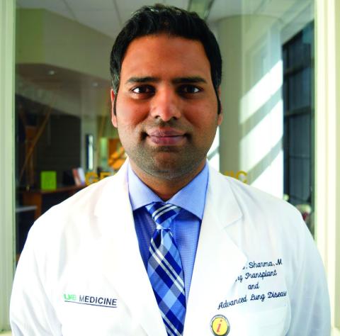
Sepsis is a complex and hard-to-diagnose condition, noted two members of the editorial advisory board of CHEST Physician® in interviews. To make things more complicated, there’s not even a standard definition of sepsis, explained board member Nirmal S. Sharma, MD, of the University of South Florida, Tampa.

“Although newer sepsis definitions have been proposed, all of them have pitfalls and are not used universally. Additionally, the presence of inflammatory response leading to suspicion of sepsis can be due to a new infection or underlying disease processes, thus making it difficult to identify the possible cause,” said Dr. Sharma. “Culture-negative cases due to the use of antibiotics prior to suspicion/onset of sepsis can further muddle the picture. Finally, in certain subsets of patients, such as the immunocompromised and elderly, the signs of sepsis may be delayed due to inadequate/dampened immune response, thus making early diagnosis difficult.”
Blood testing can provide information about germs that are causing an infection, but “they often take several days, and we need to start the antibiotics before we have those results,” added Daniel R. Ouellette, MD, FCCP, the other board member interviewed.
The SeptiCyte Lab assay, which was approved by the FDA for use in diagnosing sepsis in 2017, was developed to help physicians distinguish sepsis from SIRS in patients during their first day of ICU treatment, noted the authors of the new study in the American Journal of Respiratory and Critical Care Medicine.
This new tool seems to overcome some of the obstacles encountered when other diagnostic methods are used to determine if a patient has sepsis.
Russell R. Miller III, MD, FCCM, and his colleagues performed their SeptiCyte Lab assay on patients’ blood samples; this involved real-time, reverse-transcription, quantitative polymerase chain reaction screening designed to analyze the relative expression levels of four genes. The testing procedure took approximately 6 hours from the draw of the blood sample, according to the study, which was recently published online.
comprising three members. Negative predictive values were at least 0.89, according to the researchers.
Overall, the findings show “good reliability,” wrote Dr. Miller of the Intermountain Medical Center in Murray, Utah, and the University of Utah, Salt Lake City, and his colleagues.

The test produced scores in four bands, with scores at or above 3.1 considered to be evidence of infection. Lower levels were considered to be evidence of noninfection.
Dr. Miller and his coauthors reported that 86% of patients unanimously considered to have sepsis had scores above 3.1. In contrast, only 30% of those considered to have SIRS had such high scores.
In addition, the study authors determined that the test was more reliable than were the clinical signs and laboratory variables that are commonly used to diagnose sepsis within 24 hours of arrival at the ICU.
Reaching a definitive sepsis diagnosis is challenging based on clinical signs alone, since various conditions mimic the signs of sepsis, noted Dr. Ouellette of Henry Ford Hospital and Wayne State University School of Medicine in Detroit.
In some cases, physicians simply assume that a patient has sepsis and begin antibiotics, he said, “but that’s not a free ride. Each [antibiotic] may produce side effects with consequences for patients. The other problem is that overuse of antibiotics leads to resistance.”
The study by Dr. Miller and his colleagues combined the results of three trials conducted from during 2011-2016 in the United States and the Netherlands in 447 subjects.
One trial analyzed the experiences of 198 consecutive subjects, all critically ill, who met various criteria. (They were part of a consortium trial of 7,500 patients). The second trial had 129 participants, and the third had 120. Of the total participants, 71% were white and 20% were black.
Inclusion of procalcitonin levels in the laboratory variables didn’t appear to make a significant difference. The study authors wrote that the test “differs from, and is complementary to that of procalcitonin. The latter test is cleared for predicting progression from severe sepsis to septic shock, for predicting 28-day mortality, and for managing antibiotic de-escalation.”
According to the researchers, differences in age, sex, and race/ethnicity did not significantly affect the test.
The study concludes by noting that “future studies are warranted to determine how host gene expression could most effectively be integrated into clinical decision making to ensure susceptible patients are accurately managed early in the course of disease.”
The test is “promising new technology, but I don’t think you could say it’s definitive,” noted Dr. Ouellette.“Like any test, it’s not perfect,” he explained. “That’s important because physicians wouldn’t want to guess wrong. We might err on the side of choosing to treat with antibiotics even in the face of a test that suggested they might not have infection.”
Immunexpress and the Australian Government funded the study. Fourteen authors disclosed being current or former employees of Immunexpress and/or shareholders; others reported receiving funding from the company via their institutions. Four authors declared having filed patent applications related to the study or to the diagnosis of community-acquired pneumonia upon ICU admission. Some authors reported various other disclosures.
Dr. Ouellette and Dr. Sharma said they did not have any disclosures.
SOURCE: Miller RR et al. Am J Respir Crit Care Med. 2018 Apr 6. doi: 10.1164/rccm.201712-2472OC.
A molecular host response assay, called SeptiCyte Lab, holds promise as a tool to distinguish between sepsis and noninfectious systemic inflammation (SIRS), reported researchers in an industry-funded study.
Sepsis is a complex and hard-to-diagnose condition, noted two members of the editorial advisory board of CHEST Physician® in interviews. To make things more complicated, there’s not even a standard definition of sepsis, explained board member Nirmal S. Sharma, MD, of the University of South Florida, Tampa.
“Although newer sepsis definitions have been proposed, all of them have pitfalls and are not used universally. Additionally, the presence of inflammatory response leading to suspicion of sepsis can be due to a new infection or underlying disease processes, thus making it difficult to identify the possible cause,” said Dr. Sharma. “Culture-negative cases due to the use of antibiotics prior to suspicion/onset of sepsis can further muddle the picture. Finally, in certain subsets of patients, such as the immunocompromised and elderly, the signs of sepsis may be delayed due to inadequate/dampened immune response, thus making early diagnosis difficult.”
Blood testing can provide information about germs that are causing an infection, but “they often take several days, and we need to start the antibiotics before we have those results,” added Daniel R. Ouellette, MD, FCCP, the other board member interviewed.
The SeptiCyte Lab assay, which was approved by the FDA for use in diagnosing sepsis in 2017, was developed to help physicians distinguish sepsis from SIRS in patients during their first day of ICU treatment, noted the authors of the new study in the American Journal of Respiratory and Critical Care Medicine.
This new tool seems to overcome some of the obstacles encountered when other diagnostic methods are used to determine if a patient has sepsis.
Russell R. Miller III, MD, FCCM, and his colleagues performed their SeptiCyte Lab assay on patients’ blood samples; this involved real-time, reverse-transcription, quantitative polymerase chain reaction screening designed to analyze the relative expression levels of four genes. The testing procedure took approximately 6 hours from the draw of the blood sample, according to the study, which was recently published online.
comprising three members. Negative predictive values were at least 0.89, according to the researchers.
Overall, the findings show “good reliability,” wrote Dr. Miller of the Intermountain Medical Center in Murray, Utah, and the University of Utah, Salt Lake City, and his colleagues.
The test produced scores in four bands, with scores at or above 3.1 considered to be evidence of infection. Lower levels were considered to be evidence of noninfection.
Dr. Miller and his coauthors reported that 86% of patients unanimously considered to have sepsis had scores above 3.1. In contrast, only 30% of those considered to have SIRS had such high scores.
In addition, the study authors determined that the test was more reliable than were the clinical signs and laboratory variables that are commonly used to diagnose sepsis within 24 hours of arrival at the ICU.
Reaching a definitive sepsis diagnosis is challenging based on clinical signs alone, since various conditions mimic the signs of sepsis, noted Dr. Ouellette of Henry Ford Hospital and Wayne State University School of Medicine in Detroit.
In some cases, physicians simply assume that a patient has sepsis and begin antibiotics, he said, “but that’s not a free ride. Each [antibiotic] may produce side effects with consequences for patients. The other problem is that overuse of antibiotics leads to resistance.”
The study by Dr. Miller and his colleagues combined the results of three trials conducted from during 2011-2016 in the United States and the Netherlands in 447 subjects.
One trial analyzed the experiences of 198 consecutive subjects, all critically ill, who met various criteria. (They were part of a consortium trial of 7,500 patients). The second trial had 129 participants, and the third had 120. Of the total participants, 71% were white and 20% were black.
Inclusion of procalcitonin levels in the laboratory variables didn’t appear to make a significant difference. The study authors wrote that the test “differs from, and is complementary to that of procalcitonin. The latter test is cleared for predicting progression from severe sepsis to septic shock, for predicting 28-day mortality, and for managing antibiotic de-escalation.”
According to the researchers, differences in age, sex, and race/ethnicity did not significantly affect the test.
The study concludes by noting that “future studies are warranted to determine how host gene expression could most effectively be integrated into clinical decision making to ensure susceptible patients are accurately managed early in the course of disease.”
The test is “promising new technology, but I don’t think you could say it’s definitive,” noted Dr. Ouellette.“Like any test, it’s not perfect,” he explained. “That’s important because physicians wouldn’t want to guess wrong. We might err on the side of choosing to treat with antibiotics even in the face of a test that suggested they might not have infection.”
Immunexpress and the Australian Government funded the study. Fourteen authors disclosed being current or former employees of Immunexpress and/or shareholders; others reported receiving funding from the company via their institutions. Four authors declared having filed patent applications related to the study or to the diagnosis of community-acquired pneumonia upon ICU admission. Some authors reported various other disclosures.
Dr. Ouellette and Dr. Sharma said they did not have any disclosures.
SOURCE: Miller RR et al. Am J Respir Crit Care Med. 2018 Apr 6. doi: 10.1164/rccm.201712-2472OC.
A molecular host response assay, called SeptiCyte Lab, holds promise as a tool to distinguish between sepsis and noninfectious systemic inflammation (SIRS), reported researchers in an industry-funded study.
Sepsis is a complex and hard-to-diagnose condition, noted two members of the editorial advisory board of CHEST Physician® in interviews. To make things more complicated, there’s not even a standard definition of sepsis, explained board member Nirmal S. Sharma, MD, of the University of South Florida, Tampa.
“Although newer sepsis definitions have been proposed, all of them have pitfalls and are not used universally. Additionally, the presence of inflammatory response leading to suspicion of sepsis can be due to a new infection or underlying disease processes, thus making it difficult to identify the possible cause,” said Dr. Sharma. “Culture-negative cases due to the use of antibiotics prior to suspicion/onset of sepsis can further muddle the picture. Finally, in certain subsets of patients, such as the immunocompromised and elderly, the signs of sepsis may be delayed due to inadequate/dampened immune response, thus making early diagnosis difficult.”
Blood testing can provide information about germs that are causing an infection, but “they often take several days, and we need to start the antibiotics before we have those results,” added Daniel R. Ouellette, MD, FCCP, the other board member interviewed.
The SeptiCyte Lab assay, which was approved by the FDA for use in diagnosing sepsis in 2017, was developed to help physicians distinguish sepsis from SIRS in patients during their first day of ICU treatment, noted the authors of the new study in the American Journal of Respiratory and Critical Care Medicine.
This new tool seems to overcome some of the obstacles encountered when other diagnostic methods are used to determine if a patient has sepsis.
Russell R. Miller III, MD, FCCM, and his colleagues performed their SeptiCyte Lab assay on patients’ blood samples; this involved real-time, reverse-transcription, quantitative polymerase chain reaction screening designed to analyze the relative expression levels of four genes. The testing procedure took approximately 6 hours from the draw of the blood sample, according to the study, which was recently published online.
comprising three members. Negative predictive values were at least 0.89, according to the researchers.
Overall, the findings show “good reliability,” wrote Dr. Miller of the Intermountain Medical Center in Murray, Utah, and the University of Utah, Salt Lake City, and his colleagues.
The test produced scores in four bands, with scores at or above 3.1 considered to be evidence of infection. Lower levels were considered to be evidence of noninfection.
Dr. Miller and his coauthors reported that 86% of patients unanimously considered to have sepsis had scores above 3.1. In contrast, only 30% of those considered to have SIRS had such high scores.
In addition, the study authors determined that the test was more reliable than were the clinical signs and laboratory variables that are commonly used to diagnose sepsis within 24 hours of arrival at the ICU.
Reaching a definitive sepsis diagnosis is challenging based on clinical signs alone, since various conditions mimic the signs of sepsis, noted Dr. Ouellette of Henry Ford Hospital and Wayne State University School of Medicine in Detroit.
In some cases, physicians simply assume that a patient has sepsis and begin antibiotics, he said, “but that’s not a free ride. Each [antibiotic] may produce side effects with consequences for patients. The other problem is that overuse of antibiotics leads to resistance.”
The study by Dr. Miller and his colleagues combined the results of three trials conducted from during 2011-2016 in the United States and the Netherlands in 447 subjects.
One trial analyzed the experiences of 198 consecutive subjects, all critically ill, who met various criteria. (They were part of a consortium trial of 7,500 patients). The second trial had 129 participants, and the third had 120. Of the total participants, 71% were white and 20% were black.
Inclusion of procalcitonin levels in the laboratory variables didn’t appear to make a significant difference. The study authors wrote that the test “differs from, and is complementary to that of procalcitonin. The latter test is cleared for predicting progression from severe sepsis to septic shock, for predicting 28-day mortality, and for managing antibiotic de-escalation.”
According to the researchers, differences in age, sex, and race/ethnicity did not significantly affect the test.
The study concludes by noting that “future studies are warranted to determine how host gene expression could most effectively be integrated into clinical decision making to ensure susceptible patients are accurately managed early in the course of disease.”
The test is “promising new technology, but I don’t think you could say it’s definitive,” noted Dr. Ouellette.“Like any test, it’s not perfect,” he explained. “That’s important because physicians wouldn’t want to guess wrong. We might err on the side of choosing to treat with antibiotics even in the face of a test that suggested they might not have infection.”
Immunexpress and the Australian Government funded the study. Fourteen authors disclosed being current or former employees of Immunexpress and/or shareholders; others reported receiving funding from the company via their institutions. Four authors declared having filed patent applications related to the study or to the diagnosis of community-acquired pneumonia upon ICU admission. Some authors reported various other disclosures.
Dr. Ouellette and Dr. Sharma said they did not have any disclosures.
SOURCE: Miller RR et al. Am J Respir Crit Care Med. 2018 Apr 6. doi: 10.1164/rccm.201712-2472OC.
FROM AJRCCM
Key clinical point: A blood test to distinguish sepsis from noninfectious systemic inflammation showed high sensitivity.
Major finding: The sensitivity at detecting sepsis was 0.97 in patients unanimously believed by expert panelists to have the condition.
Study details: Prospective, observational, noninterventional analysis of 447 critically ill patients in three trials.
Disclosures: Immunexpress and the Australian Government funded the study. Fourteen authors disclose they are current or were former employees of Immunexpress and/or shareholders, and others disclosed receiving funding from the company.
Source: Miller III RR et al. Am J Respir Crit Care Med. 2018 Apr 6. doi: 10.1164/rccm.201712-2472OC.
Autism spectrum disorder rate calculated at record high in 2014
a new federal report found.
The number suggests the ASD diagnosis rate has continued its steady rise since 2000-2002, when only 0.67 per 1,000 8-year-olds were believed to have the condition.
The report also found that while the gap in diagnosis rates between blacks and whites has dwindled, ASD prevalence “continues to vary among certain racial/ethnic groups and communities.” Indeed, the ASD rate approached 3% in some communities, according to the report published April 28 in Morbidity and Mortality Weekly Report.
The findings are based on statistics gathered by the Autism and Developmental Disabilities Monitoring Network, which uses multiple strategies to track ASD diagnoses among 8-year-olds in Arizona, Arkansas, Colorado, Georgia, Maryland, Minnesota, Missouri, New Jersey, North Carolina, Tennessee, and Wisconsin.
The network, which started its work in 2000, monitors 8-year-old children because that’s the age when ASD prevalence is thought to be at its highest.
The new report, by Jon Baio of the National Center on Birth Defects and Developmental Disabilities and his associates relied upon ASD definitions from DSM-IV-TR and DSM-5. While the definitions seem to be quite different, the report states, “the prevalence of ASD and characteristics of children identified by each case definition were similar in 2014.” Prevalence estimates in the report are only based on DSM-IV-TR criteria.
In total, the report for 2014 tracked 325,483 children aged 8 years, which accounted for 8% of the entire U.S. population in that age group. Of those, 5,473 were determined to have ASD.
The overall ASD prevalence was 16.8 per 1,000 (1 in 59) children. In 2000, the rate was estimated at 6.7 per 1,000. (The researchers caution that the states included in the monitoring network have changed over time.)
New Jersey had the highest prevalence of ASD (29.3 per 1,000, or 2.93%), and the difference when compared with each of the other 10 states was statistically significant (P less than .01).
The overall ASD rate was 26.6 per 1,000 for boys and 6.6 per 1,000 for girls (P less than .01). For whites, it was 17.2 per 1,000, while blacks (16.0 per 1,000) and Hispanics (14.0 per 1,000) had lower rates.
When intelligence quotient information was available, 44% of those with ASD had IQ greater than 85. Blacks (44%) and Hispanics (35%) with ASD were more likely than whites (22%) to have IQs less than or equal to 70, a sign of intellectual disability, the researcher reported.
Of the children with ASD, 80% had previously been diagnosed with the condition or determined to be eligible for autism services.
In light of the report findings, the authors wrote, “With prevalence of ASD reaching nearly 3% in some communities and representing an increase of 150% since 2000, ASD is an urgent public health concern that could benefit from enhanced strategies to help identify ASD earlier; to determine possible risk factors; and to address the growing behavioral, educational, residential and occupational needs of this population.”
Funding and disclosures were not reported.
SOURCE: Baio J et al. Morb Mortal Wkly Rep. 2018 Apr 27;67(6):1-28.
a new federal report found.
The number suggests the ASD diagnosis rate has continued its steady rise since 2000-2002, when only 0.67 per 1,000 8-year-olds were believed to have the condition.
The report also found that while the gap in diagnosis rates between blacks and whites has dwindled, ASD prevalence “continues to vary among certain racial/ethnic groups and communities.” Indeed, the ASD rate approached 3% in some communities, according to the report published April 28 in Morbidity and Mortality Weekly Report.
The findings are based on statistics gathered by the Autism and Developmental Disabilities Monitoring Network, which uses multiple strategies to track ASD diagnoses among 8-year-olds in Arizona, Arkansas, Colorado, Georgia, Maryland, Minnesota, Missouri, New Jersey, North Carolina, Tennessee, and Wisconsin.
The network, which started its work in 2000, monitors 8-year-old children because that’s the age when ASD prevalence is thought to be at its highest.
The new report, by Jon Baio of the National Center on Birth Defects and Developmental Disabilities and his associates relied upon ASD definitions from DSM-IV-TR and DSM-5. While the definitions seem to be quite different, the report states, “the prevalence of ASD and characteristics of children identified by each case definition were similar in 2014.” Prevalence estimates in the report are only based on DSM-IV-TR criteria.
In total, the report for 2014 tracked 325,483 children aged 8 years, which accounted for 8% of the entire U.S. population in that age group. Of those, 5,473 were determined to have ASD.
The overall ASD prevalence was 16.8 per 1,000 (1 in 59) children. In 2000, the rate was estimated at 6.7 per 1,000. (The researchers caution that the states included in the monitoring network have changed over time.)
New Jersey had the highest prevalence of ASD (29.3 per 1,000, or 2.93%), and the difference when compared with each of the other 10 states was statistically significant (P less than .01).
The overall ASD rate was 26.6 per 1,000 for boys and 6.6 per 1,000 for girls (P less than .01). For whites, it was 17.2 per 1,000, while blacks (16.0 per 1,000) and Hispanics (14.0 per 1,000) had lower rates.
When intelligence quotient information was available, 44% of those with ASD had IQ greater than 85. Blacks (44%) and Hispanics (35%) with ASD were more likely than whites (22%) to have IQs less than or equal to 70, a sign of intellectual disability, the researcher reported.
Of the children with ASD, 80% had previously been diagnosed with the condition or determined to be eligible for autism services.
In light of the report findings, the authors wrote, “With prevalence of ASD reaching nearly 3% in some communities and representing an increase of 150% since 2000, ASD is an urgent public health concern that could benefit from enhanced strategies to help identify ASD earlier; to determine possible risk factors; and to address the growing behavioral, educational, residential and occupational needs of this population.”
Funding and disclosures were not reported.
SOURCE: Baio J et al. Morb Mortal Wkly Rep. 2018 Apr 27;67(6):1-28.
a new federal report found.
The number suggests the ASD diagnosis rate has continued its steady rise since 2000-2002, when only 0.67 per 1,000 8-year-olds were believed to have the condition.
The report also found that while the gap in diagnosis rates between blacks and whites has dwindled, ASD prevalence “continues to vary among certain racial/ethnic groups and communities.” Indeed, the ASD rate approached 3% in some communities, according to the report published April 28 in Morbidity and Mortality Weekly Report.
The findings are based on statistics gathered by the Autism and Developmental Disabilities Monitoring Network, which uses multiple strategies to track ASD diagnoses among 8-year-olds in Arizona, Arkansas, Colorado, Georgia, Maryland, Minnesota, Missouri, New Jersey, North Carolina, Tennessee, and Wisconsin.
The network, which started its work in 2000, monitors 8-year-old children because that’s the age when ASD prevalence is thought to be at its highest.
The new report, by Jon Baio of the National Center on Birth Defects and Developmental Disabilities and his associates relied upon ASD definitions from DSM-IV-TR and DSM-5. While the definitions seem to be quite different, the report states, “the prevalence of ASD and characteristics of children identified by each case definition were similar in 2014.” Prevalence estimates in the report are only based on DSM-IV-TR criteria.
In total, the report for 2014 tracked 325,483 children aged 8 years, which accounted for 8% of the entire U.S. population in that age group. Of those, 5,473 were determined to have ASD.
The overall ASD prevalence was 16.8 per 1,000 (1 in 59) children. In 2000, the rate was estimated at 6.7 per 1,000. (The researchers caution that the states included in the monitoring network have changed over time.)
New Jersey had the highest prevalence of ASD (29.3 per 1,000, or 2.93%), and the difference when compared with each of the other 10 states was statistically significant (P less than .01).
The overall ASD rate was 26.6 per 1,000 for boys and 6.6 per 1,000 for girls (P less than .01). For whites, it was 17.2 per 1,000, while blacks (16.0 per 1,000) and Hispanics (14.0 per 1,000) had lower rates.
When intelligence quotient information was available, 44% of those with ASD had IQ greater than 85. Blacks (44%) and Hispanics (35%) with ASD were more likely than whites (22%) to have IQs less than or equal to 70, a sign of intellectual disability, the researcher reported.
Of the children with ASD, 80% had previously been diagnosed with the condition or determined to be eligible for autism services.
In light of the report findings, the authors wrote, “With prevalence of ASD reaching nearly 3% in some communities and representing an increase of 150% since 2000, ASD is an urgent public health concern that could benefit from enhanced strategies to help identify ASD earlier; to determine possible risk factors; and to address the growing behavioral, educational, residential and occupational needs of this population.”
Funding and disclosures were not reported.
SOURCE: Baio J et al. Morb Mortal Wkly Rep. 2018 Apr 27;67(6):1-28.
FROM MMWR
Key clinical point: The ASD rate continues to grow, at least in a sampling of states.
Major finding: An estimated 1.68 per 1,000 (1 in 59) 8-year-old children in 11 states are believed to have ASD.
Study details: The Autism and Developmental Disabilities Monitoring Network uses multiple strategies to track ASD diagnoses among 8-year-olds in 11 states. In 2014, the network tracked 325,483 children.
Disclosures: Funding and disclosures were not reported.
Source: Baio J et al. Morb Mortal Wkly Rep. 2018 Apr 27;67(6):1-28.

Sucralose sparks appetite in obese, not lean, individuals
CHICAGO – but not in lean participants of a recent study, even though hunger and satiety hormone levels didn’t change. Those with obesity also consumed more calories after ingesting the artificial sweetener, though lean participants did not.
The study compared acute effects of consuming a set amount of glucose, sucralose, or water as the control, finding that sucralose consumption resulted in a significant increase in blood flow to certain areas of the brains of study participants with obesity but not in lean individuals (2.10 mL/100g per min vs. –.079 mL/100g per min; P = .002).

Whether responses to caloric and noncaloric sweeteners are different between individuals with and without obesity has not been well established, though recent in vitro and in vivo studies have suggested an association.
“A proposed mechanism is that noncaloric sweeteners uncouple sweetness from calorie intake, which may impact neurophysiological regulators of feeding behavior,” wrote Mr. Ge and his collaborators in an abstract presented at the annual meeting of the Endocrine Society. Still, the work attempts to fill a knowledge gap: “Little evidence, however, has determined the relationship between obesity status and neurophysiological and feeding responses to caloric and non-caloric sweetener consumption,” they wrote.
Of the 30 participants aged 19-24 years, 16 were female; half were lean, with a body mass index of 19-25 kg/m2; the remainder met obesity criteria, with BMIs greater than 30 kg/m2.
For the brain-imaging portion of the study, arterial spin labeling fMRI was used to examine blood flow in a number of predetermined regions of interest. These included the hypothalamus, amygdala, dorsal striatum, insula, and anterior cingulate cortex.
Participants had three scans, spaced at least 2 days apart and occurring after a 12-hour overnight fast. A scan with arterial spin labeling acquisition was taken before and 10 minutes after participants drank a 300-mL beverage consisting of just water, or either a 75-g glucose solution or 2 mmol/L sucralose.
Twenty-five of the participants had blood drawn at 0, 40, and 60 minutes after drinking the study beverage, to track levels of serum insulin, ghrelin, GLP-1, and peptide YY – all hormones that help regulate appetite and satiety.
Hormone levels for individuals who had the non–glucose beverages were similar, regardless of BMI. However, there were significant differences in cerebral blood flow between obese and nonobese participants. Mr. Ge, an undergraduate student, and his collaborators looked at the contributions of the individual brain structures to the significantly higher activation seen after sucralose consumption by the high-BMI participants. Individuals with obesity had significantly more activity in the amygdala than did the lean participants (P = .0088) after drinking the sucralose beverage; also, in lean individuals, hypothalamic activity decreased after sucralose consumption, while activity increased slightly in the high-BMI participants (P = .017).
Eating behavior after drinking the various beverages also differed depending on beverage type and BMI status. After the overnight fast and study beverage consumption, participants were offered unlimited access to a buffet-style meal. The beverage type had no significant effect on calorie consumption at the buffet for the lean study participants. However, obese individuals consumed significantly more calories than did lean individuals after ingesting sucralose (1,191 kcal vs. 731 kcal; P = .01). Caloric intake was not significantly different between the high- and low-BMI groups after consumption of water or glucose.
None of the study authors reported conflicts of interest.
SOURCE: Ge B et al. ENDO 2018, Abstract SUN-070.
CHICAGO – but not in lean participants of a recent study, even though hunger and satiety hormone levels didn’t change. Those with obesity also consumed more calories after ingesting the artificial sweetener, though lean participants did not.
The study compared acute effects of consuming a set amount of glucose, sucralose, or water as the control, finding that sucralose consumption resulted in a significant increase in blood flow to certain areas of the brains of study participants with obesity but not in lean individuals (2.10 mL/100g per min vs. –.079 mL/100g per min; P = .002).
Whether responses to caloric and noncaloric sweeteners are different between individuals with and without obesity has not been well established, though recent in vitro and in vivo studies have suggested an association.
“A proposed mechanism is that noncaloric sweeteners uncouple sweetness from calorie intake, which may impact neurophysiological regulators of feeding behavior,” wrote Mr. Ge and his collaborators in an abstract presented at the annual meeting of the Endocrine Society. Still, the work attempts to fill a knowledge gap: “Little evidence, however, has determined the relationship between obesity status and neurophysiological and feeding responses to caloric and non-caloric sweetener consumption,” they wrote.
Of the 30 participants aged 19-24 years, 16 were female; half were lean, with a body mass index of 19-25 kg/m2; the remainder met obesity criteria, with BMIs greater than 30 kg/m2.
For the brain-imaging portion of the study, arterial spin labeling fMRI was used to examine blood flow in a number of predetermined regions of interest. These included the hypothalamus, amygdala, dorsal striatum, insula, and anterior cingulate cortex.
Participants had three scans, spaced at least 2 days apart and occurring after a 12-hour overnight fast. A scan with arterial spin labeling acquisition was taken before and 10 minutes after participants drank a 300-mL beverage consisting of just water, or either a 75-g glucose solution or 2 mmol/L sucralose.
Twenty-five of the participants had blood drawn at 0, 40, and 60 minutes after drinking the study beverage, to track levels of serum insulin, ghrelin, GLP-1, and peptide YY – all hormones that help regulate appetite and satiety.
Hormone levels for individuals who had the non–glucose beverages were similar, regardless of BMI. However, there were significant differences in cerebral blood flow between obese and nonobese participants. Mr. Ge, an undergraduate student, and his collaborators looked at the contributions of the individual brain structures to the significantly higher activation seen after sucralose consumption by the high-BMI participants. Individuals with obesity had significantly more activity in the amygdala than did the lean participants (P = .0088) after drinking the sucralose beverage; also, in lean individuals, hypothalamic activity decreased after sucralose consumption, while activity increased slightly in the high-BMI participants (P = .017).
Eating behavior after drinking the various beverages also differed depending on beverage type and BMI status. After the overnight fast and study beverage consumption, participants were offered unlimited access to a buffet-style meal. The beverage type had no significant effect on calorie consumption at the buffet for the lean study participants. However, obese individuals consumed significantly more calories than did lean individuals after ingesting sucralose (1,191 kcal vs. 731 kcal; P = .01). Caloric intake was not significantly different between the high- and low-BMI groups after consumption of water or glucose.
None of the study authors reported conflicts of interest.
SOURCE: Ge B et al. ENDO 2018, Abstract SUN-070.
CHICAGO – but not in lean participants of a recent study, even though hunger and satiety hormone levels didn’t change. Those with obesity also consumed more calories after ingesting the artificial sweetener, though lean participants did not.
The study compared acute effects of consuming a set amount of glucose, sucralose, or water as the control, finding that sucralose consumption resulted in a significant increase in blood flow to certain areas of the brains of study participants with obesity but not in lean individuals (2.10 mL/100g per min vs. –.079 mL/100g per min; P = .002).
Whether responses to caloric and noncaloric sweeteners are different between individuals with and without obesity has not been well established, though recent in vitro and in vivo studies have suggested an association.
“A proposed mechanism is that noncaloric sweeteners uncouple sweetness from calorie intake, which may impact neurophysiological regulators of feeding behavior,” wrote Mr. Ge and his collaborators in an abstract presented at the annual meeting of the Endocrine Society. Still, the work attempts to fill a knowledge gap: “Little evidence, however, has determined the relationship between obesity status and neurophysiological and feeding responses to caloric and non-caloric sweetener consumption,” they wrote.
Of the 30 participants aged 19-24 years, 16 were female; half were lean, with a body mass index of 19-25 kg/m2; the remainder met obesity criteria, with BMIs greater than 30 kg/m2.
For the brain-imaging portion of the study, arterial spin labeling fMRI was used to examine blood flow in a number of predetermined regions of interest. These included the hypothalamus, amygdala, dorsal striatum, insula, and anterior cingulate cortex.
Participants had three scans, spaced at least 2 days apart and occurring after a 12-hour overnight fast. A scan with arterial spin labeling acquisition was taken before and 10 minutes after participants drank a 300-mL beverage consisting of just water, or either a 75-g glucose solution or 2 mmol/L sucralose.
Twenty-five of the participants had blood drawn at 0, 40, and 60 minutes after drinking the study beverage, to track levels of serum insulin, ghrelin, GLP-1, and peptide YY – all hormones that help regulate appetite and satiety.
Hormone levels for individuals who had the non–glucose beverages were similar, regardless of BMI. However, there were significant differences in cerebral blood flow between obese and nonobese participants. Mr. Ge, an undergraduate student, and his collaborators looked at the contributions of the individual brain structures to the significantly higher activation seen after sucralose consumption by the high-BMI participants. Individuals with obesity had significantly more activity in the amygdala than did the lean participants (P = .0088) after drinking the sucralose beverage; also, in lean individuals, hypothalamic activity decreased after sucralose consumption, while activity increased slightly in the high-BMI participants (P = .017).
Eating behavior after drinking the various beverages also differed depending on beverage type and BMI status. After the overnight fast and study beverage consumption, participants were offered unlimited access to a buffet-style meal. The beverage type had no significant effect on calorie consumption at the buffet for the lean study participants. However, obese individuals consumed significantly more calories than did lean individuals after ingesting sucralose (1,191 kcal vs. 731 kcal; P = .01). Caloric intake was not significantly different between the high- and low-BMI groups after consumption of water or glucose.
None of the study authors reported conflicts of interest.
SOURCE: Ge B et al. ENDO 2018, Abstract SUN-070.
REPORTING FROM ENDO 2018
Key clinical point: Sucralose ingestion upped activity in brain appetite centers only for those with obesity.
Major finding: Cerebral blood flow for regions of interest was 2.10 mL/100g per min versus –0.79 mL/100g per min after sucralose consumption by obese individuals (P = .002).
Study details: Randomized placebo-controlled trial in 15 lean participants and 15 with obesity.
Disclosures: The authors reported no external sources of funding and no conflicts of interest.
Source: Ge B et al. ENDO 2018, Abstract SUN-070.

Twin study highlights environmental factors that may aggravate acne
A survey conducted at the world’s largest twin celebration provides more evidence that twins share a genetic propensity toward acne, and provides information about several aggravating factors.
The study “further supports that there may be a genetic phenotypic link, though social and environmental factors may also have an influence in the disease process,” the authors wrote.
Previous twin research has linked genetic factors to 80% of acne variance, with environmental factors, such as stress and low intake of produce, believed to account for the rest of the risk (J Invest Dermat. 2002;119[6]:1317-22). For the new study, researchers surveyed twins at the 2016 Twins Day Festival in Twinsburg, Ohio. Thousand of twins – and triplets and quadruplets – from around the world attend the annual event.
After incomplete surveys were discarded, the survey population included 202 identical twins (101 pairs) and 53 fraternal twins or triplets. (A set of triplets was included in addition to 25 pairs of twins.) The majority of participants were female: 23% of identical twins and 17% of the fraternal twins and triplets were male. The mean age was 29 years among the identical twins and 21 years among fraternal twins.
that suggest “acne is largely attributable to genetics,” the authors observed. Among identical twins, those with acne were more likely to have polycystic ovarian syndrome (P = .045), anxiety (P = .014), and asthma (P = .026).
“Identical twin pairs with acne had a higher BMI [body mass index] and exercised less than those without,” the researchers added. These two associations were statistically significant, both for higher BMI (P = .020) and for less exercise (P = .001). “This suggests that a higher BMI and lack of exercise may contribute [along with genetics of course] to acne development. Thus, regular exercise and lower BMI may keep acne at bay,” they noted.
They also analyzed 56 pairs of identical twins with acne, who reported different severities, and found that the twin with more severe acne was more likely to report that sun exposure (P = .048), cosmetic product use (P = .002), and sugar intake (P = .048) aggravated their acne. Refined carbohydrates, as an aggravating factor, approached statistical significance, they said.
A separate analysis of 45 pairs of female identical twins with different degrees of acne severity produced similar findings. There were no significant difference between acne severity groups in terms of menstruation flare frequency or with oral contraceptive use. The twin with more severe acne, however, “was more likely to report aggravation of acne with sun exposure,” cosmetic use, and sugar intake, all associations which reached statistical significance. They were also more likely to report that refined carbohydrates and intake of fried foods aggravated their acne, associations that approached statistical significance.
“This twin study provides further support for reducing intake of sugar and refined carbohydrates to decrease acne severity in susceptible individuals,” the authors wrote. “For females, reducing intake of fried foods may also help,” they added.
There’s a twist to their results: The finding that those with more severe acne reported worsening symptoms with sun exposure “conflicts with prior research, which has found that acne improves with sun,” the authors wrote, adding that “perhaps the data was confounded by comedogenic sunscreen use.”
No specific study funding was reported. The study authors reported no disclosures.
SOURCE: Suggs A et al. J Drugs Dermatol. 2018 Apr;17(4):380-2.
A survey conducted at the world’s largest twin celebration provides more evidence that twins share a genetic propensity toward acne, and provides information about several aggravating factors.
The study “further supports that there may be a genetic phenotypic link, though social and environmental factors may also have an influence in the disease process,” the authors wrote.
Previous twin research has linked genetic factors to 80% of acne variance, with environmental factors, such as stress and low intake of produce, believed to account for the rest of the risk (J Invest Dermat. 2002;119[6]:1317-22). For the new study, researchers surveyed twins at the 2016 Twins Day Festival in Twinsburg, Ohio. Thousand of twins – and triplets and quadruplets – from around the world attend the annual event.
After incomplete surveys were discarded, the survey population included 202 identical twins (101 pairs) and 53 fraternal twins or triplets. (A set of triplets was included in addition to 25 pairs of twins.) The majority of participants were female: 23% of identical twins and 17% of the fraternal twins and triplets were male. The mean age was 29 years among the identical twins and 21 years among fraternal twins.
that suggest “acne is largely attributable to genetics,” the authors observed. Among identical twins, those with acne were more likely to have polycystic ovarian syndrome (P = .045), anxiety (P = .014), and asthma (P = .026).
“Identical twin pairs with acne had a higher BMI [body mass index] and exercised less than those without,” the researchers added. These two associations were statistically significant, both for higher BMI (P = .020) and for less exercise (P = .001). “This suggests that a higher BMI and lack of exercise may contribute [along with genetics of course] to acne development. Thus, regular exercise and lower BMI may keep acne at bay,” they noted.
They also analyzed 56 pairs of identical twins with acne, who reported different severities, and found that the twin with more severe acne was more likely to report that sun exposure (P = .048), cosmetic product use (P = .002), and sugar intake (P = .048) aggravated their acne. Refined carbohydrates, as an aggravating factor, approached statistical significance, they said.
A separate analysis of 45 pairs of female identical twins with different degrees of acne severity produced similar findings. There were no significant difference between acne severity groups in terms of menstruation flare frequency or with oral contraceptive use. The twin with more severe acne, however, “was more likely to report aggravation of acne with sun exposure,” cosmetic use, and sugar intake, all associations which reached statistical significance. They were also more likely to report that refined carbohydrates and intake of fried foods aggravated their acne, associations that approached statistical significance.
“This twin study provides further support for reducing intake of sugar and refined carbohydrates to decrease acne severity in susceptible individuals,” the authors wrote. “For females, reducing intake of fried foods may also help,” they added.
There’s a twist to their results: The finding that those with more severe acne reported worsening symptoms with sun exposure “conflicts with prior research, which has found that acne improves with sun,” the authors wrote, adding that “perhaps the data was confounded by comedogenic sunscreen use.”
No specific study funding was reported. The study authors reported no disclosures.
SOURCE: Suggs A et al. J Drugs Dermatol. 2018 Apr;17(4):380-2.
A survey conducted at the world’s largest twin celebration provides more evidence that twins share a genetic propensity toward acne, and provides information about several aggravating factors.
The study “further supports that there may be a genetic phenotypic link, though social and environmental factors may also have an influence in the disease process,” the authors wrote.
Previous twin research has linked genetic factors to 80% of acne variance, with environmental factors, such as stress and low intake of produce, believed to account for the rest of the risk (J Invest Dermat. 2002;119[6]:1317-22). For the new study, researchers surveyed twins at the 2016 Twins Day Festival in Twinsburg, Ohio. Thousand of twins – and triplets and quadruplets – from around the world attend the annual event.
After incomplete surveys were discarded, the survey population included 202 identical twins (101 pairs) and 53 fraternal twins or triplets. (A set of triplets was included in addition to 25 pairs of twins.) The majority of participants were female: 23% of identical twins and 17% of the fraternal twins and triplets were male. The mean age was 29 years among the identical twins and 21 years among fraternal twins.
that suggest “acne is largely attributable to genetics,” the authors observed. Among identical twins, those with acne were more likely to have polycystic ovarian syndrome (P = .045), anxiety (P = .014), and asthma (P = .026).
“Identical twin pairs with acne had a higher BMI [body mass index] and exercised less than those without,” the researchers added. These two associations were statistically significant, both for higher BMI (P = .020) and for less exercise (P = .001). “This suggests that a higher BMI and lack of exercise may contribute [along with genetics of course] to acne development. Thus, regular exercise and lower BMI may keep acne at bay,” they noted.
They also analyzed 56 pairs of identical twins with acne, who reported different severities, and found that the twin with more severe acne was more likely to report that sun exposure (P = .048), cosmetic product use (P = .002), and sugar intake (P = .048) aggravated their acne. Refined carbohydrates, as an aggravating factor, approached statistical significance, they said.
A separate analysis of 45 pairs of female identical twins with different degrees of acne severity produced similar findings. There were no significant difference between acne severity groups in terms of menstruation flare frequency or with oral contraceptive use. The twin with more severe acne, however, “was more likely to report aggravation of acne with sun exposure,” cosmetic use, and sugar intake, all associations which reached statistical significance. They were also more likely to report that refined carbohydrates and intake of fried foods aggravated their acne, associations that approached statistical significance.
“This twin study provides further support for reducing intake of sugar and refined carbohydrates to decrease acne severity in susceptible individuals,” the authors wrote. “For females, reducing intake of fried foods may also help,” they added.
There’s a twist to their results: The finding that those with more severe acne reported worsening symptoms with sun exposure “conflicts with prior research, which has found that acne improves with sun,” the authors wrote, adding that “perhaps the data was confounded by comedogenic sunscreen use.”
No specific study funding was reported. The study authors reported no disclosures.
SOURCE: Suggs A et al. J Drugs Dermatol. 2018 Apr;17(4):380-2.
FROM THE JOURNAL OF DRUGS IN DERMATOLOGY
Key clinical point: Moderating environmental factors, such as sugar intake and refined carbohydrates, may help reduce the severity of acne.
Major finding: Sun exposure (P = .048), cosmetic product use (P = .002), and sugar intake (P = .048) were among the factors identified that aggravated acne.
Study details: A survey of 202 identical twins (101 pairs) and 53 fraternal twins or triplets conducted at the annual Twins Day Festival in 2016.
Disclosures: No specific study funding was reported. The study authors reported no disclosures.
Source: Suggs A et al. J Drugs Dermatol. 2018 Apr;17(4):380-2.

Anticholinergics’ link to dementia calls for vigilance in elderly
Antidepressant, urologic, and antiparkinson in a large observational study published in The BMJ.
Kathryn Richardson, PhD, of the University of East Anglia, Norwich, England, and her colleagues said that while the associations were “moderate” given the high incidence of dementia observed in the study, they nevertheless reflected an “appreciable risk” for patients.
According to a linked editorial by Shelly L. Gray, PharmD, of the University of Washington, Seattle, and Joseph T. Hanlon, PharmD, of the University of Pittsburgh, the findings suggest that anticholinergics in general should be avoided in older adults (BMJ. 2018;361:k1722. doi: 10.1136/bmj.k1722).
“Specifically, for most highly anticholinergic drugs, nonpharmacological and pharmacological alternatives are available and should be considered,” they said.
The nested case-control study involved 40,770 patients from the United Kingdom’s Clinical Practice Research Database who were aged 65-99 years and diagnosed with dementia between April 2006 and July 2015. The research team matched the patients to 283,933 similar controls without dementia.
They scored drugs according to their anticholinergic activity using the Anticholinergic Cognitive Burden (ACB) scale: A score of 1 was classified as possibly anticholinergic, while a score of 2 or 3 was classified as having “definite” anticholinergic activity. Daily doses of each drug were then compared for both cases and controls over an exposure period of 4-20 years before a diagnosis of dementia.
Overall, 14,453 cases (35%) and 86,403 controls (30%) were prescribed at least one anticholinergic drug with an ACB score of 3 (definite anticholinergic activity) during the exposure period. People prescribed greater dosage quantities over time of probable (ACB category 2) and definite (ACB category 3) anticholinergics had a higher risk of dementia, the researchers reported.
For example, anticholinergic use consistent with the highest dose category (more than 1,460 defined daily doses) was associated with an adjusted odds ratio for dementia of 1.57 (95% confidence interval, 1.18-2.09) for probable and 1.31 (95% CI, 1.22-1.41) for definite anticholinergics.
However, no increased risk was found for anticholinergics used to treat gastrointestinal, cardiovascular, or respiratory conditions. The research team also found no evidence for a cumulative harm of drugs considered “possibly” anticholinergic.
“A typical patient aged 65-70 might normally expect a period incidence of dementia of around 10% over the next 15 years, so this odds ratio would be consistent with an absolute risk increase of 2% (1% to 3%) over that period, corresponding to a number needed to harm of 50 (33 to 100),” they wrote.
They suggested that their findings could be explained by the drugs being markers of prodromal symptoms or dementia risk factors. The class effect observed might also reflect differences in the way anticholinergics crossed the blood-brain barrier.
The Alzheimer’s Society supported the research. Several of the authors reported receiving personal fees from Astellas. One author declared personal fees from Thame Pharmaceuticals.
SOURCE: Richardson K et al. BMJ. 2018;360:k1315. doi: 10.1136/bmj.k1315.
Antidepressant, urologic, and antiparkinson in a large observational study published in The BMJ.
Kathryn Richardson, PhD, of the University of East Anglia, Norwich, England, and her colleagues said that while the associations were “moderate” given the high incidence of dementia observed in the study, they nevertheless reflected an “appreciable risk” for patients.
According to a linked editorial by Shelly L. Gray, PharmD, of the University of Washington, Seattle, and Joseph T. Hanlon, PharmD, of the University of Pittsburgh, the findings suggest that anticholinergics in general should be avoided in older adults (BMJ. 2018;361:k1722. doi: 10.1136/bmj.k1722).
“Specifically, for most highly anticholinergic drugs, nonpharmacological and pharmacological alternatives are available and should be considered,” they said.
The nested case-control study involved 40,770 patients from the United Kingdom’s Clinical Practice Research Database who were aged 65-99 years and diagnosed with dementia between April 2006 and July 2015. The research team matched the patients to 283,933 similar controls without dementia.
They scored drugs according to their anticholinergic activity using the Anticholinergic Cognitive Burden (ACB) scale: A score of 1 was classified as possibly anticholinergic, while a score of 2 or 3 was classified as having “definite” anticholinergic activity. Daily doses of each drug were then compared for both cases and controls over an exposure period of 4-20 years before a diagnosis of dementia.
Overall, 14,453 cases (35%) and 86,403 controls (30%) were prescribed at least one anticholinergic drug with an ACB score of 3 (definite anticholinergic activity) during the exposure period. People prescribed greater dosage quantities over time of probable (ACB category 2) and definite (ACB category 3) anticholinergics had a higher risk of dementia, the researchers reported.
For example, anticholinergic use consistent with the highest dose category (more than 1,460 defined daily doses) was associated with an adjusted odds ratio for dementia of 1.57 (95% confidence interval, 1.18-2.09) for probable and 1.31 (95% CI, 1.22-1.41) for definite anticholinergics.
However, no increased risk was found for anticholinergics used to treat gastrointestinal, cardiovascular, or respiratory conditions. The research team also found no evidence for a cumulative harm of drugs considered “possibly” anticholinergic.
“A typical patient aged 65-70 might normally expect a period incidence of dementia of around 10% over the next 15 years, so this odds ratio would be consistent with an absolute risk increase of 2% (1% to 3%) over that period, corresponding to a number needed to harm of 50 (33 to 100),” they wrote.
They suggested that their findings could be explained by the drugs being markers of prodromal symptoms or dementia risk factors. The class effect observed might also reflect differences in the way anticholinergics crossed the blood-brain barrier.
The Alzheimer’s Society supported the research. Several of the authors reported receiving personal fees from Astellas. One author declared personal fees from Thame Pharmaceuticals.
SOURCE: Richardson K et al. BMJ. 2018;360:k1315. doi: 10.1136/bmj.k1315.
Antidepressant, urologic, and antiparkinson in a large observational study published in The BMJ.
Kathryn Richardson, PhD, of the University of East Anglia, Norwich, England, and her colleagues said that while the associations were “moderate” given the high incidence of dementia observed in the study, they nevertheless reflected an “appreciable risk” for patients.
According to a linked editorial by Shelly L. Gray, PharmD, of the University of Washington, Seattle, and Joseph T. Hanlon, PharmD, of the University of Pittsburgh, the findings suggest that anticholinergics in general should be avoided in older adults (BMJ. 2018;361:k1722. doi: 10.1136/bmj.k1722).
“Specifically, for most highly anticholinergic drugs, nonpharmacological and pharmacological alternatives are available and should be considered,” they said.
The nested case-control study involved 40,770 patients from the United Kingdom’s Clinical Practice Research Database who were aged 65-99 years and diagnosed with dementia between April 2006 and July 2015. The research team matched the patients to 283,933 similar controls without dementia.
They scored drugs according to their anticholinergic activity using the Anticholinergic Cognitive Burden (ACB) scale: A score of 1 was classified as possibly anticholinergic, while a score of 2 or 3 was classified as having “definite” anticholinergic activity. Daily doses of each drug were then compared for both cases and controls over an exposure period of 4-20 years before a diagnosis of dementia.
Overall, 14,453 cases (35%) and 86,403 controls (30%) were prescribed at least one anticholinergic drug with an ACB score of 3 (definite anticholinergic activity) during the exposure period. People prescribed greater dosage quantities over time of probable (ACB category 2) and definite (ACB category 3) anticholinergics had a higher risk of dementia, the researchers reported.
For example, anticholinergic use consistent with the highest dose category (more than 1,460 defined daily doses) was associated with an adjusted odds ratio for dementia of 1.57 (95% confidence interval, 1.18-2.09) for probable and 1.31 (95% CI, 1.22-1.41) for definite anticholinergics.
However, no increased risk was found for anticholinergics used to treat gastrointestinal, cardiovascular, or respiratory conditions. The research team also found no evidence for a cumulative harm of drugs considered “possibly” anticholinergic.
“A typical patient aged 65-70 might normally expect a period incidence of dementia of around 10% over the next 15 years, so this odds ratio would be consistent with an absolute risk increase of 2% (1% to 3%) over that period, corresponding to a number needed to harm of 50 (33 to 100),” they wrote.
They suggested that their findings could be explained by the drugs being markers of prodromal symptoms or dementia risk factors. The class effect observed might also reflect differences in the way anticholinergics crossed the blood-brain barrier.
The Alzheimer’s Society supported the research. Several of the authors reported receiving personal fees from Astellas. One author declared personal fees from Thame Pharmaceuticals.
SOURCE: Richardson K et al. BMJ. 2018;360:k1315. doi: 10.1136/bmj.k1315.
FROM THE BMJ
Key clinical point: Clinicians should continue to be mindful of the long-term cognitive effects, as well as short-term effects, of using of anticholinergic drugs, particularly in elderly patients.
Main finding: The highest dose category of probable anticholinergic use was associated with an adjusted odds ratio for dementia of 1.57 (95% confidence interval, 1.18-2.09) while definite anticholinergic use gave an odds ratio of 1.31 (95% CI, 1.22-1.41).
Study details: Nested case-control study involving 40,770 patients aged 65-99 from the U.K.’s Clinical Practice Research Database.
Disclosures: The Alzheimer’s Society supported the research. Several of the authors reported receiving personal fees from Astellas. One author declared personal fees from Thame Pharmaceuticals.
Source: Richardson K et al. BMJ. 2018;360:k1315. doi: 10.1136/bmj.k1315.

Bubble Trouble
A 13-year-old girl is referred to dermatology by her pediatrician for evaluation of “warts” that manifested several months ago. The asymptomatic lesions are simply a cosmetic concern—albeit a persistent one.
Treatments including liquid nitrogen, salicylic acid–based OTC products, and an electric device (purchased online) have been tried, but none have helped. The topical products caused pain and blistering, and although they did eliminate several of the lesions, more soon appeared to take their place.
Further history-taking reveals that the child (like her family) is highly atopic, with seasonal allergies and a history of eczema, hives, and asthma. Two of her siblings have had similar lesions, which cleared fairly quickly without treatment.
EXAMINATION
Approximately 20 papules are randomly arranged on the patient’s anterior neck. They are pink and round, each measuring 2 to 3 mm. Closer inspection reveals that most display a central umbilication. The lesions are firm on palpation.
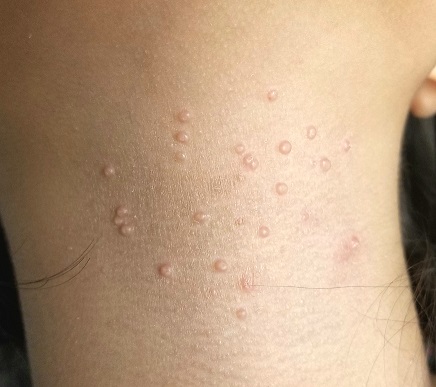
The child is multiracial; her type IV skin is quite dry but otherwise free of lesions.
What is the diagnosis?
DISCUSSION
One of the most frequently encountered skin lesions in primary care, molluscum contagiosum (MC) is seen mostly in children. It is strongly associated with immune suppression, especially atopy, which affects around 20% of newborns. Commonly called “water warts,” MC lesions have no connection to human papillomavirus and are instead caused by the molluscum contagiosum virus—the largest virus to infect humans.
This virus is transmitted through direct contact with an infected individual, which often occurs during the summertime when many children swim. Though the warts cause little if any harm, they can be a source of embarrassment for the child and can be concerning to parents, who are often given erroneous information about the diagnosis.
There’s also the unfortunate fact that the occasional MC lesion fills with pus, turning red and swollen—a fair imitation of bacterial infection. This is simply a sign that the lesion is dying and will soon disappear, but it’s understandably frightening to parents.
As this case illustrates so well, destroying a few MC lesions does nothing to keep a whole new crop from taking their place. And although the condition is self-limiting, it is common for the warts to take two or more years to go away.
The truth is, to date, there has been no proven, safe, painless, effective treatment for MC. Modalities include laser, electrodessication, and simple curettage.
A new treatment that combines dilute povidone-iodine with dimethyl sulfoxide in an OTC compounded liquid mixture (applied bid) has shown some promise in limited trials (Capriotti et al. J Clin Aesthet Dermatol. 2017;10[3]:41). This is what the case patient was treated with. I have given it to perhaps a dozen patients over the past several months, but to date, none have returned to report results. (This, as far as I know, is not a proprietary product and I have no financial interest in it.)
TAKE-HOME LEARNING POINTS
- Firm, 2- to 3-mm, umbilicated papules on children are almost certainly molluscum contagiosum (MC), usually related to atopy.
- MC is acquired by direct contact but can be spread by scratching or picking.
- It often appears admixed with eczema, especially in the antecubital and popliteal areas.
- Although MC eventually resolves with or without treatment, the process can take a while, making patient/parent education important.
- The newest treatment (that I am aware of) is a mixture of povidone-iodine and dimethyl sulfoxide, to be applied bid.
A 13-year-old girl is referred to dermatology by her pediatrician for evaluation of “warts” that manifested several months ago. The asymptomatic lesions are simply a cosmetic concern—albeit a persistent one.
Treatments including liquid nitrogen, salicylic acid–based OTC products, and an electric device (purchased online) have been tried, but none have helped. The topical products caused pain and blistering, and although they did eliminate several of the lesions, more soon appeared to take their place.
Further history-taking reveals that the child (like her family) is highly atopic, with seasonal allergies and a history of eczema, hives, and asthma. Two of her siblings have had similar lesions, which cleared fairly quickly without treatment.
EXAMINATION
Approximately 20 papules are randomly arranged on the patient’s anterior neck. They are pink and round, each measuring 2 to 3 mm. Closer inspection reveals that most display a central umbilication. The lesions are firm on palpation.
The child is multiracial; her type IV skin is quite dry but otherwise free of lesions.
What is the diagnosis?
DISCUSSION
One of the most frequently encountered skin lesions in primary care, molluscum contagiosum (MC) is seen mostly in children. It is strongly associated with immune suppression, especially atopy, which affects around 20% of newborns. Commonly called “water warts,” MC lesions have no connection to human papillomavirus and are instead caused by the molluscum contagiosum virus—the largest virus to infect humans.
This virus is transmitted through direct contact with an infected individual, which often occurs during the summertime when many children swim. Though the warts cause little if any harm, they can be a source of embarrassment for the child and can be concerning to parents, who are often given erroneous information about the diagnosis.
There’s also the unfortunate fact that the occasional MC lesion fills with pus, turning red and swollen—a fair imitation of bacterial infection. This is simply a sign that the lesion is dying and will soon disappear, but it’s understandably frightening to parents.
As this case illustrates so well, destroying a few MC lesions does nothing to keep a whole new crop from taking their place. And although the condition is self-limiting, it is common for the warts to take two or more years to go away.
The truth is, to date, there has been no proven, safe, painless, effective treatment for MC. Modalities include laser, electrodessication, and simple curettage.
A new treatment that combines dilute povidone-iodine with dimethyl sulfoxide in an OTC compounded liquid mixture (applied bid) has shown some promise in limited trials (Capriotti et al. J Clin Aesthet Dermatol. 2017;10[3]:41). This is what the case patient was treated with. I have given it to perhaps a dozen patients over the past several months, but to date, none have returned to report results. (This, as far as I know, is not a proprietary product and I have no financial interest in it.)
TAKE-HOME LEARNING POINTS
- Firm, 2- to 3-mm, umbilicated papules on children are almost certainly molluscum contagiosum (MC), usually related to atopy.
- MC is acquired by direct contact but can be spread by scratching or picking.
- It often appears admixed with eczema, especially in the antecubital and popliteal areas.
- Although MC eventually resolves with or without treatment, the process can take a while, making patient/parent education important.
- The newest treatment (that I am aware of) is a mixture of povidone-iodine and dimethyl sulfoxide, to be applied bid.
A 13-year-old girl is referred to dermatology by her pediatrician for evaluation of “warts” that manifested several months ago. The asymptomatic lesions are simply a cosmetic concern—albeit a persistent one.
Treatments including liquid nitrogen, salicylic acid–based OTC products, and an electric device (purchased online) have been tried, but none have helped. The topical products caused pain and blistering, and although they did eliminate several of the lesions, more soon appeared to take their place.
Further history-taking reveals that the child (like her family) is highly atopic, with seasonal allergies and a history of eczema, hives, and asthma. Two of her siblings have had similar lesions, which cleared fairly quickly without treatment.
EXAMINATION
Approximately 20 papules are randomly arranged on the patient’s anterior neck. They are pink and round, each measuring 2 to 3 mm. Closer inspection reveals that most display a central umbilication. The lesions are firm on palpation.
The child is multiracial; her type IV skin is quite dry but otherwise free of lesions.
What is the diagnosis?
DISCUSSION
One of the most frequently encountered skin lesions in primary care, molluscum contagiosum (MC) is seen mostly in children. It is strongly associated with immune suppression, especially atopy, which affects around 20% of newborns. Commonly called “water warts,” MC lesions have no connection to human papillomavirus and are instead caused by the molluscum contagiosum virus—the largest virus to infect humans.
This virus is transmitted through direct contact with an infected individual, which often occurs during the summertime when many children swim. Though the warts cause little if any harm, they can be a source of embarrassment for the child and can be concerning to parents, who are often given erroneous information about the diagnosis.
There’s also the unfortunate fact that the occasional MC lesion fills with pus, turning red and swollen—a fair imitation of bacterial infection. This is simply a sign that the lesion is dying and will soon disappear, but it’s understandably frightening to parents.
As this case illustrates so well, destroying a few MC lesions does nothing to keep a whole new crop from taking their place. And although the condition is self-limiting, it is common for the warts to take two or more years to go away.
The truth is, to date, there has been no proven, safe, painless, effective treatment for MC. Modalities include laser, electrodessication, and simple curettage.
A new treatment that combines dilute povidone-iodine with dimethyl sulfoxide in an OTC compounded liquid mixture (applied bid) has shown some promise in limited trials (Capriotti et al. J Clin Aesthet Dermatol. 2017;10[3]:41). This is what the case patient was treated with. I have given it to perhaps a dozen patients over the past several months, but to date, none have returned to report results. (This, as far as I know, is not a proprietary product and I have no financial interest in it.)
TAKE-HOME LEARNING POINTS
- Firm, 2- to 3-mm, umbilicated papules on children are almost certainly molluscum contagiosum (MC), usually related to atopy.
- MC is acquired by direct contact but can be spread by scratching or picking.
- It often appears admixed with eczema, especially in the antecubital and popliteal areas.
- Although MC eventually resolves with or without treatment, the process can take a while, making patient/parent education important.
- The newest treatment (that I am aware of) is a mixture of povidone-iodine and dimethyl sulfoxide, to be applied bid.
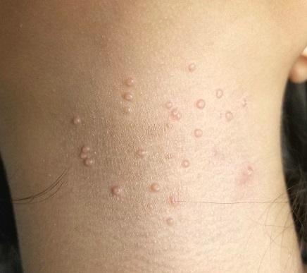
Don’t delay hip-fracture surgery
Background: Guidelines from the American College of Surgeons and Canadian Institute for Health recommend hip fracture surgery within 48 hours. However, a time-to-surgery threshold after which mortality and complications are increased has not been determined. This study aims to determine a time to surgery threshold for hip-fracture surgery.
Study design: Retrospective cohort trial.
Setting: 72 hospitals in Ontario, Ca., during April 1, 2009-March 31, 2014.
Synopsis: Of the 42,230 adult patients in this study, 14,174 (33.6%) received hip-fracture surgery within 24 hours of emergency department arrival. A matched patient analysis of early surgery (within 24 hours of ED arrival) vs. delayed surgery determined that patients undergoing early operation experienced lower 30-day mortality (5.8% vs 6.5%) and fewer complications (myocardial infarction, deep vein thrombosis, pulmonary embolism, and pneumonia). Major bleeding was not assessed as a complication. Also omitted from analysis were patients undergoing nonoperative hip-fracture management.
These findings suggest a time to surgery of 24 hours may represent a threshold defining higher risk. Two-thirds of patients in this study surpassed this threshold. Hospitalists seeing patients with hip fracture should balance time delay risks with the need for medical optimization.
Bottom line: Wait time greater than 24 hours for adults undergoing hip fracture surgery is associated with an increased risk of 30-day mortality and complications.
Citation: Pincus D et al. Association between wait time and 30-day mortality in adults undergoing hip fracture surgery. JAMA. 2017 Nov 28;318(20):1994-2003.
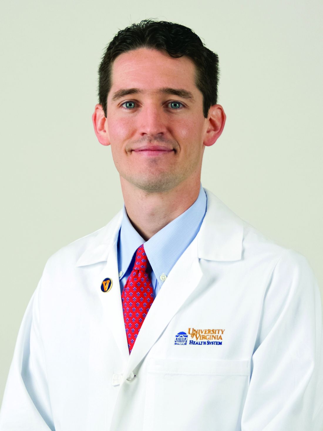
Dr. Moulder is assistant professor, University of Virginia Health System.
Background: Guidelines from the American College of Surgeons and Canadian Institute for Health recommend hip fracture surgery within 48 hours. However, a time-to-surgery threshold after which mortality and complications are increased has not been determined. This study aims to determine a time to surgery threshold for hip-fracture surgery.
Study design: Retrospective cohort trial.
Setting: 72 hospitals in Ontario, Ca., during April 1, 2009-March 31, 2014.
Synopsis: Of the 42,230 adult patients in this study, 14,174 (33.6%) received hip-fracture surgery within 24 hours of emergency department arrival. A matched patient analysis of early surgery (within 24 hours of ED arrival) vs. delayed surgery determined that patients undergoing early operation experienced lower 30-day mortality (5.8% vs 6.5%) and fewer complications (myocardial infarction, deep vein thrombosis, pulmonary embolism, and pneumonia). Major bleeding was not assessed as a complication. Also omitted from analysis were patients undergoing nonoperative hip-fracture management.
These findings suggest a time to surgery of 24 hours may represent a threshold defining higher risk. Two-thirds of patients in this study surpassed this threshold. Hospitalists seeing patients with hip fracture should balance time delay risks with the need for medical optimization.
Bottom line: Wait time greater than 24 hours for adults undergoing hip fracture surgery is associated with an increased risk of 30-day mortality and complications.
Citation: Pincus D et al. Association between wait time and 30-day mortality in adults undergoing hip fracture surgery. JAMA. 2017 Nov 28;318(20):1994-2003.
Dr. Moulder is assistant professor, University of Virginia Health System.
Background: Guidelines from the American College of Surgeons and Canadian Institute for Health recommend hip fracture surgery within 48 hours. However, a time-to-surgery threshold after which mortality and complications are increased has not been determined. This study aims to determine a time to surgery threshold for hip-fracture surgery.
Study design: Retrospective cohort trial.
Setting: 72 hospitals in Ontario, Ca., during April 1, 2009-March 31, 2014.
Synopsis: Of the 42,230 adult patients in this study, 14,174 (33.6%) received hip-fracture surgery within 24 hours of emergency department arrival. A matched patient analysis of early surgery (within 24 hours of ED arrival) vs. delayed surgery determined that patients undergoing early operation experienced lower 30-day mortality (5.8% vs 6.5%) and fewer complications (myocardial infarction, deep vein thrombosis, pulmonary embolism, and pneumonia). Major bleeding was not assessed as a complication. Also omitted from analysis were patients undergoing nonoperative hip-fracture management.
These findings suggest a time to surgery of 24 hours may represent a threshold defining higher risk. Two-thirds of patients in this study surpassed this threshold. Hospitalists seeing patients with hip fracture should balance time delay risks with the need for medical optimization.
Bottom line: Wait time greater than 24 hours for adults undergoing hip fracture surgery is associated with an increased risk of 30-day mortality and complications.
Citation: Pincus D et al. Association between wait time and 30-day mortality in adults undergoing hip fracture surgery. JAMA. 2017 Nov 28;318(20):1994-2003.
Dr. Moulder is assistant professor, University of Virginia Health System.
Coming soon: CAR T-cell approvals in multiple myeloma
, and will “completely transform oncology,” according to Carl June, MD.
That approval is anticipated sometime in 2019.
“Myeloma is the most common blood cancer in adults, and there’s never been a curative therapy, but now there is a subset of patients who look like they’re cured with CAR T cells,” Dr. June, the Richard W. Vague Professor in Immunotherapy and a pioneer in CAR T-cell research at the University of Pennsylvania, Philadelphia, said in an interview.
The first treated patient in a trial of a novel anti-B-cell maturation antigen (BCMA)-specific CAR T-cell therapy (CART-BCMA) developed by University of Pennsylvania researchers in collaboration with Novartis is part of that subset.
Woodring Wright, MD, a professor of cell biology and medicine at the University of Texas Southwestern Medical Center (UT Southwestern) in Dallas recently outed himself as that first patient, announcing in a Feb. 14, 2018, UT Southwestern press report that CART-BCMA saved his life.
Dr. Wright, who holds the Southland Financial Corporation Distinguished Chair in Geriatrics at UT Southwestern, was diagnosed with multiple myeloma about 12 years ago and failed 11 prior chemotherapies before he was enrolled in the CART-BCMA trial.
“Now he considers himself cured,” Dr. June said.
More than 2 years after receiving CART-BCMA he remains cancer free, and is now conducting CAR T-cell-related research in his lab at UT Southwestern in an effort to broaden the effectiveness of current CAR T-cell therapies. Specifically, he is looking at whether the small percentage of patients in whom CAR T-cell therapy does not work might benefit from telomerase to lengthen telomeres, as most patients who fail CAR T-cell therapy are elderly patients who might have terminally short telomeres, UT Southwestern reported.
The ongoing University of Pennsylvania trial led by Adam D. Cohen, MD, director of myeloma immunotherapy at the Abramson Cancer Center, has an overall response rate of 64%; initial phase 1 efficacy and safety results were reported at the American Society of Hematology (ASH) annual meeting in 2016, and multiple companies are currently pursuing registration trials for CAR T therapies in myeloma, Dr. June said.
Among them are bluebird bio and Celgene, which together are developing an anti-BCMA CAR T-cell therapy known as bb2121. That product was granted breakthrough therapy designation by the Food and Drug Administration in November 2017, and will thus receive expedited review. It has also been fast-tracked in Europe.
The decision to fast-track bb2121 in the United States was based on preliminary results from the CRB-410 trial. Updated findings from that trial were presented in December 2017 at ASH and showed an overall response rate of 94% in 21 patients, with 17 of 18 patients who received doses above 50 x 106 CAR+ T cells having an overall response, and 10 of the 18 achieving complete remission. The progression-free survival rates were 81% at 6 months, and 71% at 9 months, with responses deepening over time. The complete response rates were 27% and 56% in May and October of 2017, respectively.
Responses were durable, lasting more than 1 year in several patients, the investigators reported. Phase 2 of the trial – the global pivotal KarMMA trial – is currently enrolling and will dose patients at between 150 and 350 x 106 CAR+ T cells.
Janssen Biotech Inc. (a Johnson & Johnson company) and Legend Biotech USA Inc./Legend Biotech Ireland Limited (of Genscript Biotech Corporation) have also joined forces to develop an anti-BCMA CAR T-cell product for multiple myeloma, Dr. June said.

The companies announced in December that they had entered into “a worldwide collaboration and license agreement” to develop the CAR T-cell drug candidate.
LCAR-B38M is currently accepted for review by the China Food and Drug Administration and is in the planning phase of clinical studies in the United States for multiple myeloma, according to that announcement.
The “race between companies” for a CAR T myeloma approval will lead to a welcome addition to the treatment armamentarium, because while myeloma represents only about 2% of all cancers, it is responsible for 7% of cancer costs, Dr. June said.
Since many patients live with their disease for a long time, that can mean huge “financial toxicity” associated with treatment and patients still usually have “an awful outcome involving a long death,” he said.
“So CAR T-cell therapy for myeloma will bring a huge change to the practice of oncology,” he added, explaining that the first CAR T-cell therapy approved (tisagenlecleucel, in August 2017) was for pediatric acute lymphoblastic leukemia that had relapsed at least twice. “That’s only about 600 kids a year in the U.S., so it’s an ultra-orphan market,” he said.
With the subsequent approval of axicabtagene ciloleucel (in October 2017) and the anticipated myeloma approval, CAR T-cell therapy will move away from orphan status.
“There are a lot of difficulties whenever you change to something new,” he said, comparing the CAR T-cell therapy evolution to that of bone marrow transplantation in the 1980s.
Early on, there were only two places in the country where a patient could get a bone marrow transplant – Fred Hutchinson Cancer Center in Seattle and Johns Hopkins University in Baltimore. “Everyone said ‘you’ll never be able to do it routinely, it’s only at these two referral centers,’ because of the skill needed and the intensity of it,” he said. “But over the years, millions of transplants have now been done; they’re done at many community centers. And it’s the same thing with CARs; Novartis now has 30 centers and people have to be trained. It’s a new skill set, and it will take time,” he said.
That can be particularly frustrating because there are many patients with diseases that “might benefit in a major way” from CAR T-cell therapy, but who can’t get on a clinical trial, Dr. June noted.
“There’s more demand than availability, and it’s going to take awhile ... it’s like liver transplants – there aren’t enough donors to go around, so people die, they get on lists, and it’s really hard to see that, but eventually it will get solved,” he said, adding that the solution will most likely involve the complementary use of off-the-shelf CAR T cells in certain patients to induce remission and perhaps provide a bridge to some other definitive therapy, and ultra-personalized CAR T therapy in others, as well as combinations that include CAR T cells and targeted agents or checkpoint inhibitors.
CRISPR-Cas9 gene editing is also being looked at as a tool for engineering multiple myeloma cellular immunotherapy (and other cancer treatments), as in the Parker Institute–funded NYCE study, Dr. June said.
“We’re actually removing the [programmed death-1] gene and the T-cell receptors ... it shows enormous potential for gene editing. CRISPR is going to be used for a lot of things, but the first use is with T-cell therapies, so we’re really excited about that trial,” he said. “We just opened and we’re screening patients now.”
Dr. June reported royalties and research funding from Novartis and an ownership interest in Tmunity Therapeutics.
, and will “completely transform oncology,” according to Carl June, MD.
That approval is anticipated sometime in 2019.
“Myeloma is the most common blood cancer in adults, and there’s never been a curative therapy, but now there is a subset of patients who look like they’re cured with CAR T cells,” Dr. June, the Richard W. Vague Professor in Immunotherapy and a pioneer in CAR T-cell research at the University of Pennsylvania, Philadelphia, said in an interview.
The first treated patient in a trial of a novel anti-B-cell maturation antigen (BCMA)-specific CAR T-cell therapy (CART-BCMA) developed by University of Pennsylvania researchers in collaboration with Novartis is part of that subset.
Woodring Wright, MD, a professor of cell biology and medicine at the University of Texas Southwestern Medical Center (UT Southwestern) in Dallas recently outed himself as that first patient, announcing in a Feb. 14, 2018, UT Southwestern press report that CART-BCMA saved his life.
Dr. Wright, who holds the Southland Financial Corporation Distinguished Chair in Geriatrics at UT Southwestern, was diagnosed with multiple myeloma about 12 years ago and failed 11 prior chemotherapies before he was enrolled in the CART-BCMA trial.
“Now he considers himself cured,” Dr. June said.
More than 2 years after receiving CART-BCMA he remains cancer free, and is now conducting CAR T-cell-related research in his lab at UT Southwestern in an effort to broaden the effectiveness of current CAR T-cell therapies. Specifically, he is looking at whether the small percentage of patients in whom CAR T-cell therapy does not work might benefit from telomerase to lengthen telomeres, as most patients who fail CAR T-cell therapy are elderly patients who might have terminally short telomeres, UT Southwestern reported.
The ongoing University of Pennsylvania trial led by Adam D. Cohen, MD, director of myeloma immunotherapy at the Abramson Cancer Center, has an overall response rate of 64%; initial phase 1 efficacy and safety results were reported at the American Society of Hematology (ASH) annual meeting in 2016, and multiple companies are currently pursuing registration trials for CAR T therapies in myeloma, Dr. June said.
Among them are bluebird bio and Celgene, which together are developing an anti-BCMA CAR T-cell therapy known as bb2121. That product was granted breakthrough therapy designation by the Food and Drug Administration in November 2017, and will thus receive expedited review. It has also been fast-tracked in Europe.
The decision to fast-track bb2121 in the United States was based on preliminary results from the CRB-410 trial. Updated findings from that trial were presented in December 2017 at ASH and showed an overall response rate of 94% in 21 patients, with 17 of 18 patients who received doses above 50 x 106 CAR+ T cells having an overall response, and 10 of the 18 achieving complete remission. The progression-free survival rates were 81% at 6 months, and 71% at 9 months, with responses deepening over time. The complete response rates were 27% and 56% in May and October of 2017, respectively.
Responses were durable, lasting more than 1 year in several patients, the investigators reported. Phase 2 of the trial – the global pivotal KarMMA trial – is currently enrolling and will dose patients at between 150 and 350 x 106 CAR+ T cells.
Janssen Biotech Inc. (a Johnson & Johnson company) and Legend Biotech USA Inc./Legend Biotech Ireland Limited (of Genscript Biotech Corporation) have also joined forces to develop an anti-BCMA CAR T-cell product for multiple myeloma, Dr. June said.
The companies announced in December that they had entered into “a worldwide collaboration and license agreement” to develop the CAR T-cell drug candidate.
LCAR-B38M is currently accepted for review by the China Food and Drug Administration and is in the planning phase of clinical studies in the United States for multiple myeloma, according to that announcement.
The “race between companies” for a CAR T myeloma approval will lead to a welcome addition to the treatment armamentarium, because while myeloma represents only about 2% of all cancers, it is responsible for 7% of cancer costs, Dr. June said.
Since many patients live with their disease for a long time, that can mean huge “financial toxicity” associated with treatment and patients still usually have “an awful outcome involving a long death,” he said.
“So CAR T-cell therapy for myeloma will bring a huge change to the practice of oncology,” he added, explaining that the first CAR T-cell therapy approved (tisagenlecleucel, in August 2017) was for pediatric acute lymphoblastic leukemia that had relapsed at least twice. “That’s only about 600 kids a year in the U.S., so it’s an ultra-orphan market,” he said.
With the subsequent approval of axicabtagene ciloleucel (in October 2017) and the anticipated myeloma approval, CAR T-cell therapy will move away from orphan status.
“There are a lot of difficulties whenever you change to something new,” he said, comparing the CAR T-cell therapy evolution to that of bone marrow transplantation in the 1980s.
Early on, there were only two places in the country where a patient could get a bone marrow transplant – Fred Hutchinson Cancer Center in Seattle and Johns Hopkins University in Baltimore. “Everyone said ‘you’ll never be able to do it routinely, it’s only at these two referral centers,’ because of the skill needed and the intensity of it,” he said. “But over the years, millions of transplants have now been done; they’re done at many community centers. And it’s the same thing with CARs; Novartis now has 30 centers and people have to be trained. It’s a new skill set, and it will take time,” he said.
That can be particularly frustrating because there are many patients with diseases that “might benefit in a major way” from CAR T-cell therapy, but who can’t get on a clinical trial, Dr. June noted.
“There’s more demand than availability, and it’s going to take awhile ... it’s like liver transplants – there aren’t enough donors to go around, so people die, they get on lists, and it’s really hard to see that, but eventually it will get solved,” he said, adding that the solution will most likely involve the complementary use of off-the-shelf CAR T cells in certain patients to induce remission and perhaps provide a bridge to some other definitive therapy, and ultra-personalized CAR T therapy in others, as well as combinations that include CAR T cells and targeted agents or checkpoint inhibitors.
CRISPR-Cas9 gene editing is also being looked at as a tool for engineering multiple myeloma cellular immunotherapy (and other cancer treatments), as in the Parker Institute–funded NYCE study, Dr. June said.
“We’re actually removing the [programmed death-1] gene and the T-cell receptors ... it shows enormous potential for gene editing. CRISPR is going to be used for a lot of things, but the first use is with T-cell therapies, so we’re really excited about that trial,” he said. “We just opened and we’re screening patients now.”
Dr. June reported royalties and research funding from Novartis and an ownership interest in Tmunity Therapeutics.
, and will “completely transform oncology,” according to Carl June, MD.
That approval is anticipated sometime in 2019.
“Myeloma is the most common blood cancer in adults, and there’s never been a curative therapy, but now there is a subset of patients who look like they’re cured with CAR T cells,” Dr. June, the Richard W. Vague Professor in Immunotherapy and a pioneer in CAR T-cell research at the University of Pennsylvania, Philadelphia, said in an interview.
The first treated patient in a trial of a novel anti-B-cell maturation antigen (BCMA)-specific CAR T-cell therapy (CART-BCMA) developed by University of Pennsylvania researchers in collaboration with Novartis is part of that subset.
Woodring Wright, MD, a professor of cell biology and medicine at the University of Texas Southwestern Medical Center (UT Southwestern) in Dallas recently outed himself as that first patient, announcing in a Feb. 14, 2018, UT Southwestern press report that CART-BCMA saved his life.
Dr. Wright, who holds the Southland Financial Corporation Distinguished Chair in Geriatrics at UT Southwestern, was diagnosed with multiple myeloma about 12 years ago and failed 11 prior chemotherapies before he was enrolled in the CART-BCMA trial.
“Now he considers himself cured,” Dr. June said.
More than 2 years after receiving CART-BCMA he remains cancer free, and is now conducting CAR T-cell-related research in his lab at UT Southwestern in an effort to broaden the effectiveness of current CAR T-cell therapies. Specifically, he is looking at whether the small percentage of patients in whom CAR T-cell therapy does not work might benefit from telomerase to lengthen telomeres, as most patients who fail CAR T-cell therapy are elderly patients who might have terminally short telomeres, UT Southwestern reported.
The ongoing University of Pennsylvania trial led by Adam D. Cohen, MD, director of myeloma immunotherapy at the Abramson Cancer Center, has an overall response rate of 64%; initial phase 1 efficacy and safety results were reported at the American Society of Hematology (ASH) annual meeting in 2016, and multiple companies are currently pursuing registration trials for CAR T therapies in myeloma, Dr. June said.
Among them are bluebird bio and Celgene, which together are developing an anti-BCMA CAR T-cell therapy known as bb2121. That product was granted breakthrough therapy designation by the Food and Drug Administration in November 2017, and will thus receive expedited review. It has also been fast-tracked in Europe.
The decision to fast-track bb2121 in the United States was based on preliminary results from the CRB-410 trial. Updated findings from that trial were presented in December 2017 at ASH and showed an overall response rate of 94% in 21 patients, with 17 of 18 patients who received doses above 50 x 106 CAR+ T cells having an overall response, and 10 of the 18 achieving complete remission. The progression-free survival rates were 81% at 6 months, and 71% at 9 months, with responses deepening over time. The complete response rates were 27% and 56% in May and October of 2017, respectively.
Responses were durable, lasting more than 1 year in several patients, the investigators reported. Phase 2 of the trial – the global pivotal KarMMA trial – is currently enrolling and will dose patients at between 150 and 350 x 106 CAR+ T cells.
Janssen Biotech Inc. (a Johnson & Johnson company) and Legend Biotech USA Inc./Legend Biotech Ireland Limited (of Genscript Biotech Corporation) have also joined forces to develop an anti-BCMA CAR T-cell product for multiple myeloma, Dr. June said.
The companies announced in December that they had entered into “a worldwide collaboration and license agreement” to develop the CAR T-cell drug candidate.
LCAR-B38M is currently accepted for review by the China Food and Drug Administration and is in the planning phase of clinical studies in the United States for multiple myeloma, according to that announcement.
The “race between companies” for a CAR T myeloma approval will lead to a welcome addition to the treatment armamentarium, because while myeloma represents only about 2% of all cancers, it is responsible for 7% of cancer costs, Dr. June said.
Since many patients live with their disease for a long time, that can mean huge “financial toxicity” associated with treatment and patients still usually have “an awful outcome involving a long death,” he said.
“So CAR T-cell therapy for myeloma will bring a huge change to the practice of oncology,” he added, explaining that the first CAR T-cell therapy approved (tisagenlecleucel, in August 2017) was for pediatric acute lymphoblastic leukemia that had relapsed at least twice. “That’s only about 600 kids a year in the U.S., so it’s an ultra-orphan market,” he said.
With the subsequent approval of axicabtagene ciloleucel (in October 2017) and the anticipated myeloma approval, CAR T-cell therapy will move away from orphan status.
“There are a lot of difficulties whenever you change to something new,” he said, comparing the CAR T-cell therapy evolution to that of bone marrow transplantation in the 1980s.
Early on, there were only two places in the country where a patient could get a bone marrow transplant – Fred Hutchinson Cancer Center in Seattle and Johns Hopkins University in Baltimore. “Everyone said ‘you’ll never be able to do it routinely, it’s only at these two referral centers,’ because of the skill needed and the intensity of it,” he said. “But over the years, millions of transplants have now been done; they’re done at many community centers. And it’s the same thing with CARs; Novartis now has 30 centers and people have to be trained. It’s a new skill set, and it will take time,” he said.
That can be particularly frustrating because there are many patients with diseases that “might benefit in a major way” from CAR T-cell therapy, but who can’t get on a clinical trial, Dr. June noted.
“There’s more demand than availability, and it’s going to take awhile ... it’s like liver transplants – there aren’t enough donors to go around, so people die, they get on lists, and it’s really hard to see that, but eventually it will get solved,” he said, adding that the solution will most likely involve the complementary use of off-the-shelf CAR T cells in certain patients to induce remission and perhaps provide a bridge to some other definitive therapy, and ultra-personalized CAR T therapy in others, as well as combinations that include CAR T cells and targeted agents or checkpoint inhibitors.
CRISPR-Cas9 gene editing is also being looked at as a tool for engineering multiple myeloma cellular immunotherapy (and other cancer treatments), as in the Parker Institute–funded NYCE study, Dr. June said.
“We’re actually removing the [programmed death-1] gene and the T-cell receptors ... it shows enormous potential for gene editing. CRISPR is going to be used for a lot of things, but the first use is with T-cell therapies, so we’re really excited about that trial,” he said. “We just opened and we’re screening patients now.”
Dr. June reported royalties and research funding from Novartis and an ownership interest in Tmunity Therapeutics.

World Trade Center responders face greater cancer burden, including greater risk of multiple myeloma
Rescue and recovery workers who were involved in the aftermath of the World Trade Center disaster may face a greater cancer burden than the general population, according to two studies published in JAMA Oncology.
In particular, they may be at risk of developing multiple myeloma at an earlier age.
The first study was a closed-cohort study of 14,474 employees of the Fire Department of the City of New York (FDNY) who were exposed to the World Trade Center disaster but were cancer-free as of Jan. 1, 2012. The aim was to project cancer incidence from 2012 through 2031, based on data from the FDNY World Trade Center Health Program, and compare those rates with age-, race-, and sex-specific New York cancer rates from the general population.
The modeling projected a “modestly” higher number of cancer cases in the white male subgroup of rescue and recovery workers exposed to the World Trade Center (2,714 vs. 2,596 for the general population of New York; P less than .001). Specifically, the investigators projected significantly higher case counts of prostate cancer (1,437 vs. 863), thyroid cancer (73 vs. 57), and melanoma (201 vs. 131), compared with the general population in New York, but fewer lung (237 vs. 373), colorectal (172 vs. 267), and kidney cancers (66 vs. 132) (P less than .001 for all).
“Our findings suggest that the FDNY WTC-exposed cohort may experience a greater burden of cancer than would be expected from a population with similar demographic characteristics,” wrote Rachel Zeig-Owens, DrPH, from the Montefiore Medical Center and Albert Einstein College of Medicine, both in New York, and coauthors, highlighting prostate cancer as a particular concern.
However, they also acknowledged that the elevated rates observed in people exposed to the World Trade Center disaster could be a result of increased surveillance, even though they did attempt to correct for that, and that firefighters in general might face higher risks.
“It is possible that firefighters have a higher risk of cancer than the general population owing to exposures associated with the occupation,” they wrote. However occupation could also have the opposite effect, as rescue and recovery workers tend to have lower smoking rates, which may explain the relatively low rates of certain cancers such as lung cancer, they said.
A second study examined the effect of the World Trade Center disaster on the risk of multiple myeloma and monoclonal gammopathies in exposed firefighters.
The seroprevalence study of monoclonal gammopathies of undetermined significance (MGUS) in 781 exposed firefighters revealed that the age-standardized prevalence of these was 76% higher in this population than it was in a white male reference population living in Minnesota.
In particular, the age-standardized prevalence of light-chain MGUS was more than threefold higher in exposed firefighters, compared with the reference population.
Researchers also analyzed a case series of 16 exposed white male firefighters who received a diagnosis of multiple myeloma after Sept. 11, 2001. Of the 14 patients for whom data on the monoclonal protein isotype was available, half had light-chain multiple myeloma.
“These findings are of interest due to previously observed associations between light-chain multiple myeloma and light-chain MGUS and exposure to toxins, and chronic immune stimulation,” wrote Ola Landgren, MD, PhD, from the Memorial Sloan Kettering Cancer Center and his coauthors.
Seven patients were also assessed for CD20 expression – a marker of poorer prognosis – and 71% were found to be CD20-positive, a prevalence around 3.5-fold higher than that seen in the general population.
The cohort with multiple myeloma was diagnosed on average 12 years younger than those in the general population. The authors commented that this was unlikely to be caused by lead-time bias because the time from first symptoms to clinical manifestation of the disease is usually around 1 year.
“Taken together, our results show that environmental exposure due to the WTC attacks is associated with myeloma precursor disease (MGUS and light-chain MGUS) and may be a risk factor for the development of multiple myeloma at an earlier age, particularly the light-chain subtype,” the authors wrote.
The first study was supported by the National Institute of Occupational Safety and Health; no conflicts of interest were declared.
The second study was supported by the V Foundation for Cancer Research, the Byrne Fund for the benefit of Memorial Sloan-Kettering Cancer Center, the National Cancer Institute, the Albert Einstein Cancer Center, and the National Institute for Occupational Safety and Health; no conflicts of interest were declared.
SOURCE: Zeig-Owens R et al. JAMA Oncology. 2018 April 26. doi: 10.1001/jamaoncol.2018.0504. Landgren O et al. JAMA Oncology. 2018 April 16. doi: 10.1001/jamaoncol.2018.0509.
When the heroes of the World Trade Center are diagnosed with even a common cancer, there is a natural tendency to assume that the diagnosis is the result of their service during the disaster. However, it is important to appreciate that the firefighting profession is known to be associated with higher risks of monoclonal gammopathy of undetermined significance and multiple myelomas, compared with the general population.
Given that, it would have been preferable to compare the World Trade Center–exposed populations with an equally intensively screened, age-matched cohort of firefighters from another major city.
If we apply Sir Richard Doll’s rule that a single epidemiologic study cannot be persuasive until the lower bound of the 95% confidence interval is greater than three, the relative risks in the study by Landgren and colleagues are too small to be persuasive.
The predicted increases in cancers of the prostate, thyroid, and myeloma are interesting, but these have also been previously reported in firefighters from other cities.
Despite this, we owe it to these men and women to find the truth and determine the illnesses that are associated with their service.
Otis W. Brawley, MD, is chief medical and scientific officer and executive vice president of the American Cancer Society and a professor at Emory University, Atlanta. These comments are taken from an accompanying editorial (JAMA Oncology. 2018 April 26. doi: 10.1001/jamaoncol.2018.0498.) No conflicts of interest were declared.
When the heroes of the World Trade Center are diagnosed with even a common cancer, there is a natural tendency to assume that the diagnosis is the result of their service during the disaster. However, it is important to appreciate that the firefighting profession is known to be associated with higher risks of monoclonal gammopathy of undetermined significance and multiple myelomas, compared with the general population.
Given that, it would have been preferable to compare the World Trade Center–exposed populations with an equally intensively screened, age-matched cohort of firefighters from another major city.
If we apply Sir Richard Doll’s rule that a single epidemiologic study cannot be persuasive until the lower bound of the 95% confidence interval is greater than three, the relative risks in the study by Landgren and colleagues are too small to be persuasive.
The predicted increases in cancers of the prostate, thyroid, and myeloma are interesting, but these have also been previously reported in firefighters from other cities.
Despite this, we owe it to these men and women to find the truth and determine the illnesses that are associated with their service.
Otis W. Brawley, MD, is chief medical and scientific officer and executive vice president of the American Cancer Society and a professor at Emory University, Atlanta. These comments are taken from an accompanying editorial (JAMA Oncology. 2018 April 26. doi: 10.1001/jamaoncol.2018.0498.) No conflicts of interest were declared.
When the heroes of the World Trade Center are diagnosed with even a common cancer, there is a natural tendency to assume that the diagnosis is the result of their service during the disaster. However, it is important to appreciate that the firefighting profession is known to be associated with higher risks of monoclonal gammopathy of undetermined significance and multiple myelomas, compared with the general population.
Given that, it would have been preferable to compare the World Trade Center–exposed populations with an equally intensively screened, age-matched cohort of firefighters from another major city.
If we apply Sir Richard Doll’s rule that a single epidemiologic study cannot be persuasive until the lower bound of the 95% confidence interval is greater than three, the relative risks in the study by Landgren and colleagues are too small to be persuasive.
The predicted increases in cancers of the prostate, thyroid, and myeloma are interesting, but these have also been previously reported in firefighters from other cities.
Despite this, we owe it to these men and women to find the truth and determine the illnesses that are associated with their service.
Otis W. Brawley, MD, is chief medical and scientific officer and executive vice president of the American Cancer Society and a professor at Emory University, Atlanta. These comments are taken from an accompanying editorial (JAMA Oncology. 2018 April 26. doi: 10.1001/jamaoncol.2018.0498.) No conflicts of interest were declared.
Rescue and recovery workers who were involved in the aftermath of the World Trade Center disaster may face a greater cancer burden than the general population, according to two studies published in JAMA Oncology.
In particular, they may be at risk of developing multiple myeloma at an earlier age.
The first study was a closed-cohort study of 14,474 employees of the Fire Department of the City of New York (FDNY) who were exposed to the World Trade Center disaster but were cancer-free as of Jan. 1, 2012. The aim was to project cancer incidence from 2012 through 2031, based on data from the FDNY World Trade Center Health Program, and compare those rates with age-, race-, and sex-specific New York cancer rates from the general population.
The modeling projected a “modestly” higher number of cancer cases in the white male subgroup of rescue and recovery workers exposed to the World Trade Center (2,714 vs. 2,596 for the general population of New York; P less than .001). Specifically, the investigators projected significantly higher case counts of prostate cancer (1,437 vs. 863), thyroid cancer (73 vs. 57), and melanoma (201 vs. 131), compared with the general population in New York, but fewer lung (237 vs. 373), colorectal (172 vs. 267), and kidney cancers (66 vs. 132) (P less than .001 for all).
“Our findings suggest that the FDNY WTC-exposed cohort may experience a greater burden of cancer than would be expected from a population with similar demographic characteristics,” wrote Rachel Zeig-Owens, DrPH, from the Montefiore Medical Center and Albert Einstein College of Medicine, both in New York, and coauthors, highlighting prostate cancer as a particular concern.
However, they also acknowledged that the elevated rates observed in people exposed to the World Trade Center disaster could be a result of increased surveillance, even though they did attempt to correct for that, and that firefighters in general might face higher risks.
“It is possible that firefighters have a higher risk of cancer than the general population owing to exposures associated with the occupation,” they wrote. However occupation could also have the opposite effect, as rescue and recovery workers tend to have lower smoking rates, which may explain the relatively low rates of certain cancers such as lung cancer, they said.
A second study examined the effect of the World Trade Center disaster on the risk of multiple myeloma and monoclonal gammopathies in exposed firefighters.
The seroprevalence study of monoclonal gammopathies of undetermined significance (MGUS) in 781 exposed firefighters revealed that the age-standardized prevalence of these was 76% higher in this population than it was in a white male reference population living in Minnesota.
In particular, the age-standardized prevalence of light-chain MGUS was more than threefold higher in exposed firefighters, compared with the reference population.
Researchers also analyzed a case series of 16 exposed white male firefighters who received a diagnosis of multiple myeloma after Sept. 11, 2001. Of the 14 patients for whom data on the monoclonal protein isotype was available, half had light-chain multiple myeloma.
“These findings are of interest due to previously observed associations between light-chain multiple myeloma and light-chain MGUS and exposure to toxins, and chronic immune stimulation,” wrote Ola Landgren, MD, PhD, from the Memorial Sloan Kettering Cancer Center and his coauthors.
Seven patients were also assessed for CD20 expression – a marker of poorer prognosis – and 71% were found to be CD20-positive, a prevalence around 3.5-fold higher than that seen in the general population.
The cohort with multiple myeloma was diagnosed on average 12 years younger than those in the general population. The authors commented that this was unlikely to be caused by lead-time bias because the time from first symptoms to clinical manifestation of the disease is usually around 1 year.
“Taken together, our results show that environmental exposure due to the WTC attacks is associated with myeloma precursor disease (MGUS and light-chain MGUS) and may be a risk factor for the development of multiple myeloma at an earlier age, particularly the light-chain subtype,” the authors wrote.
The first study was supported by the National Institute of Occupational Safety and Health; no conflicts of interest were declared.
The second study was supported by the V Foundation for Cancer Research, the Byrne Fund for the benefit of Memorial Sloan-Kettering Cancer Center, the National Cancer Institute, the Albert Einstein Cancer Center, and the National Institute for Occupational Safety and Health; no conflicts of interest were declared.
SOURCE: Zeig-Owens R et al. JAMA Oncology. 2018 April 26. doi: 10.1001/jamaoncol.2018.0504. Landgren O et al. JAMA Oncology. 2018 April 16. doi: 10.1001/jamaoncol.2018.0509.
Rescue and recovery workers who were involved in the aftermath of the World Trade Center disaster may face a greater cancer burden than the general population, according to two studies published in JAMA Oncology.
In particular, they may be at risk of developing multiple myeloma at an earlier age.
The first study was a closed-cohort study of 14,474 employees of the Fire Department of the City of New York (FDNY) who were exposed to the World Trade Center disaster but were cancer-free as of Jan. 1, 2012. The aim was to project cancer incidence from 2012 through 2031, based on data from the FDNY World Trade Center Health Program, and compare those rates with age-, race-, and sex-specific New York cancer rates from the general population.
The modeling projected a “modestly” higher number of cancer cases in the white male subgroup of rescue and recovery workers exposed to the World Trade Center (2,714 vs. 2,596 for the general population of New York; P less than .001). Specifically, the investigators projected significantly higher case counts of prostate cancer (1,437 vs. 863), thyroid cancer (73 vs. 57), and melanoma (201 vs. 131), compared with the general population in New York, but fewer lung (237 vs. 373), colorectal (172 vs. 267), and kidney cancers (66 vs. 132) (P less than .001 for all).
“Our findings suggest that the FDNY WTC-exposed cohort may experience a greater burden of cancer than would be expected from a population with similar demographic characteristics,” wrote Rachel Zeig-Owens, DrPH, from the Montefiore Medical Center and Albert Einstein College of Medicine, both in New York, and coauthors, highlighting prostate cancer as a particular concern.
However, they also acknowledged that the elevated rates observed in people exposed to the World Trade Center disaster could be a result of increased surveillance, even though they did attempt to correct for that, and that firefighters in general might face higher risks.
“It is possible that firefighters have a higher risk of cancer than the general population owing to exposures associated with the occupation,” they wrote. However occupation could also have the opposite effect, as rescue and recovery workers tend to have lower smoking rates, which may explain the relatively low rates of certain cancers such as lung cancer, they said.
A second study examined the effect of the World Trade Center disaster on the risk of multiple myeloma and monoclonal gammopathies in exposed firefighters.
The seroprevalence study of monoclonal gammopathies of undetermined significance (MGUS) in 781 exposed firefighters revealed that the age-standardized prevalence of these was 76% higher in this population than it was in a white male reference population living in Minnesota.
In particular, the age-standardized prevalence of light-chain MGUS was more than threefold higher in exposed firefighters, compared with the reference population.
Researchers also analyzed a case series of 16 exposed white male firefighters who received a diagnosis of multiple myeloma after Sept. 11, 2001. Of the 14 patients for whom data on the monoclonal protein isotype was available, half had light-chain multiple myeloma.
“These findings are of interest due to previously observed associations between light-chain multiple myeloma and light-chain MGUS and exposure to toxins, and chronic immune stimulation,” wrote Ola Landgren, MD, PhD, from the Memorial Sloan Kettering Cancer Center and his coauthors.
Seven patients were also assessed for CD20 expression – a marker of poorer prognosis – and 71% were found to be CD20-positive, a prevalence around 3.5-fold higher than that seen in the general population.
The cohort with multiple myeloma was diagnosed on average 12 years younger than those in the general population. The authors commented that this was unlikely to be caused by lead-time bias because the time from first symptoms to clinical manifestation of the disease is usually around 1 year.
“Taken together, our results show that environmental exposure due to the WTC attacks is associated with myeloma precursor disease (MGUS and light-chain MGUS) and may be a risk factor for the development of multiple myeloma at an earlier age, particularly the light-chain subtype,” the authors wrote.
The first study was supported by the National Institute of Occupational Safety and Health; no conflicts of interest were declared.
The second study was supported by the V Foundation for Cancer Research, the Byrne Fund for the benefit of Memorial Sloan-Kettering Cancer Center, the National Cancer Institute, the Albert Einstein Cancer Center, and the National Institute for Occupational Safety and Health; no conflicts of interest were declared.
SOURCE: Zeig-Owens R et al. JAMA Oncology. 2018 April 26. doi: 10.1001/jamaoncol.2018.0504. Landgren O et al. JAMA Oncology. 2018 April 16. doi: 10.1001/jamaoncol.2018.0509.
FROM JAMA ONCOLOGY
Key clinical point: Monoclonal gammopathies and multiple myeloma may occur more often and earlier in World Trade Center rescue workers.
Major finding: Prevalence of light-chain monoclonal gammopathies is threefold higher in exposed firefighters than in a reference population of white males.
Study details: A cohort study in 14,474 employees of the Fire Department of the City of New York exposed to the Sept. 11, 2001, World Trade Center disaster, a case series of 16 exposed white male firefighters diagnosed with multiple myeloma, and a seroprevalence study of monoclonal gammopathies of undetermined significance in 781 exposed firefighters.
Disclosures: The first study was supported by the National Institute of Occupational Safety and Health; no conflicts of interest were declared. The second study was supported by the V Foundation for Cancer Research, the Byrne Fund for the benefit of Memorial Sloan-Kettering Cancer Center, the National Cancer Institute, the Albert Einstein Cancer Center, and the National Institute for Occupational Safety and Health; no conflicts of interest were declared.
Source: Zeig-Owens R et al. JAMA Oncology 2018, Apr 26. doi: 10.1001/jamaoncol.2018.0504. Landgren O et al. JAMA Oncology 2018, Apr 26. doi: 10.1001/jamaoncol.2018.0509.