User login
Novel transplant protocol improves engraftment in severe hemoglobinopathies
Doubling total body irradiation improved rates of engraftment without altering safety in patients with severe hemoglobinopathies undergoing haploidentical hematopoietic cell transplantation, new findings suggest.
“[A]lthough our previous study showed cures in most patients and low toxicity, the graft failure rate – albeit all with full host recovery – was 50%,” Francisco Javier Bolaños-Meade, MD, of Johns Hopkins University, Baltimore, and his colleagues wrote in the Lancet Haematology. The present study set out to decrease graft failure in these patients.
The researchers conducted a single-center study of 17 consecutive patients who underwent haploidentical hematopoietic cell transplantation for a severe hemoglobinopathy. A total of 12 patients had sickle cell disease and 5 had beta-thalassemia major.
Study participants received a nonmyeloablative conditioning regimen consisting of haploidentical related donors and postprocedure cyclophosphamide.
“The primary endpoint of the study was modified to evaluate engraftment by measurement of blood chimerism,” they wrote.
After analysis, Dr. Bolaños-Meade and his colleagues found that increasing total body irradiation dose from 200 cGy to 400 cGy lowered graft failure without raising toxicity. In particular, only one participant had primary graft failure, but experienced recovery of host hematopoiesis.
Of the 17 patients, 13 patients (76%) achieved full donor chimerism and 3 patients (18%) had mixed donor-host chimerism. Three patients remained on immunosuppression, the researchers reported.
With respect to safety, five patients developed acute GVHD, which varied from grade 2 to 4; chronic GVHD was seen in three patients.
“The results of our study warrant further investigation to determine whether the curative potential of allogeneic bone marrow transplantation can extend beyond the traditionally small fraction of patients with severe hemoglobinopathies who have matched donors and are healthy enough to receive myeloablative conditioning,” they wrote.
The study was funded by the National Institutes of Health and the Maryland Stem Cell Research Fund. The researchers reported financial disclosures related to Aduro Biotech, Amgen, Alexion Pharmaceuticals, Celgene, Takeda, and others.
SOURCE: Bolaños-Meade FJ et al. Lancet Haematol. 2019 Mar 13. doi: 10.1016/S2352-3026(19)30031-6.
Doubling total body irradiation improved rates of engraftment without altering safety in patients with severe hemoglobinopathies undergoing haploidentical hematopoietic cell transplantation, new findings suggest.
“[A]lthough our previous study showed cures in most patients and low toxicity, the graft failure rate – albeit all with full host recovery – was 50%,” Francisco Javier Bolaños-Meade, MD, of Johns Hopkins University, Baltimore, and his colleagues wrote in the Lancet Haematology. The present study set out to decrease graft failure in these patients.
The researchers conducted a single-center study of 17 consecutive patients who underwent haploidentical hematopoietic cell transplantation for a severe hemoglobinopathy. A total of 12 patients had sickle cell disease and 5 had beta-thalassemia major.
Study participants received a nonmyeloablative conditioning regimen consisting of haploidentical related donors and postprocedure cyclophosphamide.
“The primary endpoint of the study was modified to evaluate engraftment by measurement of blood chimerism,” they wrote.
After analysis, Dr. Bolaños-Meade and his colleagues found that increasing total body irradiation dose from 200 cGy to 400 cGy lowered graft failure without raising toxicity. In particular, only one participant had primary graft failure, but experienced recovery of host hematopoiesis.
Of the 17 patients, 13 patients (76%) achieved full donor chimerism and 3 patients (18%) had mixed donor-host chimerism. Three patients remained on immunosuppression, the researchers reported.
With respect to safety, five patients developed acute GVHD, which varied from grade 2 to 4; chronic GVHD was seen in three patients.
“The results of our study warrant further investigation to determine whether the curative potential of allogeneic bone marrow transplantation can extend beyond the traditionally small fraction of patients with severe hemoglobinopathies who have matched donors and are healthy enough to receive myeloablative conditioning,” they wrote.
The study was funded by the National Institutes of Health and the Maryland Stem Cell Research Fund. The researchers reported financial disclosures related to Aduro Biotech, Amgen, Alexion Pharmaceuticals, Celgene, Takeda, and others.
SOURCE: Bolaños-Meade FJ et al. Lancet Haematol. 2019 Mar 13. doi: 10.1016/S2352-3026(19)30031-6.
Doubling total body irradiation improved rates of engraftment without altering safety in patients with severe hemoglobinopathies undergoing haploidentical hematopoietic cell transplantation, new findings suggest.
“[A]lthough our previous study showed cures in most patients and low toxicity, the graft failure rate – albeit all with full host recovery – was 50%,” Francisco Javier Bolaños-Meade, MD, of Johns Hopkins University, Baltimore, and his colleagues wrote in the Lancet Haematology. The present study set out to decrease graft failure in these patients.
The researchers conducted a single-center study of 17 consecutive patients who underwent haploidentical hematopoietic cell transplantation for a severe hemoglobinopathy. A total of 12 patients had sickle cell disease and 5 had beta-thalassemia major.
Study participants received a nonmyeloablative conditioning regimen consisting of haploidentical related donors and postprocedure cyclophosphamide.
“The primary endpoint of the study was modified to evaluate engraftment by measurement of blood chimerism,” they wrote.
After analysis, Dr. Bolaños-Meade and his colleagues found that increasing total body irradiation dose from 200 cGy to 400 cGy lowered graft failure without raising toxicity. In particular, only one participant had primary graft failure, but experienced recovery of host hematopoiesis.
Of the 17 patients, 13 patients (76%) achieved full donor chimerism and 3 patients (18%) had mixed donor-host chimerism. Three patients remained on immunosuppression, the researchers reported.
With respect to safety, five patients developed acute GVHD, which varied from grade 2 to 4; chronic GVHD was seen in three patients.
“The results of our study warrant further investigation to determine whether the curative potential of allogeneic bone marrow transplantation can extend beyond the traditionally small fraction of patients with severe hemoglobinopathies who have matched donors and are healthy enough to receive myeloablative conditioning,” they wrote.
The study was funded by the National Institutes of Health and the Maryland Stem Cell Research Fund. The researchers reported financial disclosures related to Aduro Biotech, Amgen, Alexion Pharmaceuticals, Celgene, Takeda, and others.
SOURCE: Bolaños-Meade FJ et al. Lancet Haematol. 2019 Mar 13. doi: 10.1016/S2352-3026(19)30031-6.
FROM LANCET HAEMATOLOGY
Intensive blood pressure lowering may not reduce risk of recurrent stroke
HONOLULU – according to research presented at the International Stroke Conference sponsored by the American Heart Association.
Combined with data from previous trials, these results support a target systolic blood pressure of less than 130 mm Hg and a diastolic blood pressure of less than 80 mm Hg for secondary stroke prevention, said Kazuo Kitagawa, MD, PhD.
Lowering blood pressure reduces the risk of recurrent stroke, but investigators have not identified the best target blood pressure for this indication. The Secondary Prevention of Small Subcortical Strokes Trial (SPS3) examined the efficacy of intensive blood pressure treatment for secondary stroke prevention. The investigators randomized more than 3,000 patients with recent lacunar stroke to intensive or standard blood pressure treatment. Intensive treatment (a target systolic blood pressure of less than 130 mm Hg) conferred a nonsignificant reduction of the risk of recurrent stroke. A 2018 meta-analysis of SPS3 and two smaller randomized controlled trials also showed that intensive treatment did not significantly reduce the risk of recurrent stroke.
A new multicenter trial
Dr. Kitagawa, of Tokyo Women’s Medical University, and colleagues conducted a new trial to evaluate whether intensive blood pressure reduction significantly reduced the risk of recurrent stroke, compared with standard treatment (a systolic target of less than 140 mm Hg and a diastolic target of less than 90 mm Hg). Between 2010 and 2016, they enrolled patients with a history of stroke within the previous 3 years at 140 hospitals in Japan. Participants were randomized to standard blood pressure treatment or intensive blood pressure treatment (defined in this study as a systolic target of less than 120 mm Hg and a diastolic target of less than 80 mm Hg). The primary end point was recurrent stroke.
Both treatment regimens were based on stepwise multidrug rationing. Step 1 was an angiotensin II receptor blockade (ARB), step 2 was the addition of diuretics, step 3 was the addition of calcium channel blockers, step 4 was an increase of the ARB, step 5 was increase of the calcium channel blocker, and step 6 was the addition of spironolactone.
This trial was stopped at the end of 2016 because of slow recruitment and funding cessation. Investigators randomized 1,280 patients out of a planned 2,000. Seventeen patients were excluded from analysis. At baseline, participants’ mean age was 67 years, and mean systolic blood pressure was 145 mm Hg. The qualifying event was ischemic stroke for 85% of patients and intracerebral hemorrhage for 15%. Mean follow-up duration was 3.9 years.
Intensive treatment reduced blood pressure
At 1 year, the mean systolic blood pressure was 132.0 mm Hg in the standard-treatment group and 123.7 mm Hg in the intensive-treatment group. Mean diastolic blood pressure was 77.5 mm Hg in the standard-treatment group and 72.8 mm Hg in the intensive-treatment group. The investigators observed a significant difference in blood pressure between the groups throughout the study period.
The annual rate of stroke recurrence was 2.26% in the standard-treatment group and 1.65% in the intensive-treatment group. Intensive treatment tended to reduce stroke recurrence (hazard ratio, 0.73), but the result was not statistically significant. “The nonsignificant finding might be due to early termination or the modest difference in blood pressure level [between groups],” said Dr. Kitagawa.
Subgroup analyses did not indicate any interaction between treatment group and age, sex, qualifying event, mean systolic blood pressure at baseline, or diabetes. The rate of ischemic stroke was similar between the two groups, but the rate of intracerebral hemorrhage was lower in the intensive treatment group than in the standard treatment group. The rate of serious adverse events was similar between treatment groups.
When Dr. Kitagawa and colleagues pooled their data with those examined in the 2018 meta-analysis, they found that intensive treatment significantly reduced the risk of recurrent stroke (hazard ratio, 0.68), compared with standard treatment.
This study was sponsored by Biomedis International.
SOURCE: Kitagawa K et al. ISC 2019, Abstract LB10.
HONOLULU – according to research presented at the International Stroke Conference sponsored by the American Heart Association.
Combined with data from previous trials, these results support a target systolic blood pressure of less than 130 mm Hg and a diastolic blood pressure of less than 80 mm Hg for secondary stroke prevention, said Kazuo Kitagawa, MD, PhD.
Lowering blood pressure reduces the risk of recurrent stroke, but investigators have not identified the best target blood pressure for this indication. The Secondary Prevention of Small Subcortical Strokes Trial (SPS3) examined the efficacy of intensive blood pressure treatment for secondary stroke prevention. The investigators randomized more than 3,000 patients with recent lacunar stroke to intensive or standard blood pressure treatment. Intensive treatment (a target systolic blood pressure of less than 130 mm Hg) conferred a nonsignificant reduction of the risk of recurrent stroke. A 2018 meta-analysis of SPS3 and two smaller randomized controlled trials also showed that intensive treatment did not significantly reduce the risk of recurrent stroke.
A new multicenter trial
Dr. Kitagawa, of Tokyo Women’s Medical University, and colleagues conducted a new trial to evaluate whether intensive blood pressure reduction significantly reduced the risk of recurrent stroke, compared with standard treatment (a systolic target of less than 140 mm Hg and a diastolic target of less than 90 mm Hg). Between 2010 and 2016, they enrolled patients with a history of stroke within the previous 3 years at 140 hospitals in Japan. Participants were randomized to standard blood pressure treatment or intensive blood pressure treatment (defined in this study as a systolic target of less than 120 mm Hg and a diastolic target of less than 80 mm Hg). The primary end point was recurrent stroke.
Both treatment regimens were based on stepwise multidrug rationing. Step 1 was an angiotensin II receptor blockade (ARB), step 2 was the addition of diuretics, step 3 was the addition of calcium channel blockers, step 4 was an increase of the ARB, step 5 was increase of the calcium channel blocker, and step 6 was the addition of spironolactone.
This trial was stopped at the end of 2016 because of slow recruitment and funding cessation. Investigators randomized 1,280 patients out of a planned 2,000. Seventeen patients were excluded from analysis. At baseline, participants’ mean age was 67 years, and mean systolic blood pressure was 145 mm Hg. The qualifying event was ischemic stroke for 85% of patients and intracerebral hemorrhage for 15%. Mean follow-up duration was 3.9 years.
Intensive treatment reduced blood pressure
At 1 year, the mean systolic blood pressure was 132.0 mm Hg in the standard-treatment group and 123.7 mm Hg in the intensive-treatment group. Mean diastolic blood pressure was 77.5 mm Hg in the standard-treatment group and 72.8 mm Hg in the intensive-treatment group. The investigators observed a significant difference in blood pressure between the groups throughout the study period.
The annual rate of stroke recurrence was 2.26% in the standard-treatment group and 1.65% in the intensive-treatment group. Intensive treatment tended to reduce stroke recurrence (hazard ratio, 0.73), but the result was not statistically significant. “The nonsignificant finding might be due to early termination or the modest difference in blood pressure level [between groups],” said Dr. Kitagawa.
Subgroup analyses did not indicate any interaction between treatment group and age, sex, qualifying event, mean systolic blood pressure at baseline, or diabetes. The rate of ischemic stroke was similar between the two groups, but the rate of intracerebral hemorrhage was lower in the intensive treatment group than in the standard treatment group. The rate of serious adverse events was similar between treatment groups.
When Dr. Kitagawa and colleagues pooled their data with those examined in the 2018 meta-analysis, they found that intensive treatment significantly reduced the risk of recurrent stroke (hazard ratio, 0.68), compared with standard treatment.
This study was sponsored by Biomedis International.
SOURCE: Kitagawa K et al. ISC 2019, Abstract LB10.
HONOLULU – according to research presented at the International Stroke Conference sponsored by the American Heart Association.
Combined with data from previous trials, these results support a target systolic blood pressure of less than 130 mm Hg and a diastolic blood pressure of less than 80 mm Hg for secondary stroke prevention, said Kazuo Kitagawa, MD, PhD.
Lowering blood pressure reduces the risk of recurrent stroke, but investigators have not identified the best target blood pressure for this indication. The Secondary Prevention of Small Subcortical Strokes Trial (SPS3) examined the efficacy of intensive blood pressure treatment for secondary stroke prevention. The investigators randomized more than 3,000 patients with recent lacunar stroke to intensive or standard blood pressure treatment. Intensive treatment (a target systolic blood pressure of less than 130 mm Hg) conferred a nonsignificant reduction of the risk of recurrent stroke. A 2018 meta-analysis of SPS3 and two smaller randomized controlled trials also showed that intensive treatment did not significantly reduce the risk of recurrent stroke.
A new multicenter trial
Dr. Kitagawa, of Tokyo Women’s Medical University, and colleagues conducted a new trial to evaluate whether intensive blood pressure reduction significantly reduced the risk of recurrent stroke, compared with standard treatment (a systolic target of less than 140 mm Hg and a diastolic target of less than 90 mm Hg). Between 2010 and 2016, they enrolled patients with a history of stroke within the previous 3 years at 140 hospitals in Japan. Participants were randomized to standard blood pressure treatment or intensive blood pressure treatment (defined in this study as a systolic target of less than 120 mm Hg and a diastolic target of less than 80 mm Hg). The primary end point was recurrent stroke.
Both treatment regimens were based on stepwise multidrug rationing. Step 1 was an angiotensin II receptor blockade (ARB), step 2 was the addition of diuretics, step 3 was the addition of calcium channel blockers, step 4 was an increase of the ARB, step 5 was increase of the calcium channel blocker, and step 6 was the addition of spironolactone.
This trial was stopped at the end of 2016 because of slow recruitment and funding cessation. Investigators randomized 1,280 patients out of a planned 2,000. Seventeen patients were excluded from analysis. At baseline, participants’ mean age was 67 years, and mean systolic blood pressure was 145 mm Hg. The qualifying event was ischemic stroke for 85% of patients and intracerebral hemorrhage for 15%. Mean follow-up duration was 3.9 years.
Intensive treatment reduced blood pressure
At 1 year, the mean systolic blood pressure was 132.0 mm Hg in the standard-treatment group and 123.7 mm Hg in the intensive-treatment group. Mean diastolic blood pressure was 77.5 mm Hg in the standard-treatment group and 72.8 mm Hg in the intensive-treatment group. The investigators observed a significant difference in blood pressure between the groups throughout the study period.
The annual rate of stroke recurrence was 2.26% in the standard-treatment group and 1.65% in the intensive-treatment group. Intensive treatment tended to reduce stroke recurrence (hazard ratio, 0.73), but the result was not statistically significant. “The nonsignificant finding might be due to early termination or the modest difference in blood pressure level [between groups],” said Dr. Kitagawa.
Subgroup analyses did not indicate any interaction between treatment group and age, sex, qualifying event, mean systolic blood pressure at baseline, or diabetes. The rate of ischemic stroke was similar between the two groups, but the rate of intracerebral hemorrhage was lower in the intensive treatment group than in the standard treatment group. The rate of serious adverse events was similar between treatment groups.
When Dr. Kitagawa and colleagues pooled their data with those examined in the 2018 meta-analysis, they found that intensive treatment significantly reduced the risk of recurrent stroke (hazard ratio, 0.68), compared with standard treatment.
This study was sponsored by Biomedis International.
SOURCE: Kitagawa K et al. ISC 2019, Abstract LB10.
REPORTING FROM ISC 2019
Stop-smoking rule before hernia repairs: Time for a rethink?
LAS VEGAS – Smoking cessation is mandatory before many hernia operations. Now, a surgeon is urging colleagues to examine the evidence and question whether this standard should still stand.

"Quality improvement is not a static process. It requires constant reassessment to make sure you’re doing a good job,” said Michael J. Rosen, MD, director of the Cleveland Clinic Comprehensive Hernia Center, in a presentation at the Annual Minimally Invasive Surgery Symposium by Global Academy for Medical Education.
It’s worth raising questions since much of the data regarding surgical risks “does not come from hernia patients,” he said. “It’s extrapolated from other surgeries and might not be applicable.”
Dr. Rosen was careful to tell the audience that he’s not an apologist for tobacco users. He listed the downsides of lighting up, including harms to pulmonary function, cardiovascular function, immune response, tissue healing,and hepatic metabolism of drugs. “I’m not crazy. I know that smoking is not healthy,” he said, “and I don’t work for a tobacco company.”
But do current smokers actually pay a price in terms of hernia repair complications? Dr. Rosen and his colleagues examined the question in a 2019 study that matched two groups of 418 ventral hernia repair patients (Surgery. 2019 Feb;165[2]:406-11).
They found no statically significant difference between current smokers and never-smokers in surgical site infections, surgical site occurrences requiring procedural intervention, reoperation, and 30-day morbidity. Seromas were more common in smokers, however (5.5% vs. 1.2%; P = .0005)
Two recent studies warned about risks in current smokers who undergo hernia operations. But, Dr. Rosen said, they actually revealed minimal differences in hernia outcomes between never-smokers and current smokers (Am J Surg. 2018 Sep;216[3]:471-4; Surg Endosc. 2017 Feb;31[2]:917-21).
Tobacco use as a risk factor for hernia complications “might not be as bad as we thought it was, at least for wound morbidity,” he said. “It might not be necessary to cancel the case” because of smoking habits, he said, adding that “you should question canceling folks.”
However, he said, amount of smoking and complexity of the operations still are important factors to consider.

In his presentation, Dr. Rosen questioned another common standard in hernia procedures: The use of postoperative epidurals in elective ventral hernia repair.
He coauthored a 2018 study that compared two matched groups of hernia patients – 763 who received epidurals and 763 who did not. Patients who received epidurals had longer length of stay (5 days vs. 4 days) and higher postop complications (26% vs. 21%; P less than .05; Ann Surg. 2018 May;26[5]:971-6). Epidurals also were linked to worse outcomes in a subset of high-risk pulmonary patients.
Factors such as high rate of improper placement, extra fluid received, and Foley catheter and thromboprophylaxis issues may explain the higher rates of problems in epidurals, he said.
According to Dr. Rosen, a study into an alternative treatment, transversus abdominis plane block, is underway.
Global Academy for Medical Education and this news organization are owned by the same parent company. Dr. Rosen disclosed having research support from Miromatrix, Intuitive, and Pacira, servicing as a board member for Ariste Medical, and serving as medical director for the Americas Hernia Society Quality Collaborative.
LAS VEGAS – Smoking cessation is mandatory before many hernia operations. Now, a surgeon is urging colleagues to examine the evidence and question whether this standard should still stand.
"Quality improvement is not a static process. It requires constant reassessment to make sure you’re doing a good job,” said Michael J. Rosen, MD, director of the Cleveland Clinic Comprehensive Hernia Center, in a presentation at the Annual Minimally Invasive Surgery Symposium by Global Academy for Medical Education.
It’s worth raising questions since much of the data regarding surgical risks “does not come from hernia patients,” he said. “It’s extrapolated from other surgeries and might not be applicable.”
Dr. Rosen was careful to tell the audience that he’s not an apologist for tobacco users. He listed the downsides of lighting up, including harms to pulmonary function, cardiovascular function, immune response, tissue healing,and hepatic metabolism of drugs. “I’m not crazy. I know that smoking is not healthy,” he said, “and I don’t work for a tobacco company.”
But do current smokers actually pay a price in terms of hernia repair complications? Dr. Rosen and his colleagues examined the question in a 2019 study that matched two groups of 418 ventral hernia repair patients (Surgery. 2019 Feb;165[2]:406-11).
They found no statically significant difference between current smokers and never-smokers in surgical site infections, surgical site occurrences requiring procedural intervention, reoperation, and 30-day morbidity. Seromas were more common in smokers, however (5.5% vs. 1.2%; P = .0005)
Two recent studies warned about risks in current smokers who undergo hernia operations. But, Dr. Rosen said, they actually revealed minimal differences in hernia outcomes between never-smokers and current smokers (Am J Surg. 2018 Sep;216[3]:471-4; Surg Endosc. 2017 Feb;31[2]:917-21).
Tobacco use as a risk factor for hernia complications “might not be as bad as we thought it was, at least for wound morbidity,” he said. “It might not be necessary to cancel the case” because of smoking habits, he said, adding that “you should question canceling folks.”
However, he said, amount of smoking and complexity of the operations still are important factors to consider.
In his presentation, Dr. Rosen questioned another common standard in hernia procedures: The use of postoperative epidurals in elective ventral hernia repair.
He coauthored a 2018 study that compared two matched groups of hernia patients – 763 who received epidurals and 763 who did not. Patients who received epidurals had longer length of stay (5 days vs. 4 days) and higher postop complications (26% vs. 21%; P less than .05; Ann Surg. 2018 May;26[5]:971-6). Epidurals also were linked to worse outcomes in a subset of high-risk pulmonary patients.
Factors such as high rate of improper placement, extra fluid received, and Foley catheter and thromboprophylaxis issues may explain the higher rates of problems in epidurals, he said.
According to Dr. Rosen, a study into an alternative treatment, transversus abdominis plane block, is underway.
Global Academy for Medical Education and this news organization are owned by the same parent company. Dr. Rosen disclosed having research support from Miromatrix, Intuitive, and Pacira, servicing as a board member for Ariste Medical, and serving as medical director for the Americas Hernia Society Quality Collaborative.
LAS VEGAS – Smoking cessation is mandatory before many hernia operations. Now, a surgeon is urging colleagues to examine the evidence and question whether this standard should still stand.
"Quality improvement is not a static process. It requires constant reassessment to make sure you’re doing a good job,” said Michael J. Rosen, MD, director of the Cleveland Clinic Comprehensive Hernia Center, in a presentation at the Annual Minimally Invasive Surgery Symposium by Global Academy for Medical Education.
It’s worth raising questions since much of the data regarding surgical risks “does not come from hernia patients,” he said. “It’s extrapolated from other surgeries and might not be applicable.”
Dr. Rosen was careful to tell the audience that he’s not an apologist for tobacco users. He listed the downsides of lighting up, including harms to pulmonary function, cardiovascular function, immune response, tissue healing,and hepatic metabolism of drugs. “I’m not crazy. I know that smoking is not healthy,” he said, “and I don’t work for a tobacco company.”
But do current smokers actually pay a price in terms of hernia repair complications? Dr. Rosen and his colleagues examined the question in a 2019 study that matched two groups of 418 ventral hernia repair patients (Surgery. 2019 Feb;165[2]:406-11).
They found no statically significant difference between current smokers and never-smokers in surgical site infections, surgical site occurrences requiring procedural intervention, reoperation, and 30-day morbidity. Seromas were more common in smokers, however (5.5% vs. 1.2%; P = .0005)
Two recent studies warned about risks in current smokers who undergo hernia operations. But, Dr. Rosen said, they actually revealed minimal differences in hernia outcomes between never-smokers and current smokers (Am J Surg. 2018 Sep;216[3]:471-4; Surg Endosc. 2017 Feb;31[2]:917-21).
Tobacco use as a risk factor for hernia complications “might not be as bad as we thought it was, at least for wound morbidity,” he said. “It might not be necessary to cancel the case” because of smoking habits, he said, adding that “you should question canceling folks.”
However, he said, amount of smoking and complexity of the operations still are important factors to consider.
In his presentation, Dr. Rosen questioned another common standard in hernia procedures: The use of postoperative epidurals in elective ventral hernia repair.
He coauthored a 2018 study that compared two matched groups of hernia patients – 763 who received epidurals and 763 who did not. Patients who received epidurals had longer length of stay (5 days vs. 4 days) and higher postop complications (26% vs. 21%; P less than .05; Ann Surg. 2018 May;26[5]:971-6). Epidurals also were linked to worse outcomes in a subset of high-risk pulmonary patients.
Factors such as high rate of improper placement, extra fluid received, and Foley catheter and thromboprophylaxis issues may explain the higher rates of problems in epidurals, he said.
According to Dr. Rosen, a study into an alternative treatment, transversus abdominis plane block, is underway.
Global Academy for Medical Education and this news organization are owned by the same parent company. Dr. Rosen disclosed having research support from Miromatrix, Intuitive, and Pacira, servicing as a board member for Ariste Medical, and serving as medical director for the Americas Hernia Society Quality Collaborative.
REPORTING FROM MISS

Worse survival seen among black patients with MCL
Black non-Hispanic patients with mantle cell lymphoma (MCL) have a lower rate of 5-year overall survival, compared with white non-Hispanic and Hispanic patients, according to a retrospective analysis of more than 18,000 cases.
However, black patients were also most likely to receive treatment at an academic center, which was an independent predictor of better survival, reported Nikesh N. Shah, MD, of Emory University, Atlanta, and his colleagues. This finding suggests that even academic centers still need to focus on overcoming demographic disparities.
“Racial and socioeconomic differences have been reported in many malignancies and certain lymphomas; however, few studies report on disparities in MCL,” the investigators wrote in Clinical Lymphoma, Myeloma & Leukemia. “To our knowledge this is the first such study to assess racial and socioeconomic disparities in this disease.”
The investigators reviewed 18,120 patients with MCL diagnosed between 2004 and 2013; data were drawn from the National Cancer Database. The primary endpoint was overall survival from the time of diagnosis, with analyses conducted to assess various associations with race/ethnicity, facility type, clinical/tumor characteristics, cancer stage, insurance type, and other factors.
Results showed that Hispanic patients had the highest rate of overall survival, at 55.8%, followed by white patients, at 50.1%. Trailing behind these groups were black patients (46.8%) and patients of other races/ethnicities (46.0%).
Along with survival disparities, race/ethnicity was tied to certain clinical and treatment characteristics. Compared with white patients, black patients were more likely to experience B symptoms (28% vs. 25%) and have Medicaid or lack insurance (15% vs. 5%). Black and Hispanic patients were also less likely than white non-Hispanic patients to receive stem cell transplant (13% vs. 10% vs. 10%).
Although black patients were more likely than white patients to receive treatment at an academic center (51% vs. 38%), a factor independently associated with best survival among center types, whatever advantage provided apparently did not exceed disadvantages associated with race.
“We report inferior overall survival in black patients after accounting for socioeconomic status, as seen in other malignancies,” the investigators wrote. “Surprisingly, these patients were more likely to be treated at academic centers, which independently showed improved overall survival in multivariable analysis that controlled for age, disease stage, insurance status, and other socioeconomic factors.”
The researchers cited a number of steps that could help close the survival gap, including providing more comprehensive supportive care between physician visits and enrollment of patients from diverse racial background on clinical trials.
The study was funded by the National Institutes of Health. The researchers reported having no conflicts of interest.
SOURCE: Shah NN et al. Clin Lymphoma Myeloma Leuk. 2019 Mar 11. doi: 10.1016/j.clml.2019.03.006.
Black non-Hispanic patients with mantle cell lymphoma (MCL) have a lower rate of 5-year overall survival, compared with white non-Hispanic and Hispanic patients, according to a retrospective analysis of more than 18,000 cases.
However, black patients were also most likely to receive treatment at an academic center, which was an independent predictor of better survival, reported Nikesh N. Shah, MD, of Emory University, Atlanta, and his colleagues. This finding suggests that even academic centers still need to focus on overcoming demographic disparities.
“Racial and socioeconomic differences have been reported in many malignancies and certain lymphomas; however, few studies report on disparities in MCL,” the investigators wrote in Clinical Lymphoma, Myeloma & Leukemia. “To our knowledge this is the first such study to assess racial and socioeconomic disparities in this disease.”
The investigators reviewed 18,120 patients with MCL diagnosed between 2004 and 2013; data were drawn from the National Cancer Database. The primary endpoint was overall survival from the time of diagnosis, with analyses conducted to assess various associations with race/ethnicity, facility type, clinical/tumor characteristics, cancer stage, insurance type, and other factors.
Results showed that Hispanic patients had the highest rate of overall survival, at 55.8%, followed by white patients, at 50.1%. Trailing behind these groups were black patients (46.8%) and patients of other races/ethnicities (46.0%).
Along with survival disparities, race/ethnicity was tied to certain clinical and treatment characteristics. Compared with white patients, black patients were more likely to experience B symptoms (28% vs. 25%) and have Medicaid or lack insurance (15% vs. 5%). Black and Hispanic patients were also less likely than white non-Hispanic patients to receive stem cell transplant (13% vs. 10% vs. 10%).
Although black patients were more likely than white patients to receive treatment at an academic center (51% vs. 38%), a factor independently associated with best survival among center types, whatever advantage provided apparently did not exceed disadvantages associated with race.
“We report inferior overall survival in black patients after accounting for socioeconomic status, as seen in other malignancies,” the investigators wrote. “Surprisingly, these patients were more likely to be treated at academic centers, which independently showed improved overall survival in multivariable analysis that controlled for age, disease stage, insurance status, and other socioeconomic factors.”
The researchers cited a number of steps that could help close the survival gap, including providing more comprehensive supportive care between physician visits and enrollment of patients from diverse racial background on clinical trials.
The study was funded by the National Institutes of Health. The researchers reported having no conflicts of interest.
SOURCE: Shah NN et al. Clin Lymphoma Myeloma Leuk. 2019 Mar 11. doi: 10.1016/j.clml.2019.03.006.
Black non-Hispanic patients with mantle cell lymphoma (MCL) have a lower rate of 5-year overall survival, compared with white non-Hispanic and Hispanic patients, according to a retrospective analysis of more than 18,000 cases.
However, black patients were also most likely to receive treatment at an academic center, which was an independent predictor of better survival, reported Nikesh N. Shah, MD, of Emory University, Atlanta, and his colleagues. This finding suggests that even academic centers still need to focus on overcoming demographic disparities.
“Racial and socioeconomic differences have been reported in many malignancies and certain lymphomas; however, few studies report on disparities in MCL,” the investigators wrote in Clinical Lymphoma, Myeloma & Leukemia. “To our knowledge this is the first such study to assess racial and socioeconomic disparities in this disease.”
The investigators reviewed 18,120 patients with MCL diagnosed between 2004 and 2013; data were drawn from the National Cancer Database. The primary endpoint was overall survival from the time of diagnosis, with analyses conducted to assess various associations with race/ethnicity, facility type, clinical/tumor characteristics, cancer stage, insurance type, and other factors.
Results showed that Hispanic patients had the highest rate of overall survival, at 55.8%, followed by white patients, at 50.1%. Trailing behind these groups were black patients (46.8%) and patients of other races/ethnicities (46.0%).
Along with survival disparities, race/ethnicity was tied to certain clinical and treatment characteristics. Compared with white patients, black patients were more likely to experience B symptoms (28% vs. 25%) and have Medicaid or lack insurance (15% vs. 5%). Black and Hispanic patients were also less likely than white non-Hispanic patients to receive stem cell transplant (13% vs. 10% vs. 10%).
Although black patients were more likely than white patients to receive treatment at an academic center (51% vs. 38%), a factor independently associated with best survival among center types, whatever advantage provided apparently did not exceed disadvantages associated with race.
“We report inferior overall survival in black patients after accounting for socioeconomic status, as seen in other malignancies,” the investigators wrote. “Surprisingly, these patients were more likely to be treated at academic centers, which independently showed improved overall survival in multivariable analysis that controlled for age, disease stage, insurance status, and other socioeconomic factors.”
The researchers cited a number of steps that could help close the survival gap, including providing more comprehensive supportive care between physician visits and enrollment of patients from diverse racial background on clinical trials.
The study was funded by the National Institutes of Health. The researchers reported having no conflicts of interest.
SOURCE: Shah NN et al. Clin Lymphoma Myeloma Leuk. 2019 Mar 11. doi: 10.1016/j.clml.2019.03.006.
FROM CLINICAL LYMPHOMA, MYELOMA & LEUKEMIA
CT may predict complications after complex ventral hernia repair
Information obtained from computed tomography (CT) scans may help predict reherniation and surgical site infection (SSI) in patients who had complex ventral hernia repair with the component separation technique (CST), according to findings published in Hernia.

In a study of 65 adults who had a CT performed before CST, visceral fat volume was a significant predictor of reherniation (P = .025, odds ratio 1.65), reported Harm Winters of the department of plastic and reconstructive surgery at Radboud University Medical Center, Nijmegen, the Netherlands, and coauthors.
Patients were 18-75 years of age, and had complex ventral hernia repair via CST between 2000 and 2013. Patients were excluded if the CT scan was performed earlier than 6 months before hernia repair, or if the scan did not cover the full abdomen.
Visceral fat volume, subcutaneous fat volume, loss of domain, rectus thickness and width, abdominal volume, hernia sac volume, total fat volume, sagittal distance, and waist circumference were measured. Mesh reinforcement during surgery was used in 45 patients (69.2%), the authors noted.
Hernia sac volume and subcutaneous fat volume per 1,000 cm3 were significant predictors of surgical site infection (P = .020, OR 2.10 and P = .034, OR 0.26, respectively).
“These findings suggest that CT measurements are a valuable tool for preoperative risk assessment in patients undergoing complex ventral hernia repair using the CST,” the researchers wrote. Future trials should “further identify the role of these CT scan-derived body morphometrics for patient-tailored risk assessment,” they concluded.
No conflicts of interest were reported.
SOURCE: Winters H et al. Hernia. 2019 Mar 7. doi: 10.1007/s10029-019-01899-8.
Information obtained from computed tomography (CT) scans may help predict reherniation and surgical site infection (SSI) in patients who had complex ventral hernia repair with the component separation technique (CST), according to findings published in Hernia.
In a study of 65 adults who had a CT performed before CST, visceral fat volume was a significant predictor of reherniation (P = .025, odds ratio 1.65), reported Harm Winters of the department of plastic and reconstructive surgery at Radboud University Medical Center, Nijmegen, the Netherlands, and coauthors.
Patients were 18-75 years of age, and had complex ventral hernia repair via CST between 2000 and 2013. Patients were excluded if the CT scan was performed earlier than 6 months before hernia repair, or if the scan did not cover the full abdomen.
Visceral fat volume, subcutaneous fat volume, loss of domain, rectus thickness and width, abdominal volume, hernia sac volume, total fat volume, sagittal distance, and waist circumference were measured. Mesh reinforcement during surgery was used in 45 patients (69.2%), the authors noted.
Hernia sac volume and subcutaneous fat volume per 1,000 cm3 were significant predictors of surgical site infection (P = .020, OR 2.10 and P = .034, OR 0.26, respectively).
“These findings suggest that CT measurements are a valuable tool for preoperative risk assessment in patients undergoing complex ventral hernia repair using the CST,” the researchers wrote. Future trials should “further identify the role of these CT scan-derived body morphometrics for patient-tailored risk assessment,” they concluded.
No conflicts of interest were reported.
SOURCE: Winters H et al. Hernia. 2019 Mar 7. doi: 10.1007/s10029-019-01899-8.
Information obtained from computed tomography (CT) scans may help predict reherniation and surgical site infection (SSI) in patients who had complex ventral hernia repair with the component separation technique (CST), according to findings published in Hernia.
In a study of 65 adults who had a CT performed before CST, visceral fat volume was a significant predictor of reherniation (P = .025, odds ratio 1.65), reported Harm Winters of the department of plastic and reconstructive surgery at Radboud University Medical Center, Nijmegen, the Netherlands, and coauthors.
Patients were 18-75 years of age, and had complex ventral hernia repair via CST between 2000 and 2013. Patients were excluded if the CT scan was performed earlier than 6 months before hernia repair, or if the scan did not cover the full abdomen.
Visceral fat volume, subcutaneous fat volume, loss of domain, rectus thickness and width, abdominal volume, hernia sac volume, total fat volume, sagittal distance, and waist circumference were measured. Mesh reinforcement during surgery was used in 45 patients (69.2%), the authors noted.
Hernia sac volume and subcutaneous fat volume per 1,000 cm3 were significant predictors of surgical site infection (P = .020, OR 2.10 and P = .034, OR 0.26, respectively).
“These findings suggest that CT measurements are a valuable tool for preoperative risk assessment in patients undergoing complex ventral hernia repair using the CST,” the researchers wrote. Future trials should “further identify the role of these CT scan-derived body morphometrics for patient-tailored risk assessment,” they concluded.
No conflicts of interest were reported.
SOURCE: Winters H et al. Hernia. 2019 Mar 7. doi: 10.1007/s10029-019-01899-8.
FROM HERNIA

Evidence weak for robotic inguinal hernia surgery
LAS VEGAS – Ajita Prabhu, MD, is intrigued enough by to study it extensively. Her verdict: In general, it’s just not ready for prime time.

“Right now, I don’t think I have any compelling evidence to tell a laparoscopic surgeon with good surgical times and good outcomes to convert to robotic surgery,” said Dr. Prabhu, an associate professor of surgery at the Cleveland Clinic Foundation, in a presentation the Annual Minimally Invasive Surgery Symposium by Global Academy for Medical Education.
According to Dr. Prabhu, the number of robotic inguinal hernia surgeries in the United States has shot up over the past 8 years, but research into the technique has remained sparse and retrospective.
“There’s not a lot out there,” she said. “If I stood here and went through every one of those studies detail by detail, I think I could do it in 15 minutes.”
It is true, she said, that robotic surgery has possible advantages, such as better ergonomics for surgeons and, perhaps, a shorter learning curve than laparoscopy. Still, she said, “for those of us who grew up on it [laparoscopy], it’s a lot less hassle for us to get in and get out and get the job done,” even though the technique can hard to both learn and teach.
Robotic surgery has some disadvantages too, she said. “We’re finding additional evidence that it adds OR [operating room] time, and it’s expensive.” She pointed to an analysis that determined the average total cost for robotic unilateral inguinal hernia repair is $5,517 versus $3,269 for laparoscopic procedures (P less than .001). The cost difference is driven by fixed costs, particularly medical device expenses (Surg Endosc. 2018 Dec 7. doi: 10.1007/s00464-018-06606-9).
Moving forward, she said, robotic inguinal hernia surgery should be tested so “we can make sure it’s actually better, not just cool. We need to be able to justify our utilization.”
To that end, a multicenter, randomized, controlled study is now comparing robotic with laparoscopic surgery in inguinal hernias with 50 patients in each group, Dr. Prabhu said. Her institution, Cleveland Clinic Foundation, is one of the centers in the study (www.clinicaltrials.gov/ct2/show/NCT02816658).
In an interview, Dr. Prabhu said the study just finished enrollment; publication is expected within the next few months.
Dr. Prabhu disclosed relationships with Intuitive Surgical (research support and honoraria), Bard Davol (honoraria) and Medtronic (advisory board).
Global Academy for Medical Education and this news organization are owned by the same parent company.
LAS VEGAS – Ajita Prabhu, MD, is intrigued enough by to study it extensively. Her verdict: In general, it’s just not ready for prime time.
“Right now, I don’t think I have any compelling evidence to tell a laparoscopic surgeon with good surgical times and good outcomes to convert to robotic surgery,” said Dr. Prabhu, an associate professor of surgery at the Cleveland Clinic Foundation, in a presentation the Annual Minimally Invasive Surgery Symposium by Global Academy for Medical Education.
According to Dr. Prabhu, the number of robotic inguinal hernia surgeries in the United States has shot up over the past 8 years, but research into the technique has remained sparse and retrospective.
“There’s not a lot out there,” she said. “If I stood here and went through every one of those studies detail by detail, I think I could do it in 15 minutes.”
It is true, she said, that robotic surgery has possible advantages, such as better ergonomics for surgeons and, perhaps, a shorter learning curve than laparoscopy. Still, she said, “for those of us who grew up on it [laparoscopy], it’s a lot less hassle for us to get in and get out and get the job done,” even though the technique can hard to both learn and teach.
Robotic surgery has some disadvantages too, she said. “We’re finding additional evidence that it adds OR [operating room] time, and it’s expensive.” She pointed to an analysis that determined the average total cost for robotic unilateral inguinal hernia repair is $5,517 versus $3,269 for laparoscopic procedures (P less than .001). The cost difference is driven by fixed costs, particularly medical device expenses (Surg Endosc. 2018 Dec 7. doi: 10.1007/s00464-018-06606-9).
Moving forward, she said, robotic inguinal hernia surgery should be tested so “we can make sure it’s actually better, not just cool. We need to be able to justify our utilization.”
To that end, a multicenter, randomized, controlled study is now comparing robotic with laparoscopic surgery in inguinal hernias with 50 patients in each group, Dr. Prabhu said. Her institution, Cleveland Clinic Foundation, is one of the centers in the study (www.clinicaltrials.gov/ct2/show/NCT02816658).
In an interview, Dr. Prabhu said the study just finished enrollment; publication is expected within the next few months.
Dr. Prabhu disclosed relationships with Intuitive Surgical (research support and honoraria), Bard Davol (honoraria) and Medtronic (advisory board).
Global Academy for Medical Education and this news organization are owned by the same parent company.
LAS VEGAS – Ajita Prabhu, MD, is intrigued enough by to study it extensively. Her verdict: In general, it’s just not ready for prime time.
“Right now, I don’t think I have any compelling evidence to tell a laparoscopic surgeon with good surgical times and good outcomes to convert to robotic surgery,” said Dr. Prabhu, an associate professor of surgery at the Cleveland Clinic Foundation, in a presentation the Annual Minimally Invasive Surgery Symposium by Global Academy for Medical Education.
According to Dr. Prabhu, the number of robotic inguinal hernia surgeries in the United States has shot up over the past 8 years, but research into the technique has remained sparse and retrospective.
“There’s not a lot out there,” she said. “If I stood here and went through every one of those studies detail by detail, I think I could do it in 15 minutes.”
It is true, she said, that robotic surgery has possible advantages, such as better ergonomics for surgeons and, perhaps, a shorter learning curve than laparoscopy. Still, she said, “for those of us who grew up on it [laparoscopy], it’s a lot less hassle for us to get in and get out and get the job done,” even though the technique can hard to both learn and teach.
Robotic surgery has some disadvantages too, she said. “We’re finding additional evidence that it adds OR [operating room] time, and it’s expensive.” She pointed to an analysis that determined the average total cost for robotic unilateral inguinal hernia repair is $5,517 versus $3,269 for laparoscopic procedures (P less than .001). The cost difference is driven by fixed costs, particularly medical device expenses (Surg Endosc. 2018 Dec 7. doi: 10.1007/s00464-018-06606-9).
Moving forward, she said, robotic inguinal hernia surgery should be tested so “we can make sure it’s actually better, not just cool. We need to be able to justify our utilization.”
To that end, a multicenter, randomized, controlled study is now comparing robotic with laparoscopic surgery in inguinal hernias with 50 patients in each group, Dr. Prabhu said. Her institution, Cleveland Clinic Foundation, is one of the centers in the study (www.clinicaltrials.gov/ct2/show/NCT02816658).
In an interview, Dr. Prabhu said the study just finished enrollment; publication is expected within the next few months.
Dr. Prabhu disclosed relationships with Intuitive Surgical (research support and honoraria), Bard Davol (honoraria) and Medtronic (advisory board).
Global Academy for Medical Education and this news organization are owned by the same parent company.
EXPERT ANALYSIS FROM MISS

PPI-fracture link can’t be ruled out
PHILADELPHIA – While a potential effect can’t be ruled out, the evidence to date is inconsistent in terms of whether proton pump inhibitors increase risk of osteoporotic fractures, an expert said at the Digestive Diseases: New Advances conference jointly provided by Rutgers and Global Academy for Medical Education.

“These associations are possible,” said David C. Metz, MD, professor of medicine in the division of gastroenterology at the University of Pennsylvania, Philadelphia.
However, Dr. Metz cautioned attendees to “be cognizant of the limitations of epidemiological data,” and highlighted a recent randomized, double-blind trial he recently published with colleagues showing a lack of proton pump inhibitors (PPI) effect on bone homeostasis in healthy, postmenopausal women.
Taking all the evidence into consideration, Dr. Metz recommended judicious use of PPIs: “Don’t deny therapy because of concerns about long-term side effects. If the patient deserves the drug, they work. Use the lowest effective long-term maintenance dose, depending on the indication.”
Dr. Metz was senior author on a frequently cited 2006 report in JAMA linking long-term PPI therapy to increased hip fracture risk, particularly when given at high doses. That study “opened up a cottage industry” of researchers examining positive or negative associations with PPI therapy, he said.
In a 2016 meta-analysis including 18 studies, investigators reported in Osteoporosis International that PPIs “modestly increased” risk of hip fracture (relative risk, 1.26; 95% confidence interval, 1.16-1.36), along with similar risk increases for spine or any-site fracture. However, many of the studies incorporated into that analysis did not conclusively associate PPIs with fractures, according to Dr. Metz.
Moreover, there had been no randomized, prospective, double-blind trials on the subject published until this month, according to Dr. Metz, when he and his coauthors reported in Gastroenterology that dexlansoprazole and esomeprazole did not affect bone homeostasis in healthy, postmenopausal women. The randomized, 26-week study, mandated by the Food and Drug Administration to evaluate long-term PPI risks, was “essentially a negative study,” Dr. Metz said in his presentation.
“I think it gives us comfort in knowing to use the drug appropriately when indicated,” he said.
In the randomized study, there were no significant differences between the PPI and placebo groups in bone mineral density, parathyroid hormone levels, serum or urine levels of minerals, or calcium absorption. There were significant increases in markers of bone turnover in women receiving PPIs versus women receiving placebo, but the levels remained within the normal limits, according to the investigators.
Nevertheless, he said that clinicians do need to be mindful of a potential risk of fracture associated with PPIs, even if studies to date are inconclusive. “I don’t think that PPIs really have a biological explanation yet at this point as to why they may make your parents a little bit more fracturable, if you happen to be a postmenopausal female who’s at risk for that.”
Dr. Metz reported receiving grant/research support from AAA, Ipsen, Lexicon, and Wren Laboratories, consulting for Takeda, and serving as a board member for the North American Neuroendocrine Tumor Society. The study was funded by Takeda, which was responsible for and sponsored the study design, data collection, data interpretation, and writing of the manuscript appearing in Gastroenterology.
This news organization and Global Academy for Medical Education are owned by the same parent company.
PHILADELPHIA – While a potential effect can’t be ruled out, the evidence to date is inconsistent in terms of whether proton pump inhibitors increase risk of osteoporotic fractures, an expert said at the Digestive Diseases: New Advances conference jointly provided by Rutgers and Global Academy for Medical Education.
“These associations are possible,” said David C. Metz, MD, professor of medicine in the division of gastroenterology at the University of Pennsylvania, Philadelphia.
However, Dr. Metz cautioned attendees to “be cognizant of the limitations of epidemiological data,” and highlighted a recent randomized, double-blind trial he recently published with colleagues showing a lack of proton pump inhibitors (PPI) effect on bone homeostasis in healthy, postmenopausal women.
Taking all the evidence into consideration, Dr. Metz recommended judicious use of PPIs: “Don’t deny therapy because of concerns about long-term side effects. If the patient deserves the drug, they work. Use the lowest effective long-term maintenance dose, depending on the indication.”
Dr. Metz was senior author on a frequently cited 2006 report in JAMA linking long-term PPI therapy to increased hip fracture risk, particularly when given at high doses. That study “opened up a cottage industry” of researchers examining positive or negative associations with PPI therapy, he said.
In a 2016 meta-analysis including 18 studies, investigators reported in Osteoporosis International that PPIs “modestly increased” risk of hip fracture (relative risk, 1.26; 95% confidence interval, 1.16-1.36), along with similar risk increases for spine or any-site fracture. However, many of the studies incorporated into that analysis did not conclusively associate PPIs with fractures, according to Dr. Metz.
Moreover, there had been no randomized, prospective, double-blind trials on the subject published until this month, according to Dr. Metz, when he and his coauthors reported in Gastroenterology that dexlansoprazole and esomeprazole did not affect bone homeostasis in healthy, postmenopausal women. The randomized, 26-week study, mandated by the Food and Drug Administration to evaluate long-term PPI risks, was “essentially a negative study,” Dr. Metz said in his presentation.
“I think it gives us comfort in knowing to use the drug appropriately when indicated,” he said.
In the randomized study, there were no significant differences between the PPI and placebo groups in bone mineral density, parathyroid hormone levels, serum or urine levels of minerals, or calcium absorption. There were significant increases in markers of bone turnover in women receiving PPIs versus women receiving placebo, but the levels remained within the normal limits, according to the investigators.
Nevertheless, he said that clinicians do need to be mindful of a potential risk of fracture associated with PPIs, even if studies to date are inconclusive. “I don’t think that PPIs really have a biological explanation yet at this point as to why they may make your parents a little bit more fracturable, if you happen to be a postmenopausal female who’s at risk for that.”
Dr. Metz reported receiving grant/research support from AAA, Ipsen, Lexicon, and Wren Laboratories, consulting for Takeda, and serving as a board member for the North American Neuroendocrine Tumor Society. The study was funded by Takeda, which was responsible for and sponsored the study design, data collection, data interpretation, and writing of the manuscript appearing in Gastroenterology.
This news organization and Global Academy for Medical Education are owned by the same parent company.
PHILADELPHIA – While a potential effect can’t be ruled out, the evidence to date is inconsistent in terms of whether proton pump inhibitors increase risk of osteoporotic fractures, an expert said at the Digestive Diseases: New Advances conference jointly provided by Rutgers and Global Academy for Medical Education.
“These associations are possible,” said David C. Metz, MD, professor of medicine in the division of gastroenterology at the University of Pennsylvania, Philadelphia.
However, Dr. Metz cautioned attendees to “be cognizant of the limitations of epidemiological data,” and highlighted a recent randomized, double-blind trial he recently published with colleagues showing a lack of proton pump inhibitors (PPI) effect on bone homeostasis in healthy, postmenopausal women.
Taking all the evidence into consideration, Dr. Metz recommended judicious use of PPIs: “Don’t deny therapy because of concerns about long-term side effects. If the patient deserves the drug, they work. Use the lowest effective long-term maintenance dose, depending on the indication.”
Dr. Metz was senior author on a frequently cited 2006 report in JAMA linking long-term PPI therapy to increased hip fracture risk, particularly when given at high doses. That study “opened up a cottage industry” of researchers examining positive or negative associations with PPI therapy, he said.
In a 2016 meta-analysis including 18 studies, investigators reported in Osteoporosis International that PPIs “modestly increased” risk of hip fracture (relative risk, 1.26; 95% confidence interval, 1.16-1.36), along with similar risk increases for spine or any-site fracture. However, many of the studies incorporated into that analysis did not conclusively associate PPIs with fractures, according to Dr. Metz.
Moreover, there had been no randomized, prospective, double-blind trials on the subject published until this month, according to Dr. Metz, when he and his coauthors reported in Gastroenterology that dexlansoprazole and esomeprazole did not affect bone homeostasis in healthy, postmenopausal women. The randomized, 26-week study, mandated by the Food and Drug Administration to evaluate long-term PPI risks, was “essentially a negative study,” Dr. Metz said in his presentation.
“I think it gives us comfort in knowing to use the drug appropriately when indicated,” he said.
In the randomized study, there were no significant differences between the PPI and placebo groups in bone mineral density, parathyroid hormone levels, serum or urine levels of minerals, or calcium absorption. There were significant increases in markers of bone turnover in women receiving PPIs versus women receiving placebo, but the levels remained within the normal limits, according to the investigators.
Nevertheless, he said that clinicians do need to be mindful of a potential risk of fracture associated with PPIs, even if studies to date are inconclusive. “I don’t think that PPIs really have a biological explanation yet at this point as to why they may make your parents a little bit more fracturable, if you happen to be a postmenopausal female who’s at risk for that.”
Dr. Metz reported receiving grant/research support from AAA, Ipsen, Lexicon, and Wren Laboratories, consulting for Takeda, and serving as a board member for the North American Neuroendocrine Tumor Society. The study was funded by Takeda, which was responsible for and sponsored the study design, data collection, data interpretation, and writing of the manuscript appearing in Gastroenterology.
This news organization and Global Academy for Medical Education are owned by the same parent company.
EXPERT ANALYSIS FROM DIGESTIVE DISEASES: NEW ADVANCES

Successful external cephalic version more likely in taller, leaner women
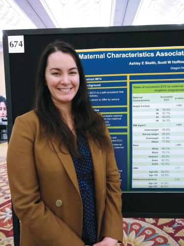
LAS VEGAS – according to data shared in a poster session at the meeting sponsored by the Society for Maternal-Fetal Medicine.
Race/ethnicity also had an impact on the likelihood that external cephalic version (ECV) would be successful.
In an interview, first author Ashley E. Skeith, a medical student at Oregon Health and Sciences University, Portland, said that various characteristics of a pregnancy may affect the success of ECV, but it wasn’t known which maternal characteristics might be associated with greater success of the maneuver.
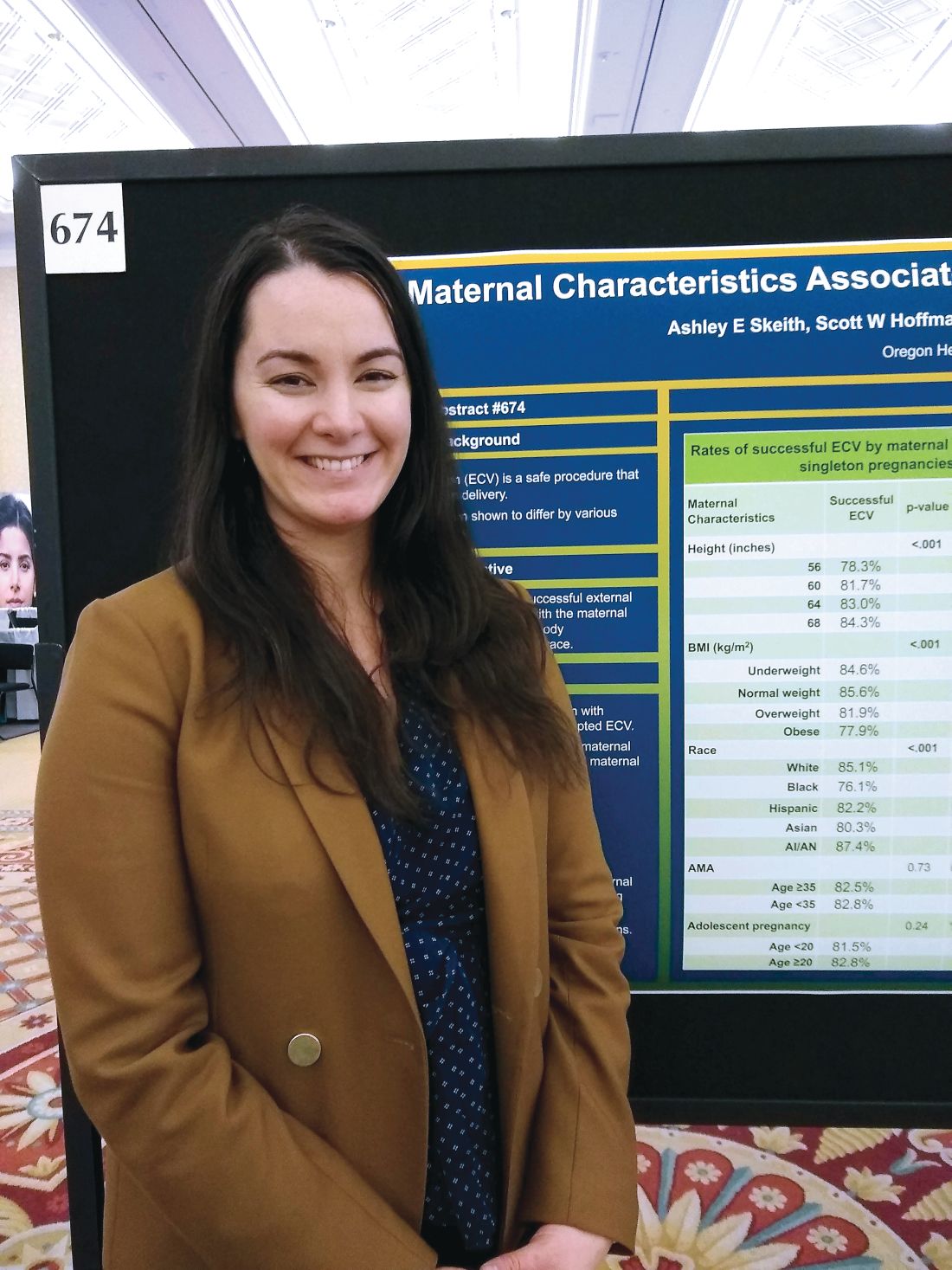
She and her collaborators found that rates of success were high overall, but that 84% of women 68 inches or taller had successful ECVs, compared with 78% of women less than 60 inches tall. Rates were 82% and 83% for women 60-64 inches and 64-68 inches tall, respectively (adjusted odds ratio, 1.03; P less than .001).
The retrospective cohort study used data from 18,896 women who had singleton, breech, term gestations for whom ECV was attempted. Variables extracted from the medical record included maternal age, height, race, and prepregnancy body mass index (BMI).
For analysis, maternal BMI was grouped into four categories: underweight (BMI, less than 18.5 kg/m2), normal weight (BMI, 18.5-24.9 kg/m2), overweight (BMI, 25-29 kg/m2), and obese (BMI, greater than 30 kg/m2).
Women who were normal weight had the highest likelihood of a successful ECV, at 86%, followed by underweight women at 85%. Women who were overweight and obese had lower success rates, at 82% and 78%, respectively (aOR, 0.86; P less than .001).
Compared with white women, black women had an aOR of 0.60 for successful ECV (P less than .001). The aOR for successful ECV for Asian women was 0.71; for Hispanic women, the aOR was 0.82. American Indian and Alaska Native women were slightly more likely to have successful ECV than white women, but the difference was not significant after statistical adjustment.
Neither advanced maternal age (greater than 35 years) nor adolescent pregnancy were associated with decreased likelihood of successful ECV.
Potential confounders included maternal education level and insurance status, how much weight was gained during pregnancy, whether an epidural was administered, and whether the mother had diabetes. Multivariable regression analysis adjusted for these variables, said Ms. Skeith.
“External cephalic version is a safe procedure that reduces risk of cesarean delivery,” wrote Ms. Skeith and her colleagues. “Though fetal positioning and analgesia have been considered in the prediction of ECV success, [these] data [suggest] that maternal stature and race/ethnicity could be incorporated into potential prediction tools.”
Ms. Skeith reported no outside sources of funding or conflicts of interest.
SOURCE: Skeith AE et al. Am J Obstet Gynecol. 2019 Jan;220(1):S445-7, Abstract 674.
LAS VEGAS – according to data shared in a poster session at the meeting sponsored by the Society for Maternal-Fetal Medicine.
Race/ethnicity also had an impact on the likelihood that external cephalic version (ECV) would be successful.
In an interview, first author Ashley E. Skeith, a medical student at Oregon Health and Sciences University, Portland, said that various characteristics of a pregnancy may affect the success of ECV, but it wasn’t known which maternal characteristics might be associated with greater success of the maneuver.
She and her collaborators found that rates of success were high overall, but that 84% of women 68 inches or taller had successful ECVs, compared with 78% of women less than 60 inches tall. Rates were 82% and 83% for women 60-64 inches and 64-68 inches tall, respectively (adjusted odds ratio, 1.03; P less than .001).
The retrospective cohort study used data from 18,896 women who had singleton, breech, term gestations for whom ECV was attempted. Variables extracted from the medical record included maternal age, height, race, and prepregnancy body mass index (BMI).
For analysis, maternal BMI was grouped into four categories: underweight (BMI, less than 18.5 kg/m2), normal weight (BMI, 18.5-24.9 kg/m2), overweight (BMI, 25-29 kg/m2), and obese (BMI, greater than 30 kg/m2).
Women who were normal weight had the highest likelihood of a successful ECV, at 86%, followed by underweight women at 85%. Women who were overweight and obese had lower success rates, at 82% and 78%, respectively (aOR, 0.86; P less than .001).
Compared with white women, black women had an aOR of 0.60 for successful ECV (P less than .001). The aOR for successful ECV for Asian women was 0.71; for Hispanic women, the aOR was 0.82. American Indian and Alaska Native women were slightly more likely to have successful ECV than white women, but the difference was not significant after statistical adjustment.
Neither advanced maternal age (greater than 35 years) nor adolescent pregnancy were associated with decreased likelihood of successful ECV.
Potential confounders included maternal education level and insurance status, how much weight was gained during pregnancy, whether an epidural was administered, and whether the mother had diabetes. Multivariable regression analysis adjusted for these variables, said Ms. Skeith.
“External cephalic version is a safe procedure that reduces risk of cesarean delivery,” wrote Ms. Skeith and her colleagues. “Though fetal positioning and analgesia have been considered in the prediction of ECV success, [these] data [suggest] that maternal stature and race/ethnicity could be incorporated into potential prediction tools.”
Ms. Skeith reported no outside sources of funding or conflicts of interest.
SOURCE: Skeith AE et al. Am J Obstet Gynecol. 2019 Jan;220(1):S445-7, Abstract 674.
LAS VEGAS – according to data shared in a poster session at the meeting sponsored by the Society for Maternal-Fetal Medicine.
Race/ethnicity also had an impact on the likelihood that external cephalic version (ECV) would be successful.
In an interview, first author Ashley E. Skeith, a medical student at Oregon Health and Sciences University, Portland, said that various characteristics of a pregnancy may affect the success of ECV, but it wasn’t known which maternal characteristics might be associated with greater success of the maneuver.
She and her collaborators found that rates of success were high overall, but that 84% of women 68 inches or taller had successful ECVs, compared with 78% of women less than 60 inches tall. Rates were 82% and 83% for women 60-64 inches and 64-68 inches tall, respectively (adjusted odds ratio, 1.03; P less than .001).
The retrospective cohort study used data from 18,896 women who had singleton, breech, term gestations for whom ECV was attempted. Variables extracted from the medical record included maternal age, height, race, and prepregnancy body mass index (BMI).
For analysis, maternal BMI was grouped into four categories: underweight (BMI, less than 18.5 kg/m2), normal weight (BMI, 18.5-24.9 kg/m2), overweight (BMI, 25-29 kg/m2), and obese (BMI, greater than 30 kg/m2).
Women who were normal weight had the highest likelihood of a successful ECV, at 86%, followed by underweight women at 85%. Women who were overweight and obese had lower success rates, at 82% and 78%, respectively (aOR, 0.86; P less than .001).
Compared with white women, black women had an aOR of 0.60 for successful ECV (P less than .001). The aOR for successful ECV for Asian women was 0.71; for Hispanic women, the aOR was 0.82. American Indian and Alaska Native women were slightly more likely to have successful ECV than white women, but the difference was not significant after statistical adjustment.
Neither advanced maternal age (greater than 35 years) nor adolescent pregnancy were associated with decreased likelihood of successful ECV.
Potential confounders included maternal education level and insurance status, how much weight was gained during pregnancy, whether an epidural was administered, and whether the mother had diabetes. Multivariable regression analysis adjusted for these variables, said Ms. Skeith.
“External cephalic version is a safe procedure that reduces risk of cesarean delivery,” wrote Ms. Skeith and her colleagues. “Though fetal positioning and analgesia have been considered in the prediction of ECV success, [these] data [suggest] that maternal stature and race/ethnicity could be incorporated into potential prediction tools.”
Ms. Skeith reported no outside sources of funding or conflicts of interest.
SOURCE: Skeith AE et al. Am J Obstet Gynecol. 2019 Jan;220(1):S445-7, Abstract 674.
REPORTING FROM THE PREGNANCY MEETING
Optical coherence tomography emerging as a promising biomarker for MS
DALLAS – Optical coherence tomography (OCT) has emerged as a promising biomarker in multiple sclerosis.

Thanks to OCT, clinicians are gaining an improved understanding of how MS affects certain eye structures. An optical analogue of ultrasound B mode imaging, OCT achieves a resolution of about 3-6 microns with commercially available devices. “That allows us to quantify the layers of the retina with quite a degree of accuracy,” Shiv Saidha, MD, said at ACTRIMS Forum 2019, the meeting held by the Americas Committee for Treatment and Research in Multiple Sclerosis.
At postmortem, up to 99% of MS patients have demyelinated plaques in their optic nerves. “This implies that optic neuropathy is an ubiquitous phenomenon as part of the MS disease process,” said Dr. Saidha, a neurologist at Johns Hopkins University, Baltimore. “The prevailing hypothesis is that there is demyelination or axonal transection related to acute inflammation that occurs within the optic nerve. There’s a retrograde degeneration of its constituent axons, and that results in thinning of the inner retinal nerve fiber layer as well as the neuronal derivative of this layer called the ganglion cell layer. In addition to neurodegenerative mechanisms in the retina, there is also perivascular inflammation, called retinal periphlebitis, which we know occurs in about 20% of MS patients. At postmortem, there are also activate microglia present within the retina of MS patients.”
One of the principal findings of OCT in MS to date is that the retinal nerve fiber layer (RNFL) and ganglion cell plus inner plexiform layer (GCIP) thinning reflects MS-related optic nerve neurodegeneration. In addition, RNFL and GCIP thinning occur after optic neuritis and also as part of the MS disease course in eyes without a history of optic neuritis. “RNFL and GCIP thinning in MS are clinically relevant and correlate with visual function, global disability, and brain atrophy,” Dr. Saidha said. Researchers have also found that rates of GCIP thinning are accelerated in MS patients exhibiting clinical and/or radiological disease activity and are altered by disease-modifying therapies, and that increased inner nuclear layer (INL) thickness correlates with T2 lesion volume and predicts clinical and radiological disease activity. “In numerous trials of putatively neuroprotective and restorative treatments, we see OCT incorporated more and more, either as a secondary or a primary outcome,” he said.
Predicting disability and brain atrophy
In a study expected to be appear in a forthcoming issue of the Annals of Translational and Clinical Neurology, colleagues of Dr. Saidha found that OCT derived retinal layer measurements and visual function predict disability at 10 years in patients with MS. The researchers used an earlier generation, lower quality OCT device to examine tertiles of total macular volume, “an old, nonspecific composite measure of all of the retinal components,” he explained. “Even with inferior technology, a single measurement at a point in time not only could predict the change in EDSS [Expanded Disability Status Scale] scores from baseline to 10 years, but the accumulation of meaningful disability.”
In an earlier study, Dr. Saidha and his colleagues conducted a 4-year study of OCT and MRI in MS (Ann Neurol 2015; 78[5]:801-13). It consisted of six monthly spectral domain OCT scans (including automated intra-retinal segmentation) and baseline and annual 3 T brain MRI (including substructure volumetrics). Patients with ocular relapses (optic neuritis) during the study were excluded. The researchers correlated individual-specific rates of change in retinal and brain measurements, adjusting for age, sex, disease duration, and optic neuritis history. They found that cerebral volume fraction (analogous to whole brain volume) “had a decent correlation between rates of GCIP atrophy and rates of whole brain volume loss,” he said. “That was predominately driven by cortical gray matter atrophy.”
Measuring effects of disease-modifying therapies
What about the effects of disease-modifying therapies? According to Dr. Saidha, there has been a paucity of studies assessing the effects of DMTs on retinal layer thickness, and they are limited by small patient numbers, cross-sectional design, and/or short periods of observation. In a retrospective analysis, he and his associates examined the effects of treatments in relapsing-remitting MS patients at his center who underwent OCT (Neurology. 2017;88[6]:525-32). Over a mean 3 years of follow-up, they examined the effects of glatiramer acetate (Copaxone), natalizumab (Tysabri), and interferon beta-1a subcutaneously (Rebif) and intramuscularly (Avonex). They adjusted for gap time, which is the interval between when a patient started a treatment and when they started to undergo retinal observation with OCT. “This is to try to account for some of the biological changes that might have occurred during that period of time,” he explained. The researchers observed that rates of GCIP atrophy as well as other retinal measures were significantly lower in people treated with natalizumab, relative to all other DMTs. “What I found fascinating was the rate of GCIP atrophy of those on natalizumab was basically the same as healthy controls,” Dr. Saidha said. “It didn’t differ.”
Retinal inflammation and treatment’s impact
Significant inflammation in the unmyelinated retina may inform clinicians about other aspects of MS, he continued. For example, retinal periphlebitis occurs in about 20% of MS patients and may be a marker of CNS inflammation. In addition, intermediate uveitis occurs in about 16% of MS patients, and postmortem studies reveal retinal inflammation with microglia. Specifically, macular microcystoid changes occur in the eyes of about 5% of MS patients and may represent a breakdown of the blood-retinal barrier and inflammation. “Since it’s a dynamic process, increased thickness of the INL in the absence of visible microcystoid changes might occur,” Dr. Saidha said. “We found that baseline INL thickness is predictive of clinico-radiologic disease activity.”
In a separate analysis of 108 MS patients and 40 healthy controls, German researchers evaluated the impact of DMTs on INL volume (Brain. 2016;11[1]:2855-63). They found that higher baseline INL volume correlated with new T2 and GAD lesions over 1 year. The reduction in INL volume was significantly associated with reduced activity, and overall, DMTs reduced INL volume over 6 months. Patients who were not treated, or who were treated and did not achieve NEDA-3 (no evidence of disease activity) did not show reductions in INL volume. They concluded that INL volume might be a novel outcome of DMT treatment.
Finding prognostic and diagnostic biomarkers
In an ongoing multisite study, Dr. Saidha and his colleagues are assessing the use of OCT in patients with progressive MS (including 186 patients from Johns Hopkins), and also determining if OCT changes differ over time between relapsing-remitting MS and different subtypes of progressive MS. So far, they have found that progressive MS is associated with accelerated inner and in particular outer layer retinal atrophy. “Although this is a decent-sized cohort, at this stage I’m not sure I would definitively say that these novel retinal biomarkers have utility specific to progressive MS, but I’m very excited about it,” he said. “The goal is to take a much deeper look at this.”
Findings from a large collaborative IMSVISUAL inter-eye asymmetry study showed that peripapillary RNFL and ganglion cell–inner plexiform layer inter-eye differences of 5 microns, respectively, were optimal for identifying patients with prior unilateral optic neuritis in the MS cohort. “In the future, the possibility of using OCT to identify subclinical optic neuropathy so we can define when a lesion is present in the optic nerve has huge diagnostic implications for MS, because the optic nerve is not currently recognized as a lesion site in current MS diagnostic criteria,” Dr. Saidha said.
Dr. Saidha disclosed that he has served on scientific advisory boards for Biogen, Genzyme, Genentech, EMD Serono, and Novartis. He has also received consulting fees from JuneBrain LLC and is the site investigator of a trial sponsored by MedDay Pharmaceuticals.
DALLAS – Optical coherence tomography (OCT) has emerged as a promising biomarker in multiple sclerosis.
Thanks to OCT, clinicians are gaining an improved understanding of how MS affects certain eye structures. An optical analogue of ultrasound B mode imaging, OCT achieves a resolution of about 3-6 microns with commercially available devices. “That allows us to quantify the layers of the retina with quite a degree of accuracy,” Shiv Saidha, MD, said at ACTRIMS Forum 2019, the meeting held by the Americas Committee for Treatment and Research in Multiple Sclerosis.
At postmortem, up to 99% of MS patients have demyelinated plaques in their optic nerves. “This implies that optic neuropathy is an ubiquitous phenomenon as part of the MS disease process,” said Dr. Saidha, a neurologist at Johns Hopkins University, Baltimore. “The prevailing hypothesis is that there is demyelination or axonal transection related to acute inflammation that occurs within the optic nerve. There’s a retrograde degeneration of its constituent axons, and that results in thinning of the inner retinal nerve fiber layer as well as the neuronal derivative of this layer called the ganglion cell layer. In addition to neurodegenerative mechanisms in the retina, there is also perivascular inflammation, called retinal periphlebitis, which we know occurs in about 20% of MS patients. At postmortem, there are also activate microglia present within the retina of MS patients.”
One of the principal findings of OCT in MS to date is that the retinal nerve fiber layer (RNFL) and ganglion cell plus inner plexiform layer (GCIP) thinning reflects MS-related optic nerve neurodegeneration. In addition, RNFL and GCIP thinning occur after optic neuritis and also as part of the MS disease course in eyes without a history of optic neuritis. “RNFL and GCIP thinning in MS are clinically relevant and correlate with visual function, global disability, and brain atrophy,” Dr. Saidha said. Researchers have also found that rates of GCIP thinning are accelerated in MS patients exhibiting clinical and/or radiological disease activity and are altered by disease-modifying therapies, and that increased inner nuclear layer (INL) thickness correlates with T2 lesion volume and predicts clinical and radiological disease activity. “In numerous trials of putatively neuroprotective and restorative treatments, we see OCT incorporated more and more, either as a secondary or a primary outcome,” he said.
Predicting disability and brain atrophy
In a study expected to be appear in a forthcoming issue of the Annals of Translational and Clinical Neurology, colleagues of Dr. Saidha found that OCT derived retinal layer measurements and visual function predict disability at 10 years in patients with MS. The researchers used an earlier generation, lower quality OCT device to examine tertiles of total macular volume, “an old, nonspecific composite measure of all of the retinal components,” he explained. “Even with inferior technology, a single measurement at a point in time not only could predict the change in EDSS [Expanded Disability Status Scale] scores from baseline to 10 years, but the accumulation of meaningful disability.”
In an earlier study, Dr. Saidha and his colleagues conducted a 4-year study of OCT and MRI in MS (Ann Neurol 2015; 78[5]:801-13). It consisted of six monthly spectral domain OCT scans (including automated intra-retinal segmentation) and baseline and annual 3 T brain MRI (including substructure volumetrics). Patients with ocular relapses (optic neuritis) during the study were excluded. The researchers correlated individual-specific rates of change in retinal and brain measurements, adjusting for age, sex, disease duration, and optic neuritis history. They found that cerebral volume fraction (analogous to whole brain volume) “had a decent correlation between rates of GCIP atrophy and rates of whole brain volume loss,” he said. “That was predominately driven by cortical gray matter atrophy.”
Measuring effects of disease-modifying therapies
What about the effects of disease-modifying therapies? According to Dr. Saidha, there has been a paucity of studies assessing the effects of DMTs on retinal layer thickness, and they are limited by small patient numbers, cross-sectional design, and/or short periods of observation. In a retrospective analysis, he and his associates examined the effects of treatments in relapsing-remitting MS patients at his center who underwent OCT (Neurology. 2017;88[6]:525-32). Over a mean 3 years of follow-up, they examined the effects of glatiramer acetate (Copaxone), natalizumab (Tysabri), and interferon beta-1a subcutaneously (Rebif) and intramuscularly (Avonex). They adjusted for gap time, which is the interval between when a patient started a treatment and when they started to undergo retinal observation with OCT. “This is to try to account for some of the biological changes that might have occurred during that period of time,” he explained. The researchers observed that rates of GCIP atrophy as well as other retinal measures were significantly lower in people treated with natalizumab, relative to all other DMTs. “What I found fascinating was the rate of GCIP atrophy of those on natalizumab was basically the same as healthy controls,” Dr. Saidha said. “It didn’t differ.”
Retinal inflammation and treatment’s impact
Significant inflammation in the unmyelinated retina may inform clinicians about other aspects of MS, he continued. For example, retinal periphlebitis occurs in about 20% of MS patients and may be a marker of CNS inflammation. In addition, intermediate uveitis occurs in about 16% of MS patients, and postmortem studies reveal retinal inflammation with microglia. Specifically, macular microcystoid changes occur in the eyes of about 5% of MS patients and may represent a breakdown of the blood-retinal barrier and inflammation. “Since it’s a dynamic process, increased thickness of the INL in the absence of visible microcystoid changes might occur,” Dr. Saidha said. “We found that baseline INL thickness is predictive of clinico-radiologic disease activity.”
In a separate analysis of 108 MS patients and 40 healthy controls, German researchers evaluated the impact of DMTs on INL volume (Brain. 2016;11[1]:2855-63). They found that higher baseline INL volume correlated with new T2 and GAD lesions over 1 year. The reduction in INL volume was significantly associated with reduced activity, and overall, DMTs reduced INL volume over 6 months. Patients who were not treated, or who were treated and did not achieve NEDA-3 (no evidence of disease activity) did not show reductions in INL volume. They concluded that INL volume might be a novel outcome of DMT treatment.
Finding prognostic and diagnostic biomarkers
In an ongoing multisite study, Dr. Saidha and his colleagues are assessing the use of OCT in patients with progressive MS (including 186 patients from Johns Hopkins), and also determining if OCT changes differ over time between relapsing-remitting MS and different subtypes of progressive MS. So far, they have found that progressive MS is associated with accelerated inner and in particular outer layer retinal atrophy. “Although this is a decent-sized cohort, at this stage I’m not sure I would definitively say that these novel retinal biomarkers have utility specific to progressive MS, but I’m very excited about it,” he said. “The goal is to take a much deeper look at this.”
Findings from a large collaborative IMSVISUAL inter-eye asymmetry study showed that peripapillary RNFL and ganglion cell–inner plexiform layer inter-eye differences of 5 microns, respectively, were optimal for identifying patients with prior unilateral optic neuritis in the MS cohort. “In the future, the possibility of using OCT to identify subclinical optic neuropathy so we can define when a lesion is present in the optic nerve has huge diagnostic implications for MS, because the optic nerve is not currently recognized as a lesion site in current MS diagnostic criteria,” Dr. Saidha said.
Dr. Saidha disclosed that he has served on scientific advisory boards for Biogen, Genzyme, Genentech, EMD Serono, and Novartis. He has also received consulting fees from JuneBrain LLC and is the site investigator of a trial sponsored by MedDay Pharmaceuticals.
DALLAS – Optical coherence tomography (OCT) has emerged as a promising biomarker in multiple sclerosis.
Thanks to OCT, clinicians are gaining an improved understanding of how MS affects certain eye structures. An optical analogue of ultrasound B mode imaging, OCT achieves a resolution of about 3-6 microns with commercially available devices. “That allows us to quantify the layers of the retina with quite a degree of accuracy,” Shiv Saidha, MD, said at ACTRIMS Forum 2019, the meeting held by the Americas Committee for Treatment and Research in Multiple Sclerosis.
At postmortem, up to 99% of MS patients have demyelinated plaques in their optic nerves. “This implies that optic neuropathy is an ubiquitous phenomenon as part of the MS disease process,” said Dr. Saidha, a neurologist at Johns Hopkins University, Baltimore. “The prevailing hypothesis is that there is demyelination or axonal transection related to acute inflammation that occurs within the optic nerve. There’s a retrograde degeneration of its constituent axons, and that results in thinning of the inner retinal nerve fiber layer as well as the neuronal derivative of this layer called the ganglion cell layer. In addition to neurodegenerative mechanisms in the retina, there is also perivascular inflammation, called retinal periphlebitis, which we know occurs in about 20% of MS patients. At postmortem, there are also activate microglia present within the retina of MS patients.”
One of the principal findings of OCT in MS to date is that the retinal nerve fiber layer (RNFL) and ganglion cell plus inner plexiform layer (GCIP) thinning reflects MS-related optic nerve neurodegeneration. In addition, RNFL and GCIP thinning occur after optic neuritis and also as part of the MS disease course in eyes without a history of optic neuritis. “RNFL and GCIP thinning in MS are clinically relevant and correlate with visual function, global disability, and brain atrophy,” Dr. Saidha said. Researchers have also found that rates of GCIP thinning are accelerated in MS patients exhibiting clinical and/or radiological disease activity and are altered by disease-modifying therapies, and that increased inner nuclear layer (INL) thickness correlates with T2 lesion volume and predicts clinical and radiological disease activity. “In numerous trials of putatively neuroprotective and restorative treatments, we see OCT incorporated more and more, either as a secondary or a primary outcome,” he said.
Predicting disability and brain atrophy
In a study expected to be appear in a forthcoming issue of the Annals of Translational and Clinical Neurology, colleagues of Dr. Saidha found that OCT derived retinal layer measurements and visual function predict disability at 10 years in patients with MS. The researchers used an earlier generation, lower quality OCT device to examine tertiles of total macular volume, “an old, nonspecific composite measure of all of the retinal components,” he explained. “Even with inferior technology, a single measurement at a point in time not only could predict the change in EDSS [Expanded Disability Status Scale] scores from baseline to 10 years, but the accumulation of meaningful disability.”
In an earlier study, Dr. Saidha and his colleagues conducted a 4-year study of OCT and MRI in MS (Ann Neurol 2015; 78[5]:801-13). It consisted of six monthly spectral domain OCT scans (including automated intra-retinal segmentation) and baseline and annual 3 T brain MRI (including substructure volumetrics). Patients with ocular relapses (optic neuritis) during the study were excluded. The researchers correlated individual-specific rates of change in retinal and brain measurements, adjusting for age, sex, disease duration, and optic neuritis history. They found that cerebral volume fraction (analogous to whole brain volume) “had a decent correlation between rates of GCIP atrophy and rates of whole brain volume loss,” he said. “That was predominately driven by cortical gray matter atrophy.”
Measuring effects of disease-modifying therapies
What about the effects of disease-modifying therapies? According to Dr. Saidha, there has been a paucity of studies assessing the effects of DMTs on retinal layer thickness, and they are limited by small patient numbers, cross-sectional design, and/or short periods of observation. In a retrospective analysis, he and his associates examined the effects of treatments in relapsing-remitting MS patients at his center who underwent OCT (Neurology. 2017;88[6]:525-32). Over a mean 3 years of follow-up, they examined the effects of glatiramer acetate (Copaxone), natalizumab (Tysabri), and interferon beta-1a subcutaneously (Rebif) and intramuscularly (Avonex). They adjusted for gap time, which is the interval between when a patient started a treatment and when they started to undergo retinal observation with OCT. “This is to try to account for some of the biological changes that might have occurred during that period of time,” he explained. The researchers observed that rates of GCIP atrophy as well as other retinal measures were significantly lower in people treated with natalizumab, relative to all other DMTs. “What I found fascinating was the rate of GCIP atrophy of those on natalizumab was basically the same as healthy controls,” Dr. Saidha said. “It didn’t differ.”
Retinal inflammation and treatment’s impact
Significant inflammation in the unmyelinated retina may inform clinicians about other aspects of MS, he continued. For example, retinal periphlebitis occurs in about 20% of MS patients and may be a marker of CNS inflammation. In addition, intermediate uveitis occurs in about 16% of MS patients, and postmortem studies reveal retinal inflammation with microglia. Specifically, macular microcystoid changes occur in the eyes of about 5% of MS patients and may represent a breakdown of the blood-retinal barrier and inflammation. “Since it’s a dynamic process, increased thickness of the INL in the absence of visible microcystoid changes might occur,” Dr. Saidha said. “We found that baseline INL thickness is predictive of clinico-radiologic disease activity.”
In a separate analysis of 108 MS patients and 40 healthy controls, German researchers evaluated the impact of DMTs on INL volume (Brain. 2016;11[1]:2855-63). They found that higher baseline INL volume correlated with new T2 and GAD lesions over 1 year. The reduction in INL volume was significantly associated with reduced activity, and overall, DMTs reduced INL volume over 6 months. Patients who were not treated, or who were treated and did not achieve NEDA-3 (no evidence of disease activity) did not show reductions in INL volume. They concluded that INL volume might be a novel outcome of DMT treatment.
Finding prognostic and diagnostic biomarkers
In an ongoing multisite study, Dr. Saidha and his colleagues are assessing the use of OCT in patients with progressive MS (including 186 patients from Johns Hopkins), and also determining if OCT changes differ over time between relapsing-remitting MS and different subtypes of progressive MS. So far, they have found that progressive MS is associated with accelerated inner and in particular outer layer retinal atrophy. “Although this is a decent-sized cohort, at this stage I’m not sure I would definitively say that these novel retinal biomarkers have utility specific to progressive MS, but I’m very excited about it,” he said. “The goal is to take a much deeper look at this.”
Findings from a large collaborative IMSVISUAL inter-eye asymmetry study showed that peripapillary RNFL and ganglion cell–inner plexiform layer inter-eye differences of 5 microns, respectively, were optimal for identifying patients with prior unilateral optic neuritis in the MS cohort. “In the future, the possibility of using OCT to identify subclinical optic neuropathy so we can define when a lesion is present in the optic nerve has huge diagnostic implications for MS, because the optic nerve is not currently recognized as a lesion site in current MS diagnostic criteria,” Dr. Saidha said.
Dr. Saidha disclosed that he has served on scientific advisory boards for Biogen, Genzyme, Genentech, EMD Serono, and Novartis. He has also received consulting fees from JuneBrain LLC and is the site investigator of a trial sponsored by MedDay Pharmaceuticals.
EXPERT ANALYSIS FROM ACTRIMS FORUM 2019

Knotless, absorbable sutures best staples for postcesarean skin closure
LAS VEGAS – compared with staples, in a single-site, retrospective study.
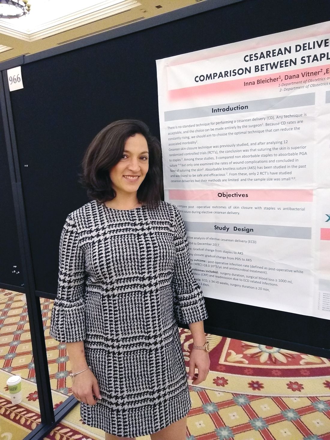
For women whose skin incisions were closed with knotless sutures, mean surgical time was 38 minutes; for women who received a staple closure, mean surgical time was 44 minutes (P less than .001). Also, fewer women whose incisions were closed with knotless sutures experienced surgical bleeding greater than 1,000 mL, compared with those who received staples (0.3% vs. 3.0%; P less than .001).
Two previous randomized, controlled trials comparing knotless sutures with staples for skin closure after cesarean delivery were small and had methodological limitations, Inna Bleicher, MD, said in an interview during a poster session at the meeting sponsored by the Society for Maternal-Fetal Medicine.
Dr. Bleicher and her colleagues reviewed records from 2,173 elective cesarean deliveries over a period of 4 years. Absorbable, antibacterial, knotless sutures were used for closure for 1,172 women, while staples were used for the remaining 1,001 women.
Over the study period, Dr. Bleicher noted that there was a gradual transition from the use of staples to absorbable, knotless sutures, which also were increasingly used for the hysterotomy closure. She added that, in conversation with peers at Bnai-Zion Medical Center, Haifa, Israel, where she practices as an ob.gyn, she’s found that physicians find the sutures easy and quick to use, because the sutures are double ended, allowing the possibility for two operators to work together in wound closure.
The study’s primary outcome measure was the rate of postoperative infection, defined as postoperative white blood count greater than 18,000 per microliter and antimicrobial treatment. Secondary outcome measures included C-reactive protein levels, hospital readmission for infection related to the delivery, duration of surgery, and surgical blood loss estimated at 1,000 mL or more.
A higher proportion of women in the staple closure group than the knotless suture group required postsurgical antibiotic treatment (11% vs. 10%), but this difference didn’t reach statistical significance (P = .243).
There were no significant differences in the groups in terms of maternal age (about 32 years), or gestational age at delivery (about 39 weeks).
“Our results suggest that cesarean scar skin closure with antibacterial knotless sutures did not increase, and may even reduce, the rates of postoperative infection, morbidity, surgical blood loss, and may shorten operation time,” wrote Dr. Bleicher and her colleagues.
Dr. Bleicher reported no outside sources of funding and no conflicts of interest.
SOURCE: Bleicher I et al. Am J Obstet Gynecol. 2019 Jan. 220;1:S622, Abstract 966.
LAS VEGAS – compared with staples, in a single-site, retrospective study.
For women whose skin incisions were closed with knotless sutures, mean surgical time was 38 minutes; for women who received a staple closure, mean surgical time was 44 minutes (P less than .001). Also, fewer women whose incisions were closed with knotless sutures experienced surgical bleeding greater than 1,000 mL, compared with those who received staples (0.3% vs. 3.0%; P less than .001).
Two previous randomized, controlled trials comparing knotless sutures with staples for skin closure after cesarean delivery were small and had methodological limitations, Inna Bleicher, MD, said in an interview during a poster session at the meeting sponsored by the Society for Maternal-Fetal Medicine.
Dr. Bleicher and her colleagues reviewed records from 2,173 elective cesarean deliveries over a period of 4 years. Absorbable, antibacterial, knotless sutures were used for closure for 1,172 women, while staples were used for the remaining 1,001 women.
Over the study period, Dr. Bleicher noted that there was a gradual transition from the use of staples to absorbable, knotless sutures, which also were increasingly used for the hysterotomy closure. She added that, in conversation with peers at Bnai-Zion Medical Center, Haifa, Israel, where she practices as an ob.gyn, she’s found that physicians find the sutures easy and quick to use, because the sutures are double ended, allowing the possibility for two operators to work together in wound closure.
The study’s primary outcome measure was the rate of postoperative infection, defined as postoperative white blood count greater than 18,000 per microliter and antimicrobial treatment. Secondary outcome measures included C-reactive protein levels, hospital readmission for infection related to the delivery, duration of surgery, and surgical blood loss estimated at 1,000 mL or more.
A higher proportion of women in the staple closure group than the knotless suture group required postsurgical antibiotic treatment (11% vs. 10%), but this difference didn’t reach statistical significance (P = .243).
There were no significant differences in the groups in terms of maternal age (about 32 years), or gestational age at delivery (about 39 weeks).
“Our results suggest that cesarean scar skin closure with antibacterial knotless sutures did not increase, and may even reduce, the rates of postoperative infection, morbidity, surgical blood loss, and may shorten operation time,” wrote Dr. Bleicher and her colleagues.
Dr. Bleicher reported no outside sources of funding and no conflicts of interest.
SOURCE: Bleicher I et al. Am J Obstet Gynecol. 2019 Jan. 220;1:S622, Abstract 966.
LAS VEGAS – compared with staples, in a single-site, retrospective study.
For women whose skin incisions were closed with knotless sutures, mean surgical time was 38 minutes; for women who received a staple closure, mean surgical time was 44 minutes (P less than .001). Also, fewer women whose incisions were closed with knotless sutures experienced surgical bleeding greater than 1,000 mL, compared with those who received staples (0.3% vs. 3.0%; P less than .001).
Two previous randomized, controlled trials comparing knotless sutures with staples for skin closure after cesarean delivery were small and had methodological limitations, Inna Bleicher, MD, said in an interview during a poster session at the meeting sponsored by the Society for Maternal-Fetal Medicine.
Dr. Bleicher and her colleagues reviewed records from 2,173 elective cesarean deliveries over a period of 4 years. Absorbable, antibacterial, knotless sutures were used for closure for 1,172 women, while staples were used for the remaining 1,001 women.
Over the study period, Dr. Bleicher noted that there was a gradual transition from the use of staples to absorbable, knotless sutures, which also were increasingly used for the hysterotomy closure. She added that, in conversation with peers at Bnai-Zion Medical Center, Haifa, Israel, where she practices as an ob.gyn, she’s found that physicians find the sutures easy and quick to use, because the sutures are double ended, allowing the possibility for two operators to work together in wound closure.
The study’s primary outcome measure was the rate of postoperative infection, defined as postoperative white blood count greater than 18,000 per microliter and antimicrobial treatment. Secondary outcome measures included C-reactive protein levels, hospital readmission for infection related to the delivery, duration of surgery, and surgical blood loss estimated at 1,000 mL or more.
A higher proportion of women in the staple closure group than the knotless suture group required postsurgical antibiotic treatment (11% vs. 10%), but this difference didn’t reach statistical significance (P = .243).
There were no significant differences in the groups in terms of maternal age (about 32 years), or gestational age at delivery (about 39 weeks).
“Our results suggest that cesarean scar skin closure with antibacterial knotless sutures did not increase, and may even reduce, the rates of postoperative infection, morbidity, surgical blood loss, and may shorten operation time,” wrote Dr. Bleicher and her colleagues.
Dr. Bleicher reported no outside sources of funding and no conflicts of interest.
SOURCE: Bleicher I et al. Am J Obstet Gynecol. 2019 Jan. 220;1:S622, Abstract 966.
REPORTING FROM THE PREGNANCY MEETING