User login
Interventions significantly improve NICU immunization rates
according to a study in Pediatrics.

Investigators led by Raymond C. Stetson, MD, of the Mayo Clinic in Rochester, Minn., identified three root causes of underimmunization in a NICU at Mayo Clinic: providers’ lack of knowledge about recommended immunization schedules; immunizations not being ordered when they were due; and parental hesitancy toward vaccination. They addressed these causes with the following five phases of intervention: an intranet resource educating providers about vaccine schedules and dosing intervals; a spreadsheet-based checklist to track and flag immunization status; an intranet resource aimed at discussion with vaccine-hesitant parents; education about safety in providing immunization and review of material from the first three interventions; and education about documentation, including parental consent.
Over the project period, 1,242 infants were discharged or transferred from the NICU. The study included a 6-month “improve phase,” during which interventions were implemented, and a “control phase,” during which the ongoing effects after implementation were observed. At baseline, the rate of fully immunized infants in the NICU was only 56% by time of discharge or transfer, but during the combined improve and control phases, it was 93% with a P value of less than .001.
One of the limitations of the study is that the first three interventions were introduced simultaneously, which makes it hard to determine how much effect each might have had.
“Infants treated in NICUs represent a vulnerable population with the potential for high morbidity and mortality from vaccine-preventable infections,” the investigators wrote. “Our [quality improvement] effort, and others, demonstrate that this population is at risk for underimmunization and that immunization rates can be improved with a small number of interventions. Additionally, we were able to significantly decrease the number of days that immunizations were delayed compared to the routine infant vaccination schedule.”
There was no external funding for the study. One of the coauthors is on safety committees of vaccine studies for Merck. The other authors have no relevant financial disclosures.
SOURCE: Stetson R et al. Pediatr. 2019. doi: 10.1542/peds.2019-0337.
according to a study in Pediatrics.
Investigators led by Raymond C. Stetson, MD, of the Mayo Clinic in Rochester, Minn., identified three root causes of underimmunization in a NICU at Mayo Clinic: providers’ lack of knowledge about recommended immunization schedules; immunizations not being ordered when they were due; and parental hesitancy toward vaccination. They addressed these causes with the following five phases of intervention: an intranet resource educating providers about vaccine schedules and dosing intervals; a spreadsheet-based checklist to track and flag immunization status; an intranet resource aimed at discussion with vaccine-hesitant parents; education about safety in providing immunization and review of material from the first three interventions; and education about documentation, including parental consent.
Over the project period, 1,242 infants were discharged or transferred from the NICU. The study included a 6-month “improve phase,” during which interventions were implemented, and a “control phase,” during which the ongoing effects after implementation were observed. At baseline, the rate of fully immunized infants in the NICU was only 56% by time of discharge or transfer, but during the combined improve and control phases, it was 93% with a P value of less than .001.
One of the limitations of the study is that the first three interventions were introduced simultaneously, which makes it hard to determine how much effect each might have had.
“Infants treated in NICUs represent a vulnerable population with the potential for high morbidity and mortality from vaccine-preventable infections,” the investigators wrote. “Our [quality improvement] effort, and others, demonstrate that this population is at risk for underimmunization and that immunization rates can be improved with a small number of interventions. Additionally, we were able to significantly decrease the number of days that immunizations were delayed compared to the routine infant vaccination schedule.”
There was no external funding for the study. One of the coauthors is on safety committees of vaccine studies for Merck. The other authors have no relevant financial disclosures.
SOURCE: Stetson R et al. Pediatr. 2019. doi: 10.1542/peds.2019-0337.
according to a study in Pediatrics.
Investigators led by Raymond C. Stetson, MD, of the Mayo Clinic in Rochester, Minn., identified three root causes of underimmunization in a NICU at Mayo Clinic: providers’ lack of knowledge about recommended immunization schedules; immunizations not being ordered when they were due; and parental hesitancy toward vaccination. They addressed these causes with the following five phases of intervention: an intranet resource educating providers about vaccine schedules and dosing intervals; a spreadsheet-based checklist to track and flag immunization status; an intranet resource aimed at discussion with vaccine-hesitant parents; education about safety in providing immunization and review of material from the first three interventions; and education about documentation, including parental consent.
Over the project period, 1,242 infants were discharged or transferred from the NICU. The study included a 6-month “improve phase,” during which interventions were implemented, and a “control phase,” during which the ongoing effects after implementation were observed. At baseline, the rate of fully immunized infants in the NICU was only 56% by time of discharge or transfer, but during the combined improve and control phases, it was 93% with a P value of less than .001.
One of the limitations of the study is that the first three interventions were introduced simultaneously, which makes it hard to determine how much effect each might have had.
“Infants treated in NICUs represent a vulnerable population with the potential for high morbidity and mortality from vaccine-preventable infections,” the investigators wrote. “Our [quality improvement] effort, and others, demonstrate that this population is at risk for underimmunization and that immunization rates can be improved with a small number of interventions. Additionally, we were able to significantly decrease the number of days that immunizations were delayed compared to the routine infant vaccination schedule.”
There was no external funding for the study. One of the coauthors is on safety committees of vaccine studies for Merck. The other authors have no relevant financial disclosures.
SOURCE: Stetson R et al. Pediatr. 2019. doi: 10.1542/peds.2019-0337.
FROM PEDIATRICS

Judge rules for insurer in doctor’s allocation lawsuit
A judge has sided with a medical malpractice insurer in a legal challenge that accused the company of misallocating blame among physicians after a liability settlement.
In a Sept. 27 decision, Judge Debra Squires-Lee of the Commonwealth of Massachusetts Superior Court ruled that Medical Professional Mutual Insurance Company (ProMutual) acted reasonably when it settled a medical liability claim for $500,000 against several health providers and allocated responsibility for 30% of the settlement ($150,000) to internist Nataly Minkina, MD. ProMutual was well within its rights and obligations when it settled the underlying claim and did not act in bad faith when assigning responsibility in the case, Judge Squires-Lee wrote in her 49-page ruling.
“At its heart, this case is about a multiple defendant malpractice lawsuit with finger pointing by Dr. Minkina against her codefendants and others, and a disagreement about ProMutual’s ultimate determination about how to allocate a global settlement with the plaintiffs amongst ProMutual’s insureds,” Judge Squires-Lee wrote in the decision. “Dr. Minkina strongly believes that she did not fail [the patient], that she acted reasonably, and that her treatment of [the patient] satisfied the standard of care. She also questions why ProMutual failed to allocate liability in the [patient’s] suit to other physicians. However ... the question for this court is whether ProMutual committed unfair or deceptive acts or practices in its settlement and allocation of its settlement. I conclude that ProMutual did not.”
The case stems from a patient’s lawsuit against Dr. Minkina and several others at Blue Hills Medical Associates in Braintree, Mass. The patient alleged that the health care professionals were responsible for a missed breast cancer diagnosis. Dr. Minkina saw the patient just once in 2002 while covering for another doctor. During the visit, she confirmed some nodularity in the 55-year-old women’s breast and referred her for a mammogram and an ultrasound. A radiologist twice reported no abnormalities, which Dr. Minkina said she relayed to the patient. Dr. Minkina left the practice shortly after.
The patient visited the practice several more times and was referred for another mammogram in 2006, the results of which revealed some signs of malignancy, according to court documents. However, a nurse at the practice misread, misunderstood, or overlooked the signs and recorded that “the benign breast condition had no changes,” according to court transcripts. Later that year, the patient visited the practice complaining of headaches and a droopy eye at which time her primary care physician diagnosed sinusitis and prescribed antibiotics. In 2007, the patient underwent magnetic resonance imaging of the brain and the breast, which revealed widespread metastatic carcinoma. She and her family sued Dr. Minkina and several others in June 2007. The patient died in 2008.
ProMutual settled the case against the defendants for $500,000 in 2008, allocating 30% of the liability to Dr. Minkina, 10% of the nurse practitioner, 60% to the medical practice, and no liability to the other doctors named. ProMutual contended Dr. Minkina bore more responsibility than the other health care professionals named for the delayed diagnosis because of causation factors and standard of care violations, namely that Dr. Minkina should have pursued a biopsy for the patient.
Dr. Minkina sued the insurer in 2012, claiming chiefly that the insurer allocated an unjustifiably high percentage of liability to her because she was no longer insured and because the company had an economic incentive to allocate a disproportionate percentage of responsibility and damages.
A lower court initially dismissed Dr. Minkina’s suit, but the Commonwealth of Massachusetts Appeals Court in 2015 overturned that decision, ruling the case could move forward. In 2018, the superior court agreed Dr. Minkina had a valid bad faith claim, stating that she had provided information about ProMutual’s conduct from which “a reasonable juror could infer the defendant’s bad faith in connection with its settling the underlying malpractice suit, including the allocation of liability.
But Judge Squires-Lee ruled that trial evidence showed that ProMutual did not act for its own benefit or favor other insureds over Dr. Minkina. The judge wrote that the insurer satisfied its contractual and legal obligations when defending the underlying legal claim.
Dr. Minkina said she was disappointed with the ruling, but that she is considering her legal avenues.
“[The] judge’s decision in my case against ProMutual is obviously disappointing, but it is not the first time [the] trial court decided against me in this case,” Dr. Minkina said in an interview. “It is a battle between David and Goliath, [a] single physician against [a] $3.6 billion insurance company with unlimited resources. The decision has just been announced and it is voluminous. I am still reading it and evaluating my options with my attorneys, but it is not the end of the road yet.”
ProMutual declined to comment about the decision.
A judge has sided with a medical malpractice insurer in a legal challenge that accused the company of misallocating blame among physicians after a liability settlement.
In a Sept. 27 decision, Judge Debra Squires-Lee of the Commonwealth of Massachusetts Superior Court ruled that Medical Professional Mutual Insurance Company (ProMutual) acted reasonably when it settled a medical liability claim for $500,000 against several health providers and allocated responsibility for 30% of the settlement ($150,000) to internist Nataly Minkina, MD. ProMutual was well within its rights and obligations when it settled the underlying claim and did not act in bad faith when assigning responsibility in the case, Judge Squires-Lee wrote in her 49-page ruling.
“At its heart, this case is about a multiple defendant malpractice lawsuit with finger pointing by Dr. Minkina against her codefendants and others, and a disagreement about ProMutual’s ultimate determination about how to allocate a global settlement with the plaintiffs amongst ProMutual’s insureds,” Judge Squires-Lee wrote in the decision. “Dr. Minkina strongly believes that she did not fail [the patient], that she acted reasonably, and that her treatment of [the patient] satisfied the standard of care. She also questions why ProMutual failed to allocate liability in the [patient’s] suit to other physicians. However ... the question for this court is whether ProMutual committed unfair or deceptive acts or practices in its settlement and allocation of its settlement. I conclude that ProMutual did not.”
The case stems from a patient’s lawsuit against Dr. Minkina and several others at Blue Hills Medical Associates in Braintree, Mass. The patient alleged that the health care professionals were responsible for a missed breast cancer diagnosis. Dr. Minkina saw the patient just once in 2002 while covering for another doctor. During the visit, she confirmed some nodularity in the 55-year-old women’s breast and referred her for a mammogram and an ultrasound. A radiologist twice reported no abnormalities, which Dr. Minkina said she relayed to the patient. Dr. Minkina left the practice shortly after.
The patient visited the practice several more times and was referred for another mammogram in 2006, the results of which revealed some signs of malignancy, according to court documents. However, a nurse at the practice misread, misunderstood, or overlooked the signs and recorded that “the benign breast condition had no changes,” according to court transcripts. Later that year, the patient visited the practice complaining of headaches and a droopy eye at which time her primary care physician diagnosed sinusitis and prescribed antibiotics. In 2007, the patient underwent magnetic resonance imaging of the brain and the breast, which revealed widespread metastatic carcinoma. She and her family sued Dr. Minkina and several others in June 2007. The patient died in 2008.
ProMutual settled the case against the defendants for $500,000 in 2008, allocating 30% of the liability to Dr. Minkina, 10% of the nurse practitioner, 60% to the medical practice, and no liability to the other doctors named. ProMutual contended Dr. Minkina bore more responsibility than the other health care professionals named for the delayed diagnosis because of causation factors and standard of care violations, namely that Dr. Minkina should have pursued a biopsy for the patient.
Dr. Minkina sued the insurer in 2012, claiming chiefly that the insurer allocated an unjustifiably high percentage of liability to her because she was no longer insured and because the company had an economic incentive to allocate a disproportionate percentage of responsibility and damages.
A lower court initially dismissed Dr. Minkina’s suit, but the Commonwealth of Massachusetts Appeals Court in 2015 overturned that decision, ruling the case could move forward. In 2018, the superior court agreed Dr. Minkina had a valid bad faith claim, stating that she had provided information about ProMutual’s conduct from which “a reasonable juror could infer the defendant’s bad faith in connection with its settling the underlying malpractice suit, including the allocation of liability.
But Judge Squires-Lee ruled that trial evidence showed that ProMutual did not act for its own benefit or favor other insureds over Dr. Minkina. The judge wrote that the insurer satisfied its contractual and legal obligations when defending the underlying legal claim.
Dr. Minkina said she was disappointed with the ruling, but that she is considering her legal avenues.
“[The] judge’s decision in my case against ProMutual is obviously disappointing, but it is not the first time [the] trial court decided against me in this case,” Dr. Minkina said in an interview. “It is a battle between David and Goliath, [a] single physician against [a] $3.6 billion insurance company with unlimited resources. The decision has just been announced and it is voluminous. I am still reading it and evaluating my options with my attorneys, but it is not the end of the road yet.”
ProMutual declined to comment about the decision.
A judge has sided with a medical malpractice insurer in a legal challenge that accused the company of misallocating blame among physicians after a liability settlement.
In a Sept. 27 decision, Judge Debra Squires-Lee of the Commonwealth of Massachusetts Superior Court ruled that Medical Professional Mutual Insurance Company (ProMutual) acted reasonably when it settled a medical liability claim for $500,000 against several health providers and allocated responsibility for 30% of the settlement ($150,000) to internist Nataly Minkina, MD. ProMutual was well within its rights and obligations when it settled the underlying claim and did not act in bad faith when assigning responsibility in the case, Judge Squires-Lee wrote in her 49-page ruling.
“At its heart, this case is about a multiple defendant malpractice lawsuit with finger pointing by Dr. Minkina against her codefendants and others, and a disagreement about ProMutual’s ultimate determination about how to allocate a global settlement with the plaintiffs amongst ProMutual’s insureds,” Judge Squires-Lee wrote in the decision. “Dr. Minkina strongly believes that she did not fail [the patient], that she acted reasonably, and that her treatment of [the patient] satisfied the standard of care. She also questions why ProMutual failed to allocate liability in the [patient’s] suit to other physicians. However ... the question for this court is whether ProMutual committed unfair or deceptive acts or practices in its settlement and allocation of its settlement. I conclude that ProMutual did not.”
The case stems from a patient’s lawsuit against Dr. Minkina and several others at Blue Hills Medical Associates in Braintree, Mass. The patient alleged that the health care professionals were responsible for a missed breast cancer diagnosis. Dr. Minkina saw the patient just once in 2002 while covering for another doctor. During the visit, she confirmed some nodularity in the 55-year-old women’s breast and referred her for a mammogram and an ultrasound. A radiologist twice reported no abnormalities, which Dr. Minkina said she relayed to the patient. Dr. Minkina left the practice shortly after.
The patient visited the practice several more times and was referred for another mammogram in 2006, the results of which revealed some signs of malignancy, according to court documents. However, a nurse at the practice misread, misunderstood, or overlooked the signs and recorded that “the benign breast condition had no changes,” according to court transcripts. Later that year, the patient visited the practice complaining of headaches and a droopy eye at which time her primary care physician diagnosed sinusitis and prescribed antibiotics. In 2007, the patient underwent magnetic resonance imaging of the brain and the breast, which revealed widespread metastatic carcinoma. She and her family sued Dr. Minkina and several others in June 2007. The patient died in 2008.
ProMutual settled the case against the defendants for $500,000 in 2008, allocating 30% of the liability to Dr. Minkina, 10% of the nurse practitioner, 60% to the medical practice, and no liability to the other doctors named. ProMutual contended Dr. Minkina bore more responsibility than the other health care professionals named for the delayed diagnosis because of causation factors and standard of care violations, namely that Dr. Minkina should have pursued a biopsy for the patient.
Dr. Minkina sued the insurer in 2012, claiming chiefly that the insurer allocated an unjustifiably high percentage of liability to her because she was no longer insured and because the company had an economic incentive to allocate a disproportionate percentage of responsibility and damages.
A lower court initially dismissed Dr. Minkina’s suit, but the Commonwealth of Massachusetts Appeals Court in 2015 overturned that decision, ruling the case could move forward. In 2018, the superior court agreed Dr. Minkina had a valid bad faith claim, stating that she had provided information about ProMutual’s conduct from which “a reasonable juror could infer the defendant’s bad faith in connection with its settling the underlying malpractice suit, including the allocation of liability.
But Judge Squires-Lee ruled that trial evidence showed that ProMutual did not act for its own benefit or favor other insureds over Dr. Minkina. The judge wrote that the insurer satisfied its contractual and legal obligations when defending the underlying legal claim.
Dr. Minkina said she was disappointed with the ruling, but that she is considering her legal avenues.
“[The] judge’s decision in my case against ProMutual is obviously disappointing, but it is not the first time [the] trial court decided against me in this case,” Dr. Minkina said in an interview. “It is a battle between David and Goliath, [a] single physician against [a] $3.6 billion insurance company with unlimited resources. The decision has just been announced and it is voluminous. I am still reading it and evaluating my options with my attorneys, but it is not the end of the road yet.”
ProMutual declined to comment about the decision.
White AAV patients post highest mortality rates
Age-adjusted mortality from antineutrophil cytoplasmic autoantibody–associated vasculitides (AAV) in the United States declined by nearly 2% each year between 1999 and 2017, based on data from the Centers for Disease Control and Prevention.
Significant morbidity and mortality are associated with untreated AAV, wrote Alexander W. Steinberg, MD, of Saint Joseph Hospital, Denver, Colo., and colleagues.
“Although population data from the United Kingdom have shown decreased AAV-related mortality during the past 20 years, it is unknown whether this pattern has occurred in the United States,” they wrote.
In a study published in Annals of Internal Medicine, the researchers identified 11,316 AAV-related deaths from 1999 to 2017 in the CDC data.
Overall, age-adjusted mortality was 1.86 per 1,000,000 persons, with highest rates among non-Hispanic whites, men, and residents of the Midwest. Mortality from AAV declined by an average of 1.6% in each year of the study period, and changes in subgroups stratified by gender, race, and geographic region were similar.
Mortality increased with age and was highest among individuals aged 75-84 years, but a significant decline in mortality occurred among individuals aged 65-74 years. “The decrease in overall mortality and mortality among persons aged 65 to 74 years may reflect increased longevity due to improved treatment of AAV and common comorbid conditions,” the researchers said.
“Surprisingly, the authors found much lower age-adjusted mortality rates for non-Hispanic black persons (0.77) and moderately lower mortality rates for Hispanic persons (1.57) than for non-Hispanic white persons (2.03),” wrote John R. Stone, MD, PhD, of Creighton University, Omaha, Neb., in an accompanying editorial.
“Suppose the mortality rate differences reported by Steinberg and colleagues are statistically significant, accurately represent death certificate diagnoses, and match people’s racial/ethnic self-identification. The data then show neither that the vasculitides actually have lower mortality rates in blacks or Hispanics compared with whites, nor that the diseases are indeed less frequent in blacks and Hispanics,” he said. “Rather, these differences probably signify how social inequities, social structural violence, and inferior health care access adversely influence diagnosis of rare diseases and promote health inequity,” Dr. Stone added. The findings suggest that clinicians should remain alert to AAV in some ethnic groups to improve diagnostic accuracy, he said.
“Moreover, improved AAV diagnosis in such groups is key to recruiting participants for research investigating whether therapies should differ among populations,” he emphasized.
The study findings were limited by possible under- or overreporting of AAV on death certificates, but they were strengthened by the large sample size, the researchers noted. “We hope that the mortality patterns presented here can be used to direct future research on the driving forces behind these trends,” they said.
Dr. Steinberg had no financial conflicts to disclose. Dr. Stone had no financial conflicts to disclose.
SOURCES: Steinberg AW et al. Ann Intern Med. 2019 Oct 8. doi: 10.7326/M19-1564; and Stone JR. Ann Intern Med. 2019 Oct 8. doi: 10.7326/M19-2755.
Age-adjusted mortality from antineutrophil cytoplasmic autoantibody–associated vasculitides (AAV) in the United States declined by nearly 2% each year between 1999 and 2017, based on data from the Centers for Disease Control and Prevention.
Significant morbidity and mortality are associated with untreated AAV, wrote Alexander W. Steinberg, MD, of Saint Joseph Hospital, Denver, Colo., and colleagues.
“Although population data from the United Kingdom have shown decreased AAV-related mortality during the past 20 years, it is unknown whether this pattern has occurred in the United States,” they wrote.
In a study published in Annals of Internal Medicine, the researchers identified 11,316 AAV-related deaths from 1999 to 2017 in the CDC data.
Overall, age-adjusted mortality was 1.86 per 1,000,000 persons, with highest rates among non-Hispanic whites, men, and residents of the Midwest. Mortality from AAV declined by an average of 1.6% in each year of the study period, and changes in subgroups stratified by gender, race, and geographic region were similar.
Mortality increased with age and was highest among individuals aged 75-84 years, but a significant decline in mortality occurred among individuals aged 65-74 years. “The decrease in overall mortality and mortality among persons aged 65 to 74 years may reflect increased longevity due to improved treatment of AAV and common comorbid conditions,” the researchers said.
“Surprisingly, the authors found much lower age-adjusted mortality rates for non-Hispanic black persons (0.77) and moderately lower mortality rates for Hispanic persons (1.57) than for non-Hispanic white persons (2.03),” wrote John R. Stone, MD, PhD, of Creighton University, Omaha, Neb., in an accompanying editorial.
“Suppose the mortality rate differences reported by Steinberg and colleagues are statistically significant, accurately represent death certificate diagnoses, and match people’s racial/ethnic self-identification. The data then show neither that the vasculitides actually have lower mortality rates in blacks or Hispanics compared with whites, nor that the diseases are indeed less frequent in blacks and Hispanics,” he said. “Rather, these differences probably signify how social inequities, social structural violence, and inferior health care access adversely influence diagnosis of rare diseases and promote health inequity,” Dr. Stone added. The findings suggest that clinicians should remain alert to AAV in some ethnic groups to improve diagnostic accuracy, he said.
“Moreover, improved AAV diagnosis in such groups is key to recruiting participants for research investigating whether therapies should differ among populations,” he emphasized.
The study findings were limited by possible under- or overreporting of AAV on death certificates, but they were strengthened by the large sample size, the researchers noted. “We hope that the mortality patterns presented here can be used to direct future research on the driving forces behind these trends,” they said.
Dr. Steinberg had no financial conflicts to disclose. Dr. Stone had no financial conflicts to disclose.
SOURCES: Steinberg AW et al. Ann Intern Med. 2019 Oct 8. doi: 10.7326/M19-1564; and Stone JR. Ann Intern Med. 2019 Oct 8. doi: 10.7326/M19-2755.
Age-adjusted mortality from antineutrophil cytoplasmic autoantibody–associated vasculitides (AAV) in the United States declined by nearly 2% each year between 1999 and 2017, based on data from the Centers for Disease Control and Prevention.
Significant morbidity and mortality are associated with untreated AAV, wrote Alexander W. Steinberg, MD, of Saint Joseph Hospital, Denver, Colo., and colleagues.
“Although population data from the United Kingdom have shown decreased AAV-related mortality during the past 20 years, it is unknown whether this pattern has occurred in the United States,” they wrote.
In a study published in Annals of Internal Medicine, the researchers identified 11,316 AAV-related deaths from 1999 to 2017 in the CDC data.
Overall, age-adjusted mortality was 1.86 per 1,000,000 persons, with highest rates among non-Hispanic whites, men, and residents of the Midwest. Mortality from AAV declined by an average of 1.6% in each year of the study period, and changes in subgroups stratified by gender, race, and geographic region were similar.
Mortality increased with age and was highest among individuals aged 75-84 years, but a significant decline in mortality occurred among individuals aged 65-74 years. “The decrease in overall mortality and mortality among persons aged 65 to 74 years may reflect increased longevity due to improved treatment of AAV and common comorbid conditions,” the researchers said.
“Surprisingly, the authors found much lower age-adjusted mortality rates for non-Hispanic black persons (0.77) and moderately lower mortality rates for Hispanic persons (1.57) than for non-Hispanic white persons (2.03),” wrote John R. Stone, MD, PhD, of Creighton University, Omaha, Neb., in an accompanying editorial.
“Suppose the mortality rate differences reported by Steinberg and colleagues are statistically significant, accurately represent death certificate diagnoses, and match people’s racial/ethnic self-identification. The data then show neither that the vasculitides actually have lower mortality rates in blacks or Hispanics compared with whites, nor that the diseases are indeed less frequent in blacks and Hispanics,” he said. “Rather, these differences probably signify how social inequities, social structural violence, and inferior health care access adversely influence diagnosis of rare diseases and promote health inequity,” Dr. Stone added. The findings suggest that clinicians should remain alert to AAV in some ethnic groups to improve diagnostic accuracy, he said.
“Moreover, improved AAV diagnosis in such groups is key to recruiting participants for research investigating whether therapies should differ among populations,” he emphasized.
The study findings were limited by possible under- or overreporting of AAV on death certificates, but they were strengthened by the large sample size, the researchers noted. “We hope that the mortality patterns presented here can be used to direct future research on the driving forces behind these trends,” they said.
Dr. Steinberg had no financial conflicts to disclose. Dr. Stone had no financial conflicts to disclose.
SOURCES: Steinberg AW et al. Ann Intern Med. 2019 Oct 8. doi: 10.7326/M19-1564; and Stone JR. Ann Intern Med. 2019 Oct 8. doi: 10.7326/M19-2755.
FROM ANNALS OF INTERNAL MEDICINE
Key clinical point: The mortality rate from antineutrophil cytoplasmic autoantibody–associated vasculitides (AAV) in the United States was 1.86 per 1,000,000 individuals from 1999 to 2017.
Major finding: Age-adjusted mortality from AAV decreased by approximately 2% each year between 1999 and 2017.
Study details: The data come from a review of 11,316 AAV-related deaths.
Disclosures: Dr. Steinberg had no financial conflicts to disclose.
Source: Steinberg AW et al. Ann Intern Med. 2019 Oct 8. doi: 10.7326/M19-1564.
Clinician burnout can impact quality of care
Burnout among health care professionals has been associated with lower quality of care, but the effect may be smaller than it seems, based on data from a meta-analysis of more than 200,000 clinicians.
Previous studies have reported associations between burnout and lower quality of care, but a standardized approach to analyze bias in the studies is lacking, wrote Daniel S. Tawfik, MD, of Stanford (Calif.) University and colleagues.
In a study published in the Annals of Internal Medicine, the researchers identified 123 publications from 1994 to 2019 with 142 study populations that included 241,553 health care providers.
Emotional exhaustion was the primary predictor for lower quality of care in 75 study populations, and overall burnout and depersonalization were the primary predictors for 56 and 11 study populations, respectively.
In an analysis of 114 unique burnout-quality combinations, 58 showed effects of burnout related to poor-quality care, 6 showed burnout related to high-quality care, and 50 showed no significant effect. Approximately one-third (33%) of the burnout-quality combinations were reported at least three times. In a review of the 46 burnout-quality combinations with primary effect sizes, 24 showed a significant effect of burnout on poor quality of care, 1 showed a significant effect of burnout on high quality of care, and 21 showed no significant effect.
The researchers also tested study bias using the Ioannidis test and found “an excess of observed versus predicted statistically significant studies (73% observed vs. 62%).”
The findings were limited by several factors, including the use of many cross-sectional, observational studies that could not show causality, the researchers noted. However, the results suggest several implications for future research including the need to consider exaggerated effects and reduce bias.
“Although the effect sizes in the published literature are modestly strong, our finding of excess significance implies that the true magnitude may be smaller than reported, and the studies that attempted to lower the risk of bias demonstrate fewer significant associations than the full evidence base,” the researchers noted.
“Whether curtailing burnout improves quality of care, or whether improving quality of care reduces burnout, is not yet known, and adequately powered and designed randomized trials will be indispensable in answering these questions,” they concluded.
The study was supported by the Stanford Maternal and Child Health Research Institute. Dr. Tawfik disclosed grants from Stanford Maternal and Child Health Research Institute during the study period.
SOURCE: Tawfik DS et al. Ann Intern Med. 2019 Oct 8. doi: 10.7326/M19-1152.
The current meta-analysis is consistent with previous research, but offers nothing new on the relationship between clinician burnout and quality of care, wrote Carolyn S. Dewa, MPH, PhD, Karen Nieuwenhuijsen, PhD, and Jeffrey S. Hoch, PhD, in an accompanying editorial.
Some of the concerns they expressed included variability in the methods used to measure provider burnout, as well as variability in measuring and defining medical error. They suggested that the researchers could have conducted a subgroup analysis based on error definition. “Such analyses might shed light on the types of errors associated with burnout and suggest directions for the design of robust psychometric studies about the error metrics,” they wrote.
The editorialists also expressed concerns about the heterogeneity of the studies included in the review and the potential for confounding. Finally, they noted that the use of observational studies in a meta-analysis can be challenging because “the assessment of observational studies is not straightforward.” They added that knowing the limitations of the studies is important in allowing readers to be confident in the estimates from any meta-analyses.
“Considering the limitations of the available literature, prior reviews, and Tawfik and colleagues’ current meta-analysis, we conclude that higher burnout is associated with lower quality, but we are left without clear answers about the magnitude or clinical significance of the relationship,” they wrote (Ann Intern Med. 2019 Oct 8. doi: 10.7326/M19-2760).
Dr. Dewa and Dr. Hoch are affiliated with the University of California, Davis. Dr. Nieuwenhuijsen is affiliated with the University of Amsterdam. The editorialists had no financial conflicts to disclose.
The current meta-analysis is consistent with previous research, but offers nothing new on the relationship between clinician burnout and quality of care, wrote Carolyn S. Dewa, MPH, PhD, Karen Nieuwenhuijsen, PhD, and Jeffrey S. Hoch, PhD, in an accompanying editorial.
Some of the concerns they expressed included variability in the methods used to measure provider burnout, as well as variability in measuring and defining medical error. They suggested that the researchers could have conducted a subgroup analysis based on error definition. “Such analyses might shed light on the types of errors associated with burnout and suggest directions for the design of robust psychometric studies about the error metrics,” they wrote.
The editorialists also expressed concerns about the heterogeneity of the studies included in the review and the potential for confounding. Finally, they noted that the use of observational studies in a meta-analysis can be challenging because “the assessment of observational studies is not straightforward.” They added that knowing the limitations of the studies is important in allowing readers to be confident in the estimates from any meta-analyses.
“Considering the limitations of the available literature, prior reviews, and Tawfik and colleagues’ current meta-analysis, we conclude that higher burnout is associated with lower quality, but we are left without clear answers about the magnitude or clinical significance of the relationship,” they wrote (Ann Intern Med. 2019 Oct 8. doi: 10.7326/M19-2760).
Dr. Dewa and Dr. Hoch are affiliated with the University of California, Davis. Dr. Nieuwenhuijsen is affiliated with the University of Amsterdam. The editorialists had no financial conflicts to disclose.
The current meta-analysis is consistent with previous research, but offers nothing new on the relationship between clinician burnout and quality of care, wrote Carolyn S. Dewa, MPH, PhD, Karen Nieuwenhuijsen, PhD, and Jeffrey S. Hoch, PhD, in an accompanying editorial.
Some of the concerns they expressed included variability in the methods used to measure provider burnout, as well as variability in measuring and defining medical error. They suggested that the researchers could have conducted a subgroup analysis based on error definition. “Such analyses might shed light on the types of errors associated with burnout and suggest directions for the design of robust psychometric studies about the error metrics,” they wrote.
The editorialists also expressed concerns about the heterogeneity of the studies included in the review and the potential for confounding. Finally, they noted that the use of observational studies in a meta-analysis can be challenging because “the assessment of observational studies is not straightforward.” They added that knowing the limitations of the studies is important in allowing readers to be confident in the estimates from any meta-analyses.
“Considering the limitations of the available literature, prior reviews, and Tawfik and colleagues’ current meta-analysis, we conclude that higher burnout is associated with lower quality, but we are left without clear answers about the magnitude or clinical significance of the relationship,” they wrote (Ann Intern Med. 2019 Oct 8. doi: 10.7326/M19-2760).
Dr. Dewa and Dr. Hoch are affiliated with the University of California, Davis. Dr. Nieuwenhuijsen is affiliated with the University of Amsterdam. The editorialists had no financial conflicts to disclose.
Burnout among health care professionals has been associated with lower quality of care, but the effect may be smaller than it seems, based on data from a meta-analysis of more than 200,000 clinicians.
Previous studies have reported associations between burnout and lower quality of care, but a standardized approach to analyze bias in the studies is lacking, wrote Daniel S. Tawfik, MD, of Stanford (Calif.) University and colleagues.
In a study published in the Annals of Internal Medicine, the researchers identified 123 publications from 1994 to 2019 with 142 study populations that included 241,553 health care providers.
Emotional exhaustion was the primary predictor for lower quality of care in 75 study populations, and overall burnout and depersonalization were the primary predictors for 56 and 11 study populations, respectively.
In an analysis of 114 unique burnout-quality combinations, 58 showed effects of burnout related to poor-quality care, 6 showed burnout related to high-quality care, and 50 showed no significant effect. Approximately one-third (33%) of the burnout-quality combinations were reported at least three times. In a review of the 46 burnout-quality combinations with primary effect sizes, 24 showed a significant effect of burnout on poor quality of care, 1 showed a significant effect of burnout on high quality of care, and 21 showed no significant effect.
The researchers also tested study bias using the Ioannidis test and found “an excess of observed versus predicted statistically significant studies (73% observed vs. 62%).”
The findings were limited by several factors, including the use of many cross-sectional, observational studies that could not show causality, the researchers noted. However, the results suggest several implications for future research including the need to consider exaggerated effects and reduce bias.
“Although the effect sizes in the published literature are modestly strong, our finding of excess significance implies that the true magnitude may be smaller than reported, and the studies that attempted to lower the risk of bias demonstrate fewer significant associations than the full evidence base,” the researchers noted.
“Whether curtailing burnout improves quality of care, or whether improving quality of care reduces burnout, is not yet known, and adequately powered and designed randomized trials will be indispensable in answering these questions,” they concluded.
The study was supported by the Stanford Maternal and Child Health Research Institute. Dr. Tawfik disclosed grants from Stanford Maternal and Child Health Research Institute during the study period.
SOURCE: Tawfik DS et al. Ann Intern Med. 2019 Oct 8. doi: 10.7326/M19-1152.
Burnout among health care professionals has been associated with lower quality of care, but the effect may be smaller than it seems, based on data from a meta-analysis of more than 200,000 clinicians.
Previous studies have reported associations between burnout and lower quality of care, but a standardized approach to analyze bias in the studies is lacking, wrote Daniel S. Tawfik, MD, of Stanford (Calif.) University and colleagues.
In a study published in the Annals of Internal Medicine, the researchers identified 123 publications from 1994 to 2019 with 142 study populations that included 241,553 health care providers.
Emotional exhaustion was the primary predictor for lower quality of care in 75 study populations, and overall burnout and depersonalization were the primary predictors for 56 and 11 study populations, respectively.
In an analysis of 114 unique burnout-quality combinations, 58 showed effects of burnout related to poor-quality care, 6 showed burnout related to high-quality care, and 50 showed no significant effect. Approximately one-third (33%) of the burnout-quality combinations were reported at least three times. In a review of the 46 burnout-quality combinations with primary effect sizes, 24 showed a significant effect of burnout on poor quality of care, 1 showed a significant effect of burnout on high quality of care, and 21 showed no significant effect.
The researchers also tested study bias using the Ioannidis test and found “an excess of observed versus predicted statistically significant studies (73% observed vs. 62%).”
The findings were limited by several factors, including the use of many cross-sectional, observational studies that could not show causality, the researchers noted. However, the results suggest several implications for future research including the need to consider exaggerated effects and reduce bias.
“Although the effect sizes in the published literature are modestly strong, our finding of excess significance implies that the true magnitude may be smaller than reported, and the studies that attempted to lower the risk of bias demonstrate fewer significant associations than the full evidence base,” the researchers noted.
“Whether curtailing burnout improves quality of care, or whether improving quality of care reduces burnout, is not yet known, and adequately powered and designed randomized trials will be indispensable in answering these questions,” they concluded.
The study was supported by the Stanford Maternal and Child Health Research Institute. Dr. Tawfik disclosed grants from Stanford Maternal and Child Health Research Institute during the study period.
SOURCE: Tawfik DS et al. Ann Intern Med. 2019 Oct 8. doi: 10.7326/M19-1152.
FROM THE ANNALS OF INTERNAL MEDICINE

F-BEVAR safe in patients with one kidney
CHICAGO – Patients who have one kidney do as well after fenestrated-branched endovascular aneurysm repair (F-BEVAR) of pararenal or thoracoabdominal aortic aneurysm as patients with both kidneys, according to a study of almost 300 patients presented at the annual meeting of the Midwestern Vascular Surgery Society.

“Despite the worse baseline renal function associated with single functioning kidney patients, F-BEVAR is safe and effective with nearly identical outcomes in patients with a SFK [single functioning kidney] as compared to patients with two functioning kidneys,” said Keouna Pather of Mayo Clinic, Rochester, Minn.
The study evaluated 287 F-BEVAR patients enrolled in a physician-sponsored investigation device exemption study from November 2013 to October 2018. Thirty of those patients had one kidney, the remaining 257 were the control group. Ms. Pather noted that characteristics were similar between both patient groups with the exception that SFK patients were younger (age 70 vs. 74 years; P = .009) and had larger renal artery diameter (6 vs. 5.7 mm; P = .05). “Patients with a SFK had enlargement of their renal artery in a compensatory fashion,” she said.
Survival at 2 years was 92% for SFK patients and 84% for controls.
“The SFK patients did start at a worse baseline of CKD [chronic kidney disease] stages as compared to controls,” she noted. In the SFK group, 63% (n = 19) had Stage III CKD versus 40% (n = 104) of controls (P = .02). Likewise, rates of Stage IV CKD were 10% (n = 3) and 2% (n = 4), respectively (P = .03).
In terms of outcomes, two patients in the control group died within 30 days but none in the SFK group did, Ms. Pather said. Also, a higher percentage of SFK patients had estimated blood loss greater than 1 L, compared with controls (20% vs. 7%; P = .02). All other outcomes, including rates of acute kidney injury (20% vs. 12%; P = .26), were not statistically different, she said.
“Between the groups, there was no significant difference in CKD progression that needed stenting,” she added, with 27% (n = 8) and 26% (n = 67) of the SFK and controls progressing to CKD Stages III to V.
The study also identified predictors of acute kidney injury in SFK patients: total fluoroscopy time (hours), which raised the risk by 78.5%, and estimated blood loss greater than 1 L, which increased risk by 109%.
Predictors of renal function deterioration in SFK patients were renal artery occlusion or reintervention for branch stenosis or kink, which raised the risk threefold; a Crawford extent II, which more than doubled the risk; and acute kidney injury, which raised chances almost fivefold. “Development of postoperative AKI [acute kidney injury] is the most important predictor for renal function deterioration,” Pather said.
When freedom from renal function deterioration at 2 years was compared between the two groups, again the results were similar because of the small sample size of the SFK group: 100% for the SFK group and 84% for controls.
Ms. Pather had no financial relationships to disclose.
CHICAGO – Patients who have one kidney do as well after fenestrated-branched endovascular aneurysm repair (F-BEVAR) of pararenal or thoracoabdominal aortic aneurysm as patients with both kidneys, according to a study of almost 300 patients presented at the annual meeting of the Midwestern Vascular Surgery Society.
“Despite the worse baseline renal function associated with single functioning kidney patients, F-BEVAR is safe and effective with nearly identical outcomes in patients with a SFK [single functioning kidney] as compared to patients with two functioning kidneys,” said Keouna Pather of Mayo Clinic, Rochester, Minn.
The study evaluated 287 F-BEVAR patients enrolled in a physician-sponsored investigation device exemption study from November 2013 to October 2018. Thirty of those patients had one kidney, the remaining 257 were the control group. Ms. Pather noted that characteristics were similar between both patient groups with the exception that SFK patients were younger (age 70 vs. 74 years; P = .009) and had larger renal artery diameter (6 vs. 5.7 mm; P = .05). “Patients with a SFK had enlargement of their renal artery in a compensatory fashion,” she said.
Survival at 2 years was 92% for SFK patients and 84% for controls.
“The SFK patients did start at a worse baseline of CKD [chronic kidney disease] stages as compared to controls,” she noted. In the SFK group, 63% (n = 19) had Stage III CKD versus 40% (n = 104) of controls (P = .02). Likewise, rates of Stage IV CKD were 10% (n = 3) and 2% (n = 4), respectively (P = .03).
In terms of outcomes, two patients in the control group died within 30 days but none in the SFK group did, Ms. Pather said. Also, a higher percentage of SFK patients had estimated blood loss greater than 1 L, compared with controls (20% vs. 7%; P = .02). All other outcomes, including rates of acute kidney injury (20% vs. 12%; P = .26), were not statistically different, she said.
“Between the groups, there was no significant difference in CKD progression that needed stenting,” she added, with 27% (n = 8) and 26% (n = 67) of the SFK and controls progressing to CKD Stages III to V.
The study also identified predictors of acute kidney injury in SFK patients: total fluoroscopy time (hours), which raised the risk by 78.5%, and estimated blood loss greater than 1 L, which increased risk by 109%.
Predictors of renal function deterioration in SFK patients were renal artery occlusion or reintervention for branch stenosis or kink, which raised the risk threefold; a Crawford extent II, which more than doubled the risk; and acute kidney injury, which raised chances almost fivefold. “Development of postoperative AKI [acute kidney injury] is the most important predictor for renal function deterioration,” Pather said.
When freedom from renal function deterioration at 2 years was compared between the two groups, again the results were similar because of the small sample size of the SFK group: 100% for the SFK group and 84% for controls.
Ms. Pather had no financial relationships to disclose.
CHICAGO – Patients who have one kidney do as well after fenestrated-branched endovascular aneurysm repair (F-BEVAR) of pararenal or thoracoabdominal aortic aneurysm as patients with both kidneys, according to a study of almost 300 patients presented at the annual meeting of the Midwestern Vascular Surgery Society.
“Despite the worse baseline renal function associated with single functioning kidney patients, F-BEVAR is safe and effective with nearly identical outcomes in patients with a SFK [single functioning kidney] as compared to patients with two functioning kidneys,” said Keouna Pather of Mayo Clinic, Rochester, Minn.
The study evaluated 287 F-BEVAR patients enrolled in a physician-sponsored investigation device exemption study from November 2013 to October 2018. Thirty of those patients had one kidney, the remaining 257 were the control group. Ms. Pather noted that characteristics were similar between both patient groups with the exception that SFK patients were younger (age 70 vs. 74 years; P = .009) and had larger renal artery diameter (6 vs. 5.7 mm; P = .05). “Patients with a SFK had enlargement of their renal artery in a compensatory fashion,” she said.
Survival at 2 years was 92% for SFK patients and 84% for controls.
“The SFK patients did start at a worse baseline of CKD [chronic kidney disease] stages as compared to controls,” she noted. In the SFK group, 63% (n = 19) had Stage III CKD versus 40% (n = 104) of controls (P = .02). Likewise, rates of Stage IV CKD were 10% (n = 3) and 2% (n = 4), respectively (P = .03).
In terms of outcomes, two patients in the control group died within 30 days but none in the SFK group did, Ms. Pather said. Also, a higher percentage of SFK patients had estimated blood loss greater than 1 L, compared with controls (20% vs. 7%; P = .02). All other outcomes, including rates of acute kidney injury (20% vs. 12%; P = .26), were not statistically different, she said.
“Between the groups, there was no significant difference in CKD progression that needed stenting,” she added, with 27% (n = 8) and 26% (n = 67) of the SFK and controls progressing to CKD Stages III to V.
The study also identified predictors of acute kidney injury in SFK patients: total fluoroscopy time (hours), which raised the risk by 78.5%, and estimated blood loss greater than 1 L, which increased risk by 109%.
Predictors of renal function deterioration in SFK patients were renal artery occlusion or reintervention for branch stenosis or kink, which raised the risk threefold; a Crawford extent II, which more than doubled the risk; and acute kidney injury, which raised chances almost fivefold. “Development of postoperative AKI [acute kidney injury] is the most important predictor for renal function deterioration,” Pather said.
When freedom from renal function deterioration at 2 years was compared between the two groups, again the results were similar because of the small sample size of the SFK group: 100% for the SFK group and 84% for controls.
Ms. Pather had no financial relationships to disclose.
REPORTING FROM MIDWESTERN VASCULAR 2019
Key clinical point: Fenestrated-branched endovascular repair of abdominal aortic aneurysm is safe and effective.
Major finding: Two-year survival rates were 92% for one-kidney patients and 84% for those with two kidneys.
Study details: Retrospective review of a prospectively collected database of 287 patients who had F-BEVAR from 2013 to 2018.
Disclosures: Ms. Pather has no financial relationships to disclose.
Source: Pather K et al. Midwestern Vascular 2019, Abstract 2.

Secukinumab continues promising axial spondyloarthritis results in PREVENT
New results for secukinumab (Cosentyx) in the PREVENT trial continue its promising trend for treatment of the full spectrum of axial spondyloarthritis (axSpA), according to a release from Novartis. The PREVENT trial is assessing this therapy in nonradiographic axSpA (nr-axSpA); it’s already approved in ankylosing spondylitis.
Results of 16-week data were presented in mid-September, and the new results represent 52-week data. Full details and data will be revealed at an upcoming scientific congress.
The PREVENT trial is an ongoing, 2-year trial with a 2-year extension phase; it enrolled 550 men and women with nr-axSpA and assessed their response to secukinumab with an induction phase, to it without induction, or to placebo. The trial met its primary endpoint of 40% improvement in Assessment in Spondyloarthritis International Society (ASAS40) criteria at 16 and 52 weeks, which indicates meaningful and sustained improvement. ASAS40 is defined as an improvement of at least 40% and both 10 out of 100 points in three of four domains (patient global assessment, pain, function, and inflammation) and no worsening in the remaining one. The safety profile in this trial was consistent with those seen in previous trials.
Overall, the most common adverse reactions seen with this interleukin-17A antagonist are nasopharyngitis, diarrhea, and upper respiratory tract infections. Patients should be evaluated for tuberculosis before initiating treatment. Serious infections have been observed, so be mindful of patients with chronic infections and discontinue secukinumab in patients who develop infections. Caution should be exercised when prescribing to patients with inflammatory bowel disease. Full prescribing information can be found on the Novartis website.
New results for secukinumab (Cosentyx) in the PREVENT trial continue its promising trend for treatment of the full spectrum of axial spondyloarthritis (axSpA), according to a release from Novartis. The PREVENT trial is assessing this therapy in nonradiographic axSpA (nr-axSpA); it’s already approved in ankylosing spondylitis.
Results of 16-week data were presented in mid-September, and the new results represent 52-week data. Full details and data will be revealed at an upcoming scientific congress.
The PREVENT trial is an ongoing, 2-year trial with a 2-year extension phase; it enrolled 550 men and women with nr-axSpA and assessed their response to secukinumab with an induction phase, to it without induction, or to placebo. The trial met its primary endpoint of 40% improvement in Assessment in Spondyloarthritis International Society (ASAS40) criteria at 16 and 52 weeks, which indicates meaningful and sustained improvement. ASAS40 is defined as an improvement of at least 40% and both 10 out of 100 points in three of four domains (patient global assessment, pain, function, and inflammation) and no worsening in the remaining one. The safety profile in this trial was consistent with those seen in previous trials.
Overall, the most common adverse reactions seen with this interleukin-17A antagonist are nasopharyngitis, diarrhea, and upper respiratory tract infections. Patients should be evaluated for tuberculosis before initiating treatment. Serious infections have been observed, so be mindful of patients with chronic infections and discontinue secukinumab in patients who develop infections. Caution should be exercised when prescribing to patients with inflammatory bowel disease. Full prescribing information can be found on the Novartis website.
New results for secukinumab (Cosentyx) in the PREVENT trial continue its promising trend for treatment of the full spectrum of axial spondyloarthritis (axSpA), according to a release from Novartis. The PREVENT trial is assessing this therapy in nonradiographic axSpA (nr-axSpA); it’s already approved in ankylosing spondylitis.
Results of 16-week data were presented in mid-September, and the new results represent 52-week data. Full details and data will be revealed at an upcoming scientific congress.
The PREVENT trial is an ongoing, 2-year trial with a 2-year extension phase; it enrolled 550 men and women with nr-axSpA and assessed their response to secukinumab with an induction phase, to it without induction, or to placebo. The trial met its primary endpoint of 40% improvement in Assessment in Spondyloarthritis International Society (ASAS40) criteria at 16 and 52 weeks, which indicates meaningful and sustained improvement. ASAS40 is defined as an improvement of at least 40% and both 10 out of 100 points in three of four domains (patient global assessment, pain, function, and inflammation) and no worsening in the remaining one. The safety profile in this trial was consistent with those seen in previous trials.
Overall, the most common adverse reactions seen with this interleukin-17A antagonist are nasopharyngitis, diarrhea, and upper respiratory tract infections. Patients should be evaluated for tuberculosis before initiating treatment. Serious infections have been observed, so be mindful of patients with chronic infections and discontinue secukinumab in patients who develop infections. Caution should be exercised when prescribing to patients with inflammatory bowel disease. Full prescribing information can be found on the Novartis website.
New York declares end to 2018 measles outbreak
New York State has reported the end of all active measles cases related to the initial outbreak in 2018, but the state is now responding to new, unrelated cases in four counties, according to the Centers for Disease Control and Prevention.
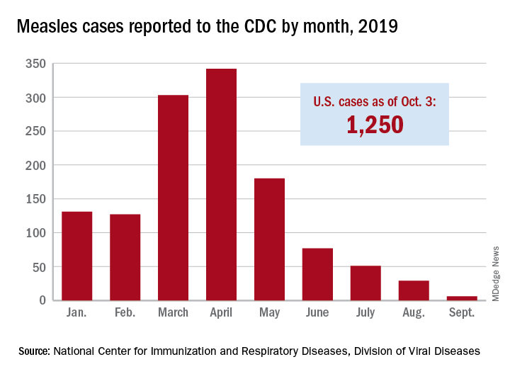
The new cases – two in Nassau County and one each in Monroe, Putnam, and Rockland counties – are “related to measles exposures from international travel but not affiliated with the 2018 outbreak,” the New York State Department of Health said in a written statement. Officials in Rockland County had declared its 2018 measles outbreak, which involved 312 cases in 2018 and 2019, over on Sept. 25.
. Of those cases, 1,163 (93%) were associated with 22 outbreaks, with the two largest occurring in New York City and Rockland County. “These two almost year-long outbreaks placed the United States at risk for losing measles elimination status,” the CDC said in a separate report, but “robust responses … ended transmission before the 1-year mark.”
New York State has reported the end of all active measles cases related to the initial outbreak in 2018, but the state is now responding to new, unrelated cases in four counties, according to the Centers for Disease Control and Prevention.
The new cases – two in Nassau County and one each in Monroe, Putnam, and Rockland counties – are “related to measles exposures from international travel but not affiliated with the 2018 outbreak,” the New York State Department of Health said in a written statement. Officials in Rockland County had declared its 2018 measles outbreak, which involved 312 cases in 2018 and 2019, over on Sept. 25.
. Of those cases, 1,163 (93%) were associated with 22 outbreaks, with the two largest occurring in New York City and Rockland County. “These two almost year-long outbreaks placed the United States at risk for losing measles elimination status,” the CDC said in a separate report, but “robust responses … ended transmission before the 1-year mark.”
New York State has reported the end of all active measles cases related to the initial outbreak in 2018, but the state is now responding to new, unrelated cases in four counties, according to the Centers for Disease Control and Prevention.
The new cases – two in Nassau County and one each in Monroe, Putnam, and Rockland counties – are “related to measles exposures from international travel but not affiliated with the 2018 outbreak,” the New York State Department of Health said in a written statement. Officials in Rockland County had declared its 2018 measles outbreak, which involved 312 cases in 2018 and 2019, over on Sept. 25.
. Of those cases, 1,163 (93%) were associated with 22 outbreaks, with the two largest occurring in New York City and Rockland County. “These two almost year-long outbreaks placed the United States at risk for losing measles elimination status,” the CDC said in a separate report, but “robust responses … ended transmission before the 1-year mark.”
Hypoxia-related discoveries net Nobel Prize
Three researchers have won the 2019 Nobel Prize in Physiology or Medicine “for their discoveries of how cells sense and adapt to oxygen availability.”

William G. Kaelin Jr., MD; Sir Peter J. Ratcliffe, MB ChB, MD; and Gregg L. Semenza, MD, PhD, described the molecular machinery that regulates gene activity in response to oxygen levels.
Their work “established the basis for our understanding of how oxygen levels affect cellular metabolism and physiological function” and “paved the way for promising new strategies to fight anemia, cancer, and many other diseases,” according to a statement by The Nobel Assembly at Karolinska Institutet.
Dr. Semenza, of Johns Hopkins Medicine in Baltimore, studied how the erythropoietin (EPO) gene is regulated by oxygen levels. Via experiments in mice, he identified DNA segments next to the EPO gene that mediate the response to hypoxia.
Dr. Ratcliffe, of the University of Oxford (England) and the Francis Crick Institute in London, also studied oxygen-dependent regulation of the EPO gene. Both his and Dr. Semenza’s groups found the oxygen-sensing mechanism was present in nearly all tissues.
Dr. Semenza also discovered a protein complex, hypoxia-inducible factor (HIF), that binds to the identified DNA segments in an oxygen-dependent manner. Additional investigation revealed that HIF consists of two transcription factors, HIF-1a and ARNT.
Several research groups found that HIF-1a is protected from degradation in hypoxia. In low-oxygen conditions, the amount of HIF-1a increases so it can bind to and regulate EPO and other genes with HIF-binding DNA segments. However, at normal oxygen levels, ubiquitin is added to HIF-1a, tagging it for degradation in the proteasome. It wasn’t clear how ubiquitin binds to HIF-1a in an oxygen-dependent manner, but Dr. Kaelin’s work provided some insight.

Dr. Kaelin, of the Dana-Farber Cancer Institute and Harvard Medical School, both in Boston, was researching von Hippel-Lindau’s (VHL) syndrome, an inherited disorder in which mutations can lead to tumors in multiple organs. He found the VHL gene encodes a protein that prevents cancer onset, and cancer cells without a functional VHL gene express high levels of hypoxia-regulated genes.
Research by other groups showed that VHL is part of a complex that labels proteins with ubiquitin, tagging them for degradation. Dr. Ratcliffe and his group found that VHL is required for the degradation of HIF-1a at normal oxygen levels.
Dr. Kaelin’s and Dr. Ratcliffe’s groups also showed that, under normal oxygen conditions, hydroxyl groups are added at two locations in HIF-1a. This modification – prolyl hydroxylation – allows VHL to bind to HIF-1a. So the researchers found that normal oxygen levels control HIF-1a degradation with the help of prolyl hydroxylases.
Additional research by Dr. Ratcliffe’s group and others revealed the specific prolyl hydroxylases involved in HIF-1a degradation. The researchers also found that HIF-1a’s gene-activating function was regulated by oxygen-dependent hydroxylation.
This work has improved the understanding of how different oxygen levels regulate physiological processes. In particular, oxygen sensing is essential for erythropoiesis, so these findings have implications for the treatment of anemia.
“There are several drugs that are now in clinical trials that serve to increase HIF activity and, as a result, will increase the production of erythropoietin and stimulate red blood cell production,” Dr. Semenza said in an interview after the announcement of his Nobel win. “These are all small molecules that can be given by mouth, and that may be a great convenience for patients who, at the present time, may require injections of recombinant human erythropoietin.”
Three researchers have won the 2019 Nobel Prize in Physiology or Medicine “for their discoveries of how cells sense and adapt to oxygen availability.”
William G. Kaelin Jr., MD; Sir Peter J. Ratcliffe, MB ChB, MD; and Gregg L. Semenza, MD, PhD, described the molecular machinery that regulates gene activity in response to oxygen levels.
Their work “established the basis for our understanding of how oxygen levels affect cellular metabolism and physiological function” and “paved the way for promising new strategies to fight anemia, cancer, and many other diseases,” according to a statement by The Nobel Assembly at Karolinska Institutet.
Dr. Semenza, of Johns Hopkins Medicine in Baltimore, studied how the erythropoietin (EPO) gene is regulated by oxygen levels. Via experiments in mice, he identified DNA segments next to the EPO gene that mediate the response to hypoxia.
Dr. Ratcliffe, of the University of Oxford (England) and the Francis Crick Institute in London, also studied oxygen-dependent regulation of the EPO gene. Both his and Dr. Semenza’s groups found the oxygen-sensing mechanism was present in nearly all tissues.
Dr. Semenza also discovered a protein complex, hypoxia-inducible factor (HIF), that binds to the identified DNA segments in an oxygen-dependent manner. Additional investigation revealed that HIF consists of two transcription factors, HIF-1a and ARNT.
Several research groups found that HIF-1a is protected from degradation in hypoxia. In low-oxygen conditions, the amount of HIF-1a increases so it can bind to and regulate EPO and other genes with HIF-binding DNA segments. However, at normal oxygen levels, ubiquitin is added to HIF-1a, tagging it for degradation in the proteasome. It wasn’t clear how ubiquitin binds to HIF-1a in an oxygen-dependent manner, but Dr. Kaelin’s work provided some insight.
Dr. Kaelin, of the Dana-Farber Cancer Institute and Harvard Medical School, both in Boston, was researching von Hippel-Lindau’s (VHL) syndrome, an inherited disorder in which mutations can lead to tumors in multiple organs. He found the VHL gene encodes a protein that prevents cancer onset, and cancer cells without a functional VHL gene express high levels of hypoxia-regulated genes.
Research by other groups showed that VHL is part of a complex that labels proteins with ubiquitin, tagging them for degradation. Dr. Ratcliffe and his group found that VHL is required for the degradation of HIF-1a at normal oxygen levels.
Dr. Kaelin’s and Dr. Ratcliffe’s groups also showed that, under normal oxygen conditions, hydroxyl groups are added at two locations in HIF-1a. This modification – prolyl hydroxylation – allows VHL to bind to HIF-1a. So the researchers found that normal oxygen levels control HIF-1a degradation with the help of prolyl hydroxylases.
Additional research by Dr. Ratcliffe’s group and others revealed the specific prolyl hydroxylases involved in HIF-1a degradation. The researchers also found that HIF-1a’s gene-activating function was regulated by oxygen-dependent hydroxylation.
This work has improved the understanding of how different oxygen levels regulate physiological processes. In particular, oxygen sensing is essential for erythropoiesis, so these findings have implications for the treatment of anemia.
“There are several drugs that are now in clinical trials that serve to increase HIF activity and, as a result, will increase the production of erythropoietin and stimulate red blood cell production,” Dr. Semenza said in an interview after the announcement of his Nobel win. “These are all small molecules that can be given by mouth, and that may be a great convenience for patients who, at the present time, may require injections of recombinant human erythropoietin.”
Three researchers have won the 2019 Nobel Prize in Physiology or Medicine “for their discoveries of how cells sense and adapt to oxygen availability.”
William G. Kaelin Jr., MD; Sir Peter J. Ratcliffe, MB ChB, MD; and Gregg L. Semenza, MD, PhD, described the molecular machinery that regulates gene activity in response to oxygen levels.
Their work “established the basis for our understanding of how oxygen levels affect cellular metabolism and physiological function” and “paved the way for promising new strategies to fight anemia, cancer, and many other diseases,” according to a statement by The Nobel Assembly at Karolinska Institutet.
Dr. Semenza, of Johns Hopkins Medicine in Baltimore, studied how the erythropoietin (EPO) gene is regulated by oxygen levels. Via experiments in mice, he identified DNA segments next to the EPO gene that mediate the response to hypoxia.
Dr. Ratcliffe, of the University of Oxford (England) and the Francis Crick Institute in London, also studied oxygen-dependent regulation of the EPO gene. Both his and Dr. Semenza’s groups found the oxygen-sensing mechanism was present in nearly all tissues.
Dr. Semenza also discovered a protein complex, hypoxia-inducible factor (HIF), that binds to the identified DNA segments in an oxygen-dependent manner. Additional investigation revealed that HIF consists of two transcription factors, HIF-1a and ARNT.
Several research groups found that HIF-1a is protected from degradation in hypoxia. In low-oxygen conditions, the amount of HIF-1a increases so it can bind to and regulate EPO and other genes with HIF-binding DNA segments. However, at normal oxygen levels, ubiquitin is added to HIF-1a, tagging it for degradation in the proteasome. It wasn’t clear how ubiquitin binds to HIF-1a in an oxygen-dependent manner, but Dr. Kaelin’s work provided some insight.
Dr. Kaelin, of the Dana-Farber Cancer Institute and Harvard Medical School, both in Boston, was researching von Hippel-Lindau’s (VHL) syndrome, an inherited disorder in which mutations can lead to tumors in multiple organs. He found the VHL gene encodes a protein that prevents cancer onset, and cancer cells without a functional VHL gene express high levels of hypoxia-regulated genes.
Research by other groups showed that VHL is part of a complex that labels proteins with ubiquitin, tagging them for degradation. Dr. Ratcliffe and his group found that VHL is required for the degradation of HIF-1a at normal oxygen levels.
Dr. Kaelin’s and Dr. Ratcliffe’s groups also showed that, under normal oxygen conditions, hydroxyl groups are added at two locations in HIF-1a. This modification – prolyl hydroxylation – allows VHL to bind to HIF-1a. So the researchers found that normal oxygen levels control HIF-1a degradation with the help of prolyl hydroxylases.
Additional research by Dr. Ratcliffe’s group and others revealed the specific prolyl hydroxylases involved in HIF-1a degradation. The researchers also found that HIF-1a’s gene-activating function was regulated by oxygen-dependent hydroxylation.
This work has improved the understanding of how different oxygen levels regulate physiological processes. In particular, oxygen sensing is essential for erythropoiesis, so these findings have implications for the treatment of anemia.
“There are several drugs that are now in clinical trials that serve to increase HIF activity and, as a result, will increase the production of erythropoietin and stimulate red blood cell production,” Dr. Semenza said in an interview after the announcement of his Nobel win. “These are all small molecules that can be given by mouth, and that may be a great convenience for patients who, at the present time, may require injections of recombinant human erythropoietin.”

C-Path and NORD team up to speed development of treatments for rare disorders
ROCKVILLE, MD – according to information provided at a launch event held Sept. 18, 2019.
By integrating data in a regulatory-grade format suitable for analytics, the RDCA-DAP hopes to accelerate the understanding of disease progression – including source of variability to optimize the characterization of subpopulations – develop clinical outcome measures and biomarkers, facilitate the development of mathematical models of disease, and promote innovative clinical trial designs.
The RDCA-DAP works as a database that will house patient-level data from a variety of sources, including clinical trials, longitudinal observational studies, patient registries, and other sources, such as real-world data collected from electronic health records across a wide range of rare diseases from all over the world. Data will then be made available to researchers to help speed the development of new treatments.
“The database and analytics we are creating will enable us to obtain new insight into these rare diseases,” C-Path President and CEO Joseph Scheeren, PharmD, said at the launch event. “Not only within specific diseases, but also we hope across related diseases.”

The key to the platform’s success will be data. “We need access to clinical data from the industry, patient groups, and academia,” Dr. Scheeren said. “Even more so, the RDCA-DAP will need to incorporate data from many sources. In addition to data from clinical trials conducted by industry and academia, we will want to access data from patients, hospitals, and any organization that can provide data.”
Dr. Scheeren said the data will take many forms, including numeric data, images, genomic information, and other forms of clinical information.
“The database will be able to handle these diverse datasets,” he said, adding that C-Path is preparing to be able to analyze the data sets “with the most sophisticated tools available.”
Dr. Scheeren made a call for all interested stakeholders with rare disease data to contribute to the platform.

NORD President and CEO Peter Saltonstall echoed that call. “We need data. We are accepting it immediately.”
Janet Woodcock, MD, director of the FDA Center for Drug Evaluation and Research, applauded the new platform. “I think foundations and patient advocacy groups and others that have been trying to help in this space have realized that simply funding basic research, although it is necessary and really important, it is not enough to get those therapies in the hands of doctors and patients,” she said. “You have to enable translation of research for that disease.”
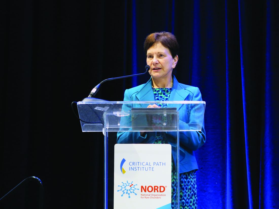
Dr. Woodcock noted that the cures may not even come from that basic research, but rather from “left field” using research into cancer or another disease state, something that will be enabled by the disease-agnostic platform being created by C-Path and NORD. She said that the platform will not only put all the data in one spot, but will help to create a standardized set of disease definitions to help make the data useful across all research.
ROCKVILLE, MD – according to information provided at a launch event held Sept. 18, 2019.
By integrating data in a regulatory-grade format suitable for analytics, the RDCA-DAP hopes to accelerate the understanding of disease progression – including source of variability to optimize the characterization of subpopulations – develop clinical outcome measures and biomarkers, facilitate the development of mathematical models of disease, and promote innovative clinical trial designs.
The RDCA-DAP works as a database that will house patient-level data from a variety of sources, including clinical trials, longitudinal observational studies, patient registries, and other sources, such as real-world data collected from electronic health records across a wide range of rare diseases from all over the world. Data will then be made available to researchers to help speed the development of new treatments.
“The database and analytics we are creating will enable us to obtain new insight into these rare diseases,” C-Path President and CEO Joseph Scheeren, PharmD, said at the launch event. “Not only within specific diseases, but also we hope across related diseases.”
The key to the platform’s success will be data. “We need access to clinical data from the industry, patient groups, and academia,” Dr. Scheeren said. “Even more so, the RDCA-DAP will need to incorporate data from many sources. In addition to data from clinical trials conducted by industry and academia, we will want to access data from patients, hospitals, and any organization that can provide data.”
Dr. Scheeren said the data will take many forms, including numeric data, images, genomic information, and other forms of clinical information.
“The database will be able to handle these diverse datasets,” he said, adding that C-Path is preparing to be able to analyze the data sets “with the most sophisticated tools available.”
Dr. Scheeren made a call for all interested stakeholders with rare disease data to contribute to the platform.
NORD President and CEO Peter Saltonstall echoed that call. “We need data. We are accepting it immediately.”
Janet Woodcock, MD, director of the FDA Center for Drug Evaluation and Research, applauded the new platform. “I think foundations and patient advocacy groups and others that have been trying to help in this space have realized that simply funding basic research, although it is necessary and really important, it is not enough to get those therapies in the hands of doctors and patients,” she said. “You have to enable translation of research for that disease.”
Dr. Woodcock noted that the cures may not even come from that basic research, but rather from “left field” using research into cancer or another disease state, something that will be enabled by the disease-agnostic platform being created by C-Path and NORD. She said that the platform will not only put all the data in one spot, but will help to create a standardized set of disease definitions to help make the data useful across all research.
ROCKVILLE, MD – according to information provided at a launch event held Sept. 18, 2019.
By integrating data in a regulatory-grade format suitable for analytics, the RDCA-DAP hopes to accelerate the understanding of disease progression – including source of variability to optimize the characterization of subpopulations – develop clinical outcome measures and biomarkers, facilitate the development of mathematical models of disease, and promote innovative clinical trial designs.
The RDCA-DAP works as a database that will house patient-level data from a variety of sources, including clinical trials, longitudinal observational studies, patient registries, and other sources, such as real-world data collected from electronic health records across a wide range of rare diseases from all over the world. Data will then be made available to researchers to help speed the development of new treatments.
“The database and analytics we are creating will enable us to obtain new insight into these rare diseases,” C-Path President and CEO Joseph Scheeren, PharmD, said at the launch event. “Not only within specific diseases, but also we hope across related diseases.”
The key to the platform’s success will be data. “We need access to clinical data from the industry, patient groups, and academia,” Dr. Scheeren said. “Even more so, the RDCA-DAP will need to incorporate data from many sources. In addition to data from clinical trials conducted by industry and academia, we will want to access data from patients, hospitals, and any organization that can provide data.”
Dr. Scheeren said the data will take many forms, including numeric data, images, genomic information, and other forms of clinical information.
“The database will be able to handle these diverse datasets,” he said, adding that C-Path is preparing to be able to analyze the data sets “with the most sophisticated tools available.”
Dr. Scheeren made a call for all interested stakeholders with rare disease data to contribute to the platform.
NORD President and CEO Peter Saltonstall echoed that call. “We need data. We are accepting it immediately.”
Janet Woodcock, MD, director of the FDA Center for Drug Evaluation and Research, applauded the new platform. “I think foundations and patient advocacy groups and others that have been trying to help in this space have realized that simply funding basic research, although it is necessary and really important, it is not enough to get those therapies in the hands of doctors and patients,” she said. “You have to enable translation of research for that disease.”
Dr. Woodcock noted that the cures may not even come from that basic research, but rather from “left field” using research into cancer or another disease state, something that will be enabled by the disease-agnostic platform being created by C-Path and NORD. She said that the platform will not only put all the data in one spot, but will help to create a standardized set of disease definitions to help make the data useful across all research.

Federal Health Care Cancer Data Trends 2019
Click here to access Federal Health Care Cancer Data Trends 2019
Table of Contents
- Introduction
- Cancer in the VA: The Big Picture
- Hepatocellular Carcinoma
- Prostate Cancer
- Multiple Myeloma
- Lung Cancer in the VA at a National Level
- Lung Cancer – Regional Snapshot: Kansas City VA
- Colorectal Cancer
- Leukemias
- Lymphomas
- Renal Cell Carcinoma
- Bladder Cancer
- Afterword

Click here to access Federal Health Care Cancer Data Trends 2019
Table of Contents
- Introduction
- Cancer in the VA: The Big Picture
- Hepatocellular Carcinoma
- Prostate Cancer
- Multiple Myeloma
- Lung Cancer in the VA at a National Level
- Lung Cancer – Regional Snapshot: Kansas City VA
- Colorectal Cancer
- Leukemias
- Lymphomas
- Renal Cell Carcinoma
- Bladder Cancer
- Afterword
Click here to access Federal Health Care Cancer Data Trends 2019
Table of Contents
- Introduction
- Cancer in the VA: The Big Picture
- Hepatocellular Carcinoma
- Prostate Cancer
- Multiple Myeloma
- Lung Cancer in the VA at a National Level
- Lung Cancer – Regional Snapshot: Kansas City VA
- Colorectal Cancer
- Leukemias
- Lymphomas
- Renal Cell Carcinoma
- Bladder Cancer
- Afterword