User login
Daily cookie makes no dent in ADHD diet effect
SAN ANTONIO – Though restriction and elimination diets have been studied for decades in the treatment of children with attention deficit hyperactive disorder, their therapeutic role and level of efficacy remain controversial in part because of blinding difficulties in dietary studies.
In a study presented at the annual meeting of the American Academy of Child & Adolescent Psychiatry Dr. Steven Pliszka of the University of Texas Health Center in San Antonio reported that among children with ADHD on a gluten- and additive-free diet, those given a daily snack inconsistent with the prescribed diet still showed behavioral improvement over 5 weeks.

In the study, Dr. Pliszka and colleagues randomized 29 children with ADHD, ages 8-12, to a restricted diet with a daily cookie or with a snack consistent with the diet. Stimulants were the only psychotropic medications allowed in the study. “The hypothesis was that if you’re on the restriction diet and getting better, and you get a snack that has gluten in it, you ought to deteriorate,” Dr. Pliszka said in an interview. “What we thought we would see was only one group would improve, but we saw improvement across the board.” This could mean that one cookie was not enough to corrupt the beneficial effects of the diet, he said, or that something besides the diet, such as a placebo effect, was responsible for the children’s improvement.
ADHD behavioral rating scores for both groups of children improved over the course of the 5 weeks; however, neither group improved faster. Children were also screened on functional magnetic resonance imaging (fMRI) at baseline and 5 weeks to evaluate activity in the ventral striatum related to images of high and low-calorie foods. Because not enough children were available for fMRI screening at the study’s end, Dr. Pliszka and colleagues combined the cohorts for analysis. Both groups also showed imaging evidence of increased activity in the ventral striatum, the part of the brain associated with reward response, and where activity is known to be lower in children with ADHD.
The researchers found regional activation within the ventral striatum to be significantly greater at follow-up than baseline for the cohort as a whole.
Dr. Pliszka said that the findings required more follow-up to understand.
“What we need to do for a follow-up study is get these groups to separate. If it is a low-calorie gluten free [diet], then probably we need to get the snack to be even more inconsistent than it was in this study, or give it several times a day, for example.”
If more snacks produced different results, “we could say it’s a dose-response issue. If there’s still no difference, we’d be able to say the diet’s not working. Because you give people all the bad stuff, but they think they’re getting the good stuff and they do ok, it’s clearly not the diet. “
Dr. Pliszka disclosed research support from Shire and Purdue Pharma and a consulting relationship with Ironshore Pharma. His co-authors in the study disclosed no conflicts of interest.
SAN ANTONIO – Though restriction and elimination diets have been studied for decades in the treatment of children with attention deficit hyperactive disorder, their therapeutic role and level of efficacy remain controversial in part because of blinding difficulties in dietary studies.
In a study presented at the annual meeting of the American Academy of Child & Adolescent Psychiatry Dr. Steven Pliszka of the University of Texas Health Center in San Antonio reported that among children with ADHD on a gluten- and additive-free diet, those given a daily snack inconsistent with the prescribed diet still showed behavioral improvement over 5 weeks.
In the study, Dr. Pliszka and colleagues randomized 29 children with ADHD, ages 8-12, to a restricted diet with a daily cookie or with a snack consistent with the diet. Stimulants were the only psychotropic medications allowed in the study. “The hypothesis was that if you’re on the restriction diet and getting better, and you get a snack that has gluten in it, you ought to deteriorate,” Dr. Pliszka said in an interview. “What we thought we would see was only one group would improve, but we saw improvement across the board.” This could mean that one cookie was not enough to corrupt the beneficial effects of the diet, he said, or that something besides the diet, such as a placebo effect, was responsible for the children’s improvement.
ADHD behavioral rating scores for both groups of children improved over the course of the 5 weeks; however, neither group improved faster. Children were also screened on functional magnetic resonance imaging (fMRI) at baseline and 5 weeks to evaluate activity in the ventral striatum related to images of high and low-calorie foods. Because not enough children were available for fMRI screening at the study’s end, Dr. Pliszka and colleagues combined the cohorts for analysis. Both groups also showed imaging evidence of increased activity in the ventral striatum, the part of the brain associated with reward response, and where activity is known to be lower in children with ADHD.
The researchers found regional activation within the ventral striatum to be significantly greater at follow-up than baseline for the cohort as a whole.
Dr. Pliszka said that the findings required more follow-up to understand.
“What we need to do for a follow-up study is get these groups to separate. If it is a low-calorie gluten free [diet], then probably we need to get the snack to be even more inconsistent than it was in this study, or give it several times a day, for example.”
If more snacks produced different results, “we could say it’s a dose-response issue. If there’s still no difference, we’d be able to say the diet’s not working. Because you give people all the bad stuff, but they think they’re getting the good stuff and they do ok, it’s clearly not the diet. “
Dr. Pliszka disclosed research support from Shire and Purdue Pharma and a consulting relationship with Ironshore Pharma. His co-authors in the study disclosed no conflicts of interest.
AT THE AACAP ANNUAL MEETING
Key clinical point:An inconsistent snack food does not appear to alter effects of restricted diet in children with ADHD.
Major finding: Children with ADHD who were allowed a cookie inconsistent with their restricted diets still improved on behavioral scores as well as those who received a diet-consistent snack; no significant differences found in rate and degree of improvement between groups.
Data source: A non-blinded trial enrolling 29 children and randomizing them to a restricted calorie and gluten diet plus a consistent or inconsistent snack, behavioral measures and fMRI taken at baseline and 5 weeks.
Disclosures: Lead investigator disclosed financial relationships with Shire, Purdue Pharma and Ironshore Pharma.

EC approves drug for acquired hemophilia A
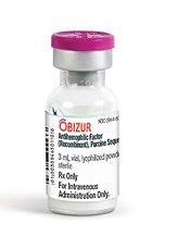
Photo courtesy of
Baxter International Inc.
The European Commission (EC) has approved a recombinant porcine factor VIII (FVIII) product, Obizur, to treat bleeding episodes in adults with acquired hemophilia A caused by autoantibodies to FVIII.
Obizur is the first recombinant porcine treatment to be made available for acquired hemophilia A in Europe.
It is specifically designed so physicians can monitor treatment response by measuring FVIII activity levels in addition to making clinical assessments.
The EC’s approval is based on a phase 2/3 trial in which patients with acquired hemophilia A received Obizur as treatment for serious bleeding episodes.
Twenty-nine patients were enrolled in this trial and evaluated for safety. Twenty-eight patients were evaluated for efficacy, as researchers determined that one of the patients did not actually have acquired hemophilia A.
At 24 hours after the initial infusion, all 28 patients in the efficacy analysis had a positive response to Obizur. This meant that bleeding stopped or decreased, the patients experienced clinical stabilization or improvement, and FVIII levels were 20% or higher.
Eighty-six percent of patients (24/28) had successful treatment of their initial bleeding episode. The overall treatment success was determined by the investigator based on the ability to discontinue or reduce the dose and/or dosing frequency of Obizur.
The adverse event most frequently reported in the 29 patients in the safety analysis was the development of inhibitors to porcine FVIII.
Nineteen patients were negative for anti-porcine FVIII antibodies at baseline, and 5 of these patients (26%) developed anti-porcine FVIII antibodies following exposure to Obizur.
Of the 10 patients with detectable anti-porcine FVIII antibodies at baseline, 2 (20%) experienced an increase in titer, and 8 (80%) decreased to a non-detectable titer.
Obizur is under development by Baxalta Incorporated. The drug is approved for use in the US and Canada as well as the European Union. It is under regulatory review in Switzerland, Australia, and Colombia. ![]()
Tocilizumab effective in giant cell arteritis
SAN FRANCISCO – Mounting evidence supports the use of tocilizumab in giant cell arteritis (GCA), and a new study supports use of the drug for induction and maintenance therapy in patients with newly diagnosed or relapsed GCA in the context of rapidly tapered glucocorticoid therapy.
“This is the first randomized controlled trial to prove efficacy of tocilizumab in induction and maintenance of remission in GCA. Tocilizumab reached the primary and secondary endpoints in our trial compared with placebo. After 12 months of therapy, more serious adverse events were observed in the placebo group,” said lead author Dr. Sabine Adler, University Hospital, Bern, Switzerland. Dr. Adler presented results of this late-breaking abstract at a special session during the annual meeting of the American College of Rheumatology.

A recent small open-label trial showed that tocilizumab was effective in patients with refractory GCA and unacceptable side effects on glucocorticoid (GC) therapy (Sem Arthr Rheum. Dec. 26, 2014. DOI:10.1016/j.semarthrit.2014.12.005). The present study was of newly diagnosed patients or those who had relapsed.
GCA is characterized by destructive inflammation in the walls of medium and large arteries. GC treatment has been the mainstay of therapy, controlling symptoms and reducing the risk of dreaded complications such as blindness, stroke, and claudication. But many patients require prolonged treatment, and cumulative doses of GC lead to substantial toxicity and morbidity. Thus, a better treatment is needed.
IL-6, the target of tocilizumab, is one of the cytokines involved in the pathogenesis of GCA. It is elevated in the serum and tissue of affected patients, Dr. Adler explained.
Dr. Adler and colleagues conducted a single-center, randomized, placebo-controlled trial evaluating tocilizumab in 30 patients with newly diagnosed or recurrent GCA. Patients were randomized in a 2:1 ratio to receive tocilizumab 8 mg/kg IV plus oral GC or IV placebo and oral GC. Infusions were given every 4 weeks for 1 year, over which time GC dose was slowly tapered to 0 mg in all patients. Some patients underwent angiography to rule out or define aortic involvement. Most patients in the trial had new onset GCA, she said.
The primary endpoint was complete remission at week 12 with GC dose of 0.1 mg/kg/day. Complete remission was defined as the absence of clinical signs and symptoms of GCA plus negative values for C-reactive protein (CRP) and erythrocyte sedimentation rate. At 12 weeks, the complete response rate was 85% for tocilizumab versus 40% for placebo, a significant difference. At the end of the trial, 85% and 20%, respectively, had not had a relapse; this difference was significant.
Placebo patients received a higher cumulative dose of GC, she continued.
There were 7 serious adverse events reported in the tocilizumab arm and 5 in the placebo arm; 1 death occurred in the placebo arm due to myocardial infarction.
“A closer look at serious adverse events reveals few cardiovascular problems,” Dr. Adler continued. Back pain and psychosis were reported as reasons for hospitalization.
During the question and answer session, an audience member noted that it was difficult to understand how a short-term treatment is effective in a chronic disease. “Probably patients will relapse when they stop the medication, so we are not on the ‘green’ side yet,” she replied. Dr. Adler noted that it could be possible that a reduced dose of tocilizumab could be effective but have fewer side effects, but that has not been studied.
Tocilizumab was provided by Roche for this investigator-initiated trial. Dr. Adler reported no financial disclosures.
SAN FRANCISCO – Mounting evidence supports the use of tocilizumab in giant cell arteritis (GCA), and a new study supports use of the drug for induction and maintenance therapy in patients with newly diagnosed or relapsed GCA in the context of rapidly tapered glucocorticoid therapy.
“This is the first randomized controlled trial to prove efficacy of tocilizumab in induction and maintenance of remission in GCA. Tocilizumab reached the primary and secondary endpoints in our trial compared with placebo. After 12 months of therapy, more serious adverse events were observed in the placebo group,” said lead author Dr. Sabine Adler, University Hospital, Bern, Switzerland. Dr. Adler presented results of this late-breaking abstract at a special session during the annual meeting of the American College of Rheumatology.
A recent small open-label trial showed that tocilizumab was effective in patients with refractory GCA and unacceptable side effects on glucocorticoid (GC) therapy (Sem Arthr Rheum. Dec. 26, 2014. DOI:10.1016/j.semarthrit.2014.12.005). The present study was of newly diagnosed patients or those who had relapsed.
GCA is characterized by destructive inflammation in the walls of medium and large arteries. GC treatment has been the mainstay of therapy, controlling symptoms and reducing the risk of dreaded complications such as blindness, stroke, and claudication. But many patients require prolonged treatment, and cumulative doses of GC lead to substantial toxicity and morbidity. Thus, a better treatment is needed.
IL-6, the target of tocilizumab, is one of the cytokines involved in the pathogenesis of GCA. It is elevated in the serum and tissue of affected patients, Dr. Adler explained.
Dr. Adler and colleagues conducted a single-center, randomized, placebo-controlled trial evaluating tocilizumab in 30 patients with newly diagnosed or recurrent GCA. Patients were randomized in a 2:1 ratio to receive tocilizumab 8 mg/kg IV plus oral GC or IV placebo and oral GC. Infusions were given every 4 weeks for 1 year, over which time GC dose was slowly tapered to 0 mg in all patients. Some patients underwent angiography to rule out or define aortic involvement. Most patients in the trial had new onset GCA, she said.
The primary endpoint was complete remission at week 12 with GC dose of 0.1 mg/kg/day. Complete remission was defined as the absence of clinical signs and symptoms of GCA plus negative values for C-reactive protein (CRP) and erythrocyte sedimentation rate. At 12 weeks, the complete response rate was 85% for tocilizumab versus 40% for placebo, a significant difference. At the end of the trial, 85% and 20%, respectively, had not had a relapse; this difference was significant.
Placebo patients received a higher cumulative dose of GC, she continued.
There were 7 serious adverse events reported in the tocilizumab arm and 5 in the placebo arm; 1 death occurred in the placebo arm due to myocardial infarction.
“A closer look at serious adverse events reveals few cardiovascular problems,” Dr. Adler continued. Back pain and psychosis were reported as reasons for hospitalization.
During the question and answer session, an audience member noted that it was difficult to understand how a short-term treatment is effective in a chronic disease. “Probably patients will relapse when they stop the medication, so we are not on the ‘green’ side yet,” she replied. Dr. Adler noted that it could be possible that a reduced dose of tocilizumab could be effective but have fewer side effects, but that has not been studied.
Tocilizumab was provided by Roche for this investigator-initiated trial. Dr. Adler reported no financial disclosures.
AT THE ACR ANNUAL MEETING
Key clinical point: Tocilizumab was effective in newly diagnosed and recurrent giant cell arteritis.
Major finding: At week 12, 85% of the tocilizumab-treated patients were in complete remission versus 40% of placebo patients.
Data source: Single-center, randomized, double-blind trial of 30 patients.
Disclosures: Tocilizumab was provided by Roche for this investigator-initiated trial. Dr. Adler reported no financial disclosures.

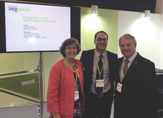
AGA, UEG celebrate young scholars in Barcelona
Leaders from AGA and United European Gastroenterology (UEG) gathered Tuesday, Oct. 27, 2015, in Barcelona, to celebrate the AGA Research Scholar/UEG Rising Star Scientific Exchange Program during UEG Week 2015 (http://live.ueg.eu/week). During the session, AGA honoree, Dr. Khalili, presented on oral contraceptive use in the etiopathogenesis of Crohn’s disease.
This recurring program fosters collaboration and goodwill with the international community and offers a unique opportunity for GI’s best and brightest to share research advances in a small group setting.
It is the ninth year of the program, which is held twice a year – once at Digestive Disease Week® (DDW) in May and once at UEG Week in October.

Lusutrombopag is effective for thrombocytopenia in liver disease
Lusutrombopag, an oral thrombopoietin receptor agonist, was found to reduce the need for platelet transfusion in patients with chronic liver disease with a planned invasive procedure, according to results in an abstract of a phase III trial that will be presented as a latebreaker at the annual meeting of the American Association for the Study of Liver Disease in San Francisco.
The novel therapy, which upregulates platelet production, was “efficacious and well tolerated,” producing a reduced risk of overall adverse events, including bleeding events, according to Dr. Namiki Izumi, Musashino Red Cross Hospital, Tokyo.

In this ongoing global phase III trial, called L-PLUS 2, 96 patients with chronic liver disease, a platelet count less than 50,000/microL, and a planned invasive procedure were randomized to receive a once-daily 3-mg tablet of lusutrombopag or a matching placebo for 7 days. The primary endpoint was the need for a preoperative platelet transfusion.
“The proportion of patients who required no preoperative platelet transfusion was significantly greater with lusutrombopag [29.2% vs. 12.5%; P less than .0001],” Dr. Izumi reported. The proportion of responders, defined by a platelet count greater than or equal to 50,000/microL and a greater than or equal to 20,000/microL-increase from baseline, was also significantly greater in the lusutrombopag arm (77.1% vs. 6.3%; P less than .0001).
In addition, the median time with a platelet count greater than or equal to 50,000/microL was 22.1 days in those who received lusutrombopag but no platelet transfusion versus 3.3 days in the placebo patients who did receive transfusion (P less than 0.0001).
Many adverse events occurred less frequently in the arm randomized to lusutrombopag. This included bleeding events (14.6% vs. 27.1%) and postoperative fever (39.6% vs. 56.3%). The rates of procedural hypertension (41.7% vs. 37.5%) and procedural pain (45.8% vs. 41.7%) were slightly greater in the group randomized to lusutrombopag, but elevations in liver enzymes, such as aspartate aminotransferase (22.9% vs. 31.3%) were somewhat lower.
“Protocol-required imaging revealed one thromboembolic event of the portal venous system in each study arm, neither of which was related to platelets,” according to Dr. Izumi, who reported that no patient discontinued therapy as a result of an adverse event.
Because of the frequency with which thrombocytopenia is observed in patients with chronic liver disease, platelet transfusion is considered a standard procedure when an invasive intervention is planned, according to Dr. Izumi. The data from this trial suggest that preoperative treatment with lusutrombopag may be an alternative.
Dr. Izumi reported financial relationships with Bayer, Daiichi Sankyo, Gilead, Merck, and Shionogi.
Lusutrombopag, an oral thrombopoietin receptor agonist, was found to reduce the need for platelet transfusion in patients with chronic liver disease with a planned invasive procedure, according to results in an abstract of a phase III trial that will be presented as a latebreaker at the annual meeting of the American Association for the Study of Liver Disease in San Francisco.
The novel therapy, which upregulates platelet production, was “efficacious and well tolerated,” producing a reduced risk of overall adverse events, including bleeding events, according to Dr. Namiki Izumi, Musashino Red Cross Hospital, Tokyo.
In this ongoing global phase III trial, called L-PLUS 2, 96 patients with chronic liver disease, a platelet count less than 50,000/microL, and a planned invasive procedure were randomized to receive a once-daily 3-mg tablet of lusutrombopag or a matching placebo for 7 days. The primary endpoint was the need for a preoperative platelet transfusion.
“The proportion of patients who required no preoperative platelet transfusion was significantly greater with lusutrombopag [29.2% vs. 12.5%; P less than .0001],” Dr. Izumi reported. The proportion of responders, defined by a platelet count greater than or equal to 50,000/microL and a greater than or equal to 20,000/microL-increase from baseline, was also significantly greater in the lusutrombopag arm (77.1% vs. 6.3%; P less than .0001).
In addition, the median time with a platelet count greater than or equal to 50,000/microL was 22.1 days in those who received lusutrombopag but no platelet transfusion versus 3.3 days in the placebo patients who did receive transfusion (P less than 0.0001).
Many adverse events occurred less frequently in the arm randomized to lusutrombopag. This included bleeding events (14.6% vs. 27.1%) and postoperative fever (39.6% vs. 56.3%). The rates of procedural hypertension (41.7% vs. 37.5%) and procedural pain (45.8% vs. 41.7%) were slightly greater in the group randomized to lusutrombopag, but elevations in liver enzymes, such as aspartate aminotransferase (22.9% vs. 31.3%) were somewhat lower.
“Protocol-required imaging revealed one thromboembolic event of the portal venous system in each study arm, neither of which was related to platelets,” according to Dr. Izumi, who reported that no patient discontinued therapy as a result of an adverse event.
Because of the frequency with which thrombocytopenia is observed in patients with chronic liver disease, platelet transfusion is considered a standard procedure when an invasive intervention is planned, according to Dr. Izumi. The data from this trial suggest that preoperative treatment with lusutrombopag may be an alternative.
Dr. Izumi reported financial relationships with Bayer, Daiichi Sankyo, Gilead, Merck, and Shionogi.
FROM THE LIVER MEETING 2015
Key clinical point: In a phase III trial, lusutrombopag was found to reduce the need for platelet transfusion in chronic liver disease patients requiring surgery.
Major finding: Prior to a planned invasive procedure, only 20.8% of lusutrombopag versus 87.5% of placebo patients (P less than .0001) required platelet transfusion.
Data source: A multicenter, double-blind phase III trial.
Disclosures: Dr. Izumi reported financial relationships with Bayer, Daiichi Sankyo, Gilead, Merck, and Shionogi.
Role of cyclins in malaria parasite development

Photo by James Gathany
Researchers say they have characterized the cyclin protein family in the rodent malaria parasite Plasmodium berghei.
The team found there are only 3 cyclins in this parasite, but one of these, the single P-type cyclin CYC3, plays a “vital role” in parasite development in the mosquito.
The researchers believe this work, published in PLoS Pathogens, could pave the way to a better understanding of malaria parasites and lead to potential new treatments.
“This first functional study of cyclin in the malaria parasite and its consequences in parasite development within pathogen-carrying mosquitoes will definitely further our understanding of parasite cell division, which I hope will lead to the elimination of this disease in the future,” said study author Magali Roques, PhD, of the University of Nottingham in the UK. ![]()
Denosumab better at building bone than zoledronic acid
SAN FRANCISCO – Denosumab was superior to zoledronic acid (ZA) in building bone at the lumber spine, total hip, and femoral neck in postmenopausal women with osteoporosis previously treated with oral bisphosphonate therapy in a randomized phase III trial. This study is one more piece of level I evidence to support the effectiveness of transitioning to denosumab in such patients.
“In postmenopausal women with osteoporosis on prior oral bisphosphonates for 2 or more years, transitioning to denosumab resulted in significantly greater increases in bone mineral density [BMD], compared with zoledronic acid at all measured skeletal sites,” said lead author Dr. Paul D. Miller of the Colorado Center for Bone Research, Lakewood, Colo.

Speaking at the annual meeting of the American College of Rheumatology, Dr. Miller said: “This study completes a suite of trials that show transitioning from oral bisphosphonates to denosumab provides greater increases in bone mineral density and reduction in bone turnover markers, compared to maintaining therapy with another bisphosphonate.”
“Adherence rates to oral bisphosphonates are low,” he continued. “Patients who are intolerant to or fail other bisphosphonates may cycle from one bisphosphonate to another, but clinical benefits are not shown. Previous trials have shown that denosumab increased bone mineral density in women previously treated with oral bisphosphonates, whereas zoledronic acid did not, in women previously treated with alendronate.”
A prospective, double-blind, double-dummy, randomized trial was mounted to compare denosumab given subcutaneously at 60 mg every 6 months (n = 321) vs. ZA 5 mg once a year (n = 322) in postmenopausal women with osteoporosis who were intolerant to or who failed oral bisphosphonate therapy after at least 2 years of treatment.
Baseline characteristics were similar between both treatment arms. Median age was 69 years. About 38% had previous osteoporotic fracture. Bone mineral density T scores were similar between groups. Prior oral bisphosphonate exposure was a median of 6.4 years. Bone turnover marker levels were similar between the two arms.
For the primary endpoint, at 12 months, denosumab achieved a greater increase in BMD at all sites, compared with ZA: a 2.1% difference was observed at the lumbar spine and a 1.4% difference at the total hip.
Changes in bone turnover markers over time were more favorable with denosumab, he continued. Serum C-terminal cross-linking telopeptide (CTX) level was maintained in the denosumab arm and increased with ZA over 12 months. A similar pattern was observed with procollagen type 1 N-terminal propeptide (P1NP).
Adverse events were comparable between both groups. Serious adverse events were reported in 7.8% in the denosumab arm versus 9.1% in the ZA arm, but this difference was not statistically significant. Slightly more hypersensitivity adverse events were reported in the denosumab group: 3.8% versus 1.9% for ZA. The number of osteoporotic-related fractures was doubled in the ZA arm: 7 for denosumab vs. 15 for ZA.
Dr. Miller told listeners that this is the fourth study to show that denosumab is effective in osteoporotic patients previously on oral bisphosphonates, including prior risedronate, ibandronate, and alendronate.
SAN FRANCISCO – Denosumab was superior to zoledronic acid (ZA) in building bone at the lumber spine, total hip, and femoral neck in postmenopausal women with osteoporosis previously treated with oral bisphosphonate therapy in a randomized phase III trial. This study is one more piece of level I evidence to support the effectiveness of transitioning to denosumab in such patients.
“In postmenopausal women with osteoporosis on prior oral bisphosphonates for 2 or more years, transitioning to denosumab resulted in significantly greater increases in bone mineral density [BMD], compared with zoledronic acid at all measured skeletal sites,” said lead author Dr. Paul D. Miller of the Colorado Center for Bone Research, Lakewood, Colo.
Speaking at the annual meeting of the American College of Rheumatology, Dr. Miller said: “This study completes a suite of trials that show transitioning from oral bisphosphonates to denosumab provides greater increases in bone mineral density and reduction in bone turnover markers, compared to maintaining therapy with another bisphosphonate.”
“Adherence rates to oral bisphosphonates are low,” he continued. “Patients who are intolerant to or fail other bisphosphonates may cycle from one bisphosphonate to another, but clinical benefits are not shown. Previous trials have shown that denosumab increased bone mineral density in women previously treated with oral bisphosphonates, whereas zoledronic acid did not, in women previously treated with alendronate.”
A prospective, double-blind, double-dummy, randomized trial was mounted to compare denosumab given subcutaneously at 60 mg every 6 months (n = 321) vs. ZA 5 mg once a year (n = 322) in postmenopausal women with osteoporosis who were intolerant to or who failed oral bisphosphonate therapy after at least 2 years of treatment.
Baseline characteristics were similar between both treatment arms. Median age was 69 years. About 38% had previous osteoporotic fracture. Bone mineral density T scores were similar between groups. Prior oral bisphosphonate exposure was a median of 6.4 years. Bone turnover marker levels were similar between the two arms.
For the primary endpoint, at 12 months, denosumab achieved a greater increase in BMD at all sites, compared with ZA: a 2.1% difference was observed at the lumbar spine and a 1.4% difference at the total hip.
Changes in bone turnover markers over time were more favorable with denosumab, he continued. Serum C-terminal cross-linking telopeptide (CTX) level was maintained in the denosumab arm and increased with ZA over 12 months. A similar pattern was observed with procollagen type 1 N-terminal propeptide (P1NP).
Adverse events were comparable between both groups. Serious adverse events were reported in 7.8% in the denosumab arm versus 9.1% in the ZA arm, but this difference was not statistically significant. Slightly more hypersensitivity adverse events were reported in the denosumab group: 3.8% versus 1.9% for ZA. The number of osteoporotic-related fractures was doubled in the ZA arm: 7 for denosumab vs. 15 for ZA.
Dr. Miller told listeners that this is the fourth study to show that denosumab is effective in osteoporotic patients previously on oral bisphosphonates, including prior risedronate, ibandronate, and alendronate.
AT THE ACR ANNUAL MEETING
Key clinical point: Denosumab was superior to zoledronic acid in building bone mineral density in postmenopausal women with osteoporosis who had previously been treated with oral bisphosphonates.
Major finding: Denosumab achieved a 2.1% difference in BMD at the lumbar spine and a 1.4% difference at the hip, compared with zoledronic acid.
Data source: Prospective, randomized, double-blind, double-dummy trial that included 643 patients.
Disclosures: Dr. Miller’s financial disclosures include Alexion, Amgen, Merck, Merck Serono, Boehringer Ingelheim, Regeneron, National Bone Health Alliance, Eli Lilly, Pfizer, Radius, and he is owner and director of the Colorado Center for Bone Research. The study was supported by Amgen.
ACR: Study confirms potential genetic basis for poor response to allopurinol in gout
SAN FRANCISCO – Patients with gout who fail to sufficiently lower their serum urate level despite adherence to a regimen of allopurinol 300 mg/day may have a genetic polymorphism affecting their response to the medication, according to new findings presented at the annual meeting of the American College of Rheumatology.
“ABCG2 genotyping may identify patients who will not reach target serum urate levels on standard allopurinol doses,” said study presenter and principal investigator Dr. Lisa K. Stamp of the University of Otago, Christchurch, New Zealand.
Sustained lowering of serum urate (SU) to less than 6 mg/dL – achieved most commonly by inhibition of xanthine oxidase using allopurinol – is crucial for the long-term management of gout. Yet, in clinical practice, less than half of patients treated with allopurinol achieve this SU target. This is usually because of poor adherence or an inappropriately low dose of the drug. But, in some patients, the response can be poor even despite allopurinol 300 mg daily. The reason for failure of allopurinol treatment in these poor responders has been unknown.

“It is important to consider genes that might help predict response or toxicity. To date, pharmacogenetic studies of allopurinol have focused on adverse effects. However, we were more interested in predicting response. The ability to predict poor response is important because it can influence drug choice and shorten the time to achieve target SU,” Dr. Stamp said.
The present study helps to validate the results of a previous genomewide association study in which the single nucleotide polymorphism (SNP) rs2231142 in ABCG2 was associated with poor response to allopurinol (Clin Pharmacol Ther. 2015 May;97[5]:518-25). Adherence to allopurinol was not examined in that study, so Dr. Stamp and her colleagues set out to make sure that it was not a confounder. The current study examined the SNP’s association with allopurinol response more closely in 264 gout patients who were well characterized phenotypically. All were being treated with allopurinol and all adhered to their treatment regimen. Overall, 120 were deemed good responders, with SU of 6 mg/dL or less on daily allopurinol of 300 mg or less, and 68 were poor responders, with SU of 6 mg/dL or higher despite a daily dose of allopurinol exceeding 300 mg. Another 76 patients were nonadherent/nonclassifiable. The poor responders tended to be younger and were more likely to be male, nonwhite, and heavier than their counterparts who responded well.
Genotyping for the rs2231142 SNP revealed a greater preponderance of the minor genotype in poor responders, compared with good responders (39% vs. 18%). This genotype was significantly associated with poor response after adjusting for age, sex, body-mass index, and ethnicity (odds ratio, 2.91; 95% confidence interval, 1.71-5.17; P = .00015). The mechanism – still to be proven – may involve reduced urate excretion from the gut.
There may not be a case for screening for the genotype just yet, according to Dr. Christopher M. Burns, a rheumatologist at Dartmouth Hitchcock Medical Center, Lebanon, N.H., who was not involved in the study. He said “it’s hard to see why you would screen for this variant, as it doesn’t account for all resistance, and you would still end up pushing the allopurinol dose up to achieve a SU of less than 6.0 mg/dL anyway. Since it’s not an adverse event warning signal, but simply one marker of possibly requiring a higher allopurinol dose, and allopurinol is an inexpensive drug, why would you check it? At least, that’s my sense. The findings may be more important simply for a better understanding of urate metabolism and the mechanism of action of allopurinol and oxypurinol.”
Dr. Stamp reported a financial disclosure with AstraZeneca.
SAN FRANCISCO – Patients with gout who fail to sufficiently lower their serum urate level despite adherence to a regimen of allopurinol 300 mg/day may have a genetic polymorphism affecting their response to the medication, according to new findings presented at the annual meeting of the American College of Rheumatology.
“ABCG2 genotyping may identify patients who will not reach target serum urate levels on standard allopurinol doses,” said study presenter and principal investigator Dr. Lisa K. Stamp of the University of Otago, Christchurch, New Zealand.
Sustained lowering of serum urate (SU) to less than 6 mg/dL – achieved most commonly by inhibition of xanthine oxidase using allopurinol – is crucial for the long-term management of gout. Yet, in clinical practice, less than half of patients treated with allopurinol achieve this SU target. This is usually because of poor adherence or an inappropriately low dose of the drug. But, in some patients, the response can be poor even despite allopurinol 300 mg daily. The reason for failure of allopurinol treatment in these poor responders has been unknown.
“It is important to consider genes that might help predict response or toxicity. To date, pharmacogenetic studies of allopurinol have focused on adverse effects. However, we were more interested in predicting response. The ability to predict poor response is important because it can influence drug choice and shorten the time to achieve target SU,” Dr. Stamp said.
The present study helps to validate the results of a previous genomewide association study in which the single nucleotide polymorphism (SNP) rs2231142 in ABCG2 was associated with poor response to allopurinol (Clin Pharmacol Ther. 2015 May;97[5]:518-25). Adherence to allopurinol was not examined in that study, so Dr. Stamp and her colleagues set out to make sure that it was not a confounder. The current study examined the SNP’s association with allopurinol response more closely in 264 gout patients who were well characterized phenotypically. All were being treated with allopurinol and all adhered to their treatment regimen. Overall, 120 were deemed good responders, with SU of 6 mg/dL or less on daily allopurinol of 300 mg or less, and 68 were poor responders, with SU of 6 mg/dL or higher despite a daily dose of allopurinol exceeding 300 mg. Another 76 patients were nonadherent/nonclassifiable. The poor responders tended to be younger and were more likely to be male, nonwhite, and heavier than their counterparts who responded well.
Genotyping for the rs2231142 SNP revealed a greater preponderance of the minor genotype in poor responders, compared with good responders (39% vs. 18%). This genotype was significantly associated with poor response after adjusting for age, sex, body-mass index, and ethnicity (odds ratio, 2.91; 95% confidence interval, 1.71-5.17; P = .00015). The mechanism – still to be proven – may involve reduced urate excretion from the gut.
There may not be a case for screening for the genotype just yet, according to Dr. Christopher M. Burns, a rheumatologist at Dartmouth Hitchcock Medical Center, Lebanon, N.H., who was not involved in the study. He said “it’s hard to see why you would screen for this variant, as it doesn’t account for all resistance, and you would still end up pushing the allopurinol dose up to achieve a SU of less than 6.0 mg/dL anyway. Since it’s not an adverse event warning signal, but simply one marker of possibly requiring a higher allopurinol dose, and allopurinol is an inexpensive drug, why would you check it? At least, that’s my sense. The findings may be more important simply for a better understanding of urate metabolism and the mechanism of action of allopurinol and oxypurinol.”
Dr. Stamp reported a financial disclosure with AstraZeneca.
AT THE ACR ANNUAL MEETING
Key clinical point: Screening could identify gout patients who require a dose adjustment of allopurinol.
Major finding: Presence of the rs2231142 SNP in the ABCG2 gene was significantly associated with poor response to allopurinol after adjusting for age, sex, body-mass index, and ethnicity (OR, 2.91; 95% CI, 1.71-5.17; P = .00015).
Data source: An analysis of 264 gout patients participating in clinical trials with allopurinol.
Disclosures: Dr. Stamp reported a financial disclosure with AstraZeneca.
ACR: Ozone injections reduce pain and improve function in knee OA
SAN FRANCISCO – Intra-articular ozone injections were effective in reducing pain, improving function, and improving quality of life in patients with knee osteoarthritis in the first randomized study to evaluate this approach.
Patients treated with a series of ozone injections achieved significant improvements on all measures, except for the Timed Up and Go Test, compared with patients given placebo, according to study results presented at the annual meeting of the American College of Rheumatology.
“After 8 weeks of treatment, ozone can give patients with knee osteoarthritis [OA] better quality of life with less pain and more independence in performing daily activities. More studies are needed to validate this option in patients with OA. Intra-articular ozone injections are safe with similar complications to placebo. In elderly people with comorbidities requiring chronic medication, this approach is a good option because it doesn’t interact with medications. The only restriction is anticoagulant therapy, as there may be bleeding at the injection site,” said Dr. Virginia Trevisani, professor at the Federal University of São Paulo.

The next series of studies Dr. Trevisani and her coauthors are planning will incorporate MRI imaging to assess the effect of the ozone injections on structural progression in knee OA.
Ozone is thought to have anti-inflammatory effects by reducing oxidative stress. Ozone is being used for medical purposes in countries such as Russia, Germany, and Spain, but it is not currently used clinically in the United States. “You can’t perform these injections in patients without experience. The only requirement is a machine to make ozone that costs about $1,000 USD,” she said.
Before this study, evidence in support of ozone injections in knee OA was anecdotal and from observational studies. The present study is the first randomized trial to evaluate intra-articular ozone injections in patients with knee OA.
The study enrolled 98 patients with documented knee OA between the ages of 60 and 85 years; 63 patients were randomized to intra-articular injections of ozone in the knee with the most pain (one injection per week for 8 consecutive weeks), and 35 were randomized to placebo injections of a small amount of air.
Patients were evaluated at baseline, after 4 and 8 injections, and 8 weeks following the last injection. Two patients in the ozone group withdrew from the study. The only adverse events were three puncture-site wounds – two in the ozone group and one in the placebo group.
Significant improvement was observed on all measures, except for the Timed Up and Go Test (getting up from a chair), at every time point for the ozone injections. Dr. Trevisani said that the ability to get up from a chair without help depends on balance and muscle strength, which may explain why the results were not significant.
Measures of pain on the Western Ontario and McMaster Universities Osteoarthritis Index (WOMAC) and the visual analog scale improved significantly with ozone, compared with placebo, and by week 16, the P value was .000 for both measures. WOMAC joint stiffness intensity was significantly improved by ozone (P = .075 at week 4; P = .002 at week 8). Quality of life on the Short Form–36 for pain and functional capacity were significantly improved by ozone, compared with placebo (P = .000 at week 16 for both measures).
Dr. Trevisani said ozone injections may be able to delay the need for total joint replacement surgery, and that they are cost effective, compared with surgery and other pharmacologic treatments.
Dr. Trevisani had no relevant financial disclosures.
SAN FRANCISCO – Intra-articular ozone injections were effective in reducing pain, improving function, and improving quality of life in patients with knee osteoarthritis in the first randomized study to evaluate this approach.
Patients treated with a series of ozone injections achieved significant improvements on all measures, except for the Timed Up and Go Test, compared with patients given placebo, according to study results presented at the annual meeting of the American College of Rheumatology.
“After 8 weeks of treatment, ozone can give patients with knee osteoarthritis [OA] better quality of life with less pain and more independence in performing daily activities. More studies are needed to validate this option in patients with OA. Intra-articular ozone injections are safe with similar complications to placebo. In elderly people with comorbidities requiring chronic medication, this approach is a good option because it doesn’t interact with medications. The only restriction is anticoagulant therapy, as there may be bleeding at the injection site,” said Dr. Virginia Trevisani, professor at the Federal University of São Paulo.
The next series of studies Dr. Trevisani and her coauthors are planning will incorporate MRI imaging to assess the effect of the ozone injections on structural progression in knee OA.
Ozone is thought to have anti-inflammatory effects by reducing oxidative stress. Ozone is being used for medical purposes in countries such as Russia, Germany, and Spain, but it is not currently used clinically in the United States. “You can’t perform these injections in patients without experience. The only requirement is a machine to make ozone that costs about $1,000 USD,” she said.
Before this study, evidence in support of ozone injections in knee OA was anecdotal and from observational studies. The present study is the first randomized trial to evaluate intra-articular ozone injections in patients with knee OA.
The study enrolled 98 patients with documented knee OA between the ages of 60 and 85 years; 63 patients were randomized to intra-articular injections of ozone in the knee with the most pain (one injection per week for 8 consecutive weeks), and 35 were randomized to placebo injections of a small amount of air.
Patients were evaluated at baseline, after 4 and 8 injections, and 8 weeks following the last injection. Two patients in the ozone group withdrew from the study. The only adverse events were three puncture-site wounds – two in the ozone group and one in the placebo group.
Significant improvement was observed on all measures, except for the Timed Up and Go Test (getting up from a chair), at every time point for the ozone injections. Dr. Trevisani said that the ability to get up from a chair without help depends on balance and muscle strength, which may explain why the results were not significant.
Measures of pain on the Western Ontario and McMaster Universities Osteoarthritis Index (WOMAC) and the visual analog scale improved significantly with ozone, compared with placebo, and by week 16, the P value was .000 for both measures. WOMAC joint stiffness intensity was significantly improved by ozone (P = .075 at week 4; P = .002 at week 8). Quality of life on the Short Form–36 for pain and functional capacity were significantly improved by ozone, compared with placebo (P = .000 at week 16 for both measures).
Dr. Trevisani said ozone injections may be able to delay the need for total joint replacement surgery, and that they are cost effective, compared with surgery and other pharmacologic treatments.
Dr. Trevisani had no relevant financial disclosures.
AT THE ACR ANNUAL MEETING
Key clinical point:Intra-articular ozone injections reduce pain, improve function, and improve quality of life in patients with knee osteoarthritis.
Major finding: On all measures of pain, function, and quality of life, ozone injections were significantly superior to placebo.
Data source: A randomized, double-blind placebo-controlled trial of 98 patients with knee OA.
Disclosures: Dr. Trevisani had no relevant financial disclosures.

The Optum termination thunderbolt
One afternoon, after seeing your last patient, you’re doing the old-school thing, with your feet up, opening and reading your paper mail after a hard day’s work at the dermatology ranch. You see an odd form letter – a green sticker on the outside, certified mail – stating that you have been terminated from a Medicare advantage plan, no reason given.
At first, you don’t care so much. After all, this plan pays you only 95% of Medicare. Then you think about it and realize that this plan represents 50% of all Medicare beneficiaries in your area. You start to freak out, and you immediately go to the American Academy of Dermatology website where you read Rob Portman’s article about how to fight a termination notice and respond expeditiously.
Later that night, your spouse asks why you were singled out. “Are you a bad doctor? What did you do wrong? Can the kids still go to college?
The answer, most often, is that you did nothing wrong. You’ve just been caught up in the insurer’s network management software, Optum 360.
Optum 360 is a large health care information and management subsidiary of UnitedHealthcare. It was created as a joint venture by the Optum insight (health technology) unit of UnitedHealthcare, and Dignity Health (claims processing), forming Optum 360.
Optum claims that its software measures the efficiency of providers, saving insurers money and improving the quality of care – the Valhalla of health care managers everywhere. Unfortunately, Optum doesn’t deliver this vision of heaven on earth, at least not for dermatology.
Optum 360does little more than aggregate and average the costs of individual providers with no recognition of severity of disease or case mix. The physician with the most reimbursement during an episode of care for a given ICD-9 code (now for a group of ICD-10 codes) gets credited with all the expenses under that code. For example, you do two stages of Mohs surgery on a big basal cell on the nose that you send to plastics for repair. If the reimbursement for the Mohs surgery exceeds the plastics reimbursement, the Optum software designates you as the responsible provider. The cost of the plastics repair accrues to you; the hospital OR facility charges accrue to you (facilities are not considered a provider); and the anesthesiologist charges are credited to you. In addition, the superficial basal cell carcinoma on the patient’s back, treated a week later by the referring doctor, is also attributed to you as part of the original episode of care. There are no quality parameters and no subspecialty recognition.
If your patient load regularly includes patients who have Mohs surgery, the dermatologist down the street who does Mohs only once a week looks much better to Optum than you do. The referral-only medical dermatologist in town, who treats very sick patients and routinely prescribes biologics, and (heaven forbid) intravenous immunoglobulin, is similarly tagged for termination from the insurer’s network.
So it looks as though dermatologists who handle the toughest patients lose out. But who really loses the most? The sickest patients! The dermatologist can always fill the schedule with patients with other insurance coverage and reduce the backlog or, if worse comes to worse, he can take the Canadian cure and go on vacation. The sickest patients, however, get eliminated from the system when their doctors get eliminated by UnitedHealthcare’s software. Then patients often cannot find another doctor because most insurer’s physician rosters are 70% inaccurate (JAMA Dermatol. 2014 Dec;150[12]:1290-7).
In some circumstances, the sickest patients have reported either being unable to find other dermatologists willing to provide the special service they need or having to wait up to 6 months for an open appointment. These patients then try to drop back into fee-for-service Medicare, only to find they cannot afford the gap insurance, which costs five times as much. Why? Because those patients now have a preexisting condition. Yes, preexisting conditions still apply in the world of gap insurance.
This is obviously not optimal nor even acceptable. To quote Michael Keaton from the 1982 movie “Night Shift”: “Is this a great country or what?” The answer is a resounding “yes” for medical insurance companies who are booking record profits, but “no” for the sickest patients.
Dr. Coldiron is a past president of the American Academy of Dermatology. He is currently in private practice, but maintains a clinical assistant professorship at the University of Cincinnati. He cares for patients, teaches medical students and residents, and has several active clinical research projects. Dr. Coldiron is the author of more than 80 scientific letters, papers, and several book chapters, and he speaks frequently on a variety of topics. Reach him at dermnews@frontlinemedcom.com.