User login
Bringing you the latest news, research and reviews, exclusive interviews, podcasts, quizzes, and more.
div[contains(@class, 'header__large-screen')]
div[contains(@class, 'read-next-article')]
div[contains(@class, 'nav-primary')]
nav[contains(@class, 'nav-primary')]
section[contains(@class, 'footer-nav-section-wrapper')]
footer[@id='footer']
div[contains(@class, 'main-prefix')]
section[contains(@class, 'nav-hidden')]
div[contains(@class, 'ce-card-content')]
nav[contains(@class, 'nav-ce-stack')]
Patient Navigators for Serious Illnesses Can Now Bill Under New Medicare Codes
In a move that acknowledges the gauntlet the US health system poses for people facing serious and fatal illnesses, Medicare will pay for a new class of workers to help patients manage treatments for conditions like cancer and heart failure.
The 2024 Medicare physician fee schedule includes new billing codes, including G0023, to pay for 60 minutes a month of care coordination by certified or trained auxiliary personnel working under the direction of a clinician.
A diagnosis of cancer or another serious illness takes a toll beyond the physical effects of the disease. Patients often scramble to make adjustments in family and work schedules to manage treatment, said Samyukta Mullangi, MD, MBA, medical director of oncology at Thyme Care, a Nashville, Tennessee–based firm that provides navigation and coordination services to oncology practices and insurers.

“It just really does create a bit of a pressure cooker for patients,” Dr. Mullangi told this news organization.
Medicare has for many years paid for medical professionals to help patients cope with the complexities of disease, such as chronic care management (CCM) provided by physicians, nurses, and physician assistants.
The new principal illness navigation (PIN) payments are intended to pay for work that to date typically has been done by people without medical degrees, including those involved in peer support networks and community health programs. The US Centers for Medicare and Medicaid Services(CMS) expects these navigators will undergo training and work under the supervision of clinicians.
The new navigators may coordinate care transitions between medical settings, follow up with patients after emergency department (ED) visits, or communicate with skilled nursing facilities regarding the psychosocial needs and functional deficits of a patient, among other functions.
CMS expects the new navigators may:
- Conduct assessments to understand a patient’s life story, strengths, needs, goals, preferences, and desired outcomes, including understanding cultural and linguistic factors.
- Provide support to accomplish the clinician’s treatment plan.
- Coordinate the receipt of needed services from healthcare facilities, home- and community-based service providers, and caregivers.
Peers as Navigators
The new navigators can be former patients who have undergone similar treatments for serious diseases, CMS said. This approach sets the new program apart from other care management services Medicare already covers, program officials wrote in the 2024 physician fee schedule.
“For some conditions, patients are best able to engage with the healthcare system and access care if they have assistance from a single, dedicated individual who has ‘lived experience,’ ” according to the rule.
The agency has taken a broad initial approach in defining what kinds of illnesses a patient may have to qualify for services. Patients must have a serious condition that is expected to last at least 3 months, such as cancer, heart failure, or substance use disorder.
But those without a definitive diagnosis may also qualify to receive navigator services.
In the rule, CMS cited a case in which a CT scan identified a suspicious mass in a patient’s colon. A clinician might decide this person would benefit from navigation services due to the potential risks for an undiagnosed illness.
“Regardless of the definitive diagnosis of the mass, presence of a colonic mass for that patient may be a serious high-risk condition that could, for example, cause obstruction and lead the patient to present to the emergency department, as well as be potentially indicative of an underlying life-threatening illness such as colon cancer,” CMS wrote in the rule.
Navigators often start their work when cancer patients are screened and guide them through initial diagnosis, potential surgery, radiation, or chemotherapy, said Sharon Gentry, MSN, RN, a former nurse navigator who is now the editor in chief of the Journal of the Academy of Oncology Nurse & Patient Navigators.
The navigators are meant to be a trusted and continual presence for patients, who otherwise might be left to start anew in finding help at each phase of care.
The navigators “see the whole picture. They see the whole journey the patient takes, from pre-diagnosis all the way through diagnosis care out through survival,” Ms. Gentry said.

Gaining a special Medicare payment for these kinds of services will elevate this work, she said.
Many newer drugs can target specific mechanisms and proteins of cancer. Often, oncology treatment involves testing to find out if mutations are allowing the cancer cells to evade a patient’s immune system.
Checking these biomarkers takes time, however. Patients sometimes become frustrated because they are anxious to begin treatment. Patients may receive inaccurate information from friends or family who went through treatment previously. Navigators can provide knowledge on the current state of care for a patient’s disease, helping them better manage anxieties.
“You have to explain to them that things have changed since the guy you drink coffee with was diagnosed with cancer, and there may be a drug that could target that,” Ms. Gentry said.
Potential Challenges
Initial uptake of the new PIN codes may be slow going, however, as clinicians and health systems may already use well-established codes. These include CCM and principal care management services, which may pay higher rates, Mullangi said.
“There might be sensitivity around not wanting to cannibalize existing programs with a new program,” Dr. Mullangi said.
In addition, many patients will have a copay for the services of principal illness navigators, Dr. Mullangi said.
While many patients have additional insurance that would cover the service, not all do. People with traditional Medicare coverage can sometimes pay 20% of the cost of some medical services.
“I think that may give patients pause, particularly if they’re already feeling the financial burden of a cancer treatment journey,” Dr. Mullangi said.
Pay rates for PIN services involve calculations of regional price differences, which are posted publicly by CMS, and potential added fees for services provided by hospital-affiliated organizations.
Consider payments for code G0023, covering 60 minutes of principal navigation services provided in a single month.
A set reimbursement for patients cared for in independent medical practices exists, with variation for local costs. Medicare’s non-facility price for G0023 would be $102.41 in some parts of Silicon Valley in California, including San Jose. In Arkansas, where costs are lower, reimbursement would be $73.14 for this same service.
Patients who get services covered by code G0023 in independent medical practices would have monthly copays of about $15-$20, depending on where they live.
The tab for patients tends to be higher for these same services if delivered through a medical practice owned by a hospital, as this would trigger the addition of facility fees to the payments made to cover the services. Facility fees are difficult for the public to ascertain before getting a treatment or service.
Dr. Mullangi and Ms. Gentry reported no relevant financial disclosures outside of their employers.
A version of this article first appeared on Medscape.com.
In a move that acknowledges the gauntlet the US health system poses for people facing serious and fatal illnesses, Medicare will pay for a new class of workers to help patients manage treatments for conditions like cancer and heart failure.
The 2024 Medicare physician fee schedule includes new billing codes, including G0023, to pay for 60 minutes a month of care coordination by certified or trained auxiliary personnel working under the direction of a clinician.
A diagnosis of cancer or another serious illness takes a toll beyond the physical effects of the disease. Patients often scramble to make adjustments in family and work schedules to manage treatment, said Samyukta Mullangi, MD, MBA, medical director of oncology at Thyme Care, a Nashville, Tennessee–based firm that provides navigation and coordination services to oncology practices and insurers.
“It just really does create a bit of a pressure cooker for patients,” Dr. Mullangi told this news organization.
Medicare has for many years paid for medical professionals to help patients cope with the complexities of disease, such as chronic care management (CCM) provided by physicians, nurses, and physician assistants.
The new principal illness navigation (PIN) payments are intended to pay for work that to date typically has been done by people without medical degrees, including those involved in peer support networks and community health programs. The US Centers for Medicare and Medicaid Services(CMS) expects these navigators will undergo training and work under the supervision of clinicians.
The new navigators may coordinate care transitions between medical settings, follow up with patients after emergency department (ED) visits, or communicate with skilled nursing facilities regarding the psychosocial needs and functional deficits of a patient, among other functions.
CMS expects the new navigators may:
- Conduct assessments to understand a patient’s life story, strengths, needs, goals, preferences, and desired outcomes, including understanding cultural and linguistic factors.
- Provide support to accomplish the clinician’s treatment plan.
- Coordinate the receipt of needed services from healthcare facilities, home- and community-based service providers, and caregivers.
Peers as Navigators
The new navigators can be former patients who have undergone similar treatments for serious diseases, CMS said. This approach sets the new program apart from other care management services Medicare already covers, program officials wrote in the 2024 physician fee schedule.
“For some conditions, patients are best able to engage with the healthcare system and access care if they have assistance from a single, dedicated individual who has ‘lived experience,’ ” according to the rule.
The agency has taken a broad initial approach in defining what kinds of illnesses a patient may have to qualify for services. Patients must have a serious condition that is expected to last at least 3 months, such as cancer, heart failure, or substance use disorder.
But those without a definitive diagnosis may also qualify to receive navigator services.
In the rule, CMS cited a case in which a CT scan identified a suspicious mass in a patient’s colon. A clinician might decide this person would benefit from navigation services due to the potential risks for an undiagnosed illness.
“Regardless of the definitive diagnosis of the mass, presence of a colonic mass for that patient may be a serious high-risk condition that could, for example, cause obstruction and lead the patient to present to the emergency department, as well as be potentially indicative of an underlying life-threatening illness such as colon cancer,” CMS wrote in the rule.
Navigators often start their work when cancer patients are screened and guide them through initial diagnosis, potential surgery, radiation, or chemotherapy, said Sharon Gentry, MSN, RN, a former nurse navigator who is now the editor in chief of the Journal of the Academy of Oncology Nurse & Patient Navigators.
The navigators are meant to be a trusted and continual presence for patients, who otherwise might be left to start anew in finding help at each phase of care.
The navigators “see the whole picture. They see the whole journey the patient takes, from pre-diagnosis all the way through diagnosis care out through survival,” Ms. Gentry said.
Gaining a special Medicare payment for these kinds of services will elevate this work, she said.
Many newer drugs can target specific mechanisms and proteins of cancer. Often, oncology treatment involves testing to find out if mutations are allowing the cancer cells to evade a patient’s immune system.
Checking these biomarkers takes time, however. Patients sometimes become frustrated because they are anxious to begin treatment. Patients may receive inaccurate information from friends or family who went through treatment previously. Navigators can provide knowledge on the current state of care for a patient’s disease, helping them better manage anxieties.
“You have to explain to them that things have changed since the guy you drink coffee with was diagnosed with cancer, and there may be a drug that could target that,” Ms. Gentry said.
Potential Challenges
Initial uptake of the new PIN codes may be slow going, however, as clinicians and health systems may already use well-established codes. These include CCM and principal care management services, which may pay higher rates, Mullangi said.
“There might be sensitivity around not wanting to cannibalize existing programs with a new program,” Dr. Mullangi said.
In addition, many patients will have a copay for the services of principal illness navigators, Dr. Mullangi said.
While many patients have additional insurance that would cover the service, not all do. People with traditional Medicare coverage can sometimes pay 20% of the cost of some medical services.
“I think that may give patients pause, particularly if they’re already feeling the financial burden of a cancer treatment journey,” Dr. Mullangi said.
Pay rates for PIN services involve calculations of regional price differences, which are posted publicly by CMS, and potential added fees for services provided by hospital-affiliated organizations.
Consider payments for code G0023, covering 60 minutes of principal navigation services provided in a single month.
A set reimbursement for patients cared for in independent medical practices exists, with variation for local costs. Medicare’s non-facility price for G0023 would be $102.41 in some parts of Silicon Valley in California, including San Jose. In Arkansas, where costs are lower, reimbursement would be $73.14 for this same service.
Patients who get services covered by code G0023 in independent medical practices would have monthly copays of about $15-$20, depending on where they live.
The tab for patients tends to be higher for these same services if delivered through a medical practice owned by a hospital, as this would trigger the addition of facility fees to the payments made to cover the services. Facility fees are difficult for the public to ascertain before getting a treatment or service.
Dr. Mullangi and Ms. Gentry reported no relevant financial disclosures outside of their employers.
A version of this article first appeared on Medscape.com.

How to explain physician compounding to legislators
In Ohio, new limits on drug compounding in physicians’ offices went into effect in April and have become a real hindrance to care for dermatology patients. The State of Ohio Board of Pharmacy has defined compounding as combining two or more prescription drugs and has required that physicians who perform this “compounding” must obtain a “Terminal Distributor of Dangerous Drugs” license. Ohio is the “test state,” and these rules, unless vigorously opposed, will be coming to your state.
[polldaddy:9779752]
The rules state that “compounded” drugs used within 6 hours of preparation must be prepared in a designated clean medication area with proper hand hygiene and the use of powder-free gloves. “Compounded” drugs that are used more than 6 hours after preparation, require a designated clean room with access limited to authorized personnel, environmental control devices such as a laminar flow hood, and additional equipment and training of personnel to maintain an aseptic environment. A separate license is required for each office location.
The state pharmacy boards are eager to restrict physicians – as well as dentists and veterinarians – and to collect annual licensing fees. Additionally, according to an article from the Ohio State Medical Association, noncompliant physicians can be fined by the pharmacy board.
We are talking big money, power, and dreams of clinical relevancy (and billable activities) here.
What can dermatologists do to prevent this regulatory overreach? I encourage you to plan a visit to your state representative, where you can demonstrate how these restrictions affect you and your patients – an exercise that should be both fun and compelling. All you need to illustrate your case is a simple kit that includes a syringe (but no needles in the statehouse!), a bottle of lidocaine with epinephrine, a bottle of 8.4% bicarbonate, alcohol pads, and gloves.
First, explain to your audience that there is a skin cancer epidemic with more than 5.4 million new cases a year and that, over the past 20 years, the incidence of skin cancer has doubled and is projected to double again over the next 20 years. Further, explain that dermatologists treat more than 70% of these cases in the office setting, under local anesthesia, at a huge cost savings to the public and government (it costs an average of 12 times as much to remove these cancers in the outpatient department at the hospital). Remember, states foot most of the bill for Medicaid and Medicare gap indigent coverage.
Take the bottle of lidocaine with epinephrine and open the syringe pack (Staffers love this demonstration; everyone is fascinated with shots.). Put on your gloves, wipe the top of the lidocaine bottle with an alcohol swab, and explain that this medicine is the anesthetic preferred for skin cancer surgery. Explain how it not only numbs the skin, but also causes vasoconstriction, so that the cancer can be easily and safely removed in the office.
Then explain that, in order for the epinephrine to be stable, the solution has to be very acidic (a pH of 4.2, in fact). Explain that this makes it burn like hell unless you add 0.1 cc per cc of 8.4% bicarbonate, in which case the perceived pain on a 10-point scale will drop from 8 to 2. Then pick up the bottle of bicarbonate and explain that you will no longer be able to mix these two components anymore without a “Terminal Distributor of Dangerous Drugs” license because your state pharmacy board considers this compounding. Your representative is likely to give you looks of astonishment, disbelief, and then a dawning realization of the absurdity of the situation.
Follow-up questions may include “Why can’t you buy buffered lidocaine with epinephrine from the compounding pharmacy?” Easy answer: because each patient needs an individual prescription, and you may not know in advance which patient will need it, and how much the patient will need, and it becomes unstable once it has been buffered. It also will cost the patient $45 per 5-cc syringe, and it will be degraded by the time the patient returns from the compounding pharmacy. Explain further that it costs you only 84 cents to make a 5-cc syringe of buffered lidocaine; that some patients may need as many as 10 syringes; and that these costs are all included in the surgery (free!) if the physician draws it up in the office.
A simple summary is – less pain, less cost – and no history of infections or complications.
It is an eye-opener when you demonstrate how ridiculous the compounding rules being imposed are for physicians and patients. I’ve used this demonstration at the state and federal legislative level, and more recently, at the Food and Drug Administration.
If you get the chance, when a state legislator is in your office, become an advocate for your patients and fellow physicians. Make sure physician offices are excluded from these definitions of com

This column was updated June 22, 2017.
Dr. Coldiron is in private practice but maintains a clinical assistant professorship at the University of Cincinnati. He cares for patients, teaches medical students and residents, and has several active clinical research projects. Dr. Coldiron is the author of more than 80 scientific letters, papers, and several book chapters, and he speaks frequently on a variety of topics. He is a past president of the American Academy of Dermatology. Write to him at dermnews@frontlinemedcom.com.
In Ohio, new limits on drug compounding in physicians’ offices went into effect in April and have become a real hindrance to care for dermatology patients. The State of Ohio Board of Pharmacy has defined compounding as combining two or more prescription drugs and has required that physicians who perform this “compounding” must obtain a “Terminal Distributor of Dangerous Drugs” license. Ohio is the “test state,” and these rules, unless vigorously opposed, will be coming to your state.
[polldaddy:9779752]
The rules state that “compounded” drugs used within 6 hours of preparation must be prepared in a designated clean medication area with proper hand hygiene and the use of powder-free gloves. “Compounded” drugs that are used more than 6 hours after preparation, require a designated clean room with access limited to authorized personnel, environmental control devices such as a laminar flow hood, and additional equipment and training of personnel to maintain an aseptic environment. A separate license is required for each office location.
The state pharmacy boards are eager to restrict physicians – as well as dentists and veterinarians – and to collect annual licensing fees. Additionally, according to an article from the Ohio State Medical Association, noncompliant physicians can be fined by the pharmacy board.
We are talking big money, power, and dreams of clinical relevancy (and billable activities) here.
What can dermatologists do to prevent this regulatory overreach? I encourage you to plan a visit to your state representative, where you can demonstrate how these restrictions affect you and your patients – an exercise that should be both fun and compelling. All you need to illustrate your case is a simple kit that includes a syringe (but no needles in the statehouse!), a bottle of lidocaine with epinephrine, a bottle of 8.4% bicarbonate, alcohol pads, and gloves.
First, explain to your audience that there is a skin cancer epidemic with more than 5.4 million new cases a year and that, over the past 20 years, the incidence of skin cancer has doubled and is projected to double again over the next 20 years. Further, explain that dermatologists treat more than 70% of these cases in the office setting, under local anesthesia, at a huge cost savings to the public and government (it costs an average of 12 times as much to remove these cancers in the outpatient department at the hospital). Remember, states foot most of the bill for Medicaid and Medicare gap indigent coverage.
Take the bottle of lidocaine with epinephrine and open the syringe pack (Staffers love this demonstration; everyone is fascinated with shots.). Put on your gloves, wipe the top of the lidocaine bottle with an alcohol swab, and explain that this medicine is the anesthetic preferred for skin cancer surgery. Explain how it not only numbs the skin, but also causes vasoconstriction, so that the cancer can be easily and safely removed in the office.
Then explain that, in order for the epinephrine to be stable, the solution has to be very acidic (a pH of 4.2, in fact). Explain that this makes it burn like hell unless you add 0.1 cc per cc of 8.4% bicarbonate, in which case the perceived pain on a 10-point scale will drop from 8 to 2. Then pick up the bottle of bicarbonate and explain that you will no longer be able to mix these two components anymore without a “Terminal Distributor of Dangerous Drugs” license because your state pharmacy board considers this compounding. Your representative is likely to give you looks of astonishment, disbelief, and then a dawning realization of the absurdity of the situation.
Follow-up questions may include “Why can’t you buy buffered lidocaine with epinephrine from the compounding pharmacy?” Easy answer: because each patient needs an individual prescription, and you may not know in advance which patient will need it, and how much the patient will need, and it becomes unstable once it has been buffered. It also will cost the patient $45 per 5-cc syringe, and it will be degraded by the time the patient returns from the compounding pharmacy. Explain further that it costs you only 84 cents to make a 5-cc syringe of buffered lidocaine; that some patients may need as many as 10 syringes; and that these costs are all included in the surgery (free!) if the physician draws it up in the office.
A simple summary is – less pain, less cost – and no history of infections or complications.
It is an eye-opener when you demonstrate how ridiculous the compounding rules being imposed are for physicians and patients. I’ve used this demonstration at the state and federal legislative level, and more recently, at the Food and Drug Administration.
If you get the chance, when a state legislator is in your office, become an advocate for your patients and fellow physicians. Make sure physician offices are excluded from these definitions of com
This column was updated June 22, 2017.
Dr. Coldiron is in private practice but maintains a clinical assistant professorship at the University of Cincinnati. He cares for patients, teaches medical students and residents, and has several active clinical research projects. Dr. Coldiron is the author of more than 80 scientific letters, papers, and several book chapters, and he speaks frequently on a variety of topics. He is a past president of the American Academy of Dermatology. Write to him at dermnews@frontlinemedcom.com.
Best Practices: Protecting Dry Vulnerable Skin with CeraVe® Healing Ointment
A supplement to Dermatology News. This advertising supplement is sponsored by Valeant Pharmaceuticals.

- Reinforcing the Skin Barrier
- NEA Seal of Acceptance
- A Preventative Approach to Dry, Cracked Skin
- CeraVe Ointment in the Clinical Setting
Faculty/Faculty Disclosure
Sheila Fallon Friedlander, MD
Professor of Clinical Dermatology & Pediatrics
Director, Pediatric Dermatology Fellowship Training Program
University of California at San Diego School of Medicine
Rady Children’s Hospital,
San Diego, California
Dr. Friedlander was compensated for her participation in the development of this article.
CeraVe is a registered trademark of Valeant Pharmaceuticals International, Inc. or its affiliates.
A supplement to Dermatology News. This advertising supplement is sponsored by Valeant Pharmaceuticals.
- Reinforcing the Skin Barrier
- NEA Seal of Acceptance
- A Preventative Approach to Dry, Cracked Skin
- CeraVe Ointment in the Clinical Setting
Faculty/Faculty Disclosure
Sheila Fallon Friedlander, MD
Professor of Clinical Dermatology & Pediatrics
Director, Pediatric Dermatology Fellowship Training Program
University of California at San Diego School of Medicine
Rady Children’s Hospital,
San Diego, California
Dr. Friedlander was compensated for her participation in the development of this article.
CeraVe is a registered trademark of Valeant Pharmaceuticals International, Inc. or its affiliates.

Age- and Sex-Related Differences in Primary Cutaneous Lymphoma
Age- and Sex-Related Differences in Primary Cutaneous Lymphoma
Non-Hodgkin lymphomas (NHLs) are a heterogeneous group of lymphoproliferative malignancies originating from T, B, or natural killer (NK) lymphocytes.1 Compared to Hodgkin lymphomas, NHLs exhibit a broader clinical spectrum and have a poorer prognosis and frequent extranodal involvement, with the skin being the second most frequent extranodal site.2 Primary cutaneous lymphomas (PCLs) are NHLs that are first evident on the skin without evidence of extracutaneous disease at diagnosis. They include a heterogeneous group of cutaneous T-cell lymphomas (CTCLs) and cutaneous B-cell lymphomas. Among CTCLs, mycosis fungoides (MF) is the most prevalent subtype, generally following an indolent course. Cutaneous B-cell lymphomas primarily include follicle center lymphoma and diffuse large B-cell lymphoma.3,4
The clinical behavior and incidence of PCLs vary dramatically between children and adults, suggesting underlying biologic, immunologic, and genetic differences.5,6 However, there is a notable lack of comparative studies in the literature addressing these variations, limiting a comprehensive understanding of PCLs and hindering the development of tailored therapeutic strategies. The main objective of this study was to evaluate the different clinical characteristics, subtypes, treatment options, and prognosis in PCLs between pediatric and adult populations.7,8
Methods
This retrospective observational study included pediatric (aged ≤18 years at diagnosis) and adult (aged >18 years at diagnosis) patients with a clinical and histopathologic diagnosis of PCL who were diagnosed and treated at either of 2 tertiary institutions (Dr. Manuel Gea Gonzalez General Hospital or the National Institute of Pediatrics, both in Mexico City, Mexico) between January 1, 1999, and December 31, 2019. The data analysis included demographic and clinical characteristics, type of PCL (according to World Health Organization [WHO]/European Organisation for Research and Treatment of Cancer [EORTC]) classification,3,4 treatment administered, and outcome. Disease remission was defined as remittance of clinical PCL manifestations for 3 months or more. Disease control was defined as stable clinical manifestations with no change in PCL stage following treatment initiation. Disease progression was defined as worsening clinical manifestations with an increase in PCL stage and/or development of systemic lymphoma. Beyond similarities and differences across age groups, we also analyzed differences by sex. χ² and Mann-Whitney U tests were used to assess differences between groups. Statistical significance was set at P≤.05. Institutional approval was obtained from both tertiary centers.
Results
Our analysis included a total of 158 patients: 54 (34.2%) were children and 104 (65.8%) were adults. Eighty-eight (55.7%) patients were male (eTable 1). The mean (SD) age at diagnosis was 9.5 (1.9) years in children and 49.7 (18) years in adults. Regarding differences between age groups, adult patients had a similar sex distribution, while in children, the majority of patients were male (37/54 [68.5%]; P=.01).
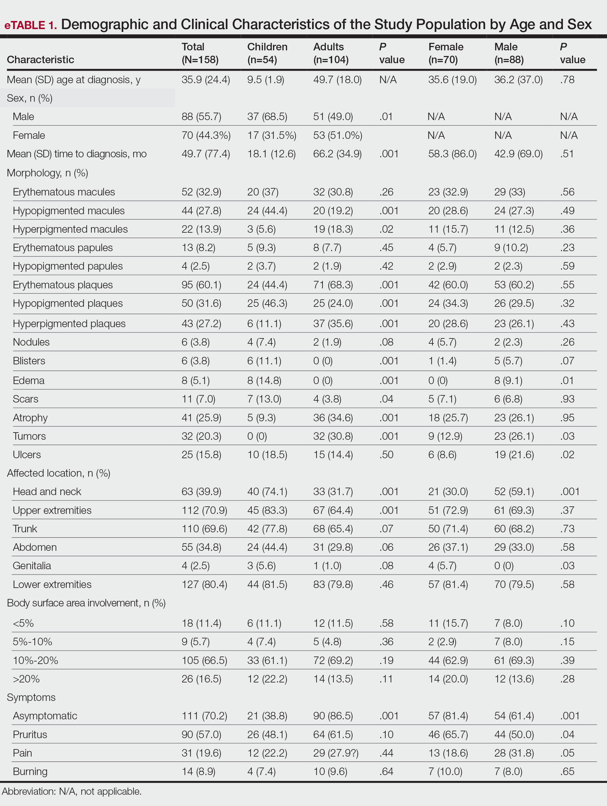
Overall, the most frequent diagnosis was MF, which occurred in 119 (75.3%) patients, and the most common lesions were erythematous plaques, noted in 95 (60.1%) patients. The lower extremities were the most affected body sites, impacting 127 (80.4%) patients, and the most common treatment was phototherapy, used to treat 110 (69.6%) patients. Reported outcomes included disease control in 45 (28.5%) patients and progression in 36 (22.8%) patients; 50 (31.6%) patients were lost to follow-up. Only 15 (9.5%) patients experienced disease remission, and 12 (7.6%) died.
The mean (SD) time between the onset of symptoms and diagnosis was shorter in children than in adults (18.1 [12.6] months vs 66.2 [34.9] months; P<.001). Regarding involved body sites, the head and neck more frequently affected children than adults (40 [74.1%] vs 33 [31.7%]; P<.001), while the upper extremities were more frequently involved in adults than in children (67 [64.4%] vs 45 [83.3%]; P<.001).
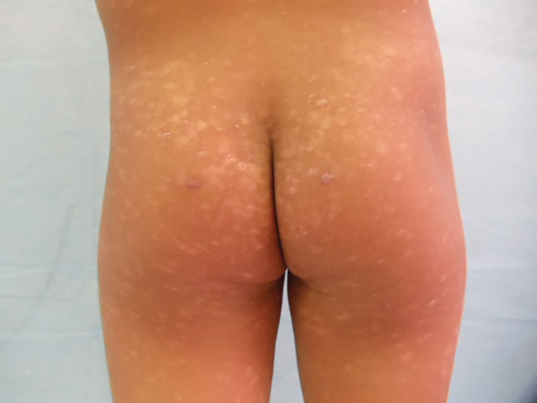
There were significant differences in the morphology of lesions and diagnoses. Children most frequently had hypopigmented plaques (25 [46.3%]) and macules (24 [44.4%])(P<.001) associated with MF, and scars (7 [13.0%]), blisters (6 [11.1%]), and edema (8 [14.8%])(P<.001) associated with positive Epstein-Barr virus (EBV) infection. Adults presented more frequently with hyperpigmented macules (19 [18.3%]) and plaques (37 [35.6%]), erythematous plaques (71 [68.3%]), atrophy (36 [34.6%]), and tumors (32 [30.8%])(P<.001). Adults were more often asymptomatic (86.5% vs 38.8%; P<.001).
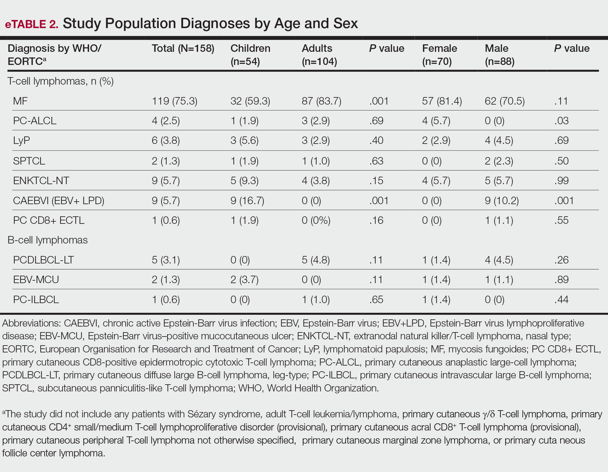
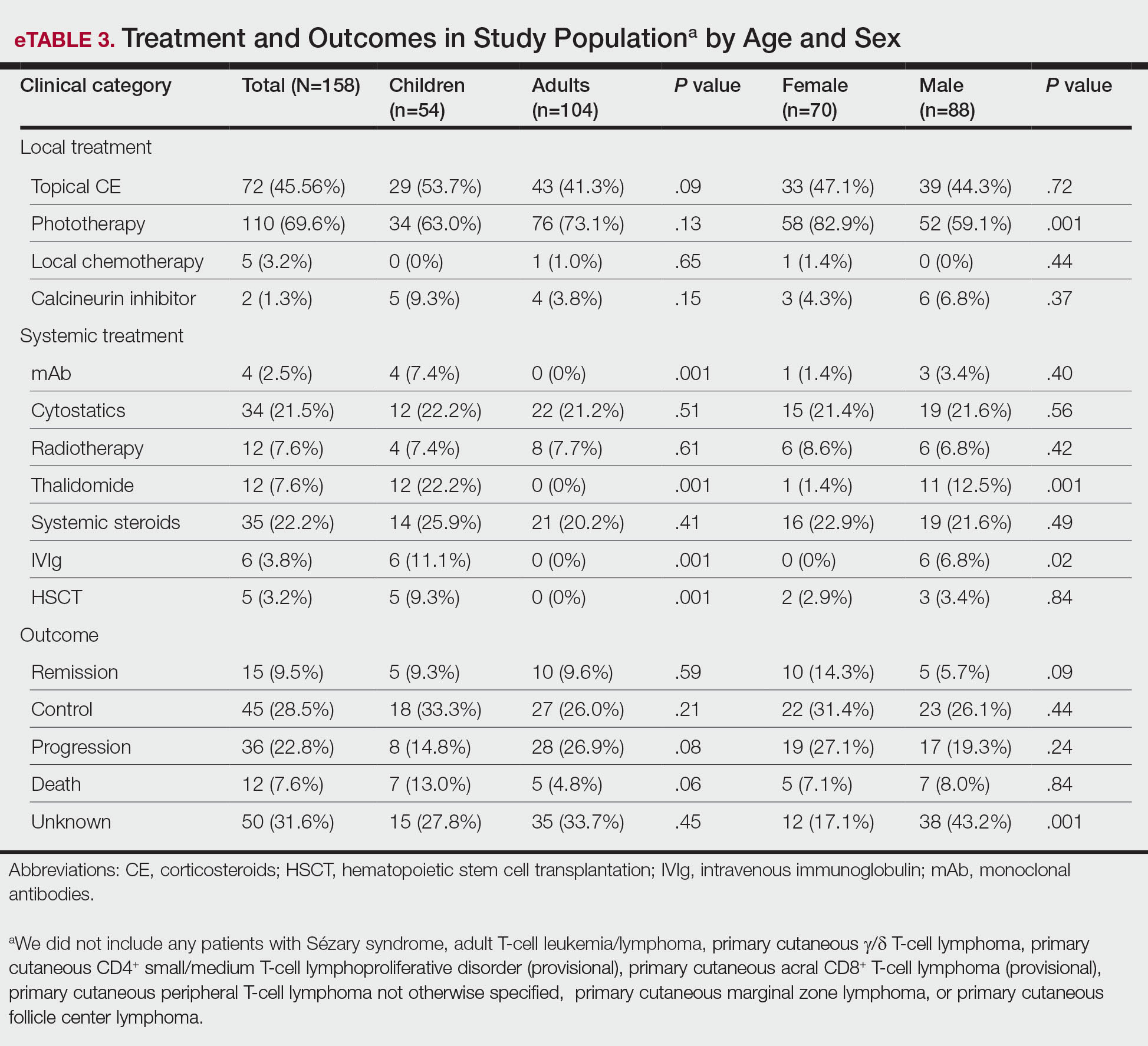
Subtypes of PCL differed by age group (eTable 2), with MF being more frequent in adults (87 [83.6%] vs 32 [59.2%]; P<.001), while chronic active positive EBV infection (CAEBVI) manifesting as lymphoproliferative disease (LPD) was exclusive to male children (9 [16.6%])(P<.001). Accordingly, we found variations in systemic treatments used: monoclonal antibodies (mAb), thalidomide, intravenous immunoglobulin (IVIg), and hematopoietic stem cell transplantation (HSCT) were used exclusively for treating children (all P<.001). Outcomes were distributed similarly by age group.
The type of PCL, clinical manifestations, and treatment also varied by sex (eTables 2 and 3). Only males had CAEBVI and presented with edema (8 [9.1%] vs 0%; P<.01). Males also had tumors (23 [26.1%] vs 9 [12.9%]; P=.03) and lesions affecting the head and neck (52 [59.1%] vs 21 [30.0%]; P<.001) more frequently than females. Males were more likely than females to report pain (28 [31.8%] vs 13 [18.6%]; P=.05) and receive systemic treatment with thalidomide (11 [12.5%] vs 1 [1.4%]; P<.001) and IVIg (6 [6.8%] vs 0%; P=.02). Only females were diagnosed with primary cutaneous anaplastic large cell lymphoma (PC-ALCL)(4 [5.7%] vs 0%; P=.03) and had genital involvement (4 [5.7%] vs 0%; P=.03). Females were more likely to be asymptomatic (57 [81.4%] vs 54 [61.4%]; P<.001) or report pruritus (46 [65.7%] vs 44 [50%]; P<.001) and receive local treatment with phototherapy (58 [82.9%] vs 52 [59.1%]; P<.001) compared to males. Although distribution of outcomes was similar by sex, we found males were more frequently lost to follow-up (38 [43.2%] vs 12 [17.1%]; P<.001).
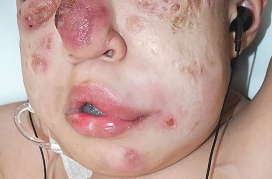
We further searched for differences in clinical manifestations according to the PCL subtype in each age group. The most frequent lesions in 32 children with MF were hypopigmented macules (21 [65.6%]; P<.001)(Figure 1). Three of 5 (60%) children with extranodal NK/T-cell lymphoma, nasal type (ENKTCL), had nodules (P=.002). Of 9 children with CAEBVI, 8 (88.9%) had edema (P<.001), ulcers (P<.001), erythematous plaques (P=.007), and hypopigmented plaques (P=.008); 5 (55.6%) children presented with scars (P<.001) and atrophy (P<.001); and 4 (44.4%) children had blisters (P=.005)(Figure 2). The 2 children with EBV-positive mucocutaneous ulcer (EBV-MCU) had crusts (P=.002) and blisters (P=.01).
Of 87 adults with MF, 54 (62%) had scaly lesions (P=.006)(Figure 3). Of 3 adult patients with PC-ALCL, 2 (66.7%) had crusts (P=.04) and ulcers (P=.05). Two of 3 (66.7%) adults with lymphomatoid papulosis (LyP) had erythematous papules (P<.001). All 4 adult patients with ENKTCL (P=.008) and all 5 patients with primary cutaneous diffuse large B-cell lymphoma, leg-type, had tumors (P<.001).
Finally, we found no differences in outcomes according to types of PCL overall or by age group; however, when categorized by sex, we found males with EBV-MCU more frequently had disease progression than females with EBV-MCU (P=.04).
Comment
Primary cutaneous lymphomas were similarly distributed among males and females (55.6% vs 44.3%, respectively). This slight male predominance was similar to other reports in the literature: one US study reported a male-to-female incidence rate ratio of 1.72, indicating a significantly higher incidence in males (P<.001).6 Similar trends have been observed in other geographic regions, with reported male-to-female ratios of 1.56 in Greece and 1.35 in Argentina.9,10 These findings suggest that PCLs are slightly more prevalent in males across different populations; however, when we stratified by age, pediatric cases of PCL were significantly more frequent in males than in females (68.5% vs. 31.4%; P=.01).
Our analysis revealed that the time to diagnosis was significantly longer in adults than in children (66.2 months vs 18.1 months) for all PCLs (P<.001). The most common type of PCL in both age groups was MF, with a notably higher prevalence in adults than in children (87 [83.7%] vs 32 [59.2%]). The prolonged course of MF in adults, often taking years to progress from early to advanced stages (47.0-52.7 months) may contribute to delayed diagnosis.8,11,12 Additionally, classic lesions of MF (erythematous scaly plaques) may resemble other common dermatologic conditions, further complicating early recognition and treatment in adults more than in children.11,12 Conversely, more aggressive and rapidly progressing PCL subtypes, including CAEBVI and ENKTCL-NT, were more frequent in children and would likely be diagnosed more promptly because of the acute onset and fast evolution of signs and symptoms.13
Mycosis fungoides is the most common CTCL, followed by CD30+ T-cell disorders such as LyP and PC-ALCL.14 While MF was the predominant subtype in both age groups, similar to previous reports,6,7 pediatric patients in our study exhibited distinctive features, such as hypopigmented macules and plaques. Hypopigmented lesions may suggest an underlying immunologic mechanism unique to younger patients, specifically children, in contrast to the hyperpigmented or violaceous lesions predominating in adults.7 The hypopigmented variant of MF has been reported to be more prevalent in children, similar to our data, accounting for 54.5% of all pediatric MF cases. These hypopigmented lesions typically manifest at an early stage and follow an indolent course.15,16 Jung et al8 conducted a systematic review of children with MF and reported a mean age at diagnosis of 12.2 years, whereas in our cohort, the mean age was 9.5 years. These findings highlight the different clinical manifestations of MF in children, which may aid in early recognition and diagnosis.
In adults, MF most commonly manifested as hyperpigmented macules and plaques, erythematous plaques, atrophic lesions, and tumors. In this population, MF remained the most frequently diagnosed PCL subtype, followed by PC-ALCL.
There were notable differences in symptom presentation between age groups and sexes. Adults were more often asymptomatic, and males reported pain more frequently.
When analyzing PCL subtypes in our study, we found that CAEBVI was exclusive to children, specifically males. The updated WHO/EORTC classification contains a new section on EBV-positive LPD in childhood, including hydroa vacciniforme–like LPD and hypersensitivity reactions to mosquito bites.4 Both are cutaneous manifestations of CAEBVI with a risk for progression to systemic EBV-positive T-cell or NK-cell lymphoma. These disorders mainly affect children and adolescents from Asia or Indigenous populations from Mexico and Central and South America.3,4,17 Cases in both female and male patients have been reported, without a clear sex predominance; however, mutations in the Src homology 2 domain containing 1A (SH2D1A) gene recently have been shown to cause X-linked lymphoproliferative disease, which is associated with predilection for EBV infection and subsequent EBV-positive LPD, including CAEBVI.18,19 Thus, it is possible some of the children with CAEBVI in our study may have an underlying X-linked lymphoproliferative disease, accounting for the male predominance.
Similarly, ENKTCL-NT tended to occur more in children than in adults in our study. Both CAEBVI-LPD and ENKTCL-NT are aggressive lymphomas with a suboptimal prognosis. Although treatment with immunomodulatory agents may lead to temporary remission in most cases, disease progression has been reported in larger cohorts, emphasizing the need for long-term follow-up and more aggressive treatments in severe cases of CAEBVI.13,17 Treatment options for EBV-positive LPD include mAb such as rituximab, IVIg, HSCT, antiviral agents, interferons α and γ, and corticosteroids.20
Treatment approaches were notably different between children and adults (P<.0001). Monoclonal antibodies, immunomodulatory agents such as thalidomide, IVIg, and HSCT were exclusively used in pediatric patients, reflecting the presence of CAEBVI and ENKTCL-NT (P<.0001). Additionally, edema, blisters, and scars were observed more frequently in children, likely due to the clinical manifestations of these EBV-related disorders.
A recent review of systemic NHL highlighted various age-related differences in clinical presentation, biology, and outcomes.13 In general, children tend to present with more aggressive subtypes and achieve better outcomes compared to adults.13 These differences may be attributed to variations in tumor biology, immune responses, and/or the benefits of early and intensive interventions in pediatric populations. These findings resonate with our results, as pediatric PCL patients received more aggressive treatments—including mAb, thalidomide, IVIg, and HSCT—likely due to the higher frequency of CAEBVI/ENKTCL-NT; however, we were unable to properly assess treatment outcomes, as many patients, both adults and children, were lost to follow-up.
Overall, our findings and comparisons with existing studies highlight the need for age-specific research and management approaches for PCL. The distinct clinical and biological profiles across age groups highlight opportunities for personalized therapies and further investigation into the molecular drivers of these differences to optimize outcomes for pediatric and adult patients.
When analyzing differences by sex, we found that CAEBVI was diagnosed exclusively in male children, who correspondingly exhibited clinical features such as edema and tumors, with lesions predominantly located on the head and neck. This also influenced treatment approach, as these patients were more likely to receive thalidomide and IVIg. In contrast, PC-ALCL was observed only in female patients, a finding that deviates from previously reported epidemiology.14 Females with PC-ALCL were more likely to have genital involvement and be asymptomatic, which could suggest a sex-related bias in disease recognition and seeking health care.
Recognizing distinctive clinical manifestations across different diagnoses and age groups can aid health care providers in early identification and accurate diagnosis of PCL. Our findings revealed several notable differences: adults with MF more frequently had scaly lesions, those with PC-ALCL had crusts and ulcers, and those with LyP had erythematous papules. All adults with ENKTCL and primary cutaneous diffuse large B-cell lymphoma, leg-type, presented with tumors. While children with MF had an increased frequency of hypopigmented macules, those with ENKTCL more frequently developed nodules, and children with EBV-MCU often had crusts and ulcers. As has already been mentioned, children with CAEBVI displayed a broad range of lesions, including edema, ulcers, erythematous and hypopigmented plaques, atrophy, blisters, and scars.
Our study was limited by the retrospective design and missing data from one-third of patients, which prevented outcome comparison. We also lacked molecular profiling of patients, which could help refine therapeutic strategies for PCL.13 Finally, as both centers included are reference institutions, results may be biased and overestimated and could differ from the rest of the population.
Conclusion
This comparative study of PCL highlighted age-related differences in clinical presentation, diagnostic distribution, and treatment patterns, including a higher prevalence among male children than female children. Adult patients with PCL had a notably longer time to diagnosis than children. The most common type of PCL identified in both age groups and sex categories was MF, but hypopigmented lesions predominated in children with this condition. Epstein-Barr virus–associated PCL occurred almost exclusively in children and manifested with nodules, edema, blisters, and scars. In terms of treatment, children received more aggressive and advanced therapies, including mAb, thalidomide, IVIg, and HSCT. Further prospective research is needed to establish variations in clinical manifestations, diagnoses, treatments, and outcomes.
- Singh R, Shaik S, Negi BS, et al. Non-Hodgkin’s lymphoma: a review.J Family Med Prim Care. 2020;9:1834-1840.
- Armitage JO, Gascoyne RD, Lunning MA, et al. Non-Hodgkin lymphoma. Lancet. 2017;390:298-310.
- Swerdlow SH, Campo E, Pileri SA, et al. The 2016 revision of the World Health Organization classification of lymphoid neoplasms. Blood. 2016;127:2375-2390.
- Willemze R, Cerroni L, Kempf W, et al. The 2018 update of the WHO-EORTC classification for primary cutaneous lymphomas. Blood. 2019;133:1703-1714.
- Willemze R, Hodak E, Zinzani PL, et al; ESMO Guidelines Committee. Primary cutaneous lymphomas: ESMO Clinical Practice Guidelines for diagnosis, treatment and follow-up. Ann Oncol. 2018;29:iv30-iv40.
- Bradford PT, Devesa SS, Anderson WF, et al. Cutaneous lymphoma incidence patterns in the United States: a population-based study of 3884 cases. Blood. 2009;113:5064-5073.
- Moon IJ, Won CH, Chang SE, et al. Prevalence, clinical features, and survival outcome trends of 627 patients with primary cutaneous lymphoma over 29 years: a retrospective review from a single tertiary center in Korea. Sci Rep. 2024;14:20118.
- Jung JM, Lim DJ, Won CH, et al. Mycosis fungoides in children and adolescents: a systematic review. JAMA Dermatol. 2021;157:431-438.
- Kaliampou S, Nikolaou V, Niforou A, et al. Epidemiological trends in cutaneous lymphomas in Greece. Eur J Dermatol. 2023;33:664-673.
- Abeldaño A, Enz P, Maskin M, et al. Primary cutaneous lymphoma in Argentina: a report of a nationwide study of 416 patients. Int J Dermatol. 2019;58:449-455.
- Cervini AB, Torres-Huamani AN, Sanchez-La-Rosa C, et al. Mycosis fungoides: experience in a pediatric hospital. Actas Dermosifiliogr. 2017;108:564-570.
- Welfringer-Morin A, Barroil M, Fraitag S, et al. Clinical features, histological characteristics, and disease outcomes of mycosis fungoides in children and adolescents: a nationwide multicentre cohort of 46 patients. Dermatology. 2023;239:132-139.
- Sandlund JT, Martin MG. Non-Hodgkin lymphoma across the pediatric and adolescent and young adult age spectrum. Hematology Am Soc Hematol Educ Program. 2016;2016:589-597.
- Ortiz-Hidalgo C, Pina-Oviedo S. Primary cutaneous anaplastic large cell lymphoma-a review of clinical, morphological, immunohistochemical, and molecular features. Cancers (Basel). 2023;15:4098.
- Nielsen PR, Eriksen JO, Wehkamp U, et al. Clinical and histological characteristics of mycosis fungoides and Sézary syndrome: a retrospective, single-centre study of 43 patients from eastern Denmark. Acta Derm Venereol. 2019;99:1231-1236.
- Suh KS, Jang MS, Jung JH, et al. Clinical characteristics and long-term outcome of 223 patients with mycosis fungoides at a single tertiary center in Korea: a 29-year review. J Am Acad Dermatol. 2022;86:1275-1284.
- Quintanilla-Martinez L, Ridaura C, Nagl F, et al. Hydroa vacciniforme-like lymphoma: a chronic EBV+ lymphoproliferative disorder with risk to develop a systemic lymphoma. Blood. 2013;122:3101-3110.
- Fujiwara S, Nakamura H. Chronic active Epstein-Barr virus infection: is it immunodeficiency, malignancy, or both? Cancers (Basel). 2020;12:3202.
- Sumazaki R, Kanegane H, Osaki M, et al. SH2D1A mutations in Japanese males with severe Epstein-Barr virus–associated illnesses. Blood. 2001;98:1268-1270.
- Kimura H. Pathogenesis of chronic active Epstein-Barr virus infection: is this an infectious disease, lymphoproliferative disorder, or immunodeficiency? Rev Med Virol. 2006;16:251-261.
Non-Hodgkin lymphomas (NHLs) are a heterogeneous group of lymphoproliferative malignancies originating from T, B, or natural killer (NK) lymphocytes.1 Compared to Hodgkin lymphomas, NHLs exhibit a broader clinical spectrum and have a poorer prognosis and frequent extranodal involvement, with the skin being the second most frequent extranodal site.2 Primary cutaneous lymphomas (PCLs) are NHLs that are first evident on the skin without evidence of extracutaneous disease at diagnosis. They include a heterogeneous group of cutaneous T-cell lymphomas (CTCLs) and cutaneous B-cell lymphomas. Among CTCLs, mycosis fungoides (MF) is the most prevalent subtype, generally following an indolent course. Cutaneous B-cell lymphomas primarily include follicle center lymphoma and diffuse large B-cell lymphoma.3,4
The clinical behavior and incidence of PCLs vary dramatically between children and adults, suggesting underlying biologic, immunologic, and genetic differences.5,6 However, there is a notable lack of comparative studies in the literature addressing these variations, limiting a comprehensive understanding of PCLs and hindering the development of tailored therapeutic strategies. The main objective of this study was to evaluate the different clinical characteristics, subtypes, treatment options, and prognosis in PCLs between pediatric and adult populations.7,8
Methods
This retrospective observational study included pediatric (aged ≤18 years at diagnosis) and adult (aged >18 years at diagnosis) patients with a clinical and histopathologic diagnosis of PCL who were diagnosed and treated at either of 2 tertiary institutions (Dr. Manuel Gea Gonzalez General Hospital or the National Institute of Pediatrics, both in Mexico City, Mexico) between January 1, 1999, and December 31, 2019. The data analysis included demographic and clinical characteristics, type of PCL (according to World Health Organization [WHO]/European Organisation for Research and Treatment of Cancer [EORTC]) classification,3,4 treatment administered, and outcome. Disease remission was defined as remittance of clinical PCL manifestations for 3 months or more. Disease control was defined as stable clinical manifestations with no change in PCL stage following treatment initiation. Disease progression was defined as worsening clinical manifestations with an increase in PCL stage and/or development of systemic lymphoma. Beyond similarities and differences across age groups, we also analyzed differences by sex. χ² and Mann-Whitney U tests were used to assess differences between groups. Statistical significance was set at P≤.05. Institutional approval was obtained from both tertiary centers.
Results
Our analysis included a total of 158 patients: 54 (34.2%) were children and 104 (65.8%) were adults. Eighty-eight (55.7%) patients were male (eTable 1). The mean (SD) age at diagnosis was 9.5 (1.9) years in children and 49.7 (18) years in adults. Regarding differences between age groups, adult patients had a similar sex distribution, while in children, the majority of patients were male (37/54 [68.5%]; P=.01).
Overall, the most frequent diagnosis was MF, which occurred in 119 (75.3%) patients, and the most common lesions were erythematous plaques, noted in 95 (60.1%) patients. The lower extremities were the most affected body sites, impacting 127 (80.4%) patients, and the most common treatment was phototherapy, used to treat 110 (69.6%) patients. Reported outcomes included disease control in 45 (28.5%) patients and progression in 36 (22.8%) patients; 50 (31.6%) patients were lost to follow-up. Only 15 (9.5%) patients experienced disease remission, and 12 (7.6%) died.
The mean (SD) time between the onset of symptoms and diagnosis was shorter in children than in adults (18.1 [12.6] months vs 66.2 [34.9] months; P<.001). Regarding involved body sites, the head and neck more frequently affected children than adults (40 [74.1%] vs 33 [31.7%]; P<.001), while the upper extremities were more frequently involved in adults than in children (67 [64.4%] vs 45 [83.3%]; P<.001).
There were significant differences in the morphology of lesions and diagnoses. Children most frequently had hypopigmented plaques (25 [46.3%]) and macules (24 [44.4%])(P<.001) associated with MF, and scars (7 [13.0%]), blisters (6 [11.1%]), and edema (8 [14.8%])(P<.001) associated with positive Epstein-Barr virus (EBV) infection. Adults presented more frequently with hyperpigmented macules (19 [18.3%]) and plaques (37 [35.6%]), erythematous plaques (71 [68.3%]), atrophy (36 [34.6%]), and tumors (32 [30.8%])(P<.001). Adults were more often asymptomatic (86.5% vs 38.8%; P<.001).
Subtypes of PCL differed by age group (eTable 2), with MF being more frequent in adults (87 [83.6%] vs 32 [59.2%]; P<.001), while chronic active positive EBV infection (CAEBVI) manifesting as lymphoproliferative disease (LPD) was exclusive to male children (9 [16.6%])(P<.001). Accordingly, we found variations in systemic treatments used: monoclonal antibodies (mAb), thalidomide, intravenous immunoglobulin (IVIg), and hematopoietic stem cell transplantation (HSCT) were used exclusively for treating children (all P<.001). Outcomes were distributed similarly by age group.
The type of PCL, clinical manifestations, and treatment also varied by sex (eTables 2 and 3). Only males had CAEBVI and presented with edema (8 [9.1%] vs 0%; P<.01). Males also had tumors (23 [26.1%] vs 9 [12.9%]; P=.03) and lesions affecting the head and neck (52 [59.1%] vs 21 [30.0%]; P<.001) more frequently than females. Males were more likely than females to report pain (28 [31.8%] vs 13 [18.6%]; P=.05) and receive systemic treatment with thalidomide (11 [12.5%] vs 1 [1.4%]; P<.001) and IVIg (6 [6.8%] vs 0%; P=.02). Only females were diagnosed with primary cutaneous anaplastic large cell lymphoma (PC-ALCL)(4 [5.7%] vs 0%; P=.03) and had genital involvement (4 [5.7%] vs 0%; P=.03). Females were more likely to be asymptomatic (57 [81.4%] vs 54 [61.4%]; P<.001) or report pruritus (46 [65.7%] vs 44 [50%]; P<.001) and receive local treatment with phototherapy (58 [82.9%] vs 52 [59.1%]; P<.001) compared to males. Although distribution of outcomes was similar by sex, we found males were more frequently lost to follow-up (38 [43.2%] vs 12 [17.1%]; P<.001).
We further searched for differences in clinical manifestations according to the PCL subtype in each age group. The most frequent lesions in 32 children with MF were hypopigmented macules (21 [65.6%]; P<.001)(Figure 1). Three of 5 (60%) children with extranodal NK/T-cell lymphoma, nasal type (ENKTCL), had nodules (P=.002). Of 9 children with CAEBVI, 8 (88.9%) had edema (P<.001), ulcers (P<.001), erythematous plaques (P=.007), and hypopigmented plaques (P=.008); 5 (55.6%) children presented with scars (P<.001) and atrophy (P<.001); and 4 (44.4%) children had blisters (P=.005)(Figure 2). The 2 children with EBV-positive mucocutaneous ulcer (EBV-MCU) had crusts (P=.002) and blisters (P=.01).
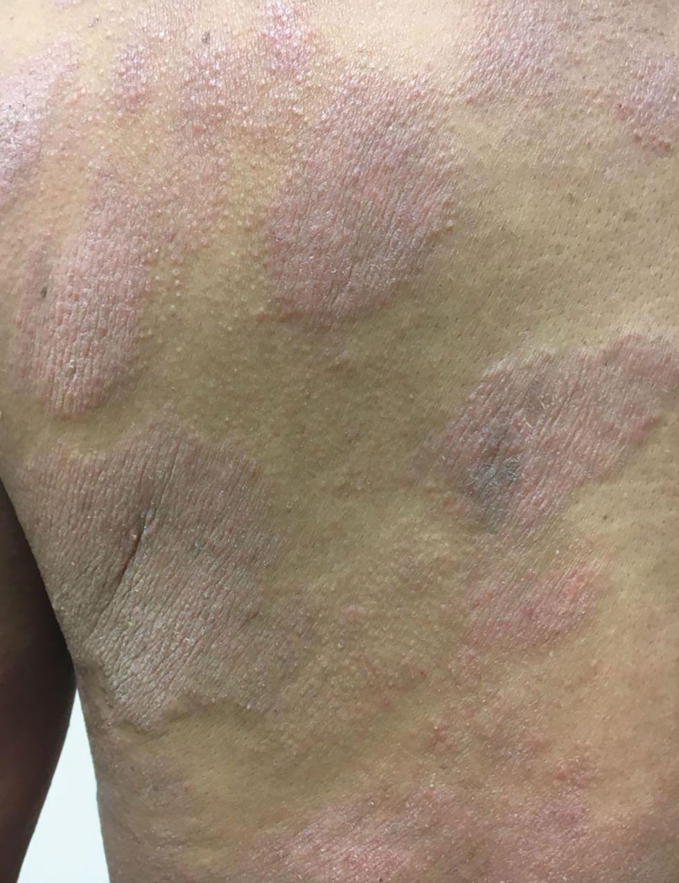
Of 87 adults with MF, 54 (62%) had scaly lesions (P=.006)(Figure 3). Of 3 adult patients with PC-ALCL, 2 (66.7%) had crusts (P=.04) and ulcers (P=.05). Two of 3 (66.7%) adults with lymphomatoid papulosis (LyP) had erythematous papules (P<.001). All 4 adult patients with ENKTCL (P=.008) and all 5 patients with primary cutaneous diffuse large B-cell lymphoma, leg-type, had tumors (P<.001).
Finally, we found no differences in outcomes according to types of PCL overall or by age group; however, when categorized by sex, we found males with EBV-MCU more frequently had disease progression than females with EBV-MCU (P=.04).
Comment
Primary cutaneous lymphomas were similarly distributed among males and females (55.6% vs 44.3%, respectively). This slight male predominance was similar to other reports in the literature: one US study reported a male-to-female incidence rate ratio of 1.72, indicating a significantly higher incidence in males (P<.001).6 Similar trends have been observed in other geographic regions, with reported male-to-female ratios of 1.56 in Greece and 1.35 in Argentina.9,10 These findings suggest that PCLs are slightly more prevalent in males across different populations; however, when we stratified by age, pediatric cases of PCL were significantly more frequent in males than in females (68.5% vs. 31.4%; P=.01).
Our analysis revealed that the time to diagnosis was significantly longer in adults than in children (66.2 months vs 18.1 months) for all PCLs (P<.001). The most common type of PCL in both age groups was MF, with a notably higher prevalence in adults than in children (87 [83.7%] vs 32 [59.2%]). The prolonged course of MF in adults, often taking years to progress from early to advanced stages (47.0-52.7 months) may contribute to delayed diagnosis.8,11,12 Additionally, classic lesions of MF (erythematous scaly plaques) may resemble other common dermatologic conditions, further complicating early recognition and treatment in adults more than in children.11,12 Conversely, more aggressive and rapidly progressing PCL subtypes, including CAEBVI and ENKTCL-NT, were more frequent in children and would likely be diagnosed more promptly because of the acute onset and fast evolution of signs and symptoms.13
Mycosis fungoides is the most common CTCL, followed by CD30+ T-cell disorders such as LyP and PC-ALCL.14 While MF was the predominant subtype in both age groups, similar to previous reports,6,7 pediatric patients in our study exhibited distinctive features, such as hypopigmented macules and plaques. Hypopigmented lesions may suggest an underlying immunologic mechanism unique to younger patients, specifically children, in contrast to the hyperpigmented or violaceous lesions predominating in adults.7 The hypopigmented variant of MF has been reported to be more prevalent in children, similar to our data, accounting for 54.5% of all pediatric MF cases. These hypopigmented lesions typically manifest at an early stage and follow an indolent course.15,16 Jung et al8 conducted a systematic review of children with MF and reported a mean age at diagnosis of 12.2 years, whereas in our cohort, the mean age was 9.5 years. These findings highlight the different clinical manifestations of MF in children, which may aid in early recognition and diagnosis.
In adults, MF most commonly manifested as hyperpigmented macules and plaques, erythematous plaques, atrophic lesions, and tumors. In this population, MF remained the most frequently diagnosed PCL subtype, followed by PC-ALCL.
There were notable differences in symptom presentation between age groups and sexes. Adults were more often asymptomatic, and males reported pain more frequently.
When analyzing PCL subtypes in our study, we found that CAEBVI was exclusive to children, specifically males. The updated WHO/EORTC classification contains a new section on EBV-positive LPD in childhood, including hydroa vacciniforme–like LPD and hypersensitivity reactions to mosquito bites.4 Both are cutaneous manifestations of CAEBVI with a risk for progression to systemic EBV-positive T-cell or NK-cell lymphoma. These disorders mainly affect children and adolescents from Asia or Indigenous populations from Mexico and Central and South America.3,4,17 Cases in both female and male patients have been reported, without a clear sex predominance; however, mutations in the Src homology 2 domain containing 1A (SH2D1A) gene recently have been shown to cause X-linked lymphoproliferative disease, which is associated with predilection for EBV infection and subsequent EBV-positive LPD, including CAEBVI.18,19 Thus, it is possible some of the children with CAEBVI in our study may have an underlying X-linked lymphoproliferative disease, accounting for the male predominance.
Similarly, ENKTCL-NT tended to occur more in children than in adults in our study. Both CAEBVI-LPD and ENKTCL-NT are aggressive lymphomas with a suboptimal prognosis. Although treatment with immunomodulatory agents may lead to temporary remission in most cases, disease progression has been reported in larger cohorts, emphasizing the need for long-term follow-up and more aggressive treatments in severe cases of CAEBVI.13,17 Treatment options for EBV-positive LPD include mAb such as rituximab, IVIg, HSCT, antiviral agents, interferons α and γ, and corticosteroids.20
Treatment approaches were notably different between children and adults (P<.0001). Monoclonal antibodies, immunomodulatory agents such as thalidomide, IVIg, and HSCT were exclusively used in pediatric patients, reflecting the presence of CAEBVI and ENKTCL-NT (P<.0001). Additionally, edema, blisters, and scars were observed more frequently in children, likely due to the clinical manifestations of these EBV-related disorders.
A recent review of systemic NHL highlighted various age-related differences in clinical presentation, biology, and outcomes.13 In general, children tend to present with more aggressive subtypes and achieve better outcomes compared to adults.13 These differences may be attributed to variations in tumor biology, immune responses, and/or the benefits of early and intensive interventions in pediatric populations. These findings resonate with our results, as pediatric PCL patients received more aggressive treatments—including mAb, thalidomide, IVIg, and HSCT—likely due to the higher frequency of CAEBVI/ENKTCL-NT; however, we were unable to properly assess treatment outcomes, as many patients, both adults and children, were lost to follow-up.
Overall, our findings and comparisons with existing studies highlight the need for age-specific research and management approaches for PCL. The distinct clinical and biological profiles across age groups highlight opportunities for personalized therapies and further investigation into the molecular drivers of these differences to optimize outcomes for pediatric and adult patients.
When analyzing differences by sex, we found that CAEBVI was diagnosed exclusively in male children, who correspondingly exhibited clinical features such as edema and tumors, with lesions predominantly located on the head and neck. This also influenced treatment approach, as these patients were more likely to receive thalidomide and IVIg. In contrast, PC-ALCL was observed only in female patients, a finding that deviates from previously reported epidemiology.14 Females with PC-ALCL were more likely to have genital involvement and be asymptomatic, which could suggest a sex-related bias in disease recognition and seeking health care.
Recognizing distinctive clinical manifestations across different diagnoses and age groups can aid health care providers in early identification and accurate diagnosis of PCL. Our findings revealed several notable differences: adults with MF more frequently had scaly lesions, those with PC-ALCL had crusts and ulcers, and those with LyP had erythematous papules. All adults with ENKTCL and primary cutaneous diffuse large B-cell lymphoma, leg-type, presented with tumors. While children with MF had an increased frequency of hypopigmented macules, those with ENKTCL more frequently developed nodules, and children with EBV-MCU often had crusts and ulcers. As has already been mentioned, children with CAEBVI displayed a broad range of lesions, including edema, ulcers, erythematous and hypopigmented plaques, atrophy, blisters, and scars.
Our study was limited by the retrospective design and missing data from one-third of patients, which prevented outcome comparison. We also lacked molecular profiling of patients, which could help refine therapeutic strategies for PCL.13 Finally, as both centers included are reference institutions, results may be biased and overestimated and could differ from the rest of the population.
Conclusion
This comparative study of PCL highlighted age-related differences in clinical presentation, diagnostic distribution, and treatment patterns, including a higher prevalence among male children than female children. Adult patients with PCL had a notably longer time to diagnosis than children. The most common type of PCL identified in both age groups and sex categories was MF, but hypopigmented lesions predominated in children with this condition. Epstein-Barr virus–associated PCL occurred almost exclusively in children and manifested with nodules, edema, blisters, and scars. In terms of treatment, children received more aggressive and advanced therapies, including mAb, thalidomide, IVIg, and HSCT. Further prospective research is needed to establish variations in clinical manifestations, diagnoses, treatments, and outcomes.
Non-Hodgkin lymphomas (NHLs) are a heterogeneous group of lymphoproliferative malignancies originating from T, B, or natural killer (NK) lymphocytes.1 Compared to Hodgkin lymphomas, NHLs exhibit a broader clinical spectrum and have a poorer prognosis and frequent extranodal involvement, with the skin being the second most frequent extranodal site.2 Primary cutaneous lymphomas (PCLs) are NHLs that are first evident on the skin without evidence of extracutaneous disease at diagnosis. They include a heterogeneous group of cutaneous T-cell lymphomas (CTCLs) and cutaneous B-cell lymphomas. Among CTCLs, mycosis fungoides (MF) is the most prevalent subtype, generally following an indolent course. Cutaneous B-cell lymphomas primarily include follicle center lymphoma and diffuse large B-cell lymphoma.3,4
The clinical behavior and incidence of PCLs vary dramatically between children and adults, suggesting underlying biologic, immunologic, and genetic differences.5,6 However, there is a notable lack of comparative studies in the literature addressing these variations, limiting a comprehensive understanding of PCLs and hindering the development of tailored therapeutic strategies. The main objective of this study was to evaluate the different clinical characteristics, subtypes, treatment options, and prognosis in PCLs between pediatric and adult populations.7,8
Methods
This retrospective observational study included pediatric (aged ≤18 years at diagnosis) and adult (aged >18 years at diagnosis) patients with a clinical and histopathologic diagnosis of PCL who were diagnosed and treated at either of 2 tertiary institutions (Dr. Manuel Gea Gonzalez General Hospital or the National Institute of Pediatrics, both in Mexico City, Mexico) between January 1, 1999, and December 31, 2019. The data analysis included demographic and clinical characteristics, type of PCL (according to World Health Organization [WHO]/European Organisation for Research and Treatment of Cancer [EORTC]) classification,3,4 treatment administered, and outcome. Disease remission was defined as remittance of clinical PCL manifestations for 3 months or more. Disease control was defined as stable clinical manifestations with no change in PCL stage following treatment initiation. Disease progression was defined as worsening clinical manifestations with an increase in PCL stage and/or development of systemic lymphoma. Beyond similarities and differences across age groups, we also analyzed differences by sex. χ² and Mann-Whitney U tests were used to assess differences between groups. Statistical significance was set at P≤.05. Institutional approval was obtained from both tertiary centers.
Results
Our analysis included a total of 158 patients: 54 (34.2%) were children and 104 (65.8%) were adults. Eighty-eight (55.7%) patients were male (eTable 1). The mean (SD) age at diagnosis was 9.5 (1.9) years in children and 49.7 (18) years in adults. Regarding differences between age groups, adult patients had a similar sex distribution, while in children, the majority of patients were male (37/54 [68.5%]; P=.01).
Overall, the most frequent diagnosis was MF, which occurred in 119 (75.3%) patients, and the most common lesions were erythematous plaques, noted in 95 (60.1%) patients. The lower extremities were the most affected body sites, impacting 127 (80.4%) patients, and the most common treatment was phototherapy, used to treat 110 (69.6%) patients. Reported outcomes included disease control in 45 (28.5%) patients and progression in 36 (22.8%) patients; 50 (31.6%) patients were lost to follow-up. Only 15 (9.5%) patients experienced disease remission, and 12 (7.6%) died.
The mean (SD) time between the onset of symptoms and diagnosis was shorter in children than in adults (18.1 [12.6] months vs 66.2 [34.9] months; P<.001). Regarding involved body sites, the head and neck more frequently affected children than adults (40 [74.1%] vs 33 [31.7%]; P<.001), while the upper extremities were more frequently involved in adults than in children (67 [64.4%] vs 45 [83.3%]; P<.001).
There were significant differences in the morphology of lesions and diagnoses. Children most frequently had hypopigmented plaques (25 [46.3%]) and macules (24 [44.4%])(P<.001) associated with MF, and scars (7 [13.0%]), blisters (6 [11.1%]), and edema (8 [14.8%])(P<.001) associated with positive Epstein-Barr virus (EBV) infection. Adults presented more frequently with hyperpigmented macules (19 [18.3%]) and plaques (37 [35.6%]), erythematous plaques (71 [68.3%]), atrophy (36 [34.6%]), and tumors (32 [30.8%])(P<.001). Adults were more often asymptomatic (86.5% vs 38.8%; P<.001).
Subtypes of PCL differed by age group (eTable 2), with MF being more frequent in adults (87 [83.6%] vs 32 [59.2%]; P<.001), while chronic active positive EBV infection (CAEBVI) manifesting as lymphoproliferative disease (LPD) was exclusive to male children (9 [16.6%])(P<.001). Accordingly, we found variations in systemic treatments used: monoclonal antibodies (mAb), thalidomide, intravenous immunoglobulin (IVIg), and hematopoietic stem cell transplantation (HSCT) were used exclusively for treating children (all P<.001). Outcomes were distributed similarly by age group.
The type of PCL, clinical manifestations, and treatment also varied by sex (eTables 2 and 3). Only males had CAEBVI and presented with edema (8 [9.1%] vs 0%; P<.01). Males also had tumors (23 [26.1%] vs 9 [12.9%]; P=.03) and lesions affecting the head and neck (52 [59.1%] vs 21 [30.0%]; P<.001) more frequently than females. Males were more likely than females to report pain (28 [31.8%] vs 13 [18.6%]; P=.05) and receive systemic treatment with thalidomide (11 [12.5%] vs 1 [1.4%]; P<.001) and IVIg (6 [6.8%] vs 0%; P=.02). Only females were diagnosed with primary cutaneous anaplastic large cell lymphoma (PC-ALCL)(4 [5.7%] vs 0%; P=.03) and had genital involvement (4 [5.7%] vs 0%; P=.03). Females were more likely to be asymptomatic (57 [81.4%] vs 54 [61.4%]; P<.001) or report pruritus (46 [65.7%] vs 44 [50%]; P<.001) and receive local treatment with phototherapy (58 [82.9%] vs 52 [59.1%]; P<.001) compared to males. Although distribution of outcomes was similar by sex, we found males were more frequently lost to follow-up (38 [43.2%] vs 12 [17.1%]; P<.001).
We further searched for differences in clinical manifestations according to the PCL subtype in each age group. The most frequent lesions in 32 children with MF were hypopigmented macules (21 [65.6%]; P<.001)(Figure 1). Three of 5 (60%) children with extranodal NK/T-cell lymphoma, nasal type (ENKTCL), had nodules (P=.002). Of 9 children with CAEBVI, 8 (88.9%) had edema (P<.001), ulcers (P<.001), erythematous plaques (P=.007), and hypopigmented plaques (P=.008); 5 (55.6%) children presented with scars (P<.001) and atrophy (P<.001); and 4 (44.4%) children had blisters (P=.005)(Figure 2). The 2 children with EBV-positive mucocutaneous ulcer (EBV-MCU) had crusts (P=.002) and blisters (P=.01).
Of 87 adults with MF, 54 (62%) had scaly lesions (P=.006)(Figure 3). Of 3 adult patients with PC-ALCL, 2 (66.7%) had crusts (P=.04) and ulcers (P=.05). Two of 3 (66.7%) adults with lymphomatoid papulosis (LyP) had erythematous papules (P<.001). All 4 adult patients with ENKTCL (P=.008) and all 5 patients with primary cutaneous diffuse large B-cell lymphoma, leg-type, had tumors (P<.001).
Finally, we found no differences in outcomes according to types of PCL overall or by age group; however, when categorized by sex, we found males with EBV-MCU more frequently had disease progression than females with EBV-MCU (P=.04).
Comment
Primary cutaneous lymphomas were similarly distributed among males and females (55.6% vs 44.3%, respectively). This slight male predominance was similar to other reports in the literature: one US study reported a male-to-female incidence rate ratio of 1.72, indicating a significantly higher incidence in males (P<.001).6 Similar trends have been observed in other geographic regions, with reported male-to-female ratios of 1.56 in Greece and 1.35 in Argentina.9,10 These findings suggest that PCLs are slightly more prevalent in males across different populations; however, when we stratified by age, pediatric cases of PCL were significantly more frequent in males than in females (68.5% vs. 31.4%; P=.01).
Our analysis revealed that the time to diagnosis was significantly longer in adults than in children (66.2 months vs 18.1 months) for all PCLs (P<.001). The most common type of PCL in both age groups was MF, with a notably higher prevalence in adults than in children (87 [83.7%] vs 32 [59.2%]). The prolonged course of MF in adults, often taking years to progress from early to advanced stages (47.0-52.7 months) may contribute to delayed diagnosis.8,11,12 Additionally, classic lesions of MF (erythematous scaly plaques) may resemble other common dermatologic conditions, further complicating early recognition and treatment in adults more than in children.11,12 Conversely, more aggressive and rapidly progressing PCL subtypes, including CAEBVI and ENKTCL-NT, were more frequent in children and would likely be diagnosed more promptly because of the acute onset and fast evolution of signs and symptoms.13
Mycosis fungoides is the most common CTCL, followed by CD30+ T-cell disorders such as LyP and PC-ALCL.14 While MF was the predominant subtype in both age groups, similar to previous reports,6,7 pediatric patients in our study exhibited distinctive features, such as hypopigmented macules and plaques. Hypopigmented lesions may suggest an underlying immunologic mechanism unique to younger patients, specifically children, in contrast to the hyperpigmented or violaceous lesions predominating in adults.7 The hypopigmented variant of MF has been reported to be more prevalent in children, similar to our data, accounting for 54.5% of all pediatric MF cases. These hypopigmented lesions typically manifest at an early stage and follow an indolent course.15,16 Jung et al8 conducted a systematic review of children with MF and reported a mean age at diagnosis of 12.2 years, whereas in our cohort, the mean age was 9.5 years. These findings highlight the different clinical manifestations of MF in children, which may aid in early recognition and diagnosis.
In adults, MF most commonly manifested as hyperpigmented macules and plaques, erythematous plaques, atrophic lesions, and tumors. In this population, MF remained the most frequently diagnosed PCL subtype, followed by PC-ALCL.
There were notable differences in symptom presentation between age groups and sexes. Adults were more often asymptomatic, and males reported pain more frequently.
When analyzing PCL subtypes in our study, we found that CAEBVI was exclusive to children, specifically males. The updated WHO/EORTC classification contains a new section on EBV-positive LPD in childhood, including hydroa vacciniforme–like LPD and hypersensitivity reactions to mosquito bites.4 Both are cutaneous manifestations of CAEBVI with a risk for progression to systemic EBV-positive T-cell or NK-cell lymphoma. These disorders mainly affect children and adolescents from Asia or Indigenous populations from Mexico and Central and South America.3,4,17 Cases in both female and male patients have been reported, without a clear sex predominance; however, mutations in the Src homology 2 domain containing 1A (SH2D1A) gene recently have been shown to cause X-linked lymphoproliferative disease, which is associated with predilection for EBV infection and subsequent EBV-positive LPD, including CAEBVI.18,19 Thus, it is possible some of the children with CAEBVI in our study may have an underlying X-linked lymphoproliferative disease, accounting for the male predominance.
Similarly, ENKTCL-NT tended to occur more in children than in adults in our study. Both CAEBVI-LPD and ENKTCL-NT are aggressive lymphomas with a suboptimal prognosis. Although treatment with immunomodulatory agents may lead to temporary remission in most cases, disease progression has been reported in larger cohorts, emphasizing the need for long-term follow-up and more aggressive treatments in severe cases of CAEBVI.13,17 Treatment options for EBV-positive LPD include mAb such as rituximab, IVIg, HSCT, antiviral agents, interferons α and γ, and corticosteroids.20
Treatment approaches were notably different between children and adults (P<.0001). Monoclonal antibodies, immunomodulatory agents such as thalidomide, IVIg, and HSCT were exclusively used in pediatric patients, reflecting the presence of CAEBVI and ENKTCL-NT (P<.0001). Additionally, edema, blisters, and scars were observed more frequently in children, likely due to the clinical manifestations of these EBV-related disorders.
A recent review of systemic NHL highlighted various age-related differences in clinical presentation, biology, and outcomes.13 In general, children tend to present with more aggressive subtypes and achieve better outcomes compared to adults.13 These differences may be attributed to variations in tumor biology, immune responses, and/or the benefits of early and intensive interventions in pediatric populations. These findings resonate with our results, as pediatric PCL patients received more aggressive treatments—including mAb, thalidomide, IVIg, and HSCT—likely due to the higher frequency of CAEBVI/ENKTCL-NT; however, we were unable to properly assess treatment outcomes, as many patients, both adults and children, were lost to follow-up.
Overall, our findings and comparisons with existing studies highlight the need for age-specific research and management approaches for PCL. The distinct clinical and biological profiles across age groups highlight opportunities for personalized therapies and further investigation into the molecular drivers of these differences to optimize outcomes for pediatric and adult patients.
When analyzing differences by sex, we found that CAEBVI was diagnosed exclusively in male children, who correspondingly exhibited clinical features such as edema and tumors, with lesions predominantly located on the head and neck. This also influenced treatment approach, as these patients were more likely to receive thalidomide and IVIg. In contrast, PC-ALCL was observed only in female patients, a finding that deviates from previously reported epidemiology.14 Females with PC-ALCL were more likely to have genital involvement and be asymptomatic, which could suggest a sex-related bias in disease recognition and seeking health care.
Recognizing distinctive clinical manifestations across different diagnoses and age groups can aid health care providers in early identification and accurate diagnosis of PCL. Our findings revealed several notable differences: adults with MF more frequently had scaly lesions, those with PC-ALCL had crusts and ulcers, and those with LyP had erythematous papules. All adults with ENKTCL and primary cutaneous diffuse large B-cell lymphoma, leg-type, presented with tumors. While children with MF had an increased frequency of hypopigmented macules, those with ENKTCL more frequently developed nodules, and children with EBV-MCU often had crusts and ulcers. As has already been mentioned, children with CAEBVI displayed a broad range of lesions, including edema, ulcers, erythematous and hypopigmented plaques, atrophy, blisters, and scars.
Our study was limited by the retrospective design and missing data from one-third of patients, which prevented outcome comparison. We also lacked molecular profiling of patients, which could help refine therapeutic strategies for PCL.13 Finally, as both centers included are reference institutions, results may be biased and overestimated and could differ from the rest of the population.
Conclusion
This comparative study of PCL highlighted age-related differences in clinical presentation, diagnostic distribution, and treatment patterns, including a higher prevalence among male children than female children. Adult patients with PCL had a notably longer time to diagnosis than children. The most common type of PCL identified in both age groups and sex categories was MF, but hypopigmented lesions predominated in children with this condition. Epstein-Barr virus–associated PCL occurred almost exclusively in children and manifested with nodules, edema, blisters, and scars. In terms of treatment, children received more aggressive and advanced therapies, including mAb, thalidomide, IVIg, and HSCT. Further prospective research is needed to establish variations in clinical manifestations, diagnoses, treatments, and outcomes.
- Singh R, Shaik S, Negi BS, et al. Non-Hodgkin’s lymphoma: a review.J Family Med Prim Care. 2020;9:1834-1840.
- Armitage JO, Gascoyne RD, Lunning MA, et al. Non-Hodgkin lymphoma. Lancet. 2017;390:298-310.
- Swerdlow SH, Campo E, Pileri SA, et al. The 2016 revision of the World Health Organization classification of lymphoid neoplasms. Blood. 2016;127:2375-2390.
- Willemze R, Cerroni L, Kempf W, et al. The 2018 update of the WHO-EORTC classification for primary cutaneous lymphomas. Blood. 2019;133:1703-1714.
- Willemze R, Hodak E, Zinzani PL, et al; ESMO Guidelines Committee. Primary cutaneous lymphomas: ESMO Clinical Practice Guidelines for diagnosis, treatment and follow-up. Ann Oncol. 2018;29:iv30-iv40.
- Bradford PT, Devesa SS, Anderson WF, et al. Cutaneous lymphoma incidence patterns in the United States: a population-based study of 3884 cases. Blood. 2009;113:5064-5073.
- Moon IJ, Won CH, Chang SE, et al. Prevalence, clinical features, and survival outcome trends of 627 patients with primary cutaneous lymphoma over 29 years: a retrospective review from a single tertiary center in Korea. Sci Rep. 2024;14:20118.
- Jung JM, Lim DJ, Won CH, et al. Mycosis fungoides in children and adolescents: a systematic review. JAMA Dermatol. 2021;157:431-438.
- Kaliampou S, Nikolaou V, Niforou A, et al. Epidemiological trends in cutaneous lymphomas in Greece. Eur J Dermatol. 2023;33:664-673.
- Abeldaño A, Enz P, Maskin M, et al. Primary cutaneous lymphoma in Argentina: a report of a nationwide study of 416 patients. Int J Dermatol. 2019;58:449-455.
- Cervini AB, Torres-Huamani AN, Sanchez-La-Rosa C, et al. Mycosis fungoides: experience in a pediatric hospital. Actas Dermosifiliogr. 2017;108:564-570.
- Welfringer-Morin A, Barroil M, Fraitag S, et al. Clinical features, histological characteristics, and disease outcomes of mycosis fungoides in children and adolescents: a nationwide multicentre cohort of 46 patients. Dermatology. 2023;239:132-139.
- Sandlund JT, Martin MG. Non-Hodgkin lymphoma across the pediatric and adolescent and young adult age spectrum. Hematology Am Soc Hematol Educ Program. 2016;2016:589-597.
- Ortiz-Hidalgo C, Pina-Oviedo S. Primary cutaneous anaplastic large cell lymphoma-a review of clinical, morphological, immunohistochemical, and molecular features. Cancers (Basel). 2023;15:4098.
- Nielsen PR, Eriksen JO, Wehkamp U, et al. Clinical and histological characteristics of mycosis fungoides and Sézary syndrome: a retrospective, single-centre study of 43 patients from eastern Denmark. Acta Derm Venereol. 2019;99:1231-1236.
- Suh KS, Jang MS, Jung JH, et al. Clinical characteristics and long-term outcome of 223 patients with mycosis fungoides at a single tertiary center in Korea: a 29-year review. J Am Acad Dermatol. 2022;86:1275-1284.
- Quintanilla-Martinez L, Ridaura C, Nagl F, et al. Hydroa vacciniforme-like lymphoma: a chronic EBV+ lymphoproliferative disorder with risk to develop a systemic lymphoma. Blood. 2013;122:3101-3110.
- Fujiwara S, Nakamura H. Chronic active Epstein-Barr virus infection: is it immunodeficiency, malignancy, or both? Cancers (Basel). 2020;12:3202.
- Sumazaki R, Kanegane H, Osaki M, et al. SH2D1A mutations in Japanese males with severe Epstein-Barr virus–associated illnesses. Blood. 2001;98:1268-1270.
- Kimura H. Pathogenesis of chronic active Epstein-Barr virus infection: is this an infectious disease, lymphoproliferative disorder, or immunodeficiency? Rev Med Virol. 2006;16:251-261.
Age- and Sex-Related Differences in Primary Cutaneous Lymphoma
Age- and Sex-Related Differences in Primary Cutaneous Lymphoma
Practice Points
- The clinical behavior and incidence of primary cutaneous lymphomas vary dramatically between children and adults, suggesting underlying biologic, immunologic, and genetic differences.
- Primary cutaneous lymphoma is more common among males, particularly in children.
- Adults experience longer diagnostic delays due to slower symptom progression and overlap with benign skin conditions, whereas children often present with rapidly progressive, severe variants requiring more aggressive therapies.
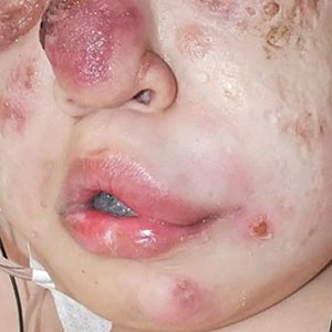
The Hair-Cut Sign for Scalp Biopsy Site Identification
The Hair-Cut Sign for Scalp Biopsy Site Identification
Practice Gap
Accurate biopsy site identification prior to Mohs micrographic surgery (MMS) can be challenging, particularly on the scalp, where excellent healing, field cancerization, prior procedures, large surface area, and hair density may complicate localization. Accurate site identification is further hindered when patients are unable to recall the biopsy location, when the biopsy and MMS are performed by different clinicians, or when photographic documentation is absent. Scalp lesions pose an additional challenge because even high-quality photographs may fail to capture subtle landmarks that are obscured by hair.
Wrong-site surgery was the most common serious error reported in a survey of 150 dermatologists, accounting for 19.0% of these errors.1 Research has shown that dermatologists may misidentify biopsy sites in up to 17.6% of cases, and patients (eg, if a patient goes to a dermatologic surgeon and is asked to identify the biopsy site for the surgeon) in up to 25.5% (N=278).2 Additionally, a survey of Mohs surgeons revealed that 14.0% of 42 malpractice cases were due to wrong-site surgery.3 A Delphi consensus study assessing possible preventive interventions found that the best way to document the site is with a photograph in association with landmarks and diagrams.4
The Technique
The hair-cut sign is a simple and effective technique for identifying biopsy sites on the scalp. Hair at the biopsy site typically is cut during biopsy; as the surrounding hair grows, the resulting short tuft serves as an anatomic marker that can persist for weeks to months to facilitate accurate site localization by the Mohs surgeon (Figure). This noninvasive, equipment-free method may be particularly useful when documentation or photography is limited. To maximize its effectiveness, clinical staff should avoid trimming the area before physician verification of the site. The technique is most effective in patients with longer hair and when the interval between biopsy and surgery is less than 3 months, as hair regrowth may reduce contrast with the surrounding scalp.
Practice Implications
Accurate biopsy site identification is a critical component of patient safety in dermatologic surgery, particularly on the scalp, where traditional localization strategies may be unreliable. The hair-cut sign is a simple, reliable, no-cost, and reproducible adjunct to photography, anatomic documentation, and patient verification that may improve site localization and help reduce the risk for wrong-site surgery in select patients to complement standard practices. Incorporating the hair-cut sign into routine scalp surgery workflow represents a low-burden intervention that may improve accuracy when other documentation is limited.
- Watson AJ, Redbord K, Taylor JS, et al. Medical error in dermatology practice: development of a classification system to drive priority setting in patient safety efforts. J Am Acad Dermatol. 2013;68:729-737. doi:10.1016/j.jaad.2012.10.058
- Nijhawan RI, Lee EH , Nehal KS. Biopsy site selfies—a quality improvement pilot study to assist with correct surgical site identification. Dermatol Surg. 2015;41:499-504. doi:10.1097/DSS.0000000000000305
- Perlis CS, Campbell RM, Perlis RH, et al. Incidence of and risk factors for medical malpractice lawsuits among Mohs surgeons. Dermatol Surg. 2006;32:79-83. doi:10.1111/1524-4725.2006.32009
- Alam M, Lee A, Ibrahimi OA, et al. A multistep approach to improving biopsy site identification in dermatology: physician, staff, and patient roles based on a Delphi consensus. JAMA Dermatol. 2014;150:550-558. doi:10.1001/jamadermatol.2013.9804
Practice Gap
Accurate biopsy site identification prior to Mohs micrographic surgery (MMS) can be challenging, particularly on the scalp, where excellent healing, field cancerization, prior procedures, large surface area, and hair density may complicate localization. Accurate site identification is further hindered when patients are unable to recall the biopsy location, when the biopsy and MMS are performed by different clinicians, or when photographic documentation is absent. Scalp lesions pose an additional challenge because even high-quality photographs may fail to capture subtle landmarks that are obscured by hair.
Wrong-site surgery was the most common serious error reported in a survey of 150 dermatologists, accounting for 19.0% of these errors.1 Research has shown that dermatologists may misidentify biopsy sites in up to 17.6% of cases, and patients (eg, if a patient goes to a dermatologic surgeon and is asked to identify the biopsy site for the surgeon) in up to 25.5% (N=278).2 Additionally, a survey of Mohs surgeons revealed that 14.0% of 42 malpractice cases were due to wrong-site surgery.3 A Delphi consensus study assessing possible preventive interventions found that the best way to document the site is with a photograph in association with landmarks and diagrams.4
The Technique
The hair-cut sign is a simple and effective technique for identifying biopsy sites on the scalp. Hair at the biopsy site typically is cut during biopsy; as the surrounding hair grows, the resulting short tuft serves as an anatomic marker that can persist for weeks to months to facilitate accurate site localization by the Mohs surgeon (Figure). This noninvasive, equipment-free method may be particularly useful when documentation or photography is limited. To maximize its effectiveness, clinical staff should avoid trimming the area before physician verification of the site. The technique is most effective in patients with longer hair and when the interval between biopsy and surgery is less than 3 months, as hair regrowth may reduce contrast with the surrounding scalp.
Practice Implications
Accurate biopsy site identification is a critical component of patient safety in dermatologic surgery, particularly on the scalp, where traditional localization strategies may be unreliable. The hair-cut sign is a simple, reliable, no-cost, and reproducible adjunct to photography, anatomic documentation, and patient verification that may improve site localization and help reduce the risk for wrong-site surgery in select patients to complement standard practices. Incorporating the hair-cut sign into routine scalp surgery workflow represents a low-burden intervention that may improve accuracy when other documentation is limited.
- Watson AJ, Redbord K, Taylor JS, et al. Medical error in dermatology practice: development of a classification system to drive priority setting in patient safety efforts. J Am Acad Dermatol. 2013;68:729-737. doi:10.1016/j.jaad.2012.10.058
- Nijhawan RI, Lee EH , Nehal KS. Biopsy site selfies—a quality improvement pilot study to assist with correct surgical site identification. Dermatol Surg. 2015;41:499-504. doi:10.1097/DSS.0000000000000305
- Perlis CS, Campbell RM, Perlis RH, et al. Incidence of and risk factors for medical malpractice lawsuits among Mohs surgeons. Dermatol Surg. 2006;32:79-83. doi:10.1111/1524-4725.2006.32009
- Alam M, Lee A, Ibrahimi OA, et al. A multistep approach to improving biopsy site identification in dermatology: physician, staff, and patient roles based on a Delphi consensus. JAMA Dermatol. 2014;150:550-558. doi:10.1001/jamadermatol.2013.9804
The Hair-Cut Sign for Scalp Biopsy Site Identification
The Hair-Cut Sign for Scalp Biopsy Site Identification
When Fungal Infections Mimic Acne: Diagnostic Pitfalls and Practical Approaches
When Fungal Infections Mimic Acne: Diagnostic Pitfalls and Practical Approaches
Dermatophyte infections, commonly referred to as tinea, involve the superficial epidermis and are caused by fungi belonging primarily to the genera Trichophyton, Epidermophyton, and Microsporum.1 Malassezia are lipophilic yeasts found in the normal skin flora that can overgrow within hair follicles and trigger an inflammatory response. While both fungal infections are associated with several classic clinical features, they can demonstrate variable clinical morphology, especially when modified by previous topical treatments. In such cases, fungal infections may mimic other forms of inflammatory dermatitis and can be misdiagnosed.
Acne vulgaris is one of the most prevalent dermatologic conditions and typically is diagnosed clinically based on characteristic morphology and distribution. Despite their distinct etiologies, dermatophyte infections and acne vulgaris may manifest with overlapping features, particularly in acne-prone regions such as the face, chest, and back, which may result in diagnostic errors and inappropriate management. This review highlights dermatophyte infections as an underrecognized mimic of acne vulgaris, emphasizing key clinical distinguishing features, common diagnostic pitfalls, and a practical approach to evaluation.
Clinical Overlap of Dermatophyte Infections and Acne
Despite their fundamentally different etiologies, dermatophyte infections and acne may demonstrate overlapping clinical morphology and anatomic distribution, creating diagnostic challenges and increasing misdiagnosis risk. Clinically, acne is characterized by the presence of open and closed comedones as well as inflammatory papules, pustules, nodules, and occasionally cysts.2 In contrast, dermatophyte infections classically manifest as annular erythematous plaques with peripheral scale and central clearing, primarily due to their superficial confinement to the stratum corneum; however, in certain cases the dermatophyte invades the hair follicle, which can lead to the formation of folliculocentric pustules.1 This is known as dermatophyte folliculitis and may closely resemble the pustules observed in acne.
Follicular invasion by dermatophytes is more likely in cases in which infection has been misdiagnosed as a noninfectious inflammatory dermatosis, (eg, atopic dermatitis) and treated with topical corticosteroids. Corticosteroid-induced local immunosuppression facilitates deeper and more extensive proliferation of the invading fungus, including into the hair follicle. Topical corticosteroid use may further obscure the diagnosis of a dermatophyte infection by masking its hallmark features such as scale and annularity.3 This steroid-altered dermatophyte infection is referred to as tinea incognita and may be misdiagnosed as acne or another inflammatory dermatosis. When dermatophytes extend from the stratum corneum into the dermis due to local immunosuppression (eg, corticosteroid use), trauma, shaving, or occlusion, the resulting deep follicular infection is known as Majocchi granuloma.
Further complicating the diagnostic picture is the substantial anatomic and epidemiologic overlap between dermatophyte infections and acne vulgaris. Acne preferentially affects sebum-rich areas, including the face, chest, and back.2 Dermatophytes, by contrast, thrive in keratinized tissue.1 Because areas with a higher density of hair follicles contain abundant keratin, dermatophyte infections often involve the same sebum-rich regions affected by acne. Both acne and tinea are observed frequently in adolescents, possibly due to hormonal changes that increase sebum production and create an environment conducive to fungal growth.4
Pityrosporum Folliculitis Manifesting as Acne Vulgaris
Although it has been widely popularized in lay and social media, the term fungal acne is a misnomer; this entity more accurately represents a fungal folliculitis manifesting as an acneform eruption. In most cases, fungal acne refers to Malassezia folliculitis, also called pityrosporum folliculitis, which is caused by Malassezia species. Malassezia are not dermatophytes but rather lipophilic yeasts found in the normal skin flora. Whereas dermatophytes are drawn to highly keratinized tissue, Malassezia are drawn to lipid-rich environments of the skin. In these conditions, including sweating and hot or humid environments, Malassezia may proliferate to pathogenic levels within the hair follicle.5
Clinically, Malassezia folliculitis manifests as monomorphic, folliculocentric, dome-shaped papules and pustules with occasional progression to nodules or cysts in more severe cases.5 Lesions typically are intensely pruritic, a distinguishing feature that helps differentiate them from acne vulgaris.6 The eruption predominantly involves sebum-rich areas, including the face, hairline, chest, and upper back (Figure 1).5 Overall, the clinical presentation often more closely resembles steroid-induced acne than classic acne vulgaris. Antibiotic exposure is an important risk factor, potentially due to disruption of the normal skin microbiome and subsequent yeast overgrowth; for example, in a retrospective review of 110 patients (age range, 0-21 years) with Malassezia folliculitis, more than 75% had recently received antibiotics for treatment of acne.6 Additional predisposing factors include corticosteroid use and immunosuppression.5
Importantly, Malassezia folliculitis and acne vulgaris may coexist, further complicating diagnosis. In a study of 217 patients with acne vulgaris, cytologic evaluation demonstrated Malassezia overgrowth (defined as >6 spores per high-power field) in approximately 25% of patients, of whom 70% responded to antifungal therapy.7 Similarly, a study of 300 patients with newly diagnosed acne found a prevalence of Malassezia folliculitis of almost 30%. Patients with concurrent Malassezia folliculitis and acne were more likely to report pruritus and have involvement of the scalp, hairline, and upper back compared to those with acne alone.8
Tinea Faciei and Tinea Barbae Manifesting as Facial Acne
Tinea faciei describes a dermatophyte infection of the nonbearded area of the face, whereas infection of the beard-bearing region is known as tinea barbae. In North America, Trichophyton species are the leading cause of tinea faciei.1 Clinically, tinea faciei manifests as one or more erythematous scaly plaques on the face, often associated with pruritus. Lesions often assume an annular shape with an advancing border along which pustules, vesicles, or crusting can be observed. In cases of inappropriate treatment with topical corticosteroids, lesions may lose their characteristic scale and annularity and instead become papular, mimicking the acneform eruptions of facial acne vulgaris (Figure 2).
Like tinea faciei, tinea barbae most commonly develops from infection with Trichophyton species but differs in its clinical presentation. While superficial scaly variants exist, tinea barbae more frequently manifests as a deep, papular, inflammatory folliculitis. This deeper form typically is caused by follicular infiltration by zoophilic dermatophytes such as Trichophyton verrucosum and Microsporum canis. Not surprisingly, infection by these zoophilic dermatophytes is associated with exposure to animals such as cattle, horses, dogs, and cats, and a history of agricultural work may offer a helpful clue to diagnosis.9 The tender, nodular, or nodulocystic lesions of severe tinea barbae infections may closely resemble nodulocystic acne.
Misdiagnosis, inappropriate treatment, and diagnostic delays are common in patients with tinea faciei and tinea barbae. In a retrospective study of 818 cases of tinea faciei, approximately 30% of patients had received prior corticosteroid treatment at the time of diagnosis.10 Similarly, a cross-sectional study of 7 adult patients with tinea barbae in a Portuguese hospital found that 3 cases initially were misdiagnosed and that in 2 cases potent topical steroids were previously applied.11 Finally, in a retrospective review of 38 patients with mycologically confirmed tinea faciei, the mean duration from symptom onset to diagnosis was 3.4 months.10 Notably, nearly 60% of patients had concomitant dermatophyte infections at other body sites, most commonly involving the feet and toes, highlighting that recognition of dermatophyte infections elsewhere on physical examination may provide an important diagnostic clue.12
Tinea Corporis Manifesting as Truncal Acne
Tinea corporis refers to a dermatophyte infection involving the glabrous, or hairless, skin of the trunk and extremities. Trichophyton rubrum accounts for 80% to 90% of the pathogenic strains that cause tinea corporis.1 As with other variants of superficial dermatophyte infections, tinea corporis classically manifests as annular erythematous plaques with peripheral scale and central clearing, distinguished by its involvement of the trunk. Pruritus of lesions is variable.1
Inappropriate treatment of tinea corporis with topical corticosteroids may induce a morphologic change in the infection so that it resembles the lesions of truncal acne, which characteristically involves the chest, upper back, and shoulders, and less frequently the lower back and abdomen.2 As in other forms of acne vulgaris, the lesions are characterized by a mixture of inflammatory papules, pustules, and comedones. When differentiating tinea corporis and truncal acne, consider the distribution and symmetry of the lesions. Dermatophyte infections often are localized to one area of the trunk and are asymmetric. In contrast, acne typically is generalized and manifests more symmetrically.
Additional clinical clues may aid in differentiation. An acneform eruption involving other seborrheic areas of the body (eg, the face) supports a diagnosis of truncal acne. Conversely, the presence of tinea elsewhere, particularly on the hands or feet, may suggest tinea corporis. Finally, although pustules can be seen with tinea corporis, the presence of true comedones is a key distinguishing factor favoring acne vulgaris.
Importantly, resistant dermatophyte infections have emerged as a growing concern among public health experts over the past decade. A recently described species, Trichophyton indotineae, has played a substantial role in driving these cases.13 While early US cases largely were limited to patients who had travelled to Bangladesh, infections now are increasingly reported in individuals without travel history.13 Trichophyton indotineae most commonly involves the trunk, extremities, and groin, which mirrors the distribution of truncal acne. Further complicating the clinical picture is the lack of response to standard antifungal therapies such as oral terbinafine in these patients.13 Failure to consider this diagnosis, particularly given its recent recognition, may lead physicians to empirically switch treatment to topical or systemic corticosteroids. This can further alter lesion morphology and increase the likelihood of misdiagnosis.
Helpful Bedside Diagnostic Tools
When clinical findings are equivocal, bedside diagnostic tools, including dermoscopy, a Wood lamp, potassium hydroxide (KOH) preparation, and histopathology, may be helpful in differentiating cutaneous fungal infections from acne.
Dermoscopy—In an observational study of 81 patients with fungal folliculitis, dermoscopy demonstrated a diagnostic accuracy of 76.5%.14 Dermoscopic findings in a cohort of 45 patients with KOH-confirmed Malassezia folliculitis included folliculocentric lesions and background erythema (100%); dotted, linear, or tortuous vessels (89%); fine white scale (78%); perifollicular hypopigmentation (64%); coiled or looped hairs (58%); and broken hairs (13%).15 Moreover, in a study comparing 36 microscopically confirmed tinea cases with 40 negative cases, peripheral scales (odds ratio [OR], 5.2; 95% CI, 2.0-13.5), moth-eaten scales (OR, 3.9; 95% CI, 1.9-8.1), broken hairs (OR, 5.8; 95% CI, 2.0-16.6), and outward-peeling scales (OR, 14.3; 95% CI, 1.3-155.2) were predictive of tinea.16 Dermoscopic findings in a cross-sectional study of 100 clinically diagnosed tinea cases included diffuse erythema with whitish scars (100.0%), follicular micropustules (36.7%), brown spots with a white-yellow halo (20.0%), wavy or broken hairs (13.0%), and Morse code– like vellus hairs (3.0%).17 In tinea incognito, features such as Morse code–like hairs, deformable translucent hairs, comma and corkscrew hairs, and perifollicular scaling may persist despite corticosteroid use.18,19
Wood Lamp Examination—Wood lamp examination may be a helpful adjunctive tool for diagnosis of Malassezia folliculitis. In a study of 264 patients with folliculitis (49 of whom were diagnosed with Malassezia folliculitis), Wood lamp examination demonstrated yellow-green fluorescence in 66.7% of cases.20 In contrast, this method has limited utility in diagnosing common dermatophyte infections, as only Microsporum and a small subset of Trichophyton species fluoresce.21 In a study of 50 pediatric patients with tinea capitis, Microsporum cases were identified via Wood lamp examination by bright green fluorescence. Wood lamp examination demonstrated 73% sensitivity and 100% specificity for Microsporum canis, confirmed by microscopy and culture, indicating that positive results are highly reliable for this genus, though false negatives may occur.22
Some dermatoscopes incorporate a Wood lamp, enabling UV-induced fluorescence dermoscopy (UVFD). In a study of 208 patients with nonneoplastic dermatoses, UVFD of tinea showed light green hair shaft concretions in 27% (4/15) of patients and no fluorescence in 73% (11/15), whereas Malassezia folliculitis demonstrated blue follicular concretions in 85% (11/13) and acne showed disruption of uniform follicular red fluorescence in 81% (13/16).23 However, these dermatoscopes are not widely available.
KOH Preparation—While the aforementioned tests are useful and require minimal effort, the diagnostic test of choice for cutaneous fungal infections remains the KOH preparation, which is fast and inexpensive and offers immediate results, often while the patient is still in the office. The test should be performed by obtaining scale, ideally from an active lesion border, by gently scraping the stratum corneum, often with a #15 blade. For sampling of pustules or when there is concern for Malassezia folliculitis, optimal technique involves unroofing a pustule and transferring its contents onto a slide for KOH preparation. The specimen then is treated with KOH, a keratolytic agent that dissolves keratinocytes and facilitates visualization of fungal elements under light microscopy. Reported sensitivity and specificity of KOH preparation are approximately 73% and 78%, respectively.24 Notably, sensitivity and specificity of KOH is highly dependent on expertise. A fungal culture also can be collected and sent for microbiologic analysis, although results often are delayed. In one pooled analysis of tinea pedis using clinical assessment as the reference standard, fungal culture demonstrated a sensitivity of 42% and specificity of 78%, though these estimates are highly dependent on study design and sampling technique.24
Histopathology—Finally, histopathologic evaluation may be considered in diagnostically challenging cases. Histology of Malassezia folliculitis demonstrates fungal spores within the follicular lumen, while histology of acne shows irregular keratin plugging, nuclear debris within the follicular lumen, and intrafollicular inflammation. Notably, perifollicular inflammatory infiltrates are histologically similar in acne and Malassezia folliculitis.25
Practical Diagnostic Approach to Differentiating Dermatophyte Infections from Acne
For physicians encountering papulopustular eruptions in acne-prone regions, distinguishing acne vulgaris from dermatophyte infection can be challenging. A stepwise approach incorporating history, morphology, and distribution can improve diagnostic accuracy and guide appropriate management.
First, obtain a thorough treatment history. Presumed acne that has failed to respond to appropriate acne therapies should prompt reconsideration of the diagnosis. Prior treatment with topical corticosteroids should be specifically assessed. Patients may not volunteer this history unless directly asked. Corticosteroid use can alter the clinical appearance of dermatophyte infections, leading to diagnostic confusion.
Second, use morphologic features and lesion distribution as diagnostic clues. The presence of comedones favors acne vulgaris, whereas their absence should raise suspicion for tinea. It is important to note, however, that certain dermatophyte infections may manifest with folliculocentric pustules, which can mimic closed comedones or inflammatory lesions seen in acne. Acne vulgaris also typically demonstrates a bilateral and relatively symmetric distribution, particularly on the face, chest, and upper back. In contrast, dermatophyte infections are more often asymmetric or localized, especially in early stages.
Patient-reported symptoms and a complete skin examination can further aid in differentiation. While acne may occasionally be pruritic, pain or tenderness is more commonly reported. In contrast, dermatophyte infections often will have prominent pruritus, which frequently is the patient’s primary complaint. The presence of tinea on the hands or feet supports a diagnosis of dermatophyte infection, whereas concurrent acneform lesions in classic seborrheic regions favor acne vulgaris. The Table outlines key clinical features that help distinguish dermatophyte infections from acne vulgaris.
When the diagnosis remains unclear after clinical assessment, physicians may utilize both bedside and laboratory tests, including dermoscopy, Wood lamp examination, in-office KOH preparation, and/or fungal culture, as discussed previously. In cases of diagnostic uncertainty, empiric antifungal therapy is preferred over topical corticosteroid therapy, as corticosteroids may exacerbate an underlying dermatophyte infection. In refractory or diagnostically challenging cases, skin biopsy with periodic acid–Schiff staining may be considered to confirm the presence of fungal organisms. Biopsy generally is reserved for cases that fail to respond to empiric therapy or when diagnostic confirmation is strongly desired. Figure 3 provides an algorithmic approach to distinguishing acne vulgaris from dermatophyte infection.
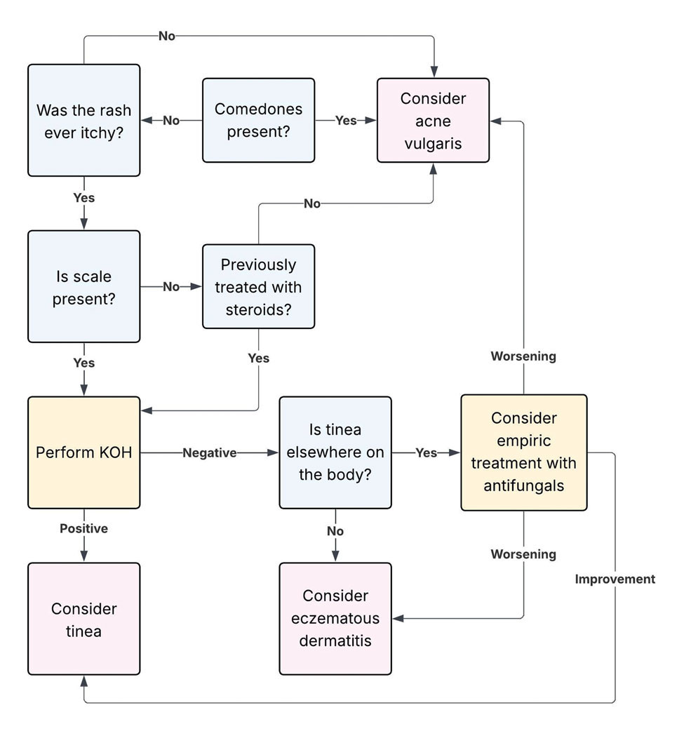
Final Thoughts
Dermatophyte infections are a common but often overlooked mimic of acne vulgaris. Clinically, acne is characterized by comedones, whereas dermatophyte infections typically demonstrate scale, though these features can be less apparent in modified presentations. In cases of diagnostic uncertainty, physicians should keep dermatophyte infections in mind and be comfortable performing bedside KOH preparations to support timely diagnosis. Early recognition is important to reduce morbidity and avoid inappropriate treatments, particularly corticosteroids, which can worsen the infection and delay improvement.
- Yee G, Syed HA, Al Aboud AM. Tinea corporis. StatPearls (Internet). Updated February 14, 2025. Accessed June 5, 2026. https://www.ncbi. nlm.nih.gov/books/NBK544360/
- Sutaria AH, Masood S, Saleh HM, et al. Acne vulgaris. StatPearls (Internet). Updated August 17, 2023. Accessed June 5, 2026. https://www. ncbi.nlm.nih.gov/books/NBK459173/
- Ive FA, Marks R. Tinea incognito. Br Med J. 1968;3:149-152.
- Zarzeka D, Benedict K, McCloskey M, et al. Current epidemiology of tinea corporis and tinea cruris causative species: analysis of data from a major commercial laboratory, United States. J Am Acad Dermatol. 2024;91:559-562.
- Vlachos C, Henning MAS, Gaitanis G, et al. Critical synthesis of available data in Malassezia folliculitis and a systematic review of treatments. J Eur Acad Dermatol Venereol. 2020;34:1672-1683.
- Prindaville B, Belazarian L, Levin NA, et al. Pityrosporum folliculitis: a retrospective review of 110 cases. J Am Acad Dermatol. 2018;78:511-514.
- Pürnak S, Durdu M, Tekindal MA, et al. The prevalence of Malassezia folliculitis in patients with papulopustular/comedonal acne, and their response to antifungal treatment. Skinmed. 2018;16:99-104.
- Paichitrojjana A, Chalermchai T. The prevalence, associated factors, and clinical characterization of Malassezia folliculitis in patients clinically diagnosed with acne vulgaris. Clin Cosmet Investig Dermatol. 2022;15:2647-2654.
- Kuruvella T, Saleh HM, Pandey S. Tinea barbae. StatPearls (Internet). Updated December 5, 2024. Accessed June 5, 2026. https://www.ncbi .nlm.nih.gov/books/NBK563204/
- del Boz J, Crespo V, de Troya M. Pediatric tinea faciei in southern Spain: a 30-year survey. Pediatr Dermatol. 2012;29:249-253.
- Duarte B, Galhardas C, Cabete J. Adult tinea capitis and tinea barbae in a tertiary Portuguese hospital: a 11-year audit. Mycoses. 2019;62:1079-1083.
- Kwak HB, Lee SK, Yoo HH, et al. Facial tinea incognito: a clinical, dermoscopic and mycological study of 38 cases. Eur J Dermatol. 2023;33:101-108.
- Caplan AS, Todd GC, Zhu Y, et al. Clinical course, antifungal susceptibility, and genomic sequencing of Trichophyton indotineae. JAMA Dermatol. 2024;160:701-709.
- Durdu M, Errichetti E, Eskiocak AH, et al. High accuracy of recognition of common forms of folliculitis by dermoscopy: an observational study. J Am Acad Dermatol. 2019;81:463-471.
- Jakhar D, Bhatia V, Gupta RK, et al. Dermoscopy as an auxiliary tool in the assessment of Malassezia folliculitis: an observational study. Actas Dermosifiliogr. 2022;113:T78-T81.
- Lekkas D, Ioannides D, Lazaridou E, et al. Dermatoscopy of tinea corporis. J Eur Acad Dermatol Venereol. 2020;34:E278-E280.
- Bhat YJ, Keen A, Hassan I, et al. Can dermoscopy serve as a diagnostic tool in dermatophytosis? a pilot study. Indian Dermatol Online J. 2019;10:530-535.
- Gómez Moyano E, Crespo Erchiga V, Martínez Pilar L, et al. Correlation between dermoscopy and direct microscopy of morse code hairs in tinea incognito. J Am Acad Dermatol. 2016;74:E7-E8.
- Sonthalia S, Ankad BS, Goldust M, et al. Dermoscopy—a simple and rapid in vivo diagnostic technique for tinea incognito. An Bras Dermatol. 2019;94:612-614.
- Durdu M, Güran M, Ilkit M. Epidemiological characteristics of Malassezia folliculitis and use of the May-Grünwald-Giemsa stain to diagnose the infection. Diagn Microbiol Infect Dis. 2013;76:450-457.
- Dyer JM, Foy VM. Revealing the unseen: a review of Wood’s lamp in dermatology. J Clin Aesthet Dermatol. 2022;15:25-30.
- Sun D, Lu J, Liu T, Wang J. Wood’s lamp for early detection of Microsporum canis tinea capitis in children. Photodiagnosis Photodyn Ther. 2025;51:104428.
- Errichetti E, Pietkiewicz P, Bhat YJ, et al. Diagnostic accuracy of ultraviolet- induced fluorescence dermoscopy in non-neoplastic dermatoses (general dermatology): a multicentric retrospective comparative study. J Eur Acad Dermatol Venereol. 2025;39:97-108.
- Levitt JO, Levitt BH, Akhavan A, et al. The sensitivity and specificity of potassium hydroxide smear and fungal culture relative to clinical assessment in the evaluation of tinea pedis: a pooled analysis. Dermatol Res Pract. 2010;2010:764843.
- An MK, Hong EH, Cho EB, et al. Clinicopathological differentiation between Pityrosporum folliculitis and acneiform eruption. J Dermatol. 2019;46:978-984.
Dermatophyte infections, commonly referred to as tinea, involve the superficial epidermis and are caused by fungi belonging primarily to the genera Trichophyton, Epidermophyton, and Microsporum.1 Malassezia are lipophilic yeasts found in the normal skin flora that can overgrow within hair follicles and trigger an inflammatory response. While both fungal infections are associated with several classic clinical features, they can demonstrate variable clinical morphology, especially when modified by previous topical treatments. In such cases, fungal infections may mimic other forms of inflammatory dermatitis and can be misdiagnosed.
Acne vulgaris is one of the most prevalent dermatologic conditions and typically is diagnosed clinically based on characteristic morphology and distribution. Despite their distinct etiologies, dermatophyte infections and acne vulgaris may manifest with overlapping features, particularly in acne-prone regions such as the face, chest, and back, which may result in diagnostic errors and inappropriate management. This review highlights dermatophyte infections as an underrecognized mimic of acne vulgaris, emphasizing key clinical distinguishing features, common diagnostic pitfalls, and a practical approach to evaluation.
Clinical Overlap of Dermatophyte Infections and Acne
Despite their fundamentally different etiologies, dermatophyte infections and acne may demonstrate overlapping clinical morphology and anatomic distribution, creating diagnostic challenges and increasing misdiagnosis risk. Clinically, acne is characterized by the presence of open and closed comedones as well as inflammatory papules, pustules, nodules, and occasionally cysts.2 In contrast, dermatophyte infections classically manifest as annular erythematous plaques with peripheral scale and central clearing, primarily due to their superficial confinement to the stratum corneum; however, in certain cases the dermatophyte invades the hair follicle, which can lead to the formation of folliculocentric pustules.1 This is known as dermatophyte folliculitis and may closely resemble the pustules observed in acne.
Follicular invasion by dermatophytes is more likely in cases in which infection has been misdiagnosed as a noninfectious inflammatory dermatosis, (eg, atopic dermatitis) and treated with topical corticosteroids. Corticosteroid-induced local immunosuppression facilitates deeper and more extensive proliferation of the invading fungus, including into the hair follicle. Topical corticosteroid use may further obscure the diagnosis of a dermatophyte infection by masking its hallmark features such as scale and annularity.3 This steroid-altered dermatophyte infection is referred to as tinea incognita and may be misdiagnosed as acne or another inflammatory dermatosis. When dermatophytes extend from the stratum corneum into the dermis due to local immunosuppression (eg, corticosteroid use), trauma, shaving, or occlusion, the resulting deep follicular infection is known as Majocchi granuloma.
Further complicating the diagnostic picture is the substantial anatomic and epidemiologic overlap between dermatophyte infections and acne vulgaris. Acne preferentially affects sebum-rich areas, including the face, chest, and back.2 Dermatophytes, by contrast, thrive in keratinized tissue.1 Because areas with a higher density of hair follicles contain abundant keratin, dermatophyte infections often involve the same sebum-rich regions affected by acne. Both acne and tinea are observed frequently in adolescents, possibly due to hormonal changes that increase sebum production and create an environment conducive to fungal growth.4
Pityrosporum Folliculitis Manifesting as Acne Vulgaris
Although it has been widely popularized in lay and social media, the term fungal acne is a misnomer; this entity more accurately represents a fungal folliculitis manifesting as an acneform eruption. In most cases, fungal acne refers to Malassezia folliculitis, also called pityrosporum folliculitis, which is caused by Malassezia species. Malassezia are not dermatophytes but rather lipophilic yeasts found in the normal skin flora. Whereas dermatophytes are drawn to highly keratinized tissue, Malassezia are drawn to lipid-rich environments of the skin. In these conditions, including sweating and hot or humid environments, Malassezia may proliferate to pathogenic levels within the hair follicle.5
Clinically, Malassezia folliculitis manifests as monomorphic, folliculocentric, dome-shaped papules and pustules with occasional progression to nodules or cysts in more severe cases.5 Lesions typically are intensely pruritic, a distinguishing feature that helps differentiate them from acne vulgaris.6 The eruption predominantly involves sebum-rich areas, including the face, hairline, chest, and upper back (Figure 1).5 Overall, the clinical presentation often more closely resembles steroid-induced acne than classic acne vulgaris. Antibiotic exposure is an important risk factor, potentially due to disruption of the normal skin microbiome and subsequent yeast overgrowth; for example, in a retrospective review of 110 patients (age range, 0-21 years) with Malassezia folliculitis, more than 75% had recently received antibiotics for treatment of acne.6 Additional predisposing factors include corticosteroid use and immunosuppression.5
Importantly, Malassezia folliculitis and acne vulgaris may coexist, further complicating diagnosis. In a study of 217 patients with acne vulgaris, cytologic evaluation demonstrated Malassezia overgrowth (defined as >6 spores per high-power field) in approximately 25% of patients, of whom 70% responded to antifungal therapy.7 Similarly, a study of 300 patients with newly diagnosed acne found a prevalence of Malassezia folliculitis of almost 30%. Patients with concurrent Malassezia folliculitis and acne were more likely to report pruritus and have involvement of the scalp, hairline, and upper back compared to those with acne alone.8
Tinea Faciei and Tinea Barbae Manifesting as Facial Acne
Tinea faciei describes a dermatophyte infection of the nonbearded area of the face, whereas infection of the beard-bearing region is known as tinea barbae. In North America, Trichophyton species are the leading cause of tinea faciei.1 Clinically, tinea faciei manifests as one or more erythematous scaly plaques on the face, often associated with pruritus. Lesions often assume an annular shape with an advancing border along which pustules, vesicles, or crusting can be observed. In cases of inappropriate treatment with topical corticosteroids, lesions may lose their characteristic scale and annularity and instead become papular, mimicking the acneform eruptions of facial acne vulgaris (Figure 2).
Like tinea faciei, tinea barbae most commonly develops from infection with Trichophyton species but differs in its clinical presentation. While superficial scaly variants exist, tinea barbae more frequently manifests as a deep, papular, inflammatory folliculitis. This deeper form typically is caused by follicular infiltration by zoophilic dermatophytes such as Trichophyton verrucosum and Microsporum canis. Not surprisingly, infection by these zoophilic dermatophytes is associated with exposure to animals such as cattle, horses, dogs, and cats, and a history of agricultural work may offer a helpful clue to diagnosis.9 The tender, nodular, or nodulocystic lesions of severe tinea barbae infections may closely resemble nodulocystic acne.
Misdiagnosis, inappropriate treatment, and diagnostic delays are common in patients with tinea faciei and tinea barbae. In a retrospective study of 818 cases of tinea faciei, approximately 30% of patients had received prior corticosteroid treatment at the time of diagnosis.10 Similarly, a cross-sectional study of 7 adult patients with tinea barbae in a Portuguese hospital found that 3 cases initially were misdiagnosed and that in 2 cases potent topical steroids were previously applied.11 Finally, in a retrospective review of 38 patients with mycologically confirmed tinea faciei, the mean duration from symptom onset to diagnosis was 3.4 months.10 Notably, nearly 60% of patients had concomitant dermatophyte infections at other body sites, most commonly involving the feet and toes, highlighting that recognition of dermatophyte infections elsewhere on physical examination may provide an important diagnostic clue.12
Tinea Corporis Manifesting as Truncal Acne
Tinea corporis refers to a dermatophyte infection involving the glabrous, or hairless, skin of the trunk and extremities. Trichophyton rubrum accounts for 80% to 90% of the pathogenic strains that cause tinea corporis.1 As with other variants of superficial dermatophyte infections, tinea corporis classically manifests as annular erythematous plaques with peripheral scale and central clearing, distinguished by its involvement of the trunk. Pruritus of lesions is variable.1
Inappropriate treatment of tinea corporis with topical corticosteroids may induce a morphologic change in the infection so that it resembles the lesions of truncal acne, which characteristically involves the chest, upper back, and shoulders, and less frequently the lower back and abdomen.2 As in other forms of acne vulgaris, the lesions are characterized by a mixture of inflammatory papules, pustules, and comedones. When differentiating tinea corporis and truncal acne, consider the distribution and symmetry of the lesions. Dermatophyte infections often are localized to one area of the trunk and are asymmetric. In contrast, acne typically is generalized and manifests more symmetrically.
Additional clinical clues may aid in differentiation. An acneform eruption involving other seborrheic areas of the body (eg, the face) supports a diagnosis of truncal acne. Conversely, the presence of tinea elsewhere, particularly on the hands or feet, may suggest tinea corporis. Finally, although pustules can be seen with tinea corporis, the presence of true comedones is a key distinguishing factor favoring acne vulgaris.
Importantly, resistant dermatophyte infections have emerged as a growing concern among public health experts over the past decade. A recently described species, Trichophyton indotineae, has played a substantial role in driving these cases.13 While early US cases largely were limited to patients who had travelled to Bangladesh, infections now are increasingly reported in individuals without travel history.13 Trichophyton indotineae most commonly involves the trunk, extremities, and groin, which mirrors the distribution of truncal acne. Further complicating the clinical picture is the lack of response to standard antifungal therapies such as oral terbinafine in these patients.13 Failure to consider this diagnosis, particularly given its recent recognition, may lead physicians to empirically switch treatment to topical or systemic corticosteroids. This can further alter lesion morphology and increase the likelihood of misdiagnosis.
Helpful Bedside Diagnostic Tools
When clinical findings are equivocal, bedside diagnostic tools, including dermoscopy, a Wood lamp, potassium hydroxide (KOH) preparation, and histopathology, may be helpful in differentiating cutaneous fungal infections from acne.
Dermoscopy—In an observational study of 81 patients with fungal folliculitis, dermoscopy demonstrated a diagnostic accuracy of 76.5%.14 Dermoscopic findings in a cohort of 45 patients with KOH-confirmed Malassezia folliculitis included folliculocentric lesions and background erythema (100%); dotted, linear, or tortuous vessels (89%); fine white scale (78%); perifollicular hypopigmentation (64%); coiled or looped hairs (58%); and broken hairs (13%).15 Moreover, in a study comparing 36 microscopically confirmed tinea cases with 40 negative cases, peripheral scales (odds ratio [OR], 5.2; 95% CI, 2.0-13.5), moth-eaten scales (OR, 3.9; 95% CI, 1.9-8.1), broken hairs (OR, 5.8; 95% CI, 2.0-16.6), and outward-peeling scales (OR, 14.3; 95% CI, 1.3-155.2) were predictive of tinea.16 Dermoscopic findings in a cross-sectional study of 100 clinically diagnosed tinea cases included diffuse erythema with whitish scars (100.0%), follicular micropustules (36.7%), brown spots with a white-yellow halo (20.0%), wavy or broken hairs (13.0%), and Morse code– like vellus hairs (3.0%).17 In tinea incognito, features such as Morse code–like hairs, deformable translucent hairs, comma and corkscrew hairs, and perifollicular scaling may persist despite corticosteroid use.18,19
Wood Lamp Examination—Wood lamp examination may be a helpful adjunctive tool for diagnosis of Malassezia folliculitis. In a study of 264 patients with folliculitis (49 of whom were diagnosed with Malassezia folliculitis), Wood lamp examination demonstrated yellow-green fluorescence in 66.7% of cases.20 In contrast, this method has limited utility in diagnosing common dermatophyte infections, as only Microsporum and a small subset of Trichophyton species fluoresce.21 In a study of 50 pediatric patients with tinea capitis, Microsporum cases were identified via Wood lamp examination by bright green fluorescence. Wood lamp examination demonstrated 73% sensitivity and 100% specificity for Microsporum canis, confirmed by microscopy and culture, indicating that positive results are highly reliable for this genus, though false negatives may occur.22
Some dermatoscopes incorporate a Wood lamp, enabling UV-induced fluorescence dermoscopy (UVFD). In a study of 208 patients with nonneoplastic dermatoses, UVFD of tinea showed light green hair shaft concretions in 27% (4/15) of patients and no fluorescence in 73% (11/15), whereas Malassezia folliculitis demonstrated blue follicular concretions in 85% (11/13) and acne showed disruption of uniform follicular red fluorescence in 81% (13/16).23 However, these dermatoscopes are not widely available.
KOH Preparation—While the aforementioned tests are useful and require minimal effort, the diagnostic test of choice for cutaneous fungal infections remains the KOH preparation, which is fast and inexpensive and offers immediate results, often while the patient is still in the office. The test should be performed by obtaining scale, ideally from an active lesion border, by gently scraping the stratum corneum, often with a #15 blade. For sampling of pustules or when there is concern for Malassezia folliculitis, optimal technique involves unroofing a pustule and transferring its contents onto a slide for KOH preparation. The specimen then is treated with KOH, a keratolytic agent that dissolves keratinocytes and facilitates visualization of fungal elements under light microscopy. Reported sensitivity and specificity of KOH preparation are approximately 73% and 78%, respectively.24 Notably, sensitivity and specificity of KOH is highly dependent on expertise. A fungal culture also can be collected and sent for microbiologic analysis, although results often are delayed. In one pooled analysis of tinea pedis using clinical assessment as the reference standard, fungal culture demonstrated a sensitivity of 42% and specificity of 78%, though these estimates are highly dependent on study design and sampling technique.24
Histopathology—Finally, histopathologic evaluation may be considered in diagnostically challenging cases. Histology of Malassezia folliculitis demonstrates fungal spores within the follicular lumen, while histology of acne shows irregular keratin plugging, nuclear debris within the follicular lumen, and intrafollicular inflammation. Notably, perifollicular inflammatory infiltrates are histologically similar in acne and Malassezia folliculitis.25
Practical Diagnostic Approach to Differentiating Dermatophyte Infections from Acne
For physicians encountering papulopustular eruptions in acne-prone regions, distinguishing acne vulgaris from dermatophyte infection can be challenging. A stepwise approach incorporating history, morphology, and distribution can improve diagnostic accuracy and guide appropriate management.
First, obtain a thorough treatment history. Presumed acne that has failed to respond to appropriate acne therapies should prompt reconsideration of the diagnosis. Prior treatment with topical corticosteroids should be specifically assessed. Patients may not volunteer this history unless directly asked. Corticosteroid use can alter the clinical appearance of dermatophyte infections, leading to diagnostic confusion.
Second, use morphologic features and lesion distribution as diagnostic clues. The presence of comedones favors acne vulgaris, whereas their absence should raise suspicion for tinea. It is important to note, however, that certain dermatophyte infections may manifest with folliculocentric pustules, which can mimic closed comedones or inflammatory lesions seen in acne. Acne vulgaris also typically demonstrates a bilateral and relatively symmetric distribution, particularly on the face, chest, and upper back. In contrast, dermatophyte infections are more often asymmetric or localized, especially in early stages.
Patient-reported symptoms and a complete skin examination can further aid in differentiation. While acne may occasionally be pruritic, pain or tenderness is more commonly reported. In contrast, dermatophyte infections often will have prominent pruritus, which frequently is the patient’s primary complaint. The presence of tinea on the hands or feet supports a diagnosis of dermatophyte infection, whereas concurrent acneform lesions in classic seborrheic regions favor acne vulgaris. The Table outlines key clinical features that help distinguish dermatophyte infections from acne vulgaris.
When the diagnosis remains unclear after clinical assessment, physicians may utilize both bedside and laboratory tests, including dermoscopy, Wood lamp examination, in-office KOH preparation, and/or fungal culture, as discussed previously. In cases of diagnostic uncertainty, empiric antifungal therapy is preferred over topical corticosteroid therapy, as corticosteroids may exacerbate an underlying dermatophyte infection. In refractory or diagnostically challenging cases, skin biopsy with periodic acid–Schiff staining may be considered to confirm the presence of fungal organisms. Biopsy generally is reserved for cases that fail to respond to empiric therapy or when diagnostic confirmation is strongly desired. Figure 3 provides an algorithmic approach to distinguishing acne vulgaris from dermatophyte infection.
Final Thoughts
Dermatophyte infections are a common but often overlooked mimic of acne vulgaris. Clinically, acne is characterized by comedones, whereas dermatophyte infections typically demonstrate scale, though these features can be less apparent in modified presentations. In cases of diagnostic uncertainty, physicians should keep dermatophyte infections in mind and be comfortable performing bedside KOH preparations to support timely diagnosis. Early recognition is important to reduce morbidity and avoid inappropriate treatments, particularly corticosteroids, which can worsen the infection and delay improvement.
- Yee G, Syed HA, Al Aboud AM. Tinea corporis. StatPearls (Internet). Updated February 14, 2025. Accessed June 5, 2026. https://www.ncbi. nlm.nih.gov/books/NBK544360/
- Sutaria AH, Masood S, Saleh HM, et al. Acne vulgaris. StatPearls (Internet). Updated August 17, 2023. Accessed June 5, 2026. https://www. ncbi.nlm.nih.gov/books/NBK459173/
- Ive FA, Marks R. Tinea incognito. Br Med J. 1968;3:149-152.
- Zarzeka D, Benedict K, McCloskey M, et al. Current epidemiology of tinea corporis and tinea cruris causative species: analysis of data from a major commercial laboratory, United States. J Am Acad Dermatol. 2024;91:559-562.
- Vlachos C, Henning MAS, Gaitanis G, et al. Critical synthesis of available data in Malassezia folliculitis and a systematic review of treatments. J Eur Acad Dermatol Venereol. 2020;34:1672-1683.
- Prindaville B, Belazarian L, Levin NA, et al. Pityrosporum folliculitis: a retrospective review of 110 cases. J Am Acad Dermatol. 2018;78:511-514.
- Pürnak S, Durdu M, Tekindal MA, et al. The prevalence of Malassezia folliculitis in patients with papulopustular/comedonal acne, and their response to antifungal treatment. Skinmed. 2018;16:99-104.
- Paichitrojjana A, Chalermchai T. The prevalence, associated factors, and clinical characterization of Malassezia folliculitis in patients clinically diagnosed with acne vulgaris. Clin Cosmet Investig Dermatol. 2022;15:2647-2654.
- Kuruvella T, Saleh HM, Pandey S. Tinea barbae. StatPearls (Internet). Updated December 5, 2024. Accessed June 5, 2026. https://www.ncbi .nlm.nih.gov/books/NBK563204/
- del Boz J, Crespo V, de Troya M. Pediatric tinea faciei in southern Spain: a 30-year survey. Pediatr Dermatol. 2012;29:249-253.
- Duarte B, Galhardas C, Cabete J. Adult tinea capitis and tinea barbae in a tertiary Portuguese hospital: a 11-year audit. Mycoses. 2019;62:1079-1083.
- Kwak HB, Lee SK, Yoo HH, et al. Facial tinea incognito: a clinical, dermoscopic and mycological study of 38 cases. Eur J Dermatol. 2023;33:101-108.
- Caplan AS, Todd GC, Zhu Y, et al. Clinical course, antifungal susceptibility, and genomic sequencing of Trichophyton indotineae. JAMA Dermatol. 2024;160:701-709.
- Durdu M, Errichetti E, Eskiocak AH, et al. High accuracy of recognition of common forms of folliculitis by dermoscopy: an observational study. J Am Acad Dermatol. 2019;81:463-471.
- Jakhar D, Bhatia V, Gupta RK, et al. Dermoscopy as an auxiliary tool in the assessment of Malassezia folliculitis: an observational study. Actas Dermosifiliogr. 2022;113:T78-T81.
- Lekkas D, Ioannides D, Lazaridou E, et al. Dermatoscopy of tinea corporis. J Eur Acad Dermatol Venereol. 2020;34:E278-E280.
- Bhat YJ, Keen A, Hassan I, et al. Can dermoscopy serve as a diagnostic tool in dermatophytosis? a pilot study. Indian Dermatol Online J. 2019;10:530-535.
- Gómez Moyano E, Crespo Erchiga V, Martínez Pilar L, et al. Correlation between dermoscopy and direct microscopy of morse code hairs in tinea incognito. J Am Acad Dermatol. 2016;74:E7-E8.
- Sonthalia S, Ankad BS, Goldust M, et al. Dermoscopy—a simple and rapid in vivo diagnostic technique for tinea incognito. An Bras Dermatol. 2019;94:612-614.
- Durdu M, Güran M, Ilkit M. Epidemiological characteristics of Malassezia folliculitis and use of the May-Grünwald-Giemsa stain to diagnose the infection. Diagn Microbiol Infect Dis. 2013;76:450-457.
- Dyer JM, Foy VM. Revealing the unseen: a review of Wood’s lamp in dermatology. J Clin Aesthet Dermatol. 2022;15:25-30.
- Sun D, Lu J, Liu T, Wang J. Wood’s lamp for early detection of Microsporum canis tinea capitis in children. Photodiagnosis Photodyn Ther. 2025;51:104428.
- Errichetti E, Pietkiewicz P, Bhat YJ, et al. Diagnostic accuracy of ultraviolet- induced fluorescence dermoscopy in non-neoplastic dermatoses (general dermatology): a multicentric retrospective comparative study. J Eur Acad Dermatol Venereol. 2025;39:97-108.
- Levitt JO, Levitt BH, Akhavan A, et al. The sensitivity and specificity of potassium hydroxide smear and fungal culture relative to clinical assessment in the evaluation of tinea pedis: a pooled analysis. Dermatol Res Pract. 2010;2010:764843.
- An MK, Hong EH, Cho EB, et al. Clinicopathological differentiation between Pityrosporum folliculitis and acneiform eruption. J Dermatol. 2019;46:978-984.
When Fungal Infections Mimic Acne: Diagnostic Pitfalls and Practical Approaches
When Fungal Infections Mimic Acne: Diagnostic Pitfalls and Practical Approaches
Practice Points
- Folliculocentric pustules and/or papules from dermatophytes (including tinea incognita and Majocchi granuloma) or Malassezia folliculitis can closely mimic acne, especially following steroid use.
- Key red flags for dermatophyte infection include an absence of comedones, pruritus, and asymmetric or localized lesions in sebum‐rich zones.
- Bedside tools such as dermoscopy, Wood lamp examination, and in-office potassium hydroxide preparation can provide rapid differentiation between dermatophyte infections and acne.
- Prompt antifungal treatment and avoidance of topical steroids can prevent deeper fungal invasion.
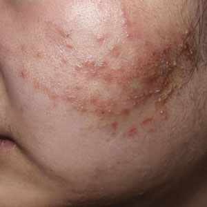
Black Malar Papules Following Use of Hydroquinone Cream 2% Purchased Online
Black Malar Papules Following Use of Hydroquinone Cream 2% Purchased Online
To the Editor:
Exogenous ochronosis (EO) is a rare dyschromia that affects individuals with darker skin tones and results from the application of skin-lightening topical agents, most commonly those containing hydroquinone. Hydroquinone-induced EO usually is reported outside the United States, mainly in African and South Asian patients,1 as well as after long-term use of high‑concentration hydroquinone. We report a case of stage II EO that developed in a patient in the United States following 6 months' use of hydroquinone cream 2% that was purchased online.
A 75-year-old Black man presented to the dermatology clinic with dark discoloration on the cheeks of 6 months’ duration. He reported that the hyperpigmentation had developed after 6 months of daily use of an over-the-counter (OTC) cream containing 2% hydroquinone, which he purchased online to treat melasma of the bilateral cheeks and temples that had been present for at least a decade. The patient was otherwise healthy and did not take any other daily medications. He was a landscaper by occupation and spent most days outdoors. He reported a history of recurrent blistering sunburns and reported that he applied sunscreen with a sun protection factor of 30 to the face on most days.
Physical examination revealed gray-black, coarsely textured papules and plaques on a background of brown pigmentation involving the malar cheeks. Reticulated, blue-black, caviarlike papules were present within the plaques (Figure 1). Dermoscopy revealed blue-black globules and globularlike structures on a brown background with the absence of follicular openings (Figure 2). A punch biopsy from the left malar cheek revealed banana-shaped, yellow-brown deposits with heavy elastosis in the superficial dermis (Figure 3). The patient was advised to permanently discontinue hydroquinone and was prescribed topical adapalene. He was evaluated for treatment with a fractional erbium-doped yttrium aluminum garnet laser but ultimately elected not to pursue treatment.
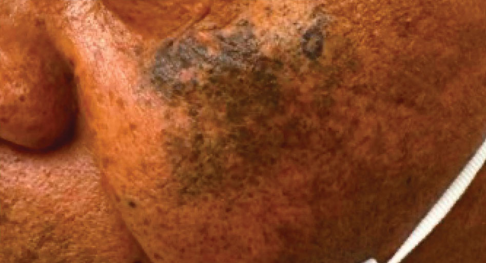
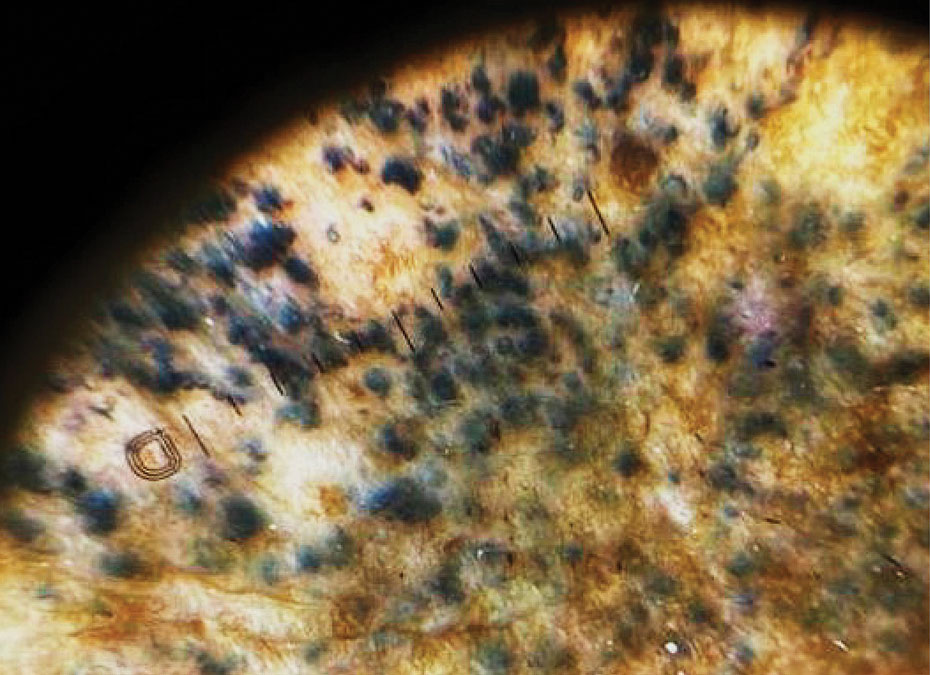
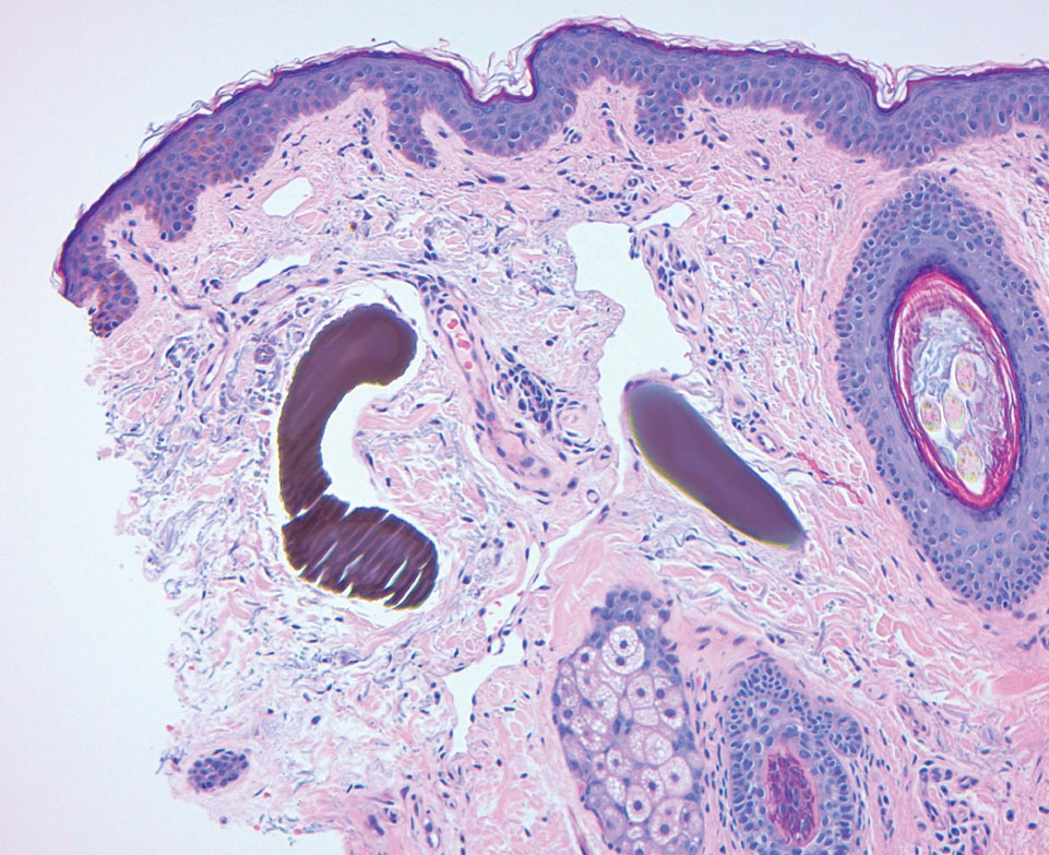
Hydroquinone has long been a first-line topical agent for the treatment of skin dyschromia. It generally is regarded as a safe and effective therapy when used under the supervision of a dermatologist. Prolonged use of topical hydroquinone is known to induce paradoxical EO. This acquired dyschromia is characterized by the development of blue-black or gray-blue pigmented patches as well as caviarlike black papules, papulonodules, and skin atrophy in more severe cases.2 Most cases are documented in African and South Asian countries, with fewer than 100 cases described in the United States.2 Despite the rarity of EO in the United States, concerns regarding the unsupervised use of hydroquinone contributed to regulatory changes. In response to the CARES Act and Over-the-Counter Drug Monograph Reform, the sale of OTC hydroquinone-based products became prohibited in September 2020 in the United States.³ Prior to the CARES Act, an estimated 10 million tubes of OTC hydroquinone were sold, and approximately 330,000 prescriptions were written in 2019.2,4 Subsequently, its availability was converted to prescription only in the United States.
Exogenous ochronosis occurs in 3 clinical stages: (I) erythema and hyperpigmentation, (II) black colloid milia (caviarlike papules) and atrophy, and (III) papulonodules.1 Our patient’s case was unusual, as EO is most commonly described after exposure to high concentrations and prolonged hydroquinone treatment. In a global retrospective review of 126 patients of varied racial and ethnic backgrounds with hydroquinone-induced ochronosis, EO was most frequently reported with concentrations exceeding 4%. The median duration of use was 5 years, and only 12 cases (9.5%) of EO were reported with 1 year of use or less, including 4 cases with 3 months or less of use.1
While large epidemiologic studies from South Africa have reported EO prevalence rates as high as 69% among users of skin-lightening products,6 a search of the PubMed, Scopus, and Web of Science databases using the terms exogenous ochronosis and hydroquinone identified only 39 cases in US-based literature between 1983 and 2020.2 In a retrospective review of 25 cases of EO in the United States, the average length of product use was 9.2 years, and the shortest duration was 1.5 years.7
The pathogenesis of EO is thought to occur due to the inhibitive effect of topical hydroquinone on homogentisic acid oxidase, resulting in the accumulation of homogentisic acid in tissues and ochronotic deposits.1,5 We suspect that our patient’s occupation as a landscaper and associated chronic sun exposure may have accelerated the development of EO, as UV exposure and inadequate photoprotection have been described as risk factors in the development and severity of EO.8 Although the patient reported daily application of sun protection factor 30 sunscreen, the extent of his outdoor occupational exposure may have exceeded the protection afforded.
Treatment of EO is notoriously challenging, and skin changes often are permanent. Of the 25 patients identified in a 10-year retrospective analysis of EO at a single US center, none responded to topical treatment alone.7 Among those who underwent procedural interventions, 3 patients treated with microneedling and 3 patients treated with Q-switched Alexandrite laser demonstrated favorable outcomes; however, this was a retrospective chart review, not a controlled trial with predefined treatment arms.7 Bellew et al9 reported 2 patients with EO who showed progressive lightening in response to Q-switched Alexandrite laser treatments. In this case series, the first patient received 6 treatments at 2-month intervals and the second received 4 treatments at 4-month intervals. Posttreatment biopsies demonstrated decreased dermal pigmentation, and neither patient experienced scarring or textural changes. Méndez Baca et al10 demonstrated successful lightening of pigmentation in a patient with medium-toned skin following 9 sessions with a fractional nonablative picosecond laser. Ceglio et al11 described a stepwise treatment approach in a patient with dark brown skin with EO, beginning with 12 sessions of a 1064-nm Q-switched Nd:YAG laser that yielded slight improvement, followed by 7 sessions of intense pulsed light therapy with partial response, and ultimately achieving marked improvement after 5 bimonthly sessions of fractional 10,600-nm carbon dioxide laser.
This case highlights the potential for EO to develop even with low-concentration (2%) hydroquinone obtained through unregulated online sources, underscoring the need for dermatologist supervision when hydroquinone is used. Despite the 2020 prohibition of OTC hydroquinone sales in the United States, patients continue to access these products online. Clinicians should maintain a high index of suspicion for EO in patients presenting with paradoxical facial hyperpigmentation and a history of unsupervised hydroquinone use.
- Ishack S, Lipner SR. Exogenous ochronosis associated with hydroquinone: a systematic review. Int J Dermatol. 2022;61:675-684. doi:10.1111/ijd.15878
- Qorbani A, Mubasher A, Sarantopoulos GP, et al. Exogenous ochronosis (EO): skin lightening cream causing rare caviar-like lesion with banana-like pigments; review of literature and histological comparison with endogenous counterpart. Autops Case Rep. 2020;10:E2020197. doi:10.4322/acr.2020.197
- Coronavirus Aid, Relief, and Economic Security (CARES) Act, Pub L No. 116-136, §3851, 134 Stat 281 (2020).
- Hydroquinone, found in skin lightening agents worldwide, linked with increased skin cancer risk. Medscape. August 26, 2022. Accessed June 5, 2026.
- Dogliotti M, Leibowitz M. Granulomatous ochronosis—a cosmetic-induced skin disorder in Blacks. S Afr Med J. 1979;56:757-760.
- Hardwick N, Van Gelder LW, Van der Merwe CA, Van der Merwe MP. Exogenous ochronosis: an epidemiological study. Br J Dermatol. 1989;120:229-238. doi:10.1111/j.1365-2133.1989.tb07787.x
- Lazar M, De La Garza H, Vashi NA. Exogenous ochronosis: characterizing a rare disorder in skin of color. J Clin Med. 2023;12:4341. doi:10.3390/jcm12134341
- Findlay GH, de Beer HA. Chronic hydroquinone poisoning of the skin from skin-lightening cosmetics. A South African epidemic of ochronosis of the face in dark-skinned individuals. S Afr Med J. 1980;57:187-190.
- Bellew SG, Alster TS. Treatment of exogenous ochronosis with a Q-switched alexandrite (755 nm) laser. Dermatol Surg. 2004;30:555-558. doi:10.1111/j.1524-4725.2004.30177.x
- Méndez Baca I, Al-Niaimi F, Colina C, et al. A case of ochronosis successfully treated with the picosecond laser. J Cosmet Dermatol. 2019;18:1322-1325. doi:10.1111/jocd.12834
- Ceglio WW, Careta MF, Patriota R, et al. Exogenous ochronosis successfully treated with the combination of intense pulsed light and fractional CO2 laser. An Bras Dermatol. 2023;98:138-140. doi:10.1016/j.abd.2021.08.013
To the Editor:
Exogenous ochronosis (EO) is a rare dyschromia that affects individuals with darker skin tones and results from the application of skin-lightening topical agents, most commonly those containing hydroquinone. Hydroquinone-induced EO usually is reported outside the United States, mainly in African and South Asian patients,1 as well as after long-term use of high‑concentration hydroquinone. We report a case of stage II EO that developed in a patient in the United States following 6 months' use of hydroquinone cream 2% that was purchased online.
A 75-year-old Black man presented to the dermatology clinic with dark discoloration on the cheeks of 6 months’ duration. He reported that the hyperpigmentation had developed after 6 months of daily use of an over-the-counter (OTC) cream containing 2% hydroquinone, which he purchased online to treat melasma of the bilateral cheeks and temples that had been present for at least a decade. The patient was otherwise healthy and did not take any other daily medications. He was a landscaper by occupation and spent most days outdoors. He reported a history of recurrent blistering sunburns and reported that he applied sunscreen with a sun protection factor of 30 to the face on most days.
Physical examination revealed gray-black, coarsely textured papules and plaques on a background of brown pigmentation involving the malar cheeks. Reticulated, blue-black, caviarlike papules were present within the plaques (Figure 1). Dermoscopy revealed blue-black globules and globularlike structures on a brown background with the absence of follicular openings (Figure 2). A punch biopsy from the left malar cheek revealed banana-shaped, yellow-brown deposits with heavy elastosis in the superficial dermis (Figure 3). The patient was advised to permanently discontinue hydroquinone and was prescribed topical adapalene. He was evaluated for treatment with a fractional erbium-doped yttrium aluminum garnet laser but ultimately elected not to pursue treatment.
Hydroquinone has long been a first-line topical agent for the treatment of skin dyschromia. It generally is regarded as a safe and effective therapy when used under the supervision of a dermatologist. Prolonged use of topical hydroquinone is known to induce paradoxical EO. This acquired dyschromia is characterized by the development of blue-black or gray-blue pigmented patches as well as caviarlike black papules, papulonodules, and skin atrophy in more severe cases.2 Most cases are documented in African and South Asian countries, with fewer than 100 cases described in the United States.2 Despite the rarity of EO in the United States, concerns regarding the unsupervised use of hydroquinone contributed to regulatory changes. In response to the CARES Act and Over-the-Counter Drug Monograph Reform, the sale of OTC hydroquinone-based products became prohibited in September 2020 in the United States.³ Prior to the CARES Act, an estimated 10 million tubes of OTC hydroquinone were sold, and approximately 330,000 prescriptions were written in 2019.2,4 Subsequently, its availability was converted to prescription only in the United States.
Exogenous ochronosis occurs in 3 clinical stages: (I) erythema and hyperpigmentation, (II) black colloid milia (caviarlike papules) and atrophy, and (III) papulonodules.1 Our patient’s case was unusual, as EO is most commonly described after exposure to high concentrations and prolonged hydroquinone treatment. In a global retrospective review of 126 patients of varied racial and ethnic backgrounds with hydroquinone-induced ochronosis, EO was most frequently reported with concentrations exceeding 4%. The median duration of use was 5 years, and only 12 cases (9.5%) of EO were reported with 1 year of use or less, including 4 cases with 3 months or less of use.1
While large epidemiologic studies from South Africa have reported EO prevalence rates as high as 69% among users of skin-lightening products,6 a search of the PubMed, Scopus, and Web of Science databases using the terms exogenous ochronosis and hydroquinone identified only 39 cases in US-based literature between 1983 and 2020.2 In a retrospective review of 25 cases of EO in the United States, the average length of product use was 9.2 years, and the shortest duration was 1.5 years.7
The pathogenesis of EO is thought to occur due to the inhibitive effect of topical hydroquinone on homogentisic acid oxidase, resulting in the accumulation of homogentisic acid in tissues and ochronotic deposits.1,5 We suspect that our patient’s occupation as a landscaper and associated chronic sun exposure may have accelerated the development of EO, as UV exposure and inadequate photoprotection have been described as risk factors in the development and severity of EO.8 Although the patient reported daily application of sun protection factor 30 sunscreen, the extent of his outdoor occupational exposure may have exceeded the protection afforded.
Treatment of EO is notoriously challenging, and skin changes often are permanent. Of the 25 patients identified in a 10-year retrospective analysis of EO at a single US center, none responded to topical treatment alone.7 Among those who underwent procedural interventions, 3 patients treated with microneedling and 3 patients treated with Q-switched Alexandrite laser demonstrated favorable outcomes; however, this was a retrospective chart review, not a controlled trial with predefined treatment arms.7 Bellew et al9 reported 2 patients with EO who showed progressive lightening in response to Q-switched Alexandrite laser treatments. In this case series, the first patient received 6 treatments at 2-month intervals and the second received 4 treatments at 4-month intervals. Posttreatment biopsies demonstrated decreased dermal pigmentation, and neither patient experienced scarring or textural changes. Méndez Baca et al10 demonstrated successful lightening of pigmentation in a patient with medium-toned skin following 9 sessions with a fractional nonablative picosecond laser. Ceglio et al11 described a stepwise treatment approach in a patient with dark brown skin with EO, beginning with 12 sessions of a 1064-nm Q-switched Nd:YAG laser that yielded slight improvement, followed by 7 sessions of intense pulsed light therapy with partial response, and ultimately achieving marked improvement after 5 bimonthly sessions of fractional 10,600-nm carbon dioxide laser.
This case highlights the potential for EO to develop even with low-concentration (2%) hydroquinone obtained through unregulated online sources, underscoring the need for dermatologist supervision when hydroquinone is used. Despite the 2020 prohibition of OTC hydroquinone sales in the United States, patients continue to access these products online. Clinicians should maintain a high index of suspicion for EO in patients presenting with paradoxical facial hyperpigmentation and a history of unsupervised hydroquinone use.
- Ishack S, Lipner SR. Exogenous ochronosis associated with hydroquinone: a systematic review. Int J Dermatol. 2022;61:675-684. doi:10.1111/ijd.15878
- Qorbani A, Mubasher A, Sarantopoulos GP, et al. Exogenous ochronosis (EO): skin lightening cream causing rare caviar-like lesion with banana-like pigments; review of literature and histological comparison with endogenous counterpart. Autops Case Rep. 2020;10:E2020197. doi:10.4322/acr.2020.197
- Coronavirus Aid, Relief, and Economic Security (CARES) Act, Pub L No. 116-136, §3851, 134 Stat 281 (2020).
- Hydroquinone, found in skin lightening agents worldwide, linked with increased skin cancer risk. Medscape. August 26, 2022. Accessed June 5, 2026.
- Dogliotti M, Leibowitz M. Granulomatous ochronosis—a cosmetic-induced skin disorder in Blacks. S Afr Med J. 1979;56:757-760.
- Hardwick N, Van Gelder LW, Van der Merwe CA, Van der Merwe MP. Exogenous ochronosis: an epidemiological study. Br J Dermatol. 1989;120:229-238. doi:10.1111/j.1365-2133.1989.tb07787.x
- Lazar M, De La Garza H, Vashi NA. Exogenous ochronosis: characterizing a rare disorder in skin of color. J Clin Med. 2023;12:4341. doi:10.3390/jcm12134341
- Findlay GH, de Beer HA. Chronic hydroquinone poisoning of the skin from skin-lightening cosmetics. A South African epidemic of ochronosis of the face in dark-skinned individuals. S Afr Med J. 1980;57:187-190.
- Bellew SG, Alster TS. Treatment of exogenous ochronosis with a Q-switched alexandrite (755 nm) laser. Dermatol Surg. 2004;30:555-558. doi:10.1111/j.1524-4725.2004.30177.x
- Méndez Baca I, Al-Niaimi F, Colina C, et al. A case of ochronosis successfully treated with the picosecond laser. J Cosmet Dermatol. 2019;18:1322-1325. doi:10.1111/jocd.12834
- Ceglio WW, Careta MF, Patriota R, et al. Exogenous ochronosis successfully treated with the combination of intense pulsed light and fractional CO2 laser. An Bras Dermatol. 2023;98:138-140. doi:10.1016/j.abd.2021.08.013
Black Malar Papules Following Use of Hydroquinone Cream 2% Purchased Online
Black Malar Papules Following Use of Hydroquinone Cream 2% Purchased Online
Practice Points
- Exogenous ochronosis (EO) is a rare adverse effect of topical hydroquinone characterized by blue-black or gray-blue pigmentation, caviarlike papules, and distinctive banana-shaped dermal deposits on histopathology.
- Most reported cases of hydroquinone-induced EO occur after prolonged use of concentrations exceeding 4%; this case developed after only 6 months of exposure to hydroquinone cream 2% purchased online.
- Available evidence suggests that laser-based therapies may offer greater improvement than topical treatments in affected patients.
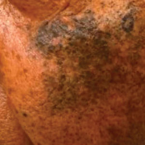
Rosacea: We Still Haven’t Found What We’re Looking For
Rosacea: We Still Haven’t Found What We’re Looking For
In May 1987, U2 released “I Still Haven’t Found What I’m Looking For” as a single from their legendary fifth album, The Joshua Tree. The song became their second consecutive #1 hit in the United States to chart on the Billboard Hot 100.1 Here I am, almost 4 decades later, composing an editorial focused on where we are now in dermatology with rosacea. The first thought that came to mind is that U2 was right—and still is—because with rosacea, we still haven’t found what we’re looking for.
In this editorial, I am discussing cutaneous rosacea, not including ocular disease and phymatous changes. The first challenge with cutaneous rosacea is to get the dermatology community on the same page with what the clinical disease state includes, as the individual “dots” that comprise the pathophysiology of rosacea are not similarly connected in each affected patient. This confounds our ability to effectively treat the disease state in many cases, as the clinical manifestations of rosacea are highly variable, reflecting relative contributions from different components of the pathophysiology.2-5
On top of this challenge, the vast majority of US Food and Drug Administration (FDA)–approved therapies for rosacea have been directed at reducing papules and pustules, which are not present in all patients, and their associated perilesional erythema.6,7 We have only had topical alpha agonists as FDA-approved therapies for persistent facial erythema, the superficial vascular component of rosacea that is distinct from the papulopustular component and can intensify during flares with flushing episodes. A major limitation of the alpha-agonist topical approach is the short duration of their effects, which last only hours, and the need for consistent application over time, which can be frustrating for patients. Physical modalities such as lights and lasers offer marked assistance, especially for the superficial vascular component, but may be limited by availability, cost, and differences in expertise. Is the above challenging enough? Keep in mind that I still have not addressed the skin barrier aspects of the disease, symptomatology, and patient education challenges.
I don’t want to sound as if dermatology has been defeated, as we have actually been able to provide marked improvement for many patients with rosacea.6-9 This is especially true in those who present with papules and pustules and also are compliant and patient enough to allow for the integration of the topical and/or physical therapies needed to treat the vascular components including telangiectasias. What has lagged has been the ability to develop a more comprehensive selection of pharmaceutical options, as most have targeted the papulopustular manifestation of the disease. There are many highly effective options in this “papules and pustules of rosacea” space, including topical ivermectin, azelaic acid, and metronidazole, along with the addition of a foam formulation of minocycline, a microencapsulated formulation of benzoyl peroxide, and low-dose oral minocycline. What mostly limits experience with newer FDA-approved therapies is access for patients due to insurance coverage issues and generic substitutions. Overall, the development of newer FDA-approved pharmacologic therapies has become relatively stagnant and remains so at the present time.6-9
So where are we now? Much of the emphasis on rosacea management has transitioned to alternative areas of consideration, including skin care and barrier dysfunction, the role of vitamins and other nutrients, and use of natural compounds.
An algorithm created by a consensus panel of dermatologists has provided foundational measures recommended for use in all patients with rosacea and rosacea-prone skin, including education, behavioral modifications, recognition and avoidance of triggers, avoidance of skin irritants, preventive skin care, and sun photoprotection measures.10 The algorithm emphasizes that assessment of an individual’s facial skin condition and grading of cutaneous rosacea are very important at the first visit and continually thereafter during treatment while the preventive measures continue. Individual dermatology panels have published on skin care nuances suggested for specific patient subgroups such as skin of color and Asian and Latino populations.11-13 While the importance of skin care and skin barrier function in rosacea are well known to the dermatology community, it is essential for clinicians to consistently emphasize this importance to their patients and their staff.
A relatively recent publication has thoroughly addressed the potential roles of vitamins and nutrients in rosacea, discussing both systemic supplementation and topical approaches. A variety of ingredients were evaluated—including vitamin B3, a vitamin A derivative, vitamin C, vitamin D, zinc formulations, and omega-3 fatty acids—demonstrating a range of outcomes.14 There are data that support the potential of vitamins and nutrients as an adjunctive approach; however, at present, this area holds potential for future advancement without any definitive products or well-established recommendations available. More well-designed studies that are targeted for specific rosacea populations are needed.
A large array of natural compounds, primarily botanicals and phytochemicals, have been discussed in the literature for both short- and long-term management of rosacea.15 Many cosmeceutical products are gaining public notoriety, as the concept of “natural” is appealing to the lay population due to the assumption that natural implies greater safety, which is not necessarily true. Semenescu et al15 provided a very comprehensive review of the applicable characteristics of several plant-based agents and potential mechanisms that may be beneficial as adjunctive agents in rosacea. Similar to vitamins and nutrients, there is some scientific basis for the routine integration of natural phytochemicals, but research is needed with well-designed studies used in targeted specific rosacea populations.
Unfortunately, I cannot finish this editorial with ground-breaking news about a new gamechanger approach or product; however, I can tell you that dermatology never gives up. Advances in developing adjunctive therapies and future disease targets such as mast cell activation, microvesicle particles, NLRP3 inflammasome pathway, and Janus kinase inhibition are always in motion, and we continue to try to find what we’re looking for.16
- U2. “I Still Haven’t Found What I’m Looking For.” Wikipedia. Accessed May 17, 2026. https://en.wikipedia.org/wiki/I_Still_Haven%27t_Found_What_I%27m_Looking_For
- Geng RSQ, Bourkas AN, Mufti A, et al. Rosacea: pathogenesis and therapeutic correlates. J Cutan Med Surg. 2024;28:178-189. doi:10.1177/12034754241229365
- Yang F, Wang L, Song D, et al. Signaling pathways and targeted therapy for rosacea. Front Immunol. 2024;16:15:1367994. doi:10.3389/fimmu.2024.1367994
- Andrusiewicz A, Khimuk S, Mijas D, et al. Molecular mechanisms in the etiopathology of rosacea-systematic review. Int J Mol Sci. 2025;26:11292. doi:10.3390/ijms262311292
- Schaller M, Almeida LMC, Bewley A, et al. Recommendations for rosacea diagnosis, classification and management: update from the global ROSacea COnsensus 2019 panel. Br J Dermatol. 2020;182:1269-1276. doi:10.1111/bjd.18420
- Del Rosso JQ, Tanghetti E, Webster G, et al. Update on the management of rosacea from the American Acne & Rosacea Society (AARS). Clin Aesthet Dermatol. 2020;13(6 suppl):S17-S24.
- van Zuuren EJ, Arents BWM, van der Linden MMD, et al. Rosacea: new concepts in classification and treatment. Am J Clin Dermatol. 2021;22:457-465. doi:10.1007/s40257-021-00595-7
- Tu KY, Jung CJ, Shih YH, et al. Therapeutic strategies focusing on immune dysregulation and neuroinflammation in rosacea. Front Immunol. 2024;15:1403798. doi:10.3389/fimmu.2024.1403798
- Schaller M, Almeida LMC, Bewley A, et al. Recommendations for rosacea diagnosis, classification and management: update from the global ROSacea COnsensus 2019 panel. Br J Dermatol. 2020;182:1269-1276. doi:10.1111/bjd.18420
- Baldwin H, Alexis A, Andriessen A, et al. Skin barrier deficiency in rosacea: an algorithm integrating OTC skincare products into treatment regimens. Drugs Dermatol. 2022;21:SF3595563-SF35955610. doi:10.36849/JDD.m0922
- Alexis A, Woolery-Lloyd H, Andriessen A, et al. Improving rosacea outcomes in skin of color patients: a review on the nuances in the treatment and the use of cleansers and moisturizers. J Drugs Dermatol. 2022;21:574-580. doi:10.36849/JDD.6838
- Kulthanan K, Andriessen A, Jiang X, et al. A review of the challenges and nuances in treating rosacea in Asian skin types using cleansers and moisturizers as adjuncts. J Drugs Dermatol. 2023;22:45-53. doi:10.36849/JDD.7021
- Gonzalez C, Andriessen A, Antelo D, et al. Treatment and maintenance of cutaneous rosacea in Latino skin types with prescription medications and non-prescription cleansers and moisturizers as adjuncts: a review. J Drugs Dermatol. 2022;21:1111-1118. doi:10.36849/JDD.7010
- Algarin YA, Pulumati A, Jaalouk D, et al. The role of vitamins and nutrients in rosacea. Arch Dermatol Res. 2024;316:142. doi:10.1007/s00403-024-02895-4
- Semenescu J, Similie D, Diaconeasa Z, et al. Recent advances in the management of rosacea through natural compounds. Pharmaceuticals (Basel). 2024;17:212. doi:10.3390/ph17020212
- Fisher GW, Travers JB, Rohan CA. Rosacea pathogenesis and therapeutics: current treatments and a look at future targets. Front Med (Lausanne). 2023;10:1292722. doi:10.3389/fmed.2023.1292722
In May 1987, U2 released “I Still Haven’t Found What I’m Looking For” as a single from their legendary fifth album, The Joshua Tree. The song became their second consecutive #1 hit in the United States to chart on the Billboard Hot 100.1 Here I am, almost 4 decades later, composing an editorial focused on where we are now in dermatology with rosacea. The first thought that came to mind is that U2 was right—and still is—because with rosacea, we still haven’t found what we’re looking for.
In this editorial, I am discussing cutaneous rosacea, not including ocular disease and phymatous changes. The first challenge with cutaneous rosacea is to get the dermatology community on the same page with what the clinical disease state includes, as the individual “dots” that comprise the pathophysiology of rosacea are not similarly connected in each affected patient. This confounds our ability to effectively treat the disease state in many cases, as the clinical manifestations of rosacea are highly variable, reflecting relative contributions from different components of the pathophysiology.2-5
On top of this challenge, the vast majority of US Food and Drug Administration (FDA)–approved therapies for rosacea have been directed at reducing papules and pustules, which are not present in all patients, and their associated perilesional erythema.6,7 We have only had topical alpha agonists as FDA-approved therapies for persistent facial erythema, the superficial vascular component of rosacea that is distinct from the papulopustular component and can intensify during flares with flushing episodes. A major limitation of the alpha-agonist topical approach is the short duration of their effects, which last only hours, and the need for consistent application over time, which can be frustrating for patients. Physical modalities such as lights and lasers offer marked assistance, especially for the superficial vascular component, but may be limited by availability, cost, and differences in expertise. Is the above challenging enough? Keep in mind that I still have not addressed the skin barrier aspects of the disease, symptomatology, and patient education challenges.
I don’t want to sound as if dermatology has been defeated, as we have actually been able to provide marked improvement for many patients with rosacea.6-9 This is especially true in those who present with papules and pustules and also are compliant and patient enough to allow for the integration of the topical and/or physical therapies needed to treat the vascular components including telangiectasias. What has lagged has been the ability to develop a more comprehensive selection of pharmaceutical options, as most have targeted the papulopustular manifestation of the disease. There are many highly effective options in this “papules and pustules of rosacea” space, including topical ivermectin, azelaic acid, and metronidazole, along with the addition of a foam formulation of minocycline, a microencapsulated formulation of benzoyl peroxide, and low-dose oral minocycline. What mostly limits experience with newer FDA-approved therapies is access for patients due to insurance coverage issues and generic substitutions. Overall, the development of newer FDA-approved pharmacologic therapies has become relatively stagnant and remains so at the present time.6-9
So where are we now? Much of the emphasis on rosacea management has transitioned to alternative areas of consideration, including skin care and barrier dysfunction, the role of vitamins and other nutrients, and use of natural compounds.
An algorithm created by a consensus panel of dermatologists has provided foundational measures recommended for use in all patients with rosacea and rosacea-prone skin, including education, behavioral modifications, recognition and avoidance of triggers, avoidance of skin irritants, preventive skin care, and sun photoprotection measures.10 The algorithm emphasizes that assessment of an individual’s facial skin condition and grading of cutaneous rosacea are very important at the first visit and continually thereafter during treatment while the preventive measures continue. Individual dermatology panels have published on skin care nuances suggested for specific patient subgroups such as skin of color and Asian and Latino populations.11-13 While the importance of skin care and skin barrier function in rosacea are well known to the dermatology community, it is essential for clinicians to consistently emphasize this importance to their patients and their staff.
A relatively recent publication has thoroughly addressed the potential roles of vitamins and nutrients in rosacea, discussing both systemic supplementation and topical approaches. A variety of ingredients were evaluated—including vitamin B3, a vitamin A derivative, vitamin C, vitamin D, zinc formulations, and omega-3 fatty acids—demonstrating a range of outcomes.14 There are data that support the potential of vitamins and nutrients as an adjunctive approach; however, at present, this area holds potential for future advancement without any definitive products or well-established recommendations available. More well-designed studies that are targeted for specific rosacea populations are needed.
A large array of natural compounds, primarily botanicals and phytochemicals, have been discussed in the literature for both short- and long-term management of rosacea.15 Many cosmeceutical products are gaining public notoriety, as the concept of “natural” is appealing to the lay population due to the assumption that natural implies greater safety, which is not necessarily true. Semenescu et al15 provided a very comprehensive review of the applicable characteristics of several plant-based agents and potential mechanisms that may be beneficial as adjunctive agents in rosacea. Similar to vitamins and nutrients, there is some scientific basis for the routine integration of natural phytochemicals, but research is needed with well-designed studies used in targeted specific rosacea populations.
Unfortunately, I cannot finish this editorial with ground-breaking news about a new gamechanger approach or product; however, I can tell you that dermatology never gives up. Advances in developing adjunctive therapies and future disease targets such as mast cell activation, microvesicle particles, NLRP3 inflammasome pathway, and Janus kinase inhibition are always in motion, and we continue to try to find what we’re looking for.16
- U2. “I Still Haven’t Found What I’m Looking For.” Wikipedia. Accessed May 17, 2026. https://en.wikipedia.org/wiki/I_Still_Haven%27t_Found_What_I%27m_Looking_For
- Geng RSQ, Bourkas AN, Mufti A, et al. Rosacea: pathogenesis and therapeutic correlates. J Cutan Med Surg. 2024;28:178-189. doi:10.1177/12034754241229365
- Yang F, Wang L, Song D, et al. Signaling pathways and targeted therapy for rosacea. Front Immunol. 2024;16:15:1367994. doi:10.3389/fimmu.2024.1367994
- Andrusiewicz A, Khimuk S, Mijas D, et al. Molecular mechanisms in the etiopathology of rosacea-systematic review. Int J Mol Sci. 2025;26:11292. doi:10.3390/ijms262311292
- Schaller M, Almeida LMC, Bewley A, et al. Recommendations for rosacea diagnosis, classification and management: update from the global ROSacea COnsensus 2019 panel. Br J Dermatol. 2020;182:1269-1276. doi:10.1111/bjd.18420
- Del Rosso JQ, Tanghetti E, Webster G, et al. Update on the management of rosacea from the American Acne & Rosacea Society (AARS). Clin Aesthet Dermatol. 2020;13(6 suppl):S17-S24.
- van Zuuren EJ, Arents BWM, van der Linden MMD, et al. Rosacea: new concepts in classification and treatment. Am J Clin Dermatol. 2021;22:457-465. doi:10.1007/s40257-021-00595-7
- Tu KY, Jung CJ, Shih YH, et al. Therapeutic strategies focusing on immune dysregulation and neuroinflammation in rosacea. Front Immunol. 2024;15:1403798. doi:10.3389/fimmu.2024.1403798
- Schaller M, Almeida LMC, Bewley A, et al. Recommendations for rosacea diagnosis, classification and management: update from the global ROSacea COnsensus 2019 panel. Br J Dermatol. 2020;182:1269-1276. doi:10.1111/bjd.18420
- Baldwin H, Alexis A, Andriessen A, et al. Skin barrier deficiency in rosacea: an algorithm integrating OTC skincare products into treatment regimens. Drugs Dermatol. 2022;21:SF3595563-SF35955610. doi:10.36849/JDD.m0922
- Alexis A, Woolery-Lloyd H, Andriessen A, et al. Improving rosacea outcomes in skin of color patients: a review on the nuances in the treatment and the use of cleansers and moisturizers. J Drugs Dermatol. 2022;21:574-580. doi:10.36849/JDD.6838
- Kulthanan K, Andriessen A, Jiang X, et al. A review of the challenges and nuances in treating rosacea in Asian skin types using cleansers and moisturizers as adjuncts. J Drugs Dermatol. 2023;22:45-53. doi:10.36849/JDD.7021
- Gonzalez C, Andriessen A, Antelo D, et al. Treatment and maintenance of cutaneous rosacea in Latino skin types with prescription medications and non-prescription cleansers and moisturizers as adjuncts: a review. J Drugs Dermatol. 2022;21:1111-1118. doi:10.36849/JDD.7010
- Algarin YA, Pulumati A, Jaalouk D, et al. The role of vitamins and nutrients in rosacea. Arch Dermatol Res. 2024;316:142. doi:10.1007/s00403-024-02895-4
- Semenescu J, Similie D, Diaconeasa Z, et al. Recent advances in the management of rosacea through natural compounds. Pharmaceuticals (Basel). 2024;17:212. doi:10.3390/ph17020212
- Fisher GW, Travers JB, Rohan CA. Rosacea pathogenesis and therapeutics: current treatments and a look at future targets. Front Med (Lausanne). 2023;10:1292722. doi:10.3389/fmed.2023.1292722
Rosacea: We Still Haven’t Found What We’re Looking For
Rosacea: We Still Haven’t Found What We’re Looking For
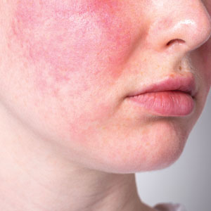
Topical Hypochlorous Acid for Acne Vulgaris: Mechanisms, Clinical Evidence, and Therapeutic Potential
Topical Hypochlorous Acid for Acne Vulgaris: Mechanisms, Clinical Evidence, and Therapeutic Potential
Acne vulgaris, a chronic inflammatory disease of the pilosebaceous unit, is among the most prevalent dermatologic conditions worldwide. Though symptoms range in severity, patients can experience painful irritation and scarring that can lead to substantial psychological distress and impact quality of life. Cutibacterium acnes plays a central role in acne development through biofilm formation, lipase activity, and activation of innate immune pathways, which together contribute to a cycle of inflammation and comedogenesis.1
First-line treatments for acne vulgaris include topical benzoyl peroxide, topical retinoids, and topical antibiotics, while oral spironolactone and tetracyclines can be used alongside topical therapies for more extensive disease. Additionally, isotretinoin is generally reserved for severe or refractory cases. While these therapies are effective, each has notable limitations and adverse effects that in some cases limit adherence and efficacy. The most common adverse effects seen with topical acne therapies include irritation and dryness. Systemic therapies such as spironolactone can cause fatigue, dizziness, and birth defects, while prolonged antibiotic use can promote the risk for antimicrobial resistance.2
Hypochlorous acid (HOCl) is a naturally occurring weak acid produced by neutrophils and currently is approved by the US Food and Drug Administration for wound cleansing, burn management, and dermal lesion irrigation. Although it is not approved for the treatment of acne, stabilized HOCl formulations have been used off label in dermatology for this purpose. Interest in HOCl stems from its broad-spectrum antimicrobial activity against C acnes, anti-inflammatory properties, and favorable safety profile. This literature review examines the mechanism of action, clinical evidence, and potential role of HOCl in acne management, contextualizing its use relative to current standard therapies.
Methods
A narrative literature review was conducted to identify and synthesize peer-reviewed evidence on the use of HOCl in dermatology, with emphasis on its potential role in acne management. Searches were performed using PubMed and Scopus databases. Search terms included combinations of hypochlorous acid, acne, acne vulgaris, dermatology, antimicrobial, anti-inflammatory, biofilm, and skin barrier. Eligible publications included original research articles, randomized controlled trials, retrospective studies, preclinical in vitro and in vivo studies, and systematic reviews published in English between January 2000 and December 2024. Titles and abstracts were screened for relevance, and the final selection included 16 peer-reviewed articles that met the inclusion criteria.
Results
Hypochlorous acid exhibits rapid, broad-spectrum antimicrobial activity against gram-positive and gram-negative bacteria and fungi. In vitro time-kill assays demonstrated that stabilized HOCl was bactericidal against a variety of pathogens, including methicillin-resistant Staphylococcus aureus, methicillin-sensitive S aureus, Staphylococcus epidermidis, Corynebacterium species, Streptococcus pyogenes, Pseudomonas aeruginosa, Candida albicans, and C acnes. Specifically, HOCl achieved 99.99% or greater kill within 2 minutes.3 Moreover, HOCl’s antimicrobial efficacy against this panel of organisms was found to be comparable to or greater than that of commonly used antiseptics, including povidone-iodine, chlorhexidine gluconate, and isopropyl alcohol.3 An additional study using HOCl stabilized in 0.9% saline (pH, 3.5-4.0) confirmed its rapid activity across gram-positive, gram-negative, and fungal species, again demonstrating 99.99 % or greater reduction within 1 to 2 minutes of exposure.4
Hypochlorous acid also has demonstrated substantial biofilm-disruptive properties. In vitro studies demonstrated that HOCl can penetrate and disrupt early-stage biofilms by oxidizing extracellular polymeric substances and damaging bacterial membranes; however, while HOCl was effective at destroying immature biofilms and preventing biofilm formation, its efficacy against mature, fully established biofilms was more limited.5 Thus, topical HOCl may be most effective during the early colonization phase of acne, helping to prevent biofilm maturation and subsequent inflammatory lesion formation. Unlike traditional topical and oral antibiotics, HOCl’s nonspecific oxidative mechanism of action is less likely to contribute to microbial resistance. These findings highlight HOCl as a rapid, broad-spectrum antimicrobial with additional biofilm-disruptive activity, supporting its potential role as an early-intervention therapeutic in acne treatment.
In addition to its antimicrobial effects, HOCl is a potent anti-inflammatory molecule that exerts its anti-inflammatory effects through several mechanisms. HOCl acts as a mast cell membrane stabilizer, inhibiting degranulation. Hypochlorous acid also has been demonstrated to reduce levels of leukotriene B4 and interleukin (IL) 2, which supports that HOCl has both antipruritic and anti-inflammatory properties.6 In keratinocytes and immune cells, HOCl has been shown to suppress the transcription of multiple proinflammatory cytokines by oxidizing IκB kinase β, which then prevents the activation of the nuclear factor kappa B (NF-κB) signaling pathway.7 Additionally, in a murine model of atopic dermatitis, treatment with HOCl resulted in a downregulation of key proinflammatory and Th2-associated cytokines, including IL-1β, IL-4, IL-6, IL-13, tumor necrosis factor α, thymus and activation-regulated chemokine, thymic stromal lymphopoietin, and IL-31. Parallel in vitro assays revealed that HOCl inhibited phosphorylation of mitogen-activated protein kinase (MAPK) and inhibitor of κB, which inhibits the downstream proinflammatory pathways, thereby elucidating a mechanistic basis for its anti-inflammatory effects.8 C acnes has been shown to activate the toll-like receptor 2 pathway on keratinocytes and macrophages, triggering NF-κB–dependent release of IL-1β and tumor necrosis factor α.9
By inhibiting these same signaling pathways, HOCl may attenuate the inflammatory response associated with acne lesions while simultaneously reducing microbial load. These combined anti-inflammatory and antimicrobial effects also may contribute to improved healing outcomes. Emerging clinical evidence supports HOCl’s benefit in minimizing scarring and postinflammatory sequelae. A comparative study evaluating a silicone-based scar gel containing HOCl vs silicone gel alone found that the HOCl-containing formulation produced greater improvement in hypertrophic and keloid scar appearance and overall scar texture.10 These findings suggest that HOCl may have beneficial effects on wound healing and scar remodeling.
In murine models of acute radiation dermatitis, topical HOCl reduced NF-κB–dependent gene expression, decreased epidermal ulceration, and promoted re-epithelialization to near-normal histologic appearance.7 A double-blind, randomized controlled trial evaluating topical sodium hypochlorite 0.005%, which is a compound in equilibrium with HOCl under physiologic pH, demonstrated a statistically significant reduction in papules among patients with mild to moderate acne after 1 month of treatment (P<.0001). Female participants exhibited greater lesion improvement, suggesting possible hormonal or immunologic modulation of response.11 Although limited in scale, this literature review provides preliminary clinical support for the therapeutic potential of HOCl in the treatment of acne. Collectively, these findings highlight the potential of HOCl as an emerging treatment in acne and other dermatologic conditions.
Comment
Traditional acne therapies include topical agents such as benzoyl peroxide, topical retinoids (eg, tretinoin, adapalene), and salicylic acid, as well as systemic agents such as oral antibiotics, spironolactone, and isotretinoin. While these treatments are effective, their use may be limited by irritation, antibiotic resistance, and systemic adverse effects.
Hypochlorous acid is a potential adjunctive option that acts locally with minimal irritation and without hormonal or systemic activity.12 Its antimicrobial and anti-inflammatory mechanisms target key pathogenic pathways in acne while maintaining excellent cutaneous tolerability. In a randomized, double-blind, placebo-controlled trial of 89 patients comparing topical HOCl solution with benzoyl peroxide for mild to moderate acne, HOCl demonstrated comparable improvement in lesion counts.13 Importantly, no local adverse effects were reported in either group and no dose adjustments were needed during the 12-week treatment period. Although both agents were effective, the absence of irritation with HOCl contrasts with the dryness and erythema frequently associated with benzoyl peroxide.
Additionally, a clinical trial comparing HOCl 0.01% with standard antiseptics, including isopropyl alcohol, povidone-iodine, and chlorhexidine gluconate, showed that HOCl achieved comparable antibacterial reductions while remaining well tolerated and free of facial adverse effects.14 Similarly, studies evaluating HOCl’s antimicrobial efficacy have confirmed that it is nontoxic to periocular and facial tissues, further supporting its safety for use on delicate skin regions.3 Importantly, in an experimental model evaluating both healthy skin and skin with experimentally induced irritant contact dermatitis, repeated application of an HOCl-based formulation did not impair skin barrier function, underscoring its excellent cutaneous compatibility even under inflammatory conditions.15 Ultimately, these findings suggest that HOCl offers efficacy comparable to benzoyl peroxide and retinoids while eliminating the irritation and barrier disruption that can limit the use of these first-line agents.
Topical antibiotics such as clindamycin and erythromycin are used widely for their antimicrobial and anti-inflammatory properties but increasingly are undermined by antibiotic resistance. In contrast, HOCl has been shown to reduce bacterial load without altering microbial diversity, supporting its role as a resistance-neutral antimicrobial option for acne management.16 These characteristics position HOCl as a well-tolerated, resistance-neutral adjunctive treatment that warrants further investigation through larger, controlled trials.
Topical HOCl formulations, particularly those available as sprays or misting solutions, have gained attention on social media for their ease of use and versatility. Although formal studies evaluating adherence or outcomes in this context are currently limited, this emerging consumer trend underscores the perceived convenience of HOCl compared with traditional acne therapies. These formulations can be applied throughout the day, including between exercise and work, supporting adherence among patients with active lifestyles. In contrast to many conventional topical agents that require specific application timing, cleansing routines, or avoidance of cosmetic products, HOCl sprays offer flexible use without disrupting daily activities. These characteristics highlight HOCl’s potential as a user-friendly option that may support consistent application and optimize therapeutic outcomes.
Conclusion
The addition of HOCl to acne treatment regimens offers several potential benefits. Its antimicrobial and anti-inflammatory properties may help prevent new papules and pustules, while its favorable tolerability profile minimizes irritation and systemic adverse effects. Preliminary data also suggest efficacy in androgen-mediated acne, though additional studies are needed to confirm these findings.¹¹ Current evidence remains limited by small sample sizes, short follow-up durations, and a lack of comparative studies among available formulations. Accordingly, HOCl should be considered an adjunctive rather than replacement therapy pending larger studies with longer follow-up.
- Vasam M, Korutla S, Bohara RA. Acne vulgaris: a review of the pathophysiology, treatment, and recent nanotechnology based advances. Biochem Biophys Rep. 2023;36:101578.
- Reynolds RV, Yeung H, Cheng CE, et al. Guidelines of care for the management of acne vulgaris. J Am Acad Dermatol. 2024;90:1006.E1-1006.E30.
- Anagnostopoulos AG, Rong A, Miller D, et al. 0.01% hypochlorous acid as an alternative skin antiseptic: an in vitro comparison. Dermatol Surg. 2018;44:1489-1493.
- Wang L, Bassiri M, Najafi R, et al. Hypochlorous acid as a potential wound care agent: part I. stabilized hypochlorous acid: a component of the inorganic armamentarium of innate immunity. J Burns Wounds. 2007;6:E5.
- Ortega-Peña S, Hidalgo-González C, Robson MC, et al. In vitro microbicidal, anti-biofilm and cytotoxic effects of different commercial antiseptics. Int Wound J. 2017;14:470-479.
- Gold MH, Andriessen A, Bhatia AC, et al. Topical stabilized hypochlorous acid: the future gold standard for wound care and scar management in dermatologic and plastic surgery procedures. J Cosmet Dermatol. 2020;19:270-277.
- Leung TH, Zhang LF, Wang J, et al. Topical hypochlorite ameliorates NF-κB–mediated skin diseases in mice. J Clin Invest. 2013;123:5361-5370.
- Fukuyama T, Martel BC, Linder KE, et al. Hypochlorous acid is antipruritic and anti‐inflammatory in a mouse model of atopic dermatitis. Clin Exp Allergy. 2018;48:78-88.
- Lheure C, Grange PA, Ollagnier G, et al. TLR-2 recognizes Propionibacterium acnes CAMP factor 1 from highly inflammatory strains. PLoS ONE. 2016;11:E0167237.
- Gold MH, Andriessen A, Dayan SH, et al. Hypochlorous acid gel technology—its impact on postprocedure treatment and scar prevention. J Cosmet Dermatol. 2017;16:162-167.
- Dorostkar A, Ghahartars M, Namazi MR, et al. Sodium hypochlorite 0.005% versus placebo in the treatment of mild to moderate acne: a double-blind randomized controlled trial. Dermatol Pract Concept. Published online May 20, 2021.
- del Rosso JQ, Bhatia N. Status report on topical hypochlorous acid: clinical relevance of specific formulations, potential modes of action, and study outcomes. J Clin Aesthet Dermatol. 2018;11:36-39.
- Tirado-Sánchez A, Ponce-Olivera RM. Efficacy and tolerance of superoxidized solution in the treatment of mild to moderate inflammatory acne. a double-blinded, placebo- controlled, parallel-group, randomized, clinical trial. J Dermatolog Treat. 2009;20:289-292.
- Tran AQ, Topilow N, Rong A, et al. Comparison of skin antiseptic agents and the role of 0.01% hypochlorous acid. Aesthet Surg J. 2021;41:1170-1175.
- Yüksel YT, Sonne M, Nørreslet LB, et al. Skin barrier response to active chlorine hand disinfectant—an experimental study comparing skin barrier response to active chlorine hand disinfectant and alcohol-based hand rub on healthy skin and eczematous skin. Skin Res Technol. 2022;28:89-97.
- Stroman D, Mintun K, Epstein A, et al. Reduction in bacterial load using hypochlorous acid hygiene solution on ocular skin. OPTH. 2017;11:707-714.
Acne vulgaris, a chronic inflammatory disease of the pilosebaceous unit, is among the most prevalent dermatologic conditions worldwide. Though symptoms range in severity, patients can experience painful irritation and scarring that can lead to substantial psychological distress and impact quality of life. Cutibacterium acnes plays a central role in acne development through biofilm formation, lipase activity, and activation of innate immune pathways, which together contribute to a cycle of inflammation and comedogenesis.1
First-line treatments for acne vulgaris include topical benzoyl peroxide, topical retinoids, and topical antibiotics, while oral spironolactone and tetracyclines can be used alongside topical therapies for more extensive disease. Additionally, isotretinoin is generally reserved for severe or refractory cases. While these therapies are effective, each has notable limitations and adverse effects that in some cases limit adherence and efficacy. The most common adverse effects seen with topical acne therapies include irritation and dryness. Systemic therapies such as spironolactone can cause fatigue, dizziness, and birth defects, while prolonged antibiotic use can promote the risk for antimicrobial resistance.2
Hypochlorous acid (HOCl) is a naturally occurring weak acid produced by neutrophils and currently is approved by the US Food and Drug Administration for wound cleansing, burn management, and dermal lesion irrigation. Although it is not approved for the treatment of acne, stabilized HOCl formulations have been used off label in dermatology for this purpose. Interest in HOCl stems from its broad-spectrum antimicrobial activity against C acnes, anti-inflammatory properties, and favorable safety profile. This literature review examines the mechanism of action, clinical evidence, and potential role of HOCl in acne management, contextualizing its use relative to current standard therapies.
Methods
A narrative literature review was conducted to identify and synthesize peer-reviewed evidence on the use of HOCl in dermatology, with emphasis on its potential role in acne management. Searches were performed using PubMed and Scopus databases. Search terms included combinations of hypochlorous acid, acne, acne vulgaris, dermatology, antimicrobial, anti-inflammatory, biofilm, and skin barrier. Eligible publications included original research articles, randomized controlled trials, retrospective studies, preclinical in vitro and in vivo studies, and systematic reviews published in English between January 2000 and December 2024. Titles and abstracts were screened for relevance, and the final selection included 16 peer-reviewed articles that met the inclusion criteria.
Results
Hypochlorous acid exhibits rapid, broad-spectrum antimicrobial activity against gram-positive and gram-negative bacteria and fungi. In vitro time-kill assays demonstrated that stabilized HOCl was bactericidal against a variety of pathogens, including methicillin-resistant Staphylococcus aureus, methicillin-sensitive S aureus, Staphylococcus epidermidis, Corynebacterium species, Streptococcus pyogenes, Pseudomonas aeruginosa, Candida albicans, and C acnes. Specifically, HOCl achieved 99.99% or greater kill within 2 minutes.3 Moreover, HOCl’s antimicrobial efficacy against this panel of organisms was found to be comparable to or greater than that of commonly used antiseptics, including povidone-iodine, chlorhexidine gluconate, and isopropyl alcohol.3 An additional study using HOCl stabilized in 0.9% saline (pH, 3.5-4.0) confirmed its rapid activity across gram-positive, gram-negative, and fungal species, again demonstrating 99.99 % or greater reduction within 1 to 2 minutes of exposure.4
Hypochlorous acid also has demonstrated substantial biofilm-disruptive properties. In vitro studies demonstrated that HOCl can penetrate and disrupt early-stage biofilms by oxidizing extracellular polymeric substances and damaging bacterial membranes; however, while HOCl was effective at destroying immature biofilms and preventing biofilm formation, its efficacy against mature, fully established biofilms was more limited.5 Thus, topical HOCl may be most effective during the early colonization phase of acne, helping to prevent biofilm maturation and subsequent inflammatory lesion formation. Unlike traditional topical and oral antibiotics, HOCl’s nonspecific oxidative mechanism of action is less likely to contribute to microbial resistance. These findings highlight HOCl as a rapid, broad-spectrum antimicrobial with additional biofilm-disruptive activity, supporting its potential role as an early-intervention therapeutic in acne treatment.
In addition to its antimicrobial effects, HOCl is a potent anti-inflammatory molecule that exerts its anti-inflammatory effects through several mechanisms. HOCl acts as a mast cell membrane stabilizer, inhibiting degranulation. Hypochlorous acid also has been demonstrated to reduce levels of leukotriene B4 and interleukin (IL) 2, which supports that HOCl has both antipruritic and anti-inflammatory properties.6 In keratinocytes and immune cells, HOCl has been shown to suppress the transcription of multiple proinflammatory cytokines by oxidizing IκB kinase β, which then prevents the activation of the nuclear factor kappa B (NF-κB) signaling pathway.7 Additionally, in a murine model of atopic dermatitis, treatment with HOCl resulted in a downregulation of key proinflammatory and Th2-associated cytokines, including IL-1β, IL-4, IL-6, IL-13, tumor necrosis factor α, thymus and activation-regulated chemokine, thymic stromal lymphopoietin, and IL-31. Parallel in vitro assays revealed that HOCl inhibited phosphorylation of mitogen-activated protein kinase (MAPK) and inhibitor of κB, which inhibits the downstream proinflammatory pathways, thereby elucidating a mechanistic basis for its anti-inflammatory effects.8 C acnes has been shown to activate the toll-like receptor 2 pathway on keratinocytes and macrophages, triggering NF-κB–dependent release of IL-1β and tumor necrosis factor α.9
By inhibiting these same signaling pathways, HOCl may attenuate the inflammatory response associated with acne lesions while simultaneously reducing microbial load. These combined anti-inflammatory and antimicrobial effects also may contribute to improved healing outcomes. Emerging clinical evidence supports HOCl’s benefit in minimizing scarring and postinflammatory sequelae. A comparative study evaluating a silicone-based scar gel containing HOCl vs silicone gel alone found that the HOCl-containing formulation produced greater improvement in hypertrophic and keloid scar appearance and overall scar texture.10 These findings suggest that HOCl may have beneficial effects on wound healing and scar remodeling.
In murine models of acute radiation dermatitis, topical HOCl reduced NF-κB–dependent gene expression, decreased epidermal ulceration, and promoted re-epithelialization to near-normal histologic appearance.7 A double-blind, randomized controlled trial evaluating topical sodium hypochlorite 0.005%, which is a compound in equilibrium with HOCl under physiologic pH, demonstrated a statistically significant reduction in papules among patients with mild to moderate acne after 1 month of treatment (P<.0001). Female participants exhibited greater lesion improvement, suggesting possible hormonal or immunologic modulation of response.11 Although limited in scale, this literature review provides preliminary clinical support for the therapeutic potential of HOCl in the treatment of acne. Collectively, these findings highlight the potential of HOCl as an emerging treatment in acne and other dermatologic conditions.
Comment
Traditional acne therapies include topical agents such as benzoyl peroxide, topical retinoids (eg, tretinoin, adapalene), and salicylic acid, as well as systemic agents such as oral antibiotics, spironolactone, and isotretinoin. While these treatments are effective, their use may be limited by irritation, antibiotic resistance, and systemic adverse effects.
Hypochlorous acid is a potential adjunctive option that acts locally with minimal irritation and without hormonal or systemic activity.12 Its antimicrobial and anti-inflammatory mechanisms target key pathogenic pathways in acne while maintaining excellent cutaneous tolerability. In a randomized, double-blind, placebo-controlled trial of 89 patients comparing topical HOCl solution with benzoyl peroxide for mild to moderate acne, HOCl demonstrated comparable improvement in lesion counts.13 Importantly, no local adverse effects were reported in either group and no dose adjustments were needed during the 12-week treatment period. Although both agents were effective, the absence of irritation with HOCl contrasts with the dryness and erythema frequently associated with benzoyl peroxide.
Additionally, a clinical trial comparing HOCl 0.01% with standard antiseptics, including isopropyl alcohol, povidone-iodine, and chlorhexidine gluconate, showed that HOCl achieved comparable antibacterial reductions while remaining well tolerated and free of facial adverse effects.14 Similarly, studies evaluating HOCl’s antimicrobial efficacy have confirmed that it is nontoxic to periocular and facial tissues, further supporting its safety for use on delicate skin regions.3 Importantly, in an experimental model evaluating both healthy skin and skin with experimentally induced irritant contact dermatitis, repeated application of an HOCl-based formulation did not impair skin barrier function, underscoring its excellent cutaneous compatibility even under inflammatory conditions.15 Ultimately, these findings suggest that HOCl offers efficacy comparable to benzoyl peroxide and retinoids while eliminating the irritation and barrier disruption that can limit the use of these first-line agents.
Topical antibiotics such as clindamycin and erythromycin are used widely for their antimicrobial and anti-inflammatory properties but increasingly are undermined by antibiotic resistance. In contrast, HOCl has been shown to reduce bacterial load without altering microbial diversity, supporting its role as a resistance-neutral antimicrobial option for acne management.16 These characteristics position HOCl as a well-tolerated, resistance-neutral adjunctive treatment that warrants further investigation through larger, controlled trials.
Topical HOCl formulations, particularly those available as sprays or misting solutions, have gained attention on social media for their ease of use and versatility. Although formal studies evaluating adherence or outcomes in this context are currently limited, this emerging consumer trend underscores the perceived convenience of HOCl compared with traditional acne therapies. These formulations can be applied throughout the day, including between exercise and work, supporting adherence among patients with active lifestyles. In contrast to many conventional topical agents that require specific application timing, cleansing routines, or avoidance of cosmetic products, HOCl sprays offer flexible use without disrupting daily activities. These characteristics highlight HOCl’s potential as a user-friendly option that may support consistent application and optimize therapeutic outcomes.
Conclusion
The addition of HOCl to acne treatment regimens offers several potential benefits. Its antimicrobial and anti-inflammatory properties may help prevent new papules and pustules, while its favorable tolerability profile minimizes irritation and systemic adverse effects. Preliminary data also suggest efficacy in androgen-mediated acne, though additional studies are needed to confirm these findings.¹¹ Current evidence remains limited by small sample sizes, short follow-up durations, and a lack of comparative studies among available formulations. Accordingly, HOCl should be considered an adjunctive rather than replacement therapy pending larger studies with longer follow-up.
- Vasam M, Korutla S, Bohara RA. Acne vulgaris: a review of the pathophysiology, treatment, and recent nanotechnology based advances. Biochem Biophys Rep. 2023;36:101578.
- Reynolds RV, Yeung H, Cheng CE, et al. Guidelines of care for the management of acne vulgaris. J Am Acad Dermatol. 2024;90:1006.E1-1006.E30.
- Anagnostopoulos AG, Rong A, Miller D, et al. 0.01% hypochlorous acid as an alternative skin antiseptic: an in vitro comparison. Dermatol Surg. 2018;44:1489-1493.
- Wang L, Bassiri M, Najafi R, et al. Hypochlorous acid as a potential wound care agent: part I. stabilized hypochlorous acid: a component of the inorganic armamentarium of innate immunity. J Burns Wounds. 2007;6:E5.
- Ortega-Peña S, Hidalgo-González C, Robson MC, et al. In vitro microbicidal, anti-biofilm and cytotoxic effects of different commercial antiseptics. Int Wound J. 2017;14:470-479.
- Gold MH, Andriessen A, Bhatia AC, et al. Topical stabilized hypochlorous acid: the future gold standard for wound care and scar management in dermatologic and plastic surgery procedures. J Cosmet Dermatol. 2020;19:270-277.
- Leung TH, Zhang LF, Wang J, et al. Topical hypochlorite ameliorates NF-κB–mediated skin diseases in mice. J Clin Invest. 2013;123:5361-5370.
- Fukuyama T, Martel BC, Linder KE, et al. Hypochlorous acid is antipruritic and anti‐inflammatory in a mouse model of atopic dermatitis. Clin Exp Allergy. 2018;48:78-88.
- Lheure C, Grange PA, Ollagnier G, et al. TLR-2 recognizes Propionibacterium acnes CAMP factor 1 from highly inflammatory strains. PLoS ONE. 2016;11:E0167237.
- Gold MH, Andriessen A, Dayan SH, et al. Hypochlorous acid gel technology—its impact on postprocedure treatment and scar prevention. J Cosmet Dermatol. 2017;16:162-167.
- Dorostkar A, Ghahartars M, Namazi MR, et al. Sodium hypochlorite 0.005% versus placebo in the treatment of mild to moderate acne: a double-blind randomized controlled trial. Dermatol Pract Concept. Published online May 20, 2021.
- del Rosso JQ, Bhatia N. Status report on topical hypochlorous acid: clinical relevance of specific formulations, potential modes of action, and study outcomes. J Clin Aesthet Dermatol. 2018;11:36-39.
- Tirado-Sánchez A, Ponce-Olivera RM. Efficacy and tolerance of superoxidized solution in the treatment of mild to moderate inflammatory acne. a double-blinded, placebo- controlled, parallel-group, randomized, clinical trial. J Dermatolog Treat. 2009;20:289-292.
- Tran AQ, Topilow N, Rong A, et al. Comparison of skin antiseptic agents and the role of 0.01% hypochlorous acid. Aesthet Surg J. 2021;41:1170-1175.
- Yüksel YT, Sonne M, Nørreslet LB, et al. Skin barrier response to active chlorine hand disinfectant—an experimental study comparing skin barrier response to active chlorine hand disinfectant and alcohol-based hand rub on healthy skin and eczematous skin. Skin Res Technol. 2022;28:89-97.
- Stroman D, Mintun K, Epstein A, et al. Reduction in bacterial load using hypochlorous acid hygiene solution on ocular skin. OPTH. 2017;11:707-714.
Topical Hypochlorous Acid for Acne Vulgaris: Mechanisms, Clinical Evidence, and Therapeutic Potential
Topical Hypochlorous Acid for Acne Vulgaris: Mechanisms, Clinical Evidence, and Therapeutic Potential
Practice Points
- First-line treatments for acne vulgaris are effective but often limited by local irritation, systemic adverse effects, and antibiotic resistance.
- Hypochlorous acid (HOCl) shows rapid, broad-spectrum antimicrobial and biofilm-disruptive activity against Cutibacterium acnes and other pathogens, with a low propensity for resistance.
- Emerging clinical data indicate HOCl formulations deliver efficacy comparable to standard topical treatments with superior tolerability and no barrier disruption, supporting its use as a well-tolerated adjunct in acne management.
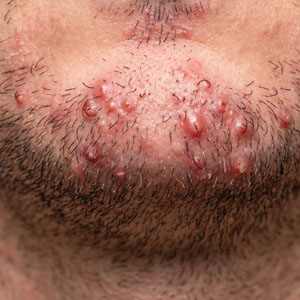
Equity Evaluation: Analysis of the Prescribing Patterns of Isotretinoin Based on Reproductive Potential
Equity Evaluation: Analysis of the Prescribing Patterns of Isotretinoin Based on Reproductive Potential
To the Editor:
Isotretinoin is the most effective treatment option for acne vulgaris and is considered the first-line therapy for severe scarring acne.1-3 Isotretinoin therapy is indicated for moderate acne when other treatments have failed or when the condition causes distress.3 While there are treatment guidelines for moderate to severe acne, there is no gold standard, and most treatment options include a combination of topical and/or oral therapies.4 Isotretinoin is one of the few monotherapy options available, but its use often is limited by concerns for severe internal and external birth defects if taken during pregnancy.
To manage teratogenicity risks associated with isotretinoin, the US Food and Drug Administration implemented the iPLEDGE Risk Evaluation and Mitigation Strategy in 2006.5 The iPLEDGE compliance requirements differ vastly depending on the reproductive potential of the patient, and the process of obtaining an isotretinoin prescription is much more cumbersome for patients who can get pregnant. Previous studies have demonstrated that males are more likely to be prescribed isotretinoin compared to females, even though females attended more acne-related office visits than males.6,7 Additionally, the administrative burden of iPLEDGE may influence the prescribing patterns of isotretinoin. In a survey of 510 dermatologists, approximately 30% reported that they have at times chosen not to prescribe isotretinoin to patients with severe acne due to the administrative burden of iPLEDGE.8 In this study, we sought to further analyze the prescribing patterns of isotretinoin based on a patient’s reproductive potential.
In this single-center, retrospective cohort study, electronic medical records from 3292 patients diagnosed with acne vulgaris at the Department of Dermatology at Rush University Medical Center (Chicago, Illinois) between January 2013 and December 2019 were reviewed. A total of 188 patients who were prescribed isotretinoin for acne were identified, but only 171 met the study criteria. Eligible patients were aged 12 to 25 years and were prescribed oral isotretinoin for acne vulgaris during the study period. Patients younger than 12 or older than 25 years, those who were prescribed isotretinoin for indications other than acne vulgaris, and those who had previously received isotretinoin and either initiated an additional course or continued treatment at our institution were excluded. Eligible patients then were grouped by reproductive potential: patients who can get pregnant (positive reproductive potential [RP+]) and patients who cannot get pregnant (negative reproductive potential [RP–]). The number of months from initial acne visit, total number of office visits attended, and number of alternative medications that failed before isotretinoin therapy was discussed and initiated were compared between the 2 groups. To standardize between groups, the office visit at which patients were enrolled in iPLEDGE served as the date that isotretinoin therapy was initiated. Alternative medication type and sex of the prescriber were evaluated as secondary end points.
Eighty-nine RP+ patients and 82 RP– patients were prescribed isotretinoin for acne, including 85 females, 2 transgender males with female reproductive organs, 2 females with a history of tubal ligation, and 82 males. Of note, the iPLEDGE program considers tubal ligation as a primary form of contraception and classifies these patients as RP+. Patient demographics are summarized in eTable 1. There was a higher proportion of RP– patients aged 10 to 20 years compared to RP+ patients, for whom there was a higher number of patients aged 21 to 30 years. Most RP+ patients were White, while most RP– patients were Hispanic/Latino.
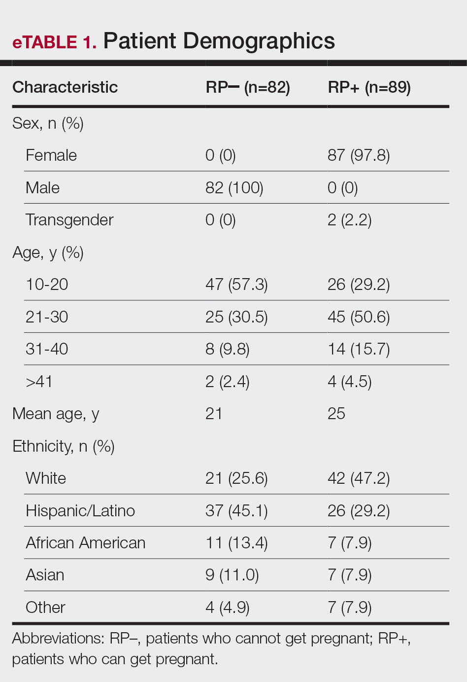
Overall isotretinoin prescribing patterns as well as prescribing patterns stratified by reproductive potential are presented in eTable 2. Overall, the average number of months before isotretinoin was discussed as a treatment option was 11.5 months and before isotretinoin therapy was initiated was 15 months. There was no significant difference between the number of months before isotretinoin was discussed (P=.83) or initiated (P=.56) between RP+ and RP– patients. On average, patients attended 2 office visits before isotretinoin was discussed as a treatment option and 3 office visits before isotretinoin therapy was initiated. The difference between the number of office visits patients attended before isotretinoin was discussed (P=.44) and before isotretinoin therapy was initiated (P=.11) was not significant between RP+ and RP– patients. The number of alternative medications that failed before initiation of isotretinoin therapy was comparable between groups. Patients in the RP– group experienced failure with an average of 5 alternative medications compared with 6 alternative medications in the RP+ group (P=.48).
As shown in eTable 3, oral antibiotic therapy commonly was prescribed prior to isotretinoin in both groups, with doxycycline being prescribed most often. Hormonal therapy, including oral contraceptives and spironolactone, was trialed in 55 of 87 (63.2%) RP+ patients. Most patients experienced failure with other oral antiacne medications before isotretinoin therapy, with only 6 (6.7%) RP+ and 5 (6.1%) RP– patients trying topical therapy only. Prior isotretinoin therapy was documented in 13 (14.6%) RP+ patients compared with only 5 (6.1%) RP– patients. Interestingly, isotretinoin was initiated at the patient’s first office visit more often in the RP+ group.
At the time isotretinoin therapy was initiated, the sex of the prescribing provider was similar within the RP+ group, with 52% (46/89) of female and 48% (43/89) of male prescribers. In the RP– group, there was a slightly higher proportion of male prescribers, with 57% (47/82) male compared to 43% (35/82) female prescribers.
iPLEDGE (https://ipledgeprogram.com) is an online system mandated by the US Food and Drug Administration to be used by patients, physicians, and pharmacists for the duration of isotretinoin therapy to prevent and track isotretinoin-associated pregnancies.9 At its inception, the iPLEDGE program categorized patients as females of child-bearing potential, females not of child-bearing potential, or males. The program recently moved toward transgender-inclusive categorization of patients based on reproductive potential.10 Patients who can get pregnant include cisgender females and transgender males, and patients who cannot get pregnant include cisgender males, transgender females, and females and transgender males who have undergone a hysterectomy or bilateral oophorectomy or who are postmenopausal.9
The iPLEDGE compliance requirements for patients who can get pregnant are extensive. Patients with reproductive potential must obtain a negative baseline pregnancy test, enroll in iPLEDGE, undergo a 30-day waiting period, and obtain a second negative pregnancy test before they can start the medication. Each month thereafter, patients must obtain a negative pregnancy test, demonstrate risk comprehension, and report their methods of contraception on iPLEDGE before they can pick up their prescription. In addition, physicians and pharmacists must confirm patient counseling and obtain authorization codes from iPLEDGE to dispense the medication. If any of these steps are not completed by the patient, physician, or pharmacist within 7 days of the patient’s negative pregnancy test, all steps must be repeated by all parties.9,11
The efficacy and utility of iPLEDGE have been criticized and debated in the literature. Although there has been a general decrease in the number of fetal exposures to isotretinoin since the implementation of iPLEDGE in 2006, the average number of fetal exposures only decreased for 2 years until it plateaued in 2008.5 Some physicians have argued that the decrease in the number of fetal exposures is not attributed to the efficacy of iPLEDGE but rather because the program has made it difficult for patients who can get pregnant to obtain necessary isotretinoin prescriptions.12 Other physicians have reported that they have chosen not to prescribe isotretinoin due to the administrative burden of iPLEDGE.8 Although we expected to observe similar trends in our study, we ultimately had more eligible patients with reproductive potential than patients who could not get pregnant. Additionally, there was no difference in the number of months from initial acne visit, total number of office visits, and number of alternative medications that failed before isotretinoin initiation between patients who could and could not get pregnant. These findings suggest that iPLEDGE requirements did not dissuade prescribers from treating acne with isotretinoin in patients who could get pregnant and that the prescribing patterns of isotretinoin were similar regardless of reproductive potential.
Across all primary outcomes, the standard deviation was high in the overall dataset and in the RP– and RP+ subsets, indicating substantial variability in number of months from initial acne visit, total number of visits, and number of alternative medications that failed prior to initiation of isotretinoin. This implies that the prescribing patterns of isotretinoin may be patient and prescriber dependent, and other variables may influence these outcomes aside from the reproductive potential of the patient. Although there was a marginally higher percentage of male prescribers in the RP– group, the sex of the prescriber did not seem to have a major impact on the prescribing patterns of isotretinoin. Further research is indicated to investigate the impact of other factors that may influence the prescribing patterns of isotretinoin, including insurance coverage, access to contraception, and patient concerns about adverse effects.
The types of alternative medications that patients tried prior to isotretinoin were similar among patients who could get pregnant and patients who could not get pregnant. Hormonal therapy, including oral contraception pills and spironolactone, can be very effective in treating acne in patients with reproductive potential,13 as evidenced by the 55 (31.1%) prescriptions written for hormonal therapy in the RP+ group. Spironolactone for acne is contraindicated in male patients due to its antiandrogenic properties and risk for gynecomastia.14 As such, males have fewer alternative medication options for acne, and this may contribute to the higher prevalence of isotretinoin therapy in males that has been demonstrated in prior studies.6,7 Of note, patients obtained more than 100 prescriptions for oral antimicrobials in both groups. Although patients can see benefit with oral antimicrobials for acne, the volume of antimicrobial prescriptions seen in our cohort raises concerns about antibiotic stewardship.15 Whether isotretinoin is a safer therapeutic option compared to antibiotics is up for debate.2,16
Our study included a small sample size at a single institution, which may limit the external validity of our results. Additionally, our study focused on patients who were prescribed isotretinoin prior to 2020 to control for the influence of the COVID-19 pandemic on prescribing patterns. With this, our data may not reflect postpandemic prescribing trends. Further multi-institution studies that include postpandemic patient cohorts can be conducted to validate our findings.
There were no significant differences in the number of months from initial acne visit, total number of office visits, and number of alternative medications that failed prior to discussing isotretinoin as a treatment option or initiating isotretinoin therapy between patients who can get pregnant and those who cannot get pregnant. We observed substantial variability in these outcomes across datasets, indicating that the prescribing patterns of isotretinoin may be patient and prescriber dependent regardless of the reproductive potential of the patient. Follow-up studies are warranted to further investigate the specific influence of iPLEDGE on the utilization of isotretinoin.
- Aslam I, Fleischer A, Feldman S. Emerging drugs for the treatment of acne. Expert Opin Emerg Drugs. 2015;20:91-101. doi:10.1517/14728214.2015.990373
- Huang CY, Chang IJ, Bolick N, et al. Comparative efficacy of pharmacological treatments for acne vulgaris: a network meta-analysis of 221 randomized controlled trials. Ann Fam Med. 2023;21:358-369. doi:10.1370/afm.2995
- Hauk L. Acne vulgaris: treatment guidelines from the AAD. Am Fam Physician. 2017;95:740-741.
- Habeshian KA, Cohen BA. Current issues in the treatment of acne vulgaris. Pediatrics. 2020;145(suppl 2):S225-S230. doi:10.1542/peds.2019-2056L
- Tkachenko E, Singer S, Sharma P, et al. US Food and Drug Administration reports of pregnancy and pregnancy-related adverse events associated with isotretinoin. JAMA Dermatol. 2019;155:1175-1179. doi:10.1001/jamadermatol.2019.1388
- Fleischer AB Jr, Simpson JK, McMichael A, et al. Are there racial and sex differences in the use of oral isotretinoin for acne management in the United States? J Am Acad Dermatol. 2003;49:662-666. doi:10.1067/s0190-9622(03)01584-6
- Barbieri JS, Shin DB, Wang S, et al. Association of race/ethnicity and sex with differences in health care use and treatment for acne. JAMA Dermatol. 2020;156:312-319. doi:10.1001/jamadermatol.2019.4818
- Lee G, Wolf JR, Somers KE. Administrative burden of iPLEDGE deters isotretinoin prescriptions: results from a survey of dermatologists. Cutis. 2022;110:44-47. doi:10.12788/cutis.0558
- The iPLEDGE REMS Prescriber Guide. iPLEDGE. Updated March 2023. https://ipledgeprogram.com/ResourceDownloadRaw/GuideBestPractices
- Boos MD, Ginsberg BA, Peebles JK. Prescribing isotretinoin for transgender youth: a pledge for more inclusive care. Pediatr Dermatol. 2019;36:169-171. doi:10.1111/pde.13694
- iPLEDGE REMS Guide for Patients Who Can Get Pregnant: The Importance of Avoiding Pregnancy on Isotretinoin. iPLEDGE Program. Updated March 2023. https://ipledgeprogram.com/#Main/Resources
- Nagler AR. Early strides for necessary data-driven improvement in iPLEDGE. JAMA Dermatol. 2019;155:1111-1112. doi:10.1001/jamadermatol.2019.1247
- Reynolds RV, Yeung H, Cheng CE, et al. Guidelines of care for the management of acne vulgaris. J Am Acad Dermatol. 2024;90:1006.E1-1006.E30. doi:10.1016/j.jaad.2023.12.017
- Sato K, Matsumoto D, Iizuka F, et al. Anti-androgenic therapy using oral spironolactone for acne vulgaris in Asians. Aesthetic Plast Surg. 2006;30:689-694. doi:10.1007/s00266-006-0081-0
- Issa NT, Kircik LH. Antibiotic stewardship in acne: 2023 update. J Drugs Dermatol. 2024;23:SF37896s4-SF378969s10. doi:10.36849/JDD.SF378969
- Vallerand IA, Lewinson RT, Farris MS, et al. Efficacy and adverse events of oral isotretinoin for acne: a systematic review. Br J Dermatol. 2018;178:76-85. doi:10.1111/bjd.15668
To the Editor:
Isotretinoin is the most effective treatment option for acne vulgaris and is considered the first-line therapy for severe scarring acne.1-3 Isotretinoin therapy is indicated for moderate acne when other treatments have failed or when the condition causes distress.3 While there are treatment guidelines for moderate to severe acne, there is no gold standard, and most treatment options include a combination of topical and/or oral therapies.4 Isotretinoin is one of the few monotherapy options available, but its use often is limited by concerns for severe internal and external birth defects if taken during pregnancy.
To manage teratogenicity risks associated with isotretinoin, the US Food and Drug Administration implemented the iPLEDGE Risk Evaluation and Mitigation Strategy in 2006.5 The iPLEDGE compliance requirements differ vastly depending on the reproductive potential of the patient, and the process of obtaining an isotretinoin prescription is much more cumbersome for patients who can get pregnant. Previous studies have demonstrated that males are more likely to be prescribed isotretinoin compared to females, even though females attended more acne-related office visits than males.6,7 Additionally, the administrative burden of iPLEDGE may influence the prescribing patterns of isotretinoin. In a survey of 510 dermatologists, approximately 30% reported that they have at times chosen not to prescribe isotretinoin to patients with severe acne due to the administrative burden of iPLEDGE.8 In this study, we sought to further analyze the prescribing patterns of isotretinoin based on a patient’s reproductive potential.
In this single-center, retrospective cohort study, electronic medical records from 3292 patients diagnosed with acne vulgaris at the Department of Dermatology at Rush University Medical Center (Chicago, Illinois) between January 2013 and December 2019 were reviewed. A total of 188 patients who were prescribed isotretinoin for acne were identified, but only 171 met the study criteria. Eligible patients were aged 12 to 25 years and were prescribed oral isotretinoin for acne vulgaris during the study period. Patients younger than 12 or older than 25 years, those who were prescribed isotretinoin for indications other than acne vulgaris, and those who had previously received isotretinoin and either initiated an additional course or continued treatment at our institution were excluded. Eligible patients then were grouped by reproductive potential: patients who can get pregnant (positive reproductive potential [RP+]) and patients who cannot get pregnant (negative reproductive potential [RP–]). The number of months from initial acne visit, total number of office visits attended, and number of alternative medications that failed before isotretinoin therapy was discussed and initiated were compared between the 2 groups. To standardize between groups, the office visit at which patients were enrolled in iPLEDGE served as the date that isotretinoin therapy was initiated. Alternative medication type and sex of the prescriber were evaluated as secondary end points.
Eighty-nine RP+ patients and 82 RP– patients were prescribed isotretinoin for acne, including 85 females, 2 transgender males with female reproductive organs, 2 females with a history of tubal ligation, and 82 males. Of note, the iPLEDGE program considers tubal ligation as a primary form of contraception and classifies these patients as RP+. Patient demographics are summarized in eTable 1. There was a higher proportion of RP– patients aged 10 to 20 years compared to RP+ patients, for whom there was a higher number of patients aged 21 to 30 years. Most RP+ patients were White, while most RP– patients were Hispanic/Latino.
Overall isotretinoin prescribing patterns as well as prescribing patterns stratified by reproductive potential are presented in eTable 2. Overall, the average number of months before isotretinoin was discussed as a treatment option was 11.5 months and before isotretinoin therapy was initiated was 15 months. There was no significant difference between the number of months before isotretinoin was discussed (P=.83) or initiated (P=.56) between RP+ and RP– patients. On average, patients attended 2 office visits before isotretinoin was discussed as a treatment option and 3 office visits before isotretinoin therapy was initiated. The difference between the number of office visits patients attended before isotretinoin was discussed (P=.44) and before isotretinoin therapy was initiated (P=.11) was not significant between RP+ and RP– patients. The number of alternative medications that failed before initiation of isotretinoin therapy was comparable between groups. Patients in the RP– group experienced failure with an average of 5 alternative medications compared with 6 alternative medications in the RP+ group (P=.48).
As shown in eTable 3, oral antibiotic therapy commonly was prescribed prior to isotretinoin in both groups, with doxycycline being prescribed most often. Hormonal therapy, including oral contraceptives and spironolactone, was trialed in 55 of 87 (63.2%) RP+ patients. Most patients experienced failure with other oral antiacne medications before isotretinoin therapy, with only 6 (6.7%) RP+ and 5 (6.1%) RP– patients trying topical therapy only. Prior isotretinoin therapy was documented in 13 (14.6%) RP+ patients compared with only 5 (6.1%) RP– patients. Interestingly, isotretinoin was initiated at the patient’s first office visit more often in the RP+ group.
At the time isotretinoin therapy was initiated, the sex of the prescribing provider was similar within the RP+ group, with 52% (46/89) of female and 48% (43/89) of male prescribers. In the RP– group, there was a slightly higher proportion of male prescribers, with 57% (47/82) male compared to 43% (35/82) female prescribers.
iPLEDGE (https://ipledgeprogram.com) is an online system mandated by the US Food and Drug Administration to be used by patients, physicians, and pharmacists for the duration of isotretinoin therapy to prevent and track isotretinoin-associated pregnancies.9 At its inception, the iPLEDGE program categorized patients as females of child-bearing potential, females not of child-bearing potential, or males. The program recently moved toward transgender-inclusive categorization of patients based on reproductive potential.10 Patients who can get pregnant include cisgender females and transgender males, and patients who cannot get pregnant include cisgender males, transgender females, and females and transgender males who have undergone a hysterectomy or bilateral oophorectomy or who are postmenopausal.9
The iPLEDGE compliance requirements for patients who can get pregnant are extensive. Patients with reproductive potential must obtain a negative baseline pregnancy test, enroll in iPLEDGE, undergo a 30-day waiting period, and obtain a second negative pregnancy test before they can start the medication. Each month thereafter, patients must obtain a negative pregnancy test, demonstrate risk comprehension, and report their methods of contraception on iPLEDGE before they can pick up their prescription. In addition, physicians and pharmacists must confirm patient counseling and obtain authorization codes from iPLEDGE to dispense the medication. If any of these steps are not completed by the patient, physician, or pharmacist within 7 days of the patient’s negative pregnancy test, all steps must be repeated by all parties.9,11
The efficacy and utility of iPLEDGE have been criticized and debated in the literature. Although there has been a general decrease in the number of fetal exposures to isotretinoin since the implementation of iPLEDGE in 2006, the average number of fetal exposures only decreased for 2 years until it plateaued in 2008.5 Some physicians have argued that the decrease in the number of fetal exposures is not attributed to the efficacy of iPLEDGE but rather because the program has made it difficult for patients who can get pregnant to obtain necessary isotretinoin prescriptions.12 Other physicians have reported that they have chosen not to prescribe isotretinoin due to the administrative burden of iPLEDGE.8 Although we expected to observe similar trends in our study, we ultimately had more eligible patients with reproductive potential than patients who could not get pregnant. Additionally, there was no difference in the number of months from initial acne visit, total number of office visits, and number of alternative medications that failed before isotretinoin initiation between patients who could and could not get pregnant. These findings suggest that iPLEDGE requirements did not dissuade prescribers from treating acne with isotretinoin in patients who could get pregnant and that the prescribing patterns of isotretinoin were similar regardless of reproductive potential.
Across all primary outcomes, the standard deviation was high in the overall dataset and in the RP– and RP+ subsets, indicating substantial variability in number of months from initial acne visit, total number of visits, and number of alternative medications that failed prior to initiation of isotretinoin. This implies that the prescribing patterns of isotretinoin may be patient and prescriber dependent, and other variables may influence these outcomes aside from the reproductive potential of the patient. Although there was a marginally higher percentage of male prescribers in the RP– group, the sex of the prescriber did not seem to have a major impact on the prescribing patterns of isotretinoin. Further research is indicated to investigate the impact of other factors that may influence the prescribing patterns of isotretinoin, including insurance coverage, access to contraception, and patient concerns about adverse effects.
The types of alternative medications that patients tried prior to isotretinoin were similar among patients who could get pregnant and patients who could not get pregnant. Hormonal therapy, including oral contraception pills and spironolactone, can be very effective in treating acne in patients with reproductive potential,13 as evidenced by the 55 (31.1%) prescriptions written for hormonal therapy in the RP+ group. Spironolactone for acne is contraindicated in male patients due to its antiandrogenic properties and risk for gynecomastia.14 As such, males have fewer alternative medication options for acne, and this may contribute to the higher prevalence of isotretinoin therapy in males that has been demonstrated in prior studies.6,7 Of note, patients obtained more than 100 prescriptions for oral antimicrobials in both groups. Although patients can see benefit with oral antimicrobials for acne, the volume of antimicrobial prescriptions seen in our cohort raises concerns about antibiotic stewardship.15 Whether isotretinoin is a safer therapeutic option compared to antibiotics is up for debate.2,16
Our study included a small sample size at a single institution, which may limit the external validity of our results. Additionally, our study focused on patients who were prescribed isotretinoin prior to 2020 to control for the influence of the COVID-19 pandemic on prescribing patterns. With this, our data may not reflect postpandemic prescribing trends. Further multi-institution studies that include postpandemic patient cohorts can be conducted to validate our findings.
There were no significant differences in the number of months from initial acne visit, total number of office visits, and number of alternative medications that failed prior to discussing isotretinoin as a treatment option or initiating isotretinoin therapy between patients who can get pregnant and those who cannot get pregnant. We observed substantial variability in these outcomes across datasets, indicating that the prescribing patterns of isotretinoin may be patient and prescriber dependent regardless of the reproductive potential of the patient. Follow-up studies are warranted to further investigate the specific influence of iPLEDGE on the utilization of isotretinoin.
- Aslam I, Fleischer A, Feldman S. Emerging drugs for the treatment of acne. Expert Opin Emerg Drugs. 2015;20:91-101. doi:10.1517/14728214.2015.990373
- Huang CY, Chang IJ, Bolick N, et al. Comparative efficacy of pharmacological treatments for acne vulgaris: a network meta-analysis of 221 randomized controlled trials. Ann Fam Med. 2023;21:358-369. doi:10.1370/afm.2995
- Hauk L. Acne vulgaris: treatment guidelines from the AAD. Am Fam Physician. 2017;95:740-741.
- Habeshian KA, Cohen BA. Current issues in the treatment of acne vulgaris. Pediatrics. 2020;145(suppl 2):S225-S230. doi:10.1542/peds.2019-2056L
- Tkachenko E, Singer S, Sharma P, et al. US Food and Drug Administration reports of pregnancy and pregnancy-related adverse events associated with isotretinoin. JAMA Dermatol. 2019;155:1175-1179. doi:10.1001/jamadermatol.2019.1388
- Fleischer AB Jr, Simpson JK, McMichael A, et al. Are there racial and sex differences in the use of oral isotretinoin for acne management in the United States? J Am Acad Dermatol. 2003;49:662-666. doi:10.1067/s0190-9622(03)01584-6
- Barbieri JS, Shin DB, Wang S, et al. Association of race/ethnicity and sex with differences in health care use and treatment for acne. JAMA Dermatol. 2020;156:312-319. doi:10.1001/jamadermatol.2019.4818
- Lee G, Wolf JR, Somers KE. Administrative burden of iPLEDGE deters isotretinoin prescriptions: results from a survey of dermatologists. Cutis. 2022;110:44-47. doi:10.12788/cutis.0558
- The iPLEDGE REMS Prescriber Guide. iPLEDGE. Updated March 2023. https://ipledgeprogram.com/ResourceDownloadRaw/GuideBestPractices
- Boos MD, Ginsberg BA, Peebles JK. Prescribing isotretinoin for transgender youth: a pledge for more inclusive care. Pediatr Dermatol. 2019;36:169-171. doi:10.1111/pde.13694
- iPLEDGE REMS Guide for Patients Who Can Get Pregnant: The Importance of Avoiding Pregnancy on Isotretinoin. iPLEDGE Program. Updated March 2023. https://ipledgeprogram.com/#Main/Resources
- Nagler AR. Early strides for necessary data-driven improvement in iPLEDGE. JAMA Dermatol. 2019;155:1111-1112. doi:10.1001/jamadermatol.2019.1247
- Reynolds RV, Yeung H, Cheng CE, et al. Guidelines of care for the management of acne vulgaris. J Am Acad Dermatol. 2024;90:1006.E1-1006.E30. doi:10.1016/j.jaad.2023.12.017
- Sato K, Matsumoto D, Iizuka F, et al. Anti-androgenic therapy using oral spironolactone for acne vulgaris in Asians. Aesthetic Plast Surg. 2006;30:689-694. doi:10.1007/s00266-006-0081-0
- Issa NT, Kircik LH. Antibiotic stewardship in acne: 2023 update. J Drugs Dermatol. 2024;23:SF37896s4-SF378969s10. doi:10.36849/JDD.SF378969
- Vallerand IA, Lewinson RT, Farris MS, et al. Efficacy and adverse events of oral isotretinoin for acne: a systematic review. Br J Dermatol. 2018;178:76-85. doi:10.1111/bjd.15668
Equity Evaluation: Analysis of the Prescribing Patterns of Isotretinoin Based on Reproductive Potential
Equity Evaluation: Analysis of the Prescribing Patterns of Isotretinoin Based on Reproductive Potential
Practice Points
- Isotretinoin is one of the few monotherapy options available for acne, but its use often is limited by concerns for severe internal and external birth defects if taken during pregnancy.
- The number of office visits attended, alternative acne medications used, and duration of acne treatment prior to isotretinoin therapy were similar between patients who can become pregnant and patients who cannot become pregnant.
- The reproductive potential of the patient did not appear to have a major impact on the prescribing patterns of isotretinoin; rather, the utilization of isotretinoin likely is dependent on individual prescriber preferences and other patient factors.