User login
AVAHO
div[contains(@class, 'header__large-screen')]
div[contains(@class, 'read-next-article')]
div[contains(@class, 'nav-primary')]
nav[contains(@class, 'nav-primary')]
section[contains(@class, 'footer-nav-section-wrapper')]
footer[@id='footer']
div[contains(@class, 'main-prefix')]
section[contains(@class, 'nav-hidden')]
div[contains(@class, 'ce-card-content')]
nav[contains(@class, 'nav-ce-stack')]

Similar results for once- or twice-weekly carfilzomib in MM
Patients with newly diagnosed multiple myeloma have similar outcomes whether they receive carfilzomib once or twice a week, according to a pooled analysis of trial data.
Researchers found no significant difference in safety, progression-free survival (PFS), or overall survival (OS) whether patients received carfilzomib at 70 mg/m2 once a week or 36 mg/m2 twice a week.
Sara Bringhen, MD, PhD, of University of Turin, Italy, and her colleagues conducted this analysis and detailed the results in Haematologica.
The researchers pooled data from a phase 1/2 trial (NCT01857115) and a phase 2 trial (NCT01346787), both enrolling transplant-ineligible patients with newly diagnosed multiple myeloma.
In both studies, induction consisted of nine 4-week cycles of carfilzomib (given once or twice weekly), cyclophosphamide (300 mg on days 1, 8, and 15), and dexamethasone (40 mg on days 1, 8, 15, and 22). After induction, patients received carfilzomib maintenance (at either dose) until progression or intolerable toxicity.
The pooled analysis included 121 patients: 63 who received carfilzomib at 70 mg/m2 once weekly and 58 who received carfilzomib at 36 mg/m2 twice weekly.
There were no significant differences in baseline characteristics between the dosing groups. For the entire cohort, the median age at diagnosis was 72 years (range, 55-86), and the median follow-up was 39 months.
A total of 119 patients started induction (63 in the once-weekly group and 56 in the twice-weekly group), and 90 patients received maintenance (47 and 43, respectively). Patients received maintenance for a median of 17 months in the once-weekly group and 20 months in the twice-weekly group (P = .17).
There was no significant difference between the groups with regard to PFS or OS, either from enrollment or the start of maintenance.
From enrollment, the median PFS was 35.7 months in the once-weekly group and 35.5 months in the twice-weekly group (hazard ratio [HR] = 1.39; P = .26). The 3-year OS was 70% and 72%, respectively (HR = 1.27; P = .5).
From the start of maintenance, the 3-year PFS was 47% in the once-weekly group and 51% in the twice-weekly group (HR = 1.04; P = .92). The 3-year OS was 72% and 73%, respectively (HR = 0.82; P = .71).
There were no significant between-group differences in the rates of grade 3-5 adverse events (AEs) or the need for carfilzomib dose reduction or discontinuation.
Grade 3-5 hematologic AEs occurred in 24% of patients in the once-weekly group and 30% of those in the twice-weekly group. Grade 3-5 nonhematologic AEs occurred in 38% and 41%, respectively.
Twenty-nine percent of patients in the once-weekly group required a reduction in carfilzomib dose, as did 30% of patients in the twice-weekly group. Common AEs leading to dose reduction were acute kidney injury, infections, and hypertension.
AEs leading to carfilzomib discontinuation occurred in 27% of patients in the once-weekly group and 30% of those in the twice-weekly group. Common AEs leading to discontinuation were cardiac injury, infections, and thromboembolism.
Both trials were sponsored by Stichting Hemato-Oncologie voor Volwassenen Nederland in collaboration with Fondazione Neoplasie Sangue ONLUS and supported by funding from Amgen (Onyx Pharmaceuticals). Dr. Bringhen reported relationships with Amgen and other companies. Coauthor Antonio Palumbo, MD, is an employee of Takeda, and other authors reported relationships with a range of companies.
SOURCE: Bringhen S et al. Haematologica. 2019 Feb 7. doi: 10.3324/haematol.2018.208272.
Patients with newly diagnosed multiple myeloma have similar outcomes whether they receive carfilzomib once or twice a week, according to a pooled analysis of trial data.
Researchers found no significant difference in safety, progression-free survival (PFS), or overall survival (OS) whether patients received carfilzomib at 70 mg/m2 once a week or 36 mg/m2 twice a week.
Sara Bringhen, MD, PhD, of University of Turin, Italy, and her colleagues conducted this analysis and detailed the results in Haematologica.
The researchers pooled data from a phase 1/2 trial (NCT01857115) and a phase 2 trial (NCT01346787), both enrolling transplant-ineligible patients with newly diagnosed multiple myeloma.
In both studies, induction consisted of nine 4-week cycles of carfilzomib (given once or twice weekly), cyclophosphamide (300 mg on days 1, 8, and 15), and dexamethasone (40 mg on days 1, 8, 15, and 22). After induction, patients received carfilzomib maintenance (at either dose) until progression or intolerable toxicity.
The pooled analysis included 121 patients: 63 who received carfilzomib at 70 mg/m2 once weekly and 58 who received carfilzomib at 36 mg/m2 twice weekly.
There were no significant differences in baseline characteristics between the dosing groups. For the entire cohort, the median age at diagnosis was 72 years (range, 55-86), and the median follow-up was 39 months.
A total of 119 patients started induction (63 in the once-weekly group and 56 in the twice-weekly group), and 90 patients received maintenance (47 and 43, respectively). Patients received maintenance for a median of 17 months in the once-weekly group and 20 months in the twice-weekly group (P = .17).
There was no significant difference between the groups with regard to PFS or OS, either from enrollment or the start of maintenance.
From enrollment, the median PFS was 35.7 months in the once-weekly group and 35.5 months in the twice-weekly group (hazard ratio [HR] = 1.39; P = .26). The 3-year OS was 70% and 72%, respectively (HR = 1.27; P = .5).
From the start of maintenance, the 3-year PFS was 47% in the once-weekly group and 51% in the twice-weekly group (HR = 1.04; P = .92). The 3-year OS was 72% and 73%, respectively (HR = 0.82; P = .71).
There were no significant between-group differences in the rates of grade 3-5 adverse events (AEs) or the need for carfilzomib dose reduction or discontinuation.
Grade 3-5 hematologic AEs occurred in 24% of patients in the once-weekly group and 30% of those in the twice-weekly group. Grade 3-5 nonhematologic AEs occurred in 38% and 41%, respectively.
Twenty-nine percent of patients in the once-weekly group required a reduction in carfilzomib dose, as did 30% of patients in the twice-weekly group. Common AEs leading to dose reduction were acute kidney injury, infections, and hypertension.
AEs leading to carfilzomib discontinuation occurred in 27% of patients in the once-weekly group and 30% of those in the twice-weekly group. Common AEs leading to discontinuation were cardiac injury, infections, and thromboembolism.
Both trials were sponsored by Stichting Hemato-Oncologie voor Volwassenen Nederland in collaboration with Fondazione Neoplasie Sangue ONLUS and supported by funding from Amgen (Onyx Pharmaceuticals). Dr. Bringhen reported relationships with Amgen and other companies. Coauthor Antonio Palumbo, MD, is an employee of Takeda, and other authors reported relationships with a range of companies.
SOURCE: Bringhen S et al. Haematologica. 2019 Feb 7. doi: 10.3324/haematol.2018.208272.
Patients with newly diagnosed multiple myeloma have similar outcomes whether they receive carfilzomib once or twice a week, according to a pooled analysis of trial data.
Researchers found no significant difference in safety, progression-free survival (PFS), or overall survival (OS) whether patients received carfilzomib at 70 mg/m2 once a week or 36 mg/m2 twice a week.
Sara Bringhen, MD, PhD, of University of Turin, Italy, and her colleagues conducted this analysis and detailed the results in Haematologica.
The researchers pooled data from a phase 1/2 trial (NCT01857115) and a phase 2 trial (NCT01346787), both enrolling transplant-ineligible patients with newly diagnosed multiple myeloma.
In both studies, induction consisted of nine 4-week cycles of carfilzomib (given once or twice weekly), cyclophosphamide (300 mg on days 1, 8, and 15), and dexamethasone (40 mg on days 1, 8, 15, and 22). After induction, patients received carfilzomib maintenance (at either dose) until progression or intolerable toxicity.
The pooled analysis included 121 patients: 63 who received carfilzomib at 70 mg/m2 once weekly and 58 who received carfilzomib at 36 mg/m2 twice weekly.
There were no significant differences in baseline characteristics between the dosing groups. For the entire cohort, the median age at diagnosis was 72 years (range, 55-86), and the median follow-up was 39 months.
A total of 119 patients started induction (63 in the once-weekly group and 56 in the twice-weekly group), and 90 patients received maintenance (47 and 43, respectively). Patients received maintenance for a median of 17 months in the once-weekly group and 20 months in the twice-weekly group (P = .17).
There was no significant difference between the groups with regard to PFS or OS, either from enrollment or the start of maintenance.
From enrollment, the median PFS was 35.7 months in the once-weekly group and 35.5 months in the twice-weekly group (hazard ratio [HR] = 1.39; P = .26). The 3-year OS was 70% and 72%, respectively (HR = 1.27; P = .5).
From the start of maintenance, the 3-year PFS was 47% in the once-weekly group and 51% in the twice-weekly group (HR = 1.04; P = .92). The 3-year OS was 72% and 73%, respectively (HR = 0.82; P = .71).
There were no significant between-group differences in the rates of grade 3-5 adverse events (AEs) or the need for carfilzomib dose reduction or discontinuation.
Grade 3-5 hematologic AEs occurred in 24% of patients in the once-weekly group and 30% of those in the twice-weekly group. Grade 3-5 nonhematologic AEs occurred in 38% and 41%, respectively.
Twenty-nine percent of patients in the once-weekly group required a reduction in carfilzomib dose, as did 30% of patients in the twice-weekly group. Common AEs leading to dose reduction were acute kidney injury, infections, and hypertension.
AEs leading to carfilzomib discontinuation occurred in 27% of patients in the once-weekly group and 30% of those in the twice-weekly group. Common AEs leading to discontinuation were cardiac injury, infections, and thromboembolism.
Both trials were sponsored by Stichting Hemato-Oncologie voor Volwassenen Nederland in collaboration with Fondazione Neoplasie Sangue ONLUS and supported by funding from Amgen (Onyx Pharmaceuticals). Dr. Bringhen reported relationships with Amgen and other companies. Coauthor Antonio Palumbo, MD, is an employee of Takeda, and other authors reported relationships with a range of companies.
SOURCE: Bringhen S et al. Haematologica. 2019 Feb 7. doi: 10.3324/haematol.2018.208272.
FROM HAEMATOLOGICA
Haplo-HCT shows viability in DLBCL
For patients with diffuse large B-cell lymphoma (DLBCL) who need allogeneic hematopoietic cell transplantation (allo-HCT), a haploidentical family member could be a viable donor, according to a retrospective study of 1,438 patients.
When combined with nonmyeloablative/reduced intensity conditioning (NMC/RIC) and posttransplant cyclophosphamide (PTCy), patients treated with haploidentical HCT (haplo-HCT) had outcomes similar to those seen in patients with matched donors, reported Peter Dreger, MD, of the University of Heidelberg (Germany) and his colleagues.
“Using well-matched sibling donors (MSDs) or unrelated donors (MUDs), allo-HCT can result in sustained disease control in 30% to 45% of patients with DLBCL who have early disease recurrence after standard chemoimmunotherapy or have failed auto-HCT [autologous HCT],” the investigators wrote in Blood Advances. “However, the search for a well-matched unrelated donor could be time-consuming and unsuccessful in up to 50% of the patients in need.”
But the present findings suggest that haplo-HCT may one day improve these odds by providing a larger pool of potential donors.
The patients in the study were divided into four treatment groups: haplo-HCT (n = 132), MSD (n = 525), MUD with T-cell depletion (n = 403), and MUD without T-cell depletion (n = 378). For graft-versus-host disease (GVHD) prophylaxis, patients in the haplo-HCT group received PTCy, with or without a calcineurin inhibitor and mycophenolate mofetil, whereas all patients with matched donors received a calcineurin inhibitor. T-cell depletion was accomplished by in vivo antithymocyte globulin and alemtuzumab.
The primary end point was overall survival (OS). Secondary end points were progression-free survival (PFS), progression/relapse, and nonrelapse mortality (NRM).
After a median follow-up of 4.1 years, all groups had similar outcomes, without statistical differences in multivariable analysis.
In the haplo-HCT group, the 3-year OS rate was 46%, the NRM rate was 22%, the PFS rate was 38%, and the relapse/progression rate was 41%.
Of note, patients receiving haplo-HCT did have a lower cumulative incidence of chronic GVHD, at 15% after 1 year and 18% after 2 years. These rates were significantly lower than the other groups’ 1- and 2-year GVHD rates, which were as follows: MSD, 41% and 48%; MUD with T-cell depletion, 23% and 27%; and MUD without T-cell depletion, 48% and 57%.
The investigators noted that these disparities may actually be caused by the use of bone marrow grafts in the haplo-HCT group instead of peripheral blood grafts, which were used in most of the patients in the other groups.
Overall, the findings were encouraging, but the investigators cautioned that “additional studies are needed before haploidentical donors can be considered as equivalent to well-matched related or unrelated donors in patients with DLBCL.”
The study was funded by the Center for International Blood & Marrow Transplant Research (CIBMTR) and the European Society for Blood and Marrow Transplantation. CIBMTR is supported by grants from the U.S. government and the pharmaceutical industry. The authors reported having no competing financial interests.
SOURCE: Dreger P et al. Blood Adv. 2019 Feb 12;3(3):360-9.
For patients with diffuse large B-cell lymphoma (DLBCL) who need allogeneic hematopoietic cell transplantation (allo-HCT), a haploidentical family member could be a viable donor, according to a retrospective study of 1,438 patients.
When combined with nonmyeloablative/reduced intensity conditioning (NMC/RIC) and posttransplant cyclophosphamide (PTCy), patients treated with haploidentical HCT (haplo-HCT) had outcomes similar to those seen in patients with matched donors, reported Peter Dreger, MD, of the University of Heidelberg (Germany) and his colleagues.
“Using well-matched sibling donors (MSDs) or unrelated donors (MUDs), allo-HCT can result in sustained disease control in 30% to 45% of patients with DLBCL who have early disease recurrence after standard chemoimmunotherapy or have failed auto-HCT [autologous HCT],” the investigators wrote in Blood Advances. “However, the search for a well-matched unrelated donor could be time-consuming and unsuccessful in up to 50% of the patients in need.”
But the present findings suggest that haplo-HCT may one day improve these odds by providing a larger pool of potential donors.
The patients in the study were divided into four treatment groups: haplo-HCT (n = 132), MSD (n = 525), MUD with T-cell depletion (n = 403), and MUD without T-cell depletion (n = 378). For graft-versus-host disease (GVHD) prophylaxis, patients in the haplo-HCT group received PTCy, with or without a calcineurin inhibitor and mycophenolate mofetil, whereas all patients with matched donors received a calcineurin inhibitor. T-cell depletion was accomplished by in vivo antithymocyte globulin and alemtuzumab.
The primary end point was overall survival (OS). Secondary end points were progression-free survival (PFS), progression/relapse, and nonrelapse mortality (NRM).
After a median follow-up of 4.1 years, all groups had similar outcomes, without statistical differences in multivariable analysis.
In the haplo-HCT group, the 3-year OS rate was 46%, the NRM rate was 22%, the PFS rate was 38%, and the relapse/progression rate was 41%.
Of note, patients receiving haplo-HCT did have a lower cumulative incidence of chronic GVHD, at 15% after 1 year and 18% after 2 years. These rates were significantly lower than the other groups’ 1- and 2-year GVHD rates, which were as follows: MSD, 41% and 48%; MUD with T-cell depletion, 23% and 27%; and MUD without T-cell depletion, 48% and 57%.
The investigators noted that these disparities may actually be caused by the use of bone marrow grafts in the haplo-HCT group instead of peripheral blood grafts, which were used in most of the patients in the other groups.
Overall, the findings were encouraging, but the investigators cautioned that “additional studies are needed before haploidentical donors can be considered as equivalent to well-matched related or unrelated donors in patients with DLBCL.”
The study was funded by the Center for International Blood & Marrow Transplant Research (CIBMTR) and the European Society for Blood and Marrow Transplantation. CIBMTR is supported by grants from the U.S. government and the pharmaceutical industry. The authors reported having no competing financial interests.
SOURCE: Dreger P et al. Blood Adv. 2019 Feb 12;3(3):360-9.
For patients with diffuse large B-cell lymphoma (DLBCL) who need allogeneic hematopoietic cell transplantation (allo-HCT), a haploidentical family member could be a viable donor, according to a retrospective study of 1,438 patients.
When combined with nonmyeloablative/reduced intensity conditioning (NMC/RIC) and posttransplant cyclophosphamide (PTCy), patients treated with haploidentical HCT (haplo-HCT) had outcomes similar to those seen in patients with matched donors, reported Peter Dreger, MD, of the University of Heidelberg (Germany) and his colleagues.
“Using well-matched sibling donors (MSDs) or unrelated donors (MUDs), allo-HCT can result in sustained disease control in 30% to 45% of patients with DLBCL who have early disease recurrence after standard chemoimmunotherapy or have failed auto-HCT [autologous HCT],” the investigators wrote in Blood Advances. “However, the search for a well-matched unrelated donor could be time-consuming and unsuccessful in up to 50% of the patients in need.”
But the present findings suggest that haplo-HCT may one day improve these odds by providing a larger pool of potential donors.
The patients in the study were divided into four treatment groups: haplo-HCT (n = 132), MSD (n = 525), MUD with T-cell depletion (n = 403), and MUD without T-cell depletion (n = 378). For graft-versus-host disease (GVHD) prophylaxis, patients in the haplo-HCT group received PTCy, with or without a calcineurin inhibitor and mycophenolate mofetil, whereas all patients with matched donors received a calcineurin inhibitor. T-cell depletion was accomplished by in vivo antithymocyte globulin and alemtuzumab.
The primary end point was overall survival (OS). Secondary end points were progression-free survival (PFS), progression/relapse, and nonrelapse mortality (NRM).
After a median follow-up of 4.1 years, all groups had similar outcomes, without statistical differences in multivariable analysis.
In the haplo-HCT group, the 3-year OS rate was 46%, the NRM rate was 22%, the PFS rate was 38%, and the relapse/progression rate was 41%.
Of note, patients receiving haplo-HCT did have a lower cumulative incidence of chronic GVHD, at 15% after 1 year and 18% after 2 years. These rates were significantly lower than the other groups’ 1- and 2-year GVHD rates, which were as follows: MSD, 41% and 48%; MUD with T-cell depletion, 23% and 27%; and MUD without T-cell depletion, 48% and 57%.
The investigators noted that these disparities may actually be caused by the use of bone marrow grafts in the haplo-HCT group instead of peripheral blood grafts, which were used in most of the patients in the other groups.
Overall, the findings were encouraging, but the investigators cautioned that “additional studies are needed before haploidentical donors can be considered as equivalent to well-matched related or unrelated donors in patients with DLBCL.”
The study was funded by the Center for International Blood & Marrow Transplant Research (CIBMTR) and the European Society for Blood and Marrow Transplantation. CIBMTR is supported by grants from the U.S. government and the pharmaceutical industry. The authors reported having no competing financial interests.
SOURCE: Dreger P et al. Blood Adv. 2019 Feb 12;3(3):360-9.
FROM BLOOD ADVANCES
First-line avelumab/axitinib for RCC benefits wide range of patients
SAN FRANCISCO – subgroup analyses of the JAVELIN Renal 101 trial have shown. Results were reported at the 2019 Genitourinary Cancers Symposium sponsored by the American Society of Clinical Oncology, ASTRO, and the Society of Urologic Oncology.
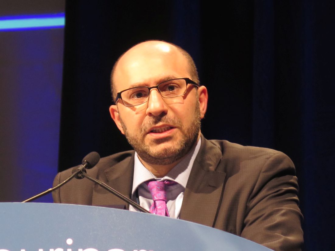
Previous research had shown that avelumab (Bavencio), an immune checkpoint inhibitor targeting programmed death-ligand 1 (PD-L1), is active when used alone for advanced RCC, noted lead investigator Toni K. Choueiri, MD, director of the Lank Center for Genitourinary Oncology at the Dana-Farber Cancer Institute, Boston. And axitinib (Inlyta), a vascular endothelial growth factor (VEGF) tyrosine kinase inhibitor, is approved for use in the second line. In an early-phase trial among patients being treated in the first line, a combination of the two drugs led to an impressive 58% objective response rate (ORR) and had a favorable safety profile (Lancet Oncol. 2018 Apr;19(4):451-60).
JAVELIN Renal 101 (NCT02684006), a phase 3 randomized, controlled trial, enrolled 886 patients with treatment-naive advanced RCC having a clear cell component regardless of their tumor’s PD-L1 status. They were randomized to the combination of avelumab/axitinib or to the VEGF tyrosine kinase inhibitor sunitinib (Sutent) alone.
Full trial results, published during the symposium in the New England Journal of Medicine (2019 Feb 16. doi: 10.1056/NEJMoa1816047), showed significant progression-free and overall survival benefits of avelumab/axitinib over sunitinib in the 63.2% of patients with PD-L1–positive tumors – the trial’s primary endpoints – as well as a progression-free survival benefit in the entire trial population.
In the subgroup analyses reported at the symposium, the combination reduced risk of progression or death by roughly 20%-50% across patients having different statuses in regard to International Metastatic Renal Cell Carcinoma Database Consortium (IMDC) risk group, Memorial Sloan Kettering Cancer Center (MSKCC) risk group, and tumor PD-L1, as well as other characteristics. Findings were similar for ORR, with the combination roughly doubling to quadrupling the odds of response, irrespective of patient and disease characteristics.
“The progression-free survival and response rate benefit was observed in all patients, regardless of PD-L1 status, regardless of prognostic risk group. At this time, the study continues to follow up for overall survival,” Dr. Choueiri commented. Taken together, “the results do support avelumab plus axitinib as a new first-line standard of care for patients with advanced RCC.”
JAVELIN Renal 101 complemented two other noteworthy trials exploring first-line checkpoint inhibitors for which new data were reported at the symposium. One, KEYNOTE-426 (NCT02853331), established that the combination of the immune checkpoint inhibitor pembrolizumab (Keytruda) and axitinib was superior to sunitinib. The other, CheckMate 214 (NCT02231749), established that the combination of two immune checkpoint inhibitors, nivolumab (Opdivo) and ipilimumab (Yervoy), was superior to sunitinib.
Weighing new options
“So a new standard of care in 2019 is present: The majority of patients with advanced clear cell RCC will be eligible to receive the combination of a checkpoint inhibitor and axitinib,” commented invited discussant Lori Wood, MD, a professor in the division of medical oncology at Dalhousie University, Halifax, Canada. “The questions now are: Which treatment should we choose? can we afford it? and perhaps more importantly, can we safely deliver this therapy to all patients?”

When it comes to selecting among the three combinations above, “I don’t think PD-L1 expression is going to help us at all,” she said. In contrast, IMDC risk category is likely still helpful because, in CheckMate 214, there was no progression-free or overall survival benefit of ipilimumab/nivolumab in patients with favorable-risk disease.
Differences in rates of discontinuation of all treatment because of treatment-related adverse events are hard to assess because CheckMate 214 had restrictions on allowing patients in the combination group to receive single-agent nivolumab, according to Dr. Wood. Financial costs are a major consideration, but so are time and staffing costs: Compared with single-agent sunitinib, the combinations as much as triple physician visits, nurse visits, infusions, and unscheduled visits.
Safely administering the combinations – through use of education, judicious patient selection, and attention to logistics – is a challenge, she maintained. “I tell the residents, you can probably give cisplatin/gemcitabine to 10 patients and you can probably give sunitinib to 20 patients and get a good sense of what’s going to happen. But every single patient that I have put on immune therapy, I learn something new.”
Evolving issues, such as nuanced differences among the immune checkpoint inhibitors and whether the doses used in trials are really needed, have yet to be worked through. But combining these agents is likely better than sequencing them because only about half of RCC patients given first-line therapy go on to get second-line therapy, “so we might as well use our best therapy up front,” Dr. Wood said. Finally, it’s unclear whether cytoreductive nephrectomy is needed to achieve a complete response with these combinations because all trials predated the CARMENA trial (NCT00930033), so most patients underwent this surgery.
“These are exciting times. I think that for the first-line metastatic renal cell patient with favorable-, intermediate-, or poor-risk disease, a checkpoint inhibitor/axitinib combination will be a new standard of care in many parts of the world, not all,” she summarized. “For intermediate- and poor-risk patients, there’s no clear winner in my mind at this current time between ipilimumab/nivolumab and checkpoint inhibitor/axitinib. Decisions will need to be based on overall survival, complete response rates, toxicities, and then practical aspects, as well as costs.”
“But we cannot safely and effectively deliver this new standard of care without true infrastructure and system changes to accommodate more doctor and nurse visits, more infusion time, all of these extra visits, and more education for everybody who is both delivering and receiving these agents,” Dr. Wood concluded.
Study details
The subgroup analyses showed that, compared with sunitinib, avelumab/axitinib yielded better progression-free survival across patients differing with respect to IMDC risk group (range of hazard ratios, 0.539-0.736), MSKCC risk group (range of HRs, 0.495-0.715), tumor PD-L1 status (range of HRs, 0.626-0.827), prior nephrectomy status (range of HRs, 0.673-0.748), smoking status (range of HRs, 0.663-0.711), and body mass index (range of HRs, 0.667-0.674), Dr. Choueiri reported at the symposium. However, the 95% confidence intervals crossed 1 in some cases.
Overall, 20.8% of the avelumab/axitinib group and 39.2% of the sunitinib group went on to receive a follow-up anticancer drug therapy. The most common was cabozantinib (Cabometyx) in the former and nivolumab (Opdivo) in the latter.
The rate of progression-free survival 2 could not be estimated for the avelumab/axitinib group and was 18.4 months for the sunitinib group (HR, 0.56). “In theory, the first-line treatment could change the biology of the disease and therefore lead to substantially shorter benefit of second-line treatment, and progression-free survival 2 is actually a potentially important endpoint for regulatory and reimbursement evaluation,” Dr. Choueiri explained. “This suggests at least no negative impact of first-line treatment with the combination on subsequent benefit from second-line treatment.”
Compared with sunitinib, avelumab/axitinib also yielded better odds of objective response regardless of IMDC risk group (range of odds ratios, 3.099-3.556), MSKCC risk group (range of ORs, 3.061-4.686), PD-L1 status (range of ORs, 2.240-3.594), prior nephrectomy status (range of ORs, 2.592-3.249), smoking status (range of ORs, 2.649-3.798), and body mass index (range of ORs, 3.086-3.292). Here, virtually all 95% confidence intervals excluded 1.
Mean duration of response was more than 4 months longer with the combination than with sunitinib. Moreover, responses were deeper for the combination patients.
In updated safety results, the avelumab/axitinib group had higher rates of any-grade treatment-related diarrhea (54% vs. 45%) and hypothyroidism (24% vs. 13%). But there were few of these adverse events of grade 3 or 4 in either group.
Dr. Choueiri disclosed that he receives honoraria from, has a consulting or advisory role with, and receives institutional research funding from Merck and Pfizer – among other disclosures. The trial was sponsored by Pfizer.
SOURCE: Choueiri TK et al. GUCS 2019, Abstract 544.
“For first-line therapy of metastatic clear-cell renal cancer, we now have two regimens that have demonstrated a survival advantage over first-line sunitinib,” Walter M. Stadler, MD, said in an interview. For first-line therapy, there is the combination of nivolumab and ipilimumab in intermediate- and poor-risk patients and also the combination of pembrolizumab and axitinib.

Dr. Stadler is the Fred C. Buffett Professor of Medicine and Surgery, chief of the section of hematology/oncology, director of the genitourinary oncology program, and deputy director of the Comprehensive Cancer Center at the University of Chicago.
“For first-line therapy of metastatic clear-cell renal cancer, we now have two regimens that have demonstrated a survival advantage over first-line sunitinib,” Walter M. Stadler, MD, said in an interview. For first-line therapy, there is the combination of nivolumab and ipilimumab in intermediate- and poor-risk patients and also the combination of pembrolizumab and axitinib.
Dr. Stadler is the Fred C. Buffett Professor of Medicine and Surgery, chief of the section of hematology/oncology, director of the genitourinary oncology program, and deputy director of the Comprehensive Cancer Center at the University of Chicago.
“For first-line therapy of metastatic clear-cell renal cancer, we now have two regimens that have demonstrated a survival advantage over first-line sunitinib,” Walter M. Stadler, MD, said in an interview. For first-line therapy, there is the combination of nivolumab and ipilimumab in intermediate- and poor-risk patients and also the combination of pembrolizumab and axitinib.
Dr. Stadler is the Fred C. Buffett Professor of Medicine and Surgery, chief of the section of hematology/oncology, director of the genitourinary oncology program, and deputy director of the Comprehensive Cancer Center at the University of Chicago.
SAN FRANCISCO – subgroup analyses of the JAVELIN Renal 101 trial have shown. Results were reported at the 2019 Genitourinary Cancers Symposium sponsored by the American Society of Clinical Oncology, ASTRO, and the Society of Urologic Oncology.
Previous research had shown that avelumab (Bavencio), an immune checkpoint inhibitor targeting programmed death-ligand 1 (PD-L1), is active when used alone for advanced RCC, noted lead investigator Toni K. Choueiri, MD, director of the Lank Center for Genitourinary Oncology at the Dana-Farber Cancer Institute, Boston. And axitinib (Inlyta), a vascular endothelial growth factor (VEGF) tyrosine kinase inhibitor, is approved for use in the second line. In an early-phase trial among patients being treated in the first line, a combination of the two drugs led to an impressive 58% objective response rate (ORR) and had a favorable safety profile (Lancet Oncol. 2018 Apr;19(4):451-60).
JAVELIN Renal 101 (NCT02684006), a phase 3 randomized, controlled trial, enrolled 886 patients with treatment-naive advanced RCC having a clear cell component regardless of their tumor’s PD-L1 status. They were randomized to the combination of avelumab/axitinib or to the VEGF tyrosine kinase inhibitor sunitinib (Sutent) alone.
Full trial results, published during the symposium in the New England Journal of Medicine (2019 Feb 16. doi: 10.1056/NEJMoa1816047), showed significant progression-free and overall survival benefits of avelumab/axitinib over sunitinib in the 63.2% of patients with PD-L1–positive tumors – the trial’s primary endpoints – as well as a progression-free survival benefit in the entire trial population.
In the subgroup analyses reported at the symposium, the combination reduced risk of progression or death by roughly 20%-50% across patients having different statuses in regard to International Metastatic Renal Cell Carcinoma Database Consortium (IMDC) risk group, Memorial Sloan Kettering Cancer Center (MSKCC) risk group, and tumor PD-L1, as well as other characteristics. Findings were similar for ORR, with the combination roughly doubling to quadrupling the odds of response, irrespective of patient and disease characteristics.
“The progression-free survival and response rate benefit was observed in all patients, regardless of PD-L1 status, regardless of prognostic risk group. At this time, the study continues to follow up for overall survival,” Dr. Choueiri commented. Taken together, “the results do support avelumab plus axitinib as a new first-line standard of care for patients with advanced RCC.”
JAVELIN Renal 101 complemented two other noteworthy trials exploring first-line checkpoint inhibitors for which new data were reported at the symposium. One, KEYNOTE-426 (NCT02853331), established that the combination of the immune checkpoint inhibitor pembrolizumab (Keytruda) and axitinib was superior to sunitinib. The other, CheckMate 214 (NCT02231749), established that the combination of two immune checkpoint inhibitors, nivolumab (Opdivo) and ipilimumab (Yervoy), was superior to sunitinib.
Weighing new options
“So a new standard of care in 2019 is present: The majority of patients with advanced clear cell RCC will be eligible to receive the combination of a checkpoint inhibitor and axitinib,” commented invited discussant Lori Wood, MD, a professor in the division of medical oncology at Dalhousie University, Halifax, Canada. “The questions now are: Which treatment should we choose? can we afford it? and perhaps more importantly, can we safely deliver this therapy to all patients?”
When it comes to selecting among the three combinations above, “I don’t think PD-L1 expression is going to help us at all,” she said. In contrast, IMDC risk category is likely still helpful because, in CheckMate 214, there was no progression-free or overall survival benefit of ipilimumab/nivolumab in patients with favorable-risk disease.
Differences in rates of discontinuation of all treatment because of treatment-related adverse events are hard to assess because CheckMate 214 had restrictions on allowing patients in the combination group to receive single-agent nivolumab, according to Dr. Wood. Financial costs are a major consideration, but so are time and staffing costs: Compared with single-agent sunitinib, the combinations as much as triple physician visits, nurse visits, infusions, and unscheduled visits.
Safely administering the combinations – through use of education, judicious patient selection, and attention to logistics – is a challenge, she maintained. “I tell the residents, you can probably give cisplatin/gemcitabine to 10 patients and you can probably give sunitinib to 20 patients and get a good sense of what’s going to happen. But every single patient that I have put on immune therapy, I learn something new.”
Evolving issues, such as nuanced differences among the immune checkpoint inhibitors and whether the doses used in trials are really needed, have yet to be worked through. But combining these agents is likely better than sequencing them because only about half of RCC patients given first-line therapy go on to get second-line therapy, “so we might as well use our best therapy up front,” Dr. Wood said. Finally, it’s unclear whether cytoreductive nephrectomy is needed to achieve a complete response with these combinations because all trials predated the CARMENA trial (NCT00930033), so most patients underwent this surgery.
“These are exciting times. I think that for the first-line metastatic renal cell patient with favorable-, intermediate-, or poor-risk disease, a checkpoint inhibitor/axitinib combination will be a new standard of care in many parts of the world, not all,” she summarized. “For intermediate- and poor-risk patients, there’s no clear winner in my mind at this current time between ipilimumab/nivolumab and checkpoint inhibitor/axitinib. Decisions will need to be based on overall survival, complete response rates, toxicities, and then practical aspects, as well as costs.”
“But we cannot safely and effectively deliver this new standard of care without true infrastructure and system changes to accommodate more doctor and nurse visits, more infusion time, all of these extra visits, and more education for everybody who is both delivering and receiving these agents,” Dr. Wood concluded.
Study details
The subgroup analyses showed that, compared with sunitinib, avelumab/axitinib yielded better progression-free survival across patients differing with respect to IMDC risk group (range of hazard ratios, 0.539-0.736), MSKCC risk group (range of HRs, 0.495-0.715), tumor PD-L1 status (range of HRs, 0.626-0.827), prior nephrectomy status (range of HRs, 0.673-0.748), smoking status (range of HRs, 0.663-0.711), and body mass index (range of HRs, 0.667-0.674), Dr. Choueiri reported at the symposium. However, the 95% confidence intervals crossed 1 in some cases.
Overall, 20.8% of the avelumab/axitinib group and 39.2% of the sunitinib group went on to receive a follow-up anticancer drug therapy. The most common was cabozantinib (Cabometyx) in the former and nivolumab (Opdivo) in the latter.
The rate of progression-free survival 2 could not be estimated for the avelumab/axitinib group and was 18.4 months for the sunitinib group (HR, 0.56). “In theory, the first-line treatment could change the biology of the disease and therefore lead to substantially shorter benefit of second-line treatment, and progression-free survival 2 is actually a potentially important endpoint for regulatory and reimbursement evaluation,” Dr. Choueiri explained. “This suggests at least no negative impact of first-line treatment with the combination on subsequent benefit from second-line treatment.”
Compared with sunitinib, avelumab/axitinib also yielded better odds of objective response regardless of IMDC risk group (range of odds ratios, 3.099-3.556), MSKCC risk group (range of ORs, 3.061-4.686), PD-L1 status (range of ORs, 2.240-3.594), prior nephrectomy status (range of ORs, 2.592-3.249), smoking status (range of ORs, 2.649-3.798), and body mass index (range of ORs, 3.086-3.292). Here, virtually all 95% confidence intervals excluded 1.
Mean duration of response was more than 4 months longer with the combination than with sunitinib. Moreover, responses were deeper for the combination patients.
In updated safety results, the avelumab/axitinib group had higher rates of any-grade treatment-related diarrhea (54% vs. 45%) and hypothyroidism (24% vs. 13%). But there were few of these adverse events of grade 3 or 4 in either group.
Dr. Choueiri disclosed that he receives honoraria from, has a consulting or advisory role with, and receives institutional research funding from Merck and Pfizer – among other disclosures. The trial was sponsored by Pfizer.
SOURCE: Choueiri TK et al. GUCS 2019, Abstract 544.
SAN FRANCISCO – subgroup analyses of the JAVELIN Renal 101 trial have shown. Results were reported at the 2019 Genitourinary Cancers Symposium sponsored by the American Society of Clinical Oncology, ASTRO, and the Society of Urologic Oncology.
Previous research had shown that avelumab (Bavencio), an immune checkpoint inhibitor targeting programmed death-ligand 1 (PD-L1), is active when used alone for advanced RCC, noted lead investigator Toni K. Choueiri, MD, director of the Lank Center for Genitourinary Oncology at the Dana-Farber Cancer Institute, Boston. And axitinib (Inlyta), a vascular endothelial growth factor (VEGF) tyrosine kinase inhibitor, is approved for use in the second line. In an early-phase trial among patients being treated in the first line, a combination of the two drugs led to an impressive 58% objective response rate (ORR) and had a favorable safety profile (Lancet Oncol. 2018 Apr;19(4):451-60).
JAVELIN Renal 101 (NCT02684006), a phase 3 randomized, controlled trial, enrolled 886 patients with treatment-naive advanced RCC having a clear cell component regardless of their tumor’s PD-L1 status. They were randomized to the combination of avelumab/axitinib or to the VEGF tyrosine kinase inhibitor sunitinib (Sutent) alone.
Full trial results, published during the symposium in the New England Journal of Medicine (2019 Feb 16. doi: 10.1056/NEJMoa1816047), showed significant progression-free and overall survival benefits of avelumab/axitinib over sunitinib in the 63.2% of patients with PD-L1–positive tumors – the trial’s primary endpoints – as well as a progression-free survival benefit in the entire trial population.
In the subgroup analyses reported at the symposium, the combination reduced risk of progression or death by roughly 20%-50% across patients having different statuses in regard to International Metastatic Renal Cell Carcinoma Database Consortium (IMDC) risk group, Memorial Sloan Kettering Cancer Center (MSKCC) risk group, and tumor PD-L1, as well as other characteristics. Findings were similar for ORR, with the combination roughly doubling to quadrupling the odds of response, irrespective of patient and disease characteristics.
“The progression-free survival and response rate benefit was observed in all patients, regardless of PD-L1 status, regardless of prognostic risk group. At this time, the study continues to follow up for overall survival,” Dr. Choueiri commented. Taken together, “the results do support avelumab plus axitinib as a new first-line standard of care for patients with advanced RCC.”
JAVELIN Renal 101 complemented two other noteworthy trials exploring first-line checkpoint inhibitors for which new data were reported at the symposium. One, KEYNOTE-426 (NCT02853331), established that the combination of the immune checkpoint inhibitor pembrolizumab (Keytruda) and axitinib was superior to sunitinib. The other, CheckMate 214 (NCT02231749), established that the combination of two immune checkpoint inhibitors, nivolumab (Opdivo) and ipilimumab (Yervoy), was superior to sunitinib.
Weighing new options
“So a new standard of care in 2019 is present: The majority of patients with advanced clear cell RCC will be eligible to receive the combination of a checkpoint inhibitor and axitinib,” commented invited discussant Lori Wood, MD, a professor in the division of medical oncology at Dalhousie University, Halifax, Canada. “The questions now are: Which treatment should we choose? can we afford it? and perhaps more importantly, can we safely deliver this therapy to all patients?”
When it comes to selecting among the three combinations above, “I don’t think PD-L1 expression is going to help us at all,” she said. In contrast, IMDC risk category is likely still helpful because, in CheckMate 214, there was no progression-free or overall survival benefit of ipilimumab/nivolumab in patients with favorable-risk disease.
Differences in rates of discontinuation of all treatment because of treatment-related adverse events are hard to assess because CheckMate 214 had restrictions on allowing patients in the combination group to receive single-agent nivolumab, according to Dr. Wood. Financial costs are a major consideration, but so are time and staffing costs: Compared with single-agent sunitinib, the combinations as much as triple physician visits, nurse visits, infusions, and unscheduled visits.
Safely administering the combinations – through use of education, judicious patient selection, and attention to logistics – is a challenge, she maintained. “I tell the residents, you can probably give cisplatin/gemcitabine to 10 patients and you can probably give sunitinib to 20 patients and get a good sense of what’s going to happen. But every single patient that I have put on immune therapy, I learn something new.”
Evolving issues, such as nuanced differences among the immune checkpoint inhibitors and whether the doses used in trials are really needed, have yet to be worked through. But combining these agents is likely better than sequencing them because only about half of RCC patients given first-line therapy go on to get second-line therapy, “so we might as well use our best therapy up front,” Dr. Wood said. Finally, it’s unclear whether cytoreductive nephrectomy is needed to achieve a complete response with these combinations because all trials predated the CARMENA trial (NCT00930033), so most patients underwent this surgery.
“These are exciting times. I think that for the first-line metastatic renal cell patient with favorable-, intermediate-, or poor-risk disease, a checkpoint inhibitor/axitinib combination will be a new standard of care in many parts of the world, not all,” she summarized. “For intermediate- and poor-risk patients, there’s no clear winner in my mind at this current time between ipilimumab/nivolumab and checkpoint inhibitor/axitinib. Decisions will need to be based on overall survival, complete response rates, toxicities, and then practical aspects, as well as costs.”
“But we cannot safely and effectively deliver this new standard of care without true infrastructure and system changes to accommodate more doctor and nurse visits, more infusion time, all of these extra visits, and more education for everybody who is both delivering and receiving these agents,” Dr. Wood concluded.
Study details
The subgroup analyses showed that, compared with sunitinib, avelumab/axitinib yielded better progression-free survival across patients differing with respect to IMDC risk group (range of hazard ratios, 0.539-0.736), MSKCC risk group (range of HRs, 0.495-0.715), tumor PD-L1 status (range of HRs, 0.626-0.827), prior nephrectomy status (range of HRs, 0.673-0.748), smoking status (range of HRs, 0.663-0.711), and body mass index (range of HRs, 0.667-0.674), Dr. Choueiri reported at the symposium. However, the 95% confidence intervals crossed 1 in some cases.
Overall, 20.8% of the avelumab/axitinib group and 39.2% of the sunitinib group went on to receive a follow-up anticancer drug therapy. The most common was cabozantinib (Cabometyx) in the former and nivolumab (Opdivo) in the latter.
The rate of progression-free survival 2 could not be estimated for the avelumab/axitinib group and was 18.4 months for the sunitinib group (HR, 0.56). “In theory, the first-line treatment could change the biology of the disease and therefore lead to substantially shorter benefit of second-line treatment, and progression-free survival 2 is actually a potentially important endpoint for regulatory and reimbursement evaluation,” Dr. Choueiri explained. “This suggests at least no negative impact of first-line treatment with the combination on subsequent benefit from second-line treatment.”
Compared with sunitinib, avelumab/axitinib also yielded better odds of objective response regardless of IMDC risk group (range of odds ratios, 3.099-3.556), MSKCC risk group (range of ORs, 3.061-4.686), PD-L1 status (range of ORs, 2.240-3.594), prior nephrectomy status (range of ORs, 2.592-3.249), smoking status (range of ORs, 2.649-3.798), and body mass index (range of ORs, 3.086-3.292). Here, virtually all 95% confidence intervals excluded 1.
Mean duration of response was more than 4 months longer with the combination than with sunitinib. Moreover, responses were deeper for the combination patients.
In updated safety results, the avelumab/axitinib group had higher rates of any-grade treatment-related diarrhea (54% vs. 45%) and hypothyroidism (24% vs. 13%). But there were few of these adverse events of grade 3 or 4 in either group.
Dr. Choueiri disclosed that he receives honoraria from, has a consulting or advisory role with, and receives institutional research funding from Merck and Pfizer – among other disclosures. The trial was sponsored by Pfizer.
SOURCE: Choueiri TK et al. GUCS 2019, Abstract 544.
REPORTING FROM GUCS 2019
How to review scientific literature
And Ilana Yurkiewicz, MD, talks about chaos and opportunity. Dr. Yurkiewicz is a fellow in hematology and oncology at Stanford (Calif.) University and is also a columnist for Hematology News. More from Dr. Yurkiewicz here.
Subscribe to Blood & Cancer here:
Show notes
By Hitomi Hosoya, MD, PhD,
Resident in the department of internal medicine, University of Pennsylvania Health System
- If you are a peer reviewer of a manuscript submitted to a journal, you should be unbiased, consistent, constructive, and focused on the research. COPE guideline is a good resource.
- If you are a reader of a published article, it is important to ensure that the abstract has the same conclusion as the body of the article.
- If you are a clinical practitioner and wondering how to apply findings of published data, the editorial section is a good source.
- If you are a trainee and wondering how to stay up-to-date, Oxford Textbook of Oncology or ASCO University are recommended.
Reference:
https://publicationethics.org/resources/guidelines-new/cope-ethical-guidelines-peer-reviewers
And Ilana Yurkiewicz, MD, talks about chaos and opportunity. Dr. Yurkiewicz is a fellow in hematology and oncology at Stanford (Calif.) University and is also a columnist for Hematology News. More from Dr. Yurkiewicz here.
Subscribe to Blood & Cancer here:
Show notes
By Hitomi Hosoya, MD, PhD,
Resident in the department of internal medicine, University of Pennsylvania Health System
- If you are a peer reviewer of a manuscript submitted to a journal, you should be unbiased, consistent, constructive, and focused on the research. COPE guideline is a good resource.
- If you are a reader of a published article, it is important to ensure that the abstract has the same conclusion as the body of the article.
- If you are a clinical practitioner and wondering how to apply findings of published data, the editorial section is a good source.
- If you are a trainee and wondering how to stay up-to-date, Oxford Textbook of Oncology or ASCO University are recommended.
Reference:
https://publicationethics.org/resources/guidelines-new/cope-ethical-guidelines-peer-reviewers
And Ilana Yurkiewicz, MD, talks about chaos and opportunity. Dr. Yurkiewicz is a fellow in hematology and oncology at Stanford (Calif.) University and is also a columnist for Hematology News. More from Dr. Yurkiewicz here.
Subscribe to Blood & Cancer here:
Show notes
By Hitomi Hosoya, MD, PhD,
Resident in the department of internal medicine, University of Pennsylvania Health System
- If you are a peer reviewer of a manuscript submitted to a journal, you should be unbiased, consistent, constructive, and focused on the research. COPE guideline is a good resource.
- If you are a reader of a published article, it is important to ensure that the abstract has the same conclusion as the body of the article.
- If you are a clinical practitioner and wondering how to apply findings of published data, the editorial section is a good source.
- If you are a trainee and wondering how to stay up-to-date, Oxford Textbook of Oncology or ASCO University are recommended.
Reference:
https://publicationethics.org/resources/guidelines-new/cope-ethical-guidelines-peer-reviewers

ICYMI: Rivaroxaban reduces VTE incidence in ambulatory cancer patients
While treatment with rivaroxaban did not significantly reduce venous thromboembolism incidence in high-risk ambulatory patients with cancer over the entire course of a 180-day intervention period (6.0% vs. 8.8% in controls; hazard ratio, 0.66; 95% confidence interval, 0.40-1.09), it did reduce major bleeding incidence while patients were on treatment (2.0% vs. 6.4%; HR, 0.40; 95% CI, 0.20 0.80), according to results from the multicenter, randomized, double-blind, placebo-controlled, parallel-group, phase 3b CASSINI trial published in the New England Journal of Medicine (2019 Feb 20. doi: 10.1056/NEJMoa1814630).
We reported this story at the annual meeting of the American Society of Hematology before it was published in the journal. Find our coverage at the link below.
While treatment with rivaroxaban did not significantly reduce venous thromboembolism incidence in high-risk ambulatory patients with cancer over the entire course of a 180-day intervention period (6.0% vs. 8.8% in controls; hazard ratio, 0.66; 95% confidence interval, 0.40-1.09), it did reduce major bleeding incidence while patients were on treatment (2.0% vs. 6.4%; HR, 0.40; 95% CI, 0.20 0.80), according to results from the multicenter, randomized, double-blind, placebo-controlled, parallel-group, phase 3b CASSINI trial published in the New England Journal of Medicine (2019 Feb 20. doi: 10.1056/NEJMoa1814630).
We reported this story at the annual meeting of the American Society of Hematology before it was published in the journal. Find our coverage at the link below.
While treatment with rivaroxaban did not significantly reduce venous thromboembolism incidence in high-risk ambulatory patients with cancer over the entire course of a 180-day intervention period (6.0% vs. 8.8% in controls; hazard ratio, 0.66; 95% confidence interval, 0.40-1.09), it did reduce major bleeding incidence while patients were on treatment (2.0% vs. 6.4%; HR, 0.40; 95% CI, 0.20 0.80), according to results from the multicenter, randomized, double-blind, placebo-controlled, parallel-group, phase 3b CASSINI trial published in the New England Journal of Medicine (2019 Feb 20. doi: 10.1056/NEJMoa1814630).
We reported this story at the annual meeting of the American Society of Hematology before it was published in the journal. Find our coverage at the link below.
FROM THE NEW ENGLAND JOURNAL OF MEDICINE
Researchers characterize new subtype of high-grade DLBCL
(DLBCL)
Patients with this subtype, dubbed “molecular high-grade” (MHG) DLBCL, were more likely to have germinal center B-cell-like (GCB) DLBCL, MYC rearrangements, and double-hit lymphoma.
When compared to other DLBCL patients, those with MHG DLBCL had inferior progression-free and overall survival.
Chulin Sha, PhD, of the University of Leeds (England), and colleagues reported these findings in the Journal of Clinical Oncology. The findings were published alongside a related editorial and a similar study from another group.
Dr. Sha and colleagues began their study by applying a previously developed gene expression classifier (Genome Med. 2015 Jul 1;7[1]:64) to 928 DLBCL patients enrolled in the REMoDL-B trial. REMoDL-B was designed to compare rituximab, cyclophosphamide, doxorubicin, vincristine, and prednisolone (R-CHOP) to R-CHOP plus bortezomib (Hematol Oncol. 2017;35:130-1).
Dr. Sha and colleagues looked for somatic mutations in 400 REMoDL-B patient samples that were sequenced for a 70-gene panel.
The team also tested 360 samples for MYC, BCL2, and BCL6 chromosomal rearrangements using fluorescent in situ hybridization, and they tested 355 samples for MYC and BCL2 protein expression with immunohistochemistry.
Characteristics of MHG DLBCL
The researchers identified 83 REMoDL-B patients as having MHG DLBCL (9%). Most of the MHG patients had GCB DLBCL (90%), 48.6% had MYC rearrangements, and 36.1% had double-hit lymphoma.
Patients with MHG DLBCL had higher International Prognostic Index scores (P = .004), greater tumor bulk (P = .007), higher disease stage (P = .06), and higher lactate dehydrogenase levels (P less than .001) than patients with non-MHG DLBCL.
Although most MHG patients had GCB DLBCL, the researchers found key differences between patients with MHG DLBCL and non-MHG GCB DLBCL. MHG patients were significantly more likely than patients with non-MHG GCB DLBCL to have mutations in KMT2D, BCL2, MYC, and DDX3X. Additionally, some genes frequently mutated in GCB DLBCL — such as B2M, SGK1, and NFKBIA — were rare in MHG DLBCL.
Dr. Sha and colleagues also compared the MHG patients to 70 patients with Burkitt lymphoma (BL) who had been analyzed in a previous study (Genome Med. 2015 Jul 1;7[1]:64).
The researchers found that BL has more upregulated genes than GCB (2,483 genes) and MHG DLBCL (1,784 genes), and MHG DLBCL has more upregulated genes than GCB DLBCL (382 genes). The team observed a similar pattern with downregulated genes and said this suggests “MHG is an intermediate group but closer to GCB than to BL.”
The researchers also found, however, that “MHG and BL share high expression of signatures that contain cell-cycle genes, ribosome biogenesis, MYC overexpression, and TCF3 targets, which suggests a shared proliferative phenotype.”
The team determined that MHG has “a highly proliferative phenotype and shares features with centroblasts of the germinal center dark zone.”
Another discovery was that MHG patients in the REMoDL-B trial had worse progression-free survival (PFS) than their peers.
Among patients who received R-CHOP, the estimated 3-year PFS was:
- 37% for MHG patients
- 78% for patients with GCB DLBCL
- 64% for patients with activated B-cell like (ABC) DLBCL
- 65% for patients with unclassified DLBCL.
Among patients who received bortezomib plus R-CHOP, there was a trend toward improved PFS for patients with MHG DLBCL (58%; P = .08).
Validation cohort
Dr. Sha and colleagues validated their initial findings using RNA sequencing data from another group of DLBCL patients (Cell. 2017 Oct 5;171[2]:481-94.e15). This data set included 624 patients who received rituximab-based therapy.
Seventy-two patients in this group had MHG DLBCL (11.5%), and most MHG patients had GCB DLBCL (82%).
The researchers said the MHG group in this cohort “showed similar associations with clinical variables” and a “similar mutation spectrum” as the MHG group in the REMoDL-B cohort. Additionally, MHG patients in the validation cohort had inferior overall survival (P less than .001) compared to patients with non-MHG GCB DLBCL.
Dr. Sha and colleagues said the poor prognosis in MHG patients in both cohorts suggests a need for different treatment approaches in this group.
In the related editorial, Wing C. Chan, MD, of City of Hope Medical Center in Duarte, Calif., echoed that sentiment and said it will be important to include patients with high-risk DLBCL in clinical trials.
“Their tumors should be comprehensively characterize[d] for correlative analysis to determine the molecular lesions that underlie their biology and response to treatment,” Dr. Chan wrote.
Dr. Chan disclosed a patent for a diagnostic algorithm on GCB/ABC-type DLBCL and a patent on a diagnostic algorithm for peripheral T-cell lymphoma.
Dr. Sha and colleagues disclosed relationships with a range of pharmaceutical companies. The team’s research was supported by a grant from Bloodwise.
The REMoDL-B trial was endorsed by Cancer Research UK and was funded by Janssen-Cillag.
SOURCES: Sha C et al. J Clin Oncol. 2019 Jan 20;37(3):202-12. doi: 10.1200/JCO.18.01314; Chan WC. J Clin Oncol. 2019 Jan 20;37(3):175-7. doi: 10.1200/JCO.18.01910; Ennishi D et al. J Clin Oncol. 2019 Jan 20;37(3):190-201. doi: 10.1200/JCO.18.01583
(DLBCL)
Patients with this subtype, dubbed “molecular high-grade” (MHG) DLBCL, were more likely to have germinal center B-cell-like (GCB) DLBCL, MYC rearrangements, and double-hit lymphoma.
When compared to other DLBCL patients, those with MHG DLBCL had inferior progression-free and overall survival.
Chulin Sha, PhD, of the University of Leeds (England), and colleagues reported these findings in the Journal of Clinical Oncology. The findings were published alongside a related editorial and a similar study from another group.
Dr. Sha and colleagues began their study by applying a previously developed gene expression classifier (Genome Med. 2015 Jul 1;7[1]:64) to 928 DLBCL patients enrolled in the REMoDL-B trial. REMoDL-B was designed to compare rituximab, cyclophosphamide, doxorubicin, vincristine, and prednisolone (R-CHOP) to R-CHOP plus bortezomib (Hematol Oncol. 2017;35:130-1).
Dr. Sha and colleagues looked for somatic mutations in 400 REMoDL-B patient samples that were sequenced for a 70-gene panel.
The team also tested 360 samples for MYC, BCL2, and BCL6 chromosomal rearrangements using fluorescent in situ hybridization, and they tested 355 samples for MYC and BCL2 protein expression with immunohistochemistry.
Characteristics of MHG DLBCL
The researchers identified 83 REMoDL-B patients as having MHG DLBCL (9%). Most of the MHG patients had GCB DLBCL (90%), 48.6% had MYC rearrangements, and 36.1% had double-hit lymphoma.
Patients with MHG DLBCL had higher International Prognostic Index scores (P = .004), greater tumor bulk (P = .007), higher disease stage (P = .06), and higher lactate dehydrogenase levels (P less than .001) than patients with non-MHG DLBCL.
Although most MHG patients had GCB DLBCL, the researchers found key differences between patients with MHG DLBCL and non-MHG GCB DLBCL. MHG patients were significantly more likely than patients with non-MHG GCB DLBCL to have mutations in KMT2D, BCL2, MYC, and DDX3X. Additionally, some genes frequently mutated in GCB DLBCL — such as B2M, SGK1, and NFKBIA — were rare in MHG DLBCL.
Dr. Sha and colleagues also compared the MHG patients to 70 patients with Burkitt lymphoma (BL) who had been analyzed in a previous study (Genome Med. 2015 Jul 1;7[1]:64).
The researchers found that BL has more upregulated genes than GCB (2,483 genes) and MHG DLBCL (1,784 genes), and MHG DLBCL has more upregulated genes than GCB DLBCL (382 genes). The team observed a similar pattern with downregulated genes and said this suggests “MHG is an intermediate group but closer to GCB than to BL.”
The researchers also found, however, that “MHG and BL share high expression of signatures that contain cell-cycle genes, ribosome biogenesis, MYC overexpression, and TCF3 targets, which suggests a shared proliferative phenotype.”
The team determined that MHG has “a highly proliferative phenotype and shares features with centroblasts of the germinal center dark zone.”
Another discovery was that MHG patients in the REMoDL-B trial had worse progression-free survival (PFS) than their peers.
Among patients who received R-CHOP, the estimated 3-year PFS was:
- 37% for MHG patients
- 78% for patients with GCB DLBCL
- 64% for patients with activated B-cell like (ABC) DLBCL
- 65% for patients with unclassified DLBCL.
Among patients who received bortezomib plus R-CHOP, there was a trend toward improved PFS for patients with MHG DLBCL (58%; P = .08).
Validation cohort
Dr. Sha and colleagues validated their initial findings using RNA sequencing data from another group of DLBCL patients (Cell. 2017 Oct 5;171[2]:481-94.e15). This data set included 624 patients who received rituximab-based therapy.
Seventy-two patients in this group had MHG DLBCL (11.5%), and most MHG patients had GCB DLBCL (82%).
The researchers said the MHG group in this cohort “showed similar associations with clinical variables” and a “similar mutation spectrum” as the MHG group in the REMoDL-B cohort. Additionally, MHG patients in the validation cohort had inferior overall survival (P less than .001) compared to patients with non-MHG GCB DLBCL.
Dr. Sha and colleagues said the poor prognosis in MHG patients in both cohorts suggests a need for different treatment approaches in this group.
In the related editorial, Wing C. Chan, MD, of City of Hope Medical Center in Duarte, Calif., echoed that sentiment and said it will be important to include patients with high-risk DLBCL in clinical trials.
“Their tumors should be comprehensively characterize[d] for correlative analysis to determine the molecular lesions that underlie their biology and response to treatment,” Dr. Chan wrote.
Dr. Chan disclosed a patent for a diagnostic algorithm on GCB/ABC-type DLBCL and a patent on a diagnostic algorithm for peripheral T-cell lymphoma.
Dr. Sha and colleagues disclosed relationships with a range of pharmaceutical companies. The team’s research was supported by a grant from Bloodwise.
The REMoDL-B trial was endorsed by Cancer Research UK and was funded by Janssen-Cillag.
SOURCES: Sha C et al. J Clin Oncol. 2019 Jan 20;37(3):202-12. doi: 10.1200/JCO.18.01314; Chan WC. J Clin Oncol. 2019 Jan 20;37(3):175-7. doi: 10.1200/JCO.18.01910; Ennishi D et al. J Clin Oncol. 2019 Jan 20;37(3):190-201. doi: 10.1200/JCO.18.01583
(DLBCL)
Patients with this subtype, dubbed “molecular high-grade” (MHG) DLBCL, were more likely to have germinal center B-cell-like (GCB) DLBCL, MYC rearrangements, and double-hit lymphoma.
When compared to other DLBCL patients, those with MHG DLBCL had inferior progression-free and overall survival.
Chulin Sha, PhD, of the University of Leeds (England), and colleagues reported these findings in the Journal of Clinical Oncology. The findings were published alongside a related editorial and a similar study from another group.
Dr. Sha and colleagues began their study by applying a previously developed gene expression classifier (Genome Med. 2015 Jul 1;7[1]:64) to 928 DLBCL patients enrolled in the REMoDL-B trial. REMoDL-B was designed to compare rituximab, cyclophosphamide, doxorubicin, vincristine, and prednisolone (R-CHOP) to R-CHOP plus bortezomib (Hematol Oncol. 2017;35:130-1).
Dr. Sha and colleagues looked for somatic mutations in 400 REMoDL-B patient samples that were sequenced for a 70-gene panel.
The team also tested 360 samples for MYC, BCL2, and BCL6 chromosomal rearrangements using fluorescent in situ hybridization, and they tested 355 samples for MYC and BCL2 protein expression with immunohistochemistry.
Characteristics of MHG DLBCL
The researchers identified 83 REMoDL-B patients as having MHG DLBCL (9%). Most of the MHG patients had GCB DLBCL (90%), 48.6% had MYC rearrangements, and 36.1% had double-hit lymphoma.
Patients with MHG DLBCL had higher International Prognostic Index scores (P = .004), greater tumor bulk (P = .007), higher disease stage (P = .06), and higher lactate dehydrogenase levels (P less than .001) than patients with non-MHG DLBCL.
Although most MHG patients had GCB DLBCL, the researchers found key differences between patients with MHG DLBCL and non-MHG GCB DLBCL. MHG patients were significantly more likely than patients with non-MHG GCB DLBCL to have mutations in KMT2D, BCL2, MYC, and DDX3X. Additionally, some genes frequently mutated in GCB DLBCL — such as B2M, SGK1, and NFKBIA — were rare in MHG DLBCL.
Dr. Sha and colleagues also compared the MHG patients to 70 patients with Burkitt lymphoma (BL) who had been analyzed in a previous study (Genome Med. 2015 Jul 1;7[1]:64).
The researchers found that BL has more upregulated genes than GCB (2,483 genes) and MHG DLBCL (1,784 genes), and MHG DLBCL has more upregulated genes than GCB DLBCL (382 genes). The team observed a similar pattern with downregulated genes and said this suggests “MHG is an intermediate group but closer to GCB than to BL.”
The researchers also found, however, that “MHG and BL share high expression of signatures that contain cell-cycle genes, ribosome biogenesis, MYC overexpression, and TCF3 targets, which suggests a shared proliferative phenotype.”
The team determined that MHG has “a highly proliferative phenotype and shares features with centroblasts of the germinal center dark zone.”
Another discovery was that MHG patients in the REMoDL-B trial had worse progression-free survival (PFS) than their peers.
Among patients who received R-CHOP, the estimated 3-year PFS was:
- 37% for MHG patients
- 78% for patients with GCB DLBCL
- 64% for patients with activated B-cell like (ABC) DLBCL
- 65% for patients with unclassified DLBCL.
Among patients who received bortezomib plus R-CHOP, there was a trend toward improved PFS for patients with MHG DLBCL (58%; P = .08).
Validation cohort
Dr. Sha and colleagues validated their initial findings using RNA sequencing data from another group of DLBCL patients (Cell. 2017 Oct 5;171[2]:481-94.e15). This data set included 624 patients who received rituximab-based therapy.
Seventy-two patients in this group had MHG DLBCL (11.5%), and most MHG patients had GCB DLBCL (82%).
The researchers said the MHG group in this cohort “showed similar associations with clinical variables” and a “similar mutation spectrum” as the MHG group in the REMoDL-B cohort. Additionally, MHG patients in the validation cohort had inferior overall survival (P less than .001) compared to patients with non-MHG GCB DLBCL.
Dr. Sha and colleagues said the poor prognosis in MHG patients in both cohorts suggests a need for different treatment approaches in this group.
In the related editorial, Wing C. Chan, MD, of City of Hope Medical Center in Duarte, Calif., echoed that sentiment and said it will be important to include patients with high-risk DLBCL in clinical trials.
“Their tumors should be comprehensively characterize[d] for correlative analysis to determine the molecular lesions that underlie their biology and response to treatment,” Dr. Chan wrote.
Dr. Chan disclosed a patent for a diagnostic algorithm on GCB/ABC-type DLBCL and a patent on a diagnostic algorithm for peripheral T-cell lymphoma.
Dr. Sha and colleagues disclosed relationships with a range of pharmaceutical companies. The team’s research was supported by a grant from Bloodwise.
The REMoDL-B trial was endorsed by Cancer Research UK and was funded by Janssen-Cillag.
SOURCES: Sha C et al. J Clin Oncol. 2019 Jan 20;37(3):202-12. doi: 10.1200/JCO.18.01314; Chan WC. J Clin Oncol. 2019 Jan 20;37(3):175-7. doi: 10.1200/JCO.18.01910; Ennishi D et al. J Clin Oncol. 2019 Jan 20;37(3):190-201. doi: 10.1200/JCO.18.01583
FROM THE JOURNAL OF CLINICAL ONCOLOGY
Culture change needed to improve gender inequalities in medicine
LONDON –
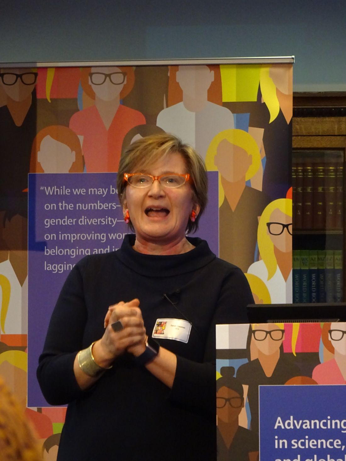
“Gender equality is everyone’s business,” Sarah Hawkes, MBBS, PhD, a professor of global public health at University College London (England), said at the event.
“We’re not talking about women taking over the shop, but women being given an equal opportunity to run the shop. It doesn’t matter where we place ourselves on the gender spectrum as far as advancing equality in science is concerned. What matters is that we all, irrespective of gender, call ourselves feminists.”
For years, women have been “underrepresented in positions of power and leadership, undervalued, and experience discrimination and gender-based violence in scientific and health disciplines across the world,” according to an editorial in the British-based journal (Lancet. 2019;393:493). Such inequalities are compounded and hard to separate from other inequalities, including ethnicity, disability, class, geography, and sexuality.
Despite efforts to readdress the predominantly male culture of medicine, the problem of gender inequality remains “stubbornly persistent,” the editorial said.
“We have spent years being told that the problem lies with us as individuals and that we just need to be better, stronger, more vocal, as women,” Dr. Hawkes observed. “But what really needs to happen is for change to happen in places that hold power.” She further argued: “We don’t need any more individual change; we need organizational and institutional norm change.”
Gender inequality has a long history, and not just in medicine, said British journalist Caroline Criado-Perez OBE, who gave a keynote speech. Ms. Criado-Perez, who is a well-respected feminist campaigner, noted that the world was largely “modeled to fit men.” From architecture to transport, and even crash-test dummies, everything was largely modeled on, or to accommodate, the male rather than the female body.
“I don’t need to tell you that women are 50% more likely to be misdiagnosed following a heart attack” than men. There is no more urgent need to challenge gender inequality than in medicine, Women are dying because of the gender data gap in medicine,” she asserted. “In medical research, in medical education, in medical practice, it needs to be closed as a matter of urgency.”
Original data published in the Advancing Women in Science, Medicine and Global Health special edition of The Lancet found that only 31% of biomedical research papers published in 2016 reported outcomes for both men and women (Lancet. 2019;393:550-9). Reporting of sex differences was somewhat better in clinical or public health-related research papers, at 67% and 69%, respectively. Sex-differences were more likely to be reported if a woman was one of the key authors, Cassidy R. Sugimoto, PhD, associate professor of informatics at Indiana University in Bloomington, and associates, observed in their paper.
That said, women often have to fight to be included as an author on a paper, even when they have done the majority of the work, the event participants highlighted. Women were still less likely than men to be named as the first or last author on a paper, as well as be less likely to receive research funding to enable them to do the work in the first place (Lancet. 2018;393:531-40).
“Was it really you?” was a question sometimes asked of a woman named as a lead or first author, noted Sonia Gandhi, MD, PhD, group leader of the Neurodegeneration Biology Laboratory at the Francis Crick Institute in Cambridge (England). Women network differently to men, Dr. Gandhi observed, and not necessarily in networks that forward careers. Women were also often questioned about their productivity, and regardless of any training on unconscious bias, women were still at a disadvantage if they took a career break to have children.
Women’s credentials and capabilities were often felt to be less respected by male colleagues, and there was talk of being met with microaggressions in the workplace, as in one example given by Nana Odom, MSc, a clinical engineer at the Royal United Hospital Bath (England). She was told “you’re not an engineer, because I have not got a set of screwdrivers and sit at a computer and program.” Such comments can deeply affect a person, Dr. Odom said. “Sometimes I feel that if I don’t go into the workshop and open up a bit of kit that I am not an engineer, but it’s so unconscious, it carries on with you.” These types of stories need to be told so then they can be properly addressed when they do happen, she said.

Female representation is so important, said F. Gigi Osler, MD, head of otolaryngology-head and neck surgery at St. Boniface Hospital in Winnipeg. Dr. Osler is the 2018-2019 president of the Canadian Medical Association, the eighth woman to hold this prestigious position in the organization’s 151 years of operation. She also happens to be the first female surgeon and the first woman of color in the role. “When I stepped into the presidency last August, I thought very long and very hard about how I was going to use my voice and this platform,” Dr. Osler said. “It became very clear to me after I started how important representation was. I can’t tell you how many women, young women, and women of color...have come up to me to say, ‘I’m so excited to have you in this position. I’ve never seen someone who looks like me in that type of leadership position,’ ” she observed.
“As leaders, I think we can advocate for structures and processes,” Dr. Osler added, “I think we set the culture.” Leaders have the responsibility for creating and nurturing and fostering a professional, respectful, and inclusive environment, she said.
“We need more strong leaders, we need more diversity in leadership.” Dr. Osler was keen to point out that greater diversity does not mean only women, but other groups as well. “Look around the room. Who is not here? How can I make it easier for them to get here?”
Another strong female role model at the event was Dame Sally Davies, the Chief Medical Officer for England, a hematologist by training. Not only is she the first female in that role, she will also become the first female Master of Trinity College Cambridge starting in October 2019, a role dominated by men for more than 500 years.
All people, women and men, need to be given fair opportunity, Dame Sally said. Addressing structural issues can help, she said. “We need role models, mentors, and champions,” she said, noting that there were differences between the three. “Mentors give you advice, they get to know you, and they help you think through your issue. Champions may not have time for that, but they know you are good,” and they are putting you forward for opportunities.
“If the system isn’t right, or we are treated badly, we need to call it out,” Dame Sally said. “I do think that we often let things pass that we shouldn’t.” A classic situation is where a woman may suggest something at a meeting and it is ignored, but when a man says the same thing it is taken on board. That kind of behavior needs to stop and be addressed when it happens, by everyone at the table, she said.
Dr. Hawkes observed in her summing up of the day: “Throughout the history of health, change comes about not just through action at the top, but also from action from the bottom up.” She added: “The question is how do we make that change happen?” That’s where the next phase of research needs to take place, she suggested, “we need to actually see, in a very evidence-informed way, what actually works to make and sustain change.”
No financial disclosures were reported by any of the speakers quoted.
SOURCE: Lancet. 2019;393:493–610, e6-e28
LONDON –
“Gender equality is everyone’s business,” Sarah Hawkes, MBBS, PhD, a professor of global public health at University College London (England), said at the event.
“We’re not talking about women taking over the shop, but women being given an equal opportunity to run the shop. It doesn’t matter where we place ourselves on the gender spectrum as far as advancing equality in science is concerned. What matters is that we all, irrespective of gender, call ourselves feminists.”
For years, women have been “underrepresented in positions of power and leadership, undervalued, and experience discrimination and gender-based violence in scientific and health disciplines across the world,” according to an editorial in the British-based journal (Lancet. 2019;393:493). Such inequalities are compounded and hard to separate from other inequalities, including ethnicity, disability, class, geography, and sexuality.
Despite efforts to readdress the predominantly male culture of medicine, the problem of gender inequality remains “stubbornly persistent,” the editorial said.
“We have spent years being told that the problem lies with us as individuals and that we just need to be better, stronger, more vocal, as women,” Dr. Hawkes observed. “But what really needs to happen is for change to happen in places that hold power.” She further argued: “We don’t need any more individual change; we need organizational and institutional norm change.”
Gender inequality has a long history, and not just in medicine, said British journalist Caroline Criado-Perez OBE, who gave a keynote speech. Ms. Criado-Perez, who is a well-respected feminist campaigner, noted that the world was largely “modeled to fit men.” From architecture to transport, and even crash-test dummies, everything was largely modeled on, or to accommodate, the male rather than the female body.
“I don’t need to tell you that women are 50% more likely to be misdiagnosed following a heart attack” than men. There is no more urgent need to challenge gender inequality than in medicine, Women are dying because of the gender data gap in medicine,” she asserted. “In medical research, in medical education, in medical practice, it needs to be closed as a matter of urgency.”
Original data published in the Advancing Women in Science, Medicine and Global Health special edition of The Lancet found that only 31% of biomedical research papers published in 2016 reported outcomes for both men and women (Lancet. 2019;393:550-9). Reporting of sex differences was somewhat better in clinical or public health-related research papers, at 67% and 69%, respectively. Sex-differences were more likely to be reported if a woman was one of the key authors, Cassidy R. Sugimoto, PhD, associate professor of informatics at Indiana University in Bloomington, and associates, observed in their paper.
That said, women often have to fight to be included as an author on a paper, even when they have done the majority of the work, the event participants highlighted. Women were still less likely than men to be named as the first or last author on a paper, as well as be less likely to receive research funding to enable them to do the work in the first place (Lancet. 2018;393:531-40).
“Was it really you?” was a question sometimes asked of a woman named as a lead or first author, noted Sonia Gandhi, MD, PhD, group leader of the Neurodegeneration Biology Laboratory at the Francis Crick Institute in Cambridge (England). Women network differently to men, Dr. Gandhi observed, and not necessarily in networks that forward careers. Women were also often questioned about their productivity, and regardless of any training on unconscious bias, women were still at a disadvantage if they took a career break to have children.
Women’s credentials and capabilities were often felt to be less respected by male colleagues, and there was talk of being met with microaggressions in the workplace, as in one example given by Nana Odom, MSc, a clinical engineer at the Royal United Hospital Bath (England). She was told “you’re not an engineer, because I have not got a set of screwdrivers and sit at a computer and program.” Such comments can deeply affect a person, Dr. Odom said. “Sometimes I feel that if I don’t go into the workshop and open up a bit of kit that I am not an engineer, but it’s so unconscious, it carries on with you.” These types of stories need to be told so then they can be properly addressed when they do happen, she said.
Female representation is so important, said F. Gigi Osler, MD, head of otolaryngology-head and neck surgery at St. Boniface Hospital in Winnipeg. Dr. Osler is the 2018-2019 president of the Canadian Medical Association, the eighth woman to hold this prestigious position in the organization’s 151 years of operation. She also happens to be the first female surgeon and the first woman of color in the role. “When I stepped into the presidency last August, I thought very long and very hard about how I was going to use my voice and this platform,” Dr. Osler said. “It became very clear to me after I started how important representation was. I can’t tell you how many women, young women, and women of color...have come up to me to say, ‘I’m so excited to have you in this position. I’ve never seen someone who looks like me in that type of leadership position,’ ” she observed.
“As leaders, I think we can advocate for structures and processes,” Dr. Osler added, “I think we set the culture.” Leaders have the responsibility for creating and nurturing and fostering a professional, respectful, and inclusive environment, she said.
“We need more strong leaders, we need more diversity in leadership.” Dr. Osler was keen to point out that greater diversity does not mean only women, but other groups as well. “Look around the room. Who is not here? How can I make it easier for them to get here?”
Another strong female role model at the event was Dame Sally Davies, the Chief Medical Officer for England, a hematologist by training. Not only is she the first female in that role, she will also become the first female Master of Trinity College Cambridge starting in October 2019, a role dominated by men for more than 500 years.
All people, women and men, need to be given fair opportunity, Dame Sally said. Addressing structural issues can help, she said. “We need role models, mentors, and champions,” she said, noting that there were differences between the three. “Mentors give you advice, they get to know you, and they help you think through your issue. Champions may not have time for that, but they know you are good,” and they are putting you forward for opportunities.
“If the system isn’t right, or we are treated badly, we need to call it out,” Dame Sally said. “I do think that we often let things pass that we shouldn’t.” A classic situation is where a woman may suggest something at a meeting and it is ignored, but when a man says the same thing it is taken on board. That kind of behavior needs to stop and be addressed when it happens, by everyone at the table, she said.
Dr. Hawkes observed in her summing up of the day: “Throughout the history of health, change comes about not just through action at the top, but also from action from the bottom up.” She added: “The question is how do we make that change happen?” That’s where the next phase of research needs to take place, she suggested, “we need to actually see, in a very evidence-informed way, what actually works to make and sustain change.”
No financial disclosures were reported by any of the speakers quoted.
SOURCE: Lancet. 2019;393:493–610, e6-e28
LONDON –
“Gender equality is everyone’s business,” Sarah Hawkes, MBBS, PhD, a professor of global public health at University College London (England), said at the event.
“We’re not talking about women taking over the shop, but women being given an equal opportunity to run the shop. It doesn’t matter where we place ourselves on the gender spectrum as far as advancing equality in science is concerned. What matters is that we all, irrespective of gender, call ourselves feminists.”
For years, women have been “underrepresented in positions of power and leadership, undervalued, and experience discrimination and gender-based violence in scientific and health disciplines across the world,” according to an editorial in the British-based journal (Lancet. 2019;393:493). Such inequalities are compounded and hard to separate from other inequalities, including ethnicity, disability, class, geography, and sexuality.
Despite efforts to readdress the predominantly male culture of medicine, the problem of gender inequality remains “stubbornly persistent,” the editorial said.
“We have spent years being told that the problem lies with us as individuals and that we just need to be better, stronger, more vocal, as women,” Dr. Hawkes observed. “But what really needs to happen is for change to happen in places that hold power.” She further argued: “We don’t need any more individual change; we need organizational and institutional norm change.”
Gender inequality has a long history, and not just in medicine, said British journalist Caroline Criado-Perez OBE, who gave a keynote speech. Ms. Criado-Perez, who is a well-respected feminist campaigner, noted that the world was largely “modeled to fit men.” From architecture to transport, and even crash-test dummies, everything was largely modeled on, or to accommodate, the male rather than the female body.
“I don’t need to tell you that women are 50% more likely to be misdiagnosed following a heart attack” than men. There is no more urgent need to challenge gender inequality than in medicine, Women are dying because of the gender data gap in medicine,” she asserted. “In medical research, in medical education, in medical practice, it needs to be closed as a matter of urgency.”
Original data published in the Advancing Women in Science, Medicine and Global Health special edition of The Lancet found that only 31% of biomedical research papers published in 2016 reported outcomes for both men and women (Lancet. 2019;393:550-9). Reporting of sex differences was somewhat better in clinical or public health-related research papers, at 67% and 69%, respectively. Sex-differences were more likely to be reported if a woman was one of the key authors, Cassidy R. Sugimoto, PhD, associate professor of informatics at Indiana University in Bloomington, and associates, observed in their paper.
That said, women often have to fight to be included as an author on a paper, even when they have done the majority of the work, the event participants highlighted. Women were still less likely than men to be named as the first or last author on a paper, as well as be less likely to receive research funding to enable them to do the work in the first place (Lancet. 2018;393:531-40).
“Was it really you?” was a question sometimes asked of a woman named as a lead or first author, noted Sonia Gandhi, MD, PhD, group leader of the Neurodegeneration Biology Laboratory at the Francis Crick Institute in Cambridge (England). Women network differently to men, Dr. Gandhi observed, and not necessarily in networks that forward careers. Women were also often questioned about their productivity, and regardless of any training on unconscious bias, women were still at a disadvantage if they took a career break to have children.
Women’s credentials and capabilities were often felt to be less respected by male colleagues, and there was talk of being met with microaggressions in the workplace, as in one example given by Nana Odom, MSc, a clinical engineer at the Royal United Hospital Bath (England). She was told “you’re not an engineer, because I have not got a set of screwdrivers and sit at a computer and program.” Such comments can deeply affect a person, Dr. Odom said. “Sometimes I feel that if I don’t go into the workshop and open up a bit of kit that I am not an engineer, but it’s so unconscious, it carries on with you.” These types of stories need to be told so then they can be properly addressed when they do happen, she said.
Female representation is so important, said F. Gigi Osler, MD, head of otolaryngology-head and neck surgery at St. Boniface Hospital in Winnipeg. Dr. Osler is the 2018-2019 president of the Canadian Medical Association, the eighth woman to hold this prestigious position in the organization’s 151 years of operation. She also happens to be the first female surgeon and the first woman of color in the role. “When I stepped into the presidency last August, I thought very long and very hard about how I was going to use my voice and this platform,” Dr. Osler said. “It became very clear to me after I started how important representation was. I can’t tell you how many women, young women, and women of color...have come up to me to say, ‘I’m so excited to have you in this position. I’ve never seen someone who looks like me in that type of leadership position,’ ” she observed.
“As leaders, I think we can advocate for structures and processes,” Dr. Osler added, “I think we set the culture.” Leaders have the responsibility for creating and nurturing and fostering a professional, respectful, and inclusive environment, she said.
“We need more strong leaders, we need more diversity in leadership.” Dr. Osler was keen to point out that greater diversity does not mean only women, but other groups as well. “Look around the room. Who is not here? How can I make it easier for them to get here?”
Another strong female role model at the event was Dame Sally Davies, the Chief Medical Officer for England, a hematologist by training. Not only is she the first female in that role, she will also become the first female Master of Trinity College Cambridge starting in October 2019, a role dominated by men for more than 500 years.
All people, women and men, need to be given fair opportunity, Dame Sally said. Addressing structural issues can help, she said. “We need role models, mentors, and champions,” she said, noting that there were differences between the three. “Mentors give you advice, they get to know you, and they help you think through your issue. Champions may not have time for that, but they know you are good,” and they are putting you forward for opportunities.
“If the system isn’t right, or we are treated badly, we need to call it out,” Dame Sally said. “I do think that we often let things pass that we shouldn’t.” A classic situation is where a woman may suggest something at a meeting and it is ignored, but when a man says the same thing it is taken on board. That kind of behavior needs to stop and be addressed when it happens, by everyone at the table, she said.
Dr. Hawkes observed in her summing up of the day: “Throughout the history of health, change comes about not just through action at the top, but also from action from the bottom up.” She added: “The question is how do we make that change happen?” That’s where the next phase of research needs to take place, she suggested, “we need to actually see, in a very evidence-informed way, what actually works to make and sustain change.”
No financial disclosures were reported by any of the speakers quoted.
SOURCE: Lancet. 2019;393:493–610, e6-e28
FROM A LAUNCH EVENT HELD BY THE LANCET
CAR T-cell therapies difficult to compare
One chimeric antigen receptor (CAR) T-cell therapy may appear better than another, but confounding factors make it difficult to compare these therapies effectively, according to a review published in the Journal of Clinical Oncology.
Caron A. Jacobson, MD, of the Dana-Farber Cancer Institute in Boston, reviewed results from three trials of CAR T-cell therapies in patients with B-cell non-Hodgkin lymphoma (B-NHL).
She noted that cross-trial comparisons are always limited, but such comparisons of CAR T-cell therapies are hindered by several confounding factors.
Dr. Jacobson said differences in manufacturing procedures and turnaround time, differences in patient eligibility and management, and the complexity of CAR T-cell therapies make it difficult to compare results from three CAR-T trials in B-NHL:
• The ZUMA-1 trial (NCT02348216) of axicabtagene ciloleucel (axi-cel, Yescarta)
• The JULIET trial (NCT02445248) of tisagenlecleucel (t-cel, Kymriah)
• The TRANSCEND-NHL-001 trial (NCT02631044) of lisocabtagene maraleucel (liso-cel, JCAR017).
Looking at response rates alone, axi-cel appears the most promising. The overall response rate (ORR) was 82% with axi-cel, 75% with liso-cel, and 52% with t-cel.
When considering cytokine release syndrome (CRS), liso-cel appears the safest. The rate of CRS was 93% with axi-cel (13% grade 3 or higher), 58% with t-cel (22% grade 3 or higher), and 39% with liso-cel (1% grade 3 or higher).
However, as Dr. Jacobson pointed out, it’s impossible to know if these differences in efficacy and toxicity are “statistically meaningful.”
Dr. Jacobson also noted that bridging therapy may have affected these results, as it might reduce tumor burden and increase toxicity, but bridging therapy was not used uniformly across these trials.
Most patients received bridging therapy before t-cel, none received it before axi-cel, and the use of bridging therapy was not reported in the trial of liso-cel.
“It is not possible to know whether patients treated on the ZUMA-1 trial, who were more likely to receive their CAR T cells, were healthier and more fit than patients on other studies or, because they were not allowed to receive bridging therapy, were actually sicker with a higher tumor burden and were therefore at risk for greater toxicity,” Dr. Jacobson wrote.
The fact that ZUMA-1 patients were more likely to receive CAR T cells brings up another issue—the difference between the reported results and the intent-to-treat (ITT) results in these trials. Since most patients on ZUMA-1 received the study treatment, there isn't much difference between the reported results and ITT results. However, about a third of patients who underwent apheresis on the JULIET trial did not ultimately receive CAR T cells, which means a bigger difference between the reported results and ITT results.
In ZUMA-1, 111 patients underwent leukapheresis, and 101 received treatment with axi-cel and were evaluable for efficacy. So the ORR was 75% (83/111) in the ITT population, compared to 82% in the population evaluable for efficacy.
In JULIET, 165 patients underwent leukapheresis, 111 received t-cel, and 93 were evaluable. The ORR was 30% (48/161) in the ITT population, compared to 52% in the evaluable population.
In TRANSCEND-NHL-001, 134 patients underwent leukapheresis, 114 patients received liso-cel, and 102 were evaluable. The ORR was 63% (77/122) in the ITT population, compared to 75% in the evaluable population.
Dr. Jacobson said these differences can be explained, in part, by differences in manufacturing. The time to manufacture cells was longer on the JULIET trial than on ZUMA-1, which may have been due to differences in transfection and manufacturing procedures as well as manufacturing ability.
In addition, differences in patient eligibility may have played a role, as healthier patients might be able to tolerate a longer manufacturing period than sicker patients.
Unfortunately, these differences cannot be accounted for without a randomized trial, but Dr. Jacobson said a randomized trial of these therapies is unlikely to occur.
“[S]o perhaps the best answers will come from institutions that have experience with all three products,” she wrote. “And in these cases, physicians and institutions will have to decide to what extent they would sacrifice efficacy for improved safety or sacrifice safety for improved reliability and consistency of treatment delivery.”
Dr. Jacobson disclosed relationships with Kite Pharma/Gilead Sciences, Bayer AG, Pfizer, Precision BioSciences, Novartis, Celgene, and Cowen.
SOURCE: Jacobson CA. J Clin Oncol. 2019 Feb 1;37(4):328-35. doi: 10.1200/JCO.18.01457
One chimeric antigen receptor (CAR) T-cell therapy may appear better than another, but confounding factors make it difficult to compare these therapies effectively, according to a review published in the Journal of Clinical Oncology.
Caron A. Jacobson, MD, of the Dana-Farber Cancer Institute in Boston, reviewed results from three trials of CAR T-cell therapies in patients with B-cell non-Hodgkin lymphoma (B-NHL).
She noted that cross-trial comparisons are always limited, but such comparisons of CAR T-cell therapies are hindered by several confounding factors.
Dr. Jacobson said differences in manufacturing procedures and turnaround time, differences in patient eligibility and management, and the complexity of CAR T-cell therapies make it difficult to compare results from three CAR-T trials in B-NHL:
• The ZUMA-1 trial (NCT02348216) of axicabtagene ciloleucel (axi-cel, Yescarta)
• The JULIET trial (NCT02445248) of tisagenlecleucel (t-cel, Kymriah)
• The TRANSCEND-NHL-001 trial (NCT02631044) of lisocabtagene maraleucel (liso-cel, JCAR017).
Looking at response rates alone, axi-cel appears the most promising. The overall response rate (ORR) was 82% with axi-cel, 75% with liso-cel, and 52% with t-cel.
When considering cytokine release syndrome (CRS), liso-cel appears the safest. The rate of CRS was 93% with axi-cel (13% grade 3 or higher), 58% with t-cel (22% grade 3 or higher), and 39% with liso-cel (1% grade 3 or higher).
However, as Dr. Jacobson pointed out, it’s impossible to know if these differences in efficacy and toxicity are “statistically meaningful.”
Dr. Jacobson also noted that bridging therapy may have affected these results, as it might reduce tumor burden and increase toxicity, but bridging therapy was not used uniformly across these trials.
Most patients received bridging therapy before t-cel, none received it before axi-cel, and the use of bridging therapy was not reported in the trial of liso-cel.
“It is not possible to know whether patients treated on the ZUMA-1 trial, who were more likely to receive their CAR T cells, were healthier and more fit than patients on other studies or, because they were not allowed to receive bridging therapy, were actually sicker with a higher tumor burden and were therefore at risk for greater toxicity,” Dr. Jacobson wrote.
The fact that ZUMA-1 patients were more likely to receive CAR T cells brings up another issue—the difference between the reported results and the intent-to-treat (ITT) results in these trials. Since most patients on ZUMA-1 received the study treatment, there isn't much difference between the reported results and ITT results. However, about a third of patients who underwent apheresis on the JULIET trial did not ultimately receive CAR T cells, which means a bigger difference between the reported results and ITT results.
In ZUMA-1, 111 patients underwent leukapheresis, and 101 received treatment with axi-cel and were evaluable for efficacy. So the ORR was 75% (83/111) in the ITT population, compared to 82% in the population evaluable for efficacy.
In JULIET, 165 patients underwent leukapheresis, 111 received t-cel, and 93 were evaluable. The ORR was 30% (48/161) in the ITT population, compared to 52% in the evaluable population.
In TRANSCEND-NHL-001, 134 patients underwent leukapheresis, 114 patients received liso-cel, and 102 were evaluable. The ORR was 63% (77/122) in the ITT population, compared to 75% in the evaluable population.
Dr. Jacobson said these differences can be explained, in part, by differences in manufacturing. The time to manufacture cells was longer on the JULIET trial than on ZUMA-1, which may have been due to differences in transfection and manufacturing procedures as well as manufacturing ability.
In addition, differences in patient eligibility may have played a role, as healthier patients might be able to tolerate a longer manufacturing period than sicker patients.
Unfortunately, these differences cannot be accounted for without a randomized trial, but Dr. Jacobson said a randomized trial of these therapies is unlikely to occur.
“[S]o perhaps the best answers will come from institutions that have experience with all three products,” she wrote. “And in these cases, physicians and institutions will have to decide to what extent they would sacrifice efficacy for improved safety or sacrifice safety for improved reliability and consistency of treatment delivery.”
Dr. Jacobson disclosed relationships with Kite Pharma/Gilead Sciences, Bayer AG, Pfizer, Precision BioSciences, Novartis, Celgene, and Cowen.
SOURCE: Jacobson CA. J Clin Oncol. 2019 Feb 1;37(4):328-35. doi: 10.1200/JCO.18.01457
One chimeric antigen receptor (CAR) T-cell therapy may appear better than another, but confounding factors make it difficult to compare these therapies effectively, according to a review published in the Journal of Clinical Oncology.
Caron A. Jacobson, MD, of the Dana-Farber Cancer Institute in Boston, reviewed results from three trials of CAR T-cell therapies in patients with B-cell non-Hodgkin lymphoma (B-NHL).
She noted that cross-trial comparisons are always limited, but such comparisons of CAR T-cell therapies are hindered by several confounding factors.
Dr. Jacobson said differences in manufacturing procedures and turnaround time, differences in patient eligibility and management, and the complexity of CAR T-cell therapies make it difficult to compare results from three CAR-T trials in B-NHL:
• The ZUMA-1 trial (NCT02348216) of axicabtagene ciloleucel (axi-cel, Yescarta)
• The JULIET trial (NCT02445248) of tisagenlecleucel (t-cel, Kymriah)
• The TRANSCEND-NHL-001 trial (NCT02631044) of lisocabtagene maraleucel (liso-cel, JCAR017).
Looking at response rates alone, axi-cel appears the most promising. The overall response rate (ORR) was 82% with axi-cel, 75% with liso-cel, and 52% with t-cel.
When considering cytokine release syndrome (CRS), liso-cel appears the safest. The rate of CRS was 93% with axi-cel (13% grade 3 or higher), 58% with t-cel (22% grade 3 or higher), and 39% with liso-cel (1% grade 3 or higher).
However, as Dr. Jacobson pointed out, it’s impossible to know if these differences in efficacy and toxicity are “statistically meaningful.”
Dr. Jacobson also noted that bridging therapy may have affected these results, as it might reduce tumor burden and increase toxicity, but bridging therapy was not used uniformly across these trials.
Most patients received bridging therapy before t-cel, none received it before axi-cel, and the use of bridging therapy was not reported in the trial of liso-cel.
“It is not possible to know whether patients treated on the ZUMA-1 trial, who were more likely to receive their CAR T cells, were healthier and more fit than patients on other studies or, because they were not allowed to receive bridging therapy, were actually sicker with a higher tumor burden and were therefore at risk for greater toxicity,” Dr. Jacobson wrote.
The fact that ZUMA-1 patients were more likely to receive CAR T cells brings up another issue—the difference between the reported results and the intent-to-treat (ITT) results in these trials. Since most patients on ZUMA-1 received the study treatment, there isn't much difference between the reported results and ITT results. However, about a third of patients who underwent apheresis on the JULIET trial did not ultimately receive CAR T cells, which means a bigger difference between the reported results and ITT results.
In ZUMA-1, 111 patients underwent leukapheresis, and 101 received treatment with axi-cel and were evaluable for efficacy. So the ORR was 75% (83/111) in the ITT population, compared to 82% in the population evaluable for efficacy.
In JULIET, 165 patients underwent leukapheresis, 111 received t-cel, and 93 were evaluable. The ORR was 30% (48/161) in the ITT population, compared to 52% in the evaluable population.
In TRANSCEND-NHL-001, 134 patients underwent leukapheresis, 114 patients received liso-cel, and 102 were evaluable. The ORR was 63% (77/122) in the ITT population, compared to 75% in the evaluable population.
Dr. Jacobson said these differences can be explained, in part, by differences in manufacturing. The time to manufacture cells was longer on the JULIET trial than on ZUMA-1, which may have been due to differences in transfection and manufacturing procedures as well as manufacturing ability.
In addition, differences in patient eligibility may have played a role, as healthier patients might be able to tolerate a longer manufacturing period than sicker patients.
Unfortunately, these differences cannot be accounted for without a randomized trial, but Dr. Jacobson said a randomized trial of these therapies is unlikely to occur.
“[S]o perhaps the best answers will come from institutions that have experience with all three products,” she wrote. “And in these cases, physicians and institutions will have to decide to what extent they would sacrifice efficacy for improved safety or sacrifice safety for improved reliability and consistency of treatment delivery.”
Dr. Jacobson disclosed relationships with Kite Pharma/Gilead Sciences, Bayer AG, Pfizer, Precision BioSciences, Novartis, Celgene, and Cowen.
SOURCE: Jacobson CA. J Clin Oncol. 2019 Feb 1;37(4):328-35. doi: 10.1200/JCO.18.01457
FROM JOURNAL OF CLINICAL ONCOLOGY
What I learned from Navy SEALs about resilience
In 2017, the National Academy of Medicine recognized the urgent need to address burnout, wellness, and resilience in physicians. A consortium was subsequently put together comprising many cosponsoring organizations, including the Accreditation Council for Graduate Medical Education (ACGME), and the American Board of Medical Specialties (ABMS). One of many outputs of this consortium was a discussion paper, “A Journey to Construct an All-Encompassing Conceptual Model of Factors Affecting Clinician Well-Being and Resilience.”
The authors conceptually divided wellness and resilience drivers into external and individual factors. It turns out that a large portion of clinician well-being and resilience is related to individual factors that include personal factors, skills, and abilities. Taking personal responsibility and ownership of developing these individual factors is important, but many do not know where to begin.
My journey in this area began 5 years ago. This was a time when organizational resources were sparse and there was little local or national attention to addressing physician wellness. My life was horribly out of balance. While this should have been obvious, the “hit-on-the-head” moment was weighing myself one day and realizing that I was 30 pounds overweight. This was the ultimate sign to me that there was a problem because throughout my entire life, I was always very athletic, even during residency and fellowship training. I was using food as a reward system for several years which, in combination with a dramatic decrease in physical activity due to prioritizing everything related to work, led to this problem. A slowing metabolism that we all face as we age certainly accentuated it.
I was taking care of everybody else, but not myself. Many family members, friends, and even patients told me this over the years, which I conveniently ignored. For several years, my patients were asking me, “How are you doing?” at the end of their office visits. As a surgeon with a busy cancer practice, this should have been a signal for me – my cancer patients asking me how I am doing!
I started to think more about why this was happening. I realized that I was a victim of my own passions. In terms of my clinical practice, I cherished and absolutely loved every aspect of my practice and taking care of patients. I loved educating our next generation and thrived on conducting research, presenting at meetings, and publishing papers. And as I was accumulating more administrative roles and responsibilities at the department, hospital, and medical school levels, I realized I had a growing passion for administrative work. I found that the administrative work was uniquely challenging and allowed me to meaningfully serve others in a very special way.
In all of these areas for which I had a deep passion, I was committed to nothing short of excellence in everything I did. That is what I expected of myself. Self-compassion was almost absent. In addition, I have a people-pleasing personality and find it difficult to say no to people. As I have come to realize, this characteristic can be self-destructive.
I began to recognize that I fell into an acceptance (and almost expectation) that every 6-8 months I’d experience an episode of burnout that lasted 3-4 days. My burnout trigger was feeling a sense of helplessness. Everything seemed to come down all at once, and I felt helpless to dig out of it.
I realized I wanted to change, but I had no idea what resources were available or how to go about making a change. One day, I was talking to a colleague about these issues, and he asked, “Have you read the book, ‘Lone Survivor?’ ” I hadn’t heard of it, but I picked it up and started reading. Looking back, this was one of the most important decisions I made in my effort to help myself. “Lone Survivor” tells the the story of Marcus Luttrell, a retired U.S. Navy SEAL who received the Navy Cross for his actions facing Taliban fighters during Operation Red Wings.
When I finished reading this book, I realized that this was a remarkable story of resilience. The entirety of his story really connected with me. I then began to think there might be something I could learn from the Navy SEAL community that I could apply to my own civilian life.
Candidates who enter training for Navy SEALs are physically fit to succeed, but only approximately 20% make it through Basic Underwater Demolition/SEAL (BUD/S). Many drop out on request, largely because they don’t have the mental toughness and emotional resilience to tolerate intense stress continuously over a prolonged period of time. The ones who succeed have a deep meaning to their “Why” to become a SEAL.
I then learned about a retired Navy SEAL Commander, Mark Devine, who had a program intended to train civilians in physical fitness, mental toughness, emotional resilience, intuitional awareness, and spiritual consciousness in a manner similar to that of preparing prospective candidates for BUD/S training. The website stated that the defining attribute for enrollees was “a burning desire to better oneself.” I connected with that. After resolving my self-doubts and uncomfortable feelings about doing this, I signed up for the 3-day Fundamentals immersion program.
My 3 days with Coach Divine and his team were truly transformative. This was definitely not a “Navy SEAL Fantasy Camp,” and perhaps were the 3 most difficult days of my life in many regards.
When I got back from this program, I had a framework and toolbox for developing resilience to avoid burnout and improve my personal wellness. I immediately changed several things in my life, in an enduring way for the past 5 years. I started to train regularly. While I could not find a predictable time to do this during the week, I prioritized training during weekends. I improved my nutrition, stopped using food as a reward system, and started getting more sleep. Within 6 months after completing the program, I dropped the 30 pounds by being disciplined, not motivated, to make these changes. I also developed a morning ritual upon awakening. This consists of drinking a glass of water, doing box-breathing exercises, positive self-talk, thinking through my day, prioritizing what needs to be done, doing an ethos check-in to make sure that the priorities of the day correlate with my “Why,” engaging in further positive self-talk, and then engaging in positive visualization. I think this mindfulness activity has been critically important.
With the enduring changes I made, my regular schedule of burnout episodes every 6-8 months stopped, despite some very stressful events in my life. Go figure. My productivity was not affected, and my happiness was certainly improved. I had a definite sense that the changes I made were real and effective. One day a few years later while rounding with an intern, one of my patients said to the resident, “I remember Dr. Nussenbaum when he was fat.” The intern looked at me with a puzzled expression.
Based on my own journey, what advice can I give you to improve your own personal wellness and resilience? Most importantly, know your “Why” and your “3 Ps” (passion, purpose, and principles in life). What’s your personal ethos? Make sure that the job you do and the activities you perform tie into your ethos as much and as often as possible. Engage in mindfulness activities. There are many possibilities. For me, the mindfulness activity is my morning ritual. Talking about failures with trusted friends and colleagues rather than hiding them can also increase your resilience.
Developing and maintaining resilience is still an evolving and ongoing process for me. I consider this a lifelong learning process, rather than a one-time deal. Most difficult has been becoming disciplined and patient to learn new things and incorporate them into my life, and along the way becoming comfortable with being uncomfortable. And taking the necessary time to define a personal ethos, which took much longer than I thought it would.
I’ve continued to learn from several resources available from the Harvard Business Review, and from reading several widely available books. I have taken an academic approach to supplement what I learned from Coach Divine and his team, which is not surprising to those that know me well. Societies also now have many resources, such as The American Medical Association’s Burnout Tip-of-the Week, as one example.
One of the four guiding principles from the recent article, “Charter on Physician Well-Being,” states that physician well-being is a shared responsibility. It’s shared among the organizations we work in, society and its regulatory agencies, and individuals. It’s important to remind ourselves that taking individual responsibility for your wellness and developing resilience will still be a key component even as resources from our organizations and society continue to expand and become more available. Improving physician well-being needs to be a team sport.
Dr. Brian Nussenbaum is executive director of the American Board of Otolaryngology–Head and Neck Surgery. He lives in Houston. These remarks were adapted from a presentation that Dr. Nussenbaum gave at the Triological Society’s Combined Sections Meeting in Coronado, Calif., which was jointly sponsored by the Triological Society and the American College of Surgeons.
In 2017, the National Academy of Medicine recognized the urgent need to address burnout, wellness, and resilience in physicians. A consortium was subsequently put together comprising many cosponsoring organizations, including the Accreditation Council for Graduate Medical Education (ACGME), and the American Board of Medical Specialties (ABMS). One of many outputs of this consortium was a discussion paper, “A Journey to Construct an All-Encompassing Conceptual Model of Factors Affecting Clinician Well-Being and Resilience.”
The authors conceptually divided wellness and resilience drivers into external and individual factors. It turns out that a large portion of clinician well-being and resilience is related to individual factors that include personal factors, skills, and abilities. Taking personal responsibility and ownership of developing these individual factors is important, but many do not know where to begin.
My journey in this area began 5 years ago. This was a time when organizational resources were sparse and there was little local or national attention to addressing physician wellness. My life was horribly out of balance. While this should have been obvious, the “hit-on-the-head” moment was weighing myself one day and realizing that I was 30 pounds overweight. This was the ultimate sign to me that there was a problem because throughout my entire life, I was always very athletic, even during residency and fellowship training. I was using food as a reward system for several years which, in combination with a dramatic decrease in physical activity due to prioritizing everything related to work, led to this problem. A slowing metabolism that we all face as we age certainly accentuated it.
I was taking care of everybody else, but not myself. Many family members, friends, and even patients told me this over the years, which I conveniently ignored. For several years, my patients were asking me, “How are you doing?” at the end of their office visits. As a surgeon with a busy cancer practice, this should have been a signal for me – my cancer patients asking me how I am doing!
I started to think more about why this was happening. I realized that I was a victim of my own passions. In terms of my clinical practice, I cherished and absolutely loved every aspect of my practice and taking care of patients. I loved educating our next generation and thrived on conducting research, presenting at meetings, and publishing papers. And as I was accumulating more administrative roles and responsibilities at the department, hospital, and medical school levels, I realized I had a growing passion for administrative work. I found that the administrative work was uniquely challenging and allowed me to meaningfully serve others in a very special way.
In all of these areas for which I had a deep passion, I was committed to nothing short of excellence in everything I did. That is what I expected of myself. Self-compassion was almost absent. In addition, I have a people-pleasing personality and find it difficult to say no to people. As I have come to realize, this characteristic can be self-destructive.
I began to recognize that I fell into an acceptance (and almost expectation) that every 6-8 months I’d experience an episode of burnout that lasted 3-4 days. My burnout trigger was feeling a sense of helplessness. Everything seemed to come down all at once, and I felt helpless to dig out of it.
I realized I wanted to change, but I had no idea what resources were available or how to go about making a change. One day, I was talking to a colleague about these issues, and he asked, “Have you read the book, ‘Lone Survivor?’ ” I hadn’t heard of it, but I picked it up and started reading. Looking back, this was one of the most important decisions I made in my effort to help myself. “Lone Survivor” tells the the story of Marcus Luttrell, a retired U.S. Navy SEAL who received the Navy Cross for his actions facing Taliban fighters during Operation Red Wings.
When I finished reading this book, I realized that this was a remarkable story of resilience. The entirety of his story really connected with me. I then began to think there might be something I could learn from the Navy SEAL community that I could apply to my own civilian life.
Candidates who enter training for Navy SEALs are physically fit to succeed, but only approximately 20% make it through Basic Underwater Demolition/SEAL (BUD/S). Many drop out on request, largely because they don’t have the mental toughness and emotional resilience to tolerate intense stress continuously over a prolonged period of time. The ones who succeed have a deep meaning to their “Why” to become a SEAL.
I then learned about a retired Navy SEAL Commander, Mark Devine, who had a program intended to train civilians in physical fitness, mental toughness, emotional resilience, intuitional awareness, and spiritual consciousness in a manner similar to that of preparing prospective candidates for BUD/S training. The website stated that the defining attribute for enrollees was “a burning desire to better oneself.” I connected with that. After resolving my self-doubts and uncomfortable feelings about doing this, I signed up for the 3-day Fundamentals immersion program.
My 3 days with Coach Divine and his team were truly transformative. This was definitely not a “Navy SEAL Fantasy Camp,” and perhaps were the 3 most difficult days of my life in many regards.
When I got back from this program, I had a framework and toolbox for developing resilience to avoid burnout and improve my personal wellness. I immediately changed several things in my life, in an enduring way for the past 5 years. I started to train regularly. While I could not find a predictable time to do this during the week, I prioritized training during weekends. I improved my nutrition, stopped using food as a reward system, and started getting more sleep. Within 6 months after completing the program, I dropped the 30 pounds by being disciplined, not motivated, to make these changes. I also developed a morning ritual upon awakening. This consists of drinking a glass of water, doing box-breathing exercises, positive self-talk, thinking through my day, prioritizing what needs to be done, doing an ethos check-in to make sure that the priorities of the day correlate with my “Why,” engaging in further positive self-talk, and then engaging in positive visualization. I think this mindfulness activity has been critically important.
With the enduring changes I made, my regular schedule of burnout episodes every 6-8 months stopped, despite some very stressful events in my life. Go figure. My productivity was not affected, and my happiness was certainly improved. I had a definite sense that the changes I made were real and effective. One day a few years later while rounding with an intern, one of my patients said to the resident, “I remember Dr. Nussenbaum when he was fat.” The intern looked at me with a puzzled expression.
Based on my own journey, what advice can I give you to improve your own personal wellness and resilience? Most importantly, know your “Why” and your “3 Ps” (passion, purpose, and principles in life). What’s your personal ethos? Make sure that the job you do and the activities you perform tie into your ethos as much and as often as possible. Engage in mindfulness activities. There are many possibilities. For me, the mindfulness activity is my morning ritual. Talking about failures with trusted friends and colleagues rather than hiding them can also increase your resilience.
Developing and maintaining resilience is still an evolving and ongoing process for me. I consider this a lifelong learning process, rather than a one-time deal. Most difficult has been becoming disciplined and patient to learn new things and incorporate them into my life, and along the way becoming comfortable with being uncomfortable. And taking the necessary time to define a personal ethos, which took much longer than I thought it would.
I’ve continued to learn from several resources available from the Harvard Business Review, and from reading several widely available books. I have taken an academic approach to supplement what I learned from Coach Divine and his team, which is not surprising to those that know me well. Societies also now have many resources, such as The American Medical Association’s Burnout Tip-of-the Week, as one example.
One of the four guiding principles from the recent article, “Charter on Physician Well-Being,” states that physician well-being is a shared responsibility. It’s shared among the organizations we work in, society and its regulatory agencies, and individuals. It’s important to remind ourselves that taking individual responsibility for your wellness and developing resilience will still be a key component even as resources from our organizations and society continue to expand and become more available. Improving physician well-being needs to be a team sport.
Dr. Brian Nussenbaum is executive director of the American Board of Otolaryngology–Head and Neck Surgery. He lives in Houston. These remarks were adapted from a presentation that Dr. Nussenbaum gave at the Triological Society’s Combined Sections Meeting in Coronado, Calif., which was jointly sponsored by the Triological Society and the American College of Surgeons.
In 2017, the National Academy of Medicine recognized the urgent need to address burnout, wellness, and resilience in physicians. A consortium was subsequently put together comprising many cosponsoring organizations, including the Accreditation Council for Graduate Medical Education (ACGME), and the American Board of Medical Specialties (ABMS). One of many outputs of this consortium was a discussion paper, “A Journey to Construct an All-Encompassing Conceptual Model of Factors Affecting Clinician Well-Being and Resilience.”
The authors conceptually divided wellness and resilience drivers into external and individual factors. It turns out that a large portion of clinician well-being and resilience is related to individual factors that include personal factors, skills, and abilities. Taking personal responsibility and ownership of developing these individual factors is important, but many do not know where to begin.
My journey in this area began 5 years ago. This was a time when organizational resources were sparse and there was little local or national attention to addressing physician wellness. My life was horribly out of balance. While this should have been obvious, the “hit-on-the-head” moment was weighing myself one day and realizing that I was 30 pounds overweight. This was the ultimate sign to me that there was a problem because throughout my entire life, I was always very athletic, even during residency and fellowship training. I was using food as a reward system for several years which, in combination with a dramatic decrease in physical activity due to prioritizing everything related to work, led to this problem. A slowing metabolism that we all face as we age certainly accentuated it.
I was taking care of everybody else, but not myself. Many family members, friends, and even patients told me this over the years, which I conveniently ignored. For several years, my patients were asking me, “How are you doing?” at the end of their office visits. As a surgeon with a busy cancer practice, this should have been a signal for me – my cancer patients asking me how I am doing!
I started to think more about why this was happening. I realized that I was a victim of my own passions. In terms of my clinical practice, I cherished and absolutely loved every aspect of my practice and taking care of patients. I loved educating our next generation and thrived on conducting research, presenting at meetings, and publishing papers. And as I was accumulating more administrative roles and responsibilities at the department, hospital, and medical school levels, I realized I had a growing passion for administrative work. I found that the administrative work was uniquely challenging and allowed me to meaningfully serve others in a very special way.
In all of these areas for which I had a deep passion, I was committed to nothing short of excellence in everything I did. That is what I expected of myself. Self-compassion was almost absent. In addition, I have a people-pleasing personality and find it difficult to say no to people. As I have come to realize, this characteristic can be self-destructive.
I began to recognize that I fell into an acceptance (and almost expectation) that every 6-8 months I’d experience an episode of burnout that lasted 3-4 days. My burnout trigger was feeling a sense of helplessness. Everything seemed to come down all at once, and I felt helpless to dig out of it.
I realized I wanted to change, but I had no idea what resources were available or how to go about making a change. One day, I was talking to a colleague about these issues, and he asked, “Have you read the book, ‘Lone Survivor?’ ” I hadn’t heard of it, but I picked it up and started reading. Looking back, this was one of the most important decisions I made in my effort to help myself. “Lone Survivor” tells the the story of Marcus Luttrell, a retired U.S. Navy SEAL who received the Navy Cross for his actions facing Taliban fighters during Operation Red Wings.
When I finished reading this book, I realized that this was a remarkable story of resilience. The entirety of his story really connected with me. I then began to think there might be something I could learn from the Navy SEAL community that I could apply to my own civilian life.
Candidates who enter training for Navy SEALs are physically fit to succeed, but only approximately 20% make it through Basic Underwater Demolition/SEAL (BUD/S). Many drop out on request, largely because they don’t have the mental toughness and emotional resilience to tolerate intense stress continuously over a prolonged period of time. The ones who succeed have a deep meaning to their “Why” to become a SEAL.
I then learned about a retired Navy SEAL Commander, Mark Devine, who had a program intended to train civilians in physical fitness, mental toughness, emotional resilience, intuitional awareness, and spiritual consciousness in a manner similar to that of preparing prospective candidates for BUD/S training. The website stated that the defining attribute for enrollees was “a burning desire to better oneself.” I connected with that. After resolving my self-doubts and uncomfortable feelings about doing this, I signed up for the 3-day Fundamentals immersion program.
My 3 days with Coach Divine and his team were truly transformative. This was definitely not a “Navy SEAL Fantasy Camp,” and perhaps were the 3 most difficult days of my life in many regards.
When I got back from this program, I had a framework and toolbox for developing resilience to avoid burnout and improve my personal wellness. I immediately changed several things in my life, in an enduring way for the past 5 years. I started to train regularly. While I could not find a predictable time to do this during the week, I prioritized training during weekends. I improved my nutrition, stopped using food as a reward system, and started getting more sleep. Within 6 months after completing the program, I dropped the 30 pounds by being disciplined, not motivated, to make these changes. I also developed a morning ritual upon awakening. This consists of drinking a glass of water, doing box-breathing exercises, positive self-talk, thinking through my day, prioritizing what needs to be done, doing an ethos check-in to make sure that the priorities of the day correlate with my “Why,” engaging in further positive self-talk, and then engaging in positive visualization. I think this mindfulness activity has been critically important.
With the enduring changes I made, my regular schedule of burnout episodes every 6-8 months stopped, despite some very stressful events in my life. Go figure. My productivity was not affected, and my happiness was certainly improved. I had a definite sense that the changes I made were real and effective. One day a few years later while rounding with an intern, one of my patients said to the resident, “I remember Dr. Nussenbaum when he was fat.” The intern looked at me with a puzzled expression.
Based on my own journey, what advice can I give you to improve your own personal wellness and resilience? Most importantly, know your “Why” and your “3 Ps” (passion, purpose, and principles in life). What’s your personal ethos? Make sure that the job you do and the activities you perform tie into your ethos as much and as often as possible. Engage in mindfulness activities. There are many possibilities. For me, the mindfulness activity is my morning ritual. Talking about failures with trusted friends and colleagues rather than hiding them can also increase your resilience.
Developing and maintaining resilience is still an evolving and ongoing process for me. I consider this a lifelong learning process, rather than a one-time deal. Most difficult has been becoming disciplined and patient to learn new things and incorporate them into my life, and along the way becoming comfortable with being uncomfortable. And taking the necessary time to define a personal ethos, which took much longer than I thought it would.
I’ve continued to learn from several resources available from the Harvard Business Review, and from reading several widely available books. I have taken an academic approach to supplement what I learned from Coach Divine and his team, which is not surprising to those that know me well. Societies also now have many resources, such as The American Medical Association’s Burnout Tip-of-the Week, as one example.
One of the four guiding principles from the recent article, “Charter on Physician Well-Being,” states that physician well-being is a shared responsibility. It’s shared among the organizations we work in, society and its regulatory agencies, and individuals. It’s important to remind ourselves that taking individual responsibility for your wellness and developing resilience will still be a key component even as resources from our organizations and society continue to expand and become more available. Improving physician well-being needs to be a team sport.
Dr. Brian Nussenbaum is executive director of the American Board of Otolaryngology–Head and Neck Surgery. He lives in Houston. These remarks were adapted from a presentation that Dr. Nussenbaum gave at the Triological Society’s Combined Sections Meeting in Coronado, Calif., which was jointly sponsored by the Triological Society and the American College of Surgeons.
Breast Cancer Tumor Board
Case Presentation
This case represents a composite of many different patients and is not meant to represent an individual. Any resemblance to an actual patient is coincidental.
A 32-year-old African American woman presented with a self-palpated left breast mass (axillary tail at 9 o’clock position). The patient was a nonsmoker, was otherwise healthy, and had no family history of breast or any other cancer. She had never used oral contraceptives or hormones, was never pregnant, her menarche was at age 12 years, and she had regular menstrual periods. On physical examination she had a 1-cm left breast mass and a palpable left axillary lymph node. A complete diagnostic workup revealed a 2-cm left breast mass. An ultrasound-guided biopsy of the axillary lymph node was positive for invasive ductal carcinoma (IDC). The final diagnosis was left breast cancer, stage IIB IDC, T1N1M0, ER+, PR+, HER2 2+ by immunohistochemistry, fluorescence in situ hybridization (FISH) was 2.4, confirming a HER2+ tumor.
Anita Aggarwal, DO, PhD. What is the role of genetic counseling and testing in this young patient who does not have a family history of breast cancer?
Vickie L. Venne, MS. This patient absolutely would be a candidate for counseling and testing. From a genetic counseling perspective, one of the first points has to do with what “no family history of cancer” means. Typically, in a fast-paced clinic, a patient will be asked “Does anybody else in your family have cancer?” And it’s not uncommon to get the answer “no.” Genetic counselors collect specific information on at least the first- and second-degree relatives, so we end up with 3 generations. This includes both the maternal and the paternal histories. We find that people who initially report no family history of cancer are often just thinking of breast cancer, even if the provider’s question is broad. When we start digging, we often find other cancers because cancer is common.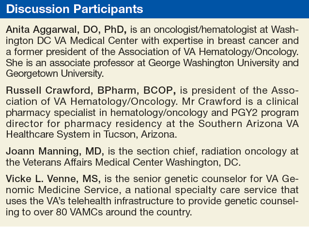
The other issue is that she was diagnosed at a young age. Clearly, 32 years is much younger than we typically see in breast cancer, and we know that individuals with hereditary cancers often have an earlier age of onset. With no other information, her a priori risk of having a BRCA1/2 mutation would be < 2.5%.
Regardless, based on current National Comprehensive Cancer Network (NCCN) guidelines, she would be a testing candidate. We would also recommend testing for more than just BRCA1/2. In the last few decades, there have been many genes identified that are associated with an increased susceptibility to cancer. Many of these genes are part of syndromes, so if you had a mutation, that also would increase the risk for a cancer in another organ. If this woman’s mother and father lived into their 70s or 80s and she had a number of aunts on both sides who never developed breast cancer, it would be less likely to be BRCA1/2. However, P53 also can present in young women and as a de novo mutation. Therefore, we would offer her a panel of actionable genes. Genes that if, in fact, we identified a mutation in one of them, would mean we could do something different for this young lady.
JoAnn Manning, MD. Let’s say she does have testing, and she comes back BRCA+. Then what would be the recommendations or guidance?
Ms. Venne. Women (and men as well!) with mutations have an increased risk for a second primary breast cancer as well as cancer in other organs. Focusing first on the breast story and all the media around BRCA1/2 mutations and surgery, this is a woman who may consider a more aggressive surgery, including prophylactic contralateral mastectomy, if she is concerned. She is young, so we also would explore her fertility plans. While her next few months will be filled with breast cancer treatment choices, women with BRCA mutations also are at an increased risk to develop ovarian cancer, so that might be a decision she makes as well. Her health care team may also eventually discuss chemotherapeutic options available specifically to women with mutations.
However, we often see young women who are extremely nervous because there is a sense that if you’re younger, your cancer must be inherited. Part of the pretest counseling is to explore psychosocial issues and help these young ladies understand that, especially if she does not have a family history of cancer and the only indication is her age, then it’s highly likely that we’re not going to find an identifiable mutation. And in that circumstance, she probably could consider a more conservative surgical decision.
Dr. Aggarwal. How common is a BRCA1 or BRCA2 mutation in African American females?
Ms. Venne. I have not paid attention to the prevalence of mutations based on ethnicity, so I don’t know. While many of the initial mutations were discovered in women of European ancestry, there are large cohorts of women with African ancestry whose specimens are now available for identifying genetic markers that will improve breast cancer risk assessment in them.1 However, because those mutations are still being characterized, it is more common to find a variant of uncertain significance (VUS) in African American women. A VUS is an alteration—a change in the gene—that we simply don’t know what it means yet. Clinicians don’t have enough information to know if that alteration is pathogenic or benign. The problem is that people try to make sense out of everything in their lives, so they also will try to make a VUS mean something. We try hard to help people understand that a VUS is really no more significant than if we had not tested in the first place, and they should not act on that information. They should use their family history, their age, their other psychosocial concerns about their experiences with cancer as they make their treatment decisions. But they also should check back periodically with their genetic counselor because VUSs can be reclassified. And if that happens, the information might be more useful for not only them, but their family members.
Dr. Manning. Would you consider this patient for any neoadjuvant chemotherapy?
Dr. Aggarwal. The patient is a young female with a small tumor that is HER2+. The indication for neoadjuvant chemotherapy is typically a big tumor or inoperable disease. Neoadjuvant chemotherapy is considered the standard of care for patients with inflammatory breast cancer and may confer a survival benefit in these patients. Of all the breast cancer subtypes, triple negative and HER2+ are considered the most chemosensitive and may benefit from neoadjuvant therapy. This patient has a small tumor, and I don’t think she’s a candidate for neoadjuvant chemotherapy unless the patient wants to see if her tumor is chemosensitive or not.
Dr. Manning, What’s the role and benefit of lumpectomy vs mastectomy?
Dr. Manning. Historically, mastectomy would have been considered the standard of care, but luckily, in the 1970s and the 1980s, we had a significant number of randomized controlled trials that demonstrated that certain women with particular characteristics would get the same overall survival if they chose mastectomy vs lumpectomy, the removal of the tumor with negative margin and whole-breast radiation. The key thing to understand is that breast-conserving surgery is now very well established with more than 20 years of data to support it. And that breast irradiation after breast-conserving surgery is essential to maximizing the local control and the overall survival (OS).
There have been a lot of major studies, but the one with the greatest follow-up now is the National Surgical Adjuvant Breast and Bowel Project (NSABP) B06 protocol, which was the only trial to compare mastectomy to lumpectomy and radiation or lumpectomy alone. It required negative margins. With 20 years of follow-up, the data still support that mastectomy or lumpectomy with radiation offers equivalent OS and local control. It’s really about patient preference if they are candidates.
Who is a candidate? Clearly, there are contraindications. We tend to look primarily at the size of the tumor. However, removing an average-sized tumor (< 2 cm) with a margin may not have a good cosmetic result for a patient with very small breasts. That patient may opt to go forward with a mastectomy instead. Young patients who are candidates must have to have negative margins. If they have persistently positive resection margins after excision or reexcision, then they need to go forward with mastectomy.
A patient who has imaging evidence of multicentric disease with 2 or more primary tumors in separate quadrants would not be a candidate for breast-conserving therapy. Diffuse malignant-appearing microcalcifications on a mammogram also would suggest multicentric disease. And a patient with a prior history of radiation therapy to the breast or chest wall cannot go through breast-conserving therapy.
In the case we are discussing, we also should make sure this young lady is not pregnant. If the patient is adamant about breast-conserving surgery and pregnant, especially in the third trimester, radiation could be deferred until after delivery. Another relative contraindication is for patients who have connective tissue disorders. Sometimes if they are given whole-breast radiation, the cosmetic result is poor. So if you’re doing this procedure to save the breast, then having a good cosmetic result is an important consideration for many patients.
When you look at the size of the tumor for this patient, she seems to be a good candidate for breast-conserving surgery. I would recommend that she go forward with lumpectomy followed by whole-breast radiation.
Ms. Venne. Although the numbers aren’t nearly as large as they were in the original trials looking at the lumpectomy vs mastectomy, there are now survival data for women with BRCA1/2 mutations. With all of the caveats that Dr. Manning mentioned, even if you have an identifiable mutation, you may not necessarily need that more aggressive surgery.2 Clearly, individuals with identifiable mutations would have a higher chance of a contralateral breast cancer, a second primary, so some individuals consider a prophylactic bilateral mastectomy. But from a survival perspective, there are a fair amount of data now available that say that lumpectomy vs mastectomy should really be the conversation based on all of the information that Dr. Manning outlines rather than using primarily the mutation status
Dr. Manning. I agree.
Dr. Aggarwal. This patient had a lumpectomy and axillary lymph node dissection. Pathology reported 1.5-cm mass, grade 3 IDC; the margins were negative. There was no skin involvement, 27 lymph nodes removed were all negative. Dr. Manning, can you please discuss the role of radiation in early stage breast cancer in patients like this case?
Dr. Manning. One of the questions that is always controversial for radiation in these early stage breast cancer cases is what do you do with the nodal irradiation? Previously, radiation oncologists based treatment plans on retrospective data, but in 2015, there were 2 major studies, 1 from Canada, and 1 from the European Organisation for Research and Treatment of Cancer (EORTC).3,4 Both studies tried to determine whether there was an advantage to doing regional nodal irradiation in early breast cancer cases. That encompassed axillary, supraclavicular, and internal mammary nodes. The studies showed that there was no survival advantage, but there was a statistically significant improvement in disease free survival and in local regional recurrence and distant mets.
Unfortunately, there are still a lot of unanswered questions, like what group potentially would benefit the most? In the MA.20 Study, some observers questioned that maybe the ER-/PR- women had the most benefit, but then, in the other study the benefit wasn’t clear.4,5 One question is which lymph node group is having the most impact? Was the benefit from radiating the supraclavicular nodes or was it from radiating the internal mammary nodes? Determining the answer is important from a technical point of view because when you radiate the internal mammary nodes, you have the potential to expose more heart and lung to radiation. You have to put all these together and make a recommendation.
Clearly, for a patient with negative nodes there is no question: You would not treat the regional nodes. However, for a patient with positive nodes you really have to individualize the approach and consider age, anatomy, tumor location, and burden of axillary disease.
I would sit down and have a discussion with this young woman to weigh the risks and the benefits. There is a slight increased risk of lymphedema in these patients, and radiation pneumonitis increases, but not significantly. A key concern is to minimize the total dose of radiation to the heart. There have been great developments in radiation oncology technology and capabilities, so the cardiac dose is now less. But when you think about a 32-year-old patient and weigh the benefit of a 2% to 3% decrease in the incidence of distant metastases and no OS advantage, then you really need to have a conversation about how to safely treat her. At a minimum, I would treat the high axilla and the supraclavicular nodes because she had a pretty extensive lymph node dissection with more than 20 nodes, and then with her getting systemic therapy, that should be more than adequate.
Dr. Aggarwal. Is there any cutoff for age or size of the tumor where you would not do any radiation to the breast?
Dr. Manning. In this particular patient absolutely not because of the lymph node. She had breast-conserving therapy, and she’s only 32-years-old. The PRIME 2 study offered lumpectomy alone vs lumpectomy and radiation for women aged ≥ 65 years with tumors ≤ 3 cm, low grade.6 The study participants had to have negative lymph nodes, be ER+, and low grade. It was a very select group. The lumpectomy patients had a recurrence rate around 4%, and the other was closer to 1.3%.
You have to look at the whole picture. Is this a healthy 70-year-old woman? Is it an inconvenience for her to get treatment? Is she going to get hormone therapy and will she be adherent? There’s a very small group of women who underwent breast-conserving surgery that I would feel safe about not offering radiation.
Dr. Aggarwal. About 15% to 20% of all breast cancers are HER2 over expressors, which used to be a poor prognostic characteristic. However, the development of anti-HER2 therapies has changed the picture of HER2 prognosis. After the initial discovery of activity, the pivotal study by Slamon et al showed benefit in terms of progression-free survival (PFS) and OS with chemotherapy and trastuzumab. The NCCN guideline recommends anti-HER2 antibody trastuzumab in combination with chemotherapy.7
Patients with tumor < 0.5 cm who are HER2+ and ER+ may not benefit from trastuzumab, but those who are ER- and HER2+ will still benefit from trastuzumab. The combination is adriamycin/cyclophosphamide followed by a taxane with trastuzumab and to complete 1 year of trastuzumab or trastuzumab in combination with carboplatin and taxanes.
Pertuzumab, in combination with trastuzumab and docetaxel (PHT) has been FDA-approved in neoadjuvant and metastatic HER2+ disease, but is not FDA approved yet in the adjuvant setting. However, these are expensive drugs, and we don’t know how long these drugs should be given.
Mr. Crawford, What are the adverse effects (AEs) of an anti-HER2 or trastuzumab treatment, and what is the cost of trastuzumab?
Russell Crawford, BPharm. The anti-HER2 antibodies have certainly changed treatment plans and outcomes for patients with breast cancer who test HER2+. There are actually 3 of these anti-HER2 drugs on the U.S. market, and they can be used in a variety of settings. Trastuzumab and pertuzumab are indicated in women or patients who have HER2+ disease, and they work by binding to the extracellular domain of the HER2 proteins and mediate antibody-dependent cellular toxicity by inhibiting proliferation of the cells that overexpress HER2.
In this patient, we would be looking at using adjuvant trastuzumab to complete a 1-year course of therapy while she’s getting her dose-dense doxorubicin and cyclophosphamide (AC) on a weekly basis for the first 12 weeks. Trastuzumab is dosed with an initial loading dose of 4 mg/kg as the first dose, and then it’s 2 mg/kg/wk until adjuvant chemotherapy is completed. We usually extend the dosing out to 6 mg/kg every 3 weeks to complete the year of treatment.
These drugs are fairly well tolerated. They are monoclonal proteins, so a lot of the AEs that patients experience are the things that we’re used to seeing with other monoclonal proteins like the infusion-related reactions and some flulike symptoms. The biggest concern with these patients is that being on the drug for a year, there is a risk of decreasing the left ventricular ejection fraction (LVEF) of the heart. That risk is increased when these drugs are combined with anthracyclines that we know are cardiotoxic. As a single agent, the impact on left ventricular function is not significant, but when it is combined with chemotherapy, it does become a problem. Usually, we recommend routine and periodic monitoring of the LVEF with a multiple-gated acquisition or an echocardiogram to make sure that we’re not causing harm related to this treatment.
The cost of these drugs depends on the frequency, is it every week, every 2 weeks, or every 3 weeks? There are different ways to give trastuzumab, but for most patients, we prefer the every 3-week dose. And it’s estimated that for a 70-kg patient, a dose of trastuzumab at 6 mg/kg at the rate of every 3 weeks costs about $2,500 per dose. The VA pays about $6 a milligram, but it’s certainly money well spent because it has changed the playing field and the outcomes for these patients.
The cost of pertuzumab is dosed a little bit differently. It’s a flat dose not a weight-based dose. Patients get an initial loading dose of 840 mg and a continuation dose of 420 mg every 3 weeks. The cost of the 420-mg dose of pertuzumab is just under $3,000, so that first-time loading dose would be a $6,000 dose, and the continuing doses are about $3,000 per dose every 3 weeks. The AE profile is no different from what you would expect with trastuzumab. There is a similar toxicity profile for these 2 drugs. It does not appear that there is any additional cardiotoxicity if you are using the combination in the neoadjuvant setting.
The third targeted agent that goes after the HER2 is ado-trastuzumab, but it is only used in the metastatic setting, so we’ll reserve that for down the road for this patient should we ever need it.
Dr. Aggarwal. The patient received adriamycin/cyclophosphamide followed by paclitaxel weekly for 12 weeks with trastuzumab. After the 12 weekly doses, she went on trastuzumab every 3 weeks. Because she was ER+, she was a candidate for additional endocrine ablation therapy. She was started on tamoxifen and leuprolide acetate for complete hormonal ablation.
Tamoxifen was the first targeted therapy for breast cancer. In women with ER+ breast cancer, with tamoxifen given for 5 years as adjuvant treatment, the odds of recurrences decreased by 39%, and death decreased by 30% in both pre- and postmenopausal women.8 Then the ATLAS data came, which randomly allocated patients to continue another 5 years of tamoxifen vs placebo, for a total of 10 years of treatment with tamoxifen. With a mean of 7.6 years of further follow-up after entry at year 5 in this trial showed that recurrence and breast cancer mortality during the second decade after diagnosis are reduced more effectively by 10 years of adjuvant tamoxifen than by 5 years.9 The current recommendation for pre- and postmenopausal is 10 years of tamoxifen.
In addition we have 3 aromatase inhibitors (AIs), anastrozole, letrozole, and exemestane, which block the production of estrogen in postmenopausal females. Anastrozole and letrozole are nonsteroidal, and exemestane is steroidal. There are countless big randomized trials using all of these drug in different combinations. In most of these trials, AIs are shown to be equal to tamoxifen when they are compared with each other, but their AE profile is different.
The recommendation by the American Society of Clinical Oncology and NCCN guidelines is to use only AIs for 5 years. There are different combinations: You can give tamoxifen for 2 to 3 years, followed by 5 years of an AI, or 5 years of tamoxifen and 5 years of an AI. Some patients wants to stop because of AEs, but others want to continue. Patients can develop osteoporosis and arthritis from an AI and hot flashes from tamoxifen.
Mr. Crawford, How would you manage of these AEs from these treatments?
Mr. Crawford. Because this woman is young, age 32, and premenopausal, tamoxifen would be the recommended endocrine therapy for her being ER+/PR+. But the role of the leuprolide acetate is to induce a chemical oophorectomy. We are putting her into ovarian ablation by using the leuprolide acetate.
The tamoxifen is relatively well tolerated, but as an ER blocker, it has a different AE profile than does an estrogen production decreaser. With tamoxifen patients tend to complain about hot flashes, edema, fluid retention, altered menses, spotting vaginal discharge, vaginal bleeding, and dryness. These medications also increase the risk of venous thromboembolism (VTE), and there is some concern about increased risk of developing endometrial cancers with these medications. We can give it either once or twice daily. There’s nothing that really says 10 mg twice daily vs 20 mg once daily is any different. So we may play with dosing to see if patients tolerate it better one way or the other.
There are medications that we can offer to help manage the hot flashes. These medications don’t necessarily make the hot flashes go away, but they can decrease the hot flash intensity or and/or frequency. Many medications have been evaluated for hot flashes. The best data are for venlafaxine, which is usually given once a day at bedtime (dosage 37.5-75.0 mg). There has been success with gabapentin titrated up to a dose of about 300 mg 3 times daily. They are fairly similar for decreasing hot flash scores and intensities, but the patient preferences were more favorable toward the venlafaxine than for the gabapentin.
The AIs, on the other hand, have a different AE profile. With tamoxifen we see vaginal discharges, bleeding, endometrial cancer risk, and VTE risk, but these are not significant problems with any of the AIs. The AE profiles for AIs include hot flashes, but more often it is complaints of bone pain, arthralgias, and myalgias. Probably the top reason why most patients discontinue taking AIs is arthralgia and myalgia.
Because we have shut off estrogen production with the AIs, and estrogen is an important component of maintaining good bone health and bone homeostasis, patients are at an increased risked of losing or declining bone mineral density (BMD). It is recommended that these patients get placed on routine calcium and vitamin D supplementation with routine dual-energy X-ray absorptiometry scans, so we know whether we will need to initiate osteoporosis treatment, whether with oral bisphosphonates, intravenous bisphosphonates, or subcutaneous rank ligand inhibitors.
With bisphosphonates there may be a slight increase in fracture rates. But we have to balance that with the BMD concerns. If the patient progresses into the metastatic setting and we know that there’s a fair chance that there’s going to be some skeletal involvement, those people are also at an increased risk of fracture. While there is a slight concern about the increased risk of fractures with bisphosphonates, I tend to believe that the benefits outweigh the risks.
Go to www.fedprac.com/AVAHO for a discussion of the next steps in the treatment for the patient after she returned 2 years later with nausea, vomiting, acute onset headache, and 2 brain lesions that were about 2 cm.
Click here to read the digital edition.
1. Feng Y, Rhie SK, Huo D, et al. Characterizing genetic susceptibility to breast cancer in women of african ancestry. Cancer Epidemiol Biomarkers. 2017;26(7):1016-1026.
2. Copson ER, Maishman TC, Tapper WJ, et al. Germline BRCA mutation and outcome in young-onset breast cancer (POSH): a prospective cohort study. Lancet Oncol. 2018;19(2):169-180.
3. Poortmans PM, Collette S, Kirkove C, et al; EORTC Radiation Oncology and Breast Cancer Groups. Internal mammary and medial supraclavicular irradiation in breast cancer. N Engl J Med. 2015;373(4):317-327.
4. Whelan TJ, Olivotto IA, Parulekar WR, et al; MA.20 Study Investigators. Regional nodal irradiation in early-stage breast cancer. N Engl J Med. 2015;373(4):307-316.
5. EBCTCG (Early Breast Cancer Trialists’ Collaborative Group), McGale P, Taylor C, Correa C, et al. Effect of radiotherapy after mastectomy and axillary surgery on 10-year recurrence and 20-year breast cancer mortality: meta-analysis of individual patient data for 8135 women in 22 randomised trials. Lancet. 2014;383(9935):2127-2135.
6. Kunkler IH, Williams LJ, Jack WJ, Cameron DA, Dixon JM; PRIME II investigators. Breast-conserving surgery with or without irradiation in women aged 65 years or older with early breast cancer (PRIME II): a randomised controlled trial. Lancet Oncol. 2015;16(3):266-273.
7. Slamon DJ, Clark GM, Wong SG, Levin WJ, Ullrich A, McGuire WL. Human breast cancer: correlation of relapse and survival with amplification of the HER-2/neu oncogene. Science. 1987;235(4785):177-182.
8. Early Breast Cancer Trialists’ Collaborative Group (EBCTCG). Effect of chemotherapy and hormonal therapy for early breast cancer on recurrence and 15 year survival: an overview of the randomised trials. Lancet. 2005;365(9472):1687-1717.
9. Davies C, Pan H, Godwin J, et al; Adjuvant Tamoxifen: Longer Against Shorter (ATLAS) Collaborative Group. Long-term effects of continuing adjuvant tamoxifen to 10 years versus stopping at 5 years after diagnosis of oestrogen receptor-positive breast cancer: ATLAS, a randomised trial. Lancet. 2013;381(9869):805-816.
Case Presentation
This case represents a composite of many different patients and is not meant to represent an individual. Any resemblance to an actual patient is coincidental.
A 32-year-old African American woman presented with a self-palpated left breast mass (axillary tail at 9 o’clock position). The patient was a nonsmoker, was otherwise healthy, and had no family history of breast or any other cancer. She had never used oral contraceptives or hormones, was never pregnant, her menarche was at age 12 years, and she had regular menstrual periods. On physical examination she had a 1-cm left breast mass and a palpable left axillary lymph node. A complete diagnostic workup revealed a 2-cm left breast mass. An ultrasound-guided biopsy of the axillary lymph node was positive for invasive ductal carcinoma (IDC). The final diagnosis was left breast cancer, stage IIB IDC, T1N1M0, ER+, PR+, HER2 2+ by immunohistochemistry, fluorescence in situ hybridization (FISH) was 2.4, confirming a HER2+ tumor.
Anita Aggarwal, DO, PhD. What is the role of genetic counseling and testing in this young patient who does not have a family history of breast cancer?
Vickie L. Venne, MS. This patient absolutely would be a candidate for counseling and testing. From a genetic counseling perspective, one of the first points has to do with what “no family history of cancer” means. Typically, in a fast-paced clinic, a patient will be asked “Does anybody else in your family have cancer?” And it’s not uncommon to get the answer “no.” Genetic counselors collect specific information on at least the first- and second-degree relatives, so we end up with 3 generations. This includes both the maternal and the paternal histories. We find that people who initially report no family history of cancer are often just thinking of breast cancer, even if the provider’s question is broad. When we start digging, we often find other cancers because cancer is common.
The other issue is that she was diagnosed at a young age. Clearly, 32 years is much younger than we typically see in breast cancer, and we know that individuals with hereditary cancers often have an earlier age of onset. With no other information, her a priori risk of having a BRCA1/2 mutation would be < 2.5%.
Regardless, based on current National Comprehensive Cancer Network (NCCN) guidelines, she would be a testing candidate. We would also recommend testing for more than just BRCA1/2. In the last few decades, there have been many genes identified that are associated with an increased susceptibility to cancer. Many of these genes are part of syndromes, so if you had a mutation, that also would increase the risk for a cancer in another organ. If this woman’s mother and father lived into their 70s or 80s and she had a number of aunts on both sides who never developed breast cancer, it would be less likely to be BRCA1/2. However, P53 also can present in young women and as a de novo mutation. Therefore, we would offer her a panel of actionable genes. Genes that if, in fact, we identified a mutation in one of them, would mean we could do something different for this young lady.
JoAnn Manning, MD. Let’s say she does have testing, and she comes back BRCA+. Then what would be the recommendations or guidance?
Ms. Venne. Women (and men as well!) with mutations have an increased risk for a second primary breast cancer as well as cancer in other organs. Focusing first on the breast story and all the media around BRCA1/2 mutations and surgery, this is a woman who may consider a more aggressive surgery, including prophylactic contralateral mastectomy, if she is concerned. She is young, so we also would explore her fertility plans. While her next few months will be filled with breast cancer treatment choices, women with BRCA mutations also are at an increased risk to develop ovarian cancer, so that might be a decision she makes as well. Her health care team may also eventually discuss chemotherapeutic options available specifically to women with mutations.
However, we often see young women who are extremely nervous because there is a sense that if you’re younger, your cancer must be inherited. Part of the pretest counseling is to explore psychosocial issues and help these young ladies understand that, especially if she does not have a family history of cancer and the only indication is her age, then it’s highly likely that we’re not going to find an identifiable mutation. And in that circumstance, she probably could consider a more conservative surgical decision.
Dr. Aggarwal. How common is a BRCA1 or BRCA2 mutation in African American females?
Ms. Venne. I have not paid attention to the prevalence of mutations based on ethnicity, so I don’t know. While many of the initial mutations were discovered in women of European ancestry, there are large cohorts of women with African ancestry whose specimens are now available for identifying genetic markers that will improve breast cancer risk assessment in them.1 However, because those mutations are still being characterized, it is more common to find a variant of uncertain significance (VUS) in African American women. A VUS is an alteration—a change in the gene—that we simply don’t know what it means yet. Clinicians don’t have enough information to know if that alteration is pathogenic or benign. The problem is that people try to make sense out of everything in their lives, so they also will try to make a VUS mean something. We try hard to help people understand that a VUS is really no more significant than if we had not tested in the first place, and they should not act on that information. They should use their family history, their age, their other psychosocial concerns about their experiences with cancer as they make their treatment decisions. But they also should check back periodically with their genetic counselor because VUSs can be reclassified. And if that happens, the information might be more useful for not only them, but their family members.
Dr. Manning. Would you consider this patient for any neoadjuvant chemotherapy?
Dr. Aggarwal. The patient is a young female with a small tumor that is HER2+. The indication for neoadjuvant chemotherapy is typically a big tumor or inoperable disease. Neoadjuvant chemotherapy is considered the standard of care for patients with inflammatory breast cancer and may confer a survival benefit in these patients. Of all the breast cancer subtypes, triple negative and HER2+ are considered the most chemosensitive and may benefit from neoadjuvant therapy. This patient has a small tumor, and I don’t think she’s a candidate for neoadjuvant chemotherapy unless the patient wants to see if her tumor is chemosensitive or not.
Dr. Manning, What’s the role and benefit of lumpectomy vs mastectomy?
Dr. Manning. Historically, mastectomy would have been considered the standard of care, but luckily, in the 1970s and the 1980s, we had a significant number of randomized controlled trials that demonstrated that certain women with particular characteristics would get the same overall survival if they chose mastectomy vs lumpectomy, the removal of the tumor with negative margin and whole-breast radiation. The key thing to understand is that breast-conserving surgery is now very well established with more than 20 years of data to support it. And that breast irradiation after breast-conserving surgery is essential to maximizing the local control and the overall survival (OS).
There have been a lot of major studies, but the one with the greatest follow-up now is the National Surgical Adjuvant Breast and Bowel Project (NSABP) B06 protocol, which was the only trial to compare mastectomy to lumpectomy and radiation or lumpectomy alone. It required negative margins. With 20 years of follow-up, the data still support that mastectomy or lumpectomy with radiation offers equivalent OS and local control. It’s really about patient preference if they are candidates.
Who is a candidate? Clearly, there are contraindications. We tend to look primarily at the size of the tumor. However, removing an average-sized tumor (< 2 cm) with a margin may not have a good cosmetic result for a patient with very small breasts. That patient may opt to go forward with a mastectomy instead. Young patients who are candidates must have to have negative margins. If they have persistently positive resection margins after excision or reexcision, then they need to go forward with mastectomy.
A patient who has imaging evidence of multicentric disease with 2 or more primary tumors in separate quadrants would not be a candidate for breast-conserving therapy. Diffuse malignant-appearing microcalcifications on a mammogram also would suggest multicentric disease. And a patient with a prior history of radiation therapy to the breast or chest wall cannot go through breast-conserving therapy.
In the case we are discussing, we also should make sure this young lady is not pregnant. If the patient is adamant about breast-conserving surgery and pregnant, especially in the third trimester, radiation could be deferred until after delivery. Another relative contraindication is for patients who have connective tissue disorders. Sometimes if they are given whole-breast radiation, the cosmetic result is poor. So if you’re doing this procedure to save the breast, then having a good cosmetic result is an important consideration for many patients.
When you look at the size of the tumor for this patient, she seems to be a good candidate for breast-conserving surgery. I would recommend that she go forward with lumpectomy followed by whole-breast radiation.
Ms. Venne. Although the numbers aren’t nearly as large as they were in the original trials looking at the lumpectomy vs mastectomy, there are now survival data for women with BRCA1/2 mutations. With all of the caveats that Dr. Manning mentioned, even if you have an identifiable mutation, you may not necessarily need that more aggressive surgery.2 Clearly, individuals with identifiable mutations would have a higher chance of a contralateral breast cancer, a second primary, so some individuals consider a prophylactic bilateral mastectomy. But from a survival perspective, there are a fair amount of data now available that say that lumpectomy vs mastectomy should really be the conversation based on all of the information that Dr. Manning outlines rather than using primarily the mutation status
Dr. Manning. I agree.
Dr. Aggarwal. This patient had a lumpectomy and axillary lymph node dissection. Pathology reported 1.5-cm mass, grade 3 IDC; the margins were negative. There was no skin involvement, 27 lymph nodes removed were all negative. Dr. Manning, can you please discuss the role of radiation in early stage breast cancer in patients like this case?
Dr. Manning. One of the questions that is always controversial for radiation in these early stage breast cancer cases is what do you do with the nodal irradiation? Previously, radiation oncologists based treatment plans on retrospective data, but in 2015, there were 2 major studies, 1 from Canada, and 1 from the European Organisation for Research and Treatment of Cancer (EORTC).3,4 Both studies tried to determine whether there was an advantage to doing regional nodal irradiation in early breast cancer cases. That encompassed axillary, supraclavicular, and internal mammary nodes. The studies showed that there was no survival advantage, but there was a statistically significant improvement in disease free survival and in local regional recurrence and distant mets.
Unfortunately, there are still a lot of unanswered questions, like what group potentially would benefit the most? In the MA.20 Study, some observers questioned that maybe the ER-/PR- women had the most benefit, but then, in the other study the benefit wasn’t clear.4,5 One question is which lymph node group is having the most impact? Was the benefit from radiating the supraclavicular nodes or was it from radiating the internal mammary nodes? Determining the answer is important from a technical point of view because when you radiate the internal mammary nodes, you have the potential to expose more heart and lung to radiation. You have to put all these together and make a recommendation.
Clearly, for a patient with negative nodes there is no question: You would not treat the regional nodes. However, for a patient with positive nodes you really have to individualize the approach and consider age, anatomy, tumor location, and burden of axillary disease.
I would sit down and have a discussion with this young woman to weigh the risks and the benefits. There is a slight increased risk of lymphedema in these patients, and radiation pneumonitis increases, but not significantly. A key concern is to minimize the total dose of radiation to the heart. There have been great developments in radiation oncology technology and capabilities, so the cardiac dose is now less. But when you think about a 32-year-old patient and weigh the benefit of a 2% to 3% decrease in the incidence of distant metastases and no OS advantage, then you really need to have a conversation about how to safely treat her. At a minimum, I would treat the high axilla and the supraclavicular nodes because she had a pretty extensive lymph node dissection with more than 20 nodes, and then with her getting systemic therapy, that should be more than adequate.
Dr. Aggarwal. Is there any cutoff for age or size of the tumor where you would not do any radiation to the breast?
Dr. Manning. In this particular patient absolutely not because of the lymph node. She had breast-conserving therapy, and she’s only 32-years-old. The PRIME 2 study offered lumpectomy alone vs lumpectomy and radiation for women aged ≥ 65 years with tumors ≤ 3 cm, low grade.6 The study participants had to have negative lymph nodes, be ER+, and low grade. It was a very select group. The lumpectomy patients had a recurrence rate around 4%, and the other was closer to 1.3%.
You have to look at the whole picture. Is this a healthy 70-year-old woman? Is it an inconvenience for her to get treatment? Is she going to get hormone therapy and will she be adherent? There’s a very small group of women who underwent breast-conserving surgery that I would feel safe about not offering radiation.
Dr. Aggarwal. About 15% to 20% of all breast cancers are HER2 over expressors, which used to be a poor prognostic characteristic. However, the development of anti-HER2 therapies has changed the picture of HER2 prognosis. After the initial discovery of activity, the pivotal study by Slamon et al showed benefit in terms of progression-free survival (PFS) and OS with chemotherapy and trastuzumab. The NCCN guideline recommends anti-HER2 antibody trastuzumab in combination with chemotherapy.7
Patients with tumor < 0.5 cm who are HER2+ and ER+ may not benefit from trastuzumab, but those who are ER- and HER2+ will still benefit from trastuzumab. The combination is adriamycin/cyclophosphamide followed by a taxane with trastuzumab and to complete 1 year of trastuzumab or trastuzumab in combination with carboplatin and taxanes.
Pertuzumab, in combination with trastuzumab and docetaxel (PHT) has been FDA-approved in neoadjuvant and metastatic HER2+ disease, but is not FDA approved yet in the adjuvant setting. However, these are expensive drugs, and we don’t know how long these drugs should be given.
Mr. Crawford, What are the adverse effects (AEs) of an anti-HER2 or trastuzumab treatment, and what is the cost of trastuzumab?
Russell Crawford, BPharm. The anti-HER2 antibodies have certainly changed treatment plans and outcomes for patients with breast cancer who test HER2+. There are actually 3 of these anti-HER2 drugs on the U.S. market, and they can be used in a variety of settings. Trastuzumab and pertuzumab are indicated in women or patients who have HER2+ disease, and they work by binding to the extracellular domain of the HER2 proteins and mediate antibody-dependent cellular toxicity by inhibiting proliferation of the cells that overexpress HER2.
In this patient, we would be looking at using adjuvant trastuzumab to complete a 1-year course of therapy while she’s getting her dose-dense doxorubicin and cyclophosphamide (AC) on a weekly basis for the first 12 weeks. Trastuzumab is dosed with an initial loading dose of 4 mg/kg as the first dose, and then it’s 2 mg/kg/wk until adjuvant chemotherapy is completed. We usually extend the dosing out to 6 mg/kg every 3 weeks to complete the year of treatment.
These drugs are fairly well tolerated. They are monoclonal proteins, so a lot of the AEs that patients experience are the things that we’re used to seeing with other monoclonal proteins like the infusion-related reactions and some flulike symptoms. The biggest concern with these patients is that being on the drug for a year, there is a risk of decreasing the left ventricular ejection fraction (LVEF) of the heart. That risk is increased when these drugs are combined with anthracyclines that we know are cardiotoxic. As a single agent, the impact on left ventricular function is not significant, but when it is combined with chemotherapy, it does become a problem. Usually, we recommend routine and periodic monitoring of the LVEF with a multiple-gated acquisition or an echocardiogram to make sure that we’re not causing harm related to this treatment.
The cost of these drugs depends on the frequency, is it every week, every 2 weeks, or every 3 weeks? There are different ways to give trastuzumab, but for most patients, we prefer the every 3-week dose. And it’s estimated that for a 70-kg patient, a dose of trastuzumab at 6 mg/kg at the rate of every 3 weeks costs about $2,500 per dose. The VA pays about $6 a milligram, but it’s certainly money well spent because it has changed the playing field and the outcomes for these patients.
The cost of pertuzumab is dosed a little bit differently. It’s a flat dose not a weight-based dose. Patients get an initial loading dose of 840 mg and a continuation dose of 420 mg every 3 weeks. The cost of the 420-mg dose of pertuzumab is just under $3,000, so that first-time loading dose would be a $6,000 dose, and the continuing doses are about $3,000 per dose every 3 weeks. The AE profile is no different from what you would expect with trastuzumab. There is a similar toxicity profile for these 2 drugs. It does not appear that there is any additional cardiotoxicity if you are using the combination in the neoadjuvant setting.
The third targeted agent that goes after the HER2 is ado-trastuzumab, but it is only used in the metastatic setting, so we’ll reserve that for down the road for this patient should we ever need it.
Dr. Aggarwal. The patient received adriamycin/cyclophosphamide followed by paclitaxel weekly for 12 weeks with trastuzumab. After the 12 weekly doses, she went on trastuzumab every 3 weeks. Because she was ER+, she was a candidate for additional endocrine ablation therapy. She was started on tamoxifen and leuprolide acetate for complete hormonal ablation.
Tamoxifen was the first targeted therapy for breast cancer. In women with ER+ breast cancer, with tamoxifen given for 5 years as adjuvant treatment, the odds of recurrences decreased by 39%, and death decreased by 30% in both pre- and postmenopausal women.8 Then the ATLAS data came, which randomly allocated patients to continue another 5 years of tamoxifen vs placebo, for a total of 10 years of treatment with tamoxifen. With a mean of 7.6 years of further follow-up after entry at year 5 in this trial showed that recurrence and breast cancer mortality during the second decade after diagnosis are reduced more effectively by 10 years of adjuvant tamoxifen than by 5 years.9 The current recommendation for pre- and postmenopausal is 10 years of tamoxifen.
In addition we have 3 aromatase inhibitors (AIs), anastrozole, letrozole, and exemestane, which block the production of estrogen in postmenopausal females. Anastrozole and letrozole are nonsteroidal, and exemestane is steroidal. There are countless big randomized trials using all of these drug in different combinations. In most of these trials, AIs are shown to be equal to tamoxifen when they are compared with each other, but their AE profile is different.
The recommendation by the American Society of Clinical Oncology and NCCN guidelines is to use only AIs for 5 years. There are different combinations: You can give tamoxifen for 2 to 3 years, followed by 5 years of an AI, or 5 years of tamoxifen and 5 years of an AI. Some patients wants to stop because of AEs, but others want to continue. Patients can develop osteoporosis and arthritis from an AI and hot flashes from tamoxifen.
Mr. Crawford, How would you manage of these AEs from these treatments?
Mr. Crawford. Because this woman is young, age 32, and premenopausal, tamoxifen would be the recommended endocrine therapy for her being ER+/PR+. But the role of the leuprolide acetate is to induce a chemical oophorectomy. We are putting her into ovarian ablation by using the leuprolide acetate.
The tamoxifen is relatively well tolerated, but as an ER blocker, it has a different AE profile than does an estrogen production decreaser. With tamoxifen patients tend to complain about hot flashes, edema, fluid retention, altered menses, spotting vaginal discharge, vaginal bleeding, and dryness. These medications also increase the risk of venous thromboembolism (VTE), and there is some concern about increased risk of developing endometrial cancers with these medications. We can give it either once or twice daily. There’s nothing that really says 10 mg twice daily vs 20 mg once daily is any different. So we may play with dosing to see if patients tolerate it better one way or the other.
There are medications that we can offer to help manage the hot flashes. These medications don’t necessarily make the hot flashes go away, but they can decrease the hot flash intensity or and/or frequency. Many medications have been evaluated for hot flashes. The best data are for venlafaxine, which is usually given once a day at bedtime (dosage 37.5-75.0 mg). There has been success with gabapentin titrated up to a dose of about 300 mg 3 times daily. They are fairly similar for decreasing hot flash scores and intensities, but the patient preferences were more favorable toward the venlafaxine than for the gabapentin.
The AIs, on the other hand, have a different AE profile. With tamoxifen we see vaginal discharges, bleeding, endometrial cancer risk, and VTE risk, but these are not significant problems with any of the AIs. The AE profiles for AIs include hot flashes, but more often it is complaints of bone pain, arthralgias, and myalgias. Probably the top reason why most patients discontinue taking AIs is arthralgia and myalgia.
Because we have shut off estrogen production with the AIs, and estrogen is an important component of maintaining good bone health and bone homeostasis, patients are at an increased risked of losing or declining bone mineral density (BMD). It is recommended that these patients get placed on routine calcium and vitamin D supplementation with routine dual-energy X-ray absorptiometry scans, so we know whether we will need to initiate osteoporosis treatment, whether with oral bisphosphonates, intravenous bisphosphonates, or subcutaneous rank ligand inhibitors.
With bisphosphonates there may be a slight increase in fracture rates. But we have to balance that with the BMD concerns. If the patient progresses into the metastatic setting and we know that there’s a fair chance that there’s going to be some skeletal involvement, those people are also at an increased risk of fracture. While there is a slight concern about the increased risk of fractures with bisphosphonates, I tend to believe that the benefits outweigh the risks.
Go to www.fedprac.com/AVAHO for a discussion of the next steps in the treatment for the patient after she returned 2 years later with nausea, vomiting, acute onset headache, and 2 brain lesions that were about 2 cm.
Click here to read the digital edition.
Case Presentation
This case represents a composite of many different patients and is not meant to represent an individual. Any resemblance to an actual patient is coincidental.
A 32-year-old African American woman presented with a self-palpated left breast mass (axillary tail at 9 o’clock position). The patient was a nonsmoker, was otherwise healthy, and had no family history of breast or any other cancer. She had never used oral contraceptives or hormones, was never pregnant, her menarche was at age 12 years, and she had regular menstrual periods. On physical examination she had a 1-cm left breast mass and a palpable left axillary lymph node. A complete diagnostic workup revealed a 2-cm left breast mass. An ultrasound-guided biopsy of the axillary lymph node was positive for invasive ductal carcinoma (IDC). The final diagnosis was left breast cancer, stage IIB IDC, T1N1M0, ER+, PR+, HER2 2+ by immunohistochemistry, fluorescence in situ hybridization (FISH) was 2.4, confirming a HER2+ tumor.
Anita Aggarwal, DO, PhD. What is the role of genetic counseling and testing in this young patient who does not have a family history of breast cancer?
Vickie L. Venne, MS. This patient absolutely would be a candidate for counseling and testing. From a genetic counseling perspective, one of the first points has to do with what “no family history of cancer” means. Typically, in a fast-paced clinic, a patient will be asked “Does anybody else in your family have cancer?” And it’s not uncommon to get the answer “no.” Genetic counselors collect specific information on at least the first- and second-degree relatives, so we end up with 3 generations. This includes both the maternal and the paternal histories. We find that people who initially report no family history of cancer are often just thinking of breast cancer, even if the provider’s question is broad. When we start digging, we often find other cancers because cancer is common.
The other issue is that she was diagnosed at a young age. Clearly, 32 years is much younger than we typically see in breast cancer, and we know that individuals with hereditary cancers often have an earlier age of onset. With no other information, her a priori risk of having a BRCA1/2 mutation would be < 2.5%.
Regardless, based on current National Comprehensive Cancer Network (NCCN) guidelines, she would be a testing candidate. We would also recommend testing for more than just BRCA1/2. In the last few decades, there have been many genes identified that are associated with an increased susceptibility to cancer. Many of these genes are part of syndromes, so if you had a mutation, that also would increase the risk for a cancer in another organ. If this woman’s mother and father lived into their 70s or 80s and she had a number of aunts on both sides who never developed breast cancer, it would be less likely to be BRCA1/2. However, P53 also can present in young women and as a de novo mutation. Therefore, we would offer her a panel of actionable genes. Genes that if, in fact, we identified a mutation in one of them, would mean we could do something different for this young lady.
JoAnn Manning, MD. Let’s say she does have testing, and she comes back BRCA+. Then what would be the recommendations or guidance?
Ms. Venne. Women (and men as well!) with mutations have an increased risk for a second primary breast cancer as well as cancer in other organs. Focusing first on the breast story and all the media around BRCA1/2 mutations and surgery, this is a woman who may consider a more aggressive surgery, including prophylactic contralateral mastectomy, if she is concerned. She is young, so we also would explore her fertility plans. While her next few months will be filled with breast cancer treatment choices, women with BRCA mutations also are at an increased risk to develop ovarian cancer, so that might be a decision she makes as well. Her health care team may also eventually discuss chemotherapeutic options available specifically to women with mutations.
However, we often see young women who are extremely nervous because there is a sense that if you’re younger, your cancer must be inherited. Part of the pretest counseling is to explore psychosocial issues and help these young ladies understand that, especially if she does not have a family history of cancer and the only indication is her age, then it’s highly likely that we’re not going to find an identifiable mutation. And in that circumstance, she probably could consider a more conservative surgical decision.
Dr. Aggarwal. How common is a BRCA1 or BRCA2 mutation in African American females?
Ms. Venne. I have not paid attention to the prevalence of mutations based on ethnicity, so I don’t know. While many of the initial mutations were discovered in women of European ancestry, there are large cohorts of women with African ancestry whose specimens are now available for identifying genetic markers that will improve breast cancer risk assessment in them.1 However, because those mutations are still being characterized, it is more common to find a variant of uncertain significance (VUS) in African American women. A VUS is an alteration—a change in the gene—that we simply don’t know what it means yet. Clinicians don’t have enough information to know if that alteration is pathogenic or benign. The problem is that people try to make sense out of everything in their lives, so they also will try to make a VUS mean something. We try hard to help people understand that a VUS is really no more significant than if we had not tested in the first place, and they should not act on that information. They should use their family history, their age, their other psychosocial concerns about their experiences with cancer as they make their treatment decisions. But they also should check back periodically with their genetic counselor because VUSs can be reclassified. And if that happens, the information might be more useful for not only them, but their family members.
Dr. Manning. Would you consider this patient for any neoadjuvant chemotherapy?
Dr. Aggarwal. The patient is a young female with a small tumor that is HER2+. The indication for neoadjuvant chemotherapy is typically a big tumor or inoperable disease. Neoadjuvant chemotherapy is considered the standard of care for patients with inflammatory breast cancer and may confer a survival benefit in these patients. Of all the breast cancer subtypes, triple negative and HER2+ are considered the most chemosensitive and may benefit from neoadjuvant therapy. This patient has a small tumor, and I don’t think she’s a candidate for neoadjuvant chemotherapy unless the patient wants to see if her tumor is chemosensitive or not.
Dr. Manning, What’s the role and benefit of lumpectomy vs mastectomy?
Dr. Manning. Historically, mastectomy would have been considered the standard of care, but luckily, in the 1970s and the 1980s, we had a significant number of randomized controlled trials that demonstrated that certain women with particular characteristics would get the same overall survival if they chose mastectomy vs lumpectomy, the removal of the tumor with negative margin and whole-breast radiation. The key thing to understand is that breast-conserving surgery is now very well established with more than 20 years of data to support it. And that breast irradiation after breast-conserving surgery is essential to maximizing the local control and the overall survival (OS).
There have been a lot of major studies, but the one with the greatest follow-up now is the National Surgical Adjuvant Breast and Bowel Project (NSABP) B06 protocol, which was the only trial to compare mastectomy to lumpectomy and radiation or lumpectomy alone. It required negative margins. With 20 years of follow-up, the data still support that mastectomy or lumpectomy with radiation offers equivalent OS and local control. It’s really about patient preference if they are candidates.
Who is a candidate? Clearly, there are contraindications. We tend to look primarily at the size of the tumor. However, removing an average-sized tumor (< 2 cm) with a margin may not have a good cosmetic result for a patient with very small breasts. That patient may opt to go forward with a mastectomy instead. Young patients who are candidates must have to have negative margins. If they have persistently positive resection margins after excision or reexcision, then they need to go forward with mastectomy.
A patient who has imaging evidence of multicentric disease with 2 or more primary tumors in separate quadrants would not be a candidate for breast-conserving therapy. Diffuse malignant-appearing microcalcifications on a mammogram also would suggest multicentric disease. And a patient with a prior history of radiation therapy to the breast or chest wall cannot go through breast-conserving therapy.
In the case we are discussing, we also should make sure this young lady is not pregnant. If the patient is adamant about breast-conserving surgery and pregnant, especially in the third trimester, radiation could be deferred until after delivery. Another relative contraindication is for patients who have connective tissue disorders. Sometimes if they are given whole-breast radiation, the cosmetic result is poor. So if you’re doing this procedure to save the breast, then having a good cosmetic result is an important consideration for many patients.
When you look at the size of the tumor for this patient, she seems to be a good candidate for breast-conserving surgery. I would recommend that she go forward with lumpectomy followed by whole-breast radiation.
Ms. Venne. Although the numbers aren’t nearly as large as they were in the original trials looking at the lumpectomy vs mastectomy, there are now survival data for women with BRCA1/2 mutations. With all of the caveats that Dr. Manning mentioned, even if you have an identifiable mutation, you may not necessarily need that more aggressive surgery.2 Clearly, individuals with identifiable mutations would have a higher chance of a contralateral breast cancer, a second primary, so some individuals consider a prophylactic bilateral mastectomy. But from a survival perspective, there are a fair amount of data now available that say that lumpectomy vs mastectomy should really be the conversation based on all of the information that Dr. Manning outlines rather than using primarily the mutation status
Dr. Manning. I agree.
Dr. Aggarwal. This patient had a lumpectomy and axillary lymph node dissection. Pathology reported 1.5-cm mass, grade 3 IDC; the margins were negative. There was no skin involvement, 27 lymph nodes removed were all negative. Dr. Manning, can you please discuss the role of radiation in early stage breast cancer in patients like this case?
Dr. Manning. One of the questions that is always controversial for radiation in these early stage breast cancer cases is what do you do with the nodal irradiation? Previously, radiation oncologists based treatment plans on retrospective data, but in 2015, there were 2 major studies, 1 from Canada, and 1 from the European Organisation for Research and Treatment of Cancer (EORTC).3,4 Both studies tried to determine whether there was an advantage to doing regional nodal irradiation in early breast cancer cases. That encompassed axillary, supraclavicular, and internal mammary nodes. The studies showed that there was no survival advantage, but there was a statistically significant improvement in disease free survival and in local regional recurrence and distant mets.
Unfortunately, there are still a lot of unanswered questions, like what group potentially would benefit the most? In the MA.20 Study, some observers questioned that maybe the ER-/PR- women had the most benefit, but then, in the other study the benefit wasn’t clear.4,5 One question is which lymph node group is having the most impact? Was the benefit from radiating the supraclavicular nodes or was it from radiating the internal mammary nodes? Determining the answer is important from a technical point of view because when you radiate the internal mammary nodes, you have the potential to expose more heart and lung to radiation. You have to put all these together and make a recommendation.
Clearly, for a patient with negative nodes there is no question: You would not treat the regional nodes. However, for a patient with positive nodes you really have to individualize the approach and consider age, anatomy, tumor location, and burden of axillary disease.
I would sit down and have a discussion with this young woman to weigh the risks and the benefits. There is a slight increased risk of lymphedema in these patients, and radiation pneumonitis increases, but not significantly. A key concern is to minimize the total dose of radiation to the heart. There have been great developments in radiation oncology technology and capabilities, so the cardiac dose is now less. But when you think about a 32-year-old patient and weigh the benefit of a 2% to 3% decrease in the incidence of distant metastases and no OS advantage, then you really need to have a conversation about how to safely treat her. At a minimum, I would treat the high axilla and the supraclavicular nodes because she had a pretty extensive lymph node dissection with more than 20 nodes, and then with her getting systemic therapy, that should be more than adequate.
Dr. Aggarwal. Is there any cutoff for age or size of the tumor where you would not do any radiation to the breast?
Dr. Manning. In this particular patient absolutely not because of the lymph node. She had breast-conserving therapy, and she’s only 32-years-old. The PRIME 2 study offered lumpectomy alone vs lumpectomy and radiation for women aged ≥ 65 years with tumors ≤ 3 cm, low grade.6 The study participants had to have negative lymph nodes, be ER+, and low grade. It was a very select group. The lumpectomy patients had a recurrence rate around 4%, and the other was closer to 1.3%.
You have to look at the whole picture. Is this a healthy 70-year-old woman? Is it an inconvenience for her to get treatment? Is she going to get hormone therapy and will she be adherent? There’s a very small group of women who underwent breast-conserving surgery that I would feel safe about not offering radiation.
Dr. Aggarwal. About 15% to 20% of all breast cancers are HER2 over expressors, which used to be a poor prognostic characteristic. However, the development of anti-HER2 therapies has changed the picture of HER2 prognosis. After the initial discovery of activity, the pivotal study by Slamon et al showed benefit in terms of progression-free survival (PFS) and OS with chemotherapy and trastuzumab. The NCCN guideline recommends anti-HER2 antibody trastuzumab in combination with chemotherapy.7
Patients with tumor < 0.5 cm who are HER2+ and ER+ may not benefit from trastuzumab, but those who are ER- and HER2+ will still benefit from trastuzumab. The combination is adriamycin/cyclophosphamide followed by a taxane with trastuzumab and to complete 1 year of trastuzumab or trastuzumab in combination with carboplatin and taxanes.
Pertuzumab, in combination with trastuzumab and docetaxel (PHT) has been FDA-approved in neoadjuvant and metastatic HER2+ disease, but is not FDA approved yet in the adjuvant setting. However, these are expensive drugs, and we don’t know how long these drugs should be given.
Mr. Crawford, What are the adverse effects (AEs) of an anti-HER2 or trastuzumab treatment, and what is the cost of trastuzumab?
Russell Crawford, BPharm. The anti-HER2 antibodies have certainly changed treatment plans and outcomes for patients with breast cancer who test HER2+. There are actually 3 of these anti-HER2 drugs on the U.S. market, and they can be used in a variety of settings. Trastuzumab and pertuzumab are indicated in women or patients who have HER2+ disease, and they work by binding to the extracellular domain of the HER2 proteins and mediate antibody-dependent cellular toxicity by inhibiting proliferation of the cells that overexpress HER2.
In this patient, we would be looking at using adjuvant trastuzumab to complete a 1-year course of therapy while she’s getting her dose-dense doxorubicin and cyclophosphamide (AC) on a weekly basis for the first 12 weeks. Trastuzumab is dosed with an initial loading dose of 4 mg/kg as the first dose, and then it’s 2 mg/kg/wk until adjuvant chemotherapy is completed. We usually extend the dosing out to 6 mg/kg every 3 weeks to complete the year of treatment.
These drugs are fairly well tolerated. They are monoclonal proteins, so a lot of the AEs that patients experience are the things that we’re used to seeing with other monoclonal proteins like the infusion-related reactions and some flulike symptoms. The biggest concern with these patients is that being on the drug for a year, there is a risk of decreasing the left ventricular ejection fraction (LVEF) of the heart. That risk is increased when these drugs are combined with anthracyclines that we know are cardiotoxic. As a single agent, the impact on left ventricular function is not significant, but when it is combined with chemotherapy, it does become a problem. Usually, we recommend routine and periodic monitoring of the LVEF with a multiple-gated acquisition or an echocardiogram to make sure that we’re not causing harm related to this treatment.
The cost of these drugs depends on the frequency, is it every week, every 2 weeks, or every 3 weeks? There are different ways to give trastuzumab, but for most patients, we prefer the every 3-week dose. And it’s estimated that for a 70-kg patient, a dose of trastuzumab at 6 mg/kg at the rate of every 3 weeks costs about $2,500 per dose. The VA pays about $6 a milligram, but it’s certainly money well spent because it has changed the playing field and the outcomes for these patients.
The cost of pertuzumab is dosed a little bit differently. It’s a flat dose not a weight-based dose. Patients get an initial loading dose of 840 mg and a continuation dose of 420 mg every 3 weeks. The cost of the 420-mg dose of pertuzumab is just under $3,000, so that first-time loading dose would be a $6,000 dose, and the continuing doses are about $3,000 per dose every 3 weeks. The AE profile is no different from what you would expect with trastuzumab. There is a similar toxicity profile for these 2 drugs. It does not appear that there is any additional cardiotoxicity if you are using the combination in the neoadjuvant setting.
The third targeted agent that goes after the HER2 is ado-trastuzumab, but it is only used in the metastatic setting, so we’ll reserve that for down the road for this patient should we ever need it.
Dr. Aggarwal. The patient received adriamycin/cyclophosphamide followed by paclitaxel weekly for 12 weeks with trastuzumab. After the 12 weekly doses, she went on trastuzumab every 3 weeks. Because she was ER+, she was a candidate for additional endocrine ablation therapy. She was started on tamoxifen and leuprolide acetate for complete hormonal ablation.
Tamoxifen was the first targeted therapy for breast cancer. In women with ER+ breast cancer, with tamoxifen given for 5 years as adjuvant treatment, the odds of recurrences decreased by 39%, and death decreased by 30% in both pre- and postmenopausal women.8 Then the ATLAS data came, which randomly allocated patients to continue another 5 years of tamoxifen vs placebo, for a total of 10 years of treatment with tamoxifen. With a mean of 7.6 years of further follow-up after entry at year 5 in this trial showed that recurrence and breast cancer mortality during the second decade after diagnosis are reduced more effectively by 10 years of adjuvant tamoxifen than by 5 years.9 The current recommendation for pre- and postmenopausal is 10 years of tamoxifen.
In addition we have 3 aromatase inhibitors (AIs), anastrozole, letrozole, and exemestane, which block the production of estrogen in postmenopausal females. Anastrozole and letrozole are nonsteroidal, and exemestane is steroidal. There are countless big randomized trials using all of these drug in different combinations. In most of these trials, AIs are shown to be equal to tamoxifen when they are compared with each other, but their AE profile is different.
The recommendation by the American Society of Clinical Oncology and NCCN guidelines is to use only AIs for 5 years. There are different combinations: You can give tamoxifen for 2 to 3 years, followed by 5 years of an AI, or 5 years of tamoxifen and 5 years of an AI. Some patients wants to stop because of AEs, but others want to continue. Patients can develop osteoporosis and arthritis from an AI and hot flashes from tamoxifen.
Mr. Crawford, How would you manage of these AEs from these treatments?
Mr. Crawford. Because this woman is young, age 32, and premenopausal, tamoxifen would be the recommended endocrine therapy for her being ER+/PR+. But the role of the leuprolide acetate is to induce a chemical oophorectomy. We are putting her into ovarian ablation by using the leuprolide acetate.
The tamoxifen is relatively well tolerated, but as an ER blocker, it has a different AE profile than does an estrogen production decreaser. With tamoxifen patients tend to complain about hot flashes, edema, fluid retention, altered menses, spotting vaginal discharge, vaginal bleeding, and dryness. These medications also increase the risk of venous thromboembolism (VTE), and there is some concern about increased risk of developing endometrial cancers with these medications. We can give it either once or twice daily. There’s nothing that really says 10 mg twice daily vs 20 mg once daily is any different. So we may play with dosing to see if patients tolerate it better one way or the other.
There are medications that we can offer to help manage the hot flashes. These medications don’t necessarily make the hot flashes go away, but they can decrease the hot flash intensity or and/or frequency. Many medications have been evaluated for hot flashes. The best data are for venlafaxine, which is usually given once a day at bedtime (dosage 37.5-75.0 mg). There has been success with gabapentin titrated up to a dose of about 300 mg 3 times daily. They are fairly similar for decreasing hot flash scores and intensities, but the patient preferences were more favorable toward the venlafaxine than for the gabapentin.
The AIs, on the other hand, have a different AE profile. With tamoxifen we see vaginal discharges, bleeding, endometrial cancer risk, and VTE risk, but these are not significant problems with any of the AIs. The AE profiles for AIs include hot flashes, but more often it is complaints of bone pain, arthralgias, and myalgias. Probably the top reason why most patients discontinue taking AIs is arthralgia and myalgia.
Because we have shut off estrogen production with the AIs, and estrogen is an important component of maintaining good bone health and bone homeostasis, patients are at an increased risked of losing or declining bone mineral density (BMD). It is recommended that these patients get placed on routine calcium and vitamin D supplementation with routine dual-energy X-ray absorptiometry scans, so we know whether we will need to initiate osteoporosis treatment, whether with oral bisphosphonates, intravenous bisphosphonates, or subcutaneous rank ligand inhibitors.
With bisphosphonates there may be a slight increase in fracture rates. But we have to balance that with the BMD concerns. If the patient progresses into the metastatic setting and we know that there’s a fair chance that there’s going to be some skeletal involvement, those people are also at an increased risk of fracture. While there is a slight concern about the increased risk of fractures with bisphosphonates, I tend to believe that the benefits outweigh the risks.
Go to www.fedprac.com/AVAHO for a discussion of the next steps in the treatment for the patient after she returned 2 years later with nausea, vomiting, acute onset headache, and 2 brain lesions that were about 2 cm.
Click here to read the digital edition.
1. Feng Y, Rhie SK, Huo D, et al. Characterizing genetic susceptibility to breast cancer in women of african ancestry. Cancer Epidemiol Biomarkers. 2017;26(7):1016-1026.
2. Copson ER, Maishman TC, Tapper WJ, et al. Germline BRCA mutation and outcome in young-onset breast cancer (POSH): a prospective cohort study. Lancet Oncol. 2018;19(2):169-180.
3. Poortmans PM, Collette S, Kirkove C, et al; EORTC Radiation Oncology and Breast Cancer Groups. Internal mammary and medial supraclavicular irradiation in breast cancer. N Engl J Med. 2015;373(4):317-327.
4. Whelan TJ, Olivotto IA, Parulekar WR, et al; MA.20 Study Investigators. Regional nodal irradiation in early-stage breast cancer. N Engl J Med. 2015;373(4):307-316.
5. EBCTCG (Early Breast Cancer Trialists’ Collaborative Group), McGale P, Taylor C, Correa C, et al. Effect of radiotherapy after mastectomy and axillary surgery on 10-year recurrence and 20-year breast cancer mortality: meta-analysis of individual patient data for 8135 women in 22 randomised trials. Lancet. 2014;383(9935):2127-2135.
6. Kunkler IH, Williams LJ, Jack WJ, Cameron DA, Dixon JM; PRIME II investigators. Breast-conserving surgery with or without irradiation in women aged 65 years or older with early breast cancer (PRIME II): a randomised controlled trial. Lancet Oncol. 2015;16(3):266-273.
7. Slamon DJ, Clark GM, Wong SG, Levin WJ, Ullrich A, McGuire WL. Human breast cancer: correlation of relapse and survival with amplification of the HER-2/neu oncogene. Science. 1987;235(4785):177-182.
8. Early Breast Cancer Trialists’ Collaborative Group (EBCTCG). Effect of chemotherapy and hormonal therapy for early breast cancer on recurrence and 15 year survival: an overview of the randomised trials. Lancet. 2005;365(9472):1687-1717.
9. Davies C, Pan H, Godwin J, et al; Adjuvant Tamoxifen: Longer Against Shorter (ATLAS) Collaborative Group. Long-term effects of continuing adjuvant tamoxifen to 10 years versus stopping at 5 years after diagnosis of oestrogen receptor-positive breast cancer: ATLAS, a randomised trial. Lancet. 2013;381(9869):805-816.
1. Feng Y, Rhie SK, Huo D, et al. Characterizing genetic susceptibility to breast cancer in women of african ancestry. Cancer Epidemiol Biomarkers. 2017;26(7):1016-1026.
2. Copson ER, Maishman TC, Tapper WJ, et al. Germline BRCA mutation and outcome in young-onset breast cancer (POSH): a prospective cohort study. Lancet Oncol. 2018;19(2):169-180.
3. Poortmans PM, Collette S, Kirkove C, et al; EORTC Radiation Oncology and Breast Cancer Groups. Internal mammary and medial supraclavicular irradiation in breast cancer. N Engl J Med. 2015;373(4):317-327.
4. Whelan TJ, Olivotto IA, Parulekar WR, et al; MA.20 Study Investigators. Regional nodal irradiation in early-stage breast cancer. N Engl J Med. 2015;373(4):307-316.
5. EBCTCG (Early Breast Cancer Trialists’ Collaborative Group), McGale P, Taylor C, Correa C, et al. Effect of radiotherapy after mastectomy and axillary surgery on 10-year recurrence and 20-year breast cancer mortality: meta-analysis of individual patient data for 8135 women in 22 randomised trials. Lancet. 2014;383(9935):2127-2135.
6. Kunkler IH, Williams LJ, Jack WJ, Cameron DA, Dixon JM; PRIME II investigators. Breast-conserving surgery with or without irradiation in women aged 65 years or older with early breast cancer (PRIME II): a randomised controlled trial. Lancet Oncol. 2015;16(3):266-273.
7. Slamon DJ, Clark GM, Wong SG, Levin WJ, Ullrich A, McGuire WL. Human breast cancer: correlation of relapse and survival with amplification of the HER-2/neu oncogene. Science. 1987;235(4785):177-182.
8. Early Breast Cancer Trialists’ Collaborative Group (EBCTCG). Effect of chemotherapy and hormonal therapy for early breast cancer on recurrence and 15 year survival: an overview of the randomised trials. Lancet. 2005;365(9472):1687-1717.
9. Davies C, Pan H, Godwin J, et al; Adjuvant Tamoxifen: Longer Against Shorter (ATLAS) Collaborative Group. Long-term effects of continuing adjuvant tamoxifen to 10 years versus stopping at 5 years after diagnosis of oestrogen receptor-positive breast cancer: ATLAS, a randomised trial. Lancet. 2013;381(9869):805-816.