User login
Mitchel is a reporter for MDedge based in the Philadelphia area. He started with the company in 1992, when it was International Medical News Group (IMNG), and has since covered a range of medical specialties. Mitchel trained as a virologist at Roswell Park Memorial Institute in Buffalo, and then worked briefly as a researcher at Boston Children's Hospital before pivoting to journalism as a AAAS Mass Media Fellow in 1980. His first reporting job was with Science Digest magazine, and from the mid-1980s to early-1990s he was a reporter with Medical World News. @mitchelzoler
Hydroxychloroquine dosage recommendations often ignored
Evidence continues to mount that some North American rheumatologists are not following practice recommendations for minimizing the retinal toxicity risk of patients on long-term hydroxychloroquine treatment.
An audit of 100 patients seen at any of nine Canadian rheumatology clinics during early 2016 showed that 30% of patients were not on appropriate weight-based hydroxychloroquine dosages, and 13% of patients on the drug had not received a baseline retinal assessment during their first year of treatment, Sahil Koppikar, MD, reported in a poster presented at the annual meeting of the Canadian Rheumatology Association in Ottawa in February.

In a second recently reported study, researchers from the Chicago area documented that roughly half of the 554 rheumatology patients on hydroxychloroquine (HCQ) in a regional health system and seen by an ophthalmologist during 2009-2016 received an excessive dosage of the drug (Ophthalmology. 2017 Jan 30. doi: 10.1016/j.ophtha.2016.12.021).
Although his study did not examine reasons for the compliance shortfall, Dr. Koppikar proposed some possible factors.
HCQ comes only as 200-mg tablets, and prescribing intermediate dosages can be a challenge (although veteran clinicians know that a safe and easy way to dial down a dosage is to have the patient periodically skip a dose). Also, “it is more convenient to prescribe 400 mg daily rather than calculate an exact dosage,” Dr. Koppikar said in an interview. In addition, rheumatologists may be unaware that the prevalence of retinopathy in patients on HCQ is fairly common (about 8% in one large recent study), assessment for risk factors that heighten sensitivity to the drug isn’t always done, and appointments for retinal screening can fall through the cracks.

“It behooves rheumatologists to adopt the [HCQ] recommendations of the American Academy of Ophthalmology [AAO] because there is more toxicity than we previously appreciated,” he added. A new version of the AAO’s recommendations came out in March 2016.
The Committee on Rheumatologic Care of the American College of Rheumatology (ACR) has regularly updated the ACR’s position statement on screening for HCQ retinopathy and appropriate dosages, with the most recent version out in August 2016. The August statement “is very similar” to the AAO’s 2016 recommendations, said Vinicius Domingues, MD, a member of the committee and an ACR spokesman for the revision. The ACR statement acknowledges and cites the AAO 2016 recommendations.

The ACR’s 2016 statement also does not fully endorse the AAO’s 2016 firm statement that “all patients using HCQ keep daily dosage less than 5.0 mg/kg real weight,” aside from “rare instances” when a higher dosage is needed to treat a “life-threatening disease.”
The ACR 2016 statement goes on to note that other authors have recommended a dosage of 6.5 mg/kg of actual body weight but capped at 400 mg/day and adjusted for renal insufficiency, and the ACR statement stops short of specifying which dosage strategy it recommends.
“The AAO recommendations are much more definitive and state more specifically what screening is recommended and what is a safe dosage,” commented Dr. Rosenbaum.
Dr. Domingues agreed that rheumatologist compliance with HCQ best practices has been spotty.
“In the past few years, more studies have used new ways to detect macular abnormalities and have identified a higher-than-expected incidence of maculopathy. Through lectures, CME, and articles, rheumatologists have received a tremendous amount of information with regard to screening and preventing retinal toxicity,” he said in an interview. “There are still gaps, and some rheumatologists still prescribe HCQ without taking into consideration the patient’s weight.”
That was a key finding in the poster presented at the Canadian Rheumatology Association by Dr. Koppikar and his collaborator on the study, Henry Averns, MD. The 100 patients assessed through the nine-clinic audit process averaged 58 years old, 81% were women, and patients had been taking HCQ for an average of just over 6 years, primarily for rheumatoid arthritis or systemic lupus erythematosus. Nearly two-thirds had a high risk for retinal toxicity. Based on the 2011 recommendations from the AAO and ACR, 17% of the patients were receiving an HCQ overdose that was more than 10% above the recommended dosage, and another 13% received a smaller overdose. If the 2016 dosage guidelines were applied, the extent of overdosing might be even greater, Dr. Koppikar said.
Dr. Koppikar and Dr. Averns said they believe that one way to address HCQ overdosing is by giving clinicians a dosing chart to easily find the right dosage for a patient’s weight. They have distributed these charts to the practices they audited and plan to do a follow-up audit to measure the effect of the intervention on HCQ prescribing.
Results from the initial clinical audit showed that “clinicians were not meeting standards, and we needed an intervention [a dosing chart] to implement a change,” Dr. Koppikar said. “Clinical audits are easy to implement, cost effective, and help improve patient care.”
Dr. Koppikar, Dr. Rosenbaum, Dr. Domingues, and Dr. Averns had no relevant financial disclosures.
mzoler@frontlinemedcom.com
On Twitter @mitchelzoler
Evidence continues to mount that some North American rheumatologists are not following practice recommendations for minimizing the retinal toxicity risk of patients on long-term hydroxychloroquine treatment.
An audit of 100 patients seen at any of nine Canadian rheumatology clinics during early 2016 showed that 30% of patients were not on appropriate weight-based hydroxychloroquine dosages, and 13% of patients on the drug had not received a baseline retinal assessment during their first year of treatment, Sahil Koppikar, MD, reported in a poster presented at the annual meeting of the Canadian Rheumatology Association in Ottawa in February.
In a second recently reported study, researchers from the Chicago area documented that roughly half of the 554 rheumatology patients on hydroxychloroquine (HCQ) in a regional health system and seen by an ophthalmologist during 2009-2016 received an excessive dosage of the drug (Ophthalmology. 2017 Jan 30. doi: 10.1016/j.ophtha.2016.12.021).
Although his study did not examine reasons for the compliance shortfall, Dr. Koppikar proposed some possible factors.
HCQ comes only as 200-mg tablets, and prescribing intermediate dosages can be a challenge (although veteran clinicians know that a safe and easy way to dial down a dosage is to have the patient periodically skip a dose). Also, “it is more convenient to prescribe 400 mg daily rather than calculate an exact dosage,” Dr. Koppikar said in an interview. In addition, rheumatologists may be unaware that the prevalence of retinopathy in patients on HCQ is fairly common (about 8% in one large recent study), assessment for risk factors that heighten sensitivity to the drug isn’t always done, and appointments for retinal screening can fall through the cracks.
“It behooves rheumatologists to adopt the [HCQ] recommendations of the American Academy of Ophthalmology [AAO] because there is more toxicity than we previously appreciated,” he added. A new version of the AAO’s recommendations came out in March 2016.
The Committee on Rheumatologic Care of the American College of Rheumatology (ACR) has regularly updated the ACR’s position statement on screening for HCQ retinopathy and appropriate dosages, with the most recent version out in August 2016. The August statement “is very similar” to the AAO’s 2016 recommendations, said Vinicius Domingues, MD, a member of the committee and an ACR spokesman for the revision. The ACR statement acknowledges and cites the AAO 2016 recommendations.
The ACR’s 2016 statement also does not fully endorse the AAO’s 2016 firm statement that “all patients using HCQ keep daily dosage less than 5.0 mg/kg real weight,” aside from “rare instances” when a higher dosage is needed to treat a “life-threatening disease.”
The ACR 2016 statement goes on to note that other authors have recommended a dosage of 6.5 mg/kg of actual body weight but capped at 400 mg/day and adjusted for renal insufficiency, and the ACR statement stops short of specifying which dosage strategy it recommends.
“The AAO recommendations are much more definitive and state more specifically what screening is recommended and what is a safe dosage,” commented Dr. Rosenbaum.
Dr. Domingues agreed that rheumatologist compliance with HCQ best practices has been spotty.
“In the past few years, more studies have used new ways to detect macular abnormalities and have identified a higher-than-expected incidence of maculopathy. Through lectures, CME, and articles, rheumatologists have received a tremendous amount of information with regard to screening and preventing retinal toxicity,” he said in an interview. “There are still gaps, and some rheumatologists still prescribe HCQ without taking into consideration the patient’s weight.”
That was a key finding in the poster presented at the Canadian Rheumatology Association by Dr. Koppikar and his collaborator on the study, Henry Averns, MD. The 100 patients assessed through the nine-clinic audit process averaged 58 years old, 81% were women, and patients had been taking HCQ for an average of just over 6 years, primarily for rheumatoid arthritis or systemic lupus erythematosus. Nearly two-thirds had a high risk for retinal toxicity. Based on the 2011 recommendations from the AAO and ACR, 17% of the patients were receiving an HCQ overdose that was more than 10% above the recommended dosage, and another 13% received a smaller overdose. If the 2016 dosage guidelines were applied, the extent of overdosing might be even greater, Dr. Koppikar said.
Dr. Koppikar and Dr. Averns said they believe that one way to address HCQ overdosing is by giving clinicians a dosing chart to easily find the right dosage for a patient’s weight. They have distributed these charts to the practices they audited and plan to do a follow-up audit to measure the effect of the intervention on HCQ prescribing.
Results from the initial clinical audit showed that “clinicians were not meeting standards, and we needed an intervention [a dosing chart] to implement a change,” Dr. Koppikar said. “Clinical audits are easy to implement, cost effective, and help improve patient care.”
Dr. Koppikar, Dr. Rosenbaum, Dr. Domingues, and Dr. Averns had no relevant financial disclosures.
mzoler@frontlinemedcom.com
On Twitter @mitchelzoler
Key clinical point:
Major finding: A practice audit showed that 30% of rheumatology patients treated with hydroxychloroquine received an excessive dosage.
Data source: An audit of 100 rheumatology patients seen at any of nine rheumatology clinics in Eastern Ontario, Canada.
Disclosures: Dr. Koppikar, Dr. Rosenbaum, Dr. Domingues, and Dr. Averns had no relevant financial disclosures.
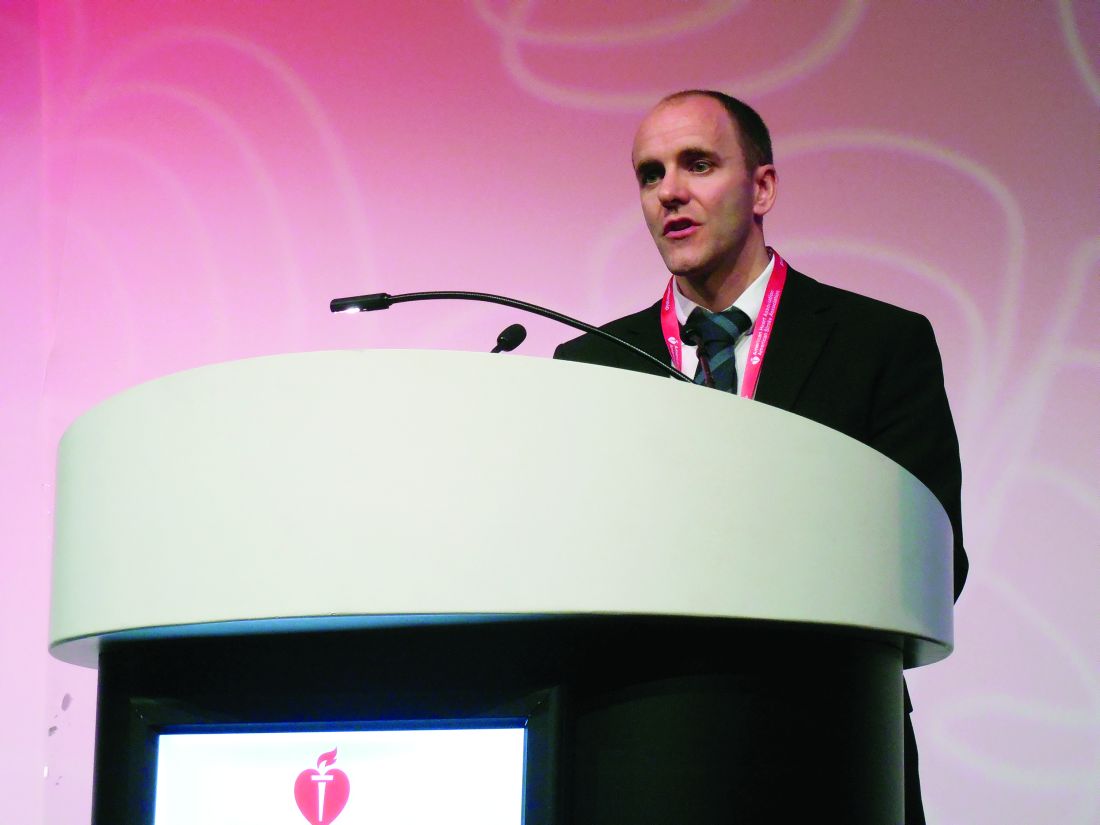
Vagus nerve stimulation shows stroke recovery promise
HOUSTON – Stroke patients with arm weakness had a clinically significant boost in arm function after about 19 weeks on a rehabilitation program that combined vagus nerve stimulation with rehabilitation training sessions in a multicenter, randomized, and sham-controlled proof-of-concept study with 17 patients.
This promising result follows a prior 21-patient study with a similar design and results (Stroke. 2016 Jan;47[1]:143-50), making the next step a pivotal trial with about 120 randomized patients that should start in 2017, Jesse Dawson, MD, said at the International Stroke Conference sponsored by the American Heart Association.
Results in the new study showed that eight poststroke patients with arm weakness who received a prolonged course of vagus nerve stimulation (VNS) and rehabilitation training had an average boost from baseline in their upper-extremity Fugl-Meyer score of 9.5 points measured 132 days after the start of the regimen, compared with an average 3.8-point rise among nine similar patients who underwent the same rehabilitation training but without VNS. A rise of 4-7 points on the upper-extremity Fugl-Meyer score is considered clinically significant for chronic stroke patients (J Physiotherapy. 2017 Jan;63[1]:53). The difference in mean scores between the VNS and control groups after 132 days was statistically significant for a secondary endpoint of the study.
The study’s primary endpoint, the difference between the control and VNS patients in mean upper-extremity Fugl-Meyer scores at the end of the initial phase of the study – a 6-week supervised training period – was 7.6 points in the VNS recipients and 5.3 points for the control patients, a difference that was not statistically significant.
The 9.5-point boost in average scores with more prolonged treatment and follow-up in the VNS patients is “highly likely to be clinically significant,” Dr. Dawson said. “We would like to see an effect earlier, with clinically important effects after 6 weeks of treatment. That would make the intervention easier to translate into clinical practice.”
The study ran at three U.S. centers and in Glasgow and enrolled patients who were 4 months to 5 years out from their index stroke and had moderate to severe arm weakness based on an upper-extremity Fugl-Meyer score of 20-50. The average age of the 17 patients in the study was 60 years. They were an average of 1.5 years removed from their index stroke.
All of the patients received an implanted device to produce VNS. The eight patients in the active arm received VNS during their 2-hour, thrice-weekly rehabilitation training sessions for the first 6 weeks of the study, with about 400 individual stimulations delivered during each training session. The nine controls received brief VNS to aid blinding, but had no meaningful VNS while they replicated the rehabilitation training regimen of the intervention group. At the end of 6 weeks, no training or VNS was done for 30 days. Then for the next 60 days, all patients did a daily program of unsupervised home rehabilitation exercises and patients in the intervention arm also self-administered 30 minutes of VNS daily.
The 17 patients who received a VNS device implant had three serious adverse events, Dr. Dawson reported: one infection, one episode of dyspnea, and one episode of vocal cord paralysis. None of the adverse events were judged definitely or likely linked to the stimulator, and all three effects were in control patients. In several patients in both arms, nonserious adverse effects occurred that are expected for the surgery used, including bruising, pain, swelling, and scarring. When the study ended, patients originally randomized to the sham group underwent active intervention with VNS and subsequently had an average 13-point increase on their upper extremity Fugl-Meyer score.
MicroTransponder, the company developing the vagus nerve stimulation device, funded the study. Dr. Dawson has received travel and meeting cost reimbursements from MicroTransponder, and several coauthors are employees of the company.
mzoler@frontlinemedcom.com
On Twitter @mitchelzoler
Arm weakness is a critical complication for patients after a stroke and is a great target for intervention. Patients with moderate to severe arm weakness such as those enrolled in this trial are seriously affected by their loss of arm function. It’s bad for patients when they cannot use an arm.

These new results are obviously exploratory, but they create a lot of hope.
Philip B. Gorelick, MD, is medical director of the Hauenstein Neuroscience Center of Saint Mary’s Health Care in Grand Rapids, Mich., and professor of translational science and molecular medicine at Michigan State University, Grand Rapids. He had no disclosures. He made these comments during a press conference.
Arm weakness is a critical complication for patients after a stroke and is a great target for intervention. Patients with moderate to severe arm weakness such as those enrolled in this trial are seriously affected by their loss of arm function. It’s bad for patients when they cannot use an arm.
These new results are obviously exploratory, but they create a lot of hope.
Philip B. Gorelick, MD, is medical director of the Hauenstein Neuroscience Center of Saint Mary’s Health Care in Grand Rapids, Mich., and professor of translational science and molecular medicine at Michigan State University, Grand Rapids. He had no disclosures. He made these comments during a press conference.
HOUSTON – Stroke patients with arm weakness had a clinically significant boost in arm function after about 19 weeks on a rehabilitation program that combined vagus nerve stimulation with rehabilitation training sessions in a multicenter, randomized, and sham-controlled proof-of-concept study with 17 patients.
This promising result follows a prior 21-patient study with a similar design and results (Stroke. 2016 Jan;47[1]:143-50), making the next step a pivotal trial with about 120 randomized patients that should start in 2017, Jesse Dawson, MD, said at the International Stroke Conference sponsored by the American Heart Association.
Results in the new study showed that eight poststroke patients with arm weakness who received a prolonged course of vagus nerve stimulation (VNS) and rehabilitation training had an average boost from baseline in their upper-extremity Fugl-Meyer score of 9.5 points measured 132 days after the start of the regimen, compared with an average 3.8-point rise among nine similar patients who underwent the same rehabilitation training but without VNS. A rise of 4-7 points on the upper-extremity Fugl-Meyer score is considered clinically significant for chronic stroke patients (J Physiotherapy. 2017 Jan;63[1]:53). The difference in mean scores between the VNS and control groups after 132 days was statistically significant for a secondary endpoint of the study.
The study’s primary endpoint, the difference between the control and VNS patients in mean upper-extremity Fugl-Meyer scores at the end of the initial phase of the study – a 6-week supervised training period – was 7.6 points in the VNS recipients and 5.3 points for the control patients, a difference that was not statistically significant.
The 9.5-point boost in average scores with more prolonged treatment and follow-up in the VNS patients is “highly likely to be clinically significant,” Dr. Dawson said. “We would like to see an effect earlier, with clinically important effects after 6 weeks of treatment. That would make the intervention easier to translate into clinical practice.”
The study ran at three U.S. centers and in Glasgow and enrolled patients who were 4 months to 5 years out from their index stroke and had moderate to severe arm weakness based on an upper-extremity Fugl-Meyer score of 20-50. The average age of the 17 patients in the study was 60 years. They were an average of 1.5 years removed from their index stroke.
All of the patients received an implanted device to produce VNS. The eight patients in the active arm received VNS during their 2-hour, thrice-weekly rehabilitation training sessions for the first 6 weeks of the study, with about 400 individual stimulations delivered during each training session. The nine controls received brief VNS to aid blinding, but had no meaningful VNS while they replicated the rehabilitation training regimen of the intervention group. At the end of 6 weeks, no training or VNS was done for 30 days. Then for the next 60 days, all patients did a daily program of unsupervised home rehabilitation exercises and patients in the intervention arm also self-administered 30 minutes of VNS daily.
The 17 patients who received a VNS device implant had three serious adverse events, Dr. Dawson reported: one infection, one episode of dyspnea, and one episode of vocal cord paralysis. None of the adverse events were judged definitely or likely linked to the stimulator, and all three effects were in control patients. In several patients in both arms, nonserious adverse effects occurred that are expected for the surgery used, including bruising, pain, swelling, and scarring. When the study ended, patients originally randomized to the sham group underwent active intervention with VNS and subsequently had an average 13-point increase on their upper extremity Fugl-Meyer score.
MicroTransponder, the company developing the vagus nerve stimulation device, funded the study. Dr. Dawson has received travel and meeting cost reimbursements from MicroTransponder, and several coauthors are employees of the company.
mzoler@frontlinemedcom.com
On Twitter @mitchelzoler
AT THE INTERNATIONAL STROKE CONFERENCE
Key clinical point:
Major finding: Vagus nerve stimulation linked with a mean 9.5-point rise in upper-extremity Fugl-Meyer score at 132-day follow-up.
Data source: A multicenter, randomized, sham-controlled study with 17 chronic stroke patients.
Disclosures: MicroTransponder, the company developing the vagus nerve stimulation device, funded the study. Dr. Dawson has received travel and meeting cost reimbursements from MicroTransponder, and several coauthors are employees of the company.
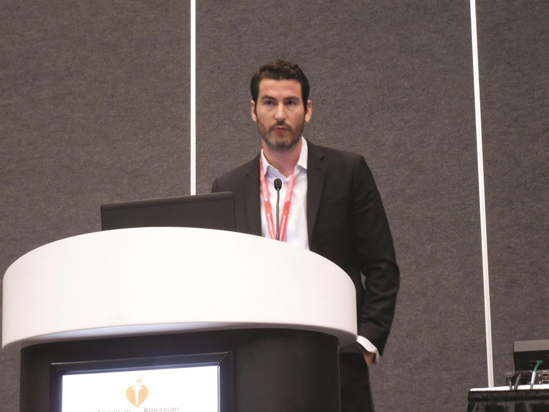
Protocol speeds thrombectomy stroke patients from primary centers
HOUSTON – A novel protocol designed to speed patients with large-vessel occlusion strokes in and out of primary stroke centers and on to centers where they can undergo definitive thrombectomy treatment produced significant improvements in treatment speed and outcomes among 22 Rhode Island patients managed with the full protocol.
Streamlining the path in and out of a primary stroke center is key for delivering mechanical thrombectomy as quickly as possible to patients with an emergent large vessel occlusion, said Ryan A. McTaggart, MD, at the International Stroke Conference sponsored by the American Heart Association. “Door-in door-out time should be the standard metric for all partnerships between primary and comprehensive stroke centers,” said Dr. McTaggart, a neuroradiologist at Rhode Island Hospital in Providence, the state’s only comprehensive stroke center.

• When a patient with a suspected large vessel occlusion with a Los Angeles Motor Score of 4 or 5 arrives soon after onset at the primary stroke center, a call immediately goes out to the EMS transfer center of Rhode Island Hospital to coordinate the transport that will move the patient from the primary center to the comprehensive when needed.
• The initial CT scan at the primary center is run as the definitive scan, including a conventional CT scan to rule out hemorrhage and allow intravenous thrombolytic therapy with tissue plasminogen activator (TPA) and CT angiography to locate the occluding clot.
• The CT images are immediately uploaded to a cloud-based library so that neurologists at Rhode Island Hospital can read the images on their phones and plan the management strategy.
During the 11 months following the start of the protocol, the Rhode Island network identified 70 patients as candidates for thrombectomy, including 22 managed using the complete protocol and 48 managed using only parts of the new protocol.
The median time from onset of stroke symptoms to revascularization with thrombectomy was 184 minutes in the 22 patients managed under the full protocol and 233 minutes among 48 similar patients who were not fully managed with the protocol, Dr. McTaggart reported. This “dramatic” difference in median times was entirely driven by a difference in the door-in door-out time at the primary stroke center, which was a median of 64 minutes for the 22 patients managed with the full protocol and a median of 104 minutes without the full protocol, a 38% relative decrease that was statistically significant.
Time to initiation of intravenous TPA at the primary stroke center also improved, from a median of 65 minutes without the full protocol to a median of 40 minutes with it, a statistically significant difference. “The primary stroke center physicians tell us they have greater confidence to start TPA when they have a consult that can identify the patient’s clot,” he said.
Consistent with the shorter time to revascularization, the prevalence after 90 days of a functionally good outcome – a modified Rankin scale score of 0-2 – occurred in 50% of patients managed with the full protocol and 25% of those managed with a partial protocol, a statistically significant difference.
To put the 184 minutes median time from stroke onset to reperfusion into perspective, Dr. McTaggart noted that it is comparable to the time to reperfusion documented recently in a U.S. registry of thrombectomy patients who had been transported directly to the comprehensive stroke centers where their thrombectomy was done.
He also acknowledged the heavy lifting he and his associates had to do to set up this network. Getting buy-in from all the regional primary strokes centers was “a ton of work,” Dr. McTaggart said in an interview. “We told the primary stroke center staffs that thrombectomy is a powerful treatment, with a number needed to treat of three to get one improved outcome. That’s a convincing argument. The thrombectomy data [that became available in early 2015] made the argument for the protocol and network more compelling.”
Primary stroke centers keep the stroke patients who don’t have a clot occlusion suitable for thrombectomy, which means the comprehensive center thrombectomy team receives fewer false-alarm patients. Dr. McTaggart’s current goal is to have primary stroke centers get incoming patients out and on the road to a thrombectomy center within 45 minutes. In the future, primary stroke centers will perform CT imaging on all patients with suspected strokes, not just the severely affected patients with a Los Angeles Motor Score of 4 or 5. Additional useful steps toward speeding appropriate stroke patients to thrombectomy is direct ambulance transport of selected, high probability patients directly to a comprehensive stroke center and use of mobile stroke units to bring CT imaging and the start of TPA treatment out into the field.
Dr. McTaggart had no disclosures.
mzoler@frontlinemedcom.com
On Twitter @mitchelzoler
HOUSTON – A novel protocol designed to speed patients with large-vessel occlusion strokes in and out of primary stroke centers and on to centers where they can undergo definitive thrombectomy treatment produced significant improvements in treatment speed and outcomes among 22 Rhode Island patients managed with the full protocol.
Streamlining the path in and out of a primary stroke center is key for delivering mechanical thrombectomy as quickly as possible to patients with an emergent large vessel occlusion, said Ryan A. McTaggart, MD, at the International Stroke Conference sponsored by the American Heart Association. “Door-in door-out time should be the standard metric for all partnerships between primary and comprehensive stroke centers,” said Dr. McTaggart, a neuroradiologist at Rhode Island Hospital in Providence, the state’s only comprehensive stroke center.
• When a patient with a suspected large vessel occlusion with a Los Angeles Motor Score of 4 or 5 arrives soon after onset at the primary stroke center, a call immediately goes out to the EMS transfer center of Rhode Island Hospital to coordinate the transport that will move the patient from the primary center to the comprehensive when needed.
• The initial CT scan at the primary center is run as the definitive scan, including a conventional CT scan to rule out hemorrhage and allow intravenous thrombolytic therapy with tissue plasminogen activator (TPA) and CT angiography to locate the occluding clot.
• The CT images are immediately uploaded to a cloud-based library so that neurologists at Rhode Island Hospital can read the images on their phones and plan the management strategy.
During the 11 months following the start of the protocol, the Rhode Island network identified 70 patients as candidates for thrombectomy, including 22 managed using the complete protocol and 48 managed using only parts of the new protocol.
The median time from onset of stroke symptoms to revascularization with thrombectomy was 184 minutes in the 22 patients managed under the full protocol and 233 minutes among 48 similar patients who were not fully managed with the protocol, Dr. McTaggart reported. This “dramatic” difference in median times was entirely driven by a difference in the door-in door-out time at the primary stroke center, which was a median of 64 minutes for the 22 patients managed with the full protocol and a median of 104 minutes without the full protocol, a 38% relative decrease that was statistically significant.
Time to initiation of intravenous TPA at the primary stroke center also improved, from a median of 65 minutes without the full protocol to a median of 40 minutes with it, a statistically significant difference. “The primary stroke center physicians tell us they have greater confidence to start TPA when they have a consult that can identify the patient’s clot,” he said.
Consistent with the shorter time to revascularization, the prevalence after 90 days of a functionally good outcome – a modified Rankin scale score of 0-2 – occurred in 50% of patients managed with the full protocol and 25% of those managed with a partial protocol, a statistically significant difference.
To put the 184 minutes median time from stroke onset to reperfusion into perspective, Dr. McTaggart noted that it is comparable to the time to reperfusion documented recently in a U.S. registry of thrombectomy patients who had been transported directly to the comprehensive stroke centers where their thrombectomy was done.
He also acknowledged the heavy lifting he and his associates had to do to set up this network. Getting buy-in from all the regional primary strokes centers was “a ton of work,” Dr. McTaggart said in an interview. “We told the primary stroke center staffs that thrombectomy is a powerful treatment, with a number needed to treat of three to get one improved outcome. That’s a convincing argument. The thrombectomy data [that became available in early 2015] made the argument for the protocol and network more compelling.”
Primary stroke centers keep the stroke patients who don’t have a clot occlusion suitable for thrombectomy, which means the comprehensive center thrombectomy team receives fewer false-alarm patients. Dr. McTaggart’s current goal is to have primary stroke centers get incoming patients out and on the road to a thrombectomy center within 45 minutes. In the future, primary stroke centers will perform CT imaging on all patients with suspected strokes, not just the severely affected patients with a Los Angeles Motor Score of 4 or 5. Additional useful steps toward speeding appropriate stroke patients to thrombectomy is direct ambulance transport of selected, high probability patients directly to a comprehensive stroke center and use of mobile stroke units to bring CT imaging and the start of TPA treatment out into the field.
Dr. McTaggart had no disclosures.
mzoler@frontlinemedcom.com
On Twitter @mitchelzoler
AT THE INTERNATIONAL STROKE CONFERENCE
Key clinical point:
Major finding: Median time from stroke onset to thrombectomy reperfusion was 184 minutes, including transfer between a primary and comprehensive stroke center.
Data source: Review of 70 acute ischemic stroke patients treated at Rhode Island Hospital.
Disclosures: Dr. McTaggart had no disclosures.
Patient transfer before thrombectomy worsens stroke outcomes
HOUSTON – Drip and ship may not be the most time-effective way to treat acute ischemic stroke patients who are candidates for endovascular thrombectomy.
Results from two separate real-world, observational studies showed that acute ischemic stroke patients with large vessel occlusions amenable to mechanical thrombectomy had significantly worse clinical outcomes when their management path included a stop at a primary stroke center followed by transfer to a comprehensive stroke center that had the capacity to perform thrombectomy, compared with going straight to the thrombectomy site.

The findings show “the system of care has room for improvement. Patients with large vessel occlusions clearly do better when we get them to mechanical thrombectomy as quickly as possible,” said Dr. Froehler, a vascular neurologist at Vanderbilt University in Nashville, Tenn. Thrombectomy “has a more powerful treatment effect than TPA [tissue plasminogen activator] and we need to adjust our standard of care to best deliver” thrombectomy, he said in an interview.

The study run by Dr. Froehler used data collected in the Systematic Evaluation of Patients Treated With Stroke Devices for Acute Ischemic Stroke (STRATIS) registry, which began in 2014 and has data for 984 acute ischemic stroke patients with large vessel occlusions treated by mechanical thrombectomy seen at any of 55 U.S. centers. The series included 445 (45%) patients first seen as a primary stroke center and then transferred to a comprehensive center and 539 (55%) who went directly to a comprehensive stroke center (direct patients). Prior to thrombectomy, 628 of all patients (64%) received TPA, with a roughly similar percentage in both the transferred and direct patients.
The data showed that the median time from symptom onset to revascularization was 202 minutes among the direct patients and 312 minutes among those first seen at a primary stroke center and then transferred, a statistically significant difference. The average time difference per patient between the two subgroups was 100 minutes, Dr. Froehler reported.
This difference in time to reperfusion led directly to significant differences in functional outcomes after 90 days measured on the modified Rankin Scale (mRS). The percentage of patients with a mRS score of 0 or 1 (an excellent functional outcome) was 38% for the patients first seen at primary stroke centers and 47% in direct patients, a 47% relative rise in excellent outcomes among the direct patients. The percentage of patients with a mRS score of 0-2, which identifies functional independence post stroke, was 52% among transferred patients and 60% in direct patients, a 38% relative improvement for this outcome among direct patients.
The second study of stroke transfer times and outcomes used data from 562 acute ischemic stroke patients with large vessel occlusions treated in the Providence Health & Services system in five western U.S. states during 2012-2016. Nearly half the patients required a transfer and the other half went directly to a center able to perform thrombectomy. The analysis used clinical outcomes scored on the mRS at the time of hospital discharge.
“Right now, the big delay at primary stroke centers is the door-in door-out time,” commented Ryan A. McTaggart, MD, an interventional neuroradiologist at Rhode Island Hospital in Providence, the only comprehensive stroke center in Rhode Island. He helped organize a partnership with 14 primary stroke centers in Rhode Island that uses a streamlined imaging, treatment (with TPA), and transfer protocol that hacked dozens of minutes off transfer times and produced a median time from onset of symptoms to revascularization by thrombectomy of 184 minutes in patients first seen at a primary stroke center. This clocking blows past the 202 minute median for stroke onset to revascularization in the direct patients from Dr. Froehler’s study.
Stroke transport and treatment networks are now undergoing refinement in Tennessee, said Dr. Froehler, based in part on the data he reported. Considerations in Tennessee include how EMS workers assess possible stroke patients, decisions by EMS on where to take patients, and how quality of care is measured at primary and comprehensive stroke centers.
The STRATIS registry is sponsored by Medtronic. Dr. Froehler is a consultant to Medtronic, Blockade, Stryker, and Control Medical. Dr. Smith, Dr. Tarpley, and Dr. McTaggart had no disclosures.
mzoler@frontlinemedcom.com
On Twitter @mitchelzoler
HOUSTON – Drip and ship may not be the most time-effective way to treat acute ischemic stroke patients who are candidates for endovascular thrombectomy.
Results from two separate real-world, observational studies showed that acute ischemic stroke patients with large vessel occlusions amenable to mechanical thrombectomy had significantly worse clinical outcomes when their management path included a stop at a primary stroke center followed by transfer to a comprehensive stroke center that had the capacity to perform thrombectomy, compared with going straight to the thrombectomy site.
The findings show “the system of care has room for improvement. Patients with large vessel occlusions clearly do better when we get them to mechanical thrombectomy as quickly as possible,” said Dr. Froehler, a vascular neurologist at Vanderbilt University in Nashville, Tenn. Thrombectomy “has a more powerful treatment effect than TPA [tissue plasminogen activator] and we need to adjust our standard of care to best deliver” thrombectomy, he said in an interview.
The study run by Dr. Froehler used data collected in the Systematic Evaluation of Patients Treated With Stroke Devices for Acute Ischemic Stroke (STRATIS) registry, which began in 2014 and has data for 984 acute ischemic stroke patients with large vessel occlusions treated by mechanical thrombectomy seen at any of 55 U.S. centers. The series included 445 (45%) patients first seen as a primary stroke center and then transferred to a comprehensive center and 539 (55%) who went directly to a comprehensive stroke center (direct patients). Prior to thrombectomy, 628 of all patients (64%) received TPA, with a roughly similar percentage in both the transferred and direct patients.
The data showed that the median time from symptom onset to revascularization was 202 minutes among the direct patients and 312 minutes among those first seen at a primary stroke center and then transferred, a statistically significant difference. The average time difference per patient between the two subgroups was 100 minutes, Dr. Froehler reported.
This difference in time to reperfusion led directly to significant differences in functional outcomes after 90 days measured on the modified Rankin Scale (mRS). The percentage of patients with a mRS score of 0 or 1 (an excellent functional outcome) was 38% for the patients first seen at primary stroke centers and 47% in direct patients, a 47% relative rise in excellent outcomes among the direct patients. The percentage of patients with a mRS score of 0-2, which identifies functional independence post stroke, was 52% among transferred patients and 60% in direct patients, a 38% relative improvement for this outcome among direct patients.
The second study of stroke transfer times and outcomes used data from 562 acute ischemic stroke patients with large vessel occlusions treated in the Providence Health & Services system in five western U.S. states during 2012-2016. Nearly half the patients required a transfer and the other half went directly to a center able to perform thrombectomy. The analysis used clinical outcomes scored on the mRS at the time of hospital discharge.
“Right now, the big delay at primary stroke centers is the door-in door-out time,” commented Ryan A. McTaggart, MD, an interventional neuroradiologist at Rhode Island Hospital in Providence, the only comprehensive stroke center in Rhode Island. He helped organize a partnership with 14 primary stroke centers in Rhode Island that uses a streamlined imaging, treatment (with TPA), and transfer protocol that hacked dozens of minutes off transfer times and produced a median time from onset of symptoms to revascularization by thrombectomy of 184 minutes in patients first seen at a primary stroke center. This clocking blows past the 202 minute median for stroke onset to revascularization in the direct patients from Dr. Froehler’s study.
Stroke transport and treatment networks are now undergoing refinement in Tennessee, said Dr. Froehler, based in part on the data he reported. Considerations in Tennessee include how EMS workers assess possible stroke patients, decisions by EMS on where to take patients, and how quality of care is measured at primary and comprehensive stroke centers.
The STRATIS registry is sponsored by Medtronic. Dr. Froehler is a consultant to Medtronic, Blockade, Stryker, and Control Medical. Dr. Smith, Dr. Tarpley, and Dr. McTaggart had no disclosures.
mzoler@frontlinemedcom.com
On Twitter @mitchelzoler
AT THE INTERNATIONAL STROKE CONFERENCE
Key clinical point:
Major finding: In STRATIS, excellent outcomes occurred in 47% of patients sent directly to a thrombectomy hospital and in 38% of transferred patients.
Data source: The STRATIS registry, with 984 U.S. acute ischemic stroke patients, and 562 U.S. acute ischemic stroke patients from the Providence Health & Services network.
Disclosures: The STRATIS registry is sponsored by Medtronic. Dr. Froehler is a consultant to Medtronic, Blockade, Stryker, and Control Medical. Dr. Smith, Dr. Tarpley, and Dr. McTaggart had no disclosures.
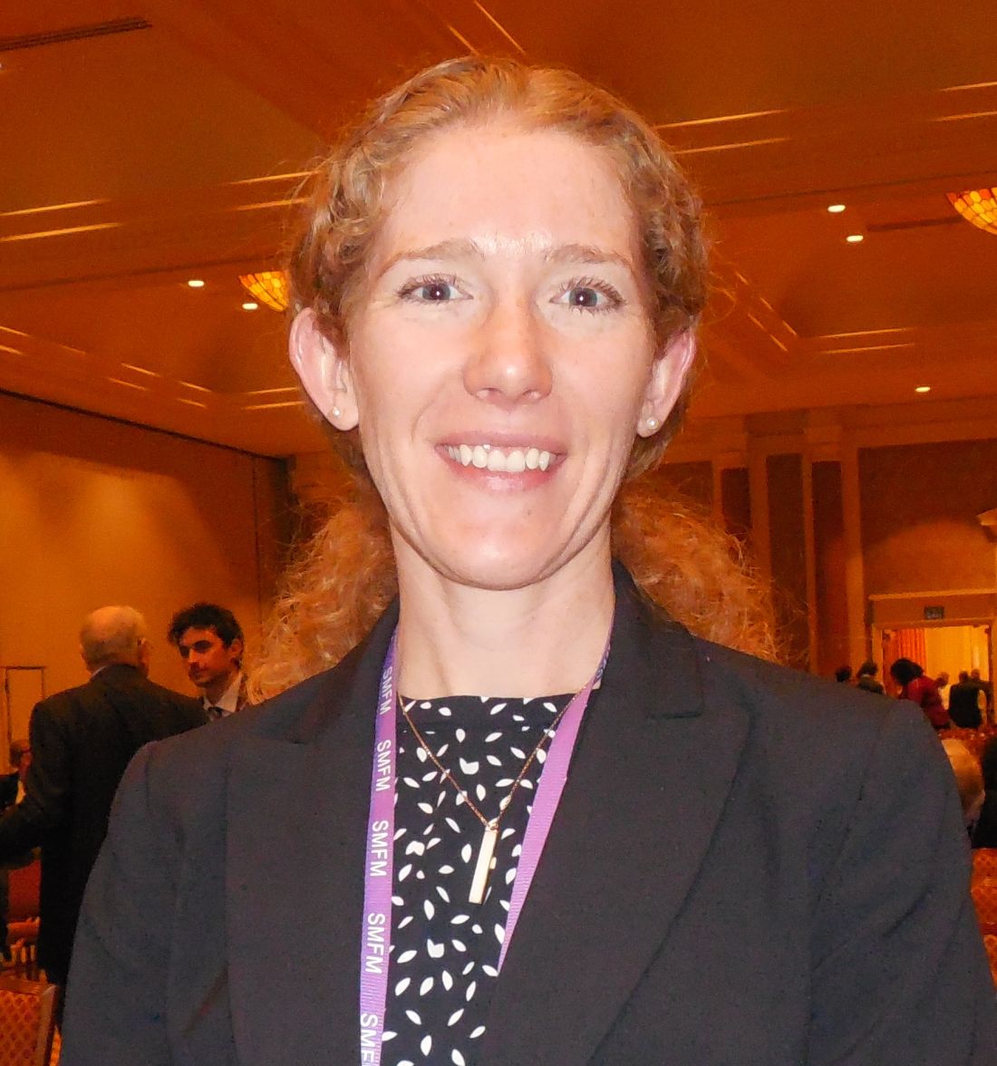
Monofilament suture works best for cesarean closure
LAS VEGAS – A monofilament suture led to substantially fewer wound complications than a braided suture for closing nonemergency cesarean incisions in a head-to-head trial with 520 evaluable women.
Cesarean incision closure with a braided, polyglactin 910 suture (Vicryl) led to 65% more wound complications than the monofilament poliglecaprone 25 suture (Monocryl), Arin M. Buresch, MD, said at the annual Pregnancy Meeting sponsored by the Society for Maternal-Fetal Medicine.
This is the first randomized, controlled trial to compare these two suture types, according to Dr. Buresch, and she highlighted the need for caution about changing practice based on results from a single study. But based in large part on these results, which were gathered at Montefiore Medical Center in New York, the obstetrical staff at Montefiore is now primarily using the monofilament, poliglecaprone 25 suture, she said.
The study enrolled 550 pregnant women at 37 weeks’ gestation or greater during May 2015 to July 2016. Participants were either scheduled for an elective cesarean delivery or underwent a nonemergency, indicated cesarean after labor began but without significant maternal or fetal distress. The study excluded emergency cesareans as well as women with a recent urogenital infection, chronic or injected steroid use, or a vertical skin incision. The enrolled women averaged 31 years old, and their average body mass index was 34 kg/m2. The demographic and clinical profile of the two randomized groups closely matched.
The study’s primary endpoint was the incidence of a wound complication during 30 days following delivery. A complication could be a surgical site infection, hematoma, seroma, or wound separation. Of the 550 women randomized, 520 were available for complete 30-day follow-up.
The results showed that wound complications occurred in 9% of the 263 women treated with the poliglecaprone 25 monofilament suture and in 14% of the 257 treated with the polyglactin 910 braided suture, a statistically significant difference, Dr. Buresch reported. The relative risk for a complication increased by 65% with the braided suture, compared with patients treated with monofilament sutures. Treating 18 patients with the monofilament suture prevented one wound complication, on average.
A subgroup analysis showed that the poliglecaprone 25 suture was effective at reducing wound complications in women who underwent elective cesarean deliveries, but among the 17% of participants who had begun labor at the time of their cesarean delivery the monofilament suture conferred no significant advantage, compared with the braided suture. Benefit from the poliglecaprone 25 monofilament occurred about equally across the entire range of body mass index among the women in the study, Dr. Buresch said.
Dr. Buresch had no disclosures.
mzoler@frontlinemedcom.com
On Twitter @mitchelzoler
Early delivery by morbidly obese moms improves outcomes
LAS VEGAS – Delivery at 38 weeks’ gestation is linked with improved perinatal survival among singleton infants born to morbidly obese mothers in a retrospective review of more than 2 million U.S. births.
“If reasonable, consider delivery at 38 weeks in morbidly obese mothers” delivering singleton pregnancies, Ruofan Yao, MD, said at the annual Pregnancy Meeting sponsored by the Society for Maternal-Fetal Medicine.

When mothers have diabetes, hypertension, or cholestasis, they receive frequent prenatal testing and fetal growth measurements, and delivery is typically at 37, 38, or 39 weeks. “This is what we also need to think about for morbidly obese mothers,” Dr. Yao said.
“Because of increased fetal growth in morbidly obese mothers there is probably earlier placental insufficiency,” he said in an interview.
The upshot is that, once a morbidly obese mother reaches 38 weeks’ gestation, induced labor should be considered, according to Dr. Yao. Induction could start immediately if the mother’s cervix is ripe, or clinicians could first take steps to hasten cervical ripening.
Induction can be especially slow in morbidly obese women, who are generally less sensitive to oxytocin and can require multiple induction strategies.
While Dr. Yao considered the evidence he reported persuasive enough to recommend this strategy, he cautioned that, ideally, the benefits of an early-delivery approach should be confirmed in a prospective, randomized trial.
The study used delivery records maintained by the state of Texas for 2006-2011. Of the more than 2.4 million births recorded during the period, Dr. Yao excluded multiple deliveries, births at less than 34 weeks’ or more than 42 weeks’ gestation, deliveries from underweight mothers (less than 18.5 kg/m2), and fetal anomalies. This left 2,181,530 births, of which 52% were by normal weight mothers (18.5-24 kg/m2), 26% by overweight mothers (25-29 kg/m2), 18% by obese mothers (30-39 kg/m2), and 4% by morbidly obese mothers (40 kg/m2 or greater). The women averaged 27 years old, 4% had preeclampsia, and 4% had pregestational diabetes.
The researchers then calculated perinatal mortality rates relative to gestational age at birth for women in each body mass index stratum. The calculations showed no significant impact of gestational age among late-term deliveries by normal weight, overweight, and obese mothers, but, among morbidly obese mothers, early deliveries made a difference and were significantly linked with reduced perinatal mortality.
Every 400 deliveries, approximately, induced at 38 weeks among morbidly obese mothers resulted in one less perinatal death, Dr. Yao reported. This relationship held even when the researchers excluded mothers with preeclampsia or pregestational diabetes (about 8% of the study group).
Dr. Yao had no disclosures.
mzoler@frontlinemedcom.com
On Twitter @mitchelzoler
LAS VEGAS – Delivery at 38 weeks’ gestation is linked with improved perinatal survival among singleton infants born to morbidly obese mothers in a retrospective review of more than 2 million U.S. births.
“If reasonable, consider delivery at 38 weeks in morbidly obese mothers” delivering singleton pregnancies, Ruofan Yao, MD, said at the annual Pregnancy Meeting sponsored by the Society for Maternal-Fetal Medicine.
When mothers have diabetes, hypertension, or cholestasis, they receive frequent prenatal testing and fetal growth measurements, and delivery is typically at 37, 38, or 39 weeks. “This is what we also need to think about for morbidly obese mothers,” Dr. Yao said.
“Because of increased fetal growth in morbidly obese mothers there is probably earlier placental insufficiency,” he said in an interview.
The upshot is that, once a morbidly obese mother reaches 38 weeks’ gestation, induced labor should be considered, according to Dr. Yao. Induction could start immediately if the mother’s cervix is ripe, or clinicians could first take steps to hasten cervical ripening.
Induction can be especially slow in morbidly obese women, who are generally less sensitive to oxytocin and can require multiple induction strategies.
While Dr. Yao considered the evidence he reported persuasive enough to recommend this strategy, he cautioned that, ideally, the benefits of an early-delivery approach should be confirmed in a prospective, randomized trial.
The study used delivery records maintained by the state of Texas for 2006-2011. Of the more than 2.4 million births recorded during the period, Dr. Yao excluded multiple deliveries, births at less than 34 weeks’ or more than 42 weeks’ gestation, deliveries from underweight mothers (less than 18.5 kg/m2), and fetal anomalies. This left 2,181,530 births, of which 52% were by normal weight mothers (18.5-24 kg/m2), 26% by overweight mothers (25-29 kg/m2), 18% by obese mothers (30-39 kg/m2), and 4% by morbidly obese mothers (40 kg/m2 or greater). The women averaged 27 years old, 4% had preeclampsia, and 4% had pregestational diabetes.
The researchers then calculated perinatal mortality rates relative to gestational age at birth for women in each body mass index stratum. The calculations showed no significant impact of gestational age among late-term deliveries by normal weight, overweight, and obese mothers, but, among morbidly obese mothers, early deliveries made a difference and were significantly linked with reduced perinatal mortality.
Every 400 deliveries, approximately, induced at 38 weeks among morbidly obese mothers resulted in one less perinatal death, Dr. Yao reported. This relationship held even when the researchers excluded mothers with preeclampsia or pregestational diabetes (about 8% of the study group).
Dr. Yao had no disclosures.
mzoler@frontlinemedcom.com
On Twitter @mitchelzoler
AT THE PREGNANCY MEETING
Key clinical point:
Major finding: Every 400 deliveries at 38 weeks is linked with one fewer perinatal death, compared with full-term deliveries.
Data source: Review of 2,181,530 Texas births during 2006-2011.
Disclosures: Dr. Yao had no disclosures.
Anticoagulant resumption after ICH aids patients
HOUSTON – Even when patients on an oral anticoagulant have the dreaded complication of an intracerebral hemorrhage, resumption of their oral anticoagulation regimen appears to produce the best midterm outcomes, based on a meta-analysis of data from more than 1,000 patients collected in three observational studies.
Resumption of oral anticoagulation therapy (OAT) is a “major dilemma” when managing patients who developed an intracerebral hemorrhage (ICH) while on OAT, said Alessandro Biffi, MD, explaining why he performed this meta-analysis that he presented at the International Stroke Conference sponsored by the American Heart Association.
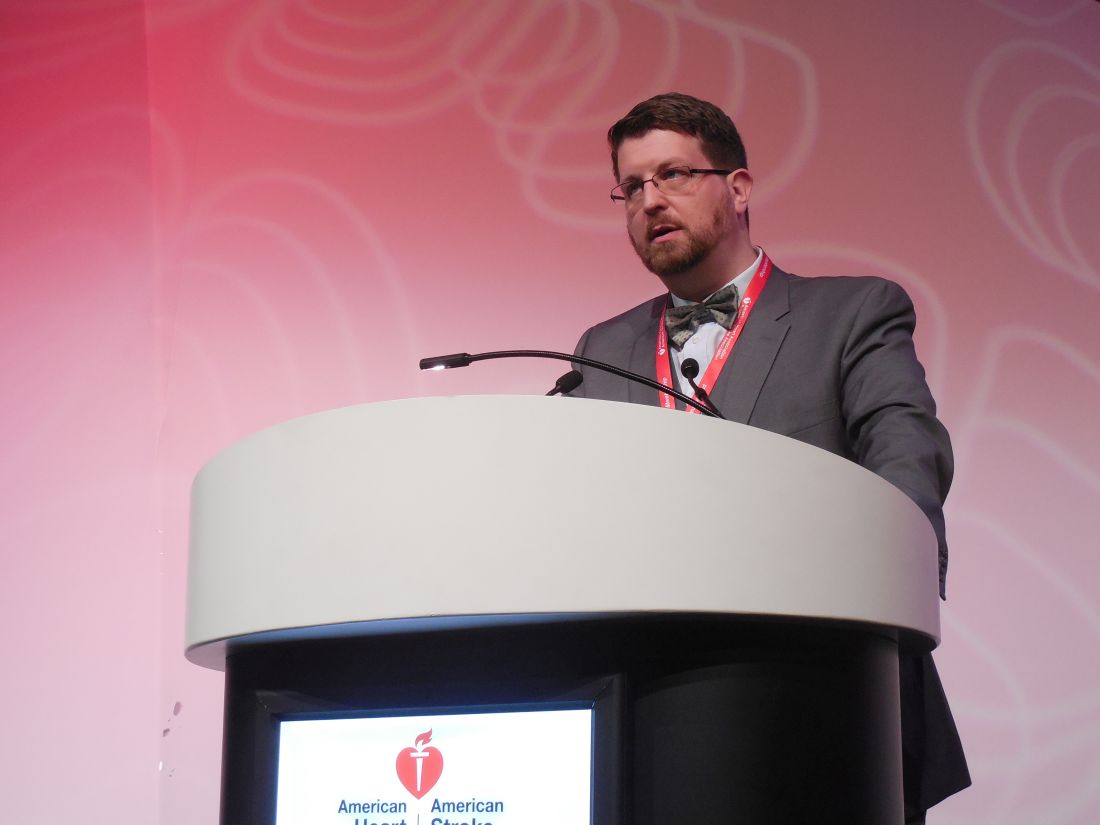
He used individual patient data collected from a total of 1,027 patients enrolled in any of three different observational studies: the German-wide Multicenter Analysis of Oral Anticoagulation Associated Intracerebral Hemorrhage (RETRACE) study, the MGH longitudinal ICH study, or the Ethnic/Racial Variations of Intracerebral Hemorrhage (ERICH) study. Overall 26% of the patients resumed OAT following their ICH, although the rate ranged from a low of 20% in one study to a high of 42% in another. The vast majority of patients received a vitamin K antagonist as their anticoagulant; very few received a new oral anticoagulant.
Using propensity score matching to compare similar patients who resumed or stayed off OAT, Dr. Biffi found that, during the year following the index ICH, mortality was 71%-74% lower among patients who resumed OAT. Recurrent all-cause stroke was 49%-55% lower with resumed OAT, and favorable functional outcomes (a score of 0-3 on the modified Rankin scale) were more than fourfold higher with OAT resumption, he reported.
Dr. Biffi calculated these relative rates, both for patients with a lumbar location of their ICH and for those with a nonlumbar location, and found that location had no influence on responsiveness to OAT. Patients with an index ICH in a lumbar location had a trend toward more recurrent ICH on OAT, a 26% higher rate relative to patients not resumed on OAT, but this difference fell short of statistical significance.
The only factor he found that linked with whether or not patients resumed OAT was the severity of their index ICH. The more severe their bleed, the less likely were patients to resume. Aside from that, “there is a lot of variation in practice,” he said. “We are gathering additional data” to try to further address this question.
Dr. Biffi had no disclosures.
mzoler@frontlinemedcom.com
On Twitter @mitchelzoler
Resuming oral anticoagulation following an intracerebral hemorrhage is one of the most vexing problems today in vascular neurology. It’s a situation that often happens, and it will grow increasingly more common as the number of patients with atrial fibrillation escalates and even more people start oral anticoagulation.

It’s also very important to remember that patients like these who need oral anticoagulation but now have a history of ICH must have all their other cardiovascular disease risk factors very well controlled: their blood pressure, their diabetes, their smoking, etc. Oral anticoagulation may be important for these patients, but tight risk factor control is even more important.
I agree with Dr. Biffi that a prospective, randomized trial is the best way to get more information to help guide resuming oral anticoagulation. Observational studies are significantly limited by ascertainment bias, and for these patients there are also many variables – at least a dozen – that can influence whether or not a patient resumes oral anticoagulation. Dr. Biffi’s findings are interesting, but the limitations of his data prevent the results from being truly compelling.
It would be very helpful to have data from a trial that randomized ICH patients who required anticoagulation to a full-dose NOAC, a reduced-dose NOAC, or aspirin and see which group had the best long-term outcome. Whatever the results, it would change practice. It’s intriguing to speculate that a reduced-dose NOAC might provide adequate ischemic protection with a reduced risk for more bleeding.
Mark J. Alberts, MD , is chief of neurology at Hartford (Conn.) Hospital. He had no disclosures. He made these comments in an interview and during a press conference.
Resuming oral anticoagulation following an intracerebral hemorrhage is one of the most vexing problems today in vascular neurology. It’s a situation that often happens, and it will grow increasingly more common as the number of patients with atrial fibrillation escalates and even more people start oral anticoagulation.
It’s also very important to remember that patients like these who need oral anticoagulation but now have a history of ICH must have all their other cardiovascular disease risk factors very well controlled: their blood pressure, their diabetes, their smoking, etc. Oral anticoagulation may be important for these patients, but tight risk factor control is even more important.
I agree with Dr. Biffi that a prospective, randomized trial is the best way to get more information to help guide resuming oral anticoagulation. Observational studies are significantly limited by ascertainment bias, and for these patients there are also many variables – at least a dozen – that can influence whether or not a patient resumes oral anticoagulation. Dr. Biffi’s findings are interesting, but the limitations of his data prevent the results from being truly compelling.
It would be very helpful to have data from a trial that randomized ICH patients who required anticoagulation to a full-dose NOAC, a reduced-dose NOAC, or aspirin and see which group had the best long-term outcome. Whatever the results, it would change practice. It’s intriguing to speculate that a reduced-dose NOAC might provide adequate ischemic protection with a reduced risk for more bleeding.
Mark J. Alberts, MD , is chief of neurology at Hartford (Conn.) Hospital. He had no disclosures. He made these comments in an interview and during a press conference.
HOUSTON – Even when patients on an oral anticoagulant have the dreaded complication of an intracerebral hemorrhage, resumption of their oral anticoagulation regimen appears to produce the best midterm outcomes, based on a meta-analysis of data from more than 1,000 patients collected in three observational studies.
Resumption of oral anticoagulation therapy (OAT) is a “major dilemma” when managing patients who developed an intracerebral hemorrhage (ICH) while on OAT, said Alessandro Biffi, MD, explaining why he performed this meta-analysis that he presented at the International Stroke Conference sponsored by the American Heart Association.
He used individual patient data collected from a total of 1,027 patients enrolled in any of three different observational studies: the German-wide Multicenter Analysis of Oral Anticoagulation Associated Intracerebral Hemorrhage (RETRACE) study, the MGH longitudinal ICH study, or the Ethnic/Racial Variations of Intracerebral Hemorrhage (ERICH) study. Overall 26% of the patients resumed OAT following their ICH, although the rate ranged from a low of 20% in one study to a high of 42% in another. The vast majority of patients received a vitamin K antagonist as their anticoagulant; very few received a new oral anticoagulant.
Using propensity score matching to compare similar patients who resumed or stayed off OAT, Dr. Biffi found that, during the year following the index ICH, mortality was 71%-74% lower among patients who resumed OAT. Recurrent all-cause stroke was 49%-55% lower with resumed OAT, and favorable functional outcomes (a score of 0-3 on the modified Rankin scale) were more than fourfold higher with OAT resumption, he reported.
Dr. Biffi calculated these relative rates, both for patients with a lumbar location of their ICH and for those with a nonlumbar location, and found that location had no influence on responsiveness to OAT. Patients with an index ICH in a lumbar location had a trend toward more recurrent ICH on OAT, a 26% higher rate relative to patients not resumed on OAT, but this difference fell short of statistical significance.
The only factor he found that linked with whether or not patients resumed OAT was the severity of their index ICH. The more severe their bleed, the less likely were patients to resume. Aside from that, “there is a lot of variation in practice,” he said. “We are gathering additional data” to try to further address this question.
Dr. Biffi had no disclosures.
mzoler@frontlinemedcom.com
On Twitter @mitchelzoler
AT THE INTERNATIONAL STROKE CONFERENCE
Key clinical point:
Major finding: One-year mortality was 71%-74% lower among patients who resumed oral anticoagulation relative to those who did not.
Data source: Meta-analysis of data from 1,027 patients collected in three observational studies.
Disclosures: Dr. Biffi had no disclosures.
VIDEO: Clot aspiration equals retrieval for ischemic stroke
HOUSTON – Intracerebral clot aspiration was as safe and effective as stent retriever thrombectomy for restoring cerebral blood flow in a French multicenter, randomized trial with 381 acute ischemic stroke patients.
This study is the “first direct comparison of aspiration versus stent retrieval” as the initial strategy for clot removal in acute ischemic stroke, and it “opens the door to add a new tool” for clot removal, Bertrand Lapergue, MD, said at the International Stroke Conference sponsored by the American Heart Association.
The new results “are the first to show that aspiration first is as good as a stent retriever, but we need to also see the results from COMPASS,” a U.S. multicenter trial that is in the process of making the same comparison, commented Ricardo A. Hanel, MD, a vascular neurosurgeon at Baptist Health in Jacksonville, Fla. The COMPASS Trial: a Direct Aspiration First Pass Technique has now enrolled about two-thirds of its target patient number, and until the study is complete the role of direct aspiration for clot removal in stroke remains investigational for U.S. practice, said Dr. Hanel, a COMPASS investigator.
The aspiration catheter tested in ASTER is marketed by Penumbra and has already received Food and Drug Administration approval for revascularization of ischemic stroke patients. U.S. use of aspiration for treating acute ischemic stroke, however, has remained limited because there is no clear evidence of the method’s efficacy. Dr. Hanel said that he occasionally uses aspiration as an adjunct to clot removal with a stent retriever.

ASTER’s primary endpoint was the percentage of patients who achieved thrombolysis in cerebral infarction (TICI) 2b or 3 flow at the end of treatment, which occurred in 85% of patients treated with aspiration first and in 83% of those treated by clot removal first, a difference that was not statistically significant, Dr. Lapergue reported. The rate of patients who achieved either TICI 2b or 3 flow after the initial strategy only was 63% with aspiration and 68% with clot removal, also a nonsignificant difference. The two strategies also showed no significant difference for any measured safety parameter. The results showed a trend toward more vasospasm with clot removal – a 6% rate, versus 3% with clot aspiration – but this did not reach statistical significance.
Results from additional analyses of the clinical outcomes of patients in the trial and of cost efficacy will be reported later in 2017, Dr. Lapergue said.
ASTER received an unrestricted research grant from Penumbra, a company that markets clot removal aspiration catheters. Dr. Lapergue had no personal disclosures. Dr. Hanel has been a consultant to and received grant support from Medtronic. He has received research grants from MicroVention and has an ownership interest in InNeuroCo.
mzoler@frontlinemedcom.com
On Twitter @mitchelzoler
ASTER is an important trial. It shows for the first time that an aspiration device is probably as safe and reasonable for opening an acute occlusion in a large cerebral artery as is a stent retriever.
ASTER, however, was done entirely in a French population, making it uncertain whether the results are applicable to other populations. For example, U.S. acute ischemic stroke patients, especially African Americans and Hispanics, generally have more intracerebral atherostenotic disease than do patients from European countries, while French patients tend to have more embolic disease. Will aspiration be as effective in U.S. patients with atherostenotic blockages? I would love to see this study repeated in a U.S. population of ischemic stroke patients, and that is now happening in the COMPASS trial. It would be helpful to know if there are selected U.S. patients who might be better treated using either aspiration or a stent retriever first.
Although aspiration catheters have already received Food and Drug Administration approval for clot removal in acute ischemic stroke patients, many U.S. interventionalists have moved to deploying stent retrievers based on the very positive results reported with these devices about 2 years ago. For the moment, stent retrievers remain the most prominent devices to open large vessel occlusions.
Ralph L. Sacco, MD, is professor and chairman of neurology at the University of Miami. He had no relevant disclosures. He made these comments in a video interview and during a press conference.
The video associated with this article is no longer available on this site. Please view all of our videos on the MDedge YouTube channel
HOUSTON – Intracerebral clot aspiration was as safe and effective as stent retriever thrombectomy for restoring cerebral blood flow in a French multicenter, randomized trial with 381 acute ischemic stroke patients.
This study is the “first direct comparison of aspiration versus stent retrieval” as the initial strategy for clot removal in acute ischemic stroke, and it “opens the door to add a new tool” for clot removal, Bertrand Lapergue, MD, said at the International Stroke Conference sponsored by the American Heart Association.
The new results “are the first to show that aspiration first is as good as a stent retriever, but we need to also see the results from COMPASS,” a U.S. multicenter trial that is in the process of making the same comparison, commented Ricardo A. Hanel, MD, a vascular neurosurgeon at Baptist Health in Jacksonville, Fla. The COMPASS Trial: a Direct Aspiration First Pass Technique has now enrolled about two-thirds of its target patient number, and until the study is complete the role of direct aspiration for clot removal in stroke remains investigational for U.S. practice, said Dr. Hanel, a COMPASS investigator.
The aspiration catheter tested in ASTER is marketed by Penumbra and has already received Food and Drug Administration approval for revascularization of ischemic stroke patients. U.S. use of aspiration for treating acute ischemic stroke, however, has remained limited because there is no clear evidence of the method’s efficacy. Dr. Hanel said that he occasionally uses aspiration as an adjunct to clot removal with a stent retriever.
ASTER’s primary endpoint was the percentage of patients who achieved thrombolysis in cerebral infarction (TICI) 2b or 3 flow at the end of treatment, which occurred in 85% of patients treated with aspiration first and in 83% of those treated by clot removal first, a difference that was not statistically significant, Dr. Lapergue reported. The rate of patients who achieved either TICI 2b or 3 flow after the initial strategy only was 63% with aspiration and 68% with clot removal, also a nonsignificant difference. The two strategies also showed no significant difference for any measured safety parameter. The results showed a trend toward more vasospasm with clot removal – a 6% rate, versus 3% with clot aspiration – but this did not reach statistical significance.
Results from additional analyses of the clinical outcomes of patients in the trial and of cost efficacy will be reported later in 2017, Dr. Lapergue said.
ASTER received an unrestricted research grant from Penumbra, a company that markets clot removal aspiration catheters. Dr. Lapergue had no personal disclosures. Dr. Hanel has been a consultant to and received grant support from Medtronic. He has received research grants from MicroVention and has an ownership interest in InNeuroCo.
mzoler@frontlinemedcom.com
On Twitter @mitchelzoler
AT THE INTERNATIONAL STROKE CONFERENCE
Key clinical point:
Major finding: Recanalization occurred in 85% of patients treated with aspiration first and 83% treated with clot removal first.
Data source: ASTER, a multicenter, randomized French trial with 381 patients.
Disclosures: ASTER received an unrestricted research grant from Penumbra, a company that markets clot removal aspiration catheters. Dr. Lapergue had no personal disclosures. Dr. Hanel has been a consultant to and received grant support from Medtronic. He has received research grants from MicroVention and has an ownership interest in InNeuroCo.
Term ultrasound shown unreliable for diagnosing macrosomia
LAS VEGAS – Fetal macrosomia can be challenging to detect by ultrasound performed just before delivery, which had 41% sensitivity and 58% positive predictive value in a prospective study of more than 2,300 pregnancies.
The results also showed that fetal macrosomia (defined as birth weight of more than 4,000 grams) is significantly linked with increased rates of prolonged labor, delivery by either operative vaginal or cesarean approaches, and postpartum hemorrhage, Daniel M. Galvin, MD, said at the annual Pregnancy Meeting sponsored by the Society for Maternal-Fetal Medicine.
Because all clinicians involved with these deliveries were blinded to the prenatal ultrasound results, the findings suggest that prolonged labor, postpartum hemorrhage, and need for either operative vaginal delivery or cesarean delivery are all outcomes driven by macrosomia itself rather than by clinical actions taken because of an expectation of macrosomia, said Dr. Galvin, an ob.gyn. with Perinatal Ireland, a Dublin-based consortium of eight Irish fetal medicine centers that is examining ways to improve delivery outcomes.
The study used “a pure population of pregnancies with unsuspected fetal macrosomia,” he explained.
Dr. Galvin and his colleagues used data collected in GENESIS, a prospective study run by the Perinatal Ireland multicenter consortium with the primary goal of determining whether late-pregnancy fetal head circumference can predict labor dystocia and intrapartum cesarean delivery. They examined two secondary outcomes: the reliability of ultrasound to estimate fetal size, and the consequences of fetal macrosomia when it is not recognized until delivery is already underway.
The study enrolled 2,336 nulliparous women with singleton pregnancies that ranged from the start of 39 weeks’ gestational age through the end of 40 weeks. The women underwent a standard ultrasound examination to assess fetal biometrics. The study excluded pregnancies with an estimated fetal size greater than 5,000 g. Mothers carrying a fetus estimated to be less than 4,000 g constituted 88% of the study group, with 12% carrying pregnancies with an estimated fetal weight greater than 4,000 g.
The ultrasound examination worked reasonably well for ruling out macrosomia, with an 89% rate of correctly identifying fetuses with a birth weight of less than 4,000 g. Near-term ultrasound was less useful for a positive identification of macrosomia; it flagged 58% of the fetuses born heavier than 4,000 g.
Analysis of delivery mode showed that infants born weighing more than 4,000 g had a statistically significant 56% reduced rate of spontaneous vaginal deliveries compared with smaller neonates, a 63% greater rate of cesarean deliveries, and a 49% greater rate of operative vaginal deliveries, compared with small babies, Dr. Galvin reported. All three between-group differences were statistically significant.
The analysis also showed that compared with the smaller babies, the larger neonates were twice as likely to be born during prolonged labor of more than 12 hours. Delivery of larger neonates was also twice as likely to trigger postpartum hemorrhage. But deliveries of larger babies had no significant link with increased rates of neonatal intensive care admissions, anal sphincter injuries, shoulder dystocias or birth injuries, compared with deliveries of smaller babies.
Dr. Galvin reported having no financial disclosures.
mzoler@frontlinemedcom.com
On Twitter @mitchelzoler
AT THE PREGNANCY MEETING
Key clinical point:
Major finding: Near-term ultrasound identified 58% of fetuses born weighing more than 4,000 g.
Data source: Prospective, multicenter study of 2,336 singleton pregnancies.
Disclosures: Dr. Galvin reported having no financial disclosures.
VIDEO: Dual antibiotic prophylaxis cuts cesarean SSIs
LAS VEGAS – Two days of prophylaxis with two oral antibiotics cut the surgical site infection rate by more than half in a randomized trial with more than 400 obese women who had cesarean deliveries.
The protective effect from combined treatment with cephalexin and metronidazole was especially powerful in the most at-risk patients, women with ruptured membranes before cesarean surgery. In this subgroup prophylaxis with the two antibiotics for 2 days cut surgical site infections (SSIs) during the 30 days after surgery, from a rate of 33% in control women who received placebo to a 10% rate, a 77% relative risk reduction that was statistically significant, Carri R. Warshak, MD, said at the annual Pregnancy Meeting sponsored by the Society for Maternal and Fetal Medicine.
“I am very excited that we found a way to help the kinds of women in the study, very-high-risk women, with an effective way to reduce their risk of infection,” Dr. Warshak of the University of Cincinnati said in a video interview. The obese women enrolled in the study, especially those with ruptured membranes, “have a very high risk of morbidity, so it’s very exciting that we found a way to help prevent” SSIs.

“Our study is the first to target postpartum interventions to reduce SSIs specifically in this high-risk population” of obese mothers, said Amy M. Valent, DO, a maternal fetal medicine clinician at Oregon Health & Science University in Portland, who ran the trial with Dr. Warshak.
The trial randomized women with a body mass index of at least 30 kg/m2 who underwent a planned or unplanned cesarean delivery at the University of Cincinnati during 2010-2015. Following standard management during cesarean delivery, the women received either 500 mg oral cephalexin and 500 mg oral metronidazole or placebo every 8 hours for 48 hours following delivery. The primary outcome was the incidence of SSIs, and randomization was stratified so that similar numbers of women with ruptured membranes got into each treatment arm. The enrolled women averaged 28 years of age, and average BMI was about 40 kg/m2. Nearly a third of the women had ruptured membranes at the time of surgery, more than a quarter of the enrolled women used tobacco, and more than a fifth had preeclampsia.

Additional analyses reported at the meeting by Dr. Valent showed that other risk factors that significantly boosted the rate of SSIs were labor prior to delivery, use of internal monitoring, and operative time of more than 90 minutes. Antibiotic prophylaxis was able to significantly reduce SSI rates in women with any of these additional risk factors, compared with placebo. A cost effectiveness analysis she ran estimated that if the antibiotic prophylaxis tested in the study were used on the roughly 460,000 obese U.S. women having cesarean deliveries annually, it would be cost saving as long as the antibiotic regimen cost no more than $357 a person. Factoring in the SSIs and long-term morbidity that prophylaxis would prevent, and the quality-adjusted life-years it would add, showed that prophylaxis would be cost-effective up to a cost of $33,557 per woman.
The prophylaxis carries a “relatively low cost and is easy to use,” Dr. Valent said.
Safety of the antibiotic combination was a question raised by Laura E. Riley, MD, director of ob.gyn. infectious disease and labor and delivery at Massachusetts General Hospital in Boston. “My biggest concern is 48 hours of these antibiotics,” and whether prophylaxis could be achieved with fewer doses, she said in an interview. “I’d want to minimize the dosage, and also try other, nondrug approaches to minimizing SSI risk in obese women.”

“I wouldn’t say that universally, every obstetrical program should do this, but clinicians should look at the comorbidities their mothers have and their SSI rates. There are populations out there at lower risk, but there are also populations like ours, with a SSI rate of 10%-20%,” Dr. Warshak said.
She also acknowledged that even her own obstetrical group in Cincinnati needs to now reach a consensus on an appropriate strategy for expanded cesarean-delivery prophylaxis. That’s because a 2016 report from a large, randomized trial documented another successful strategy for limiting infections following cesarean delivery: a preoperative intravenous dose of azithromycin as a supplement to standard cefazolin. The Cesarean Section Optimal Antibiotic Prophylaxis (C/SOAP) trial, done in women with any BMI but specifically nonelective cesarean deliveries, showed a significant reduction in the combined rate of SSIs, endometritis, or any other infection during 6 weeks of follow-up among women who received azithromycin on top of standard prophylaxis (N Engl J Med. 2016 Sept 29;375[13]:1231-41).
“The bottom line is that, a couple of grams of cefazolin [administered before the incision] isn’t enough, especially for women with risk factors for infection. We see infection rates of more than 10% because cefazolin alone is simply inadequate. The results from both our study and the 2016 study show we can do better to reduce morbidity,” said Dr. Warshak.
“In high-risk women, such as those who are obese, we probably need to expand the spectrum and duration of prophylaxis,” agreed Dr. Main. “Obesity is one high-risk group, but there are others.”
The video associated with this article is no longer available on this site. Please view all of our videos on the MDedge YouTube channel
mzoler@frontlinemedcom.com
On Twitter @mitchelzoler
LAS VEGAS – Two days of prophylaxis with two oral antibiotics cut the surgical site infection rate by more than half in a randomized trial with more than 400 obese women who had cesarean deliveries.
The protective effect from combined treatment with cephalexin and metronidazole was especially powerful in the most at-risk patients, women with ruptured membranes before cesarean surgery. In this subgroup prophylaxis with the two antibiotics for 2 days cut surgical site infections (SSIs) during the 30 days after surgery, from a rate of 33% in control women who received placebo to a 10% rate, a 77% relative risk reduction that was statistically significant, Carri R. Warshak, MD, said at the annual Pregnancy Meeting sponsored by the Society for Maternal and Fetal Medicine.
“I am very excited that we found a way to help the kinds of women in the study, very-high-risk women, with an effective way to reduce their risk of infection,” Dr. Warshak of the University of Cincinnati said in a video interview. The obese women enrolled in the study, especially those with ruptured membranes, “have a very high risk of morbidity, so it’s very exciting that we found a way to help prevent” SSIs.
“Our study is the first to target postpartum interventions to reduce SSIs specifically in this high-risk population” of obese mothers, said Amy M. Valent, DO, a maternal fetal medicine clinician at Oregon Health & Science University in Portland, who ran the trial with Dr. Warshak.
The trial randomized women with a body mass index of at least 30 kg/m2 who underwent a planned or unplanned cesarean delivery at the University of Cincinnati during 2010-2015. Following standard management during cesarean delivery, the women received either 500 mg oral cephalexin and 500 mg oral metronidazole or placebo every 8 hours for 48 hours following delivery. The primary outcome was the incidence of SSIs, and randomization was stratified so that similar numbers of women with ruptured membranes got into each treatment arm. The enrolled women averaged 28 years of age, and average BMI was about 40 kg/m2. Nearly a third of the women had ruptured membranes at the time of surgery, more than a quarter of the enrolled women used tobacco, and more than a fifth had preeclampsia.
Additional analyses reported at the meeting by Dr. Valent showed that other risk factors that significantly boosted the rate of SSIs were labor prior to delivery, use of internal monitoring, and operative time of more than 90 minutes. Antibiotic prophylaxis was able to significantly reduce SSI rates in women with any of these additional risk factors, compared with placebo. A cost effectiveness analysis she ran estimated that if the antibiotic prophylaxis tested in the study were used on the roughly 460,000 obese U.S. women having cesarean deliveries annually, it would be cost saving as long as the antibiotic regimen cost no more than $357 a person. Factoring in the SSIs and long-term morbidity that prophylaxis would prevent, and the quality-adjusted life-years it would add, showed that prophylaxis would be cost-effective up to a cost of $33,557 per woman.
The prophylaxis carries a “relatively low cost and is easy to use,” Dr. Valent said.
Safety of the antibiotic combination was a question raised by Laura E. Riley, MD, director of ob.gyn. infectious disease and labor and delivery at Massachusetts General Hospital in Boston. “My biggest concern is 48 hours of these antibiotics,” and whether prophylaxis could be achieved with fewer doses, she said in an interview. “I’d want to minimize the dosage, and also try other, nondrug approaches to minimizing SSI risk in obese women.”
“I wouldn’t say that universally, every obstetrical program should do this, but clinicians should look at the comorbidities their mothers have and their SSI rates. There are populations out there at lower risk, but there are also populations like ours, with a SSI rate of 10%-20%,” Dr. Warshak said.
She also acknowledged that even her own obstetrical group in Cincinnati needs to now reach a consensus on an appropriate strategy for expanded cesarean-delivery prophylaxis. That’s because a 2016 report from a large, randomized trial documented another successful strategy for limiting infections following cesarean delivery: a preoperative intravenous dose of azithromycin as a supplement to standard cefazolin. The Cesarean Section Optimal Antibiotic Prophylaxis (C/SOAP) trial, done in women with any BMI but specifically nonelective cesarean deliveries, showed a significant reduction in the combined rate of SSIs, endometritis, or any other infection during 6 weeks of follow-up among women who received azithromycin on top of standard prophylaxis (N Engl J Med. 2016 Sept 29;375[13]:1231-41).
“The bottom line is that, a couple of grams of cefazolin [administered before the incision] isn’t enough, especially for women with risk factors for infection. We see infection rates of more than 10% because cefazolin alone is simply inadequate. The results from both our study and the 2016 study show we can do better to reduce morbidity,” said Dr. Warshak.
“In high-risk women, such as those who are obese, we probably need to expand the spectrum and duration of prophylaxis,” agreed Dr. Main. “Obesity is one high-risk group, but there are others.”
The video associated with this article is no longer available on this site. Please view all of our videos on the MDedge YouTube channel
mzoler@frontlinemedcom.com
On Twitter @mitchelzoler
AT THE PREGNANCY MEETING
Key clinical point:
Major finding: Surgical site infections occurred in 7% of women who received oral prophylaxis and 16% of controls during 30-day follow-up.
Data source: A single-center randomized trial with 382 evaluable women.
Disclosures: Dr. Warshak had no relevant disclosures.