User login
Opiate use tied to hepatitis C risk in youth
SAN FRANCISCO – A new study indicates young adults with opioid use disorder are seldom screened for hepatitis C virus infections; yet 11% of the subjects with opioid use disorder who were tested had been exposed to hepatitis C, and 6.8% had evidence of chronic hepatitis C infection.
Overall, 2.5% (6,812 subjects) of all subjects received hepatitis C testing and 122 (1.8%) tested positive. Based on health records, 23,345 had an ICD-9 code for any illicit drug use and 8.9% of those (2,090) were tested for HCV infection. Of the 933 subjects with an ICD-9 code for opioid use disorder, 35% were tested for HCV.
The results suggest that a group at significant risk of hepatitis C – those with opioid use disorder – is being overlooked in public health efforts to control the disease.
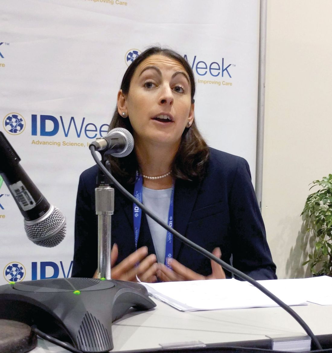
Clinicians may presume, “Oh, you just take opioids orally, you don’t inject drugs,” but oral opiate users can progress to intravenous drug use, Donna Futterman, MD, director of clinical pediatrics, Montefiore Medical Center, and professor of clinical pediatrics at Albert Einstein College of Medicine, both in New York, said during the press conference at the annual scientific meeting on infectious diseases.
Guidelines call for testing for hepatitis C only in individuals with known injected drug use, among other risk factors, but the research suggests that this significantly underestimates the population of teenagers and young adults who are at risk. Many who take opiates go on to use injectable drugs.
Another surprise finding in the study was that only 10.6% of those tested for hepatitis C had also been screened for human immunodeficiency virus (HIV).
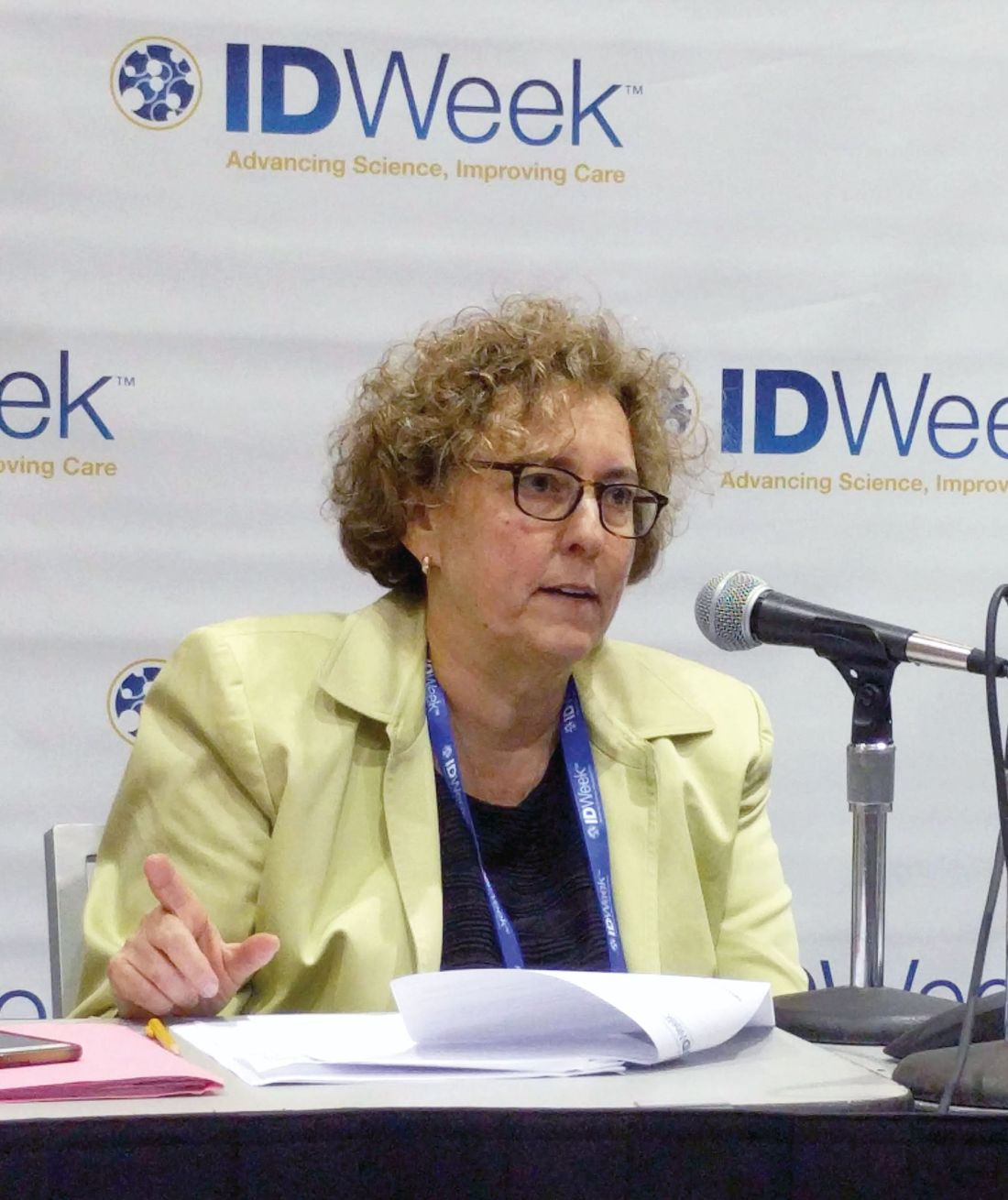
The reasons for the low frequency of screening are likely complex, including lack of time, discomfort between the physician and patient, and concerns over privacy and stigma, according to Dr. Epstein, who emphasized the importance of communication to overcome such barriers.
“As a pediatrician, I try to be as open as possible with patients and let them know that anything they tell me is confidential. I start out discussing less private issues, things that are easier to talk about,” Dr. Epstein said.
But the results of the study also suggest that preconceived notions may be holding clinicians back from testing. “How can you test for hepatitis C and not think HIV?” Dr. Futterman said. “What is that differentiator in providers’ heads that makes them focus on one thing and not the other?”
SAN FRANCISCO – A new study indicates young adults with opioid use disorder are seldom screened for hepatitis C virus infections; yet 11% of the subjects with opioid use disorder who were tested had been exposed to hepatitis C, and 6.8% had evidence of chronic hepatitis C infection.
Overall, 2.5% (6,812 subjects) of all subjects received hepatitis C testing and 122 (1.8%) tested positive. Based on health records, 23,345 had an ICD-9 code for any illicit drug use and 8.9% of those (2,090) were tested for HCV infection. Of the 933 subjects with an ICD-9 code for opioid use disorder, 35% were tested for HCV.
The results suggest that a group at significant risk of hepatitis C – those with opioid use disorder – is being overlooked in public health efforts to control the disease.
Clinicians may presume, “Oh, you just take opioids orally, you don’t inject drugs,” but oral opiate users can progress to intravenous drug use, Donna Futterman, MD, director of clinical pediatrics, Montefiore Medical Center, and professor of clinical pediatrics at Albert Einstein College of Medicine, both in New York, said during the press conference at the annual scientific meeting on infectious diseases.
Guidelines call for testing for hepatitis C only in individuals with known injected drug use, among other risk factors, but the research suggests that this significantly underestimates the population of teenagers and young adults who are at risk. Many who take opiates go on to use injectable drugs.
Another surprise finding in the study was that only 10.6% of those tested for hepatitis C had also been screened for human immunodeficiency virus (HIV).
The reasons for the low frequency of screening are likely complex, including lack of time, discomfort between the physician and patient, and concerns over privacy and stigma, according to Dr. Epstein, who emphasized the importance of communication to overcome such barriers.
“As a pediatrician, I try to be as open as possible with patients and let them know that anything they tell me is confidential. I start out discussing less private issues, things that are easier to talk about,” Dr. Epstein said.
But the results of the study also suggest that preconceived notions may be holding clinicians back from testing. “How can you test for hepatitis C and not think HIV?” Dr. Futterman said. “What is that differentiator in providers’ heads that makes them focus on one thing and not the other?”
REPORTING FROM ID WEEK 2018
Key clinical point: By focusing solely on injectable drug users, clinicians may miss many others who are at risk for hepatitis C infection.
Major finding: Among those with opiate use disorder, 11% tested positive for hepatitis C.
Study details: Survey of 269,124 teenagers and young adults visiting U.S. Federally Qualified Health Centers.
Disclosures: Dr. Epstein and Dr. Futterman have reported no conflicts of interest.
Encourage influenza vaccination in pregnant women
They are at greater risk for more severe illness, and influenza can lead to adverse outcomes in infants. The good news is that recent studies have shown that flu vaccines are safe and effective in pregnant women.
The bad news is that many women are hesitant to be vaccinated out of concerns over safety, in a trend that reflects broader societal worries over vaccination, said Dr. Chu, of the University of Washington, Seattle. In a video interview at an annual scientific meeting on infectious diseases, Dr. Chu advised steps to ensure that pregnant women are aware of the safety and efficacy of flu vaccines, and the benefits to the infant who acquires immunity through the mother. It’s also a good idea to have vaccine on hand to be able to offer it immediately during an office visit.
REPORTING FROM ID WEEK 2018
Suicide prevention in rheumatology: Engagement is key
Fibromyalgia patients with no documented suicide attempt spent far more hours in face-to-face meetings with providers than did those who made a suicide attempt over a 20-year period at a single academic medical center. The results, based on a machine-learning analysis of electronic health records, shed more light on the heavy burden of suicidality among patients with rheumatologic illnesses.

“People who didn’t have suicide attempts were present at the doctor 50 hours in a year, compared to less than an hour in a year for those who did attempt suicide. It’s a staggering difference,” said study author Lindsey McKernan, PhD, of the department of psychiatry and behavioral sciences at Vanderbilt University, Nashville, Tenn. What’s more, patients who did not have suicidal thoughts averaged about 6 office hours per year, compared with less than 2 hours for those with suicidal ideation (Arthritis Care Res. 2018 Sep 7. doi: 10.1002/acr.23748).
Fibromyalgia patients are at about ten times the risk of suicide as the general population, and rates of depression and anxiety are higher in patients with rheumatoid arthritis, ankylosing spondylitis, and psoriatic disease as well.
Still, mental health issues often go unaddressed. “Many times rheumatologists focus on the patient’s joints and their rheumatologic illness, and they don’t focus on their mental health, and as a result depression in a suicidal patient is, I think, more often missed in a rheumatologic practice than it should be,” said Rakesh Jain, MD, of the department of psychiatry at Texas Tech University, Odessa.

But that gap isn’t for lack of awareness, says Barton Wise, MD, of the departments of orthopedic surgery and internal medicine at the University of California, Davis. “In general, people recognize that depression is a major problem in their patients,” he said. He was the lead author of a study that found that rheumatologists often lack the time, confidence, and connections to properly address a patient’s mental health needs (J Clin Rheumatol. 2016 Sep;22[6]:307-11).
Together, the two studies underscore the pressing need for better mental health care among rheumatology patients. Such issues often take a back seat to a rheumatologist’s primary concern about joint and overall health, but studies have shown that mental health issues are tied to worse rheumatologic disease outcomes. “Addressing comorbid depression will just make the rheumatological outcome be better. So why not do it?” Dr. Jain said.
Screening is a key consideration, according to Dr. Jain, who recommends the Patient Health Questionnaire-9. But even when a problem arises, rheumatologists may lack the confidence to tackle mental health issues. This can be addressed through various resources, such as courses at professional meetings, but another challenge awaits. Rheumatologists may also be unsure of who should be responsible for handling mental health concerns. Even though the rheumatologist may see the patient more often than his or her other providers, “you often feel that you can’t manage everything,” Dr. Wise said.
One way to address that is to establish relationships with mental health providers who can receive referrals for patients who require it. In academic medical centers or other large institutions, relationships can be formalized, so that a patient could see a psychiatrist on the same day as the rheumatologist visit. He even suggests group sessions for patients with similar comorbidities, such as depression related to fibromyalgia or lupus.
Special challenges
Rheumatologic conditions are well documented to have heightened rates of suicide and mental health issues, and this may be related to some of the additional challenges such patients face. Many have chronic pain, which in itself is a risk factor for suicide.
Fibromyalgia can be particularly difficult because patients struggle to communicate about their condition with their clinician and even with loved ones. “Patients report feeling stigmatized and often struggle to communicate about what’s happening in their bodies. The pain can change location and intensity, and it doesn’t show up on medical tests,” Dr. McKernan said.
Another burden is that rheumatologic patients typically have high levels of inflammation, a characteristic that has been linked to lower responses to some antidepressants, according to Dr. Jain.
Antidepressants should still be considered for these patients, but they should be combined with other treatments or management techniques, he says. He emphasizes the importance of a low-inflammatory diet, exercise, and other lifestyle factors. He has also created the free Wild 5 Wellness program, which seeks to broadly improve wellness in patients with chronic pain and mental health challenges.
Physician response
The results of the study on fibromyalgia patients suggests another avenue toward improving mental health. The researchers examined data on 8,879 patients with fibromyalgia, using data collected between 1998 and 2017. There were 34 suicide attempts and 96 cases of suicidal ideation. A machine-learning algorithm spat out some factors associated with heightened suicide risk, such as fatigue, dizziness, and weakness, as well as obesity and drug dependence.
But it also generated some associations that weren’t obviously related, such as receiving a flu shot or taking vitamin supplements. “There were a lot of things associated with routine medical care in the patients who didn’t have thoughts about suicide and didn’t attempt suicide. So we looked at how much time people spent with their doctor. I think we might have found an important signal that requires further investigation in bigger samples, and also in other populations. If we can look at people who are at risk [of suicide] but who aren’t engaged with their doctors, that gives us a potential avenue to do something about it, where we can get them connected with a provider, or reconnected if they’ve fallen off, or give them a call to see how they’re doing,” Dr. McKernan said.
In fact, the research suggests that such an effort alone might be enough to reduce suicidality, since patient-provider contact appears to be so important.
“I know in the past that physicians have expressed feeling frustrated – like they don’t have some sort of [mental health] intervention to provide patients who have fibromyalgia. This might show that continuing to see the patient, to stay engaged, may have intrinsic benefit that serves almost like an intervention in itself,” Dr. McKernan said.
Dr. McKernan, Dr. Jain, and Dr. Wise had no financial conflicts of interest.
Fibromyalgia patients with no documented suicide attempt spent far more hours in face-to-face meetings with providers than did those who made a suicide attempt over a 20-year period at a single academic medical center. The results, based on a machine-learning analysis of electronic health records, shed more light on the heavy burden of suicidality among patients with rheumatologic illnesses.
“People who didn’t have suicide attempts were present at the doctor 50 hours in a year, compared to less than an hour in a year for those who did attempt suicide. It’s a staggering difference,” said study author Lindsey McKernan, PhD, of the department of psychiatry and behavioral sciences at Vanderbilt University, Nashville, Tenn. What’s more, patients who did not have suicidal thoughts averaged about 6 office hours per year, compared with less than 2 hours for those with suicidal ideation (Arthritis Care Res. 2018 Sep 7. doi: 10.1002/acr.23748).
Fibromyalgia patients are at about ten times the risk of suicide as the general population, and rates of depression and anxiety are higher in patients with rheumatoid arthritis, ankylosing spondylitis, and psoriatic disease as well.
Still, mental health issues often go unaddressed. “Many times rheumatologists focus on the patient’s joints and their rheumatologic illness, and they don’t focus on their mental health, and as a result depression in a suicidal patient is, I think, more often missed in a rheumatologic practice than it should be,” said Rakesh Jain, MD, of the department of psychiatry at Texas Tech University, Odessa.
But that gap isn’t for lack of awareness, says Barton Wise, MD, of the departments of orthopedic surgery and internal medicine at the University of California, Davis. “In general, people recognize that depression is a major problem in their patients,” he said. He was the lead author of a study that found that rheumatologists often lack the time, confidence, and connections to properly address a patient’s mental health needs (J Clin Rheumatol. 2016 Sep;22[6]:307-11).
Together, the two studies underscore the pressing need for better mental health care among rheumatology patients. Such issues often take a back seat to a rheumatologist’s primary concern about joint and overall health, but studies have shown that mental health issues are tied to worse rheumatologic disease outcomes. “Addressing comorbid depression will just make the rheumatological outcome be better. So why not do it?” Dr. Jain said.
Screening is a key consideration, according to Dr. Jain, who recommends the Patient Health Questionnaire-9. But even when a problem arises, rheumatologists may lack the confidence to tackle mental health issues. This can be addressed through various resources, such as courses at professional meetings, but another challenge awaits. Rheumatologists may also be unsure of who should be responsible for handling mental health concerns. Even though the rheumatologist may see the patient more often than his or her other providers, “you often feel that you can’t manage everything,” Dr. Wise said.
One way to address that is to establish relationships with mental health providers who can receive referrals for patients who require it. In academic medical centers or other large institutions, relationships can be formalized, so that a patient could see a psychiatrist on the same day as the rheumatologist visit. He even suggests group sessions for patients with similar comorbidities, such as depression related to fibromyalgia or lupus.
Special challenges
Rheumatologic conditions are well documented to have heightened rates of suicide and mental health issues, and this may be related to some of the additional challenges such patients face. Many have chronic pain, which in itself is a risk factor for suicide.
Fibromyalgia can be particularly difficult because patients struggle to communicate about their condition with their clinician and even with loved ones. “Patients report feeling stigmatized and often struggle to communicate about what’s happening in their bodies. The pain can change location and intensity, and it doesn’t show up on medical tests,” Dr. McKernan said.
Another burden is that rheumatologic patients typically have high levels of inflammation, a characteristic that has been linked to lower responses to some antidepressants, according to Dr. Jain.
Antidepressants should still be considered for these patients, but they should be combined with other treatments or management techniques, he says. He emphasizes the importance of a low-inflammatory diet, exercise, and other lifestyle factors. He has also created the free Wild 5 Wellness program, which seeks to broadly improve wellness in patients with chronic pain and mental health challenges.
Physician response
The results of the study on fibromyalgia patients suggests another avenue toward improving mental health. The researchers examined data on 8,879 patients with fibromyalgia, using data collected between 1998 and 2017. There were 34 suicide attempts and 96 cases of suicidal ideation. A machine-learning algorithm spat out some factors associated with heightened suicide risk, such as fatigue, dizziness, and weakness, as well as obesity and drug dependence.
But it also generated some associations that weren’t obviously related, such as receiving a flu shot or taking vitamin supplements. “There were a lot of things associated with routine medical care in the patients who didn’t have thoughts about suicide and didn’t attempt suicide. So we looked at how much time people spent with their doctor. I think we might have found an important signal that requires further investigation in bigger samples, and also in other populations. If we can look at people who are at risk [of suicide] but who aren’t engaged with their doctors, that gives us a potential avenue to do something about it, where we can get them connected with a provider, or reconnected if they’ve fallen off, or give them a call to see how they’re doing,” Dr. McKernan said.
In fact, the research suggests that such an effort alone might be enough to reduce suicidality, since patient-provider contact appears to be so important.
“I know in the past that physicians have expressed feeling frustrated – like they don’t have some sort of [mental health] intervention to provide patients who have fibromyalgia. This might show that continuing to see the patient, to stay engaged, may have intrinsic benefit that serves almost like an intervention in itself,” Dr. McKernan said.
Dr. McKernan, Dr. Jain, and Dr. Wise had no financial conflicts of interest.
Simplified SYNTAX holds promise, but still has doubters
SAN DIEGO – A simplified version of the SYNTAX score for coronary artery disease complexity strongly correlated with the unmodified SYNTAX score and could make it easier for cardiologists to employ it in everyday practice. The modified version can be easily memorized and has simplified values that a clinician can calculate and use to determine an appropriate treatment without breaking their work flow to consult a computerized system.
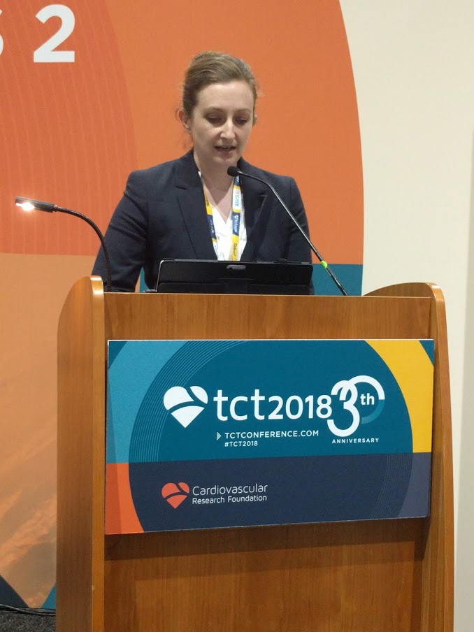
“It’s primarily simplifying the values for the location of the lesions that has allowed it to be more memorizable. That does make it a slightly blunter tool, but only slightly,” said Sonya Burgess, MBCHB, during a presentation at the Transcatheter Cardiovascular Therapeutics annual meeting. Dr. Burgess is an interventional cardiologist at the University of New South Wales, Sydney.
In common practice, SYNTAX scores often go uncalculated because of their complexity, despite guidelines that recommend it. “My guess might be that 1 in every 10 [cardiologists] do it, and that’s not right for our patients,” said Dr. Burgess.
Others at the session agreed that SYNTAX is underutilized, but not all agreed with Dr. Burgess’ solution. Notable among the attendees was Patrick Serruys, MD, of Erasmus University, Rotterdam, the Netherlands, who originally published the SYNTAX system. He doesn’t like the idea of simplifying SYNTAX, based in part on previous experience. “About 10 years ago, Boston Scientific found the score too difficult and they wanted to simplify it, but the score completely lost its prognostic value. I was very emotional about that and I said, ‘no, we have to keep all of the components until we understand [the risk factors] much better,’ ” said Dr. Serruys.
But Dr. Burgess argued that the simplified system, which she and her coinvestigators created, makes concessions to the realities of an interventional cardiology suite. To obtain a standard SYNTAX score, “they have to stop doing something or rebook something, or there’s a lesion there that they just want to treat. [With the simplified score], at least before they go on to treat that lesion, they don’t have to take their gowns and gloves off. You can even have a card hanging on the bar [to refer to].” Dr. Burgess did not provide details of the system.

Dr. Serruys agreed with the source of the problem. “Dr. Burgess is right, it is somewhat demanding,” he allowed, but he also called on clinicians to consider the needs of the patient, even going so far as picturing the patient as a family member. “People complain that doing the SYNTAX is on average 7 minutes, with a standard deviation of 7, so you could work 15 minutes just looking at film. But when you think that the future of your father or brother is depending on the surgeon and the cardiologist looking carefully, I find the argument absolutely obnoxious,” said Dr. Serruys at the meeting sponsored by the Cardiovascular Research Foundation.
Rather than simplification, he stresses teamwork. With at least three people looking at the angiogram, the results are consistent and useful. “You look at the film with the trainees, and that’s how they learn. Never do a SYNTAX score alone,” said Dr. Serruys.
To assess the simplified SYNTAX score, the researchers conducted a retrospective assessment of both the SYNTAX score and the simplified SYNTAX score in 617 patients who had multivessel disease. They performed both assessments in subgroups of patients with 169 patients with ST-segment elevation MI, 78 patients with chronic total occlusion, and 113 patients with left main coronary artery (LMCA) stenosis. They used a 100-patient derivation cohort to determine cutoffs for patients who would not be suitable for percutaneous coronary intervention. They also looked at the accuracy of the simplified version compared with standard SYNTAX in 517 patients from five tertiary centers.
The Spearman’s rho value was 0.93 overall and at least 0.91 for all subgroups (P less than .001 for all). In patients with LMCA stenosis, the simplified version had a sensitivity of 100%, specificity of 85%, negative predictive of 100%, and an area under the curve of 1.0 (P less than .001). For patients with multivessel disease and no LMCA stenosis, the values were 98%, 82%, 99%, and 0.971, respectively (P less than .001).
Dr. Burgess and Dr. Serruys reported no financial conflicts of interest.
SAN DIEGO – A simplified version of the SYNTAX score for coronary artery disease complexity strongly correlated with the unmodified SYNTAX score and could make it easier for cardiologists to employ it in everyday practice. The modified version can be easily memorized and has simplified values that a clinician can calculate and use to determine an appropriate treatment without breaking their work flow to consult a computerized system.
“It’s primarily simplifying the values for the location of the lesions that has allowed it to be more memorizable. That does make it a slightly blunter tool, but only slightly,” said Sonya Burgess, MBCHB, during a presentation at the Transcatheter Cardiovascular Therapeutics annual meeting. Dr. Burgess is an interventional cardiologist at the University of New South Wales, Sydney.
In common practice, SYNTAX scores often go uncalculated because of their complexity, despite guidelines that recommend it. “My guess might be that 1 in every 10 [cardiologists] do it, and that’s not right for our patients,” said Dr. Burgess.
Others at the session agreed that SYNTAX is underutilized, but not all agreed with Dr. Burgess’ solution. Notable among the attendees was Patrick Serruys, MD, of Erasmus University, Rotterdam, the Netherlands, who originally published the SYNTAX system. He doesn’t like the idea of simplifying SYNTAX, based in part on previous experience. “About 10 years ago, Boston Scientific found the score too difficult and they wanted to simplify it, but the score completely lost its prognostic value. I was very emotional about that and I said, ‘no, we have to keep all of the components until we understand [the risk factors] much better,’ ” said Dr. Serruys.
But Dr. Burgess argued that the simplified system, which she and her coinvestigators created, makes concessions to the realities of an interventional cardiology suite. To obtain a standard SYNTAX score, “they have to stop doing something or rebook something, or there’s a lesion there that they just want to treat. [With the simplified score], at least before they go on to treat that lesion, they don’t have to take their gowns and gloves off. You can even have a card hanging on the bar [to refer to].” Dr. Burgess did not provide details of the system.
Dr. Serruys agreed with the source of the problem. “Dr. Burgess is right, it is somewhat demanding,” he allowed, but he also called on clinicians to consider the needs of the patient, even going so far as picturing the patient as a family member. “People complain that doing the SYNTAX is on average 7 minutes, with a standard deviation of 7, so you could work 15 minutes just looking at film. But when you think that the future of your father or brother is depending on the surgeon and the cardiologist looking carefully, I find the argument absolutely obnoxious,” said Dr. Serruys at the meeting sponsored by the Cardiovascular Research Foundation.
Rather than simplification, he stresses teamwork. With at least three people looking at the angiogram, the results are consistent and useful. “You look at the film with the trainees, and that’s how they learn. Never do a SYNTAX score alone,” said Dr. Serruys.
To assess the simplified SYNTAX score, the researchers conducted a retrospective assessment of both the SYNTAX score and the simplified SYNTAX score in 617 patients who had multivessel disease. They performed both assessments in subgroups of patients with 169 patients with ST-segment elevation MI, 78 patients with chronic total occlusion, and 113 patients with left main coronary artery (LMCA) stenosis. They used a 100-patient derivation cohort to determine cutoffs for patients who would not be suitable for percutaneous coronary intervention. They also looked at the accuracy of the simplified version compared with standard SYNTAX in 517 patients from five tertiary centers.
The Spearman’s rho value was 0.93 overall and at least 0.91 for all subgroups (P less than .001 for all). In patients with LMCA stenosis, the simplified version had a sensitivity of 100%, specificity of 85%, negative predictive of 100%, and an area under the curve of 1.0 (P less than .001). For patients with multivessel disease and no LMCA stenosis, the values were 98%, 82%, 99%, and 0.971, respectively (P less than .001).
Dr. Burgess and Dr. Serruys reported no financial conflicts of interest.
REPORTING FROM TCT 2018
Key clinical point: A simplified SYNTAX score could lead to broader implementation of evidence-based decision making for complex coronary artery disease.
Major finding: The simplified score had a Spearman’s rho value of 0.93 overall.
Study details: A retrospective analysis of 617 patients with multivessel disease.
Disclosures: Dr. Burgess and Dr. Serruys reported no financial conflicts of interest.
ULTIMATE: IVUS-guided stent placement bests angiography
SAN DIEGO – Clinical outcomes were better when stent placement was guided by intravascular ultrasound rather than angiography, based on the results of a trial conducted in China. Unlike previous comparative studies, which focused on patients with more complex lesions, this trial included all patients undergoing drug-eluting stent (DES) placement.
In the ULTIMATE trial, 1,448 all-comer patients receiving DES were randomized to either intravascular ultrasound (IVUS)–guided or angiography-guided implantation, reported Junjie Zhang, PhD, of Nanjing Medical University in China, and colleagues. The study excluded patients who had a life expectancy shorter than 12 months, who were intolerant of dual antiplatelet therapy, and who had severe calcification needing rotational atherectomy.
At 30 days after stent placement, the incidence of target vessel failure was 0.8% in the IVUS group and 1.9% in the angiography group, a nonsignificant trend. However, outcomes were significantly different at 1 year, with target vessel failure occurring in 2.9% of IVUS patients and 5.4% of angiography patients (hazard ratio, 0.530; 95% confidence interval, 0.312-0.901; P = .019).
The work was simultaneously published in the Journal of the American College of Cardiology (2018 Sept. doi: 10.1016/j.jacc.2018.09.013) .
Despite good results in this and prior studies, uptake of the IVUS procedure is not high in the United States or Europe, according to members of a panel that reviewed the results at the Transcatheter Cardiovascular Therapeutics annual meeting.
“How can people continue to ignore the importance of imaging-guided stent optimization? Even with second-generation DES, the results are consistent across the studies. This is just another piece of irrefutable evidence,” said Gary S. Mintz, MD, chief medical officer at the Cardiovascular Research Foundation, and a discussant at the meeting, sponsored by the Cardiovascular Research Foundation.
That sentiment was generally echoed by the rest of the panel. John M. Hodson, MD, a professor of medicine at MetroHealth Medical Center in Cleveland, pointed out that the study included a variety of cases, and angiography was performed to a high standard in that arm of the study. “It shows that, even with a good angiographic approach, IVUS still wins. I’m amazed that there’s still some resistance to [IVUS] image guidance,” said Dr. Hodgson.
The ULTIMATE study also found that the procedural time was longer (60.88 minutes vs. 45.49 minutes; P less than .001), and the contrast volume was higher (178.29 mL vs. 161.96 mL; P less than .001) in the IVUS than in the angiography group.
A postprocedure IVUS assessment was performed to determine whether the stent was optimally deployed. The criteria for optimal deployment included minimal lumen area in the stented segment of at least 5 mm2, or 90% of the minimal lumen area at distal reference segment meeting that criteria; a less than 50% plaque burden at the 5 mm of vessel proximal or distal to the stent edge; and no edge dissection involving media greater than 3 mm in length.
In the IVUS group, 53% of patients had optimal placement. The rate of target vessel failure was 1.6% of patients with optimal placement and 4.4% of patients who failed to achieve all optimal criteria (HR, 0.349; 95% CI, 0.135-0.898; P = 0.029). Compared with angiography guidance, IVUS guidance was of similar benefit for patients with either American College of Cardiology/American Heart Association–defined B2/C lesions or A/B1 lesions in terms of the composite endpoint. The significant reduction of clinically driven target lesion revascularization or definite stent thrombosis (HR, 0.407; 95% CI: 0.188-0.880; P = 0.018) based on lesion-level analysis by IVUS guidance was not achieved when patient-level analysis was performed.
“I’m particularly impressed by the analysis of the optimal versus nonoptimal group. If you don’t use IVUS correctly, you don’t get a benefit. The ones [in the IVUS group] who did not get optimal stenting were very similar to the angiographic group,” said Dr. Mintz.
The study was funded by the National Science Foundation of China, Six Talent Peaks Project, Nanjing Health and Family Planning Commission, Nanjing Health Youth Talent Training Project, and the Nanjing Municipal Commission of Science & Technology. None of the study authors had relevant financial disclosures. Dr. Mintz reported received research support from Abbott Vascular and Boston Scientific. He has been a consultant for Boston Scientific, Volcano, and Infraredx. Dr. Hodgson reported received research support and consulted for Volcano.
REPORTING FROM TCT 2018
Key clinical point: Intravascular ultrasound–guided placement of drug-eluting stents resulted in a lower target vessel failure rate than did angiography guidance.
Major finding: The 1-year target vessel failure rate was 2.9% in the intravascular ultrasound–guided group and 5.4% in the angiography group.
Study details: A randomized, controlled trial of 1,448 all-comer patients.
Disclosures: The study was funded by the National Science Foundation of China, Six Talent Peaks Project, Nanjing Health and Family Planning Commission, Nanjing Health Youth Talent Training Project, and the Nanjing Municipal Commission of Science & Technology. None of the study authors had relevant financial disclosures. Dr. Mintz reported receiving research support from Abbott Vascular and Boston Scientific. He has been a consultant for Boston Scientific, Volcano, and Infraredx. Dr. Hodgson reported receiving research support and consulted for Volcano.
Antithrombotic strategy 1 year after stenting in AF patients leans toward oral anticoagulant alone
SAN DIEGO – In patients with atrial fibrillation and stable coronary artery disease, a randomized trial of oral anticoagulation alone versus an anticoagulant plus a single antiplatelet agent failed to establish noninferiority of the single-agent approach. The trial could not demonstrate its primary endpoint of all-cause death, myocardial infarction, stroke, or systemic embolism.
But a secondary endpoint that included major bleeding did demonstrate equivalence, leading the researchers to suggest that oral anticoagulation (OAC) alone may be sufficient in most patients.
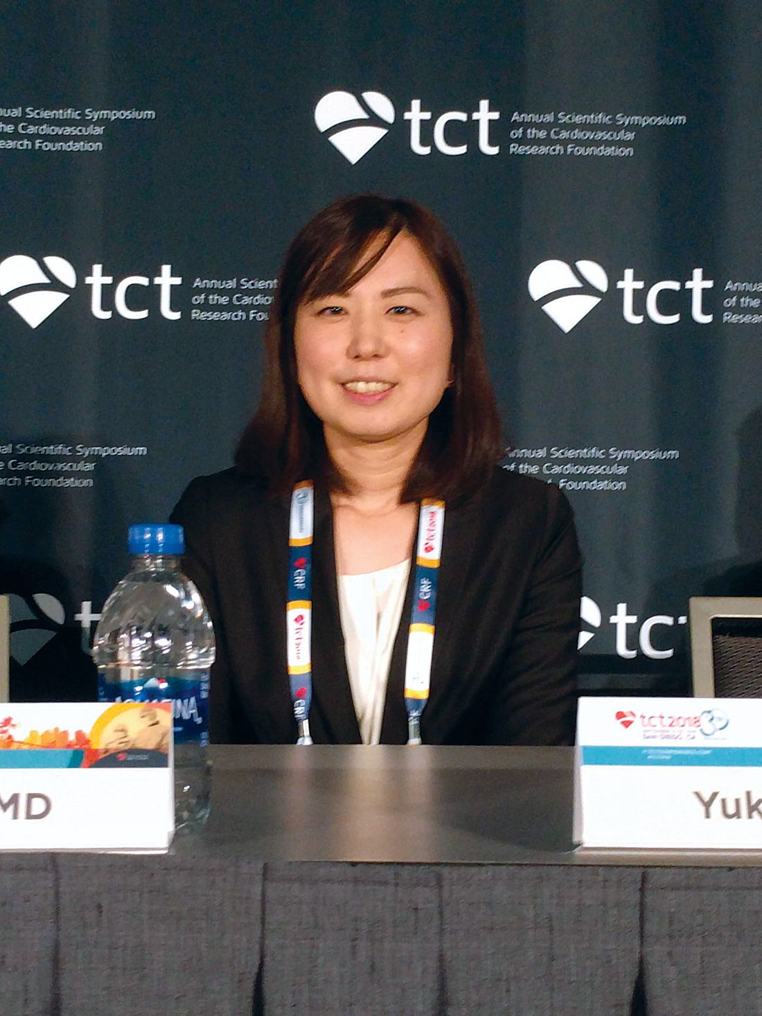
“Combined OAC and single antiplatelet therapy is unlikely to provide net clinical benefit over OAC alone. Thus, OAC alone might be reasonable for AF [atrial fibrillation] patients beyond 1 year after coronary stenting,” Yukiko Nakano, MD, of Kyoto (Japan) University Graduate School of Medicine, said during a press conference at the Transcatheter Cardiovascular Therapeutics annual meeting, sponsored by the Cardiovascular Research Foundation. The report was simultaneously published Sept. 24 in Circulation (doi: 10.1161/CIRCULATIONAHA.118.036768).
The results support the European Society of Cardiology practice guidelines, which recommend lifelong OAC without antiplatelet therapy. But physicians often continue to prescribe antiplatelet agents out of concern that stent thrombosis could occur if the therapy is stopped.
The study was stopped prematurely because of insufficient recruitment, which may have contributed to the failed primary endpoint. It’s a shortcoming that befalls many such studies, perhaps because cardiologists tend to be set in their ways when it comes to treatment of patients after a stent implant. “Cardiologists just think they know the answer, and they don’t want to expose their patients (to a clinical trial). They say, ‘I have my patients on whatever regimen. It seems to be working, and they’re not bleeding, so I don’t want to change it.’ This study suggests that we probably can stop one of the two (antiplatelet drugs) and get by with a single agent, and in this case they got by with no agent (in the monotherapy arm),” said C. Michael Gibson, MD, chief of clinical research in the division of cardiology at Beth Israel Deaconess Medical Center, Boston, who was a discussant at the press conference.
The study recruited 696 patients who were receiving OAC plus single antiplatelet therapy (SAPT) 1 year after receiving a stent. They were randomized 1:1 to continue combined therapy or to stop SAPT and then followed for a median of 2.5 years. A total of 74% of patients who received OAC alone were taking warfarin, while 26% were taking a direct oral anticoagulant. The SAPT group took aspirin or clopidogrel.
Overall, 15.7% of OAC patients experienced the primary endpoint, compared with 13.6% of the combined group (noninferiority P = .20). None of the individual components of the primary endpoint were statistically significantly different between the groups. International Society on Thrombosis and Haemostasis major bleeding and Thrombolysis in Myocardial Infarction major bleeding trended in favor of OAC alone. The secondary endpoint (primary endpoint plus major bleeding) achieved noninferiority, occurring in 19.5% of the OAC group and 19.4% of the combined therapy group (noninferiority P = .016; superiority P = .96).
Daiichi-Sankyo funded the trial. Dr. Nakano had no conflicts of interest. Dr. Gibson reported numerous financial ties to pharmaceutical companies, including Daiichi-Sankyo.
SAN DIEGO – In patients with atrial fibrillation and stable coronary artery disease, a randomized trial of oral anticoagulation alone versus an anticoagulant plus a single antiplatelet agent failed to establish noninferiority of the single-agent approach. The trial could not demonstrate its primary endpoint of all-cause death, myocardial infarction, stroke, or systemic embolism.
But a secondary endpoint that included major bleeding did demonstrate equivalence, leading the researchers to suggest that oral anticoagulation (OAC) alone may be sufficient in most patients.
“Combined OAC and single antiplatelet therapy is unlikely to provide net clinical benefit over OAC alone. Thus, OAC alone might be reasonable for AF [atrial fibrillation] patients beyond 1 year after coronary stenting,” Yukiko Nakano, MD, of Kyoto (Japan) University Graduate School of Medicine, said during a press conference at the Transcatheter Cardiovascular Therapeutics annual meeting, sponsored by the Cardiovascular Research Foundation. The report was simultaneously published Sept. 24 in Circulation (doi: 10.1161/CIRCULATIONAHA.118.036768).
The results support the European Society of Cardiology practice guidelines, which recommend lifelong OAC without antiplatelet therapy. But physicians often continue to prescribe antiplatelet agents out of concern that stent thrombosis could occur if the therapy is stopped.
The study was stopped prematurely because of insufficient recruitment, which may have contributed to the failed primary endpoint. It’s a shortcoming that befalls many such studies, perhaps because cardiologists tend to be set in their ways when it comes to treatment of patients after a stent implant. “Cardiologists just think they know the answer, and they don’t want to expose their patients (to a clinical trial). They say, ‘I have my patients on whatever regimen. It seems to be working, and they’re not bleeding, so I don’t want to change it.’ This study suggests that we probably can stop one of the two (antiplatelet drugs) and get by with a single agent, and in this case they got by with no agent (in the monotherapy arm),” said C. Michael Gibson, MD, chief of clinical research in the division of cardiology at Beth Israel Deaconess Medical Center, Boston, who was a discussant at the press conference.
The study recruited 696 patients who were receiving OAC plus single antiplatelet therapy (SAPT) 1 year after receiving a stent. They were randomized 1:1 to continue combined therapy or to stop SAPT and then followed for a median of 2.5 years. A total of 74% of patients who received OAC alone were taking warfarin, while 26% were taking a direct oral anticoagulant. The SAPT group took aspirin or clopidogrel.
Overall, 15.7% of OAC patients experienced the primary endpoint, compared with 13.6% of the combined group (noninferiority P = .20). None of the individual components of the primary endpoint were statistically significantly different between the groups. International Society on Thrombosis and Haemostasis major bleeding and Thrombolysis in Myocardial Infarction major bleeding trended in favor of OAC alone. The secondary endpoint (primary endpoint plus major bleeding) achieved noninferiority, occurring in 19.5% of the OAC group and 19.4% of the combined therapy group (noninferiority P = .016; superiority P = .96).
Daiichi-Sankyo funded the trial. Dr. Nakano had no conflicts of interest. Dr. Gibson reported numerous financial ties to pharmaceutical companies, including Daiichi-Sankyo.
REPORTING FROM TCT 2018
Key clinical point:
Major finding: A measure that included cardiac events plus major bleeding showed an oral anticoagulant alone was noninferior to an OAC plus antiplatelet therapy.
Study details: Randomized, controlled trial of 696 patients.
Disclosures: Daiichi-Sankyo funded the trial. Dr. Nakano had no conflicts of interest. Dr. Gibson reported numerous financial ties to pharmaceutical companies, including Daiichi-Sankyo.
IVUS guidance cuts target-vessel failure risk by 47%
SAN DIEGO – An all-comers trial conducted in China confirms that intravascular ultrasound (IVUS)–guided implantation of drug-eluting stents (DES) resulted in better clinical outcomes than did angiography-guided procedures, Jun-Jie Zhang, MD, said in reporting results of the ULTIMATE trial at the Transcatheter Cardiovascular Therapeutics annual meeting.
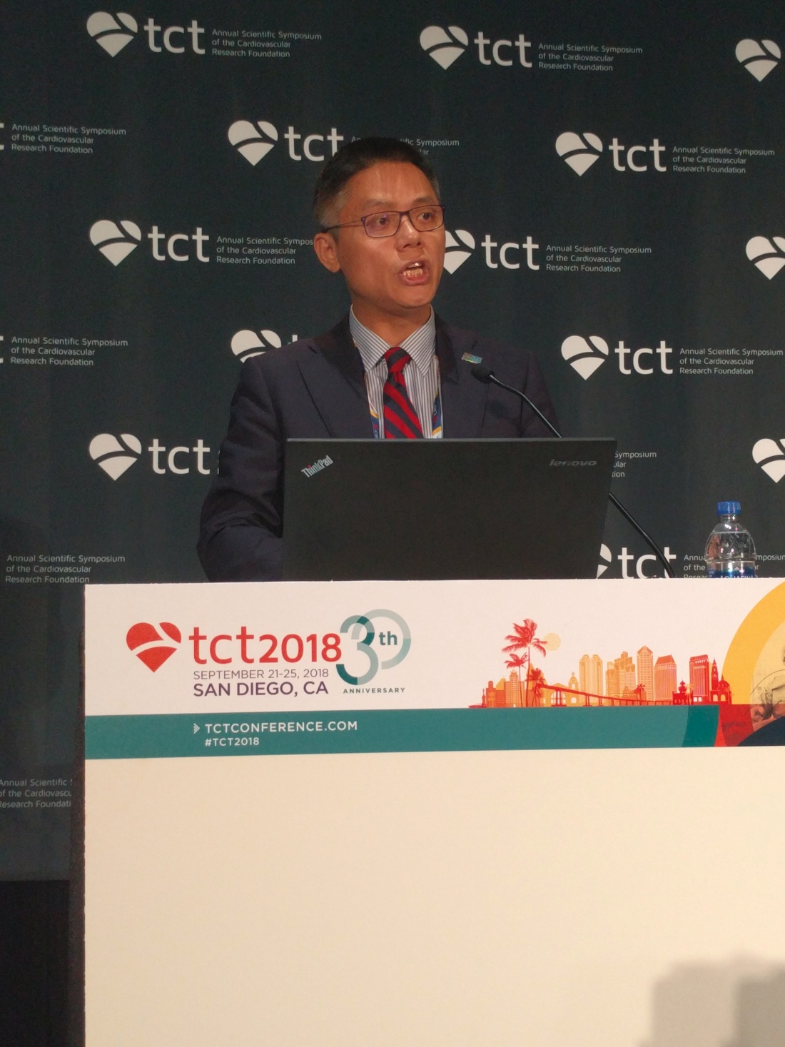
The ULTIMATE trial is the latest in a line of evidence showing the utility of IVUS guidance, though uptake of the procedure is not high in the United States or Europe. Its broader focus also rounds out the findings of earlier studies, which focused on patients with more complex lesions.
“How can people continue to ignore the importance of imaging-guided stent optimization? Even with second-generation DES, the results are consistent across the studies. This is just another piece of irrefutable evidence,” said discussant Gary Mintz, MD.
That sentiment was generally echoed by the rest of the panel. John McB Hodgson, MD, a professor of medicine at MetroHealth Medical Center in Cleveland, also pointed out the consistency of the body of evidence supporting the use of imaging. The study also represented a variety of cases, and the angiography arm of the study showed that the procedure was performed to a high standard. “It shows that even with a good angiographic approach, IVUS still wins. I’m amazed that there’s still some resistance to image guidance,” said Dr. Hodgson.
In ULTIMATE, 1,448 all-comer patients were randomized to either IVUS-guided or angiography-guided DES implantation. Patients with a life-expectancy shorter than 12 months, who were intolerant of dual-antiplatelet therapy, or who had severe calcification needing rotational atherectomy were excluded.
The procedural time was longer in the IVUS group, at 61 min, compared with 45 min in the angiography group, and the contrast volume was higher, at 178 mL and 162 mL, respectively (P less than .001 for both). At 30 days, the incidence of target vessel failure (TVF) was 0.8% in the IVUS group and 1.9% in the angiographic group, though this difference did not reach statistical significance (P = .08). The trend did reach significance at 1 year, with failure occurring in 2.9% of IVUS patients and 5.4% of angiography patients (P = .019). The hazard ratio for TVF in the IVUS group was 0.53 (95% confidence interval, 0.312-0.901), for a relative risk reduction of 47%, reported Dr. Zhang of Nanjing (China) Medical University.
Patients also underwent a postprocedure IVUS assessment to determine whether the stent was deployed optimally or suboptimally. The criteria for optimal deployment included minimal lumen area in the stented segment at least 5.0 mm2, or 90% of the minimal lumen area at distal reference segment meeting that criteria; a less than 50% plaque burden at the 5 mm of vessel proximal or distal to the stent edge; and no edge dissection involving media greater than 3 mm in length.
More than half (53%) of patients in the IVUS group had optimal placement, and 47% did not. Significantly fewer (1.6%) of patients with optimal IVUS experienced TVF at 12 months, compared with 4.4% of the suboptimal group (HR, 0.35; 95% CI, 0.135-0.898). The results were published online simultaneously with the presentation in the Journal of the American College of Cardiology.
“I’m particularly impressed by the analysis of the optimal versus nonoptimal group. If you don’t use IVUS correctly, you don’t get a benefit. The ones who did not get optimal stenting were very similar to the angiographic group,” said Dr. Mintz, chief medical officer at the Cardiovascular Research Foundation, which sponsored the meeting.
ULTIMATE was funded by the National Science Foundation of China and several other research organizations. Dr. Zhang had no relevant disclosures. Dr. Mintz has received research support and/or consulting fees from Abbott Vascular, Boston Scientific, Volcano, and Infraredx. Dr. Hodgson has received research support and consulted for Volcano.
SAN DIEGO – An all-comers trial conducted in China confirms that intravascular ultrasound (IVUS)–guided implantation of drug-eluting stents (DES) resulted in better clinical outcomes than did angiography-guided procedures, Jun-Jie Zhang, MD, said in reporting results of the ULTIMATE trial at the Transcatheter Cardiovascular Therapeutics annual meeting.
The ULTIMATE trial is the latest in a line of evidence showing the utility of IVUS guidance, though uptake of the procedure is not high in the United States or Europe. Its broader focus also rounds out the findings of earlier studies, which focused on patients with more complex lesions.
“How can people continue to ignore the importance of imaging-guided stent optimization? Even with second-generation DES, the results are consistent across the studies. This is just another piece of irrefutable evidence,” said discussant Gary Mintz, MD.
That sentiment was generally echoed by the rest of the panel. John McB Hodgson, MD, a professor of medicine at MetroHealth Medical Center in Cleveland, also pointed out the consistency of the body of evidence supporting the use of imaging. The study also represented a variety of cases, and the angiography arm of the study showed that the procedure was performed to a high standard. “It shows that even with a good angiographic approach, IVUS still wins. I’m amazed that there’s still some resistance to image guidance,” said Dr. Hodgson.
In ULTIMATE, 1,448 all-comer patients were randomized to either IVUS-guided or angiography-guided DES implantation. Patients with a life-expectancy shorter than 12 months, who were intolerant of dual-antiplatelet therapy, or who had severe calcification needing rotational atherectomy were excluded.
The procedural time was longer in the IVUS group, at 61 min, compared with 45 min in the angiography group, and the contrast volume was higher, at 178 mL and 162 mL, respectively (P less than .001 for both). At 30 days, the incidence of target vessel failure (TVF) was 0.8% in the IVUS group and 1.9% in the angiographic group, though this difference did not reach statistical significance (P = .08). The trend did reach significance at 1 year, with failure occurring in 2.9% of IVUS patients and 5.4% of angiography patients (P = .019). The hazard ratio for TVF in the IVUS group was 0.53 (95% confidence interval, 0.312-0.901), for a relative risk reduction of 47%, reported Dr. Zhang of Nanjing (China) Medical University.
Patients also underwent a postprocedure IVUS assessment to determine whether the stent was deployed optimally or suboptimally. The criteria for optimal deployment included minimal lumen area in the stented segment at least 5.0 mm2, or 90% of the minimal lumen area at distal reference segment meeting that criteria; a less than 50% plaque burden at the 5 mm of vessel proximal or distal to the stent edge; and no edge dissection involving media greater than 3 mm in length.
More than half (53%) of patients in the IVUS group had optimal placement, and 47% did not. Significantly fewer (1.6%) of patients with optimal IVUS experienced TVF at 12 months, compared with 4.4% of the suboptimal group (HR, 0.35; 95% CI, 0.135-0.898). The results were published online simultaneously with the presentation in the Journal of the American College of Cardiology.
“I’m particularly impressed by the analysis of the optimal versus nonoptimal group. If you don’t use IVUS correctly, you don’t get a benefit. The ones who did not get optimal stenting were very similar to the angiographic group,” said Dr. Mintz, chief medical officer at the Cardiovascular Research Foundation, which sponsored the meeting.
ULTIMATE was funded by the National Science Foundation of China and several other research organizations. Dr. Zhang had no relevant disclosures. Dr. Mintz has received research support and/or consulting fees from Abbott Vascular, Boston Scientific, Volcano, and Infraredx. Dr. Hodgson has received research support and consulted for Volcano.
REPORTING FROM TCT 2018
Key clinical point:
Major finding: IVUS guidance reduced target vessel failure by 47%.
Study details: ULTIMATE, a randomized, controlled trial of 1,448 patients.
Disclosures: ULTIMATE was funded by the National Science Foundation of China and several other research organizations. Dr. Zhang had no relevant disclosures. Dr. Mintz has received research support from Abbott Vascular and Boston Scientific. He has been a consultant for Boston Scientific, Volcano, and Infraredx. Dr. Hodgson has received research support and consulted for Volcano.
Novel device improves mitral regurgitation 30% in REDUCE-FMR
SAN DIEGO – In patients with heart failure and functional mitral regurgitation, implantation of an investigational device led to reduced MR and improved left ventricular remodeling at 1 year, compared with patients who received sham treatment in the REDUCE-FMR trial.
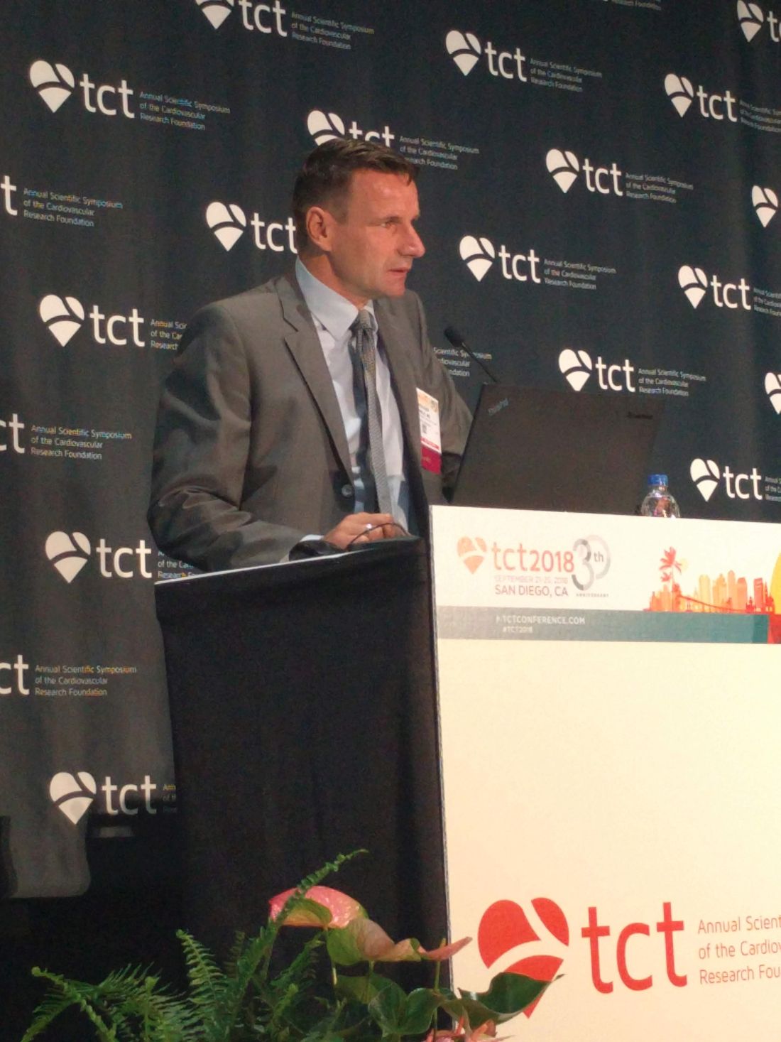
The device showed promise in this trial, despite a small sample size, and its nature makes it possible to follow up with other procedures if the disease progresses. “The advantage of this technique is that all other options are still open,” Horst Sievert, MD, director of the CardioVascular Center in Frankfurt, said during a press conference at the Transcatheter Cardiovascular Therapeutics annual meeting.
The Carillon Mitral Counter System includes two anchors, one in the great cardiac vein and one in the coronary sinus, connected by a shaping ribbon. The tension of the ribbon bolsters the mitral annulus, which in turn reduces mitral regurgitation.
REDUCE-FMR recruited 120 patients from centers in eight countries and randomized 87 to the Carillon device (73 implanted) and 33 to a sham procedure. Sham patients were sedated and received a coronary sinus angiogram. Patients were included if they had dilated ischemic or nonischemic cardiomyopathy and moderate to severe functional MR, among other requirements. Exclusion criteria included existing coronary artery stents in the implant target zone, severe mitral annular calcification, and significant organic mitral valve pathology.
The primary endpoint was the mean reduction of regurgitant volume at 1 year. The treated patients had a 22% reduction of 7.1 mL, while the sham group on average had an 8% increase of 3.3 mL (P = .03). In the as-treated subpopulation, which comprised 45 patients in the treatment group and 13 controls, the values were –7.5 mL and +3.3 mL (P = .02). A per-protocol analysis, which excluded patients who did not meet protocol criteria, led to an amplification of the effect when the study design was adhered to (–12.5 mL vs. +1.3 mL), though this result did not achieve statistical significance owing to the small sample size.
For the safety endpoints, the researchers examined the frequency of major adverse events (MAE), including death, myocardial infarction, cardiac perforation, device embolism, and surgery or percutaneous coronary intervention related to the device at 1 year. In the treatment group, 16.1% experienced a MAE, compared with 18.2% of control patients, a statistically nonsignificant difference.
A secondary efficacy endpoint of change in left ventricular end-diastolic volume showed improvements in the treatment group at 6 months (–12.4 mL) and 12 months (–8.6 mL), compared with increases in the sham group at 6 months (+5.4 mL) and 12 months (+6.5 mL). A similar trend occurred in left ventricular end-systolic volume (–7.8 mL and –4.8 mL; +3.4 mL and +6.1 mL, respectively).
The study was conducted in a patient population similar to that of the COAPT trial, which examined implantation of Abbott’s MitraClip. That study, presented here at TCT 2018 and simultaneously published in the New England Journal of Medicine, also examined patients with heart failure and secondary MR.
However, in REDUCE-FMR, many of the patients had milder heart failure than the researchers had expected: 44.8% in the treatment group had NYHA class II, as did 48.5% in the sham group. That surprise may help identify an appropriate patient population. “I think this device may have a nice spot in between medical therapy and MitraClip implantation, because we have, by chance, a patient population with mild heart insufficiency and mild MR,” said Dr. Sievert.

The two devices also showed different physiologic effects, Michael Mack, MD, said at a press conference. “One subtle difference is that, in this trial, the difference is due to both positive left ventricular remodeling in the treatment arm and continued progression in the sham control. In COAPT, the difference in improvement that we saw was totally due to prevention of progression of disease. We just stabilized the disease to where it was at. So that’s an intriguing difference here, that you actually were able to demonstrate positive left ventricular remodeling,” noted Dr. Mack, medical director for cardiovascular surgery at Baylor Scott & White Medical Center, Plano, Tex. He was a coinvestigator in the COAPT trial.
REDUCE-FMR was funded by Cardiac Dimensions. Dr. Sievert has received consulting fees, travel expenses, and study honoraria from Cardiac Dimensions, and 35 other companies. Dr. Mack has received grant support or had a research contract with Abbott Vascular, Medtronic, and Edwards Lifesciences.
SAN DIEGO – In patients with heart failure and functional mitral regurgitation, implantation of an investigational device led to reduced MR and improved left ventricular remodeling at 1 year, compared with patients who received sham treatment in the REDUCE-FMR trial.
The device showed promise in this trial, despite a small sample size, and its nature makes it possible to follow up with other procedures if the disease progresses. “The advantage of this technique is that all other options are still open,” Horst Sievert, MD, director of the CardioVascular Center in Frankfurt, said during a press conference at the Transcatheter Cardiovascular Therapeutics annual meeting.
The Carillon Mitral Counter System includes two anchors, one in the great cardiac vein and one in the coronary sinus, connected by a shaping ribbon. The tension of the ribbon bolsters the mitral annulus, which in turn reduces mitral regurgitation.
REDUCE-FMR recruited 120 patients from centers in eight countries and randomized 87 to the Carillon device (73 implanted) and 33 to a sham procedure. Sham patients were sedated and received a coronary sinus angiogram. Patients were included if they had dilated ischemic or nonischemic cardiomyopathy and moderate to severe functional MR, among other requirements. Exclusion criteria included existing coronary artery stents in the implant target zone, severe mitral annular calcification, and significant organic mitral valve pathology.
The primary endpoint was the mean reduction of regurgitant volume at 1 year. The treated patients had a 22% reduction of 7.1 mL, while the sham group on average had an 8% increase of 3.3 mL (P = .03). In the as-treated subpopulation, which comprised 45 patients in the treatment group and 13 controls, the values were –7.5 mL and +3.3 mL (P = .02). A per-protocol analysis, which excluded patients who did not meet protocol criteria, led to an amplification of the effect when the study design was adhered to (–12.5 mL vs. +1.3 mL), though this result did not achieve statistical significance owing to the small sample size.
For the safety endpoints, the researchers examined the frequency of major adverse events (MAE), including death, myocardial infarction, cardiac perforation, device embolism, and surgery or percutaneous coronary intervention related to the device at 1 year. In the treatment group, 16.1% experienced a MAE, compared with 18.2% of control patients, a statistically nonsignificant difference.
A secondary efficacy endpoint of change in left ventricular end-diastolic volume showed improvements in the treatment group at 6 months (–12.4 mL) and 12 months (–8.6 mL), compared with increases in the sham group at 6 months (+5.4 mL) and 12 months (+6.5 mL). A similar trend occurred in left ventricular end-systolic volume (–7.8 mL and –4.8 mL; +3.4 mL and +6.1 mL, respectively).
The study was conducted in a patient population similar to that of the COAPT trial, which examined implantation of Abbott’s MitraClip. That study, presented here at TCT 2018 and simultaneously published in the New England Journal of Medicine, also examined patients with heart failure and secondary MR.
However, in REDUCE-FMR, many of the patients had milder heart failure than the researchers had expected: 44.8% in the treatment group had NYHA class II, as did 48.5% in the sham group. That surprise may help identify an appropriate patient population. “I think this device may have a nice spot in between medical therapy and MitraClip implantation, because we have, by chance, a patient population with mild heart insufficiency and mild MR,” said Dr. Sievert.
The two devices also showed different physiologic effects, Michael Mack, MD, said at a press conference. “One subtle difference is that, in this trial, the difference is due to both positive left ventricular remodeling in the treatment arm and continued progression in the sham control. In COAPT, the difference in improvement that we saw was totally due to prevention of progression of disease. We just stabilized the disease to where it was at. So that’s an intriguing difference here, that you actually were able to demonstrate positive left ventricular remodeling,” noted Dr. Mack, medical director for cardiovascular surgery at Baylor Scott & White Medical Center, Plano, Tex. He was a coinvestigator in the COAPT trial.
REDUCE-FMR was funded by Cardiac Dimensions. Dr. Sievert has received consulting fees, travel expenses, and study honoraria from Cardiac Dimensions, and 35 other companies. Dr. Mack has received grant support or had a research contract with Abbott Vascular, Medtronic, and Edwards Lifesciences.
REPORTING FROM TCT 2018
Key clinical point: The device led to improvement in mitral regurgitation as well as ventricular remodeling.
Major finding:
Study details: REDUCE-FMR, a randomized, sham controlled trial of 120 patients from 8 countries.
Disclosures: REDUCE-FMR was funded by Cardiac Dimensions. Dr. Sievert has received consulting fees, travel expenses, and study honoraria from Cardiac Dimensions, and 35 other companies. Dr. Mack has received grant support or had a research contract with Abbott Vascular, Medtronic, and Edwards Lifesciences.
Cheaper drug-eluting stent fares as well as Xience
SAN DIEGO – The Supraflex drug-eluting stent is noninferior to Abbott’s Xience stent, according to results of the TALENT trial. Supfraflex elutes sirolimus from a bioabsorbable polymer, while Xience elutes everolimus from a durable polymer coating.

Stent technology has reached a “safety plateau,” which is leading to decisions based on economic factors, said Patrick Serruys, MD, during a presentation of the results at Transcatheter Cardiovascular Therapeutics annual meeting. Dr. Serruys is a professor of cardiology at the National Heart and Lung Institute of Imperial College in London.
The Indian government’s policy also requires companies to prove that their product is noninferior to proven stents. To satisfy that requirement, researchers conducted the Thin Strut Sirolimus-Eluting Stent in All Comers Population vs Everolimus-eluting Stent (TALENT) study. In this trial (NCT02870140), 1,435 patients from 23 sites were randomized to receive either the Supraflex stent, manufactured by India-based SMT, or the Xience stent.
The population was “all comers” who had symptomatic coronary artery disease that qualified for percutaneous coronary interventions, including chronic stable angina, silent ischemia, and acute coronary syndromes. The researchers followed patients out to 3 years.
The two groups were similar in their baseline characteristics, with the exception of previous coronary artery bypass graft, which was more frequent in the Xience group (7.7% versus 4.6%).
The primary endpoint of the study was a composite of cardiac death, target vessel myocardial infarction, and clinically indicated target lesion revascularization (CI-TLR). At 3 years, 5.3% of the Xience group experienced the composite outcome versus 4.9% of the Supraflex group, a nonsignificant difference. The result confirmed the noninferiority of Supraflex at a margin of 4% (P less than .001). When the adverse events were broken down individually in a per-protocol analysis, the only significant difference between the two groups was with respect to CI-TLR, which favored Supraflex (1.2% versus 3.1%; P = .021). There were seven deaths in the Supraflex arm versus two in the Xience group. However, there was no apparent explanation for the difference, and it may have been because of chance, according to Dr. Serruys.
“The performance of Xience was excellent in this trial, at 5.3% it had one of the lowest (adverse outcome rates) I have seen so far, but the Supraflex also did very well,” said Dr. Serruys at a press conference.
The results of the study have implications in countries with capped stent prices or where there are models for competitive pricing. “Market competitiveness may influence future decisions on which stent you use,” said Dr. Serruys.
Because the trial was designed with India’s regulatory environment in mind, it is unlikely to be applicable to marketing approval in the United States, but such inexpensive stents could be coming. They may drive down prices, but they could also have a dark side, according to Dr. Cohen. “The companies that make these kinds of stents don’t do the kind of innovation we’ve come to expect in the field. I don’t know if they can support the R&D programs that the companies that we currently use [can support],” he said.
TALENT was sponsored by SMT, which manufactures Supraflex, and by the European Clinical Research Institute. Dr. Serruys has consulted for, received honoraria from, or served on the speaker’s bureau for Abbott. Dr. Cohen has received grant support from Abbott Vascular, Boston Scientific, Edwards Lifesciences, Medtronic, Corvia Medical, and Svelte. He has consulted for, received honoraria, or served on the speaker’s bureau for Edwards Lifesciences and Medtronic.
SAN DIEGO – The Supraflex drug-eluting stent is noninferior to Abbott’s Xience stent, according to results of the TALENT trial. Supfraflex elutes sirolimus from a bioabsorbable polymer, while Xience elutes everolimus from a durable polymer coating.
Stent technology has reached a “safety plateau,” which is leading to decisions based on economic factors, said Patrick Serruys, MD, during a presentation of the results at Transcatheter Cardiovascular Therapeutics annual meeting. Dr. Serruys is a professor of cardiology at the National Heart and Lung Institute of Imperial College in London.
The Indian government’s policy also requires companies to prove that their product is noninferior to proven stents. To satisfy that requirement, researchers conducted the Thin Strut Sirolimus-Eluting Stent in All Comers Population vs Everolimus-eluting Stent (TALENT) study. In this trial (NCT02870140), 1,435 patients from 23 sites were randomized to receive either the Supraflex stent, manufactured by India-based SMT, or the Xience stent.
The population was “all comers” who had symptomatic coronary artery disease that qualified for percutaneous coronary interventions, including chronic stable angina, silent ischemia, and acute coronary syndromes. The researchers followed patients out to 3 years.
The two groups were similar in their baseline characteristics, with the exception of previous coronary artery bypass graft, which was more frequent in the Xience group (7.7% versus 4.6%).
The primary endpoint of the study was a composite of cardiac death, target vessel myocardial infarction, and clinically indicated target lesion revascularization (CI-TLR). At 3 years, 5.3% of the Xience group experienced the composite outcome versus 4.9% of the Supraflex group, a nonsignificant difference. The result confirmed the noninferiority of Supraflex at a margin of 4% (P less than .001). When the adverse events were broken down individually in a per-protocol analysis, the only significant difference between the two groups was with respect to CI-TLR, which favored Supraflex (1.2% versus 3.1%; P = .021). There were seven deaths in the Supraflex arm versus two in the Xience group. However, there was no apparent explanation for the difference, and it may have been because of chance, according to Dr. Serruys.
“The performance of Xience was excellent in this trial, at 5.3% it had one of the lowest (adverse outcome rates) I have seen so far, but the Supraflex also did very well,” said Dr. Serruys at a press conference.
The results of the study have implications in countries with capped stent prices or where there are models for competitive pricing. “Market competitiveness may influence future decisions on which stent you use,” said Dr. Serruys.
Because the trial was designed with India’s regulatory environment in mind, it is unlikely to be applicable to marketing approval in the United States, but such inexpensive stents could be coming. They may drive down prices, but they could also have a dark side, according to Dr. Cohen. “The companies that make these kinds of stents don’t do the kind of innovation we’ve come to expect in the field. I don’t know if they can support the R&D programs that the companies that we currently use [can support],” he said.
TALENT was sponsored by SMT, which manufactures Supraflex, and by the European Clinical Research Institute. Dr. Serruys has consulted for, received honoraria from, or served on the speaker’s bureau for Abbott. Dr. Cohen has received grant support from Abbott Vascular, Boston Scientific, Edwards Lifesciences, Medtronic, Corvia Medical, and Svelte. He has consulted for, received honoraria, or served on the speaker’s bureau for Edwards Lifesciences and Medtronic.
REPORTING FROM TCT 2018
Key clinical point: Equivalent, cheaper stents could put economic pressure on choice of stent.
Major finding: At 1 year, there was no significant difference in a composite of death and cardiac events.
Study details: TALENT, a multicenter, randomized, controlled trial of 1,435 patients.
Disclosures: TALENT was sponsored by SMT, which manufactures Supraflex, and by the European Clinical Research Institute. Dr. Serruys has consulted for, received honoraria from, or served on the speaker’s bureau for Abbott. Dr. Cohen has received grant support from Abbott Vascular, Boston Scientific, Edwards Lifesciences, Medtronic, Corvia Medical, and Svelte. He has consulted for, received honoraria from, or served on the speaker’s bureau for Edwards Lifesciences and Medtronic.
Partial nephrectomies on rise for early-stage RCC
SAN FRANCISCO – Partial nephrectomy for localized renal cell carcinomas (RCCs) is on the rise, but the procedure is still less commonly performed than radical nephrectomy and more commonly performed at high-volume surgical centers.
About half of localized renal tumors are excised through partial nephrectomy, based on results from a new analysis of The National Cancer Database. The study also indicated that patient mortality rates were lower at high volume centers, those in the top 10 percentile of treatment volume, said David Cahn, DO, who presented the results at the annual meeting of the American Urological Association.
The study looked at surgeries among 142,000 patients with pT1a-T2b RCCs (no involvement of lymph nodes or metastases) who underwent procedures during 2004-2014.
Overall, 41% of patients had partial nephrectomies, 58% had radical nephrectomies, and 1% received ablative therapy. The frequency of partial nephrectomies rose markedly over the course of the study, increasing from 24% of cases in 2004 to 53% in 2014. The vast majority of partial nephrectomies, 81%, were performed for pT1a tumors; 24% of pT1b tumors also were excised using partial nephrectomy.
Compared with the reference point of T1a tumors, T1b tumors (odds ratio, 0.22; P less than .0001), T2a (OR, 0.06; P less than .0001), and T2b tumors (OR, 0.03; P less than .0001) were progressively less likely to be treated with partial nephrectomy. A multivariate analysis showed that patients at a high volume center were significantly more likely to undergo partial nephrectomy (OR, 1.89; P less than .0001). Overall mortality was lower at high volume centers (hazard ratio, 0.92; P = .012).
“I think that regionalization of care has ... some benefits to patients, but it comes with many challenges as well. This study really did help show that increased numbers [of procedures performed] makes you more comfortable and willing to offer partial nephrectomy,” said Dr. Cahn, of Fox Chase Cancer Center, Philadelphia.
“Volume tends to show better outcomes. But the concern is: Are you overloading systems? Are you making patients travel farther, and how does that work in terms of follow-up care?” Dr. Cahn said. He cited the example of a patient who may have to travel several hours to get surgery but then returns home and develops a postoperative complication that must be treated in a different facility. That raises issues of transfer of medical records and coordination of care.
The solution, he said, is for some of the procedures and practices common at high-volume centers to be transferred to smaller centers. “For the routine partial nephrectomy ... we need to have well-trained physicians who can offer those all over the country. I think it’s too hard in terms of feasibility to push those patients just towards certain high volume centers.”
SOURCE: AUA Annual Meeting, Abstract PD07-04.
REPORTING FROM THE AUA ANNUAL MEETING
Key clinical point: Many patients who could undergo partial nephrectomy are still receiving radical nephrectomies.
Major finding: In 2014, 53% of patients with pT1a or pT1b RCC underwent partial nephrectomy.
Study details: Retrospective analysis of 142,000 patients.
Disclosures: The study was funded internally. Dr. Cahn reports having no relevant financial disclosures.
Source: Cahn D et al. AUA Annual Meeting, Abstract PD07-04.