User login
FDG-PET/CT leads pack for small-cell lung cancer staging
For pretreatment staging of small-cell lung cancer (SCLC) the use of positron-emission tomography combined with CT was more sensitive compared with several other tests, according to a new report on a review of studies.
Overall, positron emission tomography using [F]-fluorodeoxyglucose as a radiotracer combined with CT (FDG-PET/CT) had greater sensitivity to detect osseous metastases than did bone scintigraphy or CT alone, according to Dr. Jonathan R. Treadwell, Ph.D., of ECRI Institute–Penn Medicine’s Evidence-based Practice Center in Plymouth Meeting, Pa., and colleagues. In addition, the researchers concluded that adding FDG-PET/CT to the protocol for patients who have undergone standard staging increased the sensitivity for detecting additional metastases. Data on endobronchial ultrasound were insufficient to draw any conclusions.
The findings generally line up with recent guidelines from the American College of Radiology (ACR) and American College of Chest Physicians (ACCP). In 2014, the ACR gave the highest rating of “usually appropriate” (with regard to staging SCLC) to FDG-PET/CT from skull base to mid-thigh, while bone scintigraphy was rated as “may be appropriate” and not necessary if PET/CT had been done, the researchers wrote. The 2013 ACCP guideline “suggested” FDG PET instead of bone scintigraphy for patients with limited disease, they added.
The researchers reviewed data from seven studies to assess the accuracy and effectiveness of several imaging modalities for the pretreatment staging of SCLC. The report was generated for the Agency for Healthcare Research and Quality (AHRQ) as part of its Comparative Effectiveness Review series, and is not an official AHRQ position, the researchers noted.
Combining FDG-PET with CT scanning has demonstrated even greater effectiveness at identifying malignant tumors and metabolically active metastases than has PET alone, because the CT allows for more localized anatomic detail, the researchers explained. “False negative scans usually result from non–metabolically active sites of tumor or from suboptimal quality studies,” they said, while false positives using FDG-PET are usually attributed to inflammation or metabolically active infection.
The meta-analysis included data on endobronchial ultrasound, which involves ultrasound to view structures inside and adjacent to the airway; bone scintigraphy, a less expensive planar molecular imaging technique; and CT alone.
Comparative evidence on pretreatment staging for SCLC is limited, according to the researchers. The data did not allow them to determine how FDG-PET/CT compared to other imaging in terms of specificity, and any type of imaging can yield false positives, they said. However, higher sensitivity alone can benefit patients in terms of improving patient selection for optimal therapy, sparing patients chemotherapy if not needed, and improving the prediction value of ongoing research, they noted.
“Although high-quality evidence may not be voluminous, I think most physicians would agree with the conclusion that a bone scan is not mandatory in the work-up of possible SCLC, if a PET/CT has been done,” Dr. W. Michael Alberts of the Moffitt Cancer Center in Tampa, Fla., said in an interview.
Cost might play a role in why the guidelines are being issued at this time, he noted, because “the initial work-up of the patient with suspected SCLC may prove to be quite expensive, and the elimination of a superfluous test may be a fiscal winner.” However, more research is needed in this area, particularly in the areas of including the order of pretreatment testing and the incorporation of new procedures and imaging modalities, he added. “Perhaps more intellectually challenging, however, might be the question of why SCLC is becoming less common, or why has improvement in treatment been so slow compared to NSCLC,” he added.
The researchers had no financial conflicts to disclose.
Candida linked to sex-specific schizophrenia symptoms
Exposure to Candida albicans significantly increased the odds of a schizophrenia diagnosis in men, according to case-control data from two cohorts of 947 adults.
“Fungal pathogens have not been extensively evaluated in studies of psychiatric disorders,” wrote Dr. Emily G. Severance of Johns Hopkins University in Baltimore, and her colleagues, in njp (Nature Partner Journals) Schizophrenia.

The researchers compared C. albicans IgG levels of individuals with schizophrenia and bipolar disorder with controls and found no diagnostic differences. However, stratification by sex showed a significantly increased risk of schizophrenia in men with elevated C. albicans IgG levels, with a ninefold increase in risk at the highest of three levels of seropositivity (odds ratio, 9.53). Elevated C. albicans levels in males with bipolar disorder were attributed to a history of homelessness.
C. albicans antibodies in women were not significantly different between groups. However, C. albicans was significantly associated with cognitive impairment in women with schizophrenia vs. control women (OR, 1.12), but no such difference was noted in men.
High levels of C. albicans antibodies were associated with comorbid gastrointestinal, genitourinary, and neoplastic conditions among individuals with psychiatric disorders overall.
“It may be premature to list this pathogen as a risk factor for disease causation, but its status as a comorbidity requires clinical attention,” the researchers wrote. “In the long term, more research is required to understand the mechanisms that trigger pathogenicity of fungal commensals and how this might impact brain function in psychiatric disorders.”
The findings were published in npj Schizophrenia. Read the full study here (npj Schizophrenia 2016 May 4. doi: 10.1038/npjschz.2016.18).
Exposure to Candida albicans significantly increased the odds of a schizophrenia diagnosis in men, according to case-control data from two cohorts of 947 adults.
“Fungal pathogens have not been extensively evaluated in studies of psychiatric disorders,” wrote Dr. Emily G. Severance of Johns Hopkins University in Baltimore, and her colleagues, in njp (Nature Partner Journals) Schizophrenia.
The researchers compared C. albicans IgG levels of individuals with schizophrenia and bipolar disorder with controls and found no diagnostic differences. However, stratification by sex showed a significantly increased risk of schizophrenia in men with elevated C. albicans IgG levels, with a ninefold increase in risk at the highest of three levels of seropositivity (odds ratio, 9.53). Elevated C. albicans levels in males with bipolar disorder were attributed to a history of homelessness.
C. albicans antibodies in women were not significantly different between groups. However, C. albicans was significantly associated with cognitive impairment in women with schizophrenia vs. control women (OR, 1.12), but no such difference was noted in men.
High levels of C. albicans antibodies were associated with comorbid gastrointestinal, genitourinary, and neoplastic conditions among individuals with psychiatric disorders overall.
“It may be premature to list this pathogen as a risk factor for disease causation, but its status as a comorbidity requires clinical attention,” the researchers wrote. “In the long term, more research is required to understand the mechanisms that trigger pathogenicity of fungal commensals and how this might impact brain function in psychiatric disorders.”
The findings were published in npj Schizophrenia. Read the full study here (npj Schizophrenia 2016 May 4. doi: 10.1038/npjschz.2016.18).
FROM NPJ SCHIZOPHRENIA

Extended PPI use accelerates cell aging
Long-term exposure to common nonprescription heartburn medication caused cells in blood vessels to age faster in a laboratory setting, compared with exposure to placebo, according to data published online May 10 in the American Heart Association’s journal, Circulation Research.
Proton pump inhibitors (PPIs) are routinely sold over the counter, mainly for gastroesophageal reflux disease, but data suggest that as much as 70% of PPI use is inappropriate, according to Gautham Yepuri, Ph.D., a postdoctoral research fellow at Houston Methodist Research Institute, and his associates.
The researchers examined how long-term use of PPIs might increase the risk of serious illnesses by studying cultured human microvascular endothelial cells (Circulation Research 2016 [doi: 10.1161/circresaha.116.308807]). They hypothesized that PPIs contribute to endothelial dysfunction, which in turn is associated with the development of conditions including heart disease, kidney disease, and dementia. They compared endothelial cells exposed to a PPI with those exposed to placebo.
Endothelial cells exposed to the PPI esomeprazole (ESO) showed reduced fluorescence “consistent with an increase in lysosomal pH,” the researchers wrote. In addition, endothelial cells treated with ESO demonstrated increased cell senescence and impaired endothelial function. PPI exposure also was associated with significant telomere shortening.
“This adverse effect appears to be due to an inhibition of lysosomal acidification and subsequent impairment of proteostasis,” the researchers noted. “In the presence of consistent epidemiological evidence of harm and a unifying mechanism for the disparate disorders linked to PPI use, and with the knowledge that PPIs are being used by millions of people for indications and durations that were never tested or approved, it is time for the pharmaceutical industry and regulatory agencies to revisit the specificity and the safety of these agents,” they stated. However, clinical studies are needed to determine whether the damage seen in the laboratory occurs in the body as well.
The study was funded in part by the National Institutes of Health and the Swiss National Science Foundation. The researchers reported having no financial conflicts to disclose.
FROM CIRCULATION RESEARCH
Key clinical point: PPIs impair endothelial lysosomal acidification, thereby contributing to cell aging.
Major finding: Telomere length, a sign of endothelial senescence, was significantly reduced in the cells exposed to PPIs, compared with placebo.
Data source: An examination comparing human endothelial cells exposed to PPIs with those exposed to placebo.
Disclosures: The study was funded in part by the National Institutes of Health and the Swiss National Science Foundation. The researchers had no financial conflicts to disclose.
Macitentan falls short to stop digital ulcers in systemic sclerosis
Macitentan failed to reduce digital ulcers in systemic sclerosis patients in a pair of randomized trials including approximately 500 adults. The findings were published online May 10 in JAMA.
Digital ulcers occur in 35%-68% of systemic sclerosis patients, and endothelin-1 (ET-1) has demonstrated overexpression in the plasma of these patients, wrote Dr. Dinesh Khanna of the University of Michigan, Ann Arbor, and colleagues (JAMA. 2016;315:1975-88. doi: 10.1001/jama.2016.5258).

To determine whether macitentan (Opsumit), a dual ET-receptor antagonist, reduced the number of new digital ulcers and their related disabilities, the researchers randomized adults aged 18 years and older with physician-diagnosed systemic sclerosis to oral macitentan doses of 3 mg or 10 mg or to placebo for 16 weeks. The first study, DUAL-1, enrolled 289 patients at a total of 70 centers in 17 countries; the second study, DUAL-2, included 265 patients from 73 centers in 20 countries. Macitentan is approved by the Food and Drug Administration for the treatment of pulmonary arterial hypertension.
A total of 226 patients completed DUAL-1 and 216 completed DUAL-2. Patients averaged 0.94 ulcers on 3 mg of oral macitentan, 1.08 ulcers on 10 mg of macitentan, and 0.85 ulcers on placebo in DUAL-1. The results were similar in DUAL-2, which was halted prematurely because the odds of benefits to the patients were deemed small.
No treatment effects were associated with either macitentan dose or placebo with regard to other endpoints, including complete healing of digital ulcers, hand function, and outcomes reported by physicians or patients. Serious adverse events occurred in 18% of the 3-mg macitentan group, 14% of the 10-mg group, and 13% of the placebo group in DUAL-1.
The results were limited by the lack of a standard classification system for digital ulcers, the researchers noted. However, “differences in physician attitudes and standard practices, and the lower than expected number of new digital ulcers after 16 weeks may have ultimately influenced the ability to demonstrate any treatment effect in the DUAL trials,” they said.
The study was funded by Actelion Pharmaceuticals, the manufacturer of macitentan. Dr. Khanna disclosed receiving grants from multiple organizations and companies and has received consulting fees from Actelion. Several coauthors are Actelion employees and stockholders.
Macitentan failed to reduce digital ulcers in systemic sclerosis patients in a pair of randomized trials including approximately 500 adults. The findings were published online May 10 in JAMA.
Digital ulcers occur in 35%-68% of systemic sclerosis patients, and endothelin-1 (ET-1) has demonstrated overexpression in the plasma of these patients, wrote Dr. Dinesh Khanna of the University of Michigan, Ann Arbor, and colleagues (JAMA. 2016;315:1975-88. doi: 10.1001/jama.2016.5258).
To determine whether macitentan (Opsumit), a dual ET-receptor antagonist, reduced the number of new digital ulcers and their related disabilities, the researchers randomized adults aged 18 years and older with physician-diagnosed systemic sclerosis to oral macitentan doses of 3 mg or 10 mg or to placebo for 16 weeks. The first study, DUAL-1, enrolled 289 patients at a total of 70 centers in 17 countries; the second study, DUAL-2, included 265 patients from 73 centers in 20 countries. Macitentan is approved by the Food and Drug Administration for the treatment of pulmonary arterial hypertension.
A total of 226 patients completed DUAL-1 and 216 completed DUAL-2. Patients averaged 0.94 ulcers on 3 mg of oral macitentan, 1.08 ulcers on 10 mg of macitentan, and 0.85 ulcers on placebo in DUAL-1. The results were similar in DUAL-2, which was halted prematurely because the odds of benefits to the patients were deemed small.
No treatment effects were associated with either macitentan dose or placebo with regard to other endpoints, including complete healing of digital ulcers, hand function, and outcomes reported by physicians or patients. Serious adverse events occurred in 18% of the 3-mg macitentan group, 14% of the 10-mg group, and 13% of the placebo group in DUAL-1.
The results were limited by the lack of a standard classification system for digital ulcers, the researchers noted. However, “differences in physician attitudes and standard practices, and the lower than expected number of new digital ulcers after 16 weeks may have ultimately influenced the ability to demonstrate any treatment effect in the DUAL trials,” they said.
The study was funded by Actelion Pharmaceuticals, the manufacturer of macitentan. Dr. Khanna disclosed receiving grants from multiple organizations and companies and has received consulting fees from Actelion. Several coauthors are Actelion employees and stockholders.
FROM JAMA
Key clinical point: Macitentan did not reduce the number of new digital ulcers in systemic sclerosis patients in two 16-week studies.
Major finding: Patients averaged 0.94 ulcers on 3 mg of oral macitentan, 1.08 ulcers on 10 mg of macitentan, and 0.85 ulcers on placebo in DUAL-1; results were similar in DUAL-2.
Data source: A pair of randomized, double-blind, placebo-controlled trials (DUAL-1 and DUAL-2) including 289 and 265 patients, respectively, with systemic sclerosis and digital ulcers.
Disclosures: The study was funded by Actelion Pharmaceuticals, the manufacturer of macitentan. Dr. Khanna disclosed receiving grants from multiple organizations and companies and has received consulting fees from Actelion. Several coauthors are Actelion employees and stockholders.

Sharp blood pressure rise spikes stroke risk
Individuals whose blood pressure rose sharply over time had a significantly increased risk of stroke and death from nonstroke causes, compared with other blood pressure trajectories in a study of more than 6,000 adults published online May 9 in Hypertension.
The current association of blood pressure with stroke does not account for variations in blood pressure trajectories over the long term, wrote Dr. M. Arfan Ikram of Erasmus University, Rotterdam, the Netherlands, and his colleagues.

The researchers reviewed data from 6,745 adults aged 55-106 years participating in the population-based Rotterdam Study, and identified four blood pressure trajectories over 5 decades. Class 1 included individuals whose blood pressure increased from 120 to 160 mm Hg; class 2 increased from 120 to 200 mm Hg; class 3 included those with moderate midlife blood pressure averaging 140 mm Hg; class 4 included those with a high midlife blood pressure averaging 160 mm Hg.
After controlling for confounding variables, class 3 had the highest overall risk of stroke, but the lowest risk of dying from a nonstroke event. Class 2 and class 4 individuals were at the greatest risk of stroke and of dying from nonstroke disease before 80 years of age. Class 1 individuals (with a normal baseline blood pressure and gradual increase) were least likely to suffer a stroke or die from a nonstroke event (Hypertension. 2016 May 9).
Although the study was limited by its homogenous nature (small geographical region, mostly white population), the findings show the importance of regular blood pressure measurement, the researchers noted.
“Identifying the patterns described in our study is an important step, since they evoke new causal and treatment questions that can motivate future studies to explore the etiologic significance and predictive value of associations,” they said. The researchers had no financial conflicts to disclose.
Individuals whose blood pressure rose sharply over time had a significantly increased risk of stroke and death from nonstroke causes, compared with other blood pressure trajectories in a study of more than 6,000 adults published online May 9 in Hypertension.
The current association of blood pressure with stroke does not account for variations in blood pressure trajectories over the long term, wrote Dr. M. Arfan Ikram of Erasmus University, Rotterdam, the Netherlands, and his colleagues.
The researchers reviewed data from 6,745 adults aged 55-106 years participating in the population-based Rotterdam Study, and identified four blood pressure trajectories over 5 decades. Class 1 included individuals whose blood pressure increased from 120 to 160 mm Hg; class 2 increased from 120 to 200 mm Hg; class 3 included those with moderate midlife blood pressure averaging 140 mm Hg; class 4 included those with a high midlife blood pressure averaging 160 mm Hg.
After controlling for confounding variables, class 3 had the highest overall risk of stroke, but the lowest risk of dying from a nonstroke event. Class 2 and class 4 individuals were at the greatest risk of stroke and of dying from nonstroke disease before 80 years of age. Class 1 individuals (with a normal baseline blood pressure and gradual increase) were least likely to suffer a stroke or die from a nonstroke event (Hypertension. 2016 May 9).
Although the study was limited by its homogenous nature (small geographical region, mostly white population), the findings show the importance of regular blood pressure measurement, the researchers noted.
“Identifying the patterns described in our study is an important step, since they evoke new causal and treatment questions that can motivate future studies to explore the etiologic significance and predictive value of associations,” they said. The researchers had no financial conflicts to disclose.
FROM HYPERTENSION
Key clinical point: Blood pressure trajectories can help develop prevention strategies.
Major finding: The risk of stroke was significantly higher in classes 2-4 (4.7%-13.6%) vs. 0.7% for class 1.
Data source: A review of 6,745 community-dwelling adults aged 55-106 years participating in a population-based study (the Rotterdam Study).
Disclosures: The researchers had no financial conflicts to disclose.

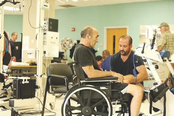
New stroke rehabilitation recommendations recognize role of interprofessional team
New guidelines issued jointly by the American Heart Association and American Stroke Association are the first to define multidisciplinary terms for stroke rehabilitation in adults. The guidelines were published online May 4 in the journal Stroke.
Previous guidelines have focused on medical issues related to stroke and its immediate aftermath, but the new document recognizes the importance of rehabilitation to quality of life, according to lead author Carolee J. Winstein, Ph.D., of the University of Southern California, Los Angeles, and colleagues (Stroke. 2016 May 4. doi: 10.1161/STR.0000000000000098).

In response to health care reform, the temptation remains to pare down postacute care and rehabilitation, “but without recognition of their clinical impact and ability to reduce the risk of downstream medical morbidity resulting from immobility, depression, loss of autonomy, and reduced functional independence,” the researchers wrote.
Considering the importance of early rehabilitation after stroke, the guidelines emphasize that initial rehabilitation occur in an inpatient facility rather than a nursing home setting, and that patients and caregivers be educated about fall prevention to reduce the risk of accidents once the patient arrives at home.
The guidelines consist of five sections: The Rehabilitation Program, Prevention and Medical Management of Comorbidities, Assessment, Sensorimotor Impairments and Activities, and Transitions in Care and Community Rehabilitation.
• The Rehabilitation Program. Options for poststroke rehabilitation should start with inpatient care, the researchers said. “It is recommended that stroke patients who are candidates for postacute rehabilitation receive organized, coordinated, interprofessional care,” they noted. Evidence suggests that such care reduces mortality and improves recovery and independence.
• Prevention and Medical Management of Comorbidities. Stroke is associated with a range of comorbid conditions, and the recommendations highlight several areas for which stroke patients should be evaluated regularly during rehabilitation, including skin breakdowns, hemiplegic limbs, urinary retention, and deep vein thrombosis.
• Assessment. Assessment of stroke patients should include communication abilities, motor ability, and ability to perform activities of daily living, the researchers wrote. “Evaluation of a stroke survivor’s rehabilitation needs is best performed by an interprofessional team that can include a physician with expertise in rehabilitation, nursing, PT, OT, speech therapy, case management, psychology, and orthotics,” they said.
• Sensorimotor Impairments and Activities. The researchers identified evidence to support management of dysphagia, which occurs in 42%-67% of stroke patients within 3 days after stroke. Screening and attention to cognitive impairment, limb apraxia, and hemispatial neglect should be part of stroke rehabilitation, they noted. Additional recommendations include tailored exercise programs; eye, speech, and balance therapy; mobility training; and a cognitively enriching environment that may include books, music, computer, and virtual reality games.
• Transitions in Care and Community Rehabilitation. The transition process should involve individualized discharge plans depending on the patient’s need for additional services, the researchers noted. Alternative communication methods such as telemedicine, web-based support, are reasonable options, especially for patients in rural areas, they noted. Resuming a previous lifestyle including work, driving, and recreational activities is recommended based on a tailored assessment of the individual’s needs and abilities for a particular job or activity.
Successful stroke rehabilitation is a team effort across medical specialties, the researchers emphasized. Communication and coordination “are paramount in maximizing the effectiveness and efficiency of rehabilitation and underlie this entire guideline,” they said.
Future directions for research in stroke rehabilitation include exploring models of care that view stroke as a chronic condition, developing more effective predictor models, and using new technology for personalized interventions, the researchers added.
The statement is the eighth and last in a set of stroke guidelines that completes the ASA recommendations on the continuum of care for stroke patients and families. The authors based the recommendations on a review of the relevant medical literature through 2014.
The guidelines are endorsed by the American Academy of Physical Medicine and Rehabilitation, the American Physical Therapy Association, the American Occupational Therapy Association, the American Society of Neurorehabilitation, and the American Congress of Rehabilitation Medicine. The American Academy of Neurology affirmed the value of the guidelines as an educational tool for neurologists.
Dr. Winstein has received research grants from the National Institutes of Health and serves as a consultant to St. Jude Medical Business Services. Some of the other authors reported receiving grants from the National Institutes of Health and/or industry, as well as serving as consultants or on an advisory board for various companies.
The guidelines are important to establish some form of common understanding or baseline of currently known best practices to treat and deliver rehabilitation to patients with stroke. Given recent health system studies suggesting significant variation in practices and cost across the United States, it is appropriate to describe guidelines that can set up standard practice. In addition, changes in the health care system toward population health make the creation of guidelines important, keeping in mind the current practices and frequent problems encountered when delivering rehabilitation to this patient population.
I think these guidelines might have a small chance to change clinical practice. On one side, it will provoke current providers to discuss what they are doing for this patient population, and what potential changes they need to do relative to the guidelines. However, it is also likely that current providers will not be compelled to change their habits for delivering care based on these guidelines.

|
Dr. Pablo Celnik |
On the other hand, while regulatory organization and insurance companies will likely look into this document to justify their practices, it is difficult to believe that they will change their policies based on this document. Nevertheless, this document, together with others, has the potential to create some momentum to advocate for best practices in stroke rehabilitation in a more homogeneous manner across the entire United States.
Rehabilitation has traditionally focused on improving the health and function of the individual. This is typically accomplished by training stroke survivors to deal with their deficits. However, many of the rehabilitation interventions have not been designed with a focus on restoring neurologic deficits. This may stem from a limited understanding of the mechanisms by which the brain recovers after stroke. However, recent studies have shown some of these mechanisms, and suggest potential approaches to augment restoration of neurologic function. Thus, more studies have tackled this problem by testing novel interventions such as high-intensity rehabilitation training using robots and games soon after the stroke (i.e., increasing the dose of training beyond that reported in the guidelines); carefully pairing noninvasive brain stimulation with training; or using exercises that take advantage of spared neurologic areas. Although these techniques have shown benefits in the laboratory setting, clinical trials are needed to determine whether they can be efficacious for broader stroke patient populations.
In sum, interventions specifically designed to restore neurologic deficits during stroke rehabilitation represent an important area of research that will need to be translated to the clinical setting and become part of best practices if proven effective. If we can reduce the amount of neurologic deficits after a stroke, it should result in a dramatic improvement of function at the individual level.
Dr. Pablo Celnik is a professor of physical medicine and rehabilitation at Johns Hopkins University in Baltimore, and medical director of the university’s outpatient neurorehabilitation program. He has received research funding from the National Institutes of Health. He was not involved in the drafting of the guidelines.
The guidelines are important to establish some form of common understanding or baseline of currently known best practices to treat and deliver rehabilitation to patients with stroke. Given recent health system studies suggesting significant variation in practices and cost across the United States, it is appropriate to describe guidelines that can set up standard practice. In addition, changes in the health care system toward population health make the creation of guidelines important, keeping in mind the current practices and frequent problems encountered when delivering rehabilitation to this patient population.
I think these guidelines might have a small chance to change clinical practice. On one side, it will provoke current providers to discuss what they are doing for this patient population, and what potential changes they need to do relative to the guidelines. However, it is also likely that current providers will not be compelled to change their habits for delivering care based on these guidelines.
|
|
Dr. Pablo Celnik |
On the other hand, while regulatory organization and insurance companies will likely look into this document to justify their practices, it is difficult to believe that they will change their policies based on this document. Nevertheless, this document, together with others, has the potential to create some momentum to advocate for best practices in stroke rehabilitation in a more homogeneous manner across the entire United States.
Rehabilitation has traditionally focused on improving the health and function of the individual. This is typically accomplished by training stroke survivors to deal with their deficits. However, many of the rehabilitation interventions have not been designed with a focus on restoring neurologic deficits. This may stem from a limited understanding of the mechanisms by which the brain recovers after stroke. However, recent studies have shown some of these mechanisms, and suggest potential approaches to augment restoration of neurologic function. Thus, more studies have tackled this problem by testing novel interventions such as high-intensity rehabilitation training using robots and games soon after the stroke (i.e., increasing the dose of training beyond that reported in the guidelines); carefully pairing noninvasive brain stimulation with training; or using exercises that take advantage of spared neurologic areas. Although these techniques have shown benefits in the laboratory setting, clinical trials are needed to determine whether they can be efficacious for broader stroke patient populations.
In sum, interventions specifically designed to restore neurologic deficits during stroke rehabilitation represent an important area of research that will need to be translated to the clinical setting and become part of best practices if proven effective. If we can reduce the amount of neurologic deficits after a stroke, it should result in a dramatic improvement of function at the individual level.
Dr. Pablo Celnik is a professor of physical medicine and rehabilitation at Johns Hopkins University in Baltimore, and medical director of the university’s outpatient neurorehabilitation program. He has received research funding from the National Institutes of Health. He was not involved in the drafting of the guidelines.
New guidelines issued jointly by the American Heart Association and American Stroke Association are the first to define multidisciplinary terms for stroke rehabilitation in adults. The guidelines were published online May 4 in the journal Stroke.
Previous guidelines have focused on medical issues related to stroke and its immediate aftermath, but the new document recognizes the importance of rehabilitation to quality of life, according to lead author Carolee J. Winstein, Ph.D., of the University of Southern California, Los Angeles, and colleagues (Stroke. 2016 May 4. doi: 10.1161/STR.0000000000000098).
In response to health care reform, the temptation remains to pare down postacute care and rehabilitation, “but without recognition of their clinical impact and ability to reduce the risk of downstream medical morbidity resulting from immobility, depression, loss of autonomy, and reduced functional independence,” the researchers wrote.
Considering the importance of early rehabilitation after stroke, the guidelines emphasize that initial rehabilitation occur in an inpatient facility rather than a nursing home setting, and that patients and caregivers be educated about fall prevention to reduce the risk of accidents once the patient arrives at home.
The guidelines consist of five sections: The Rehabilitation Program, Prevention and Medical Management of Comorbidities, Assessment, Sensorimotor Impairments and Activities, and Transitions in Care and Community Rehabilitation.
• The Rehabilitation Program. Options for poststroke rehabilitation should start with inpatient care, the researchers said. “It is recommended that stroke patients who are candidates for postacute rehabilitation receive organized, coordinated, interprofessional care,” they noted. Evidence suggests that such care reduces mortality and improves recovery and independence.
• Prevention and Medical Management of Comorbidities. Stroke is associated with a range of comorbid conditions, and the recommendations highlight several areas for which stroke patients should be evaluated regularly during rehabilitation, including skin breakdowns, hemiplegic limbs, urinary retention, and deep vein thrombosis.
• Assessment. Assessment of stroke patients should include communication abilities, motor ability, and ability to perform activities of daily living, the researchers wrote. “Evaluation of a stroke survivor’s rehabilitation needs is best performed by an interprofessional team that can include a physician with expertise in rehabilitation, nursing, PT, OT, speech therapy, case management, psychology, and orthotics,” they said.
• Sensorimotor Impairments and Activities. The researchers identified evidence to support management of dysphagia, which occurs in 42%-67% of stroke patients within 3 days after stroke. Screening and attention to cognitive impairment, limb apraxia, and hemispatial neglect should be part of stroke rehabilitation, they noted. Additional recommendations include tailored exercise programs; eye, speech, and balance therapy; mobility training; and a cognitively enriching environment that may include books, music, computer, and virtual reality games.
• Transitions in Care and Community Rehabilitation. The transition process should involve individualized discharge plans depending on the patient’s need for additional services, the researchers noted. Alternative communication methods such as telemedicine, web-based support, are reasonable options, especially for patients in rural areas, they noted. Resuming a previous lifestyle including work, driving, and recreational activities is recommended based on a tailored assessment of the individual’s needs and abilities for a particular job or activity.
Successful stroke rehabilitation is a team effort across medical specialties, the researchers emphasized. Communication and coordination “are paramount in maximizing the effectiveness and efficiency of rehabilitation and underlie this entire guideline,” they said.
Future directions for research in stroke rehabilitation include exploring models of care that view stroke as a chronic condition, developing more effective predictor models, and using new technology for personalized interventions, the researchers added.
The statement is the eighth and last in a set of stroke guidelines that completes the ASA recommendations on the continuum of care for stroke patients and families. The authors based the recommendations on a review of the relevant medical literature through 2014.
The guidelines are endorsed by the American Academy of Physical Medicine and Rehabilitation, the American Physical Therapy Association, the American Occupational Therapy Association, the American Society of Neurorehabilitation, and the American Congress of Rehabilitation Medicine. The American Academy of Neurology affirmed the value of the guidelines as an educational tool for neurologists.
Dr. Winstein has received research grants from the National Institutes of Health and serves as a consultant to St. Jude Medical Business Services. Some of the other authors reported receiving grants from the National Institutes of Health and/or industry, as well as serving as consultants or on an advisory board for various companies.
FROM STROKE
Autism screening rises after process-based training
A 3- to 6-month learning program for pediatric and family medicine providers significantly improved their screening for autism spectrum disorders (ASD) at 18- and 24-month well child visits, based on data from 26 primary care practices that participated in the program and from 43 physicians who completed surveys before and after the program, according to findings published online May 5 in Pediatrics.
“Unlike traditional continuing medical education, the LC [learning collaborative] focused on improvement of processes of care at the practice level,” wrote Dr. Paul S. Carbone and his colleagues of the University of Utah, Salt Lake City. The first signs of ASD can be present as early as 2 years of age, but often remain undiagnosed for lack of screening at 18- and 24-month visits, they noted.

Rates of documented ASD screening among toddlers increased from 16% before starting the program to 91% during the last month of the program, and 70% of the practices sustained the 91% screening rate 4 years later.
Physician self-efficacy improved significantly from baseline to after the program on the nine autism conditions (such as sleep problems, constipation, and attention deficit/hyperactivity disorder [ADHD]) and seven autism needs (such as making referrals, addressing developmental concerns, and identifying community support services) included in the survey. On a scale of 1 to 10, the average physician’s progress rating was 6.5 after completing the program.
“A LC using the methods we describe is a successful approach to improving the early identification and ongoing care of children with ASD in primary care practices,” they researchers said.
Read the whole article at Pediatrics (2016 May. doi: 10.1542/peds.2015-1850).
A 3- to 6-month learning program for pediatric and family medicine providers significantly improved their screening for autism spectrum disorders (ASD) at 18- and 24-month well child visits, based on data from 26 primary care practices that participated in the program and from 43 physicians who completed surveys before and after the program, according to findings published online May 5 in Pediatrics.
“Unlike traditional continuing medical education, the LC [learning collaborative] focused on improvement of processes of care at the practice level,” wrote Dr. Paul S. Carbone and his colleagues of the University of Utah, Salt Lake City. The first signs of ASD can be present as early as 2 years of age, but often remain undiagnosed for lack of screening at 18- and 24-month visits, they noted.
Rates of documented ASD screening among toddlers increased from 16% before starting the program to 91% during the last month of the program, and 70% of the practices sustained the 91% screening rate 4 years later.
Physician self-efficacy improved significantly from baseline to after the program on the nine autism conditions (such as sleep problems, constipation, and attention deficit/hyperactivity disorder [ADHD]) and seven autism needs (such as making referrals, addressing developmental concerns, and identifying community support services) included in the survey. On a scale of 1 to 10, the average physician’s progress rating was 6.5 after completing the program.
“A LC using the methods we describe is a successful approach to improving the early identification and ongoing care of children with ASD in primary care practices,” they researchers said.
Read the whole article at Pediatrics (2016 May. doi: 10.1542/peds.2015-1850).
FROM PEDIATRICS

Light mesh doesn’t outweigh heavy mesh for hernia repair
Lightweight mesh has no significant benefit over heavyweight mesh for inguinal hernia repair and was associated with greater pain and higher risk of recurrence, based on data from a randomized trial of 950 patients published in the May issue of the Annals of Surgery.
Although the use of mesh for hernia surgery has been associated with lower recurrence rates and less chronic pain in open hernia repair procedures, the benefits of lightweight vs. heavyweight mesh for laparoscopic procedures has not been well studied, “and there is no consensus which type of mesh is optimal in these procedures,” wrote Dr. Josephina P.J. Burgmans of Diakonessenhuis Utrecht/Zeist (the Netherlands) and colleagues (Ann. Surg. 2016;263[5]:862-6).

To compare the clinical outcomes for patients who received lightweight vs. heavyweight mesh in hernia repairs, the researchers randomized 478 patients to a lightweight-mesh group and 471 to a heavyweight-mesh group. One patient was excluded because the type of mesh was unknown. The study population included men older than 18 years with a primary, reducible, unilateral inguinal hernia who underwent surgery at a single center with one of four surgeons between March 2010 and October 2012. Demographic characteristics were similar between the two groups, as were preoperation pain, operation time, and complications during and after surgery.
At 1 year post surgery, the prevalence of reported relevant pain was significantly higher in the lightweight group, compared with the heavyweight group (2.9% vs. 0.7%). The difference remained statistically significant at 2 years’ follow-up, with relevant pain reported in 3.0% of the lightweight group and 0.9% of the heavyweight group; the difference also remained significant after controlling for factors including age, body mass index, hernia type, severe preoperative pain, recurrence, and surgeon).
Recurrence rates were similar between the two groups at 3 months (two cases in each group), but recurrences became more common in the lightweight group, compared with the heavyweight group, at 1 year (1.7% vs. 0.6%) and significantly more common at 2 years (2.7% vs. 0.8%). The difference remained significant after controlling for multiple variables including hernia type, operating time, surgeon, and body mass index.
No significant differences in foreign body feeling, testicular pain, or sexual-related pain or discomfort were reported between the two groups at the 1-year and 2-year follow-ups.
The study’s strengths include the large patient population and prospective data collection, the researchers noted. The findings were limited by the small number of patients with relevant pain at 1 and 2 years; the homogenous, male-only study population; and the use of questionnaires, which might affect the accuracy of recurrence-rate reports. The results, however, suggest that “there is no benefit for lightweight [meshes] and a conventional heavyweight standard polypropylene 10 cm × 15 cm mesh is recommended for laparoscopic inguinal hernia repair,” Dr. Burgmans and associates wrote
The study was sponsored in part by a grant to the Hernia Centre Zeist from Johnson & Johnston. The researchers had no other relevant financial conflicts to disclose.
Lightweight mesh has no significant benefit over heavyweight mesh for inguinal hernia repair and was associated with greater pain and higher risk of recurrence, based on data from a randomized trial of 950 patients published in the May issue of the Annals of Surgery.
Although the use of mesh for hernia surgery has been associated with lower recurrence rates and less chronic pain in open hernia repair procedures, the benefits of lightweight vs. heavyweight mesh for laparoscopic procedures has not been well studied, “and there is no consensus which type of mesh is optimal in these procedures,” wrote Dr. Josephina P.J. Burgmans of Diakonessenhuis Utrecht/Zeist (the Netherlands) and colleagues (Ann. Surg. 2016;263[5]:862-6).
To compare the clinical outcomes for patients who received lightweight vs. heavyweight mesh in hernia repairs, the researchers randomized 478 patients to a lightweight-mesh group and 471 to a heavyweight-mesh group. One patient was excluded because the type of mesh was unknown. The study population included men older than 18 years with a primary, reducible, unilateral inguinal hernia who underwent surgery at a single center with one of four surgeons between March 2010 and October 2012. Demographic characteristics were similar between the two groups, as were preoperation pain, operation time, and complications during and after surgery.
At 1 year post surgery, the prevalence of reported relevant pain was significantly higher in the lightweight group, compared with the heavyweight group (2.9% vs. 0.7%). The difference remained statistically significant at 2 years’ follow-up, with relevant pain reported in 3.0% of the lightweight group and 0.9% of the heavyweight group; the difference also remained significant after controlling for factors including age, body mass index, hernia type, severe preoperative pain, recurrence, and surgeon).
Recurrence rates were similar between the two groups at 3 months (two cases in each group), but recurrences became more common in the lightweight group, compared with the heavyweight group, at 1 year (1.7% vs. 0.6%) and significantly more common at 2 years (2.7% vs. 0.8%). The difference remained significant after controlling for multiple variables including hernia type, operating time, surgeon, and body mass index.
No significant differences in foreign body feeling, testicular pain, or sexual-related pain or discomfort were reported between the two groups at the 1-year and 2-year follow-ups.
The study’s strengths include the large patient population and prospective data collection, the researchers noted. The findings were limited by the small number of patients with relevant pain at 1 and 2 years; the homogenous, male-only study population; and the use of questionnaires, which might affect the accuracy of recurrence-rate reports. The results, however, suggest that “there is no benefit for lightweight [meshes] and a conventional heavyweight standard polypropylene 10 cm × 15 cm mesh is recommended for laparoscopic inguinal hernia repair,” Dr. Burgmans and associates wrote
The study was sponsored in part by a grant to the Hernia Centre Zeist from Johnson & Johnston. The researchers had no other relevant financial conflicts to disclose.
FROM ANNALS OF SURGERY
Key clinical point: Lightweight mesh was associated with more pain and higher recurrence risk 1 and 2 years after surgery.
Major finding: Two years after surgery, relevant pain was reported in 3.0% of the lightweight-mesh patients vs. 0.9% of heavyweight-mesh patients.
Data source: A randomized trial including 950 adult men with inguinal hernias.
Disclosures: The study was sponsored in part by a grant to the Hernia Centre Zeist from Johnson & Johnston. The researchers had no other relevant financial conflicts to disclose.
Cell-based strategy curbs constipation
Treatment at the nanomolecular level may become an alternative for various types of constipation, in particular for the opioid-induced constipation that is common after surgery, based on data from a proof of concept study involving mice and a small-molecule activator.
“Activation of the cystic fibrosis transmembrane conductance regulator (CFTR) chloride channel is the primary pathway that drives fluid secretion in the intestine, which maintains lubrication of luminal contents,” wrote Dr. Onur Cil of the University of California, San Francisco, and colleagues. The researchers examined whether direct activation of the CFTR would prompt fluid secretion and reverse stool dehydration when applied to constipated mice. The findings were published online in the May issue of the journal Cellular and Molecular Gastroenterology and Hepatology (2016. doi: 10.1016/j.jcmgh.2015.12.010).
The researchers identified a promising activator, the phenylquinoxalinone CFTRact-J027. Mice received up to 10 mg/kg CFTRact-J027 either orally or intraperitoneally (IP), with doses including 0.1 mg/kg, 0.3 mg/kg, 1 mg/kg, 3 mg/kg, and 10 mg/kg.
Overall, IP doses of CFTRact-J027 at 10 mg/kg normalized stool in the constipated mice, and dose-response studies showed a 50% effective dose of 2 mg/kg in these mice.
When given orally, CFTRact-J027 “normalized stool output and water content in a loperamide-induced mouse model of constipation with a 50% effective dose of approximately 0.5 mg/kg,” that was significantly lower than the IP administration, the researchers noted. An oral dose of 10 mg/kg CFTRact-J027 1 hour before inducing constipation also was effective in normalizing stool output and water content in loperamide-treated mice, with no effect in control nonconstipated mice.
The activator was not effective against constipation in cystic fibrosis mice that were missing a functional CFTR, they added.
The researchers used an in vivo closed loop model to specifically test the effects of CFTRact-J027 on intestinal fluid secretion and absorption and found that CFTRact-J027 caused “a 140% increase in loop weight/length ratio, indicating fluid secretion into the intestinal lumen in wild-type mice.” However, there was no effect in cystic fibrosis mice, further supporting the CFTR-selective mechanism of action, the researchers said.
As for potential toxic effects of the treatment, CFTRact-J027 showed no impact on the major serum chemistry and blood parameters of the mice after 7 days, and had no apparent impact on body weight. No accumulation of fluid (the most significant potential adverse effect) was noted in the airway or lungs of the treated mice.
Additional toxicity data are needed to continue preclinical development, the researchers said. However, “our data provide evidence for the prosecretory action of a CFTR activator in mouse intestine and proof of concept for its use in the treatment of various types of constipation, which could include opioid-induced constipation, chronic idiopathic constipation, and irritable bowel syndrome with constipation predominance,” they wrote. In addition, a CFTR activator similar to that used in this study may have clinical applications for other conditions including asthma, dry eye, cholestatic liver disease, chronic obstructive pulmonary disease and bronchitis, and cigarette smoke–induced lung dysfunction, they added.
Dr. Cil and two coauthors are inventors on a provisional patent filing, with rights owned by the University of California, San Francisco. The study was funded in part by several grants from organizations including the National Institutes of Health and the Cystic Fibrosis Foundation.
FROM CELLULAR AND MOLECULAR GASTROENTEROLOGY AND HEPATOLOGY
Key clinical point: Treatment at the nanomolecular level may become an alternative for various types of constipation.
Major finding: Oral doses of CFTRact-J027 normalized stool output and water content in a loperamide-induced mouse model of constipation with a 50% effective dose of approximately 0.5 mg/kg.
Data source: A proof-of-concept study in which a cell-based screen was performed for 120,000 druglike, synthetic small molecules that were then tested in constipation-induced mice and control mice.
Disclosures: Dr. Cil and two coauthors are inventors on a provisional patent filing, with rights owned by the University of California, San Francisco. The study was funded in part by several grants from organizations including the National Institutes of Health and the Cystic Fibrosis Foundation.
Norovirus costs $60 billion worldwide, making inroads in U.S.
The global economic impact of norovirus is roughly $60 billion in societal costs and $4 billion in direct health care costs per year, according to an simulation model analysis.
Most of the costs (84%-99%, varying by region) resulted from lost productivity. The total societal cost was highest in children younger than 5 years, nearly $40 billion, compared with $20 billion for all other age groups combined, but the cost per illness was highest among adults aged 55 years and older.
Disease incidence was similar across all countries in the simulation model that included 233 countries stratified by World Health Organization region and globally but high-income countries accounted for 62% of the global health system costs, wrote Sarah M. Bartsch of Johns Hopkins University, Baltimore, and colleagues (PLoS ONE 2016. doi: 10.1371/journal.pone.0151219).

In the United States, norovirus was the most common pathogen identified in cases of acute gastroenteritis (AGE, 6%), followed by rotavirus and sapovirus (2% for both) and astrovirus (1%), according to data from a population-based study, including 1,099 stool specimens collected between August 2012 and September 2013. These findings were published in a separate study (PLoS ONE 2016. doi: 10.1371/journal.pone.0148395).
Overall, U.S. children younger than 5 years of age had the highest prevalence of norovirus; outpatient incidence and community incidences were 25.6 and 152.2 per 1,000 person-years, respectively. The next highest norovirus prevalence occurred in adults older than 65 years; outpatient and community incidence were 7.8 and 7.8 per 1,000 person-years, respectively. The study provides “the first U.S. laboratory-based age-specific norovirus AGE incidence rates in both community and outpatient settings,” wrote Scott P. Grytdal of the Centers for Disease Control and Prevention in Atlanta, and colleagues.
The global economic impact of norovirus is roughly $60 billion in societal costs and $4 billion in direct health care costs per year, according to an simulation model analysis.
Most of the costs (84%-99%, varying by region) resulted from lost productivity. The total societal cost was highest in children younger than 5 years, nearly $40 billion, compared with $20 billion for all other age groups combined, but the cost per illness was highest among adults aged 55 years and older.
Disease incidence was similar across all countries in the simulation model that included 233 countries stratified by World Health Organization region and globally but high-income countries accounted for 62% of the global health system costs, wrote Sarah M. Bartsch of Johns Hopkins University, Baltimore, and colleagues (PLoS ONE 2016. doi: 10.1371/journal.pone.0151219).
In the United States, norovirus was the most common pathogen identified in cases of acute gastroenteritis (AGE, 6%), followed by rotavirus and sapovirus (2% for both) and astrovirus (1%), according to data from a population-based study, including 1,099 stool specimens collected between August 2012 and September 2013. These findings were published in a separate study (PLoS ONE 2016. doi: 10.1371/journal.pone.0148395).
Overall, U.S. children younger than 5 years of age had the highest prevalence of norovirus; outpatient incidence and community incidences were 25.6 and 152.2 per 1,000 person-years, respectively. The next highest norovirus prevalence occurred in adults older than 65 years; outpatient and community incidence were 7.8 and 7.8 per 1,000 person-years, respectively. The study provides “the first U.S. laboratory-based age-specific norovirus AGE incidence rates in both community and outpatient settings,” wrote Scott P. Grytdal of the Centers for Disease Control and Prevention in Atlanta, and colleagues.
FROM PLOS ONE
