User login
Dexlansoprazole Approved to Treat Heartburn in Younger Patients
The Food and Drug Administration has approved dexlansoprazole (Dexilant) for gastroesophageal reflux disease patients aged 12-17 years, according to a press release from Takeda Pharmaceuticals.
FDA approval of dexlansoprazole for this age group is based on adequate and well controlled studies proving its efficacy in adults, as well data on safety, pharmacokinetics, and efficacy in children 12-17 years. Dexlansoprazole is approved for younger patients to maintain healing of erosive esophagus and heartburn relief for 16 weeks, healing all grades of EE for 8 weeks, and treating heartburn associated with GERD for 4 weeks.

Ddexlansoprazole is a proton pump inhibitor, prescribed to relieve heartburn associated with GERD and maintain healed erosive esophagus. The most common side effects seen in individuals aged 12-17 were headache, abdominal pain, diarrhea, nasopharyngitis, and oropharyngeal pain.
“Takeda is pleased with the FDA’s approval to expand the access of Dexilant to younger patients with GERD. This new approval provides an alternative treatment option for appropriate patients with this condition. With our more than 20 years of experience in gastroenterology, we remain dedicated to all patients living with these conditions,” noted Thomas Gibbs, senior vice president of general medicine at Takeda.
Find the full press release on the Takeda Pharmaceuticals website.
The Food and Drug Administration has approved dexlansoprazole (Dexilant) for gastroesophageal reflux disease patients aged 12-17 years, according to a press release from Takeda Pharmaceuticals.
FDA approval of dexlansoprazole for this age group is based on adequate and well controlled studies proving its efficacy in adults, as well data on safety, pharmacokinetics, and efficacy in children 12-17 years. Dexlansoprazole is approved for younger patients to maintain healing of erosive esophagus and heartburn relief for 16 weeks, healing all grades of EE for 8 weeks, and treating heartburn associated with GERD for 4 weeks.
Ddexlansoprazole is a proton pump inhibitor, prescribed to relieve heartburn associated with GERD and maintain healed erosive esophagus. The most common side effects seen in individuals aged 12-17 were headache, abdominal pain, diarrhea, nasopharyngitis, and oropharyngeal pain.
“Takeda is pleased with the FDA’s approval to expand the access of Dexilant to younger patients with GERD. This new approval provides an alternative treatment option for appropriate patients with this condition. With our more than 20 years of experience in gastroenterology, we remain dedicated to all patients living with these conditions,” noted Thomas Gibbs, senior vice president of general medicine at Takeda.
Find the full press release on the Takeda Pharmaceuticals website.
The Food and Drug Administration has approved dexlansoprazole (Dexilant) for gastroesophageal reflux disease patients aged 12-17 years, according to a press release from Takeda Pharmaceuticals.
FDA approval of dexlansoprazole for this age group is based on adequate and well controlled studies proving its efficacy in adults, as well data on safety, pharmacokinetics, and efficacy in children 12-17 years. Dexlansoprazole is approved for younger patients to maintain healing of erosive esophagus and heartburn relief for 16 weeks, healing all grades of EE for 8 weeks, and treating heartburn associated with GERD for 4 weeks.
Ddexlansoprazole is a proton pump inhibitor, prescribed to relieve heartburn associated with GERD and maintain healed erosive esophagus. The most common side effects seen in individuals aged 12-17 were headache, abdominal pain, diarrhea, nasopharyngitis, and oropharyngeal pain.
“Takeda is pleased with the FDA’s approval to expand the access of Dexilant to younger patients with GERD. This new approval provides an alternative treatment option for appropriate patients with this condition. With our more than 20 years of experience in gastroenterology, we remain dedicated to all patients living with these conditions,” noted Thomas Gibbs, senior vice president of general medicine at Takeda.
Find the full press release on the Takeda Pharmaceuticals website.

Dexlansoprazole approved to treat heartburn in younger patients
The Food and Drug Administration has approved dexlansoprazole (Dexilant) for gastroesophageal reflux disease patients aged 12-17 years, according to a press release from Takeda Pharmaceuticals.
FDA approval of dexlansoprazole for this age group is based on adequate and well controlled studies proving its efficacy in adults, as well data on safety, pharmacokinetics, and efficacy in children 12-17 years. Dexlansoprazole is approved for younger patients to maintain healing of erosive esophagus and heartburn relief for 16 weeks, healing all grades of EE for 8 weeks, and treating heartburn associated with GERD for 4 weeks.

Ddexlansoprazole is a proton pump inhibitor, prescribed to relieve heartburn associated with GERD and maintain healed erosive esophagus. The most common side effects seen in individuals aged 12-17 were headache, abdominal pain, diarrhea, nasopharyngitis, and oropharyngeal pain.
“Takeda is pleased with the FDA’s approval to expand the access of Dexilant to younger patients with GERD. This new approval provides an alternative treatment option for appropriate patients with this condition. With our more than 20 years of experience in gastroenterology, we remain dedicated to all patients living with these conditions,” noted Thomas Gibbs, senior vice president of general medicine at Takeda.
Find the full press release on the Takeda Pharmaceuticals website.
The Food and Drug Administration has approved dexlansoprazole (Dexilant) for gastroesophageal reflux disease patients aged 12-17 years, according to a press release from Takeda Pharmaceuticals.
FDA approval of dexlansoprazole for this age group is based on adequate and well controlled studies proving its efficacy in adults, as well data on safety, pharmacokinetics, and efficacy in children 12-17 years. Dexlansoprazole is approved for younger patients to maintain healing of erosive esophagus and heartburn relief for 16 weeks, healing all grades of EE for 8 weeks, and treating heartburn associated with GERD for 4 weeks.
Ddexlansoprazole is a proton pump inhibitor, prescribed to relieve heartburn associated with GERD and maintain healed erosive esophagus. The most common side effects seen in individuals aged 12-17 were headache, abdominal pain, diarrhea, nasopharyngitis, and oropharyngeal pain.
“Takeda is pleased with the FDA’s approval to expand the access of Dexilant to younger patients with GERD. This new approval provides an alternative treatment option for appropriate patients with this condition. With our more than 20 years of experience in gastroenterology, we remain dedicated to all patients living with these conditions,” noted Thomas Gibbs, senior vice president of general medicine at Takeda.
Find the full press release on the Takeda Pharmaceuticals website.
The Food and Drug Administration has approved dexlansoprazole (Dexilant) for gastroesophageal reflux disease patients aged 12-17 years, according to a press release from Takeda Pharmaceuticals.
FDA approval of dexlansoprazole for this age group is based on adequate and well controlled studies proving its efficacy in adults, as well data on safety, pharmacokinetics, and efficacy in children 12-17 years. Dexlansoprazole is approved for younger patients to maintain healing of erosive esophagus and heartburn relief for 16 weeks, healing all grades of EE for 8 weeks, and treating heartburn associated with GERD for 4 weeks.
Ddexlansoprazole is a proton pump inhibitor, prescribed to relieve heartburn associated with GERD and maintain healed erosive esophagus. The most common side effects seen in individuals aged 12-17 were headache, abdominal pain, diarrhea, nasopharyngitis, and oropharyngeal pain.
“Takeda is pleased with the FDA’s approval to expand the access of Dexilant to younger patients with GERD. This new approval provides an alternative treatment option for appropriate patients with this condition. With our more than 20 years of experience in gastroenterology, we remain dedicated to all patients living with these conditions,” noted Thomas Gibbs, senior vice president of general medicine at Takeda.
Find the full press release on the Takeda Pharmaceuticals website.

Cervical spine injury common in children under age 2 with abusive head trauma
BALTIMORE – Cervical spine injuries may be more prevalent than previously thought among young children who sustain abusive head trauma, results of a 4-year, multicenter, retrospective study presented at the annual meeting of the Pediatric Academic Societies have shown.
Of children with abusive head trauma (AHT) who received cervical CT or MRI scans, 31% had abnormal findings. The most common cervical spine injuries were hemorrhagic (23.7%), with ligamentous abnormalities less common (8.7%).
This study helps fill a gap in the existing literature on the risk for cervical spine injuries for children who have suffered abusive head trauma, said Kate Henry, MD, in presenting the results of the study, conducted with her colleagues at the Center for Pediatric Clinical Effectiveness at the Children’s Hospital of Philadelphia.
Previous research in the area has concentrated on older children and on motor vehicle crashes, but “young children are different. They are preverbal, and the physical exam can be difficult. Also, the developing spine has a unique anatomy” that can change injury patterns and make imaging interpretation challenging, said Dr. Henry. Motor vehicle crashes make up less than 15% of the cases of traumatic brain injury (TBI) in young children, with falls being responsible for about half, and AHT accounting for 19%-25%, she said.
The study included patients younger than 2 years with an ICD-9 code for TBI associated with a hospital admission or emergency department visit. For final inclusion, patients also had to have MRI or CT confirmation of an intracranial injury. Patients who sustained a motor vehicle collision, were readmitted for complications of a prior injury, or were hospitalized following birth were excluded.
The retrospective chart review of records from four urban children’s hospitals found 3,170 patients who met all criteria except positive intracranial imaging findings; a stratified random sampling reduced the number to 664 charts eligible for review. The final TBI cohort included 329 patients who met the imaging criteria and were not excluded for other reasons.
For these 329 records, Dr. Henry and her colleagues collected physical exam findings, radiology reports of abnormal imaging findings, and the care team’s documentation of their assessment of the etiology of the injury.
The medical team’s assessment, together with the assessment of the child protection team (if it was engaged) were used to classify patients as having either “AHT” or “accidental TBI.”
The trauma-related cervical spine injuries considered as positive findings included ligamentous or soft tissue injury, spinal cord injury, vertebral dislocation or fracture, and extra-axial hemorrhage. “We had a high threshold for inclusion,” said Dr. Henry, explaining that only cervical, not high thoracic, findings were included. Additionally, she and her colleagues excluded radiologist interpretation of injuries as “nonspecific soft tissue findings” or a “possible” injury.
Overall, vertebral fractures or dislocations were seen in just 1.1% of patients with imaged AHT. Cord injuries were slightly more common, at 5.5%. Soft tissue or muscular findings were seen in 0.6% of patients. Some patients had multiple findings, so the overall 31.3% of patients with image-confirmed cervical injuries includes the 23.7% of patients with extra-axial bleed and the 8.7% with ligamentous injury.
Dr. Henry noted that the study was predicated on a certain “circularity of reasoning,” in that the results of spinal imaging could have an influence on the clinician assessment of etiology. The percentage of children diagnosed with AHT who received imaging of any kind was higher than for the all-cause TBI group, as well as for the subgroups of those with accidental TBI or indeterminate etiology. And Dr. Henry said there’s an inherent selection bias in including only those cases where imaging was performed: “The decision to image is unlikely to be random,” she said. However, if all AHT patients who did not receive imaging within the study period were considered to have normal findings, then 9.7% of AHT cases, or about 1 in 10, would still have abnormal findings.
These findings are important because currently, “There are no guidelines for use of cervical CT or MRI” for children with abusive head trauma, said Dr. Henry, and previous research in this area was based on a single-center study. Next steps should go beyond retrospective chart reviews, she said. “Prospective studies are needed to inform cervical imaging recommendations.”
Dr. Henry reported no relevant disclosures.
On Twitter @karioakes
BALTIMORE – Cervical spine injuries may be more prevalent than previously thought among young children who sustain abusive head trauma, results of a 4-year, multicenter, retrospective study presented at the annual meeting of the Pediatric Academic Societies have shown.
Of children with abusive head trauma (AHT) who received cervical CT or MRI scans, 31% had abnormal findings. The most common cervical spine injuries were hemorrhagic (23.7%), with ligamentous abnormalities less common (8.7%).
This study helps fill a gap in the existing literature on the risk for cervical spine injuries for children who have suffered abusive head trauma, said Kate Henry, MD, in presenting the results of the study, conducted with her colleagues at the Center for Pediatric Clinical Effectiveness at the Children’s Hospital of Philadelphia.
Previous research in the area has concentrated on older children and on motor vehicle crashes, but “young children are different. They are preverbal, and the physical exam can be difficult. Also, the developing spine has a unique anatomy” that can change injury patterns and make imaging interpretation challenging, said Dr. Henry. Motor vehicle crashes make up less than 15% of the cases of traumatic brain injury (TBI) in young children, with falls being responsible for about half, and AHT accounting for 19%-25%, she said.
The study included patients younger than 2 years with an ICD-9 code for TBI associated with a hospital admission or emergency department visit. For final inclusion, patients also had to have MRI or CT confirmation of an intracranial injury. Patients who sustained a motor vehicle collision, were readmitted for complications of a prior injury, or were hospitalized following birth were excluded.
The retrospective chart review of records from four urban children’s hospitals found 3,170 patients who met all criteria except positive intracranial imaging findings; a stratified random sampling reduced the number to 664 charts eligible for review. The final TBI cohort included 329 patients who met the imaging criteria and were not excluded for other reasons.
For these 329 records, Dr. Henry and her colleagues collected physical exam findings, radiology reports of abnormal imaging findings, and the care team’s documentation of their assessment of the etiology of the injury.
The medical team’s assessment, together with the assessment of the child protection team (if it was engaged) were used to classify patients as having either “AHT” or “accidental TBI.”
The trauma-related cervical spine injuries considered as positive findings included ligamentous or soft tissue injury, spinal cord injury, vertebral dislocation or fracture, and extra-axial hemorrhage. “We had a high threshold for inclusion,” said Dr. Henry, explaining that only cervical, not high thoracic, findings were included. Additionally, she and her colleagues excluded radiologist interpretation of injuries as “nonspecific soft tissue findings” or a “possible” injury.
Overall, vertebral fractures or dislocations were seen in just 1.1% of patients with imaged AHT. Cord injuries were slightly more common, at 5.5%. Soft tissue or muscular findings were seen in 0.6% of patients. Some patients had multiple findings, so the overall 31.3% of patients with image-confirmed cervical injuries includes the 23.7% of patients with extra-axial bleed and the 8.7% with ligamentous injury.
Dr. Henry noted that the study was predicated on a certain “circularity of reasoning,” in that the results of spinal imaging could have an influence on the clinician assessment of etiology. The percentage of children diagnosed with AHT who received imaging of any kind was higher than for the all-cause TBI group, as well as for the subgroups of those with accidental TBI or indeterminate etiology. And Dr. Henry said there’s an inherent selection bias in including only those cases where imaging was performed: “The decision to image is unlikely to be random,” she said. However, if all AHT patients who did not receive imaging within the study period were considered to have normal findings, then 9.7% of AHT cases, or about 1 in 10, would still have abnormal findings.
These findings are important because currently, “There are no guidelines for use of cervical CT or MRI” for children with abusive head trauma, said Dr. Henry, and previous research in this area was based on a single-center study. Next steps should go beyond retrospective chart reviews, she said. “Prospective studies are needed to inform cervical imaging recommendations.”
Dr. Henry reported no relevant disclosures.
On Twitter @karioakes
BALTIMORE – Cervical spine injuries may be more prevalent than previously thought among young children who sustain abusive head trauma, results of a 4-year, multicenter, retrospective study presented at the annual meeting of the Pediatric Academic Societies have shown.
Of children with abusive head trauma (AHT) who received cervical CT or MRI scans, 31% had abnormal findings. The most common cervical spine injuries were hemorrhagic (23.7%), with ligamentous abnormalities less common (8.7%).
This study helps fill a gap in the existing literature on the risk for cervical spine injuries for children who have suffered abusive head trauma, said Kate Henry, MD, in presenting the results of the study, conducted with her colleagues at the Center for Pediatric Clinical Effectiveness at the Children’s Hospital of Philadelphia.
Previous research in the area has concentrated on older children and on motor vehicle crashes, but “young children are different. They are preverbal, and the physical exam can be difficult. Also, the developing spine has a unique anatomy” that can change injury patterns and make imaging interpretation challenging, said Dr. Henry. Motor vehicle crashes make up less than 15% of the cases of traumatic brain injury (TBI) in young children, with falls being responsible for about half, and AHT accounting for 19%-25%, she said.
The study included patients younger than 2 years with an ICD-9 code for TBI associated with a hospital admission or emergency department visit. For final inclusion, patients also had to have MRI or CT confirmation of an intracranial injury. Patients who sustained a motor vehicle collision, were readmitted for complications of a prior injury, or were hospitalized following birth were excluded.
The retrospective chart review of records from four urban children’s hospitals found 3,170 patients who met all criteria except positive intracranial imaging findings; a stratified random sampling reduced the number to 664 charts eligible for review. The final TBI cohort included 329 patients who met the imaging criteria and were not excluded for other reasons.
For these 329 records, Dr. Henry and her colleagues collected physical exam findings, radiology reports of abnormal imaging findings, and the care team’s documentation of their assessment of the etiology of the injury.
The medical team’s assessment, together with the assessment of the child protection team (if it was engaged) were used to classify patients as having either “AHT” or “accidental TBI.”
The trauma-related cervical spine injuries considered as positive findings included ligamentous or soft tissue injury, spinal cord injury, vertebral dislocation or fracture, and extra-axial hemorrhage. “We had a high threshold for inclusion,” said Dr. Henry, explaining that only cervical, not high thoracic, findings were included. Additionally, she and her colleagues excluded radiologist interpretation of injuries as “nonspecific soft tissue findings” or a “possible” injury.
Overall, vertebral fractures or dislocations were seen in just 1.1% of patients with imaged AHT. Cord injuries were slightly more common, at 5.5%. Soft tissue or muscular findings were seen in 0.6% of patients. Some patients had multiple findings, so the overall 31.3% of patients with image-confirmed cervical injuries includes the 23.7% of patients with extra-axial bleed and the 8.7% with ligamentous injury.
Dr. Henry noted that the study was predicated on a certain “circularity of reasoning,” in that the results of spinal imaging could have an influence on the clinician assessment of etiology. The percentage of children diagnosed with AHT who received imaging of any kind was higher than for the all-cause TBI group, as well as for the subgroups of those with accidental TBI or indeterminate etiology. And Dr. Henry said there’s an inherent selection bias in including only those cases where imaging was performed: “The decision to image is unlikely to be random,” she said. However, if all AHT patients who did not receive imaging within the study period were considered to have normal findings, then 9.7% of AHT cases, or about 1 in 10, would still have abnormal findings.
These findings are important because currently, “There are no guidelines for use of cervical CT or MRI” for children with abusive head trauma, said Dr. Henry, and previous research in this area was based on a single-center study. Next steps should go beyond retrospective chart reviews, she said. “Prospective studies are needed to inform cervical imaging recommendations.”
Dr. Henry reported no relevant disclosures.
On Twitter @karioakes
AT THE PAS ANNUAL MEETING
Key clinical point: Cervical spine imaging findings were seen in 31.3% of young children with imaging-confirmed traumatic brain injury from abusive head trauma.
Major finding: Of 329 patients with TBI from abusive head trauma, 23.7% had extra-axial cervical hemorrhages and 8.7% had cervical ligamentous injuries.
Data source: Weighted sample drawn from retrospective chart review of 3,170 patients from four urban hospitals.
Disclosures: Dr. Henry reported no relevant disclosures.
Incidence of HPV-associated Cancers on the Rise
The incidence of human papillomavirus–associated cancers is increasing in the United States, but many of these cases could be prevented by the HPV vaccine, investigators from the Centers for Disease Control and Prevention report.
Between 2008 and 2012 the incidence of HPV-associated cancers was 11.7/100,000 persons, up from 10.8/100,000 persons during the previous 4-year period, according to data from population-based cancer registries. An average of 38,793 HPV-associated cancers were diagnosed in the most recent period, 30,700 (79%) of which can be attributed to HPV. Of those, 28,500 cancers are of HPV types that are preventable with the 9-valent HPV vaccine, reported Laura J. Viens, MD, and her associates.

Among the HPV-associated cancers, an average of 11,7771 cervical cancer cases were diagnosed each year. Rates of cervical cancer per 100,000 persons were higher among blacks (9.2) than among whites (7.1), and among Hispanics (9.7) than non-Hispanics (7.1).
“Increasing [HPV] vaccination coverage could decrease the cancer incidence and disparities in the United States,” the investigators concluded.
Find the full report from Morbidity and Mortality Weekly Report here.
The incidence of human papillomavirus–associated cancers is increasing in the United States, but many of these cases could be prevented by the HPV vaccine, investigators from the Centers for Disease Control and Prevention report.
Between 2008 and 2012 the incidence of HPV-associated cancers was 11.7/100,000 persons, up from 10.8/100,000 persons during the previous 4-year period, according to data from population-based cancer registries. An average of 38,793 HPV-associated cancers were diagnosed in the most recent period, 30,700 (79%) of which can be attributed to HPV. Of those, 28,500 cancers are of HPV types that are preventable with the 9-valent HPV vaccine, reported Laura J. Viens, MD, and her associates.
Among the HPV-associated cancers, an average of 11,7771 cervical cancer cases were diagnosed each year. Rates of cervical cancer per 100,000 persons were higher among blacks (9.2) than among whites (7.1), and among Hispanics (9.7) than non-Hispanics (7.1).
“Increasing [HPV] vaccination coverage could decrease the cancer incidence and disparities in the United States,” the investigators concluded.
Find the full report from Morbidity and Mortality Weekly Report here.
The incidence of human papillomavirus–associated cancers is increasing in the United States, but many of these cases could be prevented by the HPV vaccine, investigators from the Centers for Disease Control and Prevention report.
Between 2008 and 2012 the incidence of HPV-associated cancers was 11.7/100,000 persons, up from 10.8/100,000 persons during the previous 4-year period, according to data from population-based cancer registries. An average of 38,793 HPV-associated cancers were diagnosed in the most recent period, 30,700 (79%) of which can be attributed to HPV. Of those, 28,500 cancers are of HPV types that are preventable with the 9-valent HPV vaccine, reported Laura J. Viens, MD, and her associates.
Among the HPV-associated cancers, an average of 11,7771 cervical cancer cases were diagnosed each year. Rates of cervical cancer per 100,000 persons were higher among blacks (9.2) than among whites (7.1), and among Hispanics (9.7) than non-Hispanics (7.1).
“Increasing [HPV] vaccination coverage could decrease the cancer incidence and disparities in the United States,” the investigators concluded.
Find the full report from Morbidity and Mortality Weekly Report here.
FROM MORBIDITY AND MORTALITY WEEKLY REPORT

Incidence of HPV-associated cancers on the rise
The incidence of human papillomavirus–associated cancers is increasing in the United States, but many of these cases could be prevented by the HPV vaccine, investigators from the Centers for Disease Control and Prevention report.
Between 2008 and 2012 the incidence of HPV-associated cancers was 11.7/100,000 persons, up from 10.8/100,000 persons during the previous 4-year period, according to data from population-based cancer registries. An average of 38,793 HPV-associated cancers were diagnosed in the most recent period, 30,700 (79%) of which can be attributed to HPV. Of those, 28,500 cancers are of HPV types that are preventable with the 9-valent HPV vaccine, reported Laura J. Viens, MD, and her associates.

Among the HPV-associated cancers, an average of 11,7771 cervical cancer cases were diagnosed each year. Rates of cervical cancer per 100,000 persons were higher among blacks (9.2) than among whites (7.1), and among Hispanics (9.7) than non-Hispanics (7.1).
“Increasing [HPV] vaccination coverage could decrease the cancer incidence and disparities in the United States,” the investigators concluded.
Find the full report from Morbidity and Mortality Weekly Report here
The incidence of human papillomavirus–associated cancers is increasing in the United States, but many of these cases could be prevented by the HPV vaccine, investigators from the Centers for Disease Control and Prevention report.
Between 2008 and 2012 the incidence of HPV-associated cancers was 11.7/100,000 persons, up from 10.8/100,000 persons during the previous 4-year period, according to data from population-based cancer registries. An average of 38,793 HPV-associated cancers were diagnosed in the most recent period, 30,700 (79%) of which can be attributed to HPV. Of those, 28,500 cancers are of HPV types that are preventable with the 9-valent HPV vaccine, reported Laura J. Viens, MD, and her associates.
Among the HPV-associated cancers, an average of 11,7771 cervical cancer cases were diagnosed each year. Rates of cervical cancer per 100,000 persons were higher among blacks (9.2) than among whites (7.1), and among Hispanics (9.7) than non-Hispanics (7.1).
“Increasing [HPV] vaccination coverage could decrease the cancer incidence and disparities in the United States,” the investigators concluded.
Find the full report from Morbidity and Mortality Weekly Report here
The incidence of human papillomavirus–associated cancers is increasing in the United States, but many of these cases could be prevented by the HPV vaccine, investigators from the Centers for Disease Control and Prevention report.
Between 2008 and 2012 the incidence of HPV-associated cancers was 11.7/100,000 persons, up from 10.8/100,000 persons during the previous 4-year period, according to data from population-based cancer registries. An average of 38,793 HPV-associated cancers were diagnosed in the most recent period, 30,700 (79%) of which can be attributed to HPV. Of those, 28,500 cancers are of HPV types that are preventable with the 9-valent HPV vaccine, reported Laura J. Viens, MD, and her associates.
Among the HPV-associated cancers, an average of 11,7771 cervical cancer cases were diagnosed each year. Rates of cervical cancer per 100,000 persons were higher among blacks (9.2) than among whites (7.1), and among Hispanics (9.7) than non-Hispanics (7.1).
“Increasing [HPV] vaccination coverage could decrease the cancer incidence and disparities in the United States,” the investigators concluded.
Find the full report from Morbidity and Mortality Weekly Report here
FROM MORBIDITY AND MORTALITY WEEKLY REPORT

FDA’s arthritis advisory committee unanimously backs biosimilar Humira
In a 26-0 vote, the Food and Drug Administration’s arthritis advisory committee recommended July 12 that the agency license an Amgen product as biosimilar to Humira (adalimumab) for seven distinct indications: rheumatoid arthritis, juvenile idiopathic arthritis (in patients at least 4 years old), psoriatic arthritis, ankylosing spondylitis, plaque psoriasis, adult Crohn’s disease, and adult ulcerative colitis.
Developing Story
In a 26-0 vote, the Food and Drug Administration’s arthritis advisory committee recommended July 12 that the agency license an Amgen product as biosimilar to Humira (adalimumab) for seven distinct indications: rheumatoid arthritis, juvenile idiopathic arthritis (in patients at least 4 years old), psoriatic arthritis, ankylosing spondylitis, plaque psoriasis, adult Crohn’s disease, and adult ulcerative colitis.
Developing Story
In a 26-0 vote, the Food and Drug Administration’s arthritis advisory committee recommended July 12 that the agency license an Amgen product as biosimilar to Humira (adalimumab) for seven distinct indications: rheumatoid arthritis, juvenile idiopathic arthritis (in patients at least 4 years old), psoriatic arthritis, ankylosing spondylitis, plaque psoriasis, adult Crohn’s disease, and adult ulcerative colitis.
Developing Story
FDA approves first retinoid for OTC acne treatment
The Food and Drug Administration has approved adapalene gel 0.1% for over-the-counter use, making it the first retinoid for treating acne that will be available in the United States without a prescription.
The approval also makes the product, marketed as Differin Gel 0.1%, “the first new active ingredient for acne treatment for OTC use since the 1980s,” according to an FDA statement announcing the approval July 8. Differin Gel is approved for use in people aged 12 years and older.

The switch to OTC status was supported by postmarketing safety data; by consumer studies data, which included a label comprehension study, a self-selection study, and an “actual use” study; and data from a “maximal use” study submitted by the manufacturer.
“Overall, results from the consumer studies showed that consumers can understand the information on the OTC label, appropriately select whether the product is right for them, and use the product appropriately,” according to the FDA statement. “The maximal use trial, a study of absorption of the drug through acne-affected skin when applied daily over a large surface area (face, shoulders, upper back, and chest), demonstrated that absorption is limited, thus supporting safe use of Differin Gel 0.1% by people using it OTC.”
The FDA noted that “some other retinoid drugs have been shown to cause birth defects,” and it advises that women “who are pregnant, planning to become pregnant, or [are] breast-feeding” should ask a doctor before using the product.
“While there have been no adequate and well-controlled studies of Differin Gel 0.1% in pregnant women, there is no specific evidence that Differin Gel 0.1%, when used topically as directed, causes birth defects in humans,” according to the FDA.
Consumers should follow the OTC Drug Facts label “and consult with their health care providers if their symptoms do not improve,” the FDA added.
At an April 2016 meeting, the FDA’s nonprescription drugs advisory committee unanimously voted that the safety of adapalene gel 0.1% for OTC use for treating acne had been “adequately demonstrated.”
Galderma Laboratories markets Differin Gel 0.1%.
The Food and Drug Administration has approved adapalene gel 0.1% for over-the-counter use, making it the first retinoid for treating acne that will be available in the United States without a prescription.
The approval also makes the product, marketed as Differin Gel 0.1%, “the first new active ingredient for acne treatment for OTC use since the 1980s,” according to an FDA statement announcing the approval July 8. Differin Gel is approved for use in people aged 12 years and older.
The switch to OTC status was supported by postmarketing safety data; by consumer studies data, which included a label comprehension study, a self-selection study, and an “actual use” study; and data from a “maximal use” study submitted by the manufacturer.
“Overall, results from the consumer studies showed that consumers can understand the information on the OTC label, appropriately select whether the product is right for them, and use the product appropriately,” according to the FDA statement. “The maximal use trial, a study of absorption of the drug through acne-affected skin when applied daily over a large surface area (face, shoulders, upper back, and chest), demonstrated that absorption is limited, thus supporting safe use of Differin Gel 0.1% by people using it OTC.”
The FDA noted that “some other retinoid drugs have been shown to cause birth defects,” and it advises that women “who are pregnant, planning to become pregnant, or [are] breast-feeding” should ask a doctor before using the product.
“While there have been no adequate and well-controlled studies of Differin Gel 0.1% in pregnant women, there is no specific evidence that Differin Gel 0.1%, when used topically as directed, causes birth defects in humans,” according to the FDA.
Consumers should follow the OTC Drug Facts label “and consult with their health care providers if their symptoms do not improve,” the FDA added.
At an April 2016 meeting, the FDA’s nonprescription drugs advisory committee unanimously voted that the safety of adapalene gel 0.1% for OTC use for treating acne had been “adequately demonstrated.”
Galderma Laboratories markets Differin Gel 0.1%.
The Food and Drug Administration has approved adapalene gel 0.1% for over-the-counter use, making it the first retinoid for treating acne that will be available in the United States without a prescription.
The approval also makes the product, marketed as Differin Gel 0.1%, “the first new active ingredient for acne treatment for OTC use since the 1980s,” according to an FDA statement announcing the approval July 8. Differin Gel is approved for use in people aged 12 years and older.
The switch to OTC status was supported by postmarketing safety data; by consumer studies data, which included a label comprehension study, a self-selection study, and an “actual use” study; and data from a “maximal use” study submitted by the manufacturer.
“Overall, results from the consumer studies showed that consumers can understand the information on the OTC label, appropriately select whether the product is right for them, and use the product appropriately,” according to the FDA statement. “The maximal use trial, a study of absorption of the drug through acne-affected skin when applied daily over a large surface area (face, shoulders, upper back, and chest), demonstrated that absorption is limited, thus supporting safe use of Differin Gel 0.1% by people using it OTC.”
The FDA noted that “some other retinoid drugs have been shown to cause birth defects,” and it advises that women “who are pregnant, planning to become pregnant, or [are] breast-feeding” should ask a doctor before using the product.
“While there have been no adequate and well-controlled studies of Differin Gel 0.1% in pregnant women, there is no specific evidence that Differin Gel 0.1%, when used topically as directed, causes birth defects in humans,” according to the FDA.
Consumers should follow the OTC Drug Facts label “and consult with their health care providers if their symptoms do not improve,” the FDA added.
At an April 2016 meeting, the FDA’s nonprescription drugs advisory committee unanimously voted that the safety of adapalene gel 0.1% for OTC use for treating acne had been “adequately demonstrated.”
Galderma Laboratories markets Differin Gel 0.1%.

Childhood trauma, cannabis use disorders examined in schizophrenia
New findings suggest childhood trauma and cannabis use disorders have no significant interaction in outcomes for patients with schizophrenia, according to Grégoire Baudin and his associates.

The researchers recruited 366 people aged 15-84 years. Two hundred ninety-five of the subjects (80.6%) met the DSM-IV-TR criteria for schizophrenia, and 71 (19.4%) for schizoaffective disorder. They found that 30.49% of the patients reported at least one significant childhood trauma, and 13.77% reported two or more. The proportion of patients reporting childhood traumas were comparable by gender; 33.78% of women and 33% of men reported at least one trauma. The differences between those rates were not statistically significant (P = 0.449), the researchers reported. Among patients with cannabis use disorders, 99 (27.05%) had a lifetime diagnosis of cannabis use disorder. This diagnosis significantly predicted age of onset, age at first hospitalization, and Medication Adherence Rating Scale scores.
Overall, no significant interaction was found between childhood trauma and cannabis use disorders. However, after adjustment for age and gender, a correlation was found (P = 0.004). But “the proportion of patients with a history of trauma among subjects with and without cannabis use disorders did not significantly differ,” the researchers wrote (P = 0.278).
“Our results emphasize the need for clinicians to systematically inquire about the traumatic history of patients with psychotic disorders, and consider trauma-focused therapy” for people with schizophrenia and those at risk, researchers concluded.
Read the full study in Schizophrenia Research (doi: 10.1016/j.schres.2016.04.042).
New findings suggest childhood trauma and cannabis use disorders have no significant interaction in outcomes for patients with schizophrenia, according to Grégoire Baudin and his associates.
The researchers recruited 366 people aged 15-84 years. Two hundred ninety-five of the subjects (80.6%) met the DSM-IV-TR criteria for schizophrenia, and 71 (19.4%) for schizoaffective disorder. They found that 30.49% of the patients reported at least one significant childhood trauma, and 13.77% reported two or more. The proportion of patients reporting childhood traumas were comparable by gender; 33.78% of women and 33% of men reported at least one trauma. The differences between those rates were not statistically significant (P = 0.449), the researchers reported. Among patients with cannabis use disorders, 99 (27.05%) had a lifetime diagnosis of cannabis use disorder. This diagnosis significantly predicted age of onset, age at first hospitalization, and Medication Adherence Rating Scale scores.
Overall, no significant interaction was found between childhood trauma and cannabis use disorders. However, after adjustment for age and gender, a correlation was found (P = 0.004). But “the proportion of patients with a history of trauma among subjects with and without cannabis use disorders did not significantly differ,” the researchers wrote (P = 0.278).
“Our results emphasize the need for clinicians to systematically inquire about the traumatic history of patients with psychotic disorders, and consider trauma-focused therapy” for people with schizophrenia and those at risk, researchers concluded.
Read the full study in Schizophrenia Research (doi: 10.1016/j.schres.2016.04.042).
New findings suggest childhood trauma and cannabis use disorders have no significant interaction in outcomes for patients with schizophrenia, according to Grégoire Baudin and his associates.
The researchers recruited 366 people aged 15-84 years. Two hundred ninety-five of the subjects (80.6%) met the DSM-IV-TR criteria for schizophrenia, and 71 (19.4%) for schizoaffective disorder. They found that 30.49% of the patients reported at least one significant childhood trauma, and 13.77% reported two or more. The proportion of patients reporting childhood traumas were comparable by gender; 33.78% of women and 33% of men reported at least one trauma. The differences between those rates were not statistically significant (P = 0.449), the researchers reported. Among patients with cannabis use disorders, 99 (27.05%) had a lifetime diagnosis of cannabis use disorder. This diagnosis significantly predicted age of onset, age at first hospitalization, and Medication Adherence Rating Scale scores.
Overall, no significant interaction was found between childhood trauma and cannabis use disorders. However, after adjustment for age and gender, a correlation was found (P = 0.004). But “the proportion of patients with a history of trauma among subjects with and without cannabis use disorders did not significantly differ,” the researchers wrote (P = 0.278).
“Our results emphasize the need for clinicians to systematically inquire about the traumatic history of patients with psychotic disorders, and consider trauma-focused therapy” for people with schizophrenia and those at risk, researchers concluded.
Read the full study in Schizophrenia Research (doi: 10.1016/j.schres.2016.04.042).
FROM SCHIZOPHRENIA RESEARCH

New insights into infant leukemia

Photo by Matthias Zepper
Researchers may have identified the cells responsible for MLL-AF4+ infant B-cell acute lymphoblastic leukemia, according to a paper published in Cell Reports.
The team analyzed mouse embryos and discovered a “window of opportunity” during which a pre-leukemic state can take hold.
They also found evidence to suggest this pre-leukemic state is driven by lymphoid-primed multipotent progenitor cells.
“One of the most common and aggressive types of infant blood cancer is associated with the MLL-AF4 fusion gene, which arises during pregnancy,” said study author Katrin Ottersbach, PhD, of the University of Edinburgh in the UK.
“One of the main impediments to improving the survival rates in infants is the lack of knowledge on where and when during development this mutation arises and how it affects the developing blood system of the baby.”
To gain some insight, Dr Ottersbach and her colleagues bred mice where one parent carries an inactive form of the MLL-AF4 fusion gene and the other parent expresses a gene for an enzyme that activates the fusion gene.
The embryos from this pairing entered a pre-leukemic state between days 12 and 14. The researchers found that MLL-AF4 imparted enhanced B lymphoid potential and increased repopulation and self-renewal capacity during this time.
Further investigation suggested that lymphoid-primed multipotent progenitor cells were the major contributors to the enhanced B lymphoid output and may therefore be the cells of origin.
The researchers noted that the mice did not actually develop infant leukemia, so more research is needed to identify the events required for progression to MLL-AF4+ infant B-cell acute lymphoblastic leukemia.
“Our findings reveal the first changes that take place in blood development caused by the MLL-AF4 mutation during a pre-cancerous state,” Dr Ottersbach said. “This has increased our knowledge on how this aggressive disease develops and will help identify early signs of disease and points for therapeutic intervention.” 
Photo by Matthias Zepper
Researchers may have identified the cells responsible for MLL-AF4+ infant B-cell acute lymphoblastic leukemia, according to a paper published in Cell Reports.
The team analyzed mouse embryos and discovered a “window of opportunity” during which a pre-leukemic state can take hold.
They also found evidence to suggest this pre-leukemic state is driven by lymphoid-primed multipotent progenitor cells.
“One of the most common and aggressive types of infant blood cancer is associated with the MLL-AF4 fusion gene, which arises during pregnancy,” said study author Katrin Ottersbach, PhD, of the University of Edinburgh in the UK.
“One of the main impediments to improving the survival rates in infants is the lack of knowledge on where and when during development this mutation arises and how it affects the developing blood system of the baby.”
To gain some insight, Dr Ottersbach and her colleagues bred mice where one parent carries an inactive form of the MLL-AF4 fusion gene and the other parent expresses a gene for an enzyme that activates the fusion gene.
The embryos from this pairing entered a pre-leukemic state between days 12 and 14. The researchers found that MLL-AF4 imparted enhanced B lymphoid potential and increased repopulation and self-renewal capacity during this time.
Further investigation suggested that lymphoid-primed multipotent progenitor cells were the major contributors to the enhanced B lymphoid output and may therefore be the cells of origin.
The researchers noted that the mice did not actually develop infant leukemia, so more research is needed to identify the events required for progression to MLL-AF4+ infant B-cell acute lymphoblastic leukemia.
“Our findings reveal the first changes that take place in blood development caused by the MLL-AF4 mutation during a pre-cancerous state,” Dr Ottersbach said. “This has increased our knowledge on how this aggressive disease develops and will help identify early signs of disease and points for therapeutic intervention.”
Photo by Matthias Zepper
Researchers may have identified the cells responsible for MLL-AF4+ infant B-cell acute lymphoblastic leukemia, according to a paper published in Cell Reports.
The team analyzed mouse embryos and discovered a “window of opportunity” during which a pre-leukemic state can take hold.
They also found evidence to suggest this pre-leukemic state is driven by lymphoid-primed multipotent progenitor cells.
“One of the most common and aggressive types of infant blood cancer is associated with the MLL-AF4 fusion gene, which arises during pregnancy,” said study author Katrin Ottersbach, PhD, of the University of Edinburgh in the UK.
“One of the main impediments to improving the survival rates in infants is the lack of knowledge on where and when during development this mutation arises and how it affects the developing blood system of the baby.”
To gain some insight, Dr Ottersbach and her colleagues bred mice where one parent carries an inactive form of the MLL-AF4 fusion gene and the other parent expresses a gene for an enzyme that activates the fusion gene.
The embryos from this pairing entered a pre-leukemic state between days 12 and 14. The researchers found that MLL-AF4 imparted enhanced B lymphoid potential and increased repopulation and self-renewal capacity during this time.
Further investigation suggested that lymphoid-primed multipotent progenitor cells were the major contributors to the enhanced B lymphoid output and may therefore be the cells of origin.
The researchers noted that the mice did not actually develop infant leukemia, so more research is needed to identify the events required for progression to MLL-AF4+ infant B-cell acute lymphoblastic leukemia.
“Our findings reveal the first changes that take place in blood development caused by the MLL-AF4 mutation during a pre-cancerous state,” Dr Ottersbach said. “This has increased our knowledge on how this aggressive disease develops and will help identify early signs of disease and points for therapeutic intervention.”
Patch of Hair Loss on the Scalp
The Diagnosis: Temporal Triangular Alopecia
Temporal triangular alopecia (TTA), also known as congenital triangular alopecia, was first described in the early 1900s.1 It presents clinically as a triangular-shaped area of nonscarring alopecia either unilaterally or bilaterally. Limited clinical data suggest that most unilateral cases are on the left frontotemporal region of the scalp. In bilateral cases, there may be asymmetry in size of the area involved.2 Dermatoscopically, TTA is characterized by decreased terminal hair follicle density as well as the presence of vellus hairs with an absence of inflammation.3 The majority of TTA is noted between birth and 6 years of life with the areas staying stable thereafter. Large areas of TTA may suggest cerebello-trigeminal-dermal dysplasia (Gomez-Lopez-Hernandez syndrome), a rare neurocutaneous syndrome characterized by rhombencephalosynapsis, trigeminal anesthesia, and parietooccipital alopecia (Online Mendelian Inheritance in Man 601853).4 Although TTA is largely idiopathic, it has been suggested that the trait may be paradominant, whereby a postzygotic loss of the wild-type allele in a heterozygotic state causes triangular alopecia and reflects hamartomatous mosaicism.5 It also is an important mimicker of alopecia areata. Correct identification prevents unnecessary treatment to the areas of the scalp. Hair restoration surgery has been reported as a tool to treat this disorder.6
- Tosti A. Congenital triangular alopecia. report of fourteen cases. J Am Acad Dermatol. 1987;16:991-993.
- Armstrong DK, Burrows D. Congenital triangular alopecia. Pediatr Dermatol. 1996;13:394-396.
- Iorizzo M, Pazzaglia M, Starace M, et al. Videodermoscopy: a useful tool for diagnosing congenital triangular alopecia. Pediatr Dermatol. 2008;25:652-654.
- Assoly P, Happle R. A hairy paradox: congenital triangular alopecia with a central hair tuft. Dermatology. 2010;221:107-109.
- Happle R. Congenital triangular alopecia may be categorized as a paradominant trait. Eur J Dermatol. 2003;13:346-347.
- Wu WY, Otberg N, Kang H, et al. Successful treatment of temporal triangular alopecia by hair restoration surgery using follicular unit transplantation. Dermatol Surg. 2009;35:1307-1310.
The Diagnosis: Temporal Triangular Alopecia
Temporal triangular alopecia (TTA), also known as congenital triangular alopecia, was first described in the early 1900s.1 It presents clinically as a triangular-shaped area of nonscarring alopecia either unilaterally or bilaterally. Limited clinical data suggest that most unilateral cases are on the left frontotemporal region of the scalp. In bilateral cases, there may be asymmetry in size of the area involved.2 Dermatoscopically, TTA is characterized by decreased terminal hair follicle density as well as the presence of vellus hairs with an absence of inflammation.3 The majority of TTA is noted between birth and 6 years of life with the areas staying stable thereafter. Large areas of TTA may suggest cerebello-trigeminal-dermal dysplasia (Gomez-Lopez-Hernandez syndrome), a rare neurocutaneous syndrome characterized by rhombencephalosynapsis, trigeminal anesthesia, and parietooccipital alopecia (Online Mendelian Inheritance in Man 601853).4 Although TTA is largely idiopathic, it has been suggested that the trait may be paradominant, whereby a postzygotic loss of the wild-type allele in a heterozygotic state causes triangular alopecia and reflects hamartomatous mosaicism.5 It also is an important mimicker of alopecia areata. Correct identification prevents unnecessary treatment to the areas of the scalp. Hair restoration surgery has been reported as a tool to treat this disorder.6
The Diagnosis: Temporal Triangular Alopecia
Temporal triangular alopecia (TTA), also known as congenital triangular alopecia, was first described in the early 1900s.1 It presents clinically as a triangular-shaped area of nonscarring alopecia either unilaterally or bilaterally. Limited clinical data suggest that most unilateral cases are on the left frontotemporal region of the scalp. In bilateral cases, there may be asymmetry in size of the area involved.2 Dermatoscopically, TTA is characterized by decreased terminal hair follicle density as well as the presence of vellus hairs with an absence of inflammation.3 The majority of TTA is noted between birth and 6 years of life with the areas staying stable thereafter. Large areas of TTA may suggest cerebello-trigeminal-dermal dysplasia (Gomez-Lopez-Hernandez syndrome), a rare neurocutaneous syndrome characterized by rhombencephalosynapsis, trigeminal anesthesia, and parietooccipital alopecia (Online Mendelian Inheritance in Man 601853).4 Although TTA is largely idiopathic, it has been suggested that the trait may be paradominant, whereby a postzygotic loss of the wild-type allele in a heterozygotic state causes triangular alopecia and reflects hamartomatous mosaicism.5 It also is an important mimicker of alopecia areata. Correct identification prevents unnecessary treatment to the areas of the scalp. Hair restoration surgery has been reported as a tool to treat this disorder.6
- Tosti A. Congenital triangular alopecia. report of fourteen cases. J Am Acad Dermatol. 1987;16:991-993.
- Armstrong DK, Burrows D. Congenital triangular alopecia. Pediatr Dermatol. 1996;13:394-396.
- Iorizzo M, Pazzaglia M, Starace M, et al. Videodermoscopy: a useful tool for diagnosing congenital triangular alopecia. Pediatr Dermatol. 2008;25:652-654.
- Assoly P, Happle R. A hairy paradox: congenital triangular alopecia with a central hair tuft. Dermatology. 2010;221:107-109.
- Happle R. Congenital triangular alopecia may be categorized as a paradominant trait. Eur J Dermatol. 2003;13:346-347.
- Wu WY, Otberg N, Kang H, et al. Successful treatment of temporal triangular alopecia by hair restoration surgery using follicular unit transplantation. Dermatol Surg. 2009;35:1307-1310.
- Tosti A. Congenital triangular alopecia. report of fourteen cases. J Am Acad Dermatol. 1987;16:991-993.
- Armstrong DK, Burrows D. Congenital triangular alopecia. Pediatr Dermatol. 1996;13:394-396.
- Iorizzo M, Pazzaglia M, Starace M, et al. Videodermoscopy: a useful tool for diagnosing congenital triangular alopecia. Pediatr Dermatol. 2008;25:652-654.
- Assoly P, Happle R. A hairy paradox: congenital triangular alopecia with a central hair tuft. Dermatology. 2010;221:107-109.
- Happle R. Congenital triangular alopecia may be categorized as a paradominant trait. Eur J Dermatol. 2003;13:346-347.
- Wu WY, Otberg N, Kang H, et al. Successful treatment of temporal triangular alopecia by hair restoration surgery using follicular unit transplantation. Dermatol Surg. 2009;35:1307-1310.
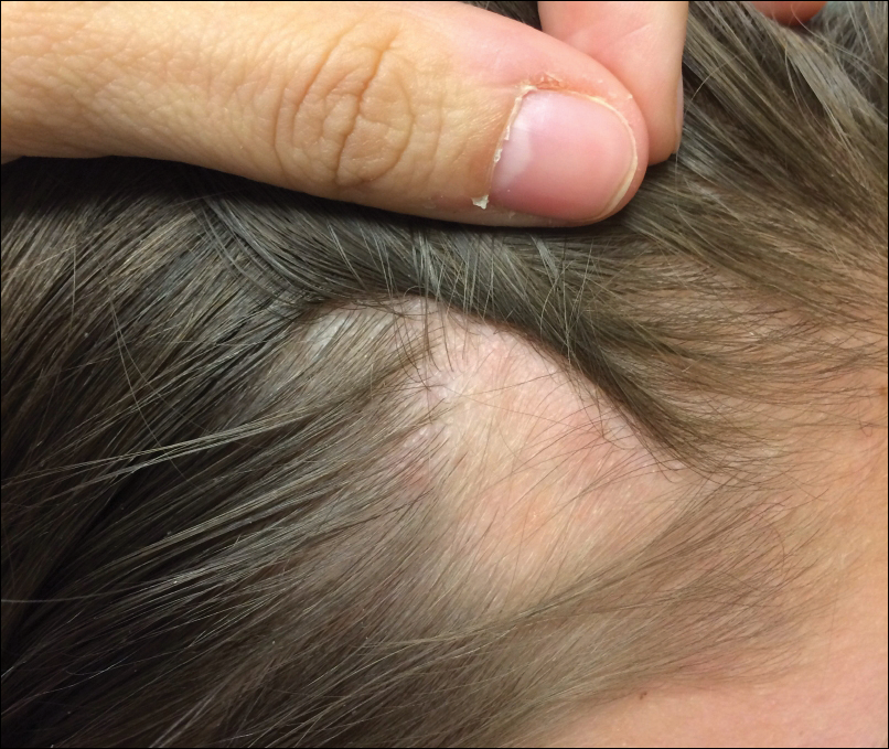
An 11-year-old girl presented for evaluation of a patch of hair loss on the right parietal scalp that had been present and stable for 2.5 years. Physical examination revealed a unilateral area of hair loss that was triangular in shape on the right parietal/temporal region, measuring 2.1×2.2 cm. Dermatoscope examination showed vellus hairs throughout. A hair-pull test was negative and the patient confirmed that the area had never been completely smooth. There were no associated symptoms and no family history of autoimmune disease or hair loss. Prior to presentation, the patient underwent a trial of intralesional steroids and topical steroids to the area without effect.