User login
PARP Inhibitors: Usurping DNA Repair to Target Cancer
The emerging role for PARP inhibitors in BRCA-mutated cancers is reviewed by Community Oncology Editor-in-Chief Dr. Lee Schwartzberg.
Editor-in-Chief Lee S. Schwartzberg, M.D., is the research medical director at The West Clinic in Memphis, TN.

The emerging role for PARP inhibitors in BRCA-mutated cancers is reviewed by Community Oncology Editor-in-Chief Dr. Lee Schwartzberg.
Editor-in-Chief Lee S. Schwartzberg, M.D., is the research medical director at The West Clinic in Memphis, TN.
The emerging role for PARP inhibitors in BRCA-mutated cancers is reviewed by Community Oncology Editor-in-Chief Dr. Lee Schwartzberg.
Editor-in-Chief Lee S. Schwartzberg, M.D., is the research medical director at The West Clinic in Memphis, TN.
Two Studies Find Beta-Blockers Help Combat Breast Cancer Progression
Beta-blockers – safe and inexpensive drugs used for 4 decades in the management of cardiovascular disease – have shown surprising promise in inhibiting breast cancer progression and metastasis.
Two retrospective observational studies, published May 31 in Journal of Clinical Oncology, offer the latest evidence that the drugs, which have a role in neuroendocrine signaling pathways, lower the risk of recurrence, metastasis, and cancer-specific mortality.
In an editorial comment (doi:10.1200/JCO.2011.35.8820) accompanying the articles, Dr. Patricia A. Ganz and Steven W. Cole, Ph.D., both of the Jonsson Comprehensive Cancer Center at the University of California, Los Angeles, wrote that the findings raised "the intriguing possibility" that these and other agents targeted to other diseases might provide "previously unappreciated opportunities for therapeutic control of disease progression, metastasis, and disease recurrence."
Because of this, they argued, future clinical treatment trials should "endeavor to collect prospective data on relevant medication exposures, weight and weight gain, comorbid conditions, and behaviors that have the potential to influence the microenvironment of the tumor, as these may be potent mediators of prognosis and survival, and may or may not be effectively accounted for in randomization."
In both studies, women with breast cancer prescribed beta-blockers for hypertension and other cardiovascular conditions were compared with women not taking the drugs.
For the first study (doi:10.1200/JCO.2010.33.5422), Thomas I. Barron, Ph.D., of Trinity College Dublin and his colleagues used linked data from an Irish national tumor registry and a pharmacy database to set up a case-control comparison of women prescribed the beta-blockers propranolol (n = 70) or atenolol (n = 525) in the year before and after their breast cancer diagnoses. These women were matched 1:2 to 4,738 women not prescribed beta-blockers, with matching inclusive of age, socioeconomic status, tumor grade and stage, and comorbidities.
After a median follow-up period of 3.5 years for the propranolol group and controls and slightly less than 3 years for the atenolol group and controls, the investigators found significant reductions in tumor size and nodal or metastatic distribution for the propranolol users, but not for the atenolol users. The cumulative probability of breast cancer–specific mortality was significantly lower among propranolol users (HR, 0.19). Propranolol users were also significantly less likely to present with a T4 (OR, 0.24) or N2/N3/M1 (OR, 0.20) tumor compared with matched controls. However, the atenolol group saw neither a significant reduction in breast-cancer specific mortality nor a difference in T4 or N2/N3/M1 tumor incidence from the matched controls.
In the second study (doi:10.1200/JCO.2010.33.4441), Dr. Amal Melhem-Bertrandt of the M.D. Anderson Cancer Center at the University of Texas, Houston, and colleagues identified 1,413 patients treated for breast cancer in a 12-year period, comparing those with (n = 102) and without (n = 1,311) concurrent beta-blocker exposure for a pathologic complete response, relapse-free survival, and overall survival. Mean follow-up was 55 months in the beta-blocker group and 63 months in the nonuser group.
The investigators saw no difference in pathologic response for those taking beta-blockers; however, after adjustment for variables including age, race, and tumor grade, women on beta-blockers showed significantly greater relapse-free survival (HR, 0.52; P = .015) and a trend toward greater overall survival that did not reach statistical significance (HR, 0.64; P = .09).
In a triple-negative subgroup (n = 377), the investigators found significant effects for beta-blocker use on relapse-free (HR, 0.30; P = .03) but not overall survival (HR, 0.35; P = .05) after adjustment.
In their editorial, Dr. Ganz and Dr. Cole noted that the findings from the second study, in which most of the beta-blockers were the beta-1–selective agents metoprolol (42% of patients) followed by atenolol (37%), would seem to contradict Dr. Barron and his colleagues’ findings, in which only propranolol, which inhibits both beta 1 and beta 2, had a significant effect. However, they wrote, "Neither metoprolol or atenolol is totally beta-1 specific; both partially inhibit beta-2–adrenergic receptors as well."
The editorialists noted that the new findings build on recent animal work indicating that beta-adrenergic receptors affect breast tumor growth and metastasis, and also on a 2010 U.K. retrospective study (n = 466) that was the first to look at a variety of antihypertensive agents in women with breast cancer. It found that women treated simultaneously with beta-blockers had a 57% reduced risk of metastasis compared with breast cancer patients receiving either other types of antihypertensive drugs or no antihypertensive drugs. Women on beta-blockers also saw 71% less cancer-specific mortality after 10 years (Oncotarget 2010; 1: 628-38).
"Beta-adrenergic signaling appears to have little effect on the biologic processes involved in breast cancer initiation, but more strongly affects the biologic processes involved in the subsequent progression and metastasis of incipient tumors. Given these results from the laboratory, and the clinical results from three recent retrospective reports suggesting the potential to limit recurrence of incident tumors, perhaps it is time to consider proof-of-concept trials testing the value of [beta-blockers] in the setting of breast cancer," Dr. Ganz and Dr. Cole wrote.
Dr. Barron and his colleagues declared that they had no financial disclosures relevant to their study, which was funded by the Irish government. Dr. Melhem-Bertrandt and colleagues said they had no financial disclosures relevant to their study, which was supported by a grant from the National Institutes of Health. Dr. Ganz and Dr. Cole, who were supported by the Breast Cancer Research Foundation, the Jonsson Comprehensive Cancer Center Foundation, Susan G. Komen for the Cure, and the National Cancer Institute, also declared that they had no relevant financial disclosures.
Beta-blockers – safe and inexpensive drugs used for 4 decades in the management of cardiovascular disease – have shown surprising promise in inhibiting breast cancer progression and metastasis.
Two retrospective observational studies, published May 31 in Journal of Clinical Oncology, offer the latest evidence that the drugs, which have a role in neuroendocrine signaling pathways, lower the risk of recurrence, metastasis, and cancer-specific mortality.
In an editorial comment (doi:10.1200/JCO.2011.35.8820) accompanying the articles, Dr. Patricia A. Ganz and Steven W. Cole, Ph.D., both of the Jonsson Comprehensive Cancer Center at the University of California, Los Angeles, wrote that the findings raised "the intriguing possibility" that these and other agents targeted to other diseases might provide "previously unappreciated opportunities for therapeutic control of disease progression, metastasis, and disease recurrence."
Because of this, they argued, future clinical treatment trials should "endeavor to collect prospective data on relevant medication exposures, weight and weight gain, comorbid conditions, and behaviors that have the potential to influence the microenvironment of the tumor, as these may be potent mediators of prognosis and survival, and may or may not be effectively accounted for in randomization."
In both studies, women with breast cancer prescribed beta-blockers for hypertension and other cardiovascular conditions were compared with women not taking the drugs.
For the first study (doi:10.1200/JCO.2010.33.5422), Thomas I. Barron, Ph.D., of Trinity College Dublin and his colleagues used linked data from an Irish national tumor registry and a pharmacy database to set up a case-control comparison of women prescribed the beta-blockers propranolol (n = 70) or atenolol (n = 525) in the year before and after their breast cancer diagnoses. These women were matched 1:2 to 4,738 women not prescribed beta-blockers, with matching inclusive of age, socioeconomic status, tumor grade and stage, and comorbidities.
After a median follow-up period of 3.5 years for the propranolol group and controls and slightly less than 3 years for the atenolol group and controls, the investigators found significant reductions in tumor size and nodal or metastatic distribution for the propranolol users, but not for the atenolol users. The cumulative probability of breast cancer–specific mortality was significantly lower among propranolol users (HR, 0.19). Propranolol users were also significantly less likely to present with a T4 (OR, 0.24) or N2/N3/M1 (OR, 0.20) tumor compared with matched controls. However, the atenolol group saw neither a significant reduction in breast-cancer specific mortality nor a difference in T4 or N2/N3/M1 tumor incidence from the matched controls.
In the second study (doi:10.1200/JCO.2010.33.4441), Dr. Amal Melhem-Bertrandt of the M.D. Anderson Cancer Center at the University of Texas, Houston, and colleagues identified 1,413 patients treated for breast cancer in a 12-year period, comparing those with (n = 102) and without (n = 1,311) concurrent beta-blocker exposure for a pathologic complete response, relapse-free survival, and overall survival. Mean follow-up was 55 months in the beta-blocker group and 63 months in the nonuser group.
The investigators saw no difference in pathologic response for those taking beta-blockers; however, after adjustment for variables including age, race, and tumor grade, women on beta-blockers showed significantly greater relapse-free survival (HR, 0.52; P = .015) and a trend toward greater overall survival that did not reach statistical significance (HR, 0.64; P = .09).
In a triple-negative subgroup (n = 377), the investigators found significant effects for beta-blocker use on relapse-free (HR, 0.30; P = .03) but not overall survival (HR, 0.35; P = .05) after adjustment.
In their editorial, Dr. Ganz and Dr. Cole noted that the findings from the second study, in which most of the beta-blockers were the beta-1–selective agents metoprolol (42% of patients) followed by atenolol (37%), would seem to contradict Dr. Barron and his colleagues’ findings, in which only propranolol, which inhibits both beta 1 and beta 2, had a significant effect. However, they wrote, "Neither metoprolol or atenolol is totally beta-1 specific; both partially inhibit beta-2–adrenergic receptors as well."
The editorialists noted that the new findings build on recent animal work indicating that beta-adrenergic receptors affect breast tumor growth and metastasis, and also on a 2010 U.K. retrospective study (n = 466) that was the first to look at a variety of antihypertensive agents in women with breast cancer. It found that women treated simultaneously with beta-blockers had a 57% reduced risk of metastasis compared with breast cancer patients receiving either other types of antihypertensive drugs or no antihypertensive drugs. Women on beta-blockers also saw 71% less cancer-specific mortality after 10 years (Oncotarget 2010; 1: 628-38).
"Beta-adrenergic signaling appears to have little effect on the biologic processes involved in breast cancer initiation, but more strongly affects the biologic processes involved in the subsequent progression and metastasis of incipient tumors. Given these results from the laboratory, and the clinical results from three recent retrospective reports suggesting the potential to limit recurrence of incident tumors, perhaps it is time to consider proof-of-concept trials testing the value of [beta-blockers] in the setting of breast cancer," Dr. Ganz and Dr. Cole wrote.
Dr. Barron and his colleagues declared that they had no financial disclosures relevant to their study, which was funded by the Irish government. Dr. Melhem-Bertrandt and colleagues said they had no financial disclosures relevant to their study, which was supported by a grant from the National Institutes of Health. Dr. Ganz and Dr. Cole, who were supported by the Breast Cancer Research Foundation, the Jonsson Comprehensive Cancer Center Foundation, Susan G. Komen for the Cure, and the National Cancer Institute, also declared that they had no relevant financial disclosures.
Beta-blockers – safe and inexpensive drugs used for 4 decades in the management of cardiovascular disease – have shown surprising promise in inhibiting breast cancer progression and metastasis.
Two retrospective observational studies, published May 31 in Journal of Clinical Oncology, offer the latest evidence that the drugs, which have a role in neuroendocrine signaling pathways, lower the risk of recurrence, metastasis, and cancer-specific mortality.
In an editorial comment (doi:10.1200/JCO.2011.35.8820) accompanying the articles, Dr. Patricia A. Ganz and Steven W. Cole, Ph.D., both of the Jonsson Comprehensive Cancer Center at the University of California, Los Angeles, wrote that the findings raised "the intriguing possibility" that these and other agents targeted to other diseases might provide "previously unappreciated opportunities for therapeutic control of disease progression, metastasis, and disease recurrence."
Because of this, they argued, future clinical treatment trials should "endeavor to collect prospective data on relevant medication exposures, weight and weight gain, comorbid conditions, and behaviors that have the potential to influence the microenvironment of the tumor, as these may be potent mediators of prognosis and survival, and may or may not be effectively accounted for in randomization."
In both studies, women with breast cancer prescribed beta-blockers for hypertension and other cardiovascular conditions were compared with women not taking the drugs.
For the first study (doi:10.1200/JCO.2010.33.5422), Thomas I. Barron, Ph.D., of Trinity College Dublin and his colleagues used linked data from an Irish national tumor registry and a pharmacy database to set up a case-control comparison of women prescribed the beta-blockers propranolol (n = 70) or atenolol (n = 525) in the year before and after their breast cancer diagnoses. These women were matched 1:2 to 4,738 women not prescribed beta-blockers, with matching inclusive of age, socioeconomic status, tumor grade and stage, and comorbidities.
After a median follow-up period of 3.5 years for the propranolol group and controls and slightly less than 3 years for the atenolol group and controls, the investigators found significant reductions in tumor size and nodal or metastatic distribution for the propranolol users, but not for the atenolol users. The cumulative probability of breast cancer–specific mortality was significantly lower among propranolol users (HR, 0.19). Propranolol users were also significantly less likely to present with a T4 (OR, 0.24) or N2/N3/M1 (OR, 0.20) tumor compared with matched controls. However, the atenolol group saw neither a significant reduction in breast-cancer specific mortality nor a difference in T4 or N2/N3/M1 tumor incidence from the matched controls.
In the second study (doi:10.1200/JCO.2010.33.4441), Dr. Amal Melhem-Bertrandt of the M.D. Anderson Cancer Center at the University of Texas, Houston, and colleagues identified 1,413 patients treated for breast cancer in a 12-year period, comparing those with (n = 102) and without (n = 1,311) concurrent beta-blocker exposure for a pathologic complete response, relapse-free survival, and overall survival. Mean follow-up was 55 months in the beta-blocker group and 63 months in the nonuser group.
The investigators saw no difference in pathologic response for those taking beta-blockers; however, after adjustment for variables including age, race, and tumor grade, women on beta-blockers showed significantly greater relapse-free survival (HR, 0.52; P = .015) and a trend toward greater overall survival that did not reach statistical significance (HR, 0.64; P = .09).
In a triple-negative subgroup (n = 377), the investigators found significant effects for beta-blocker use on relapse-free (HR, 0.30; P = .03) but not overall survival (HR, 0.35; P = .05) after adjustment.
In their editorial, Dr. Ganz and Dr. Cole noted that the findings from the second study, in which most of the beta-blockers were the beta-1–selective agents metoprolol (42% of patients) followed by atenolol (37%), would seem to contradict Dr. Barron and his colleagues’ findings, in which only propranolol, which inhibits both beta 1 and beta 2, had a significant effect. However, they wrote, "Neither metoprolol or atenolol is totally beta-1 specific; both partially inhibit beta-2–adrenergic receptors as well."
The editorialists noted that the new findings build on recent animal work indicating that beta-adrenergic receptors affect breast tumor growth and metastasis, and also on a 2010 U.K. retrospective study (n = 466) that was the first to look at a variety of antihypertensive agents in women with breast cancer. It found that women treated simultaneously with beta-blockers had a 57% reduced risk of metastasis compared with breast cancer patients receiving either other types of antihypertensive drugs or no antihypertensive drugs. Women on beta-blockers also saw 71% less cancer-specific mortality after 10 years (Oncotarget 2010; 1: 628-38).
"Beta-adrenergic signaling appears to have little effect on the biologic processes involved in breast cancer initiation, but more strongly affects the biologic processes involved in the subsequent progression and metastasis of incipient tumors. Given these results from the laboratory, and the clinical results from three recent retrospective reports suggesting the potential to limit recurrence of incident tumors, perhaps it is time to consider proof-of-concept trials testing the value of [beta-blockers] in the setting of breast cancer," Dr. Ganz and Dr. Cole wrote.
Dr. Barron and his colleagues declared that they had no financial disclosures relevant to their study, which was funded by the Irish government. Dr. Melhem-Bertrandt and colleagues said they had no financial disclosures relevant to their study, which was supported by a grant from the National Institutes of Health. Dr. Ganz and Dr. Cole, who were supported by the Breast Cancer Research Foundation, the Jonsson Comprehensive Cancer Center Foundation, Susan G. Komen for the Cure, and the National Cancer Institute, also declared that they had no relevant financial disclosures.
FROM JOURNAL OF CLINICAL ONCOLOGY
Major Finding: In the first study, the cumulative probability of breast cancer–specific mortality was significantly lower among propranolol users (HR, 0.19), compared with matched controls. Propranolol users were also significantly less likely to present with a T4 (OR, 0.24) or N2/N3/M1 (OR, 0.20) tumor. In the second study, beta-blocker users demonstrated greater relapse-free survival (HR, 0.52; P .015) and a trend toward greater overall survival that did not reach statistical significance (HR, 0.64; P = .09).
Data Source: Two retrospective observational studies (n = 5,333 and n = 1,413) of women with breast cancer who did and did not use beta-blockers.
Disclosures: Dr. Barron and his colleagues declared that they had no financial disclosures relevant to their study, which was funded by the Irish government. Dr. Melhem-Bertrandt and colleagues said they had no financial disclosures relevant to their study, which was supported by a grant from the National Institutes of Health. Dr. Ganz and Dr. Cole, who were supported by the Breast Cancer Research Foundation, the Jonsson Comprehensive Cancer Center Foundation, Susan G. Komen for the Cure, and the National Cancer Institute/National Institutes of Health, also declared that they had no relevant financial disclosures.
Point/Counterpoint: Does Surgery Improve Survival in Stage IV Breast Cancer?
Yes – It Is Time to Consider a Paradigm Shift.
In stage IV breast cancer, local therapy is not considered important except for controlling symptoms. But recent large-scale retrospective studies tell us that, if you follow enough women long enough, you see a difference in survival related to control of local disease. These emerging data tell us it’s time to consider a paradigm shift in the treatment of stage IV breast cancer.
Researchers are finding that primary site treatment regimens are important because an intact primary tumor can serve as a continued source of new metastatic lesions. There is reason, therefore, to believe that treating these can prolong life.
First explored by Dr. Larry Norton and Joan Massagué, Ph.D., (Nature Med. 2006;12:875-8), the concept of cancer self-seeding holds that cells from the primary tumor not only travel unidirectionally to seed metastases but also return to the primary tumor, self-seeding it and helping it to grow even more.
This view incorporates the microenvironment of the primary tumor. Here, fibroblasts and other cells – particularly mesenchymal stem cells derived from bone marrow – may play a very important role, creating a reactive stroma that seems to release growth-promoting substances that increase the metastatic efficiency of the circulating cells.
Robert A. Weinberg, Ph.D., published data showing that these mesenchymal stem cells are pluripotent progenitor cells. When weakly metastatic human breast cancer cells were mixed with these stem cells, the cancer cells’ metastatic potential greatly increased. The breast cancer cells then stimulated the stem cells to secrete a chemokine which – in turn – enhanced the cancer cells’ motility and their ability to invade and metastasize (Nature 2007;449:557-63). In effect, the primary tumor acts like a filling station, giving these tumor cells more energy to go out and metastasize in different sites.
Although such studies remain animal models, I think we can say there is a basis for primary tumor therapy in the metastatic setting. A dozen or more retrospective studies of large institutional series, cancer databases, and population-based studies have examined outcomes in women treated with and without surgical resection. Data on about 30,000 women have been published, with about 50% receiving some form of primary tumor therapy. The survival benefit across them is quite consistent, with the hazard ratio of death over a given time period reduced from 30% to 50%.
These studies do demonstrate selection bias: Women treated with surgical removal were more likely to be young, white, married, with smaller tumors and a lower burden of disease, and it is possible that they would have done better anyway. Studies have also shown that better survival, even in stage IV cancer, occurs in women who have a therapeutic target (that is, hormone receptors or HER2). But there was still a significant survival benefit for surgery.
Surgical resection may also confer benefit by preventing the primary tumor in the breast from becoming a quality of-life problem. At Northwestern Memorial Hospital, Chicago, we conducted a study that showed the odds of symptomatic chest wall disease were far lower in the surgical group, compared with the nonsurgical group. In this 2008 study, we found that women with good local control– surgical or systemic - did better. Time to first progression was prolonged by 50% in the surgical group, while chest wall control was associated with a 60% improvement in overall survival, regardless of whether surgical resection was performed (Cancer 2008;113:2011-9).
So the question is, do we need a randomized trial? My unbiased answer is that we do – and I am the principal investigator of such a trial open to all U.S. and Canadian institutions (NCT01242800). The end points are survival, local control, and quality of life. In addition, the trial will provide an opportunity to answer biological questions regarding the relationship between the primary tumor and metastatic site. Only when we have samples of primary and metastatic tumors can we settle this continuing debate.
Dr. Khan is the Bluhm Family Professor of Cancer Research at the Lurie Comprehensive Cancer Center, Chicago.
No – Case Selection Bias Is Behind the Survival Advantage.
It’s true that some studies show an increase in stage IV breast cancer survival after primary site resection – but this more likely reflects a bias of case selection than a true benefit of local therapy.
Any findings of positive benefit other than case selection must be biologically plausible. The suggestion that the primary tumor releases some sort of growth-promoting substance that is gone when you remove the tumor, inhibiting metastatic disease has not yet been proven in humans; nor has the proposition that continued cell shedding might further metastasis.
Decreased tumor burden has sometimes been shown to increase the usefulness of systemic therapy, but I think that, all in all, case selection simply promotes breast surgery in patients with a better prognosis.
To show improved survival, we must find causality and not simply coincidence. Findings from large case series or cancer databases are unable to provide the answer. Although registry interpretations can provide basic data, they often break down when you attempt to extract detailed data.
Sometimes a stage IV cancer is registered initially on the basis of a pulmonary or liver shadow on imaging, which later is proven not to have been metastatic disease – and the case is never amended in the registry. There are also registry misinterpretations of the type of surgery that’s been done.
We performed a matched pair analysis of stage IV breast cancer patients with and without resection of the primary tumor. This was based on the Partners Health Care Consortium cancer database, which included information on 19,464 invasive breast cancers treated between 1970 and 2002; 808 of these (4%) were recorded as stage IV. Of these, 622 were analyzed by case matching that considered age, diagnostic date, metastatic disease location, estrogen-receptor status, and systemic therapy (Ann. Surg. Oncol. 2008; 15:3384-95).
It’s absolutely true that there was a statistically significant difference in survival in the overall comparison of surgery vs. no surgery. With bone metastases only, it was still statistically significant, but with a substantially lower difference in survival. In visceral metastases, the difference became nonsignificant.
Looking at therapy sequence in those who had the same type of systemic therapy, but with chemotherapy before or after surgery, you see a suggestion that giving systemic therapy first with a clinical response, which is a favorable prognostic indication, leads to a selection bias in choosing patients for surgery.
I reviewed the 86 cases who survived 5 years with records available. In those records, 22 were actually not stage IV disease, leaving 64 with confirmed stage IV (M1) – an overall 10% 5-year survival rate. Patients who survived were significantly younger than those who did not survive, significantly more likely to have estrogen receptor-positive tumors, and significantly more likely to have bone rather than visceral metastases or oligometastatic disease – all of which are good prognostic factors.
Among the 64 5-year survivors, 25 had therapeutic breast surgery. Of these, 8 had an excellent chemotherapy response followed by breast surgery; 8 had delayed surgery following a failure of systemic therapy; 7 had an initial therapeutic and curative surgery, but staging revealed metastases because of unexpected positive nodes; and 2 had initial surgery with oligometastatic resection.
Thirty-nine patients had no therapeutic breast surgery, and, of these, 16 had an excellent response to systemic therapy; 15 had slowly progressive disease; and 8 had oligometastatic disease – a positive prognostic factor. Thus, favorable prognostic factors led to a selection of primary-site surgery, whereas poor prognostic factors mean that primary-site surgery was avoided, contradicting the assumption of its therapeutic benefit.
There were also surgical discrepancies among the 5-year survivors. The registry listed 11 as having had breast surgery, while a chart review showed that surgery was not therapeutic. Five were listed as having no breast surgery, while they had indeed undergone a therapeutic operation.
Cancer registry data are not reliable for defining stage IV disease, discriminating advanced local disease with bone involvement from true distant, imaging-confirmed metastasis, or assessing surgical procedures. Case selection bias accounts for most, if not all, of the apparent survival advantage.
Dr. Blake Cady is professor of surgery (emeritus) at Harvard Medical School, Boston, and at Brown University, Providence, R.I.
Yes – It Is Time to Consider a Paradigm Shift.
In stage IV breast cancer, local therapy is not considered important except for controlling symptoms. But recent large-scale retrospective studies tell us that, if you follow enough women long enough, you see a difference in survival related to control of local disease. These emerging data tell us it’s time to consider a paradigm shift in the treatment of stage IV breast cancer.
Researchers are finding that primary site treatment regimens are important because an intact primary tumor can serve as a continued source of new metastatic lesions. There is reason, therefore, to believe that treating these can prolong life.
First explored by Dr. Larry Norton and Joan Massagué, Ph.D., (Nature Med. 2006;12:875-8), the concept of cancer self-seeding holds that cells from the primary tumor not only travel unidirectionally to seed metastases but also return to the primary tumor, self-seeding it and helping it to grow even more.
This view incorporates the microenvironment of the primary tumor. Here, fibroblasts and other cells – particularly mesenchymal stem cells derived from bone marrow – may play a very important role, creating a reactive stroma that seems to release growth-promoting substances that increase the metastatic efficiency of the circulating cells.
Robert A. Weinberg, Ph.D., published data showing that these mesenchymal stem cells are pluripotent progenitor cells. When weakly metastatic human breast cancer cells were mixed with these stem cells, the cancer cells’ metastatic potential greatly increased. The breast cancer cells then stimulated the stem cells to secrete a chemokine which – in turn – enhanced the cancer cells’ motility and their ability to invade and metastasize (Nature 2007;449:557-63). In effect, the primary tumor acts like a filling station, giving these tumor cells more energy to go out and metastasize in different sites.
Although such studies remain animal models, I think we can say there is a basis for primary tumor therapy in the metastatic setting. A dozen or more retrospective studies of large institutional series, cancer databases, and population-based studies have examined outcomes in women treated with and without surgical resection. Data on about 30,000 women have been published, with about 50% receiving some form of primary tumor therapy. The survival benefit across them is quite consistent, with the hazard ratio of death over a given time period reduced from 30% to 50%.
These studies do demonstrate selection bias: Women treated with surgical removal were more likely to be young, white, married, with smaller tumors and a lower burden of disease, and it is possible that they would have done better anyway. Studies have also shown that better survival, even in stage IV cancer, occurs in women who have a therapeutic target (that is, hormone receptors or HER2). But there was still a significant survival benefit for surgery.
Surgical resection may also confer benefit by preventing the primary tumor in the breast from becoming a quality of-life problem. At Northwestern Memorial Hospital, Chicago, we conducted a study that showed the odds of symptomatic chest wall disease were far lower in the surgical group, compared with the nonsurgical group. In this 2008 study, we found that women with good local control– surgical or systemic - did better. Time to first progression was prolonged by 50% in the surgical group, while chest wall control was associated with a 60% improvement in overall survival, regardless of whether surgical resection was performed (Cancer 2008;113:2011-9).
So the question is, do we need a randomized trial? My unbiased answer is that we do – and I am the principal investigator of such a trial open to all U.S. and Canadian institutions (NCT01242800). The end points are survival, local control, and quality of life. In addition, the trial will provide an opportunity to answer biological questions regarding the relationship between the primary tumor and metastatic site. Only when we have samples of primary and metastatic tumors can we settle this continuing debate.
Dr. Khan is the Bluhm Family Professor of Cancer Research at the Lurie Comprehensive Cancer Center, Chicago.
No – Case Selection Bias Is Behind the Survival Advantage.
It’s true that some studies show an increase in stage IV breast cancer survival after primary site resection – but this more likely reflects a bias of case selection than a true benefit of local therapy.
Any findings of positive benefit other than case selection must be biologically plausible. The suggestion that the primary tumor releases some sort of growth-promoting substance that is gone when you remove the tumor, inhibiting metastatic disease has not yet been proven in humans; nor has the proposition that continued cell shedding might further metastasis.
Decreased tumor burden has sometimes been shown to increase the usefulness of systemic therapy, but I think that, all in all, case selection simply promotes breast surgery in patients with a better prognosis.
To show improved survival, we must find causality and not simply coincidence. Findings from large case series or cancer databases are unable to provide the answer. Although registry interpretations can provide basic data, they often break down when you attempt to extract detailed data.
Sometimes a stage IV cancer is registered initially on the basis of a pulmonary or liver shadow on imaging, which later is proven not to have been metastatic disease – and the case is never amended in the registry. There are also registry misinterpretations of the type of surgery that’s been done.
We performed a matched pair analysis of stage IV breast cancer patients with and without resection of the primary tumor. This was based on the Partners Health Care Consortium cancer database, which included information on 19,464 invasive breast cancers treated between 1970 and 2002; 808 of these (4%) were recorded as stage IV. Of these, 622 were analyzed by case matching that considered age, diagnostic date, metastatic disease location, estrogen-receptor status, and systemic therapy (Ann. Surg. Oncol. 2008; 15:3384-95).
It’s absolutely true that there was a statistically significant difference in survival in the overall comparison of surgery vs. no surgery. With bone metastases only, it was still statistically significant, but with a substantially lower difference in survival. In visceral metastases, the difference became nonsignificant.
Looking at therapy sequence in those who had the same type of systemic therapy, but with chemotherapy before or after surgery, you see a suggestion that giving systemic therapy first with a clinical response, which is a favorable prognostic indication, leads to a selection bias in choosing patients for surgery.
I reviewed the 86 cases who survived 5 years with records available. In those records, 22 were actually not stage IV disease, leaving 64 with confirmed stage IV (M1) – an overall 10% 5-year survival rate. Patients who survived were significantly younger than those who did not survive, significantly more likely to have estrogen receptor-positive tumors, and significantly more likely to have bone rather than visceral metastases or oligometastatic disease – all of which are good prognostic factors.
Among the 64 5-year survivors, 25 had therapeutic breast surgery. Of these, 8 had an excellent chemotherapy response followed by breast surgery; 8 had delayed surgery following a failure of systemic therapy; 7 had an initial therapeutic and curative surgery, but staging revealed metastases because of unexpected positive nodes; and 2 had initial surgery with oligometastatic resection.
Thirty-nine patients had no therapeutic breast surgery, and, of these, 16 had an excellent response to systemic therapy; 15 had slowly progressive disease; and 8 had oligometastatic disease – a positive prognostic factor. Thus, favorable prognostic factors led to a selection of primary-site surgery, whereas poor prognostic factors mean that primary-site surgery was avoided, contradicting the assumption of its therapeutic benefit.
There were also surgical discrepancies among the 5-year survivors. The registry listed 11 as having had breast surgery, while a chart review showed that surgery was not therapeutic. Five were listed as having no breast surgery, while they had indeed undergone a therapeutic operation.
Cancer registry data are not reliable for defining stage IV disease, discriminating advanced local disease with bone involvement from true distant, imaging-confirmed metastasis, or assessing surgical procedures. Case selection bias accounts for most, if not all, of the apparent survival advantage.
Dr. Blake Cady is professor of surgery (emeritus) at Harvard Medical School, Boston, and at Brown University, Providence, R.I.
Yes – It Is Time to Consider a Paradigm Shift.
In stage IV breast cancer, local therapy is not considered important except for controlling symptoms. But recent large-scale retrospective studies tell us that, if you follow enough women long enough, you see a difference in survival related to control of local disease. These emerging data tell us it’s time to consider a paradigm shift in the treatment of stage IV breast cancer.
Researchers are finding that primary site treatment regimens are important because an intact primary tumor can serve as a continued source of new metastatic lesions. There is reason, therefore, to believe that treating these can prolong life.
First explored by Dr. Larry Norton and Joan Massagué, Ph.D., (Nature Med. 2006;12:875-8), the concept of cancer self-seeding holds that cells from the primary tumor not only travel unidirectionally to seed metastases but also return to the primary tumor, self-seeding it and helping it to grow even more.
This view incorporates the microenvironment of the primary tumor. Here, fibroblasts and other cells – particularly mesenchymal stem cells derived from bone marrow – may play a very important role, creating a reactive stroma that seems to release growth-promoting substances that increase the metastatic efficiency of the circulating cells.
Robert A. Weinberg, Ph.D., published data showing that these mesenchymal stem cells are pluripotent progenitor cells. When weakly metastatic human breast cancer cells were mixed with these stem cells, the cancer cells’ metastatic potential greatly increased. The breast cancer cells then stimulated the stem cells to secrete a chemokine which – in turn – enhanced the cancer cells’ motility and their ability to invade and metastasize (Nature 2007;449:557-63). In effect, the primary tumor acts like a filling station, giving these tumor cells more energy to go out and metastasize in different sites.
Although such studies remain animal models, I think we can say there is a basis for primary tumor therapy in the metastatic setting. A dozen or more retrospective studies of large institutional series, cancer databases, and population-based studies have examined outcomes in women treated with and without surgical resection. Data on about 30,000 women have been published, with about 50% receiving some form of primary tumor therapy. The survival benefit across them is quite consistent, with the hazard ratio of death over a given time period reduced from 30% to 50%.
These studies do demonstrate selection bias: Women treated with surgical removal were more likely to be young, white, married, with smaller tumors and a lower burden of disease, and it is possible that they would have done better anyway. Studies have also shown that better survival, even in stage IV cancer, occurs in women who have a therapeutic target (that is, hormone receptors or HER2). But there was still a significant survival benefit for surgery.
Surgical resection may also confer benefit by preventing the primary tumor in the breast from becoming a quality of-life problem. At Northwestern Memorial Hospital, Chicago, we conducted a study that showed the odds of symptomatic chest wall disease were far lower in the surgical group, compared with the nonsurgical group. In this 2008 study, we found that women with good local control– surgical or systemic - did better. Time to first progression was prolonged by 50% in the surgical group, while chest wall control was associated with a 60% improvement in overall survival, regardless of whether surgical resection was performed (Cancer 2008;113:2011-9).
So the question is, do we need a randomized trial? My unbiased answer is that we do – and I am the principal investigator of such a trial open to all U.S. and Canadian institutions (NCT01242800). The end points are survival, local control, and quality of life. In addition, the trial will provide an opportunity to answer biological questions regarding the relationship between the primary tumor and metastatic site. Only when we have samples of primary and metastatic tumors can we settle this continuing debate.
Dr. Khan is the Bluhm Family Professor of Cancer Research at the Lurie Comprehensive Cancer Center, Chicago.
No – Case Selection Bias Is Behind the Survival Advantage.
It’s true that some studies show an increase in stage IV breast cancer survival after primary site resection – but this more likely reflects a bias of case selection than a true benefit of local therapy.
Any findings of positive benefit other than case selection must be biologically plausible. The suggestion that the primary tumor releases some sort of growth-promoting substance that is gone when you remove the tumor, inhibiting metastatic disease has not yet been proven in humans; nor has the proposition that continued cell shedding might further metastasis.
Decreased tumor burden has sometimes been shown to increase the usefulness of systemic therapy, but I think that, all in all, case selection simply promotes breast surgery in patients with a better prognosis.
To show improved survival, we must find causality and not simply coincidence. Findings from large case series or cancer databases are unable to provide the answer. Although registry interpretations can provide basic data, they often break down when you attempt to extract detailed data.
Sometimes a stage IV cancer is registered initially on the basis of a pulmonary or liver shadow on imaging, which later is proven not to have been metastatic disease – and the case is never amended in the registry. There are also registry misinterpretations of the type of surgery that’s been done.
We performed a matched pair analysis of stage IV breast cancer patients with and without resection of the primary tumor. This was based on the Partners Health Care Consortium cancer database, which included information on 19,464 invasive breast cancers treated between 1970 and 2002; 808 of these (4%) were recorded as stage IV. Of these, 622 were analyzed by case matching that considered age, diagnostic date, metastatic disease location, estrogen-receptor status, and systemic therapy (Ann. Surg. Oncol. 2008; 15:3384-95).
It’s absolutely true that there was a statistically significant difference in survival in the overall comparison of surgery vs. no surgery. With bone metastases only, it was still statistically significant, but with a substantially lower difference in survival. In visceral metastases, the difference became nonsignificant.
Looking at therapy sequence in those who had the same type of systemic therapy, but with chemotherapy before or after surgery, you see a suggestion that giving systemic therapy first with a clinical response, which is a favorable prognostic indication, leads to a selection bias in choosing patients for surgery.
I reviewed the 86 cases who survived 5 years with records available. In those records, 22 were actually not stage IV disease, leaving 64 with confirmed stage IV (M1) – an overall 10% 5-year survival rate. Patients who survived were significantly younger than those who did not survive, significantly more likely to have estrogen receptor-positive tumors, and significantly more likely to have bone rather than visceral metastases or oligometastatic disease – all of which are good prognostic factors.
Among the 64 5-year survivors, 25 had therapeutic breast surgery. Of these, 8 had an excellent chemotherapy response followed by breast surgery; 8 had delayed surgery following a failure of systemic therapy; 7 had an initial therapeutic and curative surgery, but staging revealed metastases because of unexpected positive nodes; and 2 had initial surgery with oligometastatic resection.
Thirty-nine patients had no therapeutic breast surgery, and, of these, 16 had an excellent response to systemic therapy; 15 had slowly progressive disease; and 8 had oligometastatic disease – a positive prognostic factor. Thus, favorable prognostic factors led to a selection of primary-site surgery, whereas poor prognostic factors mean that primary-site surgery was avoided, contradicting the assumption of its therapeutic benefit.
There were also surgical discrepancies among the 5-year survivors. The registry listed 11 as having had breast surgery, while a chart review showed that surgery was not therapeutic. Five were listed as having no breast surgery, while they had indeed undergone a therapeutic operation.
Cancer registry data are not reliable for defining stage IV disease, discriminating advanced local disease with bone involvement from true distant, imaging-confirmed metastasis, or assessing surgical procedures. Case selection bias accounts for most, if not all, of the apparent survival advantage.
Dr. Blake Cady is professor of surgery (emeritus) at Harvard Medical School, Boston, and at Brown University, Providence, R.I.
A Phase II Tolerability Trial of Neoadjuvant Docetaxel with Carboplatin and Capecitabine in Locally Advanced Breast Cancer
The standard of care for locally advanced breast cancer (LABC) is neoadjuvant chemotherapy,1 with LABC including clinical stages IIA, IIB, and IIIA. The goals of preoperative chemotherapy are to downstage so as to render breast conservation feasible, to eradicate disease in the axillary nodes, and to allow in vivo testing of tumor drug sensitivity, all with the ultimate aim of improving prognosis. Clinical trials have demonstrated that the pathologic in-breast response generally correlates with pathologic response in the lymph nodes. Furthermore, nodal status at the time of surgery correlates with overall survival (OS) and disease-free survival (DFS).2,3 A combined analysis of two large prospective neoadjuvant chemotherapy trials demonstrated significantly higher 5-year OS and DFS in patients achieving in-breast pathologic complete response (pCR), compared with those who did not (OS, 89% vs 64%; DFS, 87% vs 58%, respectively).4
At the start of this trial, the most effective neoad- juvant regimen remained in question. Even now, National Comprehensive Cancer Center guidelines suggest that any recommended adjuvant regimen can be used in the neoadjuvant setting.1 Numerous phase II and III trials have evaluated single-agent5–8 and combination9– 32 chemotherapies, most of which are anthracycline- based, with pCR rates reported between 7% and 36%. In the NSABP-B27 study, patients treated preoperatively with four cycles of doxorubicin and cyclophosphamide (AC) followed by four cycles of docetaxel (Taxotere) had a 26% pCR rate versus a 13% pCR rate in those receiving preoperative AC and postoperative docetaxel. Despite the doubling of pCR with neoadjuvant docetaxel, there was no difference in DFS or OS.9 However, as reported by Kuerer et al, patients achieving a pCR after completion of neoadjuvant chemotherapy appeared to have superior survival.4
Many previous trials (including the study reported here) did not exclude patients with human epidermal growth factor receptor 2 (HER2)-positive disease. It is now well established that such patients should be treated with neoadjuvant regimens incorporating HER2-targeted therapy. In fact, an early neoadjuvant study of paclitaxel followed by fluorouracil, epirubicin, and cyclophosphamide with or without 24 weeks of concurrent trastuzumab (Herceptin) in patients with HER2-positive tumors was closed early because patients receiving trastuzumab had a pCR rate of 65%, compared with 26% in those who did not receive it.33 Expanded clinical trials of this approach are in progress.
The selection of capecitabine (Xeloda) and docetaxel in the present trial was based on the hypothesis that the upregulation of thymidine phosphorylase by docetaxel should increase the activity of capecitabine. 34–36 Single-agent docetaxel in the neoadjuvant setting has yielded pCR rates of 7%–20%.6–8 Treatment with docetaxel and capecitabine together has been reported to produce pCR rates of 10%–21%.37–39 The addition of carboplatin was based on studies by Hurley et al at the University of Miami39– 41 suggesting that platinum salts appeared quite active in the neoadjuvant setting, with the combination of docetaxel and cisplatin producing a pCR rate of 20%, with no residual disease in the breast or axilla.40 Other regimens incorporating cisplatin or carboplatin have pCR rates ranging from 16% to 24%.27,42–44
Patients and methods
Study design
In this phase II multicenter study, patients were assigned to receive docetaxel (30 mg/m2 IV) and carboplatin (AUC 2 IV) on days 1, 8, and 15 of each 28-day cycle plus capecitabine (625 mg/m2 PO) twice daily on days 5–18. The capecitabine dose was based on observations that this dose was effective and relatively nontoxic in metastatic breast cancer (C.L. Vogel, empirical observations). Patients were to receive four cycles prior to surgical resection.
Given that this neoadjuvant regimen was under study, all of the patients were scheduled to receive a proven standard postoperative adjuvant chemotherapy regimen, starting 4–6 weeks postoperatively, with doxorubicin (60 mg/m2 IV) and cyclophosphamide (600 mg/m2 IV) every 21 days for 4 cycles. This sequential design was prompted by studies such as the NSABP B-27 and Aberdeen trials.9,32
Radiation therapy after lumpectomy or mastectomy was given according to individual institution guidelines. Patients with hormone receptor–positive tumors received appropriate antihormonal therapy. Tumor measurements were assessed at baseline and on day 1 of each cycle by physical examination with calipers. No breast or other imaging was required during the period of neoadjuvant chemotherapy or immediately preoperatively. Patients were considered evaluable if they proceeded to surgery after all intended cycles of neoadjuvant chemotherapy or if they developed disease progression during neoadjuvant therapy.
Patients
Eligible patients were men and women regardless of menopausal status ≥ 18 years of age with coreneedle biopsy proven locally advanced or inflammatory breast cancer. Breast cancer characteristics such as estrogen receptor (ER), progesterone receptor (PR), or HER2 status were collected but not used for inclusion/exclusion. Eligible tumors were T2 requiring mastectomy; T3N0–2; T4; and any TN2–3 that by calipers was > 2 cm or with fixed or matted axillary or imaging-detected internal mammary nodes. Patients with prior ductal carcinoma in situ (DCIS) were included, as were those with ≤ T2N0M0 breast cancer > 5 years prior.
Other requirements were an Eastern Cooperative Oncology Group (ECOG) performance status of 0–1; life expectancy > 6 months; negative metastatic workup (bone scan and CT chest/abdomen/pelvis); adequate bone marrow, liver, and kidney function; and peripheral neuropathy ≤ grade 1. All patients of child-bearing potential were required to consent to dual methods of contraception during treatment and for 3 months afterward. A negative pregnancy test was required for these women before treatment, and any suspicion of pregnancy had to be reported to the treating physician.
Study endpoints
The primary endpoint of the study was the in-breast pCR after four cycles of platinum-based neoadjuvant chemotherapy. Pathologic complete response was defined as complete disappearance of invasive and in situ disease or invasive disease alone. During the course of this trial, it became generally acceptable to include patients with only residual DCIS as equivalent to pCR.45
The secondary endpoints were pCR in the lymph nodes; clinical response rate; tolerability; breast conservation; time to disease progression (local, regional, and distant); and OS. Also recorded was minimal residual disease (MRD), which we arbitrarily defined as ≤ 1 cm invasive carcinoma at resection. The overall treatment plan included postoperative AC to provide a standard-of-care regimen to maximize curative potential.
Statistical analysis
Data were analyzed on an intentto- treat basis. Although pCR rates with doxorubicin plus either cyclophosphamide or docetaxel have been < 15%, the studies by Smith et al26 and Hurley et al39 with in-breast pCR rates of at least 20% served as comparators (albeit imprecise).
Applying the min/max statistical design, the procedure tests the null hypothesis H0: P ≤ 0.15 against the alternative hypothesis H1: P ≥ 0.30. The overall level of significance and power for this design are 5% and 80%, respectively. The sample size needed for the first stage was 23 evaluable patients. If three or fewer pCR responses were observed, then the study would be terminated and the treatment regimen would not be investigated further. Otherwise, an additional 25 evaluable patients would be accrued for a total of 48 study patients. If 11 or fewer responses were observed, then the study would be terminated. Otherwise, this treatment regimen would be recommended to proceed to phase III for further investigation.
Tolerability assessment
At each visit, toxicities were assessed and graded according to the National Cancer Institute Common Toxicity Criteria, version 2.46 Two dose reductions were allowed for all drugs.
Ethical considerations
The investigational nature of this study was fully disclosed to each patient. In accordance with institutional and federal guidelines, the patients were guided through and subsequently signed the informed consent approved by the appropriate site Institutional Review Board.
Literature review
The terms “neoadjuvant” and “breast” were used in a literature search on PubMed, with filters “English” and “clinical trials.” Abstracts for each of the 398 results were reviewed We used phase II or III trials with at least 30 patients, at least four cycles of chemotherapy, and clearly defined pCR for comparison to this study.
Results Patients
Between June 2003 and December 2006, 50 women with a median age of 49 years (range, 28–75 years) were enrolled. One patient was ineligible due to preceding lumpectomy. The 49 eligible patients were treated with ≥ 1 cycle of neoadjuvant chemotherapy between June 27, 2003, and April 12, 2007.
The baseline characteristics of the 49 eligible patients are summarized in Table 3. Thirty-one patients (63%) were premenopausal. Twenty patients (41%) were positive for either ER or PR and were negative for HER2. Eight patients (16%) had HER2- positive tumors, and 23 (46%) had triple-negative tumors. At baseline, 22 patients (45%) had clinical lymphadenopathy, and 1 patient (2%) had inflammatory breast cancer.
The 41 patients (83%) who completed all four cycles of therapy were evaluable for response; 8 (16%) were inevaluable due to noncompliance (1), grade 3 or 4 toxicity (5), or withdrawal of consent (2). The following efficacy assessments apply to the 41 evaluable patients, whereas the toxicity assessments include the 49 patients who received at least one full cycle of chemotherapy.
Clinical response
At study onset, of the 49 eligible patients, 38 (78%) had a palpable inbreast tumor (median size, 5.5 cm); 22 (45%) had enlarged nodes, and 34 (69%) had confirmed nodal involvement (by biopsy or imaging). A clinical complete response (cCR) rate in the breast was seen in 23 of 41 (56%) evaluable patients. Of 22 patients with baseline lymphadenopathy (by imaging or physical examination), 13 had axillary assessment by physical examination throughout treatment, with 12 (92%) exhibiting a cCR in the axilla.
Pathologic response
After four cycles of chemotherapy, an in-breast pCR (the primary endpoint) was demonstrated in 6 of 41 patients (15%). One of these six patients had residual DCIS and is listed separately. All of these patients had nodal pCR, whereas overall, 20 patients (49%) had negative nodes at resection.
The pathology reports of two patients were read as having invasive tumor within lymphatics and lymphovascular invasion (one each) with no measurable disease, with tumor thus sized as Tx. Neither of these patients had involved lymph nodes. Fourteen patients (34%) had MRD in the breast, and 8 of these 14 patients (57%) had residual nodal disease. Nine patients (22%) had T1c tumors (> 1–2 cm), with five of these nine patients (55%) having nodal disease. Seven patients (17%) had T2 tumors (> 2–5 cm) tumors, with five of these seven patients (71%) having nodal disease. These findings are summarized in Table 4. The correlation between in-breast cCR and pCR was 26%.
Biologic features of responders
Of interest, five of the six patients with a pCR had triple-negative tumors. This translates to a 22% pCR rate (5 of 23) in the triple-negative subset, and a pCR rate of 6% (1 of 18) in patients with ER-positive and/ or PR-positive tumors. The remaining patient with a pCR had ER-, PR-, and HER2-positive disease.
One patient had inflammatory breast cancer at diagnosis, and another developed this during the course of chemotherapy; the latter patient was removed from the study for progressive disease. Interestingly, the patient who presented with inflammatory breast cancer was one of the six patients with a pCR. Both of these inflammatory disease patients had triple-negative tumors.
Conversion to breast conservation
Breast conservation was offered to patients if it was deemed appropriate by the treating surgeon. Preoperative imaging was not mandated and thus was not routinely performed. Mastectomy was ultimately performed in 4 of the 6 patients (67%) with pCR and in 22 of the 35 patients (63%) with less than a pCR. Thus, the choice for breast conservation did not correlate well with response to chemotherapy.
Time to disease progression
At a median follow-up of 48 months (range, 7–63), 36 of 41 patients (88%) remained free of disease (range, 19–63 months). Two patients had progressive disease while they were on study treatment and had T3 tumors on resection. Another three patients were found to have progressive disease at 10, 41, and 50 months from study day 1.
Of the nine patients with T1c disease, only one patient (who had positive nodes at resection) had a recurrence (at 41 months). Overall, the patients who had a recurrence had MRD (one patient), T1c (one patient), T2 (one patient), and T3 (the same two patients whose disease progressed while they were on treatment and continued to progress after surgery).
Disease-free and overall survival
Three patients were lost to followup, with point of last contact at 19, 34, and 59 months. Of the 41 evaluable patients, 5 patients developed progressive disease, with 2 of these patients progressing during the study treatment. Disease-free survival at 12, 24, and 36 months was 89%, 89%, and 78%, respectively. Overall survival at these same time points was 95%, 90%, and 76%. None of the patients with a pCR is known to have recurrent disease. Of the six patients achieving pCR, two were lost to follow-up after 34 and 59 months, and four continued diseasefree at 38, 39, 55, and 62 months.
Adverse events
Five patients were removed from the study secondary to toxicities. Grade 3 and 4 toxicity events are summarized in Table 5. Grade 3 toxicities were anemia (4), diarrhea (2), epigastric pain (1), fatigue (2), hand-foot syndrome (1), infection (1), leukopenia (9), pain (5), and peripheral sensory neuropathy (1). Grade 4 toxicities were depression (1) and leukopenia (4). Toxicities (all grades) occurring in ≥ 10% of the 49 treated patients were anemia (76%), leukopenia (70%), fatigue (67%), nausea (59%), alopecia (49%), thrombocytopenia (47%), diarrhea (47%), constipation (37%), pain (35%), vomiting (31%), epigastric pain (27%), nail changes (22%), epiphora (22%), hand-foot syndrome (20%), infection (18%), edema (16%), rash (16%), anorexia (16%), and depression (10%). In the intent-to-treat population, there were nine dose reductions among nine patients, and 19 dose delays among 15 patients.
Discussion
The combination of agents tested thus far in the neoadjuvant setting consistently produce pCR rates far less than 50% in unselected populations. This study was begun prior to the widespread use of personalized medicine. Most prior published trials had utilized anthracycline-based chemotherapy, with response rates generally ranging between 7% and 36%.6,9–26,28–31,41,42
The idea of thymidine phosphorylase upregulation by the combination of capecitabine and docetaxel upon which this study was largely based34–36 has since been disputed.47 The primary endpoint of this trial of a novel platinum- based regimen was a pCR rate of 15%. It is significant that 83% of the pCRs were in triple-negative tumors. A secondary endpoint of MRD was calculated, as this was in the original design of the study, but ultimately was not relevant to the primary endpoint.
Ultimately, pCR is the more relevant point of discussion for the modern era. The 15% pCR rate seen in this phase II study was within range of those achieved in numerous other phase II/III neoadjuvant chemotherapy trials with ≥ 25 patients, ≥ 3 cycles of chemotherapy, and pCR defined as absence of carcinoma in the breast and axilla. To date, no patient in our study with a pCR has been noted to have recurrent disease. However, a recently published French study found a 22% recurrence rate at 11 years in patients with triple-negative breast cancer achieving pCR, highlighting the importance of longer-term follow- up.48.
The inclusion of patients with HER2-positive disease in neoadjuvant studies without HER2-targeted therapy was standard at the time that this study was conducted, but is no longer appropriate. If we were to exclude the eight HER2-positive patients from analysis, then there would be only 34 patients evaluable for response, with a pCR rate of 18%. Buzdar et al33 demonstrated a 65% pCR rate in women with HER2-positive disease treated with neoadjuvant chemotherapy plus trastuzumab. The improvement in pCR with the addition of trastuzumab is supported by other confirmatory trials. Authors of a single-arm trial of dose-dense epirubicin and cyclophosphamide followed by dosedense docetaxel and trastuzumab in a HER2-positive population reported a pCR rate of 57%.49 The randomized NOAH study50 achieved a pCR rate of 23% in 115 patients treated with trastuzumab-based chemotherapy.
It is interesting to note that five of six patients (83%) achieving a pCR in our study had triple-negative tumors. Investigators at the University of Miami presented a retrospective review of locally advanced triple-negative breast cancer treated with docetaxel and a platinum salt, with 61% of patients also receiving AC. The authors reported a pCR rate of 34% overall and 40% for patients receiving AC.51 A pCR rate of 60% was noted in the triplenegative subset of patients in another study evaluating docetaxel, doxorubicin, and cyclophosphamide with or without vinorelbine/capecitabine (GeparTrio Study).52 Further, a pCR rate of 72% was achieved with singleagent cisplatin in a group of 25 women with BRCA1 mutations, suggesting, if confirmed by others, that this largely triple- negative population may be exquisitely sensitive to platinum salts.43 In contrast, in a previous study of cisplatin in BRCA mutation carriers, Garber et al44 reported a pCR rate of 22%, suggesting that further trials are needed specifically in BRCA carriers and in triple-negative tumors to see whether these specific patient subsets preferentially derive benefit from platinum salts in the neoadjuvant setting.
The results of the current study are consistent with others indicating a low likelihood of pCR in patients with ERpositive tumors. In fact, none of our ER-positive patients had a pCR. Neoadjuvant endocrine therapy in postmenopausal women with ER- and/ or PR-positive disease is a reasonable treatment option for selected patients, but endpoints other than pCR have often been used.53,54 It is therefore difficult to directly compare these two strategies. Currently, investigators are comparing the three aromatase inhibitors head to head in the neoadjuvant setting for postmenopausal women with hormone receptor–positive tumors.55
The historic pCR ceiling appears to be rising, albeit slowly. Where targets such as HER2 overexpression and triple- negative biology are recognized, progress is being made. Patient eligibility criteria for neoadjuvant breast cancer studies at the time of this trial were quite broad, and it is now recognized that specific subsets of breast cancer respond differently to different classes of agents. Furthermore, our knowledge about breast cancer prognostic markers continues to expand. Had this study been designed in 2011, other data points such as Ki67 would have been collected. A recently published study on neoadjuvant triplenegative breast cancer found that only patients with baseline Ki67% expression > 10% achieved pCR.56
Given the long-term implications of not achieving pCR, optimal treatment of patients in the adjuvant setting is critical. Although neoadjuvantly treated patients with ER-positive or HER2-positive disease go on to receive adjuvant agents (antihormonal therapy for ER-positive disease and trastuzumab for HER2-positive disease), patients with triple-negative disease lack long-term therapies of proven efficacy. Perhaps, as we edge closer to defining the optimal neoadjuvant agents for each subset of patients, this will be less of a concern. Many earlyphase neoadjuvant studies have been conducted, with promising reports, yet the results of larger, randomized trials continue to frustrate both investigators and clinicians. These deficits in care can only be answered by carefully planned randomized clinical trials.
Acknowledgment: Funding for this study was provided by sanofi-aventis, U.S.
1. Carlson RW, Allred DC, Anderson BO, et al. NCCN Clinical Practice Guidelines in Oncology: Breast Cancer, V.1.2009. http://www. nccn.org. Accessed May 12, 2011.
2. Gadjos C, Tartter PI, Estabrook A, et al. Relationship of clinical and pathologic response to neoadjuvant chemotherapy and outcome of locally advanced breast cancer. J Surg Oncol 2002;80:4–11.
3. Machiavelli MR, Romero AO, Perez JE, et al. Prognostic significance of pathologic response of primary tumor and metastatic axillary lymph nodes after neoadjuvant chemotherapy for locally advanced breast carcinoma. Cancer J Sci Am 1998;4:125–131.
4. Kuerer HM, Newman LA, Smith TL, et al. Clinical course of breast cancer patients with complete pathologic primary tumor and axillary lymph node response to doxorubicinbased neoadjuvant chemotherapy. J Clin Oncol 1999;17:441–444.
5. Schneeweiss A, Bastert G, Huober J, et al. Neoadjuvant therapy with gemcitabine in breast cancer. Oncology 2004;18:27–31.
6. Gradishar WJ, Wedam SB, Jahanzeb M, et al. Neoadjuvant docetaxel followed by adjuvant doxorubicin and cyclophosphamide in patients with stage III breast cancer. Ann Oncol 2005;8:1297–1304.
7. Amat S, Bougnoux P, Penault-Llorca F, et al. Neoadjuvant docetaxel for operable breast cancer induces a high pathologic response and breast conservation rate. Br J Cancer 2003;88:1339–1345.
8. Estevez LG, Cuevas JM, Anton A, et al. Weekly docetaxel as neoadjuvant chemotherapy for stage II and III breast cancer: efficacy and correlation with biologic markers in a phase II, multicenter study. Clin Cancer Res 2003;9:686–692.
9. Bear HD, Anderson S, Smith RE, et al. Sequential preoperative or postoperative docetaxel added to preoperative doxorubicin plus cyclophosphamide for operable breast cancer: National Surgical Adjuvant Breast and Bowel Protocol B-27. J Clin Oncol 2006;24:2019–2027.
10. Hamm JT, Wilson JW, Rastogi P, et al. Gemcitabine/epirubicin/paclitaxel as neoadjuvant chemotherapy in locally advanced breast cancer: a phase II trial of the NSABP Foundation Research Group. Clin Breast Cancer 2008;8:257–263
11. Steger GG, Galid A, Gnant M, et al. Pathologic complete response with six compared with three cycles of neoadjuvant epirubicin plus docetaxel and granulocyte colony- stimulating factor in operable breast cancer: results of ABCSG-14. J Clin Oncol 2007;20:2012–2018.
12. Sarid D, Ron IG, Sperber F et al. Neoadjuvant treatment with paclitaxel and epirubicin in invasive breast cancer: a phase II study. Clin Drug Investig 2006;26:691–701.
13. Thatai LC, Vishnubhotla P, Biernat L, et al. A phase II study of docetaxel, doxorubicin, and infusional 5-fluorouracil in the treatment of patients with locally advanced breast cancer. Am J Clin Oncol 2006;29:484–489.
14. Aguiar-Bujanda D, Bohn-Sarmiento U, Cabrera-Suarez MA, et al. Epirubicin, cyclophosphamide and weekly paclitaxel as neoadjuvant chemotherapy for stage II and III breast cancer. J Cancer Res Clin Oncol 2006;132:332–338.
15. Cooper BW, Radivovevitch T, Overmoyer BA, et al. Phase II study of dose-dense sequential doxorubicin and docetaxel for patients with advanced operable and inoperable breast cancer. Breast Cancer Res Treat 2006;97:311–318.
16. Ramaswamy B, Povoski SP, Rhoades C, et al. Phase II trial of neoadjuvant chemotherapy with docetaxel followed by epirubicin in stage II/III breast cancer. Breast Cancer Res Treat 2005;93:67–74.
17. Reitsamer R, Peintinger F, Prokop E, et al. Pathological complete response rates comparing 3 versus 6 cycles of epidoxorubicin and docetaxel in the neoadjuvant setting of patients with stage II and III breast cancer. Anticancer Drugs 2005;16:867–870.
18. O’Regan RM, Von Roenn JH, Carlson RW, et al. Final results of a phase II trial of preoperative TAC (docetaxel/doxorubicin/ cyclophosphamide) in stage III breast cancer. Clin Breast Cancer 2005;6:163–168.
19. Chua S, Smith IE, A’Hern RP, et al. Neoadjuvant vinorelbine/epirubicin (VE) versus standard Adriamycin/cyclophosphamide (AC) in operable breast cancer: analysis of response and tolerability in a randomized phase III trial (TOPIC 2). Ann Oncol 2005;16:1435–1441.
20. Chen SC, Chang HK, Lin YC, et al. Increased feasibility of weekly epirubicin and paclitaxel as neoadjuvant chemotherapy for locally advanced breast carcinoma. Onkologie 2005;28:339–344.
21. Evans TR, Yellowlees A, Foster E, et al. Phase III randomized trial of doxorubicin and docetaxel versus doxorubicin and cyclophosphamide as primary medical therapy in women with breast cancer: an Anglo-Celtic Cooperative Oncology Group study. J Clin Oncol 2005;23:2988–2995.
22. Abrial C, Van Praagh I, Delva R, et al. Pathologic and clinical response of a primary chemotherapy regimen combining vinorelbine, epirubicin, and paclitaxel as neoadjuvant treatment in patients with operable breast cancer. Oncologist 2005;10:242–249.
23. Dieras V, Fumoleau P, Romieu G, et al. Randomized parallel study of doxorubicin plus paclitaxel and doxorubicin plus cyclophosphamide as neoadjuvant treatment of patients with breast cancer. J Clin Oncol 2004;22:4958– 4965.
24. Mouret-Reynier MA, Abrial CJ, Ferriere JP, et al. Neoadjuvant FEC100 for operable breast cancer: eight year experience of Centre Jean Perrin. Clin Breast Cancer 2004;5:303–307.
25. Espinosa E, Morales S, Borrega P, et al. Docetaxel and high-dose epirubicin as neoadjuvant chemotherapy in locally advanced breast cancer. Cancer Chemother Pharmacol 2004;54:546–552.
26. Smith IE, A’Hern RP, Coombes GA, et al. A novel continuous infusional 5-fluorouracil- based chemotherapy regimen compared with conventional chemotherapy in the neo-adjuvant treatment of early breast cancer: 5 year results of the TOPIC trial. Ann Oncol 2004;15:751–758.
27. Ezzat AA, Ibrahim EM, Ajarim DS, et al. Phase II study of neoadjuvant paclitaxel and cisplatin for operable and locally advanced breast cancer: analysis of 126 patients. Br J Cancer 2004;90:968–974.
28. von Minckwitz G, Raab G, Caputo A, et al. Doxorubicin with cyclophosphamide followed by docetaxel every 21 days compared with doxorubicin and docetaxel every 14 days as preoperative treatment in operable breast cancer: the GEPARDUO study of the German Breast Group. J Clin Oncol 2005;23:2676–2685.
29. Gogas H, Papadimitriou C, Kalofonos HP, et al. Neoadjuvant chemotherapy with a combination of pegylated liposomal doxorubicin (Caelyx) and paclitaxel in locally advanced breast cancer: a phase II study by the Hellenic Cooperative Oncology Group. Ann Oncol 2002;13:1737–1742. 30. Braud AC, Levy E, Feuilhade F, et al. Combination of vinorelbine, epirubicin, and cyclophosphamide as neoadjuvant chemotherapy for locally advanced breast cancer: a phase II study. Am J Clin Oncol 2002;25:303–307. 31. de Matteis A, Nuzzo F, D’Aiuto G, et al. Docetaxel plus epidoxorubicin as neoadjuvant treatment in patients with large operable or locally advanced carcinoma of the breast: a single-center, phase II study. Cancer 2002;94:895–901. 32. Heys SD, Hutcheon AW, Sarkar TK, et al. Neoadjuvant docetaxel in breast cancer: 3-year survival results from the Aberdeen trial. Clin Breast Cancer 2002(suppl 3):S69–S74. 33. Buzdar AU, Ibrahim NK, Francis D, et al. Significantly higher pathologic complete remission rate after neoadjuvant therapy with trastuzumab, paclitaxel, and epirubicin chemotherapy: results of a randomized trial in human epidermal growth factor receptor 2-positive operable breast cancer. J Clin Oncol 2005;23:3676–3685. 34. Sawada N, Ishikawa T, Fukase Y, et al. Induction of thymidine phosphorylase activity and enhancement of capecitabine efficacy by Taxol/Taxotere in human cancer xenografts. Clin Cancer Res 1998;4:1013–1019.
35. Endo M, Shinbori N, Fukase Y, et al. Induction of thymidine phosphorylase expression and enhancement of efficacy of capecitabine or 5´deoxy-5-fluorouridine by cyclophosphamide in mammary tumor models. Int J Cancer 1999;83:127–134.
36. Yamamoto S, Kurebayashi J, Kurosumi M, et al. Combined effects of docetaxel and fluoropyrimidines on tumor growth and expression of interleukin-6 and thymidine phosphorylase in breast cancer xenografts. Cancer Chemother Pharmacol 2001;48:283–288.
37. Lebowitz PF, Eng-Wong J, Swain SM, et al. A phase II trial of neoadjuvant docetaxel and capecitabine for locally advanced breast cancer. Clin Cancer Res 2004;10:6764–6769.
38. Lee KS, Ro J, Nam BH, et al. A randomized phase-III trial of docetaxel/capecitabine versus doxorubicin/cyclophosphamide as primary chemotherapy for patients with stage II/III breast cancer. Breast Cancer Res Treat 2008;109:481–489.
39. Hurley J, Reis I, Silva O, et al. Weekly docetaxel/carboplatin as primary systemic therapy for Her2-negative locally advanced breast cancer. Clin Breast Cancer 2005;6:447–454.
40. Lee YJ, Doliny P, Gomez-Fernandez C, et al. Docetaxel and cisplatin as primary chemotherapy for treatment of locally advanced breast cancers. Clin Breast Cancer 2004;5:371–376.
41. Morrell LE, Lee YJ, Hurley J, et al. A phase II trial of neoadjuvant methotrexate, vinblastine, doxorubicin, and cisplatin in the treatment of patients with locally advanced breast carcinoma. Cancer 1998;82:503–511.
42. Villman K, Ohd JF, Lidbrink E, et al. A phase II study of epirubicin, cisplatin and capecitabine as neoadjuvant chemotherapy in locally advanced or inflammatory breast cancer. Eur J Cancer 2007;43:1153–1160.
43. Gronwald J, Byrski T, Huzarski T, et al. Neoadjuvant therapy with cisplatin in BRCA1-positive breast cancer patients. J Clin Oncol 2009;27(15S):502.
44. Garber J, Richardson A, Harris L, et al. Neoadjuvant cisplatin in “triple-negative” breast cancer. Presented at the 29th San Antonio Breast Cancer Symposium; December 14– 17, 2006; San Antonio, TX. Poster 3074.
45. Mazouni C, Peintinger F, Wan-Kau S, et al. Residual ductal carcinoma in situ in patients with complete eradication of invasive breast cancer after neoadjuvant chemotherapy does not adversely affect patient outcome. J Clin Oncol 2007;19:2650–2655.
46. Cancer Therapy Evaluation Program. Common Toxicity Criteria version 2.0. National Cancer Institute, 1999. http://ctep.cancer. gov. Accessed May 12, 2011.
47. Layman RM, Thomas DG, Griffith KA, et al. Neoadjuvant docetaxel and capecitabine and the use of thymidine phosphorylase as a predictive biomarker in breast cancer. Clin Cancer Res 2007;13:4092–4097.
48. Le Tourneau C, Dettwiler S, Beuzeboc P, et al. Pathologic response to short intensified taxane-free neoadjuvant chemotherapy in patients with highly proliferative operable breast cancer. Am J Clin Oncol 2011. doi: 10.1097/ COC.0b013e318209d34c (Epub ahead of print).
49. Blakely L, Somer B, Keaton M, et al. Neoadjuvant dose-dense sequential biweekly epirubicin and cyclophosphamide followed by docetaxel and trastuzumab for Her2+ operable breast cancer. J Clin Oncol 2009;27(15S):595.
50. Gianni L, Semiglazov V, Manikhas GM, et al. Neoadjuvant trastuzumab in locally advanced breast cancer (NOAH): antitumour and safety analysis. J Clin Oncol 2007;25(18S):532.
51. Leone JP, Guardiola V, Venkatraman A, et al. Neoadjuvant platinum-based chemotherapy (CT) for triple-negative locally advanced breast cancer (LABC): retrospective analysis of 125 patients. J Clin Oncol 2009;27(15S):625.
52. Huober J, von Minckwitz G, Denkert C, et al. Effect of neoadjuvant anthracyclinetaxane- based chemotherapy in different biological breast cancer phenotypes: overall results from the GeparTrio study. Breast Cancer Res Treat 2010;124:133–140.
53. Smith IE, Dowsett M, Ebbs SR, et al. Neoadjuvant treatment of postmenopausal breast cancer with anastrozole, tamoxifen, or both in combination: the Immediate Preoperative Anastrozole, Tamoxifen, or Combined with Tamoxifen (IMPACT) multicenter double-blind randomized trial. J Clin Oncol 2005;23:5108–5116.
54. Mamounas EP. Facilitating breastconserving surgery and preventing recurrence: aromatase inhibitors in the neoadjuvant and adjuvant settings. Ann Surg Oncol 2008;15:691–703.
55. American College of Surgeons, National Cancer Institute, and Cancer and Leukemia Group B. Exemestane, letrozole, or anastrozole in treating postmenopausal women who are undergoing surgery for stage II or stage III breast cancer. ClinicalTrials.gov NCT00265759. http://clinicaltrials.gov. Accessed May 12, 2011.
56. Keam B, Im SA, Lee KH, et al. Ki67 can be used for further classification of triple negative breast cancer into two subtypes with different response and prognosis. Breast Cancer Res 2011 March 2;13(2):R22 (Epub ahead of print).
ABOUT THE AUTHORS
Aruna Mani, MD; Sandra X. Franco, MD; Grace Wang, MD: Neil Abramson, MD; Lee S. Schwartzberg, MD: James Jakub, MD; Elizabeth Tan-Chiu, MD: Alisha Stein, RNC, BSN, OCN; Alejandra T. Perez, MD; and Charles L Vogel, MD.
Affiliations: Dr. Mani is a breast medical oncologist at Memorial Cancer Institute, Pembroke Pines, FL. Dr. Franco is now Chief of Oncology at the Oncology Center, Clinica del Country, Bogota, Colombia. Dr. Wang is an oncologist at Advanced Medical Specialties, Miami, FL. Dr. Abramson is Clinical Professor of Medicine and Emeritus Director of Education and Research at Baptist Cancer Institute, University of Florida, Jacksonville, FL. Dr. Schwartzberg is Medical Director of The West Clinic, Memphis, TN. Dr. Jakub is now Assistant Professor of Surgery, Division of Gastroenterology and General Surgery, Mayo Clinic, Rochester, MN. Dr. Tan-Chiu is Medical Director of Florida Cancer Care, Davie, FL. Dr. Schwartz is Principal Investigator at Mount Sinai Medical Center, Miami Beach, FL. Ms. Frankel is Director of Oncology Clinical Research and Development at Memorial Cancer Institute, Hollywood, FL. Dr. Krill-Jackson is an oncologist at Mount Sinai Comprehensive Cancer Center, Miami, FL. Ms. Stein is now Oncology Clinical Coordinator at Genentech Inc., Fort Lauderdale, FL. Dr. Perez is Director of the Breast Cancer Center at Memorial Cancer Institute, Hollywood, FL. Dr. Vogel is Professor of Clinical Medicine and Director of the Women’s Center, Sylvester Comprehensive Cancer Center, Deerfield Beach, FL.
Conflicts of interest: Dr. Vogel has served as an advisor and is a member of the speakers’ bureaus of sanofi-aventis U.S. and Roche, as well as many other companies whose products were not part of the current study plan. The other authors have no pertinent conflicts of interest to disclose.
The standard of care for locally advanced breast cancer (LABC) is neoadjuvant chemotherapy,1 with LABC including clinical stages IIA, IIB, and IIIA. The goals of preoperative chemotherapy are to downstage so as to render breast conservation feasible, to eradicate disease in the axillary nodes, and to allow in vivo testing of tumor drug sensitivity, all with the ultimate aim of improving prognosis. Clinical trials have demonstrated that the pathologic in-breast response generally correlates with pathologic response in the lymph nodes. Furthermore, nodal status at the time of surgery correlates with overall survival (OS) and disease-free survival (DFS).2,3 A combined analysis of two large prospective neoadjuvant chemotherapy trials demonstrated significantly higher 5-year OS and DFS in patients achieving in-breast pathologic complete response (pCR), compared with those who did not (OS, 89% vs 64%; DFS, 87% vs 58%, respectively).4
At the start of this trial, the most effective neoad- juvant regimen remained in question. Even now, National Comprehensive Cancer Center guidelines suggest that any recommended adjuvant regimen can be used in the neoadjuvant setting.1 Numerous phase II and III trials have evaluated single-agent5–8 and combination9– 32 chemotherapies, most of which are anthracycline- based, with pCR rates reported between 7% and 36%. In the NSABP-B27 study, patients treated preoperatively with four cycles of doxorubicin and cyclophosphamide (AC) followed by four cycles of docetaxel (Taxotere) had a 26% pCR rate versus a 13% pCR rate in those receiving preoperative AC and postoperative docetaxel. Despite the doubling of pCR with neoadjuvant docetaxel, there was no difference in DFS or OS.9 However, as reported by Kuerer et al, patients achieving a pCR after completion of neoadjuvant chemotherapy appeared to have superior survival.4
Many previous trials (including the study reported here) did not exclude patients with human epidermal growth factor receptor 2 (HER2)-positive disease. It is now well established that such patients should be treated with neoadjuvant regimens incorporating HER2-targeted therapy. In fact, an early neoadjuvant study of paclitaxel followed by fluorouracil, epirubicin, and cyclophosphamide with or without 24 weeks of concurrent trastuzumab (Herceptin) in patients with HER2-positive tumors was closed early because patients receiving trastuzumab had a pCR rate of 65%, compared with 26% in those who did not receive it.33 Expanded clinical trials of this approach are in progress.
The selection of capecitabine (Xeloda) and docetaxel in the present trial was based on the hypothesis that the upregulation of thymidine phosphorylase by docetaxel should increase the activity of capecitabine. 34–36 Single-agent docetaxel in the neoadjuvant setting has yielded pCR rates of 7%–20%.6–8 Treatment with docetaxel and capecitabine together has been reported to produce pCR rates of 10%–21%.37–39 The addition of carboplatin was based on studies by Hurley et al at the University of Miami39– 41 suggesting that platinum salts appeared quite active in the neoadjuvant setting, with the combination of docetaxel and cisplatin producing a pCR rate of 20%, with no residual disease in the breast or axilla.40 Other regimens incorporating cisplatin or carboplatin have pCR rates ranging from 16% to 24%.27,42–44
Patients and methods
Study design
In this phase II multicenter study, patients were assigned to receive docetaxel (30 mg/m2 IV) and carboplatin (AUC 2 IV) on days 1, 8, and 15 of each 28-day cycle plus capecitabine (625 mg/m2 PO) twice daily on days 5–18. The capecitabine dose was based on observations that this dose was effective and relatively nontoxic in metastatic breast cancer (C.L. Vogel, empirical observations). Patients were to receive four cycles prior to surgical resection.
Given that this neoadjuvant regimen was under study, all of the patients were scheduled to receive a proven standard postoperative adjuvant chemotherapy regimen, starting 4–6 weeks postoperatively, with doxorubicin (60 mg/m2 IV) and cyclophosphamide (600 mg/m2 IV) every 21 days for 4 cycles. This sequential design was prompted by studies such as the NSABP B-27 and Aberdeen trials.9,32
Radiation therapy after lumpectomy or mastectomy was given according to individual institution guidelines. Patients with hormone receptor–positive tumors received appropriate antihormonal therapy. Tumor measurements were assessed at baseline and on day 1 of each cycle by physical examination with calipers. No breast or other imaging was required during the period of neoadjuvant chemotherapy or immediately preoperatively. Patients were considered evaluable if they proceeded to surgery after all intended cycles of neoadjuvant chemotherapy or if they developed disease progression during neoadjuvant therapy.
Patients
Eligible patients were men and women regardless of menopausal status ≥ 18 years of age with coreneedle biopsy proven locally advanced or inflammatory breast cancer. Breast cancer characteristics such as estrogen receptor (ER), progesterone receptor (PR), or HER2 status were collected but not used for inclusion/exclusion. Eligible tumors were T2 requiring mastectomy; T3N0–2; T4; and any TN2–3 that by calipers was > 2 cm or with fixed or matted axillary or imaging-detected internal mammary nodes. Patients with prior ductal carcinoma in situ (DCIS) were included, as were those with ≤ T2N0M0 breast cancer > 5 years prior.
Other requirements were an Eastern Cooperative Oncology Group (ECOG) performance status of 0–1; life expectancy > 6 months; negative metastatic workup (bone scan and CT chest/abdomen/pelvis); adequate bone marrow, liver, and kidney function; and peripheral neuropathy ≤ grade 1. All patients of child-bearing potential were required to consent to dual methods of contraception during treatment and for 3 months afterward. A negative pregnancy test was required for these women before treatment, and any suspicion of pregnancy had to be reported to the treating physician.
Study endpoints
The primary endpoint of the study was the in-breast pCR after four cycles of platinum-based neoadjuvant chemotherapy. Pathologic complete response was defined as complete disappearance of invasive and in situ disease or invasive disease alone. During the course of this trial, it became generally acceptable to include patients with only residual DCIS as equivalent to pCR.45
The secondary endpoints were pCR in the lymph nodes; clinical response rate; tolerability; breast conservation; time to disease progression (local, regional, and distant); and OS. Also recorded was minimal residual disease (MRD), which we arbitrarily defined as ≤ 1 cm invasive carcinoma at resection. The overall treatment plan included postoperative AC to provide a standard-of-care regimen to maximize curative potential.
Statistical analysis
Data were analyzed on an intentto- treat basis. Although pCR rates with doxorubicin plus either cyclophosphamide or docetaxel have been < 15%, the studies by Smith et al26 and Hurley et al39 with in-breast pCR rates of at least 20% served as comparators (albeit imprecise).
Applying the min/max statistical design, the procedure tests the null hypothesis H0: P ≤ 0.15 against the alternative hypothesis H1: P ≥ 0.30. The overall level of significance and power for this design are 5% and 80%, respectively. The sample size needed for the first stage was 23 evaluable patients. If three or fewer pCR responses were observed, then the study would be terminated and the treatment regimen would not be investigated further. Otherwise, an additional 25 evaluable patients would be accrued for a total of 48 study patients. If 11 or fewer responses were observed, then the study would be terminated. Otherwise, this treatment regimen would be recommended to proceed to phase III for further investigation.
Tolerability assessment
At each visit, toxicities were assessed and graded according to the National Cancer Institute Common Toxicity Criteria, version 2.46 Two dose reductions were allowed for all drugs.
Ethical considerations
The investigational nature of this study was fully disclosed to each patient. In accordance with institutional and federal guidelines, the patients were guided through and subsequently signed the informed consent approved by the appropriate site Institutional Review Board.
Literature review
The terms “neoadjuvant” and “breast” were used in a literature search on PubMed, with filters “English” and “clinical trials.” Abstracts for each of the 398 results were reviewed We used phase II or III trials with at least 30 patients, at least four cycles of chemotherapy, and clearly defined pCR for comparison to this study.
Results Patients
Between June 2003 and December 2006, 50 women with a median age of 49 years (range, 28–75 years) were enrolled. One patient was ineligible due to preceding lumpectomy. The 49 eligible patients were treated with ≥ 1 cycle of neoadjuvant chemotherapy between June 27, 2003, and April 12, 2007.
The baseline characteristics of the 49 eligible patients are summarized in Table 3. Thirty-one patients (63%) were premenopausal. Twenty patients (41%) were positive for either ER or PR and were negative for HER2. Eight patients (16%) had HER2- positive tumors, and 23 (46%) had triple-negative tumors. At baseline, 22 patients (45%) had clinical lymphadenopathy, and 1 patient (2%) had inflammatory breast cancer.
The 41 patients (83%) who completed all four cycles of therapy were evaluable for response; 8 (16%) were inevaluable due to noncompliance (1), grade 3 or 4 toxicity (5), or withdrawal of consent (2). The following efficacy assessments apply to the 41 evaluable patients, whereas the toxicity assessments include the 49 patients who received at least one full cycle of chemotherapy.
Clinical response
At study onset, of the 49 eligible patients, 38 (78%) had a palpable inbreast tumor (median size, 5.5 cm); 22 (45%) had enlarged nodes, and 34 (69%) had confirmed nodal involvement (by biopsy or imaging). A clinical complete response (cCR) rate in the breast was seen in 23 of 41 (56%) evaluable patients. Of 22 patients with baseline lymphadenopathy (by imaging or physical examination), 13 had axillary assessment by physical examination throughout treatment, with 12 (92%) exhibiting a cCR in the axilla.
Pathologic response
After four cycles of chemotherapy, an in-breast pCR (the primary endpoint) was demonstrated in 6 of 41 patients (15%). One of these six patients had residual DCIS and is listed separately. All of these patients had nodal pCR, whereas overall, 20 patients (49%) had negative nodes at resection.
The pathology reports of two patients were read as having invasive tumor within lymphatics and lymphovascular invasion (one each) with no measurable disease, with tumor thus sized as Tx. Neither of these patients had involved lymph nodes. Fourteen patients (34%) had MRD in the breast, and 8 of these 14 patients (57%) had residual nodal disease. Nine patients (22%) had T1c tumors (> 1–2 cm), with five of these nine patients (55%) having nodal disease. Seven patients (17%) had T2 tumors (> 2–5 cm) tumors, with five of these seven patients (71%) having nodal disease. These findings are summarized in Table 4. The correlation between in-breast cCR and pCR was 26%.
Biologic features of responders
Of interest, five of the six patients with a pCR had triple-negative tumors. This translates to a 22% pCR rate (5 of 23) in the triple-negative subset, and a pCR rate of 6% (1 of 18) in patients with ER-positive and/ or PR-positive tumors. The remaining patient with a pCR had ER-, PR-, and HER2-positive disease.
One patient had inflammatory breast cancer at diagnosis, and another developed this during the course of chemotherapy; the latter patient was removed from the study for progressive disease. Interestingly, the patient who presented with inflammatory breast cancer was one of the six patients with a pCR. Both of these inflammatory disease patients had triple-negative tumors.
Conversion to breast conservation
Breast conservation was offered to patients if it was deemed appropriate by the treating surgeon. Preoperative imaging was not mandated and thus was not routinely performed. Mastectomy was ultimately performed in 4 of the 6 patients (67%) with pCR and in 22 of the 35 patients (63%) with less than a pCR. Thus, the choice for breast conservation did not correlate well with response to chemotherapy.
Time to disease progression
At a median follow-up of 48 months (range, 7–63), 36 of 41 patients (88%) remained free of disease (range, 19–63 months). Two patients had progressive disease while they were on study treatment and had T3 tumors on resection. Another three patients were found to have progressive disease at 10, 41, and 50 months from study day 1.
Of the nine patients with T1c disease, only one patient (who had positive nodes at resection) had a recurrence (at 41 months). Overall, the patients who had a recurrence had MRD (one patient), T1c (one patient), T2 (one patient), and T3 (the same two patients whose disease progressed while they were on treatment and continued to progress after surgery).
Disease-free and overall survival
Three patients were lost to followup, with point of last contact at 19, 34, and 59 months. Of the 41 evaluable patients, 5 patients developed progressive disease, with 2 of these patients progressing during the study treatment. Disease-free survival at 12, 24, and 36 months was 89%, 89%, and 78%, respectively. Overall survival at these same time points was 95%, 90%, and 76%. None of the patients with a pCR is known to have recurrent disease. Of the six patients achieving pCR, two were lost to follow-up after 34 and 59 months, and four continued diseasefree at 38, 39, 55, and 62 months.
Adverse events
Five patients were removed from the study secondary to toxicities. Grade 3 and 4 toxicity events are summarized in Table 5. Grade 3 toxicities were anemia (4), diarrhea (2), epigastric pain (1), fatigue (2), hand-foot syndrome (1), infection (1), leukopenia (9), pain (5), and peripheral sensory neuropathy (1). Grade 4 toxicities were depression (1) and leukopenia (4). Toxicities (all grades) occurring in ≥ 10% of the 49 treated patients were anemia (76%), leukopenia (70%), fatigue (67%), nausea (59%), alopecia (49%), thrombocytopenia (47%), diarrhea (47%), constipation (37%), pain (35%), vomiting (31%), epigastric pain (27%), nail changes (22%), epiphora (22%), hand-foot syndrome (20%), infection (18%), edema (16%), rash (16%), anorexia (16%), and depression (10%). In the intent-to-treat population, there were nine dose reductions among nine patients, and 19 dose delays among 15 patients.
Discussion
The combination of agents tested thus far in the neoadjuvant setting consistently produce pCR rates far less than 50% in unselected populations. This study was begun prior to the widespread use of personalized medicine. Most prior published trials had utilized anthracycline-based chemotherapy, with response rates generally ranging between 7% and 36%.6,9–26,28–31,41,42
The idea of thymidine phosphorylase upregulation by the combination of capecitabine and docetaxel upon which this study was largely based34–36 has since been disputed.47 The primary endpoint of this trial of a novel platinum- based regimen was a pCR rate of 15%. It is significant that 83% of the pCRs were in triple-negative tumors. A secondary endpoint of MRD was calculated, as this was in the original design of the study, but ultimately was not relevant to the primary endpoint.
Ultimately, pCR is the more relevant point of discussion for the modern era. The 15% pCR rate seen in this phase II study was within range of those achieved in numerous other phase II/III neoadjuvant chemotherapy trials with ≥ 25 patients, ≥ 3 cycles of chemotherapy, and pCR defined as absence of carcinoma in the breast and axilla. To date, no patient in our study with a pCR has been noted to have recurrent disease. However, a recently published French study found a 22% recurrence rate at 11 years in patients with triple-negative breast cancer achieving pCR, highlighting the importance of longer-term follow- up.48.
The inclusion of patients with HER2-positive disease in neoadjuvant studies without HER2-targeted therapy was standard at the time that this study was conducted, but is no longer appropriate. If we were to exclude the eight HER2-positive patients from analysis, then there would be only 34 patients evaluable for response, with a pCR rate of 18%. Buzdar et al33 demonstrated a 65% pCR rate in women with HER2-positive disease treated with neoadjuvant chemotherapy plus trastuzumab. The improvement in pCR with the addition of trastuzumab is supported by other confirmatory trials. Authors of a single-arm trial of dose-dense epirubicin and cyclophosphamide followed by dosedense docetaxel and trastuzumab in a HER2-positive population reported a pCR rate of 57%.49 The randomized NOAH study50 achieved a pCR rate of 23% in 115 patients treated with trastuzumab-based chemotherapy.
It is interesting to note that five of six patients (83%) achieving a pCR in our study had triple-negative tumors. Investigators at the University of Miami presented a retrospective review of locally advanced triple-negative breast cancer treated with docetaxel and a platinum salt, with 61% of patients also receiving AC. The authors reported a pCR rate of 34% overall and 40% for patients receiving AC.51 A pCR rate of 60% was noted in the triplenegative subset of patients in another study evaluating docetaxel, doxorubicin, and cyclophosphamide with or without vinorelbine/capecitabine (GeparTrio Study).52 Further, a pCR rate of 72% was achieved with singleagent cisplatin in a group of 25 women with BRCA1 mutations, suggesting, if confirmed by others, that this largely triple- negative population may be exquisitely sensitive to platinum salts.43 In contrast, in a previous study of cisplatin in BRCA mutation carriers, Garber et al44 reported a pCR rate of 22%, suggesting that further trials are needed specifically in BRCA carriers and in triple-negative tumors to see whether these specific patient subsets preferentially derive benefit from platinum salts in the neoadjuvant setting.
The results of the current study are consistent with others indicating a low likelihood of pCR in patients with ERpositive tumors. In fact, none of our ER-positive patients had a pCR. Neoadjuvant endocrine therapy in postmenopausal women with ER- and/ or PR-positive disease is a reasonable treatment option for selected patients, but endpoints other than pCR have often been used.53,54 It is therefore difficult to directly compare these two strategies. Currently, investigators are comparing the three aromatase inhibitors head to head in the neoadjuvant setting for postmenopausal women with hormone receptor–positive tumors.55
The historic pCR ceiling appears to be rising, albeit slowly. Where targets such as HER2 overexpression and triple- negative biology are recognized, progress is being made. Patient eligibility criteria for neoadjuvant breast cancer studies at the time of this trial were quite broad, and it is now recognized that specific subsets of breast cancer respond differently to different classes of agents. Furthermore, our knowledge about breast cancer prognostic markers continues to expand. Had this study been designed in 2011, other data points such as Ki67 would have been collected. A recently published study on neoadjuvant triplenegative breast cancer found that only patients with baseline Ki67% expression > 10% achieved pCR.56
Given the long-term implications of not achieving pCR, optimal treatment of patients in the adjuvant setting is critical. Although neoadjuvantly treated patients with ER-positive or HER2-positive disease go on to receive adjuvant agents (antihormonal therapy for ER-positive disease and trastuzumab for HER2-positive disease), patients with triple-negative disease lack long-term therapies of proven efficacy. Perhaps, as we edge closer to defining the optimal neoadjuvant agents for each subset of patients, this will be less of a concern. Many earlyphase neoadjuvant studies have been conducted, with promising reports, yet the results of larger, randomized trials continue to frustrate both investigators and clinicians. These deficits in care can only be answered by carefully planned randomized clinical trials.
Acknowledgment: Funding for this study was provided by sanofi-aventis, U.S.
1. Carlson RW, Allred DC, Anderson BO, et al. NCCN Clinical Practice Guidelines in Oncology: Breast Cancer, V.1.2009. http://www. nccn.org. Accessed May 12, 2011.
2. Gadjos C, Tartter PI, Estabrook A, et al. Relationship of clinical and pathologic response to neoadjuvant chemotherapy and outcome of locally advanced breast cancer. J Surg Oncol 2002;80:4–11.
3. Machiavelli MR, Romero AO, Perez JE, et al. Prognostic significance of pathologic response of primary tumor and metastatic axillary lymph nodes after neoadjuvant chemotherapy for locally advanced breast carcinoma. Cancer J Sci Am 1998;4:125–131.
4. Kuerer HM, Newman LA, Smith TL, et al. Clinical course of breast cancer patients with complete pathologic primary tumor and axillary lymph node response to doxorubicinbased neoadjuvant chemotherapy. J Clin Oncol 1999;17:441–444.
5. Schneeweiss A, Bastert G, Huober J, et al. Neoadjuvant therapy with gemcitabine in breast cancer. Oncology 2004;18:27–31.
6. Gradishar WJ, Wedam SB, Jahanzeb M, et al. Neoadjuvant docetaxel followed by adjuvant doxorubicin and cyclophosphamide in patients with stage III breast cancer. Ann Oncol 2005;8:1297–1304.
7. Amat S, Bougnoux P, Penault-Llorca F, et al. Neoadjuvant docetaxel for operable breast cancer induces a high pathologic response and breast conservation rate. Br J Cancer 2003;88:1339–1345.
8. Estevez LG, Cuevas JM, Anton A, et al. Weekly docetaxel as neoadjuvant chemotherapy for stage II and III breast cancer: efficacy and correlation with biologic markers in a phase II, multicenter study. Clin Cancer Res 2003;9:686–692.
9. Bear HD, Anderson S, Smith RE, et al. Sequential preoperative or postoperative docetaxel added to preoperative doxorubicin plus cyclophosphamide for operable breast cancer: National Surgical Adjuvant Breast and Bowel Protocol B-27. J Clin Oncol 2006;24:2019–2027.
10. Hamm JT, Wilson JW, Rastogi P, et al. Gemcitabine/epirubicin/paclitaxel as neoadjuvant chemotherapy in locally advanced breast cancer: a phase II trial of the NSABP Foundation Research Group. Clin Breast Cancer 2008;8:257–263
11. Steger GG, Galid A, Gnant M, et al. Pathologic complete response with six compared with three cycles of neoadjuvant epirubicin plus docetaxel and granulocyte colony- stimulating factor in operable breast cancer: results of ABCSG-14. J Clin Oncol 2007;20:2012–2018.
12. Sarid D, Ron IG, Sperber F et al. Neoadjuvant treatment with paclitaxel and epirubicin in invasive breast cancer: a phase II study. Clin Drug Investig 2006;26:691–701.
13. Thatai LC, Vishnubhotla P, Biernat L, et al. A phase II study of docetaxel, doxorubicin, and infusional 5-fluorouracil in the treatment of patients with locally advanced breast cancer. Am J Clin Oncol 2006;29:484–489.
14. Aguiar-Bujanda D, Bohn-Sarmiento U, Cabrera-Suarez MA, et al. Epirubicin, cyclophosphamide and weekly paclitaxel as neoadjuvant chemotherapy for stage II and III breast cancer. J Cancer Res Clin Oncol 2006;132:332–338.
15. Cooper BW, Radivovevitch T, Overmoyer BA, et al. Phase II study of dose-dense sequential doxorubicin and docetaxel for patients with advanced operable and inoperable breast cancer. Breast Cancer Res Treat 2006;97:311–318.
16. Ramaswamy B, Povoski SP, Rhoades C, et al. Phase II trial of neoadjuvant chemotherapy with docetaxel followed by epirubicin in stage II/III breast cancer. Breast Cancer Res Treat 2005;93:67–74.
17. Reitsamer R, Peintinger F, Prokop E, et al. Pathological complete response rates comparing 3 versus 6 cycles of epidoxorubicin and docetaxel in the neoadjuvant setting of patients with stage II and III breast cancer. Anticancer Drugs 2005;16:867–870.
18. O’Regan RM, Von Roenn JH, Carlson RW, et al. Final results of a phase II trial of preoperative TAC (docetaxel/doxorubicin/ cyclophosphamide) in stage III breast cancer. Clin Breast Cancer 2005;6:163–168.
19. Chua S, Smith IE, A’Hern RP, et al. Neoadjuvant vinorelbine/epirubicin (VE) versus standard Adriamycin/cyclophosphamide (AC) in operable breast cancer: analysis of response and tolerability in a randomized phase III trial (TOPIC 2). Ann Oncol 2005;16:1435–1441.
20. Chen SC, Chang HK, Lin YC, et al. Increased feasibility of weekly epirubicin and paclitaxel as neoadjuvant chemotherapy for locally advanced breast carcinoma. Onkologie 2005;28:339–344.
21. Evans TR, Yellowlees A, Foster E, et al. Phase III randomized trial of doxorubicin and docetaxel versus doxorubicin and cyclophosphamide as primary medical therapy in women with breast cancer: an Anglo-Celtic Cooperative Oncology Group study. J Clin Oncol 2005;23:2988–2995.
22. Abrial C, Van Praagh I, Delva R, et al. Pathologic and clinical response of a primary chemotherapy regimen combining vinorelbine, epirubicin, and paclitaxel as neoadjuvant treatment in patients with operable breast cancer. Oncologist 2005;10:242–249.
23. Dieras V, Fumoleau P, Romieu G, et al. Randomized parallel study of doxorubicin plus paclitaxel and doxorubicin plus cyclophosphamide as neoadjuvant treatment of patients with breast cancer. J Clin Oncol 2004;22:4958– 4965.
24. Mouret-Reynier MA, Abrial CJ, Ferriere JP, et al. Neoadjuvant FEC100 for operable breast cancer: eight year experience of Centre Jean Perrin. Clin Breast Cancer 2004;5:303–307.
25. Espinosa E, Morales S, Borrega P, et al. Docetaxel and high-dose epirubicin as neoadjuvant chemotherapy in locally advanced breast cancer. Cancer Chemother Pharmacol 2004;54:546–552.
26. Smith IE, A’Hern RP, Coombes GA, et al. A novel continuous infusional 5-fluorouracil- based chemotherapy regimen compared with conventional chemotherapy in the neo-adjuvant treatment of early breast cancer: 5 year results of the TOPIC trial. Ann Oncol 2004;15:751–758.
27. Ezzat AA, Ibrahim EM, Ajarim DS, et al. Phase II study of neoadjuvant paclitaxel and cisplatin for operable and locally advanced breast cancer: analysis of 126 patients. Br J Cancer 2004;90:968–974.
28. von Minckwitz G, Raab G, Caputo A, et al. Doxorubicin with cyclophosphamide followed by docetaxel every 21 days compared with doxorubicin and docetaxel every 14 days as preoperative treatment in operable breast cancer: the GEPARDUO study of the German Breast Group. J Clin Oncol 2005;23:2676–2685.
29. Gogas H, Papadimitriou C, Kalofonos HP, et al. Neoadjuvant chemotherapy with a combination of pegylated liposomal doxorubicin (Caelyx) and paclitaxel in locally advanced breast cancer: a phase II study by the Hellenic Cooperative Oncology Group. Ann Oncol 2002;13:1737–1742. 30. Braud AC, Levy E, Feuilhade F, et al. Combination of vinorelbine, epirubicin, and cyclophosphamide as neoadjuvant chemotherapy for locally advanced breast cancer: a phase II study. Am J Clin Oncol 2002;25:303–307. 31. de Matteis A, Nuzzo F, D’Aiuto G, et al. Docetaxel plus epidoxorubicin as neoadjuvant treatment in patients with large operable or locally advanced carcinoma of the breast: a single-center, phase II study. Cancer 2002;94:895–901. 32. Heys SD, Hutcheon AW, Sarkar TK, et al. Neoadjuvant docetaxel in breast cancer: 3-year survival results from the Aberdeen trial. Clin Breast Cancer 2002(suppl 3):S69–S74. 33. Buzdar AU, Ibrahim NK, Francis D, et al. Significantly higher pathologic complete remission rate after neoadjuvant therapy with trastuzumab, paclitaxel, and epirubicin chemotherapy: results of a randomized trial in human epidermal growth factor receptor 2-positive operable breast cancer. J Clin Oncol 2005;23:3676–3685. 34. Sawada N, Ishikawa T, Fukase Y, et al. Induction of thymidine phosphorylase activity and enhancement of capecitabine efficacy by Taxol/Taxotere in human cancer xenografts. Clin Cancer Res 1998;4:1013–1019.
35. Endo M, Shinbori N, Fukase Y, et al. Induction of thymidine phosphorylase expression and enhancement of efficacy of capecitabine or 5´deoxy-5-fluorouridine by cyclophosphamide in mammary tumor models. Int J Cancer 1999;83:127–134.
36. Yamamoto S, Kurebayashi J, Kurosumi M, et al. Combined effects of docetaxel and fluoropyrimidines on tumor growth and expression of interleukin-6 and thymidine phosphorylase in breast cancer xenografts. Cancer Chemother Pharmacol 2001;48:283–288.
37. Lebowitz PF, Eng-Wong J, Swain SM, et al. A phase II trial of neoadjuvant docetaxel and capecitabine for locally advanced breast cancer. Clin Cancer Res 2004;10:6764–6769.
38. Lee KS, Ro J, Nam BH, et al. A randomized phase-III trial of docetaxel/capecitabine versus doxorubicin/cyclophosphamide as primary chemotherapy for patients with stage II/III breast cancer. Breast Cancer Res Treat 2008;109:481–489.
39. Hurley J, Reis I, Silva O, et al. Weekly docetaxel/carboplatin as primary systemic therapy for Her2-negative locally advanced breast cancer. Clin Breast Cancer 2005;6:447–454.
40. Lee YJ, Doliny P, Gomez-Fernandez C, et al. Docetaxel and cisplatin as primary chemotherapy for treatment of locally advanced breast cancers. Clin Breast Cancer 2004;5:371–376.
41. Morrell LE, Lee YJ, Hurley J, et al. A phase II trial of neoadjuvant methotrexate, vinblastine, doxorubicin, and cisplatin in the treatment of patients with locally advanced breast carcinoma. Cancer 1998;82:503–511.
42. Villman K, Ohd JF, Lidbrink E, et al. A phase II study of epirubicin, cisplatin and capecitabine as neoadjuvant chemotherapy in locally advanced or inflammatory breast cancer. Eur J Cancer 2007;43:1153–1160.
43. Gronwald J, Byrski T, Huzarski T, et al. Neoadjuvant therapy with cisplatin in BRCA1-positive breast cancer patients. J Clin Oncol 2009;27(15S):502.
44. Garber J, Richardson A, Harris L, et al. Neoadjuvant cisplatin in “triple-negative” breast cancer. Presented at the 29th San Antonio Breast Cancer Symposium; December 14– 17, 2006; San Antonio, TX. Poster 3074.
45. Mazouni C, Peintinger F, Wan-Kau S, et al. Residual ductal carcinoma in situ in patients with complete eradication of invasive breast cancer after neoadjuvant chemotherapy does not adversely affect patient outcome. J Clin Oncol 2007;19:2650–2655.
46. Cancer Therapy Evaluation Program. Common Toxicity Criteria version 2.0. National Cancer Institute, 1999. http://ctep.cancer. gov. Accessed May 12, 2011.
47. Layman RM, Thomas DG, Griffith KA, et al. Neoadjuvant docetaxel and capecitabine and the use of thymidine phosphorylase as a predictive biomarker in breast cancer. Clin Cancer Res 2007;13:4092–4097.
48. Le Tourneau C, Dettwiler S, Beuzeboc P, et al. Pathologic response to short intensified taxane-free neoadjuvant chemotherapy in patients with highly proliferative operable breast cancer. Am J Clin Oncol 2011. doi: 10.1097/ COC.0b013e318209d34c (Epub ahead of print).
49. Blakely L, Somer B, Keaton M, et al. Neoadjuvant dose-dense sequential biweekly epirubicin and cyclophosphamide followed by docetaxel and trastuzumab for Her2+ operable breast cancer. J Clin Oncol 2009;27(15S):595.
50. Gianni L, Semiglazov V, Manikhas GM, et al. Neoadjuvant trastuzumab in locally advanced breast cancer (NOAH): antitumour and safety analysis. J Clin Oncol 2007;25(18S):532.
51. Leone JP, Guardiola V, Venkatraman A, et al. Neoadjuvant platinum-based chemotherapy (CT) for triple-negative locally advanced breast cancer (LABC): retrospective analysis of 125 patients. J Clin Oncol 2009;27(15S):625.
52. Huober J, von Minckwitz G, Denkert C, et al. Effect of neoadjuvant anthracyclinetaxane- based chemotherapy in different biological breast cancer phenotypes: overall results from the GeparTrio study. Breast Cancer Res Treat 2010;124:133–140.
53. Smith IE, Dowsett M, Ebbs SR, et al. Neoadjuvant treatment of postmenopausal breast cancer with anastrozole, tamoxifen, or both in combination: the Immediate Preoperative Anastrozole, Tamoxifen, or Combined with Tamoxifen (IMPACT) multicenter double-blind randomized trial. J Clin Oncol 2005;23:5108–5116.
54. Mamounas EP. Facilitating breastconserving surgery and preventing recurrence: aromatase inhibitors in the neoadjuvant and adjuvant settings. Ann Surg Oncol 2008;15:691–703.
55. American College of Surgeons, National Cancer Institute, and Cancer and Leukemia Group B. Exemestane, letrozole, or anastrozole in treating postmenopausal women who are undergoing surgery for stage II or stage III breast cancer. ClinicalTrials.gov NCT00265759. http://clinicaltrials.gov. Accessed May 12, 2011.
56. Keam B, Im SA, Lee KH, et al. Ki67 can be used for further classification of triple negative breast cancer into two subtypes with different response and prognosis. Breast Cancer Res 2011 March 2;13(2):R22 (Epub ahead of print).
ABOUT THE AUTHORS
Aruna Mani, MD; Sandra X. Franco, MD; Grace Wang, MD: Neil Abramson, MD; Lee S. Schwartzberg, MD: James Jakub, MD; Elizabeth Tan-Chiu, MD: Alisha Stein, RNC, BSN, OCN; Alejandra T. Perez, MD; and Charles L Vogel, MD.
Affiliations: Dr. Mani is a breast medical oncologist at Memorial Cancer Institute, Pembroke Pines, FL. Dr. Franco is now Chief of Oncology at the Oncology Center, Clinica del Country, Bogota, Colombia. Dr. Wang is an oncologist at Advanced Medical Specialties, Miami, FL. Dr. Abramson is Clinical Professor of Medicine and Emeritus Director of Education and Research at Baptist Cancer Institute, University of Florida, Jacksonville, FL. Dr. Schwartzberg is Medical Director of The West Clinic, Memphis, TN. Dr. Jakub is now Assistant Professor of Surgery, Division of Gastroenterology and General Surgery, Mayo Clinic, Rochester, MN. Dr. Tan-Chiu is Medical Director of Florida Cancer Care, Davie, FL. Dr. Schwartz is Principal Investigator at Mount Sinai Medical Center, Miami Beach, FL. Ms. Frankel is Director of Oncology Clinical Research and Development at Memorial Cancer Institute, Hollywood, FL. Dr. Krill-Jackson is an oncologist at Mount Sinai Comprehensive Cancer Center, Miami, FL. Ms. Stein is now Oncology Clinical Coordinator at Genentech Inc., Fort Lauderdale, FL. Dr. Perez is Director of the Breast Cancer Center at Memorial Cancer Institute, Hollywood, FL. Dr. Vogel is Professor of Clinical Medicine and Director of the Women’s Center, Sylvester Comprehensive Cancer Center, Deerfield Beach, FL.
Conflicts of interest: Dr. Vogel has served as an advisor and is a member of the speakers’ bureaus of sanofi-aventis U.S. and Roche, as well as many other companies whose products were not part of the current study plan. The other authors have no pertinent conflicts of interest to disclose.
The standard of care for locally advanced breast cancer (LABC) is neoadjuvant chemotherapy,1 with LABC including clinical stages IIA, IIB, and IIIA. The goals of preoperative chemotherapy are to downstage so as to render breast conservation feasible, to eradicate disease in the axillary nodes, and to allow in vivo testing of tumor drug sensitivity, all with the ultimate aim of improving prognosis. Clinical trials have demonstrated that the pathologic in-breast response generally correlates with pathologic response in the lymph nodes. Furthermore, nodal status at the time of surgery correlates with overall survival (OS) and disease-free survival (DFS).2,3 A combined analysis of two large prospective neoadjuvant chemotherapy trials demonstrated significantly higher 5-year OS and DFS in patients achieving in-breast pathologic complete response (pCR), compared with those who did not (OS, 89% vs 64%; DFS, 87% vs 58%, respectively).4
At the start of this trial, the most effective neoad- juvant regimen remained in question. Even now, National Comprehensive Cancer Center guidelines suggest that any recommended adjuvant regimen can be used in the neoadjuvant setting.1 Numerous phase II and III trials have evaluated single-agent5–8 and combination9– 32 chemotherapies, most of which are anthracycline- based, with pCR rates reported between 7% and 36%. In the NSABP-B27 study, patients treated preoperatively with four cycles of doxorubicin and cyclophosphamide (AC) followed by four cycles of docetaxel (Taxotere) had a 26% pCR rate versus a 13% pCR rate in those receiving preoperative AC and postoperative docetaxel. Despite the doubling of pCR with neoadjuvant docetaxel, there was no difference in DFS or OS.9 However, as reported by Kuerer et al, patients achieving a pCR after completion of neoadjuvant chemotherapy appeared to have superior survival.4
Many previous trials (including the study reported here) did not exclude patients with human epidermal growth factor receptor 2 (HER2)-positive disease. It is now well established that such patients should be treated with neoadjuvant regimens incorporating HER2-targeted therapy. In fact, an early neoadjuvant study of paclitaxel followed by fluorouracil, epirubicin, and cyclophosphamide with or without 24 weeks of concurrent trastuzumab (Herceptin) in patients with HER2-positive tumors was closed early because patients receiving trastuzumab had a pCR rate of 65%, compared with 26% in those who did not receive it.33 Expanded clinical trials of this approach are in progress.
The selection of capecitabine (Xeloda) and docetaxel in the present trial was based on the hypothesis that the upregulation of thymidine phosphorylase by docetaxel should increase the activity of capecitabine. 34–36 Single-agent docetaxel in the neoadjuvant setting has yielded pCR rates of 7%–20%.6–8 Treatment with docetaxel and capecitabine together has been reported to produce pCR rates of 10%–21%.37–39 The addition of carboplatin was based on studies by Hurley et al at the University of Miami39– 41 suggesting that platinum salts appeared quite active in the neoadjuvant setting, with the combination of docetaxel and cisplatin producing a pCR rate of 20%, with no residual disease in the breast or axilla.40 Other regimens incorporating cisplatin or carboplatin have pCR rates ranging from 16% to 24%.27,42–44
Patients and methods
Study design
In this phase II multicenter study, patients were assigned to receive docetaxel (30 mg/m2 IV) and carboplatin (AUC 2 IV) on days 1, 8, and 15 of each 28-day cycle plus capecitabine (625 mg/m2 PO) twice daily on days 5–18. The capecitabine dose was based on observations that this dose was effective and relatively nontoxic in metastatic breast cancer (C.L. Vogel, empirical observations). Patients were to receive four cycles prior to surgical resection.
Given that this neoadjuvant regimen was under study, all of the patients were scheduled to receive a proven standard postoperative adjuvant chemotherapy regimen, starting 4–6 weeks postoperatively, with doxorubicin (60 mg/m2 IV) and cyclophosphamide (600 mg/m2 IV) every 21 days for 4 cycles. This sequential design was prompted by studies such as the NSABP B-27 and Aberdeen trials.9,32
Radiation therapy after lumpectomy or mastectomy was given according to individual institution guidelines. Patients with hormone receptor–positive tumors received appropriate antihormonal therapy. Tumor measurements were assessed at baseline and on day 1 of each cycle by physical examination with calipers. No breast or other imaging was required during the period of neoadjuvant chemotherapy or immediately preoperatively. Patients were considered evaluable if they proceeded to surgery after all intended cycles of neoadjuvant chemotherapy or if they developed disease progression during neoadjuvant therapy.
Patients
Eligible patients were men and women regardless of menopausal status ≥ 18 years of age with coreneedle biopsy proven locally advanced or inflammatory breast cancer. Breast cancer characteristics such as estrogen receptor (ER), progesterone receptor (PR), or HER2 status were collected but not used for inclusion/exclusion. Eligible tumors were T2 requiring mastectomy; T3N0–2; T4; and any TN2–3 that by calipers was > 2 cm or with fixed or matted axillary or imaging-detected internal mammary nodes. Patients with prior ductal carcinoma in situ (DCIS) were included, as were those with ≤ T2N0M0 breast cancer > 5 years prior.
Other requirements were an Eastern Cooperative Oncology Group (ECOG) performance status of 0–1; life expectancy > 6 months; negative metastatic workup (bone scan and CT chest/abdomen/pelvis); adequate bone marrow, liver, and kidney function; and peripheral neuropathy ≤ grade 1. All patients of child-bearing potential were required to consent to dual methods of contraception during treatment and for 3 months afterward. A negative pregnancy test was required for these women before treatment, and any suspicion of pregnancy had to be reported to the treating physician.
Study endpoints
The primary endpoint of the study was the in-breast pCR after four cycles of platinum-based neoadjuvant chemotherapy. Pathologic complete response was defined as complete disappearance of invasive and in situ disease or invasive disease alone. During the course of this trial, it became generally acceptable to include patients with only residual DCIS as equivalent to pCR.45
The secondary endpoints were pCR in the lymph nodes; clinical response rate; tolerability; breast conservation; time to disease progression (local, regional, and distant); and OS. Also recorded was minimal residual disease (MRD), which we arbitrarily defined as ≤ 1 cm invasive carcinoma at resection. The overall treatment plan included postoperative AC to provide a standard-of-care regimen to maximize curative potential.
Statistical analysis
Data were analyzed on an intentto- treat basis. Although pCR rates with doxorubicin plus either cyclophosphamide or docetaxel have been < 15%, the studies by Smith et al26 and Hurley et al39 with in-breast pCR rates of at least 20% served as comparators (albeit imprecise).
Applying the min/max statistical design, the procedure tests the null hypothesis H0: P ≤ 0.15 against the alternative hypothesis H1: P ≥ 0.30. The overall level of significance and power for this design are 5% and 80%, respectively. The sample size needed for the first stage was 23 evaluable patients. If three or fewer pCR responses were observed, then the study would be terminated and the treatment regimen would not be investigated further. Otherwise, an additional 25 evaluable patients would be accrued for a total of 48 study patients. If 11 or fewer responses were observed, then the study would be terminated. Otherwise, this treatment regimen would be recommended to proceed to phase III for further investigation.
Tolerability assessment
At each visit, toxicities were assessed and graded according to the National Cancer Institute Common Toxicity Criteria, version 2.46 Two dose reductions were allowed for all drugs.
Ethical considerations
The investigational nature of this study was fully disclosed to each patient. In accordance with institutional and federal guidelines, the patients were guided through and subsequently signed the informed consent approved by the appropriate site Institutional Review Board.
Literature review
The terms “neoadjuvant” and “breast” were used in a literature search on PubMed, with filters “English” and “clinical trials.” Abstracts for each of the 398 results were reviewed We used phase II or III trials with at least 30 patients, at least four cycles of chemotherapy, and clearly defined pCR for comparison to this study.
Results Patients
Between June 2003 and December 2006, 50 women with a median age of 49 years (range, 28–75 years) were enrolled. One patient was ineligible due to preceding lumpectomy. The 49 eligible patients were treated with ≥ 1 cycle of neoadjuvant chemotherapy between June 27, 2003, and April 12, 2007.
The baseline characteristics of the 49 eligible patients are summarized in Table 3. Thirty-one patients (63%) were premenopausal. Twenty patients (41%) were positive for either ER or PR and were negative for HER2. Eight patients (16%) had HER2- positive tumors, and 23 (46%) had triple-negative tumors. At baseline, 22 patients (45%) had clinical lymphadenopathy, and 1 patient (2%) had inflammatory breast cancer.
The 41 patients (83%) who completed all four cycles of therapy were evaluable for response; 8 (16%) were inevaluable due to noncompliance (1), grade 3 or 4 toxicity (5), or withdrawal of consent (2). The following efficacy assessments apply to the 41 evaluable patients, whereas the toxicity assessments include the 49 patients who received at least one full cycle of chemotherapy.
Clinical response
At study onset, of the 49 eligible patients, 38 (78%) had a palpable inbreast tumor (median size, 5.5 cm); 22 (45%) had enlarged nodes, and 34 (69%) had confirmed nodal involvement (by biopsy or imaging). A clinical complete response (cCR) rate in the breast was seen in 23 of 41 (56%) evaluable patients. Of 22 patients with baseline lymphadenopathy (by imaging or physical examination), 13 had axillary assessment by physical examination throughout treatment, with 12 (92%) exhibiting a cCR in the axilla.
Pathologic response
After four cycles of chemotherapy, an in-breast pCR (the primary endpoint) was demonstrated in 6 of 41 patients (15%). One of these six patients had residual DCIS and is listed separately. All of these patients had nodal pCR, whereas overall, 20 patients (49%) had negative nodes at resection.
The pathology reports of two patients were read as having invasive tumor within lymphatics and lymphovascular invasion (one each) with no measurable disease, with tumor thus sized as Tx. Neither of these patients had involved lymph nodes. Fourteen patients (34%) had MRD in the breast, and 8 of these 14 patients (57%) had residual nodal disease. Nine patients (22%) had T1c tumors (> 1–2 cm), with five of these nine patients (55%) having nodal disease. Seven patients (17%) had T2 tumors (> 2–5 cm) tumors, with five of these seven patients (71%) having nodal disease. These findings are summarized in Table 4. The correlation between in-breast cCR and pCR was 26%.
Biologic features of responders
Of interest, five of the six patients with a pCR had triple-negative tumors. This translates to a 22% pCR rate (5 of 23) in the triple-negative subset, and a pCR rate of 6% (1 of 18) in patients with ER-positive and/ or PR-positive tumors. The remaining patient with a pCR had ER-, PR-, and HER2-positive disease.
One patient had inflammatory breast cancer at diagnosis, and another developed this during the course of chemotherapy; the latter patient was removed from the study for progressive disease. Interestingly, the patient who presented with inflammatory breast cancer was one of the six patients with a pCR. Both of these inflammatory disease patients had triple-negative tumors.
Conversion to breast conservation
Breast conservation was offered to patients if it was deemed appropriate by the treating surgeon. Preoperative imaging was not mandated and thus was not routinely performed. Mastectomy was ultimately performed in 4 of the 6 patients (67%) with pCR and in 22 of the 35 patients (63%) with less than a pCR. Thus, the choice for breast conservation did not correlate well with response to chemotherapy.
Time to disease progression
At a median follow-up of 48 months (range, 7–63), 36 of 41 patients (88%) remained free of disease (range, 19–63 months). Two patients had progressive disease while they were on study treatment and had T3 tumors on resection. Another three patients were found to have progressive disease at 10, 41, and 50 months from study day 1.
Of the nine patients with T1c disease, only one patient (who had positive nodes at resection) had a recurrence (at 41 months). Overall, the patients who had a recurrence had MRD (one patient), T1c (one patient), T2 (one patient), and T3 (the same two patients whose disease progressed while they were on treatment and continued to progress after surgery).
Disease-free and overall survival
Three patients were lost to followup, with point of last contact at 19, 34, and 59 months. Of the 41 evaluable patients, 5 patients developed progressive disease, with 2 of these patients progressing during the study treatment. Disease-free survival at 12, 24, and 36 months was 89%, 89%, and 78%, respectively. Overall survival at these same time points was 95%, 90%, and 76%. None of the patients with a pCR is known to have recurrent disease. Of the six patients achieving pCR, two were lost to follow-up after 34 and 59 months, and four continued diseasefree at 38, 39, 55, and 62 months.
Adverse events
Five patients were removed from the study secondary to toxicities. Grade 3 and 4 toxicity events are summarized in Table 5. Grade 3 toxicities were anemia (4), diarrhea (2), epigastric pain (1), fatigue (2), hand-foot syndrome (1), infection (1), leukopenia (9), pain (5), and peripheral sensory neuropathy (1). Grade 4 toxicities were depression (1) and leukopenia (4). Toxicities (all grades) occurring in ≥ 10% of the 49 treated patients were anemia (76%), leukopenia (70%), fatigue (67%), nausea (59%), alopecia (49%), thrombocytopenia (47%), diarrhea (47%), constipation (37%), pain (35%), vomiting (31%), epigastric pain (27%), nail changes (22%), epiphora (22%), hand-foot syndrome (20%), infection (18%), edema (16%), rash (16%), anorexia (16%), and depression (10%). In the intent-to-treat population, there were nine dose reductions among nine patients, and 19 dose delays among 15 patients.
Discussion
The combination of agents tested thus far in the neoadjuvant setting consistently produce pCR rates far less than 50% in unselected populations. This study was begun prior to the widespread use of personalized medicine. Most prior published trials had utilized anthracycline-based chemotherapy, with response rates generally ranging between 7% and 36%.6,9–26,28–31,41,42
The idea of thymidine phosphorylase upregulation by the combination of capecitabine and docetaxel upon which this study was largely based34–36 has since been disputed.47 The primary endpoint of this trial of a novel platinum- based regimen was a pCR rate of 15%. It is significant that 83% of the pCRs were in triple-negative tumors. A secondary endpoint of MRD was calculated, as this was in the original design of the study, but ultimately was not relevant to the primary endpoint.
Ultimately, pCR is the more relevant point of discussion for the modern era. The 15% pCR rate seen in this phase II study was within range of those achieved in numerous other phase II/III neoadjuvant chemotherapy trials with ≥ 25 patients, ≥ 3 cycles of chemotherapy, and pCR defined as absence of carcinoma in the breast and axilla. To date, no patient in our study with a pCR has been noted to have recurrent disease. However, a recently published French study found a 22% recurrence rate at 11 years in patients with triple-negative breast cancer achieving pCR, highlighting the importance of longer-term follow- up.48.
The inclusion of patients with HER2-positive disease in neoadjuvant studies without HER2-targeted therapy was standard at the time that this study was conducted, but is no longer appropriate. If we were to exclude the eight HER2-positive patients from analysis, then there would be only 34 patients evaluable for response, with a pCR rate of 18%. Buzdar et al33 demonstrated a 65% pCR rate in women with HER2-positive disease treated with neoadjuvant chemotherapy plus trastuzumab. The improvement in pCR with the addition of trastuzumab is supported by other confirmatory trials. Authors of a single-arm trial of dose-dense epirubicin and cyclophosphamide followed by dosedense docetaxel and trastuzumab in a HER2-positive population reported a pCR rate of 57%.49 The randomized NOAH study50 achieved a pCR rate of 23% in 115 patients treated with trastuzumab-based chemotherapy.
It is interesting to note that five of six patients (83%) achieving a pCR in our study had triple-negative tumors. Investigators at the University of Miami presented a retrospective review of locally advanced triple-negative breast cancer treated with docetaxel and a platinum salt, with 61% of patients also receiving AC. The authors reported a pCR rate of 34% overall and 40% for patients receiving AC.51 A pCR rate of 60% was noted in the triplenegative subset of patients in another study evaluating docetaxel, doxorubicin, and cyclophosphamide with or without vinorelbine/capecitabine (GeparTrio Study).52 Further, a pCR rate of 72% was achieved with singleagent cisplatin in a group of 25 women with BRCA1 mutations, suggesting, if confirmed by others, that this largely triple- negative population may be exquisitely sensitive to platinum salts.43 In contrast, in a previous study of cisplatin in BRCA mutation carriers, Garber et al44 reported a pCR rate of 22%, suggesting that further trials are needed specifically in BRCA carriers and in triple-negative tumors to see whether these specific patient subsets preferentially derive benefit from platinum salts in the neoadjuvant setting.
The results of the current study are consistent with others indicating a low likelihood of pCR in patients with ERpositive tumors. In fact, none of our ER-positive patients had a pCR. Neoadjuvant endocrine therapy in postmenopausal women with ER- and/ or PR-positive disease is a reasonable treatment option for selected patients, but endpoints other than pCR have often been used.53,54 It is therefore difficult to directly compare these two strategies. Currently, investigators are comparing the three aromatase inhibitors head to head in the neoadjuvant setting for postmenopausal women with hormone receptor–positive tumors.55
The historic pCR ceiling appears to be rising, albeit slowly. Where targets such as HER2 overexpression and triple- negative biology are recognized, progress is being made. Patient eligibility criteria for neoadjuvant breast cancer studies at the time of this trial were quite broad, and it is now recognized that specific subsets of breast cancer respond differently to different classes of agents. Furthermore, our knowledge about breast cancer prognostic markers continues to expand. Had this study been designed in 2011, other data points such as Ki67 would have been collected. A recently published study on neoadjuvant triplenegative breast cancer found that only patients with baseline Ki67% expression > 10% achieved pCR.56
Given the long-term implications of not achieving pCR, optimal treatment of patients in the adjuvant setting is critical. Although neoadjuvantly treated patients with ER-positive or HER2-positive disease go on to receive adjuvant agents (antihormonal therapy for ER-positive disease and trastuzumab for HER2-positive disease), patients with triple-negative disease lack long-term therapies of proven efficacy. Perhaps, as we edge closer to defining the optimal neoadjuvant agents for each subset of patients, this will be less of a concern. Many earlyphase neoadjuvant studies have been conducted, with promising reports, yet the results of larger, randomized trials continue to frustrate both investigators and clinicians. These deficits in care can only be answered by carefully planned randomized clinical trials.
Acknowledgment: Funding for this study was provided by sanofi-aventis, U.S.
1. Carlson RW, Allred DC, Anderson BO, et al. NCCN Clinical Practice Guidelines in Oncology: Breast Cancer, V.1.2009. http://www. nccn.org. Accessed May 12, 2011.
2. Gadjos C, Tartter PI, Estabrook A, et al. Relationship of clinical and pathologic response to neoadjuvant chemotherapy and outcome of locally advanced breast cancer. J Surg Oncol 2002;80:4–11.
3. Machiavelli MR, Romero AO, Perez JE, et al. Prognostic significance of pathologic response of primary tumor and metastatic axillary lymph nodes after neoadjuvant chemotherapy for locally advanced breast carcinoma. Cancer J Sci Am 1998;4:125–131.
4. Kuerer HM, Newman LA, Smith TL, et al. Clinical course of breast cancer patients with complete pathologic primary tumor and axillary lymph node response to doxorubicinbased neoadjuvant chemotherapy. J Clin Oncol 1999;17:441–444.
5. Schneeweiss A, Bastert G, Huober J, et al. Neoadjuvant therapy with gemcitabine in breast cancer. Oncology 2004;18:27–31.
6. Gradishar WJ, Wedam SB, Jahanzeb M, et al. Neoadjuvant docetaxel followed by adjuvant doxorubicin and cyclophosphamide in patients with stage III breast cancer. Ann Oncol 2005;8:1297–1304.
7. Amat S, Bougnoux P, Penault-Llorca F, et al. Neoadjuvant docetaxel for operable breast cancer induces a high pathologic response and breast conservation rate. Br J Cancer 2003;88:1339–1345.
8. Estevez LG, Cuevas JM, Anton A, et al. Weekly docetaxel as neoadjuvant chemotherapy for stage II and III breast cancer: efficacy and correlation with biologic markers in a phase II, multicenter study. Clin Cancer Res 2003;9:686–692.
9. Bear HD, Anderson S, Smith RE, et al. Sequential preoperative or postoperative docetaxel added to preoperative doxorubicin plus cyclophosphamide for operable breast cancer: National Surgical Adjuvant Breast and Bowel Protocol B-27. J Clin Oncol 2006;24:2019–2027.
10. Hamm JT, Wilson JW, Rastogi P, et al. Gemcitabine/epirubicin/paclitaxel as neoadjuvant chemotherapy in locally advanced breast cancer: a phase II trial of the NSABP Foundation Research Group. Clin Breast Cancer 2008;8:257–263
11. Steger GG, Galid A, Gnant M, et al. Pathologic complete response with six compared with three cycles of neoadjuvant epirubicin plus docetaxel and granulocyte colony- stimulating factor in operable breast cancer: results of ABCSG-14. J Clin Oncol 2007;20:2012–2018.
12. Sarid D, Ron IG, Sperber F et al. Neoadjuvant treatment with paclitaxel and epirubicin in invasive breast cancer: a phase II study. Clin Drug Investig 2006;26:691–701.
13. Thatai LC, Vishnubhotla P, Biernat L, et al. A phase II study of docetaxel, doxorubicin, and infusional 5-fluorouracil in the treatment of patients with locally advanced breast cancer. Am J Clin Oncol 2006;29:484–489.
14. Aguiar-Bujanda D, Bohn-Sarmiento U, Cabrera-Suarez MA, et al. Epirubicin, cyclophosphamide and weekly paclitaxel as neoadjuvant chemotherapy for stage II and III breast cancer. J Cancer Res Clin Oncol 2006;132:332–338.
15. Cooper BW, Radivovevitch T, Overmoyer BA, et al. Phase II study of dose-dense sequential doxorubicin and docetaxel for patients with advanced operable and inoperable breast cancer. Breast Cancer Res Treat 2006;97:311–318.
16. Ramaswamy B, Povoski SP, Rhoades C, et al. Phase II trial of neoadjuvant chemotherapy with docetaxel followed by epirubicin in stage II/III breast cancer. Breast Cancer Res Treat 2005;93:67–74.
17. Reitsamer R, Peintinger F, Prokop E, et al. Pathological complete response rates comparing 3 versus 6 cycles of epidoxorubicin and docetaxel in the neoadjuvant setting of patients with stage II and III breast cancer. Anticancer Drugs 2005;16:867–870.
18. O’Regan RM, Von Roenn JH, Carlson RW, et al. Final results of a phase II trial of preoperative TAC (docetaxel/doxorubicin/ cyclophosphamide) in stage III breast cancer. Clin Breast Cancer 2005;6:163–168.
19. Chua S, Smith IE, A’Hern RP, et al. Neoadjuvant vinorelbine/epirubicin (VE) versus standard Adriamycin/cyclophosphamide (AC) in operable breast cancer: analysis of response and tolerability in a randomized phase III trial (TOPIC 2). Ann Oncol 2005;16:1435–1441.
20. Chen SC, Chang HK, Lin YC, et al. Increased feasibility of weekly epirubicin and paclitaxel as neoadjuvant chemotherapy for locally advanced breast carcinoma. Onkologie 2005;28:339–344.
21. Evans TR, Yellowlees A, Foster E, et al. Phase III randomized trial of doxorubicin and docetaxel versus doxorubicin and cyclophosphamide as primary medical therapy in women with breast cancer: an Anglo-Celtic Cooperative Oncology Group study. J Clin Oncol 2005;23:2988–2995.
22. Abrial C, Van Praagh I, Delva R, et al. Pathologic and clinical response of a primary chemotherapy regimen combining vinorelbine, epirubicin, and paclitaxel as neoadjuvant treatment in patients with operable breast cancer. Oncologist 2005;10:242–249.
23. Dieras V, Fumoleau P, Romieu G, et al. Randomized parallel study of doxorubicin plus paclitaxel and doxorubicin plus cyclophosphamide as neoadjuvant treatment of patients with breast cancer. J Clin Oncol 2004;22:4958– 4965.
24. Mouret-Reynier MA, Abrial CJ, Ferriere JP, et al. Neoadjuvant FEC100 for operable breast cancer: eight year experience of Centre Jean Perrin. Clin Breast Cancer 2004;5:303–307.
25. Espinosa E, Morales S, Borrega P, et al. Docetaxel and high-dose epirubicin as neoadjuvant chemotherapy in locally advanced breast cancer. Cancer Chemother Pharmacol 2004;54:546–552.
26. Smith IE, A’Hern RP, Coombes GA, et al. A novel continuous infusional 5-fluorouracil- based chemotherapy regimen compared with conventional chemotherapy in the neo-adjuvant treatment of early breast cancer: 5 year results of the TOPIC trial. Ann Oncol 2004;15:751–758.
27. Ezzat AA, Ibrahim EM, Ajarim DS, et al. Phase II study of neoadjuvant paclitaxel and cisplatin for operable and locally advanced breast cancer: analysis of 126 patients. Br J Cancer 2004;90:968–974.
28. von Minckwitz G, Raab G, Caputo A, et al. Doxorubicin with cyclophosphamide followed by docetaxel every 21 days compared with doxorubicin and docetaxel every 14 days as preoperative treatment in operable breast cancer: the GEPARDUO study of the German Breast Group. J Clin Oncol 2005;23:2676–2685.
29. Gogas H, Papadimitriou C, Kalofonos HP, et al. Neoadjuvant chemotherapy with a combination of pegylated liposomal doxorubicin (Caelyx) and paclitaxel in locally advanced breast cancer: a phase II study by the Hellenic Cooperative Oncology Group. Ann Oncol 2002;13:1737–1742. 30. Braud AC, Levy E, Feuilhade F, et al. Combination of vinorelbine, epirubicin, and cyclophosphamide as neoadjuvant chemotherapy for locally advanced breast cancer: a phase II study. Am J Clin Oncol 2002;25:303–307. 31. de Matteis A, Nuzzo F, D’Aiuto G, et al. Docetaxel plus epidoxorubicin as neoadjuvant treatment in patients with large operable or locally advanced carcinoma of the breast: a single-center, phase II study. Cancer 2002;94:895–901. 32. Heys SD, Hutcheon AW, Sarkar TK, et al. Neoadjuvant docetaxel in breast cancer: 3-year survival results from the Aberdeen trial. Clin Breast Cancer 2002(suppl 3):S69–S74. 33. Buzdar AU, Ibrahim NK, Francis D, et al. Significantly higher pathologic complete remission rate after neoadjuvant therapy with trastuzumab, paclitaxel, and epirubicin chemotherapy: results of a randomized trial in human epidermal growth factor receptor 2-positive operable breast cancer. J Clin Oncol 2005;23:3676–3685. 34. Sawada N, Ishikawa T, Fukase Y, et al. Induction of thymidine phosphorylase activity and enhancement of capecitabine efficacy by Taxol/Taxotere in human cancer xenografts. Clin Cancer Res 1998;4:1013–1019.
35. Endo M, Shinbori N, Fukase Y, et al. Induction of thymidine phosphorylase expression and enhancement of efficacy of capecitabine or 5´deoxy-5-fluorouridine by cyclophosphamide in mammary tumor models. Int J Cancer 1999;83:127–134.
36. Yamamoto S, Kurebayashi J, Kurosumi M, et al. Combined effects of docetaxel and fluoropyrimidines on tumor growth and expression of interleukin-6 and thymidine phosphorylase in breast cancer xenografts. Cancer Chemother Pharmacol 2001;48:283–288.
37. Lebowitz PF, Eng-Wong J, Swain SM, et al. A phase II trial of neoadjuvant docetaxel and capecitabine for locally advanced breast cancer. Clin Cancer Res 2004;10:6764–6769.
38. Lee KS, Ro J, Nam BH, et al. A randomized phase-III trial of docetaxel/capecitabine versus doxorubicin/cyclophosphamide as primary chemotherapy for patients with stage II/III breast cancer. Breast Cancer Res Treat 2008;109:481–489.
39. Hurley J, Reis I, Silva O, et al. Weekly docetaxel/carboplatin as primary systemic therapy for Her2-negative locally advanced breast cancer. Clin Breast Cancer 2005;6:447–454.
40. Lee YJ, Doliny P, Gomez-Fernandez C, et al. Docetaxel and cisplatin as primary chemotherapy for treatment of locally advanced breast cancers. Clin Breast Cancer 2004;5:371–376.
41. Morrell LE, Lee YJ, Hurley J, et al. A phase II trial of neoadjuvant methotrexate, vinblastine, doxorubicin, and cisplatin in the treatment of patients with locally advanced breast carcinoma. Cancer 1998;82:503–511.
42. Villman K, Ohd JF, Lidbrink E, et al. A phase II study of epirubicin, cisplatin and capecitabine as neoadjuvant chemotherapy in locally advanced or inflammatory breast cancer. Eur J Cancer 2007;43:1153–1160.
43. Gronwald J, Byrski T, Huzarski T, et al. Neoadjuvant therapy with cisplatin in BRCA1-positive breast cancer patients. J Clin Oncol 2009;27(15S):502.
44. Garber J, Richardson A, Harris L, et al. Neoadjuvant cisplatin in “triple-negative” breast cancer. Presented at the 29th San Antonio Breast Cancer Symposium; December 14– 17, 2006; San Antonio, TX. Poster 3074.
45. Mazouni C, Peintinger F, Wan-Kau S, et al. Residual ductal carcinoma in situ in patients with complete eradication of invasive breast cancer after neoadjuvant chemotherapy does not adversely affect patient outcome. J Clin Oncol 2007;19:2650–2655.
46. Cancer Therapy Evaluation Program. Common Toxicity Criteria version 2.0. National Cancer Institute, 1999. http://ctep.cancer. gov. Accessed May 12, 2011.
47. Layman RM, Thomas DG, Griffith KA, et al. Neoadjuvant docetaxel and capecitabine and the use of thymidine phosphorylase as a predictive biomarker in breast cancer. Clin Cancer Res 2007;13:4092–4097.
48. Le Tourneau C, Dettwiler S, Beuzeboc P, et al. Pathologic response to short intensified taxane-free neoadjuvant chemotherapy in patients with highly proliferative operable breast cancer. Am J Clin Oncol 2011. doi: 10.1097/ COC.0b013e318209d34c (Epub ahead of print).
49. Blakely L, Somer B, Keaton M, et al. Neoadjuvant dose-dense sequential biweekly epirubicin and cyclophosphamide followed by docetaxel and trastuzumab for Her2+ operable breast cancer. J Clin Oncol 2009;27(15S):595.
50. Gianni L, Semiglazov V, Manikhas GM, et al. Neoadjuvant trastuzumab in locally advanced breast cancer (NOAH): antitumour and safety analysis. J Clin Oncol 2007;25(18S):532.
51. Leone JP, Guardiola V, Venkatraman A, et al. Neoadjuvant platinum-based chemotherapy (CT) for triple-negative locally advanced breast cancer (LABC): retrospective analysis of 125 patients. J Clin Oncol 2009;27(15S):625.
52. Huober J, von Minckwitz G, Denkert C, et al. Effect of neoadjuvant anthracyclinetaxane- based chemotherapy in different biological breast cancer phenotypes: overall results from the GeparTrio study. Breast Cancer Res Treat 2010;124:133–140.
53. Smith IE, Dowsett M, Ebbs SR, et al. Neoadjuvant treatment of postmenopausal breast cancer with anastrozole, tamoxifen, or both in combination: the Immediate Preoperative Anastrozole, Tamoxifen, or Combined with Tamoxifen (IMPACT) multicenter double-blind randomized trial. J Clin Oncol 2005;23:5108–5116.
54. Mamounas EP. Facilitating breastconserving surgery and preventing recurrence: aromatase inhibitors in the neoadjuvant and adjuvant settings. Ann Surg Oncol 2008;15:691–703.
55. American College of Surgeons, National Cancer Institute, and Cancer and Leukemia Group B. Exemestane, letrozole, or anastrozole in treating postmenopausal women who are undergoing surgery for stage II or stage III breast cancer. ClinicalTrials.gov NCT00265759. http://clinicaltrials.gov. Accessed May 12, 2011.
56. Keam B, Im SA, Lee KH, et al. Ki67 can be used for further classification of triple negative breast cancer into two subtypes with different response and prognosis. Breast Cancer Res 2011 March 2;13(2):R22 (Epub ahead of print).
ABOUT THE AUTHORS
Aruna Mani, MD; Sandra X. Franco, MD; Grace Wang, MD: Neil Abramson, MD; Lee S. Schwartzberg, MD: James Jakub, MD; Elizabeth Tan-Chiu, MD: Alisha Stein, RNC, BSN, OCN; Alejandra T. Perez, MD; and Charles L Vogel, MD.
Affiliations: Dr. Mani is a breast medical oncologist at Memorial Cancer Institute, Pembroke Pines, FL. Dr. Franco is now Chief of Oncology at the Oncology Center, Clinica del Country, Bogota, Colombia. Dr. Wang is an oncologist at Advanced Medical Specialties, Miami, FL. Dr. Abramson is Clinical Professor of Medicine and Emeritus Director of Education and Research at Baptist Cancer Institute, University of Florida, Jacksonville, FL. Dr. Schwartzberg is Medical Director of The West Clinic, Memphis, TN. Dr. Jakub is now Assistant Professor of Surgery, Division of Gastroenterology and General Surgery, Mayo Clinic, Rochester, MN. Dr. Tan-Chiu is Medical Director of Florida Cancer Care, Davie, FL. Dr. Schwartz is Principal Investigator at Mount Sinai Medical Center, Miami Beach, FL. Ms. Frankel is Director of Oncology Clinical Research and Development at Memorial Cancer Institute, Hollywood, FL. Dr. Krill-Jackson is an oncologist at Mount Sinai Comprehensive Cancer Center, Miami, FL. Ms. Stein is now Oncology Clinical Coordinator at Genentech Inc., Fort Lauderdale, FL. Dr. Perez is Director of the Breast Cancer Center at Memorial Cancer Institute, Hollywood, FL. Dr. Vogel is Professor of Clinical Medicine and Director of the Women’s Center, Sylvester Comprehensive Cancer Center, Deerfield Beach, FL.
Conflicts of interest: Dr. Vogel has served as an advisor and is a member of the speakers’ bureaus of sanofi-aventis U.S. and Roche, as well as many other companies whose products were not part of the current study plan. The other authors have no pertinent conflicts of interest to disclose.
Low Vitamin D Associated With Poor Prognostic Features in Breast Cancer
WASHINGTON – Vitamin D deficiency was not only twice as common in women undergoing breast cancer surgery, but it also was associated with poor-prognosis tumors in a case-control study that compared cancer patients with cancer-free women who had been tested for vitamin D.
The breast cancer patients had significantly lower mean vitamin D levels than did controls (33 ng/mL vs. 37 ng/mL), Dr. Kristin A. Skinner reported at the annual meeting of the American Association of Breast Surgeons. Patients were also more than twice as likely to have deficient levels (odds ratio, 2.4; P less than .01), she said.
Analyses presented by Dr. Skinner showed that mean vitamin D levels were significantly lower in the following subgroups of breast cancer patients:
• Those with estrogen receptornegative cancers vs. those with ER-positive cancers (28 ng/mL vs. 33 ng/mL; P = .04).
• Those with triple-negative cancers vs. those with cancers that were not triple negative (26 ng/mL vs.33 ng/mL; P -= .02).
• Those of the basal-like phenotype vs. those of the luminal A phenotype (24 ng/mL vs. 33 ng/mL; P = .04).
Some patient characteristics also carried significant associations with decreased vitamin D. White women, women aged 65 years and older, and those who were postmenopausal had significantly higher vitamin D levels than did nonwhite, younger, and premenopausal women, respectively. (See box.)
Although vitamin D levels were lower in patients with high Oncotype DX recurrence scores, progesterone receptornegative tumors, and invasive tumors, these differences were not statistically significant. Nor were family history or HER2, tumor, or nodal status significantly related to vitamin D levels, according to Dr. Skinner, a surgical oncologist and breast specialist at the cancer center of the University of Rochester (N.Y.).
In the case-control study, Dr. Skinner and her colleagues selected 194 women who were treated for breast cancer (stage 0-III) at the center and had total 25-hydroxy vitamin D levels drawn in the 3 months before or after their cancer surgery; the mean time of the blood draw was 30 days before surgery.
The patients were matched 1:1 with cancer-free controls who were drawn from a pool of more than 37,000 women who also underwent vitamin D testing in the university’s clinical labs in 2009-2010, the same time the cases were treated. The women were matched for age and the season of testing, since vitamin D levels can change as sun exposure varies.
The researchers divided vitamin D levels into tertiles: Optimal level was considered at least 32 ng/mL, suboptimal was 20-31 ng/mL, and deficient was less than 20 ng/mL.
The findings may argue for vitamin D testing and supplementation either in a primary care setting or in one devoted to breast health, Dr. Skinner said during a press briefing.
"At our institution, we routinely check vitamin D levels and replace them until they are well into the normal range," which is greater than 32 ng/mL, she said. "We really aim for a level of about 50 ng/mL, and titrate their replacement to those levels. In terms of taking supplements, we usually recommend starting at 1,000-2,000 IU daily, but the most effective way is to check levels, and replace accordingly."
Extant epidemiologic data have consistently found a link between more aggressive breast cancers and low vitamin D levels, she said, describing the relationship as biologically plausible. "The vitamin D receptor appears to modulate cell cycles, including the proliferation and differentiation of cells and the activation of apoptosis. Some studies have shown that vitamin D supplementation reduces the risk of breast cancer and improves survival outcomes, but very little is known about vitamin D levels and standard prognostic factors in breast cancer," she said.
"These findings may explain the associations seen in the epidemiologic studies, and may help explain why the black and other nonwhite populations tend to get more-aggressive breast cancer, and get breast cancer at a younger age," Dr. Skinner said.
She had no financial declarations with regard to the work.
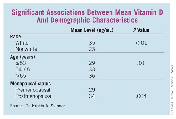
WASHINGTON – Vitamin D deficiency was not only twice as common in women undergoing breast cancer surgery, but it also was associated with poor-prognosis tumors in a case-control study that compared cancer patients with cancer-free women who had been tested for vitamin D.
The breast cancer patients had significantly lower mean vitamin D levels than did controls (33 ng/mL vs. 37 ng/mL), Dr. Kristin A. Skinner reported at the annual meeting of the American Association of Breast Surgeons. Patients were also more than twice as likely to have deficient levels (odds ratio, 2.4; P less than .01), she said.
Analyses presented by Dr. Skinner showed that mean vitamin D levels were significantly lower in the following subgroups of breast cancer patients:
• Those with estrogen receptornegative cancers vs. those with ER-positive cancers (28 ng/mL vs. 33 ng/mL; P = .04).
• Those with triple-negative cancers vs. those with cancers that were not triple negative (26 ng/mL vs.33 ng/mL; P -= .02).
• Those of the basal-like phenotype vs. those of the luminal A phenotype (24 ng/mL vs. 33 ng/mL; P = .04).
Some patient characteristics also carried significant associations with decreased vitamin D. White women, women aged 65 years and older, and those who were postmenopausal had significantly higher vitamin D levels than did nonwhite, younger, and premenopausal women, respectively. (See box.)
Although vitamin D levels were lower in patients with high Oncotype DX recurrence scores, progesterone receptornegative tumors, and invasive tumors, these differences were not statistically significant. Nor were family history or HER2, tumor, or nodal status significantly related to vitamin D levels, according to Dr. Skinner, a surgical oncologist and breast specialist at the cancer center of the University of Rochester (N.Y.).
In the case-control study, Dr. Skinner and her colleagues selected 194 women who were treated for breast cancer (stage 0-III) at the center and had total 25-hydroxy vitamin D levels drawn in the 3 months before or after their cancer surgery; the mean time of the blood draw was 30 days before surgery.
The patients were matched 1:1 with cancer-free controls who were drawn from a pool of more than 37,000 women who also underwent vitamin D testing in the university’s clinical labs in 2009-2010, the same time the cases were treated. The women were matched for age and the season of testing, since vitamin D levels can change as sun exposure varies.
The researchers divided vitamin D levels into tertiles: Optimal level was considered at least 32 ng/mL, suboptimal was 20-31 ng/mL, and deficient was less than 20 ng/mL.
The findings may argue for vitamin D testing and supplementation either in a primary care setting or in one devoted to breast health, Dr. Skinner said during a press briefing.
"At our institution, we routinely check vitamin D levels and replace them until they are well into the normal range," which is greater than 32 ng/mL, she said. "We really aim for a level of about 50 ng/mL, and titrate their replacement to those levels. In terms of taking supplements, we usually recommend starting at 1,000-2,000 IU daily, but the most effective way is to check levels, and replace accordingly."
Extant epidemiologic data have consistently found a link between more aggressive breast cancers and low vitamin D levels, she said, describing the relationship as biologically plausible. "The vitamin D receptor appears to modulate cell cycles, including the proliferation and differentiation of cells and the activation of apoptosis. Some studies have shown that vitamin D supplementation reduces the risk of breast cancer and improves survival outcomes, but very little is known about vitamin D levels and standard prognostic factors in breast cancer," she said.
"These findings may explain the associations seen in the epidemiologic studies, and may help explain why the black and other nonwhite populations tend to get more-aggressive breast cancer, and get breast cancer at a younger age," Dr. Skinner said.
She had no financial declarations with regard to the work.
WASHINGTON – Vitamin D deficiency was not only twice as common in women undergoing breast cancer surgery, but it also was associated with poor-prognosis tumors in a case-control study that compared cancer patients with cancer-free women who had been tested for vitamin D.
The breast cancer patients had significantly lower mean vitamin D levels than did controls (33 ng/mL vs. 37 ng/mL), Dr. Kristin A. Skinner reported at the annual meeting of the American Association of Breast Surgeons. Patients were also more than twice as likely to have deficient levels (odds ratio, 2.4; P less than .01), she said.
Analyses presented by Dr. Skinner showed that mean vitamin D levels were significantly lower in the following subgroups of breast cancer patients:
• Those with estrogen receptornegative cancers vs. those with ER-positive cancers (28 ng/mL vs. 33 ng/mL; P = .04).
• Those with triple-negative cancers vs. those with cancers that were not triple negative (26 ng/mL vs.33 ng/mL; P -= .02).
• Those of the basal-like phenotype vs. those of the luminal A phenotype (24 ng/mL vs. 33 ng/mL; P = .04).
Some patient characteristics also carried significant associations with decreased vitamin D. White women, women aged 65 years and older, and those who were postmenopausal had significantly higher vitamin D levels than did nonwhite, younger, and premenopausal women, respectively. (See box.)
Although vitamin D levels were lower in patients with high Oncotype DX recurrence scores, progesterone receptornegative tumors, and invasive tumors, these differences were not statistically significant. Nor were family history or HER2, tumor, or nodal status significantly related to vitamin D levels, according to Dr. Skinner, a surgical oncologist and breast specialist at the cancer center of the University of Rochester (N.Y.).
In the case-control study, Dr. Skinner and her colleagues selected 194 women who were treated for breast cancer (stage 0-III) at the center and had total 25-hydroxy vitamin D levels drawn in the 3 months before or after their cancer surgery; the mean time of the blood draw was 30 days before surgery.
The patients were matched 1:1 with cancer-free controls who were drawn from a pool of more than 37,000 women who also underwent vitamin D testing in the university’s clinical labs in 2009-2010, the same time the cases were treated. The women were matched for age and the season of testing, since vitamin D levels can change as sun exposure varies.
The researchers divided vitamin D levels into tertiles: Optimal level was considered at least 32 ng/mL, suboptimal was 20-31 ng/mL, and deficient was less than 20 ng/mL.
The findings may argue for vitamin D testing and supplementation either in a primary care setting or in one devoted to breast health, Dr. Skinner said during a press briefing.
"At our institution, we routinely check vitamin D levels and replace them until they are well into the normal range," which is greater than 32 ng/mL, she said. "We really aim for a level of about 50 ng/mL, and titrate their replacement to those levels. In terms of taking supplements, we usually recommend starting at 1,000-2,000 IU daily, but the most effective way is to check levels, and replace accordingly."
Extant epidemiologic data have consistently found a link between more aggressive breast cancers and low vitamin D levels, she said, describing the relationship as biologically plausible. "The vitamin D receptor appears to modulate cell cycles, including the proliferation and differentiation of cells and the activation of apoptosis. Some studies have shown that vitamin D supplementation reduces the risk of breast cancer and improves survival outcomes, but very little is known about vitamin D levels and standard prognostic factors in breast cancer," she said.
"These findings may explain the associations seen in the epidemiologic studies, and may help explain why the black and other nonwhite populations tend to get more-aggressive breast cancer, and get breast cancer at a younger age," Dr. Skinner said.
She had no financial declarations with regard to the work.
FROM THE ANNUAL MEETING OF THE AMERICAN SOCIETY OF BREAST SURGEONS
Finding: Vitamin D deficiency was more than twice as common as normal levels in women undergoing surgery for breast cancer (OR, 2.4).
Data Source: A case-control study of vitamin D levels in 194 women with breast cancer matched 1:1 to a control population.
Disclosures: Dr. Skinner said she had no relevant disclosures.
CDER: Avastin Breast Cancer Claim Undermines Approval Process
The Food and Drug Administration’s accelerated approval process would operate as a lower approval standard if Genentech were allowed to maintain the metastatic breast cancer claim for Avastin, the Center for Drug Evaluation and Research states in a written summary of arguments to be presented at a June 28-29 hearing on the indication.
Current evidence does not justify allowing Avastin (bevacizumab) to retain an indication for combination use with paclitaxel in first-line metastatic breast cancer (MBC); doing so while Genentech designs and conducts additional confirmatory studies would jeopardize the integrity of the accelerated approval process, CDER says.
CDER also dispatches with Genentech’s argument that it should be allowed to demonstrate that the choice of chemotherapy partner is integral to Avastin’s benefit in MBC. Rather, the regulator asserts there is no proven scientific basis for Genentech’s view that Avastin’s effect is predicated upon substantive differences between paclitaxel and other chemotherapy agents.
The drug center stands by its view that as part of Avastin’s accelerated approval Genentech was required to confirm the magnitude of benefit seen in the E2100 pivotal trial, which the company has failed to do.
Making the Case
The document, released on May 16, summarizes the arguments CDER will make to presiding officer Dr. Karen Midthun and the Oncologic Drugs Advisory Committee at the June hearing.
In its written summary of arguments, Genentech said it does not plan to focus on the AVADO and RIBBON1 studies – which were intended to serve as confirmatory studies for Avastin’s accelerated approval in MBC. Rather, it will argue that accelerated approval should be maintained pending a confirmatory study of Avastin in combination with paclitaxel that also uses a biomarker to identify those patients most likely to benefit.
CDER’s filing makes clear the regulator’s focus will be both on the specific issues relating to the existing Avastin data as well as the bigger policy issue of accelerated approval standards.
CDER observes it took the "unprecedented step" of granting accelerated approval based solely on an interim analysis of progression-free survival in the E2100 trial, which studied Avastin in combination with paclitaxel. "This was the first approval of a nonhormonal agent in which evidence of a treatment effect on PFS [progression free survival] alone was viewed not as a surrogate end point, but rather as a clinical benefit because of the magnitude of improvement in PFS," CDER says.
Data from the E2100 trial showed a 5.5-month improvement in median PFS, and CDER granted accelerated approval because "in its best scientific judgment at that time, the magnitude of PFS in the single trial suggested that Avastin held promise for patients with MBC."
Two confirmatory trials studied Avastin in combination with chemotherapeutics other than paclitaxel. Avastin’s chemotherapy partner in AVADO was docetaxel, while RIBBON1 looked at Avastin in combination with taxanes, anthracyclines, or capecitabine. These studies showed statistically significant improvements in PFS ranging from less than one month to less than three months, well shy of the 5.5-month benefit seen with paclitaxel in E2100.
The "small effect" seen in the confirmatory trials was not enough to verify Avastin’s clinical benefit, CDER asserts.
"In order for the accelerated approval system to serve its purpose and not operate as a lower approval standard, CDER must be able to withdraw approvals when it determines, based upon careful consideration of the data, that the confirmatory trials have failed to verify clinical benefit," CDER says.
Accelerated Approval Encompasses "Accelerated Withdrawal"
"Accelerated withdrawal" is an integral part of the accelerated approval process, CDER maintains.
"Once postapproval trials fail to demonstrate the expected clinical benefit, the accelerated approval rubric does not contemplate – as Genentech argues – that the agency should ‘maintain’ approval while an applicant designs and conducts more trials with the hope of eventually verifying clinical benefit," CDER says. "If CDER were forced to allow products to stay on the market when the risk-benefit analysis shows that the product is not safe and effective for its intended use, the accelerated approval program would be significantly undermined."
The agency takes issue with Genentech’s assertion that the indication should be maintained as long as the data for Avastin in combination with paclitaxel continue to be reasonably likely to predict clinical benefit.
The "reasonably likely" language in the accelerated approval regulations refers to the relationship between a surrogate end point and clinical benefit, CDER explains. "In the case of Avastin’s MBC approval, there was no surrogate end point – CDER believed that 5.5 months of PFS could be deemed a clinical benefit, subject to confirmatory trials."
Genentech does not explain how it would enroll patients in a new, blinded, controlled trial of Avastin in combination with paclitaxel if this remains a labeled use, CDER says. Furthermore, such a study would likely take many years to complete.
"Rather than serving as a basis to ‘maintain’ approval, which is not part of the accelerated approval program, the new research proposed by Genentech, if completed favorably, could be used to support a new sBLA [Supplemental Biologics License Application] requesting an MBC indication."
Chemotherapy Partner Hypothesis Does Not Hold Water
Genentech has hypothesized that chemotherapy partners that provide for prolonged combined exposure with Avastin may result in the strongest treatment effect. Yet this hypothesis has not been substantiated by either clinical or nonclinical evidence, CDER says.
"To support this argument, CDER expects that there should be some proven scientific basis for substantial differences, such as evidence of drug interactions or synergistic/overlapping toxicity between Avastin and other chemotherapy drugs. There is none," CDER says. Evidence submitted to date, such as population pharmacokinetic analyses, suggest no unique interactions between Avastin and any of the chemotherapy agents administered in the trials.
"In the absence of a scientifically supported basis for chemotherapy-specific interactions the more likely explanation for the failure of the clinical trials to verify the results of the E2100 trial is that the magnitude of the PFS treatment effect observed in E2100 is an outlier."
Genentech’s argument on the importance of chemotherapy partner used also runs contrary to its prior position, CDER says, noting that the company initially sought broad approval of Avastin in combination with all taxane-based chemotherapies based solely on the E2100 data.
No One Is Moving Goal Posts at CDER
CDER further disputes Genentech’s argument that it has changed its approval standards for MBC treatments. The regulator consistently maintained in conversations with Genentech that the adequacy of PFS as an approval end point would depend upon the overall dataset and the magnitude of benefit, it says.
"CDER has not in any way sought to ‘move the goal posts,’ " the filing states. "CDER has not determined that a set magnitude of PFS improvement is needed to support an MBC indication; however, CDER has determined that the magnitude of PFS improvement shown in Avastin’s MBC post-approval studies, and shown in the totality of the relevant data, is so small that the risk-benefit balance is unfavorable."
This coverage is provided courtesy of "The Pink Sheet." "The Pink Sheet" and Internal Medicine News Digital Network are both owned by Elsevier.
The Food and Drug Administration’s accelerated approval process would operate as a lower approval standard if Genentech were allowed to maintain the metastatic breast cancer claim for Avastin, the Center for Drug Evaluation and Research states in a written summary of arguments to be presented at a June 28-29 hearing on the indication.
Current evidence does not justify allowing Avastin (bevacizumab) to retain an indication for combination use with paclitaxel in first-line metastatic breast cancer (MBC); doing so while Genentech designs and conducts additional confirmatory studies would jeopardize the integrity of the accelerated approval process, CDER says.
CDER also dispatches with Genentech’s argument that it should be allowed to demonstrate that the choice of chemotherapy partner is integral to Avastin’s benefit in MBC. Rather, the regulator asserts there is no proven scientific basis for Genentech’s view that Avastin’s effect is predicated upon substantive differences between paclitaxel and other chemotherapy agents.
The drug center stands by its view that as part of Avastin’s accelerated approval Genentech was required to confirm the magnitude of benefit seen in the E2100 pivotal trial, which the company has failed to do.
Making the Case
The document, released on May 16, summarizes the arguments CDER will make to presiding officer Dr. Karen Midthun and the Oncologic Drugs Advisory Committee at the June hearing.
In its written summary of arguments, Genentech said it does not plan to focus on the AVADO and RIBBON1 studies – which were intended to serve as confirmatory studies for Avastin’s accelerated approval in MBC. Rather, it will argue that accelerated approval should be maintained pending a confirmatory study of Avastin in combination with paclitaxel that also uses a biomarker to identify those patients most likely to benefit.
CDER’s filing makes clear the regulator’s focus will be both on the specific issues relating to the existing Avastin data as well as the bigger policy issue of accelerated approval standards.
CDER observes it took the "unprecedented step" of granting accelerated approval based solely on an interim analysis of progression-free survival in the E2100 trial, which studied Avastin in combination with paclitaxel. "This was the first approval of a nonhormonal agent in which evidence of a treatment effect on PFS [progression free survival] alone was viewed not as a surrogate end point, but rather as a clinical benefit because of the magnitude of improvement in PFS," CDER says.
Data from the E2100 trial showed a 5.5-month improvement in median PFS, and CDER granted accelerated approval because "in its best scientific judgment at that time, the magnitude of PFS in the single trial suggested that Avastin held promise for patients with MBC."
Two confirmatory trials studied Avastin in combination with chemotherapeutics other than paclitaxel. Avastin’s chemotherapy partner in AVADO was docetaxel, while RIBBON1 looked at Avastin in combination with taxanes, anthracyclines, or capecitabine. These studies showed statistically significant improvements in PFS ranging from less than one month to less than three months, well shy of the 5.5-month benefit seen with paclitaxel in E2100.
The "small effect" seen in the confirmatory trials was not enough to verify Avastin’s clinical benefit, CDER asserts.
"In order for the accelerated approval system to serve its purpose and not operate as a lower approval standard, CDER must be able to withdraw approvals when it determines, based upon careful consideration of the data, that the confirmatory trials have failed to verify clinical benefit," CDER says.
Accelerated Approval Encompasses "Accelerated Withdrawal"
"Accelerated withdrawal" is an integral part of the accelerated approval process, CDER maintains.
"Once postapproval trials fail to demonstrate the expected clinical benefit, the accelerated approval rubric does not contemplate – as Genentech argues – that the agency should ‘maintain’ approval while an applicant designs and conducts more trials with the hope of eventually verifying clinical benefit," CDER says. "If CDER were forced to allow products to stay on the market when the risk-benefit analysis shows that the product is not safe and effective for its intended use, the accelerated approval program would be significantly undermined."
The agency takes issue with Genentech’s assertion that the indication should be maintained as long as the data for Avastin in combination with paclitaxel continue to be reasonably likely to predict clinical benefit.
The "reasonably likely" language in the accelerated approval regulations refers to the relationship between a surrogate end point and clinical benefit, CDER explains. "In the case of Avastin’s MBC approval, there was no surrogate end point – CDER believed that 5.5 months of PFS could be deemed a clinical benefit, subject to confirmatory trials."
Genentech does not explain how it would enroll patients in a new, blinded, controlled trial of Avastin in combination with paclitaxel if this remains a labeled use, CDER says. Furthermore, such a study would likely take many years to complete.
"Rather than serving as a basis to ‘maintain’ approval, which is not part of the accelerated approval program, the new research proposed by Genentech, if completed favorably, could be used to support a new sBLA [Supplemental Biologics License Application] requesting an MBC indication."
Chemotherapy Partner Hypothesis Does Not Hold Water
Genentech has hypothesized that chemotherapy partners that provide for prolonged combined exposure with Avastin may result in the strongest treatment effect. Yet this hypothesis has not been substantiated by either clinical or nonclinical evidence, CDER says.
"To support this argument, CDER expects that there should be some proven scientific basis for substantial differences, such as evidence of drug interactions or synergistic/overlapping toxicity between Avastin and other chemotherapy drugs. There is none," CDER says. Evidence submitted to date, such as population pharmacokinetic analyses, suggest no unique interactions between Avastin and any of the chemotherapy agents administered in the trials.
"In the absence of a scientifically supported basis for chemotherapy-specific interactions the more likely explanation for the failure of the clinical trials to verify the results of the E2100 trial is that the magnitude of the PFS treatment effect observed in E2100 is an outlier."
Genentech’s argument on the importance of chemotherapy partner used also runs contrary to its prior position, CDER says, noting that the company initially sought broad approval of Avastin in combination with all taxane-based chemotherapies based solely on the E2100 data.
No One Is Moving Goal Posts at CDER
CDER further disputes Genentech’s argument that it has changed its approval standards for MBC treatments. The regulator consistently maintained in conversations with Genentech that the adequacy of PFS as an approval end point would depend upon the overall dataset and the magnitude of benefit, it says.
"CDER has not in any way sought to ‘move the goal posts,’ " the filing states. "CDER has not determined that a set magnitude of PFS improvement is needed to support an MBC indication; however, CDER has determined that the magnitude of PFS improvement shown in Avastin’s MBC post-approval studies, and shown in the totality of the relevant data, is so small that the risk-benefit balance is unfavorable."
This coverage is provided courtesy of "The Pink Sheet." "The Pink Sheet" and Internal Medicine News Digital Network are both owned by Elsevier.
The Food and Drug Administration’s accelerated approval process would operate as a lower approval standard if Genentech were allowed to maintain the metastatic breast cancer claim for Avastin, the Center for Drug Evaluation and Research states in a written summary of arguments to be presented at a June 28-29 hearing on the indication.
Current evidence does not justify allowing Avastin (bevacizumab) to retain an indication for combination use with paclitaxel in first-line metastatic breast cancer (MBC); doing so while Genentech designs and conducts additional confirmatory studies would jeopardize the integrity of the accelerated approval process, CDER says.
CDER also dispatches with Genentech’s argument that it should be allowed to demonstrate that the choice of chemotherapy partner is integral to Avastin’s benefit in MBC. Rather, the regulator asserts there is no proven scientific basis for Genentech’s view that Avastin’s effect is predicated upon substantive differences between paclitaxel and other chemotherapy agents.
The drug center stands by its view that as part of Avastin’s accelerated approval Genentech was required to confirm the magnitude of benefit seen in the E2100 pivotal trial, which the company has failed to do.
Making the Case
The document, released on May 16, summarizes the arguments CDER will make to presiding officer Dr. Karen Midthun and the Oncologic Drugs Advisory Committee at the June hearing.
In its written summary of arguments, Genentech said it does not plan to focus on the AVADO and RIBBON1 studies – which were intended to serve as confirmatory studies for Avastin’s accelerated approval in MBC. Rather, it will argue that accelerated approval should be maintained pending a confirmatory study of Avastin in combination with paclitaxel that also uses a biomarker to identify those patients most likely to benefit.
CDER’s filing makes clear the regulator’s focus will be both on the specific issues relating to the existing Avastin data as well as the bigger policy issue of accelerated approval standards.
CDER observes it took the "unprecedented step" of granting accelerated approval based solely on an interim analysis of progression-free survival in the E2100 trial, which studied Avastin in combination with paclitaxel. "This was the first approval of a nonhormonal agent in which evidence of a treatment effect on PFS [progression free survival] alone was viewed not as a surrogate end point, but rather as a clinical benefit because of the magnitude of improvement in PFS," CDER says.
Data from the E2100 trial showed a 5.5-month improvement in median PFS, and CDER granted accelerated approval because "in its best scientific judgment at that time, the magnitude of PFS in the single trial suggested that Avastin held promise for patients with MBC."
Two confirmatory trials studied Avastin in combination with chemotherapeutics other than paclitaxel. Avastin’s chemotherapy partner in AVADO was docetaxel, while RIBBON1 looked at Avastin in combination with taxanes, anthracyclines, or capecitabine. These studies showed statistically significant improvements in PFS ranging from less than one month to less than three months, well shy of the 5.5-month benefit seen with paclitaxel in E2100.
The "small effect" seen in the confirmatory trials was not enough to verify Avastin’s clinical benefit, CDER asserts.
"In order for the accelerated approval system to serve its purpose and not operate as a lower approval standard, CDER must be able to withdraw approvals when it determines, based upon careful consideration of the data, that the confirmatory trials have failed to verify clinical benefit," CDER says.
Accelerated Approval Encompasses "Accelerated Withdrawal"
"Accelerated withdrawal" is an integral part of the accelerated approval process, CDER maintains.
"Once postapproval trials fail to demonstrate the expected clinical benefit, the accelerated approval rubric does not contemplate – as Genentech argues – that the agency should ‘maintain’ approval while an applicant designs and conducts more trials with the hope of eventually verifying clinical benefit," CDER says. "If CDER were forced to allow products to stay on the market when the risk-benefit analysis shows that the product is not safe and effective for its intended use, the accelerated approval program would be significantly undermined."
The agency takes issue with Genentech’s assertion that the indication should be maintained as long as the data for Avastin in combination with paclitaxel continue to be reasonably likely to predict clinical benefit.
The "reasonably likely" language in the accelerated approval regulations refers to the relationship between a surrogate end point and clinical benefit, CDER explains. "In the case of Avastin’s MBC approval, there was no surrogate end point – CDER believed that 5.5 months of PFS could be deemed a clinical benefit, subject to confirmatory trials."
Genentech does not explain how it would enroll patients in a new, blinded, controlled trial of Avastin in combination with paclitaxel if this remains a labeled use, CDER says. Furthermore, such a study would likely take many years to complete.
"Rather than serving as a basis to ‘maintain’ approval, which is not part of the accelerated approval program, the new research proposed by Genentech, if completed favorably, could be used to support a new sBLA [Supplemental Biologics License Application] requesting an MBC indication."
Chemotherapy Partner Hypothesis Does Not Hold Water
Genentech has hypothesized that chemotherapy partners that provide for prolonged combined exposure with Avastin may result in the strongest treatment effect. Yet this hypothesis has not been substantiated by either clinical or nonclinical evidence, CDER says.
"To support this argument, CDER expects that there should be some proven scientific basis for substantial differences, such as evidence of drug interactions or synergistic/overlapping toxicity between Avastin and other chemotherapy drugs. There is none," CDER says. Evidence submitted to date, such as population pharmacokinetic analyses, suggest no unique interactions between Avastin and any of the chemotherapy agents administered in the trials.
"In the absence of a scientifically supported basis for chemotherapy-specific interactions the more likely explanation for the failure of the clinical trials to verify the results of the E2100 trial is that the magnitude of the PFS treatment effect observed in E2100 is an outlier."
Genentech’s argument on the importance of chemotherapy partner used also runs contrary to its prior position, CDER says, noting that the company initially sought broad approval of Avastin in combination with all taxane-based chemotherapies based solely on the E2100 data.
No One Is Moving Goal Posts at CDER
CDER further disputes Genentech’s argument that it has changed its approval standards for MBC treatments. The regulator consistently maintained in conversations with Genentech that the adequacy of PFS as an approval end point would depend upon the overall dataset and the magnitude of benefit, it says.
"CDER has not in any way sought to ‘move the goal posts,’ " the filing states. "CDER has not determined that a set magnitude of PFS improvement is needed to support an MBC indication; however, CDER has determined that the magnitude of PFS improvement shown in Avastin’s MBC post-approval studies, and shown in the totality of the relevant data, is so small that the risk-benefit balance is unfavorable."
This coverage is provided courtesy of "The Pink Sheet." "The Pink Sheet" and Internal Medicine News Digital Network are both owned by Elsevier.
FROM THE CENTER FOR DRUG EVALUATION AND RESEARCH
Genentech Appeals to FDAs Regulatory Flexibility on Avastin
Genentech’s advance summary of evidence and arguments for the Food and Drug Administration’s upcoming hearing on the agency’s proposed withdrawal of the metastatic breast cancer approval for Avastin shows that the company is trying to keep the focus on the additional confirmatory trial it proposes to conduct.
The document, released May 13, provides an overview of the case Genentech will make to support maintaining the accelerated approval of bevacizumab, in combination with paclitaxel, for first-line metastatic breast cancer (MBC) at the June 28-29 hearing, as requested by the presiding officer, Center for Biologics Evaluation and Research Director Karen Midthun.
Dr. Midthun recently released the agenda for the hearing and the issues that are to be discussed. These include whether the AVADO and RIBBON1 trials failed to verify Avastin’s clinical benefit in MBC as seen in the pivotal E2100 study, whether the available evidence shows it is safe and effective, and whether the FDA should withdraw the approval.
In its prehearing summary of evidence, Genentech notes it has recognized the view of the Center for Drug Evaluation and Research (CDER) and the Oncologic Drugs Advisory Committee that the AVADO and RIBBON1 trials failed to confirm the benefit and has responded by tailoring its proposal to maintaining accelerated approval for use with paclitaxel "subject to" an additional confirmatory trial.
Thus, the company does not plan to focus on AVADO and RIBBON1, the summary explains. "Instead, Genentech intends to focus on the question of whether accelerated approval should be maintained for Avastin and paclitaxel subject to a further confirmatory study of this combination, even in light of the views of CDER and the ODAC on AVADO and RIBBON1."
"The only open question is whether the magnitude of benefit observed in the E2100 study is reasonably likely to be confirmed in a second study of Avastin with paclitaxel," the company argues.
"Given the meaningful probability that the chemotherapy partner has an impact on the magnitude of benefit, and given that an additional study can resolve this question, accelerated approval should be maintained pending completion of Genentech’s further study."
The company also says that continuing the approval while gaining further evidence fits with the overall goals of the accelerated approval program and that the accelerated approval regime allows the FDA that regulatory flexibility.
The New Confirmatory Trial
Genentech first proposed conducting a new confirmatory trial in August 2010 after the FDA’s Oncologic Drugs Advisory Committee voted against continued approval in MBC, and that proposal was a pivotal point in its request for a hearing on FDA’s proposed withdrawal.
The company intends to conduct a new trial – a double-blind, randomized, multicenter phase III trial of Avastin in combination with weekly paclitaxel – using a biomarker to select for patients more likely to derive a greater benefit from treatment, which was an option alluded to in CDER’s memo on the proposed withdrawal.
"Based on the company’s extensive research, including a new assay sensitive to specific isoforms, plasma concentration of VEGF-A (Avastin’s target) has been identified as a potential predictive biomarker in breast, gastric and pancreatic cancers," the summary of evidence states.
An analysis of the AVADO trial, presented as an abstract at the San Antonio Breast Cancer Symposium in December, showed that patients with high levels of VEGF-A had a hazard ratio for progression-free survival analysis of 0.49, compared with 0.87 for patients with low levels of VEGF-A. "The data suggest that patients with high VEGF-A levels may be more likely to derive a substantial benefit from Avastin, and it is important and appropriate to validate this predictive biomarker in a prospective phase III trial," Genentech argued.
The company met with the agency to discuss the trial on Feb. 22, the preparatory documents for the hearing reveal, the day before the hearing was granted. At that meeting, Genentech reports, CDER stated that "PFS [progression free survival] results confirming the magnitude of treatment effect observed in E2100 without a detriment to OS [overall survival], coupled with the E2100 data, would support Avastin’s full approval with paclitaxel."
The company has proceeded with study planning and intends to submit the protocol under a Special Protocol Assessment, the summary states.
Nothing Wrong With E2100
Genentech presented an overview of the E2100 findings that emphasize their statistical rigor and clinical significance, an essential point of agreement to support continuing the accelerated approval based upon those findings while another confirmatory trial is conducted.
In the context of Dr. Midthun’s posed question on whether Avastin’s benefit-risk ratio remains favorable in MBC, the company notes that withdrawal would "necessarily rest on a determination that the findings from the E2100 study are no longer reliable and that Avastin presents unique toxicity concerns in the MBC setting."
"Respectfully, that determination would be contrary to the most reasonable interpretation of the scientific data."
In addition to defending E2100, the summary shows that Genentech will focus on FDA’s application of the accelerated approval standard, the external support for Avastin’s use in MBC (including that of other regulatory bodies), and the agency’s potential regulatory flexibility.
In the end, it’s that flexibility that Genentech is really appealing to. "Regulatory flexibility is intended for cases like this," the summary concludes, "where a formulaic application of the withdrawal standard would deprive patients and physicians of the choice intended by the accelerated approval program – and do so in the face of continued findings of benefit, a well-understood safety profile, and a viable option for providing a clearer answer to the remaining scientific questions."
Internal Medicine News Digital Network and "The Pink Sheet" are published by Elsevier.
Genentech’s advance summary of evidence and arguments for the Food and Drug Administration’s upcoming hearing on the agency’s proposed withdrawal of the metastatic breast cancer approval for Avastin shows that the company is trying to keep the focus on the additional confirmatory trial it proposes to conduct.
The document, released May 13, provides an overview of the case Genentech will make to support maintaining the accelerated approval of bevacizumab, in combination with paclitaxel, for first-line metastatic breast cancer (MBC) at the June 28-29 hearing, as requested by the presiding officer, Center for Biologics Evaluation and Research Director Karen Midthun.
Dr. Midthun recently released the agenda for the hearing and the issues that are to be discussed. These include whether the AVADO and RIBBON1 trials failed to verify Avastin’s clinical benefit in MBC as seen in the pivotal E2100 study, whether the available evidence shows it is safe and effective, and whether the FDA should withdraw the approval.
In its prehearing summary of evidence, Genentech notes it has recognized the view of the Center for Drug Evaluation and Research (CDER) and the Oncologic Drugs Advisory Committee that the AVADO and RIBBON1 trials failed to confirm the benefit and has responded by tailoring its proposal to maintaining accelerated approval for use with paclitaxel "subject to" an additional confirmatory trial.
Thus, the company does not plan to focus on AVADO and RIBBON1, the summary explains. "Instead, Genentech intends to focus on the question of whether accelerated approval should be maintained for Avastin and paclitaxel subject to a further confirmatory study of this combination, even in light of the views of CDER and the ODAC on AVADO and RIBBON1."
"The only open question is whether the magnitude of benefit observed in the E2100 study is reasonably likely to be confirmed in a second study of Avastin with paclitaxel," the company argues.
"Given the meaningful probability that the chemotherapy partner has an impact on the magnitude of benefit, and given that an additional study can resolve this question, accelerated approval should be maintained pending completion of Genentech’s further study."
The company also says that continuing the approval while gaining further evidence fits with the overall goals of the accelerated approval program and that the accelerated approval regime allows the FDA that regulatory flexibility.
The New Confirmatory Trial
Genentech first proposed conducting a new confirmatory trial in August 2010 after the FDA’s Oncologic Drugs Advisory Committee voted against continued approval in MBC, and that proposal was a pivotal point in its request for a hearing on FDA’s proposed withdrawal.
The company intends to conduct a new trial – a double-blind, randomized, multicenter phase III trial of Avastin in combination with weekly paclitaxel – using a biomarker to select for patients more likely to derive a greater benefit from treatment, which was an option alluded to in CDER’s memo on the proposed withdrawal.
"Based on the company’s extensive research, including a new assay sensitive to specific isoforms, plasma concentration of VEGF-A (Avastin’s target) has been identified as a potential predictive biomarker in breast, gastric and pancreatic cancers," the summary of evidence states.
An analysis of the AVADO trial, presented as an abstract at the San Antonio Breast Cancer Symposium in December, showed that patients with high levels of VEGF-A had a hazard ratio for progression-free survival analysis of 0.49, compared with 0.87 for patients with low levels of VEGF-A. "The data suggest that patients with high VEGF-A levels may be more likely to derive a substantial benefit from Avastin, and it is important and appropriate to validate this predictive biomarker in a prospective phase III trial," Genentech argued.
The company met with the agency to discuss the trial on Feb. 22, the preparatory documents for the hearing reveal, the day before the hearing was granted. At that meeting, Genentech reports, CDER stated that "PFS [progression free survival] results confirming the magnitude of treatment effect observed in E2100 without a detriment to OS [overall survival], coupled with the E2100 data, would support Avastin’s full approval with paclitaxel."
The company has proceeded with study planning and intends to submit the protocol under a Special Protocol Assessment, the summary states.
Nothing Wrong With E2100
Genentech presented an overview of the E2100 findings that emphasize their statistical rigor and clinical significance, an essential point of agreement to support continuing the accelerated approval based upon those findings while another confirmatory trial is conducted.
In the context of Dr. Midthun’s posed question on whether Avastin’s benefit-risk ratio remains favorable in MBC, the company notes that withdrawal would "necessarily rest on a determination that the findings from the E2100 study are no longer reliable and that Avastin presents unique toxicity concerns in the MBC setting."
"Respectfully, that determination would be contrary to the most reasonable interpretation of the scientific data."
In addition to defending E2100, the summary shows that Genentech will focus on FDA’s application of the accelerated approval standard, the external support for Avastin’s use in MBC (including that of other regulatory bodies), and the agency’s potential regulatory flexibility.
In the end, it’s that flexibility that Genentech is really appealing to. "Regulatory flexibility is intended for cases like this," the summary concludes, "where a formulaic application of the withdrawal standard would deprive patients and physicians of the choice intended by the accelerated approval program – and do so in the face of continued findings of benefit, a well-understood safety profile, and a viable option for providing a clearer answer to the remaining scientific questions."
Internal Medicine News Digital Network and "The Pink Sheet" are published by Elsevier.
Genentech’s advance summary of evidence and arguments for the Food and Drug Administration’s upcoming hearing on the agency’s proposed withdrawal of the metastatic breast cancer approval for Avastin shows that the company is trying to keep the focus on the additional confirmatory trial it proposes to conduct.
The document, released May 13, provides an overview of the case Genentech will make to support maintaining the accelerated approval of bevacizumab, in combination with paclitaxel, for first-line metastatic breast cancer (MBC) at the June 28-29 hearing, as requested by the presiding officer, Center for Biologics Evaluation and Research Director Karen Midthun.
Dr. Midthun recently released the agenda for the hearing and the issues that are to be discussed. These include whether the AVADO and RIBBON1 trials failed to verify Avastin’s clinical benefit in MBC as seen in the pivotal E2100 study, whether the available evidence shows it is safe and effective, and whether the FDA should withdraw the approval.
In its prehearing summary of evidence, Genentech notes it has recognized the view of the Center for Drug Evaluation and Research (CDER) and the Oncologic Drugs Advisory Committee that the AVADO and RIBBON1 trials failed to confirm the benefit and has responded by tailoring its proposal to maintaining accelerated approval for use with paclitaxel "subject to" an additional confirmatory trial.
Thus, the company does not plan to focus on AVADO and RIBBON1, the summary explains. "Instead, Genentech intends to focus on the question of whether accelerated approval should be maintained for Avastin and paclitaxel subject to a further confirmatory study of this combination, even in light of the views of CDER and the ODAC on AVADO and RIBBON1."
"The only open question is whether the magnitude of benefit observed in the E2100 study is reasonably likely to be confirmed in a second study of Avastin with paclitaxel," the company argues.
"Given the meaningful probability that the chemotherapy partner has an impact on the magnitude of benefit, and given that an additional study can resolve this question, accelerated approval should be maintained pending completion of Genentech’s further study."
The company also says that continuing the approval while gaining further evidence fits with the overall goals of the accelerated approval program and that the accelerated approval regime allows the FDA that regulatory flexibility.
The New Confirmatory Trial
Genentech first proposed conducting a new confirmatory trial in August 2010 after the FDA’s Oncologic Drugs Advisory Committee voted against continued approval in MBC, and that proposal was a pivotal point in its request for a hearing on FDA’s proposed withdrawal.
The company intends to conduct a new trial – a double-blind, randomized, multicenter phase III trial of Avastin in combination with weekly paclitaxel – using a biomarker to select for patients more likely to derive a greater benefit from treatment, which was an option alluded to in CDER’s memo on the proposed withdrawal.
"Based on the company’s extensive research, including a new assay sensitive to specific isoforms, plasma concentration of VEGF-A (Avastin’s target) has been identified as a potential predictive biomarker in breast, gastric and pancreatic cancers," the summary of evidence states.
An analysis of the AVADO trial, presented as an abstract at the San Antonio Breast Cancer Symposium in December, showed that patients with high levels of VEGF-A had a hazard ratio for progression-free survival analysis of 0.49, compared with 0.87 for patients with low levels of VEGF-A. "The data suggest that patients with high VEGF-A levels may be more likely to derive a substantial benefit from Avastin, and it is important and appropriate to validate this predictive biomarker in a prospective phase III trial," Genentech argued.
The company met with the agency to discuss the trial on Feb. 22, the preparatory documents for the hearing reveal, the day before the hearing was granted. At that meeting, Genentech reports, CDER stated that "PFS [progression free survival] results confirming the magnitude of treatment effect observed in E2100 without a detriment to OS [overall survival], coupled with the E2100 data, would support Avastin’s full approval with paclitaxel."
The company has proceeded with study planning and intends to submit the protocol under a Special Protocol Assessment, the summary states.
Nothing Wrong With E2100
Genentech presented an overview of the E2100 findings that emphasize their statistical rigor and clinical significance, an essential point of agreement to support continuing the accelerated approval based upon those findings while another confirmatory trial is conducted.
In the context of Dr. Midthun’s posed question on whether Avastin’s benefit-risk ratio remains favorable in MBC, the company notes that withdrawal would "necessarily rest on a determination that the findings from the E2100 study are no longer reliable and that Avastin presents unique toxicity concerns in the MBC setting."
"Respectfully, that determination would be contrary to the most reasonable interpretation of the scientific data."
In addition to defending E2100, the summary shows that Genentech will focus on FDA’s application of the accelerated approval standard, the external support for Avastin’s use in MBC (including that of other regulatory bodies), and the agency’s potential regulatory flexibility.
In the end, it’s that flexibility that Genentech is really appealing to. "Regulatory flexibility is intended for cases like this," the summary concludes, "where a formulaic application of the withdrawal standard would deprive patients and physicians of the choice intended by the accelerated approval program – and do so in the face of continued findings of benefit, a well-understood safety profile, and a viable option for providing a clearer answer to the remaining scientific questions."
Internal Medicine News Digital Network and "The Pink Sheet" are published by Elsevier.
FROM THE FDA
Radioactive Seeds Guide Surgeons to Nonpalpable Breast Lesions
WASHINGTON – Radioactive seeds were a safe and effective method of pinpointing nonpalpable breast lesions for surgery, with an 85% rate of negative margins on the first excision and an ipsilateral recurrence rate of less than 2% over 33 months, according to a large retrospective study.
Close margins occurred in 12% of the 767 patients with nonpalpable breast lesions, and positive margins occurred in 3%. The overall re-excision rate was 15%, Dr. Lee McGhan of the Mayo Clinic, Scottsdale, Ariz., said at the annual meeting of the American Society of Breast Surgeons.
Performing the procedure is "almost intuitive," with a very low learning curve, he said. "It is now our method of choice when dealing with preoperative localization of nonpalpable breast lesions."
His retrospective review of 978 prospectively collected patient records comprised 1,000 radioactive in 2003-2010. Almost 1,150 seeds were deployed.
The patients’ mean age was 65 years; their mean lesion size was 1.2 cm. Most (550) had an invasive carcinoma; 217 had DCIS (ductal carcinoma in situ); 115 had atypical hyperplasia, and the remainder, uncertain or suspicious percutaneous biopsy results.
Dr. McGhan reported 33-month follow-up results on the 767 women with either invasive carcinoma or DCIS. Most patients (910) received just one seed; 84 received two seeds; 5 received three seeds; and 1 patient received four of the devices.
Most (76%) underwent the procedure at least 1 day before surgery. Typically, Dr. McGhan said, patients came to the clinic a few days before surgery for an evaluation. Many chose to have the localization on a Friday, stayed over the weekend, and had the seeds removed early on Monday morning.
The 4- to 5-mm seeds containing radioactive iodine-125 can be placed up to 5 days before surgery. They were deployed through an 18-gauge spinal needle under image guidance; post deployment, a mammogram or ultrasound confirmed their position near the lesion. "We used a handheld gamma probe to identify the area of greatest radioactivity at the skin surface, marking the optimal site of skin incision," Dr. McGhan said.
Intraoperative complications included 30 displaced seeds – including 3 suctioned up by operative tubing and 3 that were improperly deployed during radiology – as well as one instance of an incorrect incision site resulting from a miscommunication between the radiologist and the surgeon, Dr. McGhan said. All of these seeds were retrieved with no patient harm.
All of the localized lesions were successfully removed, along with their associated seeds; the specimens were sent to pathology. Among the 550 invasive cancers, margins were negative in 87%, close in 9%, and positive in 3%. Re-excision was required in 13% (69).
Among the 217 DCIS lesions, margins were negative in 77%, close in 19%, and positive in 3%. Re-excision was necessary in 23% (49).
Sentinel node biopsies occurred in 544 cases, and were successful in all but one, Dr. McGhan said. "There was no blue dye detected, which was determined to be due to tumor invasion of the lymphatics rather than a direct complication of the procedure."
The mean follow-up period was 33 months. Over this time, the overall ipsilateral recurrence rate was 1.6% (12 patients). The rate was slightly higher among patients with DCIS (3%; seven patients). The local recurrence rate was 1% (five patients) among those with invasive cancer. By the end of the follow-up period, there were six mastectomies secondary to recurrence: three (0.5%) in the invasive cancer group and three (1%) in the DCIS group.
Dr. McGhan had no financial declarations.
WASHINGTON – Radioactive seeds were a safe and effective method of pinpointing nonpalpable breast lesions for surgery, with an 85% rate of negative margins on the first excision and an ipsilateral recurrence rate of less than 2% over 33 months, according to a large retrospective study.
Close margins occurred in 12% of the 767 patients with nonpalpable breast lesions, and positive margins occurred in 3%. The overall re-excision rate was 15%, Dr. Lee McGhan of the Mayo Clinic, Scottsdale, Ariz., said at the annual meeting of the American Society of Breast Surgeons.
Performing the procedure is "almost intuitive," with a very low learning curve, he said. "It is now our method of choice when dealing with preoperative localization of nonpalpable breast lesions."
His retrospective review of 978 prospectively collected patient records comprised 1,000 radioactive in 2003-2010. Almost 1,150 seeds were deployed.
The patients’ mean age was 65 years; their mean lesion size was 1.2 cm. Most (550) had an invasive carcinoma; 217 had DCIS (ductal carcinoma in situ); 115 had atypical hyperplasia, and the remainder, uncertain or suspicious percutaneous biopsy results.
Dr. McGhan reported 33-month follow-up results on the 767 women with either invasive carcinoma or DCIS. Most patients (910) received just one seed; 84 received two seeds; 5 received three seeds; and 1 patient received four of the devices.
Most (76%) underwent the procedure at least 1 day before surgery. Typically, Dr. McGhan said, patients came to the clinic a few days before surgery for an evaluation. Many chose to have the localization on a Friday, stayed over the weekend, and had the seeds removed early on Monday morning.
The 4- to 5-mm seeds containing radioactive iodine-125 can be placed up to 5 days before surgery. They were deployed through an 18-gauge spinal needle under image guidance; post deployment, a mammogram or ultrasound confirmed their position near the lesion. "We used a handheld gamma probe to identify the area of greatest radioactivity at the skin surface, marking the optimal site of skin incision," Dr. McGhan said.
Intraoperative complications included 30 displaced seeds – including 3 suctioned up by operative tubing and 3 that were improperly deployed during radiology – as well as one instance of an incorrect incision site resulting from a miscommunication between the radiologist and the surgeon, Dr. McGhan said. All of these seeds were retrieved with no patient harm.
All of the localized lesions were successfully removed, along with their associated seeds; the specimens were sent to pathology. Among the 550 invasive cancers, margins were negative in 87%, close in 9%, and positive in 3%. Re-excision was required in 13% (69).
Among the 217 DCIS lesions, margins were negative in 77%, close in 19%, and positive in 3%. Re-excision was necessary in 23% (49).
Sentinel node biopsies occurred in 544 cases, and were successful in all but one, Dr. McGhan said. "There was no blue dye detected, which was determined to be due to tumor invasion of the lymphatics rather than a direct complication of the procedure."
The mean follow-up period was 33 months. Over this time, the overall ipsilateral recurrence rate was 1.6% (12 patients). The rate was slightly higher among patients with DCIS (3%; seven patients). The local recurrence rate was 1% (five patients) among those with invasive cancer. By the end of the follow-up period, there were six mastectomies secondary to recurrence: three (0.5%) in the invasive cancer group and three (1%) in the DCIS group.
Dr. McGhan had no financial declarations.
WASHINGTON – Radioactive seeds were a safe and effective method of pinpointing nonpalpable breast lesions for surgery, with an 85% rate of negative margins on the first excision and an ipsilateral recurrence rate of less than 2% over 33 months, according to a large retrospective study.
Close margins occurred in 12% of the 767 patients with nonpalpable breast lesions, and positive margins occurred in 3%. The overall re-excision rate was 15%, Dr. Lee McGhan of the Mayo Clinic, Scottsdale, Ariz., said at the annual meeting of the American Society of Breast Surgeons.
Performing the procedure is "almost intuitive," with a very low learning curve, he said. "It is now our method of choice when dealing with preoperative localization of nonpalpable breast lesions."
His retrospective review of 978 prospectively collected patient records comprised 1,000 radioactive in 2003-2010. Almost 1,150 seeds were deployed.
The patients’ mean age was 65 years; their mean lesion size was 1.2 cm. Most (550) had an invasive carcinoma; 217 had DCIS (ductal carcinoma in situ); 115 had atypical hyperplasia, and the remainder, uncertain or suspicious percutaneous biopsy results.
Dr. McGhan reported 33-month follow-up results on the 767 women with either invasive carcinoma or DCIS. Most patients (910) received just one seed; 84 received two seeds; 5 received three seeds; and 1 patient received four of the devices.
Most (76%) underwent the procedure at least 1 day before surgery. Typically, Dr. McGhan said, patients came to the clinic a few days before surgery for an evaluation. Many chose to have the localization on a Friday, stayed over the weekend, and had the seeds removed early on Monday morning.
The 4- to 5-mm seeds containing radioactive iodine-125 can be placed up to 5 days before surgery. They were deployed through an 18-gauge spinal needle under image guidance; post deployment, a mammogram or ultrasound confirmed their position near the lesion. "We used a handheld gamma probe to identify the area of greatest radioactivity at the skin surface, marking the optimal site of skin incision," Dr. McGhan said.
Intraoperative complications included 30 displaced seeds – including 3 suctioned up by operative tubing and 3 that were improperly deployed during radiology – as well as one instance of an incorrect incision site resulting from a miscommunication between the radiologist and the surgeon, Dr. McGhan said. All of these seeds were retrieved with no patient harm.
All of the localized lesions were successfully removed, along with their associated seeds; the specimens were sent to pathology. Among the 550 invasive cancers, margins were negative in 87%, close in 9%, and positive in 3%. Re-excision was required in 13% (69).
Among the 217 DCIS lesions, margins were negative in 77%, close in 19%, and positive in 3%. Re-excision was necessary in 23% (49).
Sentinel node biopsies occurred in 544 cases, and were successful in all but one, Dr. McGhan said. "There was no blue dye detected, which was determined to be due to tumor invasion of the lymphatics rather than a direct complication of the procedure."
The mean follow-up period was 33 months. Over this time, the overall ipsilateral recurrence rate was 1.6% (12 patients). The rate was slightly higher among patients with DCIS (3%; seven patients). The local recurrence rate was 1% (five patients) among those with invasive cancer. By the end of the follow-up period, there were six mastectomies secondary to recurrence: three (0.5%) in the invasive cancer group and three (1%) in the DCIS group.
Dr. McGhan had no financial declarations.
FROM THE ANNUAL MEETING OF THE AMERICAN SOCIETY OF BREAST SURGEONS
Major Finding: Radioactive seed localization resulted in initial negative margins in 85% of cases and an overall re-excision rate of 2%.
Data Source: A retrospective study of 767 women with invasive cancer or DCIS who were followed for a mean of 33 months.
Disclosures: Dr. McGhan had no financial declarations.
Sequencing Reveals MAP3K1 Mutation in Luminal-Type Breast Cancer
ORLANDO – Massively parallel sequencing of DNA from tumor samples in 50 patients with luminal-type breast cancer revealed a novel mutation in the breast cancer tumor suppressor gene MAP3K1, which normally controls programmed cell death.
Presumably, the knockout mutation – which affected about 10% of estrogen receptor–positive breast cancers in the study and which "unequivocally destroys the function of the gene" – allows cells to survive when they would normally die, Dr. Matthew J. Ellis said at the annual meeting of the American Association for Cancer Research.
This finding, along with others from the sequencing of more than 10 trillion chemical bases of DNA in this extensive genomics investigation (one of the largest to date), marks an important early step toward personalized therapy for breast cancer patients who fail to respond to estrogen-lowering therapy prior to surgery, said Dr. Ellis, professor of medicine and chief of breast oncology at the Washington University in St. Louis.
Luminal-type breast cancer is the most common form of the disease, accounting for 70%-80% of hormone receptor–positive breast cancers. Many patients have a good prognosis, but a subset has this very aggressive type of disease. Indeed, more patients die of aggressive luminal-type breast cancer than do all other breast cancer subtypes combined, he said.
"So we set out to find a molecular basis for poor outcome in receptor-positive disease," he said.
DNA from tumor samples of patients who were enrolled in ongoing neoadjuvant endocrine clinical trials – 24 of whom were resistant to estrogen receptor–targeted therapy – was used for the supercomputer-conducted analysis. The whole genomes of the tumors were compared with the matched DNA of the same patients’ healthy cells, allowing identification of mutations occurring only in the cancer cells.
In all, 1,700 mutations were identified, and most of these were unique to individuals. In addition to two previously identified, relatively common mutations (PIK3CA and TP53), Dr. Ellis and his colleagues found only three others – including MAP3K1 – that recurred at a frequency of at least 10%; the other two were ATR and MYST3.
PIK3CA and TP53 were the most frequently mutated genes in estrogen receptor–positive breast cancer in this study, occurring in about 50% and 20% of tumors, respectively. MAP3K1 was the third most commonly mutated gene.
Considering the large number of mutations found, it was "a rather shocking result" to find only three new gene mutations at the 10% recurrence level, Dr. Ellis said. "What it says is that breast cancer is highly complex, that the genetic make-up involves a large number of mutations that averages about 20 tier-1 [or coding region] mutations in each tumor, and there’s a wide range," he added.
But the findings do offer a glimpse into how therapy can be personalized.
Using a "very, very simple model" produced by this analysis, Dr. Ellis illustrated how a constellation of mutations could be used to predict response or resistance patterns: The three-gene cluster of mutated MAP3K1, mutated PIK3CA, and wild type TP53, which occurred in a small subset of patients, was shown to be significantly associated with "luminal A status [indicative of good prognosis], suppressed proliferation, and favorable small tumors at the end of neoadjuvant treatment," he said.
Although there remains "a great sea of unknown," the findings – when considered in the context of the growing list of "druggable mutations" and treatments approved for other diseases – allow for a vision of therapy that involves obtaining the genetic information in advance of treatment to allow for the design of individually appropriate therapy to address the problem of resistance.
"Very clearly, this is a big problem clinically ... and only a tailored approach will lead to a solution to the problem," he said.
Dr. Ellis disclosed that he has received grant or research support from and/or served on the speakers bureau for Novartis, AstraZeneca, and Bioclassifier LLC.
ORLANDO – Massively parallel sequencing of DNA from tumor samples in 50 patients with luminal-type breast cancer revealed a novel mutation in the breast cancer tumor suppressor gene MAP3K1, which normally controls programmed cell death.
Presumably, the knockout mutation – which affected about 10% of estrogen receptor–positive breast cancers in the study and which "unequivocally destroys the function of the gene" – allows cells to survive when they would normally die, Dr. Matthew J. Ellis said at the annual meeting of the American Association for Cancer Research.
This finding, along with others from the sequencing of more than 10 trillion chemical bases of DNA in this extensive genomics investigation (one of the largest to date), marks an important early step toward personalized therapy for breast cancer patients who fail to respond to estrogen-lowering therapy prior to surgery, said Dr. Ellis, professor of medicine and chief of breast oncology at the Washington University in St. Louis.
Luminal-type breast cancer is the most common form of the disease, accounting for 70%-80% of hormone receptor–positive breast cancers. Many patients have a good prognosis, but a subset has this very aggressive type of disease. Indeed, more patients die of aggressive luminal-type breast cancer than do all other breast cancer subtypes combined, he said.
"So we set out to find a molecular basis for poor outcome in receptor-positive disease," he said.
DNA from tumor samples of patients who were enrolled in ongoing neoadjuvant endocrine clinical trials – 24 of whom were resistant to estrogen receptor–targeted therapy – was used for the supercomputer-conducted analysis. The whole genomes of the tumors were compared with the matched DNA of the same patients’ healthy cells, allowing identification of mutations occurring only in the cancer cells.
In all, 1,700 mutations were identified, and most of these were unique to individuals. In addition to two previously identified, relatively common mutations (PIK3CA and TP53), Dr. Ellis and his colleagues found only three others – including MAP3K1 – that recurred at a frequency of at least 10%; the other two were ATR and MYST3.
PIK3CA and TP53 were the most frequently mutated genes in estrogen receptor–positive breast cancer in this study, occurring in about 50% and 20% of tumors, respectively. MAP3K1 was the third most commonly mutated gene.
Considering the large number of mutations found, it was "a rather shocking result" to find only three new gene mutations at the 10% recurrence level, Dr. Ellis said. "What it says is that breast cancer is highly complex, that the genetic make-up involves a large number of mutations that averages about 20 tier-1 [or coding region] mutations in each tumor, and there’s a wide range," he added.
But the findings do offer a glimpse into how therapy can be personalized.
Using a "very, very simple model" produced by this analysis, Dr. Ellis illustrated how a constellation of mutations could be used to predict response or resistance patterns: The three-gene cluster of mutated MAP3K1, mutated PIK3CA, and wild type TP53, which occurred in a small subset of patients, was shown to be significantly associated with "luminal A status [indicative of good prognosis], suppressed proliferation, and favorable small tumors at the end of neoadjuvant treatment," he said.
Although there remains "a great sea of unknown," the findings – when considered in the context of the growing list of "druggable mutations" and treatments approved for other diseases – allow for a vision of therapy that involves obtaining the genetic information in advance of treatment to allow for the design of individually appropriate therapy to address the problem of resistance.
"Very clearly, this is a big problem clinically ... and only a tailored approach will lead to a solution to the problem," he said.
Dr. Ellis disclosed that he has received grant or research support from and/or served on the speakers bureau for Novartis, AstraZeneca, and Bioclassifier LLC.
ORLANDO – Massively parallel sequencing of DNA from tumor samples in 50 patients with luminal-type breast cancer revealed a novel mutation in the breast cancer tumor suppressor gene MAP3K1, which normally controls programmed cell death.
Presumably, the knockout mutation – which affected about 10% of estrogen receptor–positive breast cancers in the study and which "unequivocally destroys the function of the gene" – allows cells to survive when they would normally die, Dr. Matthew J. Ellis said at the annual meeting of the American Association for Cancer Research.
This finding, along with others from the sequencing of more than 10 trillion chemical bases of DNA in this extensive genomics investigation (one of the largest to date), marks an important early step toward personalized therapy for breast cancer patients who fail to respond to estrogen-lowering therapy prior to surgery, said Dr. Ellis, professor of medicine and chief of breast oncology at the Washington University in St. Louis.
Luminal-type breast cancer is the most common form of the disease, accounting for 70%-80% of hormone receptor–positive breast cancers. Many patients have a good prognosis, but a subset has this very aggressive type of disease. Indeed, more patients die of aggressive luminal-type breast cancer than do all other breast cancer subtypes combined, he said.
"So we set out to find a molecular basis for poor outcome in receptor-positive disease," he said.
DNA from tumor samples of patients who were enrolled in ongoing neoadjuvant endocrine clinical trials – 24 of whom were resistant to estrogen receptor–targeted therapy – was used for the supercomputer-conducted analysis. The whole genomes of the tumors were compared with the matched DNA of the same patients’ healthy cells, allowing identification of mutations occurring only in the cancer cells.
In all, 1,700 mutations were identified, and most of these were unique to individuals. In addition to two previously identified, relatively common mutations (PIK3CA and TP53), Dr. Ellis and his colleagues found only three others – including MAP3K1 – that recurred at a frequency of at least 10%; the other two were ATR and MYST3.
PIK3CA and TP53 were the most frequently mutated genes in estrogen receptor–positive breast cancer in this study, occurring in about 50% and 20% of tumors, respectively. MAP3K1 was the third most commonly mutated gene.
Considering the large number of mutations found, it was "a rather shocking result" to find only three new gene mutations at the 10% recurrence level, Dr. Ellis said. "What it says is that breast cancer is highly complex, that the genetic make-up involves a large number of mutations that averages about 20 tier-1 [or coding region] mutations in each tumor, and there’s a wide range," he added.
But the findings do offer a glimpse into how therapy can be personalized.
Using a "very, very simple model" produced by this analysis, Dr. Ellis illustrated how a constellation of mutations could be used to predict response or resistance patterns: The three-gene cluster of mutated MAP3K1, mutated PIK3CA, and wild type TP53, which occurred in a small subset of patients, was shown to be significantly associated with "luminal A status [indicative of good prognosis], suppressed proliferation, and favorable small tumors at the end of neoadjuvant treatment," he said.
Although there remains "a great sea of unknown," the findings – when considered in the context of the growing list of "druggable mutations" and treatments approved for other diseases – allow for a vision of therapy that involves obtaining the genetic information in advance of treatment to allow for the design of individually appropriate therapy to address the problem of resistance.
"Very clearly, this is a big problem clinically ... and only a tailored approach will lead to a solution to the problem," he said.
Dr. Ellis disclosed that he has received grant or research support from and/or served on the speakers bureau for Novartis, AstraZeneca, and Bioclassifier LLC.
Major Finding: A three-gene cluster of mutated MAP3K1, mutated PIK3CA, and wild-type TP53 was shown to be significantly associated with "luminal A status (indicative of good prognosis), suppressed proliferation, and favorable small tumors at the end of neoadjuvant treatment."
Data Source: Parallel sequencing of DNA from tumor samples of 50 patients with luminal-type breast cancer.
Disclosures: Dr. Ellis disclosed that he has received grant or research support from and/or served on the speakers bureau for Novartis, AstraZeneca, and Bioclassifier LLC.
NCCN Breast Guidelines Stand by Bevacizumab
HOLLYWOOD, FLA. – The National Comprehensive Cancer Network is sticking by its endorsement of bevacizumab in combination with paclitaxel for the treatment of metastatic breast cancer.
Dr. Robert W. Carlson announced that the network’s breast cancer panel voted to retain the guideline recommendation after holding multiple "marathon" meetings to review data behind the Food and Drug Administration’s controversial decision to withdraw marketing approval for bevacizumab (Avastin) in metastatic breast cancer.

"The data observed in the [E2100 trial] really had not changed from its approval previously, and we thought that if the data were compelling 2 years ago, why isn’t it compelling enough today?" explained Dr. Carlson of Stanford (Calif.) University, chair of the 27-member panel, at the annual conference of the National Comprehensive Cancer Network
Bevacizumab received accelerated approval in 2008 based on results of the E2100 trial, which showed a statistically significant increase in progression-free survival but not overall survival. When subsequent trials did not match these results or show an improvement in overall survival, the FDA’s Oncologic Drugs Advisory Committee voted unanimously that the indication ought to be withdrawn.
The Food and Drug Administration announced in December that it would do so. A more than 20% increase in grade 3-5 toxicity relative to paclitaxel alone suggests the benefits of the drug do not outweigh the risks, according to the agency, which has scheduled a hearing June 28-29, 2011, to consider an appeal by drugmaker Genentech (owned by Roche).
In a footnote to a listing of preferred chemotherapy regimens for metastatic breast cancer in the updated NCCN guidelines, the breast cancer panel states: "Randomized clinical trials in metastatic breast cancer document that the addition of bevacizumab to some first- or second-line chemotherapy agents modestly improves time to progression and response rates but does not improve overall survival. The time to progression impact may vary among cytotoxic agents and appears greatest with bevacizumab in combination with weekly paclitaxel."
Other important updates to the breast cancer guidelines follow.
Axillary Lymph Node Dissection
The panel stopped short of endorsing omission of complete axillary lymph node dissection (ALND) in patients with early breast cancer who are found to be node positive or have unidentified nodal involvement after sentinel lymph node dissection (SLND).
After reviewing the American College of Surgeons Oncology Group (ACOSOG) Z011 study, which found no improvement in outcomes with the additional procedure (J. Clin. Oncol. 28:18s, 2010 [suppl; abstr CRA506]), the NCCN panel issued a "guidance" via the following footnote in the new guidelines: "The data from a single randomized trial suggests complete axillary lymph node dissection in women with clinically node negative T/1-2 tumors, fewer than 3 involved sentinel lymph nodes, and undergoing breast conserving surgery and whole breast radiation results in more morbidity, no improvement in local regional recurrence rates, and no difference in overall survival compared with sentinel lymph node procedure alone."
While the statistical and absolute numerical advantage in the observed survival curves clearly favored skipping the additional procedure, the panel declined to recommend against ALND because the study accrued only 450 of the planned 1,900 patients, according to Dr. Carlson. Nonetheless, he acknowledged that results are already changing clinical practice.
"There’s no reason to suspect that the trend wouldn’t have continued with sufficient accrual," he said. "In fact, as a result of the study, there is at least one NCCN institution that has abandoned axillary node dissection in this highly selective group of patients."
Eribulin as Third-Line Monotherapy
Based on the findings of the phase III EMBRACE (Eisai Metastatic Breast Cancer Study Assessing Physician’s Choice vs. E7389) trial, the guidelines panel included eribulin (Halaven), a synthetic microtubule blocker, on the list of preferred single-agent treatments for metastatic or recurrent breast cancer. It is FDA approved in patients previously treated with both a taxane-based therapy and an anthracycline-based treatment.
The addition of this agent to the treatment algorithm is especially noteworthy, Dr. Carlson said, "considering the limited options for women with metastatic breast cancer who have already received other therapies."
The median overall survival of women randomized to eribulin in the EMBRACE trial was 13.12 months, compared with 10.65 in the comparator group of women treated with their doctors’ choice of chemotherapy (Lancet 2011 March 3 [doi:10.1016/S0140-6736(11)60070-6]). Despite the statistically significant improvement in overall survival, there was no progression-free survival advantage with eribulin treatment – a result that Dr. Carlson deemed "confusing."
The apparent contradiction "gives me a lot of concern that we may not know what we are measuring when we measure progression-free survival," he said.
Denosumab for Skeletal Protection
Recently approved by the FDA for the prevention of skeletal events in advanced breast cancer patients with bone metastases, the monoclonal antibody denosumab (Xgeva) joins zoledronic acid (Zometa) and pamidronate (Aredia) as supportive care options in the updated guidelines. The recommendation is based on the findings of a phase III trial in which denosumab reduced the risks of experiencing a first skeletal event by 18% and multiple skeletal-related events by 23% relative to zoledronic acid (J. Clin. Oncol. 2010;10:5132-9)
Although treatment with denosumab did not improve overall survival or progression-free survival, the impact of the drug in reducing such events as fractures and bone pain could potentially minimize the need for surgery or radiation for skeletal complications and as such substantially improves patients’ quality of life, Dr. Carlson stressed.
Role of Biomarkers
While affirming the importance of determining the status of the estrogen receptor (ER), progesterone receptor (PR), and HER2 in the initial or recurrent work-up for stage IV disease, the revised guidelines do not address testing for CYP2D6 biomarkers prior to prescribing tamoxifen.
Although it has been suggested that testing for variations in the CYP2D6 gene can identify poor metabolizers of tamoxifen who will not benefit from the drug’s protective effects, "the biologic evidence is inconsistent and confusing," Dr. Carlson stated. As such, he noted, "the guidelines are silent on the issue of [CYP2D6] testing, which clinicians should interpret as a recommendation not to do it."
The updated NCCN Guidelines for Breast Cancer are available free of charge at the NCCN website.
Dr. Carlson disclosed receiving grant and research support from AstraZeneca Pharmaceuticals, Genentech, Pfizer, and Sanofi-Aventis US.
HOLLYWOOD, FLA. – The National Comprehensive Cancer Network is sticking by its endorsement of bevacizumab in combination with paclitaxel for the treatment of metastatic breast cancer.
Dr. Robert W. Carlson announced that the network’s breast cancer panel voted to retain the guideline recommendation after holding multiple "marathon" meetings to review data behind the Food and Drug Administration’s controversial decision to withdraw marketing approval for bevacizumab (Avastin) in metastatic breast cancer.
"The data observed in the [E2100 trial] really had not changed from its approval previously, and we thought that if the data were compelling 2 years ago, why isn’t it compelling enough today?" explained Dr. Carlson of Stanford (Calif.) University, chair of the 27-member panel, at the annual conference of the National Comprehensive Cancer Network
Bevacizumab received accelerated approval in 2008 based on results of the E2100 trial, which showed a statistically significant increase in progression-free survival but not overall survival. When subsequent trials did not match these results or show an improvement in overall survival, the FDA’s Oncologic Drugs Advisory Committee voted unanimously that the indication ought to be withdrawn.
The Food and Drug Administration announced in December that it would do so. A more than 20% increase in grade 3-5 toxicity relative to paclitaxel alone suggests the benefits of the drug do not outweigh the risks, according to the agency, which has scheduled a hearing June 28-29, 2011, to consider an appeal by drugmaker Genentech (owned by Roche).
In a footnote to a listing of preferred chemotherapy regimens for metastatic breast cancer in the updated NCCN guidelines, the breast cancer panel states: "Randomized clinical trials in metastatic breast cancer document that the addition of bevacizumab to some first- or second-line chemotherapy agents modestly improves time to progression and response rates but does not improve overall survival. The time to progression impact may vary among cytotoxic agents and appears greatest with bevacizumab in combination with weekly paclitaxel."
Other important updates to the breast cancer guidelines follow.
Axillary Lymph Node Dissection
The panel stopped short of endorsing omission of complete axillary lymph node dissection (ALND) in patients with early breast cancer who are found to be node positive or have unidentified nodal involvement after sentinel lymph node dissection (SLND).
After reviewing the American College of Surgeons Oncology Group (ACOSOG) Z011 study, which found no improvement in outcomes with the additional procedure (J. Clin. Oncol. 28:18s, 2010 [suppl; abstr CRA506]), the NCCN panel issued a "guidance" via the following footnote in the new guidelines: "The data from a single randomized trial suggests complete axillary lymph node dissection in women with clinically node negative T/1-2 tumors, fewer than 3 involved sentinel lymph nodes, and undergoing breast conserving surgery and whole breast radiation results in more morbidity, no improvement in local regional recurrence rates, and no difference in overall survival compared with sentinel lymph node procedure alone."
While the statistical and absolute numerical advantage in the observed survival curves clearly favored skipping the additional procedure, the panel declined to recommend against ALND because the study accrued only 450 of the planned 1,900 patients, according to Dr. Carlson. Nonetheless, he acknowledged that results are already changing clinical practice.
"There’s no reason to suspect that the trend wouldn’t have continued with sufficient accrual," he said. "In fact, as a result of the study, there is at least one NCCN institution that has abandoned axillary node dissection in this highly selective group of patients."
Eribulin as Third-Line Monotherapy
Based on the findings of the phase III EMBRACE (Eisai Metastatic Breast Cancer Study Assessing Physician’s Choice vs. E7389) trial, the guidelines panel included eribulin (Halaven), a synthetic microtubule blocker, on the list of preferred single-agent treatments for metastatic or recurrent breast cancer. It is FDA approved in patients previously treated with both a taxane-based therapy and an anthracycline-based treatment.
The addition of this agent to the treatment algorithm is especially noteworthy, Dr. Carlson said, "considering the limited options for women with metastatic breast cancer who have already received other therapies."
The median overall survival of women randomized to eribulin in the EMBRACE trial was 13.12 months, compared with 10.65 in the comparator group of women treated with their doctors’ choice of chemotherapy (Lancet 2011 March 3 [doi:10.1016/S0140-6736(11)60070-6]). Despite the statistically significant improvement in overall survival, there was no progression-free survival advantage with eribulin treatment – a result that Dr. Carlson deemed "confusing."
The apparent contradiction "gives me a lot of concern that we may not know what we are measuring when we measure progression-free survival," he said.
Denosumab for Skeletal Protection
Recently approved by the FDA for the prevention of skeletal events in advanced breast cancer patients with bone metastases, the monoclonal antibody denosumab (Xgeva) joins zoledronic acid (Zometa) and pamidronate (Aredia) as supportive care options in the updated guidelines. The recommendation is based on the findings of a phase III trial in which denosumab reduced the risks of experiencing a first skeletal event by 18% and multiple skeletal-related events by 23% relative to zoledronic acid (J. Clin. Oncol. 2010;10:5132-9)
Although treatment with denosumab did not improve overall survival or progression-free survival, the impact of the drug in reducing such events as fractures and bone pain could potentially minimize the need for surgery or radiation for skeletal complications and as such substantially improves patients’ quality of life, Dr. Carlson stressed.
Role of Biomarkers
While affirming the importance of determining the status of the estrogen receptor (ER), progesterone receptor (PR), and HER2 in the initial or recurrent work-up for stage IV disease, the revised guidelines do not address testing for CYP2D6 biomarkers prior to prescribing tamoxifen.
Although it has been suggested that testing for variations in the CYP2D6 gene can identify poor metabolizers of tamoxifen who will not benefit from the drug’s protective effects, "the biologic evidence is inconsistent and confusing," Dr. Carlson stated. As such, he noted, "the guidelines are silent on the issue of [CYP2D6] testing, which clinicians should interpret as a recommendation not to do it."
The updated NCCN Guidelines for Breast Cancer are available free of charge at the NCCN website.
Dr. Carlson disclosed receiving grant and research support from AstraZeneca Pharmaceuticals, Genentech, Pfizer, and Sanofi-Aventis US.
HOLLYWOOD, FLA. – The National Comprehensive Cancer Network is sticking by its endorsement of bevacizumab in combination with paclitaxel for the treatment of metastatic breast cancer.
Dr. Robert W. Carlson announced that the network’s breast cancer panel voted to retain the guideline recommendation after holding multiple "marathon" meetings to review data behind the Food and Drug Administration’s controversial decision to withdraw marketing approval for bevacizumab (Avastin) in metastatic breast cancer.
"The data observed in the [E2100 trial] really had not changed from its approval previously, and we thought that if the data were compelling 2 years ago, why isn’t it compelling enough today?" explained Dr. Carlson of Stanford (Calif.) University, chair of the 27-member panel, at the annual conference of the National Comprehensive Cancer Network
Bevacizumab received accelerated approval in 2008 based on results of the E2100 trial, which showed a statistically significant increase in progression-free survival but not overall survival. When subsequent trials did not match these results or show an improvement in overall survival, the FDA’s Oncologic Drugs Advisory Committee voted unanimously that the indication ought to be withdrawn.
The Food and Drug Administration announced in December that it would do so. A more than 20% increase in grade 3-5 toxicity relative to paclitaxel alone suggests the benefits of the drug do not outweigh the risks, according to the agency, which has scheduled a hearing June 28-29, 2011, to consider an appeal by drugmaker Genentech (owned by Roche).
In a footnote to a listing of preferred chemotherapy regimens for metastatic breast cancer in the updated NCCN guidelines, the breast cancer panel states: "Randomized clinical trials in metastatic breast cancer document that the addition of bevacizumab to some first- or second-line chemotherapy agents modestly improves time to progression and response rates but does not improve overall survival. The time to progression impact may vary among cytotoxic agents and appears greatest with bevacizumab in combination with weekly paclitaxel."
Other important updates to the breast cancer guidelines follow.
Axillary Lymph Node Dissection
The panel stopped short of endorsing omission of complete axillary lymph node dissection (ALND) in patients with early breast cancer who are found to be node positive or have unidentified nodal involvement after sentinel lymph node dissection (SLND).
After reviewing the American College of Surgeons Oncology Group (ACOSOG) Z011 study, which found no improvement in outcomes with the additional procedure (J. Clin. Oncol. 28:18s, 2010 [suppl; abstr CRA506]), the NCCN panel issued a "guidance" via the following footnote in the new guidelines: "The data from a single randomized trial suggests complete axillary lymph node dissection in women with clinically node negative T/1-2 tumors, fewer than 3 involved sentinel lymph nodes, and undergoing breast conserving surgery and whole breast radiation results in more morbidity, no improvement in local regional recurrence rates, and no difference in overall survival compared with sentinel lymph node procedure alone."
While the statistical and absolute numerical advantage in the observed survival curves clearly favored skipping the additional procedure, the panel declined to recommend against ALND because the study accrued only 450 of the planned 1,900 patients, according to Dr. Carlson. Nonetheless, he acknowledged that results are already changing clinical practice.
"There’s no reason to suspect that the trend wouldn’t have continued with sufficient accrual," he said. "In fact, as a result of the study, there is at least one NCCN institution that has abandoned axillary node dissection in this highly selective group of patients."
Eribulin as Third-Line Monotherapy
Based on the findings of the phase III EMBRACE (Eisai Metastatic Breast Cancer Study Assessing Physician’s Choice vs. E7389) trial, the guidelines panel included eribulin (Halaven), a synthetic microtubule blocker, on the list of preferred single-agent treatments for metastatic or recurrent breast cancer. It is FDA approved in patients previously treated with both a taxane-based therapy and an anthracycline-based treatment.
The addition of this agent to the treatment algorithm is especially noteworthy, Dr. Carlson said, "considering the limited options for women with metastatic breast cancer who have already received other therapies."
The median overall survival of women randomized to eribulin in the EMBRACE trial was 13.12 months, compared with 10.65 in the comparator group of women treated with their doctors’ choice of chemotherapy (Lancet 2011 March 3 [doi:10.1016/S0140-6736(11)60070-6]). Despite the statistically significant improvement in overall survival, there was no progression-free survival advantage with eribulin treatment – a result that Dr. Carlson deemed "confusing."
The apparent contradiction "gives me a lot of concern that we may not know what we are measuring when we measure progression-free survival," he said.
Denosumab for Skeletal Protection
Recently approved by the FDA for the prevention of skeletal events in advanced breast cancer patients with bone metastases, the monoclonal antibody denosumab (Xgeva) joins zoledronic acid (Zometa) and pamidronate (Aredia) as supportive care options in the updated guidelines. The recommendation is based on the findings of a phase III trial in which denosumab reduced the risks of experiencing a first skeletal event by 18% and multiple skeletal-related events by 23% relative to zoledronic acid (J. Clin. Oncol. 2010;10:5132-9)
Although treatment with denosumab did not improve overall survival or progression-free survival, the impact of the drug in reducing such events as fractures and bone pain could potentially minimize the need for surgery or radiation for skeletal complications and as such substantially improves patients’ quality of life, Dr. Carlson stressed.
Role of Biomarkers
While affirming the importance of determining the status of the estrogen receptor (ER), progesterone receptor (PR), and HER2 in the initial or recurrent work-up for stage IV disease, the revised guidelines do not address testing for CYP2D6 biomarkers prior to prescribing tamoxifen.
Although it has been suggested that testing for variations in the CYP2D6 gene can identify poor metabolizers of tamoxifen who will not benefit from the drug’s protective effects, "the biologic evidence is inconsistent and confusing," Dr. Carlson stated. As such, he noted, "the guidelines are silent on the issue of [CYP2D6] testing, which clinicians should interpret as a recommendation not to do it."
The updated NCCN Guidelines for Breast Cancer are available free of charge at the NCCN website.
Dr. Carlson disclosed receiving grant and research support from AstraZeneca Pharmaceuticals, Genentech, Pfizer, and Sanofi-Aventis US.
FROM THE ANNUAL CONFERENCE OF THE NATIONAL COMPREHENSIVE CANCER NETWORK