User login
Apixaban outmatches rivaroxaban in patients with AFib and valvular heart disease
Compared with rivaroxaban, apixaban cut risks nearly in half, suggesting that clinicians should consider these new data when choosing an anticoagulant, reported lead author Ghadeer K. Dawwas, PhD, of the University of Pennsylvania, Philadelphia, and colleagues.
In the new retrospective study involving almost 20,000 patients, Dr. Dawwas and her colleagues “emulated a target trial” using private insurance claims from Optum’s deidentified Clinformatics Data Mart Database. The cohort was narrowed from a screened population of 58,210 patients with concurrent AFib and VHD to 9,947 new apixaban users who could be closely matched with 9,947 new rivaroxaban users. Covariates included provider specialty, type of VHD, demographic characteristics, measures of health care use, baseline use of medications, and baseline comorbidities.
The primary effectiveness outcome was a composite of systemic embolism and ischemic stroke, while the primary safety outcome was a composite of intracranial or gastrointestinal bleeding.
“Although several ongoing trials aim to compare apixaban with warfarin in patients with AFib and VHD, none of these trials will directly compare apixaban and rivaroxaban,” the investigators wrote. Their report is in Annals of Internal Medicine.
Dr. Dawwas and colleagues previously showed that direct oral anticoagulants (DOACs) were safer and more effective than warfarin in the same patient population. Comparing apixaban and rivaroxaban – the two most common DOACs – was the next logical step, Dr. Dawwas said in an interview.
Study results
Compared with rivaroxaban, patients who received apixaban had a 43% reduced risk of stroke or embolism (hazard ratio [HR], 0.57; 95% confidence interval [CI], 0.40-0.80). Apixaban’s ability to protect against bleeding appeared even more pronounced, with a 49% reduced risk over rivaroxaban (HR, 0.51; 95% CI, 0.41-0.62).
Comparing the two agents on an absolute basis, apixaban reduced risk of embolism or stroke by 0.2% within the first 6 months of treatment initiation, and 1.1% within the first year of initiation. At the same time points, absolute risk reductions for bleeding were 1.2% and 1.9%, respectively.
The investigators noted that their results held consistent in an alternative analysis that considered separate types of VHD.
“Based on the results from our analysis, we showed that apixaban is effective and safe in patients with atrial fibrillation and valvular heart diseases,” Dr. Dawwas said.
Head-to-head trial needed to change practice
Christopher M. Bianco, DO, associate professor of medicine at West Virginia University Heart and Vascular Institute, Morgantown, said the findings “add to the growing body of literature,” but “a head-to-head trial would be necessary to make a definitive change to clinical practice.”
Dr. Bianco, who recently conducted a retrospective analysis of apixaban and rivaroxaban that found no difference in safety and efficacy among a different patient population, said these kinds of studies are helpful in generating hypotheses, but they can’t account for all relevant clinical factors.
“There are just so many things that go into the decision-making process of [prescribing] apixaban and rivaroxaban,” he said. “Even though [Dr. Dawwas and colleagues] used propensity matching, you’re never going to be able to sort that out with a retrospective analysis.”
Specifically, Dr. Bianco noted that the findings did not include dose data. This is a key gap, he said, considering how often real-world datasets have shown that providers underdose DOACs for a number of unaccountable reasons, and how frequently patients exhibit poor adherence.
The study also lacked detail concerning the degree of renal dysfunction, which can determine drug eligibility, Dr. Bianco said. Furthermore, attempts to stratify patients based on thrombosis and bleeding risk were likely “insufficient,” he added.
Dr. Bianco also cautioned that the investigators defined valvular heart disease as any valve-related disease of any severity. In contrast, previous studies have generally restricted valvular heart disease to patients with mitral stenosis or prosthetic valves.
“This is definitely not the traditional definition of valvular heart disease, so the title is a little bit misleading in that sense, although they certainly do disclose that in the methods,” Dr. Bianco said.
On a more positive note, he highlighted the size of the patient population, and the real-world data, which included many patients who would be excluded from clinical trials.
More broadly, the study helps drive research forward, Dr. Bianco concluded; namely, by attracting financial support for a more powerful head-to-head trial that drug makers are unlikely to fund due to inherent market risk.
This study was supported by the National Institutes of Health. The investigators disclosed additional relationships with Takeda, Spark, Sanofi, and others. Dr. Bianco disclosed no conflicts of interest.
FROM ANNALS OF INTERNAL MEDICINE
AFib detection by smartwatch challenging in some patients
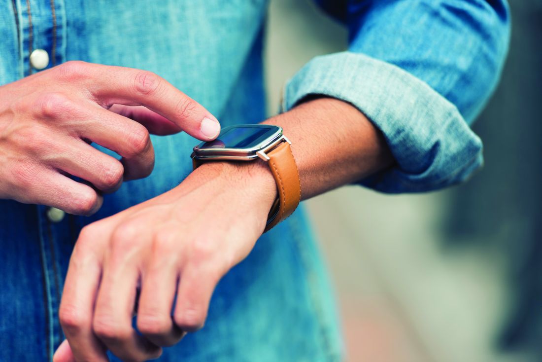
The ability of an Apple Watch to detect atrial fibrillation (AFib) is significantly affected by underlying ECG abnormalities such as sinus node dysfunction, atrioventricular (AV) block, or intraventricular conduction delay (IVCD), a single-center study suggests.
“We were surprised to find that in one in every five patients, the smartwatch ECG failed to produce an automatic diagnosis,” study author Marc Strik, MD, PhD, a clinician at Bordeaux University Hospital in Pessac, France, told this news organization. “This [failure] was mostly due to insufficient quality of the tracing [60%], but in a third of cases, [34%], it was due to bradycardia, and in some cases, tachycardia [6%].
“We were also surprised to find that the existence of ventricular conduction disease was associated with a higher likelihood of missing AFib,” he said.
The study was published in the Canadian Journal of Cardiology.
Abnormalities affected detection
The investigators tested the accuracy of the Apple Watch (Apple, Cupertino, California) in detecting AFib in patients with various ECG anomalies. All participants underwent 12-lead ECG, followed by a 30-second ECG tracing with an Apple Watch Series 5. The smartwatch’s automated AFib detection algorithm gave a result of “no signs of AFib,” “AFib,” or “not checked for AFib (unclassified).”
Unclassified recordings resulted from “low heart rate” (below 50 beats/min), “high heart rate” (above 150 beats/min), “poor recording,” or “inconclusive recording.”
The smartwatch recordings were reviewed by a blinded electrophysiologist who interpreted each tracing and assigned a diagnosis of “AFib,” “absence of AFib,” or “diagnosis unclear.” To assess interobserver agreement, a second blinded electrophysiologist interpreted 100 randomly selected tracings.
Among the 734 patients (mean age, 66; 58% men) enrolled, 539 (73%) were in normal sinus rhythm (SR), 154 (21%) in AFib, 33 in atrial flutter or atrial tachycardia, 3 in ventricular tachycardia, and 5 in junctional tachycardia.
Furthermore, 65 (8.9%) had sinus node dysfunction, 21 (2.9%) had second- or third-degree AV block, 39 (5.3%) had a ventricular paced rhythm, 54 (7.4%) had premature ventricular contractions (PVCs), and 132 (18%) had IVCD (right or left bundle branch block or nonspecific IVCD).
Of the 539 patients in normal SR, 437 recordings were correctly diagnosed by the smartwatch, 7 were diagnosed incorrectly as AFib, and 95 were not classified.
Of the 187 patients in AFib, 129 were correctly diagnosed, 17 were incorrectly diagnosed as SR, and 41 were not classified.
When unclassified ECGs were considered false results, the smartwatch had a sensitivity of 69% and specificity of 81% for AFib detection. When unclassified ECGs were excluded from the analysis, sensitivity was 88%, and specificity was 98%.
Compared with patients without the abnormality, the relative risk of having false positive tracings was higher for patients with premature atrial contractions (PACs) or PVCs (risk ratio, 2.9), sinus node dysfunction (RR, 3.71), and AV block (RR, 7.8).
Fifty-eight patients with AFib were classified as SR or inconclusive by the smartwatch. Among them, 21 (36%) had an IVCD, 7 (12%) had a ventricular paced rhythm, and 5 (9%) had PACs or PVCs.
The risk of having false negative tracings (missed AF) was higher for patients with IVCD (RR, 2.6) and pacing (RR, 2.47), compared with those without the abnormality.
‘A powerful tool’
Overall, cardiac electrophysiologists showed high agreement in differentiating between AFib and non-AFib, with high interobserver reproducibility. A manual diagnosis was not possible for 10% of tracings because of either poor ECG quality (3%) or unclear P-waves (7%).
Fifty-nine of the 580 patients in SR were misclassified as AFib by the experts, and 5 of the 154 patients in AFib were misclassified as SR.
“Our results show that the presence of sinus node dysfunction, second- or third-degree AV block, ventricular paced rhythm, PVCs, and IVCD were more frequently represented in smartwatch misdiagnoses,” wrote the authors. “Patients with PVCs were three times as likely to have false positive AFib diagnoses.”
Study limitations included the single-center nature of the study and the fact that patients were recruited in a cardiology office. The latter factor may have influenced the incidence of ECG abnormalities, which was much higher than for the average smartwatch user.
“Even with its limitations, the smartwatch remains a powerful tool that is able to detect AFib and multiple other abnormalities,” said Dr. Strik. “Missed diagnosis of AFib may be less important in real life because of repeated measurements, and algorithms will continue to improve.”
Technology improving
Richard C. Becker, MD, director and physician in chief of the University of Cincinnati Heart, Lung, and Vascular Institute, said, “This is exactly the kind of investigation required to improve upon existing detection algorithms that will someday facilitate routine use in patient care. An ability to detect AFib in a large proportion of those with the heart rhythm abnormality is encouraging.”
The findings should not detract from well-conducted studies in otherwise healthy individuals of varied age in whom AFib was accurately detected, he added. “Similarly, an automatic diagnosis algorithm for AF, pending optimization and validation in a large and diverse cohort, should be viewed as a communication tool between patients and health care providers.”
Patients at risk for developing AFib could benefit from continuous monitoring using a smartwatch, said Dr. Becker. “Pre-existing heart rhythm abnormalities must be taken into consideration. Optimal utilization of emerging technology to include wearables requires an understanding of performance and limitations. It is best undertaken in coordination with a health care provider.”
Andrés F. Miranda-Arboleda, MD, and Adrian Baranchuk, MD, of Kingston Health Sciences Center, Canada, conclude in an accompanying editorial, “In a certain manner, the smartwatch algorithms for the detection of AFib in patients with cardiovascular conditions are not yet smart enough ... but they may soon be.”
The study was supported by the French government. Dr. Strik, Dr. Miranda-Arboleda, Dr. Baranchuk, and Dr. Becker reported no conflicts of interest.
A version of this article first appeared on Medscape.com.
FROM CANADIAN JOURNAL OF CARDIOLOGY
New deep dive into Paxlovid interactions with CVD meds
Nirmatrelvir/ritonavir (Paxlovid) has been a game changer for high-risk patients with early COVID-19 symptoms but has significant interactions with commonly used cardiovascular medications, a new paper cautions.
COVID-19 patients with cardiovascular disease (CVD) or risk factors such as diabetes, hypertension, and chronic kidney disease are at high risk of severe disease and account for the lion’s share of those receiving Paxlovid. Data from the initial EPIC-HR trial and recent real-world data also suggest they’re among the most likely to benefit from the oral antiviral, regardless of their COVID-19 vaccination status.
“But at the same time, it unfortunately interacts with many very commonly prescribed cardiovascular medications and with many of them in a very clinically meaningful way, which may lead to serious adverse consequences,” senior author Sarju Ganatra, MD, said in an interview. “So, while it’s being prescribed with a good intention to help these people, we may actually end up doing more harm than good.
“We don’t want to deter people from getting their necessary COVID-19 treatment, which is excellent for the most part these days as an outpatient,” he added. “So, we felt the need to make a comprehensive list of cardiac medications and level of interactions with Paxlovid and also to help the clinicians and prescribers at the point of care to make the clinical decision of what modifications they may need to do.”
The paper, published online in the Journal of the American College of Cardiology, details drug-drug interactions with some 80 CV medications including statins, antihypertensive agents, heart failure therapies, and antiplatelet/anticoagulants.
It also includes a color-coded figure denoting whether a drug is safe to coadminister with Paxlovid, may potentially interact and require a dose adjustment or temporary discontinuation, or is contraindicated.
Among the commonly used blood thinners, for example, the paper notes that Paxlovid significantly increases drug levels of the direct oral anticoagulants (DOACs) apixaban, rivaroxaban, edoxaban, and dabigatran and, thus, increases the risk of bleeding.
“It can still be administered, if it’s necessary, but the dose of the DOAC either needs to be reduced or held depending on what they are getting it for, whether they’re getting it for pulmonary embolism or atrial fibrillation, and we adjust for all those things in the table in the paper,” said Dr. Ganatra, from Lahey Hospital and Medical Center, Burlington, Mass.
When the DOAC can’t be interrupted or dose adjusted, however, Paxlovid should not be given, the experts said. The antiviral is safe to use with enoxaparin, a low-molecular-weight heparin, but can increase or decrease levels of warfarin and should be used with close international normalized ratio monitoring.
For patients on antiplatelet agents, clinicians are advised to avoid prescribing nirmatrelvir/ritonavir to those on ticagrelor or clopidogrel unless the agents can be replaced by prasugrel.
Ritonavir – an inhibitor of cytochrome P 450 enzymes, particularly CYP3A4 – poses an increased risk of bleeding when given with ticagrelor, a CYP3A4 substrate, and decreases the active metabolite of clopidogrel, cutting its platelet inhibition by 20%. Although there’s a twofold decrease in the maximum concentration of prasugrel in patients on ritonavir, this does not affect its antiplatelet activity, the paper explains.
Among the lipid-lowering agents, experts suggested temporarily withholding atorvastatin, rosuvastatin, simvastatin, and lovastatin because of an increased risk for myopathy and liver toxicity but say that other statins, fibrates, ezetimibe, and the proprotein convertase subtilisin/kexin type 9 inhibitors evolocumab and alirocumab are safe to coadminister with Paxlovid.
While statins typically leave the body within hours, most of the antiarrhythmic drugs, except for sotalol, are not safe to give with Paxlovid, Dr. Ganatra said. It’s technically not feasible to hold these drugs because most have long half-lives, reaching about 100 days, for example, for amiodarone.
“It’s going to hang around in your system for a long time, so you don’t want to be falsely reassured that you’re holding the drug and it’s going to be fine to go back slowly,” he said. “You need to look for alternative therapies in those scenarios for COVID-19 treatment, which could be other antivirals, or a monoclonal antibody individualized to the patient’s risk.”
Although there’s limited clinical information regarding interaction-related adverse events with Paxlovid, the team used pharmacokinetics and pharmacodynamics data to provide the guidance. Serious adverse events are also well documented for ritonavir, which has been prescribed for years to treat HIV, Dr. Ganatra noted.
The Infectious Disease Society of America also published guidance on the management of potential drug interactions with Paxlovid in May and, earlier in October, the Food and Drug Administration updated its Paxlovid patient eligibility screening checklist.
Still, most prescribers are actually primary care physicians and even pharmacists, who may not be completely attuned, said Dr. Ganatra, who noted that some centers have started programs to help connect primary care physicians with their cardiology colleagues to check on CV drugs in their COVID-19 patients.
“We need to be thinking more broadly and at a system level where the hospital or health care system leverages the electronic health record systems,” he said. “Most of them are sophisticated enough to incorporate simple drug-drug interaction information, so if you try to prescribe someone Paxlovid and it’s a heart transplant patient who is on immunosuppressive therapy or a patient on a blood thinner, then it should give you a warning ... or at least give them a link to our paper or other valuable resources.
“If someone is on a blood thinner and the blood thinner level goes up by ninefold, we can only imagine what we would be dealing with,” Dr. Ganatra said. “So, these interactions should be taken very seriously and I think it’s worth the time and investment.”
The authors reported no relevant financial relationships.
A version of this article first appeared on Medscape.com.
FROM THE JOURNAL OF THE AMERICAN COLLEGE OF CARDIOLOGY
ACC calls for more career flexibility in cardiology
A new statement from the American College of Cardiology is calling for a greater degree of career flexibility in the specialty to promote cardiologists’ personal and professional well-being and preserve excellence in patient care.
The statement recommends that cardiologists, from trainees to those contemplating retirement, be granted more leeway in their careers to allow them to take time for common life events, such as child-rearing, taking care of aged parents, or reducing their workload in case of poor health or physical disabilities, without jeopardizing their careers.
The “2022 ACC Health Policy Statement on Career Flexibility in Cardiology: A Report of the American College of Cardiology Solution Set Oversight Committee” was published online in the Journal of the American College of Cardiology.
‘Hard-driving profession’
The well-being of the cardiovascular workforce is critical to the achievement of the mission of the ACC, which is to transform cardiovascular care and improve heart health, the Health Policy writing committee stated. Career flexibility is an important component of ensuring that well-being, the authors wrote.
“The ACC has critically looked at the factors that contribute to the lack of diversity and inclusion in cardiovascular practice, and one of the issues is the lack of flexibility in our profession,” writing committee chair, Mary Norine Walsh, MD, medical director of the heart failure and cardiac transplantation programs, Ascension St. Vincent Heart Center, Indianapolis, Ind., told this news organization.
The notion of work-life balance has become increasingly important but cardiology as a profession has traditionally not been open to the idea of its value, Dr. Walsh said.
“We have a very hard-driving profession. It takes many years to train to do the work we do. The need for on-call services is very significant, and we go along because we have always done it this way, but if you don’t reexamine the way that you are structuring your work, you’ll never change it,” she said.
“For example, the ‘full time, full call, come to work after you’ve been up all night’ work ethic, which is no longer allowed for trainees, is still in effect once you get into university practice or clinical practice. We have interventional cardiologists up all night doing STEMI care for patients and then having a full clinic the next day,” Dr. Walsh said. “The changes that came about for trainees have not trickled up to the faculty or clinical practice level. It’s really a patient safety issue.”
She emphasized that the new policy statement is not focused solely on women. “The need for time away or flexible time around family planning, childbirth, and parental leave is increasingly important to our younger colleagues, both men and women.”
Dr. Walsh pointed out that the writing committee was carefully composed to include representation from all stakeholders.
“We have representation from very young cardiologists, one of whom was in training at the time we began our work. We have two systems CEOs who are cardiologists, we have a chair of medicine, we have two very senior cardiologists, and someone who works in industry,” she said.
The ACC also believes that cardiologists with physically demanding roles should have pathways to transition into other opportunities in patient care, research, or education.
“Right now, there are many cardiology practices that have traditional policies, where you are either all in, or you are all out. They do not allow for what we term a ‘step down’ policy, where you perhaps stop going into the cath lab, but you still do clinic and see patients,” Dr. Walsh noted.
“One of the goals of this policy statement is to allow for such practices to look at their compensation and structure, and to realize that their most senior cardiologists may be willing to stay on for several more years and be contributing members to the practice, but they may no longer wish to stay in the cath lab or be in the night call pool,” she said.
Transparency around compensation is also very important because cardiologists contemplating a reduced work schedule need to know how this will affect the amount of money they will be earning, she added.
“Transparency about policies around compensation are crucial because if an individual cardiologist wishes to pursue a flexible scheduling at any time in their career, it’s clear that they won’t have the same compensation as someone who is a full-time employee. All of this has to be very transparent and clear on both sides, so that the person deciding toward some flexibility understands what the implications are from a financial and compensation standpoint,” Dr. Walsh said.
As an example, a senior career cardiologist who no longer wants to take night calls should know what this may cost financially.
“The practice should set a valuation of night calls, so that the individual who makes the choice to step out of the call pool understands what the impact on their compensation will be. That type of transparency is necessary for all to ensure that individuals who seek flexibility will not be blindsided by the resulting decrease in financial compensation,” she said.
A growing need
“In its new health policy statement, the American College of Cardiology addresses the growing need for career flexibility as an important component of ensuring the well-being of the cardiovascular care workforce,” Harlan M. Krumholz, MD, SM, Harold H. Hines Jr. Professor of Medicine and professor in the Institute for Social and Policy Studies at Yale University, New Haven, Conn., told this news organization.
“The writing committee reviews opportunities for offering flexibility at all career levels to combat burnout and increase retention in the field, as well as proposes system, policy, and practice solutions to allow both men and women to emphasize and embrace work-life balance,” Dr. Krumholz said.
“The document provides pathways for cardiologists looking to pursue other interests or career transitions while maintaining excellence in clinical care,” he added. “Chief among these recommendations are flexible/part-time hours, leave and reentry policies, changes in job descriptions to support overarching cultural change, and equitable compensation and opportunities. The document is intended to be used as a guide for innovation in the cardiology workforce.”
‘Thoughtful and long overdue’
“This policy statement is thoughtful and long overdue,” Steven E. Nissen, MD, Lewis and Patricia Dickey Chair in Cardiovascular Medicine and professor of medicine at Cleveland Clinic, told this news organization.
“Career flexibility will allow cardiologists to fulfill family responsibilities while continuing to advance their careers. Successfully contributing to patient care and research does not require physicians to isolate themselves from all their other responsibilities,” Dr. Nissen added.
“I am pleased that the ACC has articulated the value of a balanced approach to career and family.”
Dr. Walsh, Dr. Krumholz, and Dr. Nissen report no relevant financial relationships.
A version of this article first appeared on Medscape.com.
FROM THE JOURNAL OF THE AMERICAN COLLEGE OF CARDIOLOGY
Athletes with mild HCM can likely continue competitive sports
Athletes with mild hypertrophic cardiomyopathy (HCM) at low risk of sudden cardiac death (SCD) can safely continue to exercise at competitive levels, a retrospective study suggests.
During a mean follow-up of 4.5 years, athletes who continued to engage in high-intensity competitive sports after a mild HCM diagnosis were free of cardiac symptoms, and there were no deaths, incidents of sustained ventricular tachycardia or syncope, or changes in cardiac electrical, structural, or functional phenotypes.
“This study supports emerging evidence that HCM individuals with a low-risk profile and mild hypertrophy may engage in vigorous exercise and competitive sport,” Sanjay Sharma, MD, of St. George’s University of London, said in an interview. Current guidelines from the European Society of Cardiology and the American College of Cardiology support a more liberal approach to exercise for these individuals.
That said, he added, “it is important to emphasize that our cohort consisted of a group of adult competitive athletes who had probably been competing for several years before the diagnosis was made and therefore represented a self-selected, low-risk cohort. It is difficult to extrapolate this data to adolescent athletes, who appear to be more vulnerable to exercise-related SCD from HCM.”
The study was published online in the Journal of the American College of Cardiology.
Vigorous exercise OK for some
Dr. Sharma and colleagues analyzed data from 53 athletes with HCM who continued to participate in competitive sports. The mean age was 39 years, 98% were men, and 72% were White. About half (53%) competed as professionals, and were most commonly engaged in cycling, football, running, and rugby.
Participants underwent 6-12 monthly assessments that included electrocardiograms, echocardiograms, cardiopulmonary exercise testing, Holter monitoring (≥ 24 hours), and cardiac magnetic resonance imaging. A majority (64.2%) were evaluated because of an abnormal electrocardiograms, and one presented with an incidental abnormal echocardiogram.
About a quarter (24.5%) were symptomatic and 5 (9.4%) were identified on family screening. Eight (15%) had a family history of HCM, and six (11.3%) of SCD.
At the baseline evaluation, all athletes had a “low” ESC 5-year SCD risk score for HCM (1.9% ± 0.9%). None had syncope. Mean peak VO2 was 40.7 ± 6.8 mL/kg per minute.
The mean left ventricular wall thickness was 14.6 ± 2.3 mm; all had normal LV systolic and diastolic function and no LV outflow tract obstruction at rest or on provocation testing. In addition, none had an LV apical aneurysm.
Twenty-two (41%) showed late gadolinium enhancement on baseline cardiac magnetic resonance imaging.
A total of 19 participants underwent genotyping; 4 (21.1%) had a pathogenic/likely pathogenic sarcomeric variant. None took cardiovascular medication or had an implantable cardioverter defibrillator (ICD).
During a mean follow-up of 4.5 years, all participants continued to exercise at the same level as before their diagnosis; none underwent detraining. All stayed free of cardiac symptoms, and there were no deaths, sustained ventricular tachycardia episodes, or syncope.
Four demonstrated new, nonsustained ventricular tachycardia (NSVT) during follow-up, one of whom underwent ICD implantation because of an increased risk score and subsequently moderated exercise levels.
One participant had a 30-second atrial fibrillation (AFib) episode lasting longer than 30 seconds, started on a beta-blocker and oral anticoagulation, and also moderated exercise levels.
The event rate was 2.1% per year for asymptomatic arrhythmias (NSVT and AFib). No changes were observed in the cardiac electrical, structural, or functional phenotype during follow-up.
Dr. Sharma and colleagues stated: “Our sample size is small; however, it is nearly double the size of a previously studied Italian athletic cohort, and one-half were professional athletes. Furthermore, 17% of our cohort comprised Black athletes who are perceived to be at higher risk of SCD than White athletes.”
Daniele Massera, MD, assistant professor in the HCM program, department of medicine, Charney Division of Cardiology, New York University Langone Health, said in an interview: “Of note, these were athletes/patients at the very low end of phenotypic severity of HCM. ... It is also notable that diastolic function was normal in all of them, an uncommon finding in patients with HCM.”
Like Dr. Sharma, he said the findings are in line with recent guidelines, and cautioned: “This small study applies only to a very small subset of patients who are being evaluated at specialized HCM programs: asymptomatic male individuals who have mild, low-risk HCM and are on no medicines.
“The findings cannot be generalized to the population of symptomatic individuals with (or without) outflow obstruction, more severe hypertrophy, and who have ICDs and/or take medication for symptoms, nor to younger patients or adolescents, who may be at higher risk for adverse outcomes,” he concluded.
Individualized approach urged
Dr. Sharma was a coauthor of the recent article challenging the traditional restrictive approach to exercise for athletes diagnosed with HCM and other inherited cardiovascular diseases. The article suggested that individualized recommendations, taking risks into consideration, can help guide those who want to exercise or participate in competitive sports.
Dr. Sharma also is a coauthor of a 6-month follow-up to the SAFE-HCM study, which compared the effects of a supervised 12-week high-intensity exercise program to usual care in low-risk individuals with HCM (mean age, 45.7).
In the 6-month follow-up study, published as an abstract in the European Journal of Preventive Cardiology 2021 supplement, “exercising individuals had improved functional capacity and atherosclerotic risk profile and there were no differences in the composite safety outcomes [cardiovascular death, cardiac arrest, device therapy, exercise-induced syncope, sustained VT, NSVT, or sustained atrial arrhythmias] between exercising individuals and usual care individuals,” Dr. Sharma said.
The full study will soon be ready to submit for publication, he added.
No commercial funding or relevant conflicts of interest were disclosed.
A version of this article first appeared on Medscape.com.
FROM THE JOURNAL OF THE AMERICAN COLLEGE OF CARDIOLOGY
Novel head-up CPR position raises odds of survival of out-of-hospital heart attacks
Individuals who experience out-of-hospital cardiac arrest (OHCA) with nonshockable presentations have a better chance of survival when first responders use a novel CPR approach that includes gradual head-up positioning combined with basic but effective circulation-enhancing adjuncts, as shown from data from more than 2,000 patients.
In a study presented at the annual meeting of the American College of Emergency Physicians, Paul Pepe, MD, medical director for Dallas County Emergency Medical Services, reviewed data from five EMS systems that had adopted the new approach. Data were collected prospectively over the past 2 years from a national registry of patients who had received what Dr. Pepe called a “neuroprotective CPR bundle” (NP-CPR).
The study compared 380 NP-CPR case patients to 1,852 control patients who had received conventional CPR. Control data came from high-performance EMS systems that had participated in well-monitored, published OHCA trials funded by the National Institutes of Health. The primary outcome that was used for comparison was successful survival to hospital discharge with neurologically intact status (SURV-NI).
Traditional CPR supine chest compression techniques, if performed early and properly, can be lifesaving, but they are suboptimal, Dr. Pepe said. “Current techniques create pressure waves that run up the arterial side, but they also create back-pressure on the venous side, increasing intracranial pressure (ICP), thus compromising optimal cerebral blood flow,” he told this news organization.
For that reason, a modified physiologic approach to CPR was designed. It involves an airway adjunct called an impedance threshold device (ITD) and active compression-decompression (ACD) with a device “resembling a toilet plunger,” Dr. Pepe said.
The devices draw more blood out of the brain and into the thorax in a complementary fashion. The combination of these two adjuncts had dramatically improved SURV-NI by 50% in a clinical trial, Dr. Pepe said.
The new technology uses automated gradual head-up/torso-up positioning (AHUP) after first “priming the pump” with ITD-ACD–enhanced circulation. It was found to markedly augment that effect even further. In the laboratory setting, this synergistic NP-CPR bundle has been shown to help normalize cerebral perfusion pressure, further promoting neuro-intact survival. Normalization of end-tidal CO2 is routinely observed, according to Dr. Pepe.
In contrast to patients who present with ventricular fibrillation (shockable cases), patients with nonshockable presentations always have had grim prognoses, Dr. Pepe said. Until now, lifesaving advances had not been found, despite the fact that nonshockable presentations (asystole or electrical activity with no pulse) constitute approximately 80% of OHCA cases, or about 250,000 to 300,00 cases a year in the United States, he said.
In the study, approximately 60% of both the NP-CPR patients and control patients had asystole (flatline) presentations. The NP-CPR group had a significant threefold improvement in SURV-NI, compared with patients treated with conventional CPR in the high-functioning systems (odds ratio, 3.09). In a propensity-scored analysis matching all variables known to affect outcome, the OR increased to nearly fourfold higher (OR, 3.87; 95% confidence interval, 1.27-11.78), Dr. Pepe said.
The researchers also found that the time from receipt of a 911 call to initiation of AHUP was associated with progressively higher chances of survival. The median time for application was 11 minutes; when the elapsed time was less than 11 minutes, the SURV-NI was nearly 11-fold higher for NP-CPR patients than for control patients (OR, 10.59), with survival chances of 6% versus 0.5%. ORs were even higher when the time to treatment was less than 16 minutes (OR, 13.58), with survival rates of 5% versus 0.4%.
The findings not only demonstrate proof of concept in these most futile cases but also that implementation is feasible for the majority of patients, considering that the median time to the start of any CPR by a first responder was 8 minutes for both NP-CPR patients and control patients, “let alone 11 minutes for the AHUP initiation,” Dr. Pepe said. “This finally gives some hope for these nonshockable cases,” he emphasized.
“All of these devices have now been cleared by the Food and Drug Administration and should be adopted by all first-in responders,” said Dr. Pepe. “But they should be implemented as a bundle and in the proper sequence and as soon as feasible.”
Training and implementation efforts continue to expand, and more lives can be saved as more firefighters and first-in response teams acquire equipment and training, which can cut the time to response, he said.
The registry will continue to monitor outcomes with NP-CPR – the term was suggested by a patient who survived through this new approach – and Dr. Pepe and colleagues expect the statistics to improve further with wider adoption and faster implementation with the fastest responders.
A recent study by Dr. Pepe’s team, published in Resuscitation, showed the effectiveness of the neuroprotective bundle in improving survival for OHCA patients overall. The current study confirmed its impact on neuro-intact survival for the subgroup of patients with nonshockable cases.
One other take-home message is that head-up CPR cannot yet be performed by lay bystanders. “Also, do not implement this unless you are going to do it right,” Dr. Pepe emphasized in an interview.
Advanced CPR Solutions provided some materials and research funding for an independent data collector. No other relevant financial relationships have been disclosed.
A version of this article first appeared on Medscape.com.
FROM ACEP 2022
Like texting and driving: The human cost of AI
A recent medical meeting I attended included multiple sessions on the use of artificial intelligence (AI), a mere preview, I suspect, of what is to come for both patients and physicians.
I vow not to be a contrarian, but I have concerns. If we’d known how cell phones would permeate nearly every waking moment of our lives, would we have built in more protections from the onset?
Although anyone can see the enormous potential of AI in medicine, harnessing the wonders of it without guarding against the dangers could be paramount to texting and driving.
A palpable disruption in the common work-a-day human interaction is a given. CEOs who mind the bottom line will seek every opportunity to cut personnel whenever machine learning can deliver. As our dependence on algorithms increases, our need to understand electrocardiogram interpretation and echocardiographic calculations will wane. Subtle case information will go undetected. Nuanced subconscious alerts regarding the patient condition will go unnoticed.
These realities are never reflected in the pronouncements of companies who promote and develop AI.
The 2-minute echo
In September 2020, Carolyn Lam, MBBS, PhD, and James Hare, MBA, founders of the AI tech company US2.AI, told Healthcare Transformers that AI advances in echocardiology will turn “a manual process of 30 minutes, 250 clicks, with up to 21% variability among fully trained sonographers analyzing the same exam, into an AI-automated process taking 2 minutes, 1 click, with 0% variability.”
Let’s contrast this 2-minute human-machine interaction with the standard 20- to 30-minute human-to-human echocardiography procedure.
Take Mrs. Smith, for instance. She is referred for echocardiography for shortness of breath. She’s shown to a room and instructed to lie down on a table, where she undergoes a brief AI-directed acquisition of images and then a cheery dismissal from the imaging lab. Medical corporate chief financial officers will salivate at the efficiency, the decrease in cost for personnel, and the sharp increase in put-through for the echo lab schedule.
But what if Mrs. Smith gets a standard 30-minute sonographer-directed exam and the astute echocardiographer notes a left ventricular ejection fraction of 38%. A conversation with the patient reveals that she lost her son a few weeks ago. Upon completion of the study, the patient stands up and then adds, “I hope I can sleep in my bed tonight.” Thinking there may be more to the patient’s insomnia than grief-driven anxiety, the sonographer asks her to explain. “I had to sleep in a chair last night because I couldn’t breathe,” Mrs. Smith replies.
The sonographer reasons correctly that Mrs. Smith is likely a few weeks past an acute coronary syndrome for which she didn’t seek attention and is now in heart failure. The consulting cardiologist is alerted. Mrs. Smith is worked into the office schedule a week earlier than planned, and a costly in-patient stay for acute heart failure or worse is avoided.
Here’s a true-life example (some details have been changed to protect the patient’s identity): Mr. Rodriquez was referred for echocardiography because of dizziness. The sonographer notes significant mitral regurgitation and a decline in left ventricular ejection fraction from moderately impaired to severely reduced. When the sonographer inquires about a fresh bruise over Mr. Rodriguez’s left eye, he replies that he “must have fallen, but can’t remember.” The sonographer also notes runs of nonsustained ventricular tachycardia on the echo telemetry, and after a phone call from the echo lab to the ordering physician, Mr. Rodriquez is admitted. Instead of chancing a sudden death at home while awaiting follow-up, he undergoes catheterization and gets an implantable cardioverter defibrillator.
These scenarios illustrate that a 2-minute visit for AI-directed acquisition of echocardiogram images will never garner the protections of a conversation with a human. Any attempts at downplaying the importance of these human interactions are misguided.
Sometimes we embrace the latest advances in medicine while failing to tend to the most rudimentary necessities of data analysis and reporting. Catherine M. Otto, MD, director of the heart valve clinic and a professor of cardiology at the University of Washington Medical Center, Seattle, is a fan of the basics.
At the recent annual congress of the European Society of Cardiology, she commented on the AI-ENHANCED trial, which used an AI decision support algorithm to identify patients with moderate to severe aortic stenosis, which is associated with poor survival if left untreated. She correctly highlighted that while we are discussing the merits of AI-driven assessment of aortic stenosis, we are doing so in an era when many echo interpreters exclude critical information. The vital findings of aortic valve area, Vmax, and ejection fraction are often nowhere to be seen on reports. We should attend to our basic flaws in interpretation and reporting before we shift our focus to AI.
Flawed algorithms
Incorrect AI algorithms that are broadly adopted could negatively affect the health of millions.
Perhaps the most unsettling claim is made by causaLens: “Causal AI is the only technology that can reason and make choices like humans do,” the website states. A tantalizing tag line that is categorically untrue.
Our mysterious and complex neurophysiological function of reasoning still eludes understanding, but one thing is certain: medical reasoning originates with listening, seeing, and touching.
As AI infiltrates mainstream medicine, opportunities for hearing, observing, and palpating will be greatly reduced.
Folkert Asselbergs from University Medical Center Utrecht, the Netherlands, who has cautioned against overhyping AI, was the discussant for an ESC study on the use of causal AI to improve cardiovascular risk estimation.
He flashed a slide of a 2019 Science article on racial bias in an algorithm that U.S. health care systems use. Remedying that bias “would increase the percentage of Black people receiving additional help from 17.7% to 46.5%,” according to the authors.
Successful integration of AI-driven technology will come only if we build human interaction into every patient encounter.
I hope I don’t live to see the rise of the physician cyborg.
Artificial intelligence could be the greatest boon since the invention of the stethoscope, but it will be our downfall if we stop administering a healthy dose of humanity to every patient encounter.
Melissa Walton-Shirley, MD, is a clinical cardiologist in Nashville, Tenn., who has retired from full-time invasive cardiology. She disclosed no relevant conflicts of interest.
A version of this article first appeared on Medscape.com.
Food insecurity a growing problem for many with CVD
A growing number of Americans with cardiovascular disease (CVD) have limited or uncertain access to food, results of a new study suggest.
An analysis of data from the National Health and Nutrition Examination Survey (NHANES) representing more than 300 million American adults found that, overall, 38.1% of people with cardiovascular disease were food insecure in 2017-2019.
Twenty years earlier, that rate was 16.3%.
“What really stood out from our study is how frequent food insecurity is among people with cardiovascular disease, compared to those without cardiovascular disease,” lead author, Eric J. Brandt, MD, MHS, a cardiologist at the University of Michigan Health Frankel Cardiovascular Center, Ann Arbor, said in an interview.
“We believe that the relationship between food insecurity and cardiovascular disease is bidirectional. Food insecurity puts people at risk for cardiovascular disease, which then makes them vulnerable to events like myocardial infarction or stroke, which in turn may make them less able to work, thereby worsening their financial situation and increasing their vulnerability to food insecurity,” Dr. Brandt said.
For the analysis, Dr. Brandt and his team used an analytic sample of 57,517 adults to represent 312 million non-institutionalized adults in the United States.
Overall, 6,770 individuals (11.8%) in the analytic sample reported food insecurity.
Food insecurity was more prevalent among Hispanic people (n = 1,938, 24.0%) and non-Hispanic Black people (n = 1,202, 18.2%), compared with non-Hispanic Asian people (n = 100, 8.0%), and non-Hispanic White people (n = 3,221, 8.5%).
The prevalence of cardiovascular disease in the sample was 7.9% (n = 4,527).
Hypertension was the most prevalent CVD risk factor, reported in 49.6% of the sample. This was followed by obesity in 33.2%, dyslipidemia in 30.8%, and diabetes in 11.2%.
The findings were published online in JAMA Cardiology.
“All cardiovascular disease and cardiometabolic diseases except coronary artery disease were more prevalent among those with food insecurity,” Dr. Brandt noted.
“The results of our study are especially timely, as the White House just hosted its first conference on Hunger, Nutrition, and Health in over 50 years. Food insecurity is a focus of that conference. In the last few years, especially in relation to the pandemic, there has been expansion of some of the federal programs to prevent food insecurity. I would like to see a continued effort to solve this,” he said.
Dr. Brandt added that he hopes clinicians will be more cognizant of the problem of food insecurity and other social determinants of health when they see their patients.
“If someone is not going to be able to afford the food on their table, they’re probably not going to pay for their medications. Recognizing these social determinants in the clinical setting and helping our patients access local resources may address the underlying factors contributing to heart disease,” he said.
Uphill battle
Johanna Contreras, MD, advanced heart failure and transplant cardiologist at the Mount Sinai Hospital, New York, treats food insecure cardiovascular patients in her practice and tries to educate them about good nutrition. But it is an uphill battle.
“A lot of my patients live in the South Bronx. They have hypertension, hypercholesterolemia, and there are no grocery stores where they can buy fresh vegetables. I talk to them about eating healthy. They tell me it’s impossible. The stores only have pre-packaged foods. So even in the South Bronx, even though it is in New York, it is very hard to get fresh food. And when it is available, it is very expensive,” Dr. Contreras told this news organization.
“Fresh pineapples can cost $8. A fast-food burger costs $3. So that is what they buy: It’s what they can afford. Even the store managers don’t want to stock fresh produce because it can spoil. They open stores, like Whole Foods, but in the more affluent neighborhoods. They should open one in poor neighborhoods,” she said.
Dr. Contreras says she spends much of her time educating her patients about good nutrition. She asks them to keep a food diary and analyzes the results at each visit.
“I look at what they eat, and I try to see how I can use this information in a good way. I advise them to use frozen foods, and avoid canned, because it is a lot healthier. I am pragmatic, because I know that if I tell my patients to eat salmon, for example, they aren’t going to be able to afford it, if they can even access it.”
She also informs them about relatively healthy fast-food choices.
“I tell them to order 100% fruit juice, water, or milk when they go to McDonalds or other fast-food places. So I think this study is very important. Food insecurity is a very important component of cardiovascular disease, and unfortunately, minority communities are where this occurs.”
Dr. Brandt and Dr. Contreras report no relevant financial relationships.
A version of this article first appeared on Medscape.com.
Coffee linked to reduced cardiovascular disease and mortality risk
Drinking two to three daily cups of – ground, instant, or decaffeinated – is associated with significant reductions in new cardiovascular disease (CVD) and mortality risk, compared with avoiding coffee, a new analysis of the prospective UK Biobank suggests.
Ground and instant coffee, but not decaffeinated coffee, also was associated with reduced risk of new-onset arrhythmia, including atrial fibrillation.
“Our study is the first to look at differences in coffee subtypes to tease out important differences which may explain some of the mechanisms through which coffee works,” Peter M. Kistler, MD, of the Alfred Hospital and Baker Heart and Diabetes Institute, Melbourne, Australia, told this news organization.
“Daily coffee intake should not be discouraged by physicians but rather considered part of a healthy diet,” Dr. Kistler said.
“This study supports that coffee is safe and even potentially beneficial, which is consistent with most of the prior evidence,” Carl “Chip” Lavie, MD, who wasn’t involved in the study, told this news organization.
“We do not prescribe coffee to patients, but for the majority who like coffee, they can be encouraged it is fine to take a few cups daily,” said Dr. Lavie, with the Ochsner Heart and Vascular Institute in New Orleans.
The study was published online in the European Journal of Preventive Cardiology.
Clear cardiovascular benefits
A total of 449,563 UK Biobank participants (median age 58 years; 55% women), who were free of arrhythmias or other CVD at baseline, reported in questionnaires their level of daily coffee intake and preferred type of coffee.
During more than 12.5 years of follow-up, 27,809 participants (6.2%) died.
Drinking one to five cups per day of ground or instant coffee (but not decaffeinated coffee) was associated with a significant reduction in incident arrhythmia. The lowest risk was with four to five cups per day for ground coffee (hazard ratio [HR] 0.83; 95% confidence interval [CI], 0.76-0.91; P < .0001) and two to three cups per day for instant coffee (HR, 0.88; 95% CI, 0.85-0.92; P < .0001).
Habitual coffee drinking of up to five cups perday was also associated with significant reductions in the risk of incident CVD, when compared with nondrinkers.
Significant reductions in the risk of incident coronary heart disease (CHD) were associated with habitual coffee intake of up to five cups per day, with the lowest risk for CHD observed in those who consumed two to three cups per day (HR 0.89; 95% CI, 0.86-0.91; P < .0001).
Coffee consumption at all levels was linked to significant reduction in the risk of congestive cardiac failure (CCF) and ischemic stroke. The lowest risks were observed in those who consumed two to three cups per day, with HR, 0.83 (95% CI, 0.79-0.87; P < .0001) for CCF and HR, 0.84 (95% CI, 0.78-0.90; P < .0001) for ischemic stroke.
Death from any cause was significantly reduced for all coffee subtypes, with the greatest risk reduction seen with two to three cups per day for decaffeinated (HR, 0.86; 95% CI, 0.81-0.91; P < .0001); ground (HR, 0.73; 95% CI, 0.69-0.78; P < .0001); and instant coffee (HR, 0.89; 95% CI, 0.86-0.93; P < .0001).
“Coffee consumption is associated with cardiovascular benefits and should not empirically be discontinued in those with underlying heart rhythm disorders or cardiovascular disease,” Dr. Kistler told this news organization.
Plausible mechanisms
There are a number of proposed mechanisms to explain the benefits of coffee on CVD.
“Caffeine has antiarrhythmic properties through adenosine A1 and A2A receptor inhibition, hence the difference in effects of decaf vs. full-strength coffee on heart rhythm disorders,” Dr. Kistler explained.
Coffee has vasodilatory effects and coffee also contains antioxidant polyphenols, which reduce oxidative stress and modulate metabolism.
“The explanation for improved survival with habitual coffee consumption remains unclear,” Dr. Kistler said.
“Putative mechanisms include improved endothelial function, circulating antioxidants, improved insulin sensitivity, and reduced inflammation. Another potential mechanism includes the beneficial effects of coffee on metabolic syndrome,” he said.
“Caffeine has a role in weight loss through inhibition of gut fatty acid absorption and increase in basal metabolic rate. Furthermore, coffee has been associated with a significantly lower incidence of type 2 diabetes mellitus,” Dr. Kistler added.
Direction of relationship unclear
Charlotte Mills, PhD, University of Reading, England, said this study “adds to the body of evidence from observational trials associating moderate coffee consumption with cardioprotection, which looks promising.”
However, with the observational design, it’s unclear “which direction the relationship goes – for example, does coffee make you healthy or do inherently healthier people consume coffee? Randomized controlled trials are needed to fully understand the relationship between coffee and health before recommendations can be made,” Dr. Mills told the UK nonprofit Science Media Centre.
Annette Creedon, PhD, nutrition scientist with the British Nutrition Foundation, said it’s possible that respondents over- or underestimated the amount of coffee that they were consuming at the start of the study when they self-reported their intake.
“It is therefore difficult to determine whether the outcomes can be directly associated with the behaviors in coffee consumption reported at the start of the study,” she told the Science Media Centre.
The study had no funding. Dr. Kistler has received funding from Abbott Medical for consultancy and speaking engagements and fellowship support from Biosense Webster. Dr. Lavie has no relevant disclosures. Dr. Mills has worked in collaboration with Nestle on research relating to coffee and health funded by UKRI. Dr. Creedon has reported no relevant financial relationships.
A version of this article first appeared on Medscape.com.
FROM EUROPEAN JOURNAL OF PREVENTIVE CARDIOLOGY
Amulet, Watchman 2.5 LAAO outcomes neck and neck at 3 years
The Amplatzer Amulet (Abbott) and first-generation Watchman 2.5 (Boston Scientific) devices provide relatively comparable results out to 3 years after left atrial appendage occlusion (LAAO), longer follow-up from the Amplatzer Amulet Left Atrial Appendage Occluder Versus Watchman Device for Stroke Prophylaxis (Amulet IDE) trial shows.
“The dual-seal Amplatzer Amulet left atrial appendage occluder continued to demonstrate safety and effectiveness through 3 years,” principal investigator Dhanunjaya Lakkireddy, MD, said in a late-breaking session at the recent Transcatheter Cardiovascular Therapeutics annual meeting.
Preliminary results, reported last year, showed that procedural complications were higher with the Amplatzer but that it provided superior closure of the left atrial appendage (LAA) at 45 days and was noninferior with respect to safety at 12 months and efficacy at 18 months.
Amulet IDE is the largest head-to-head comparison of the two devices, enrolling 1,878 high-risk patients with nonvalvular atrial fibrillation undergoing LAA closure to reduce the risk of stroke.
Three-year follow-up was higher with the Amulet device than with the Watchman, at 721 vs. 659 patients, driven by increased deaths (85 vs. 63) and withdrawals (50 vs. 23) in the Watchman group within 18 months, noted Dr. Lakkireddy, Kansas City Heart Rhythm Institute and Research Foundation, Overland Park, Kan.
Use of oral anticoagulation was higher in the Watchman group at 6 months (2.8% vs. 4.7%; P = .04), 18 months (3.1% vs. 5.6%; P = .01), and 3 years (3.7% vs. 7.3%; P < .01).
This was primarily driven by more late device-related thrombus (DRT) after 6 months with the Watchman device than with the Amulet occluder (23 vs. 10). “Perhaps the dual-closure mechanism of the Amulet explains this fundamental difference, where you have a nice smooth disc that covers the ostium,” he posited.
At 3 years, rates of cardiovascular death trended lower with Amulet than with Watchman (6.6% vs. 8.5%; P = .14), as did all-cause deaths (14.6% vs. 17.9%; P = .07).
Most cardiovascular deaths in the Amulet group were not preceded by a device factor, whereas DRT (1 vs. 4) and peridevice leak 3 mm or more (5 vs. 15) frequently preceded these deaths in the Watchman group, Dr. Lakkireddy observed. No pericardial effusion-related deaths occurred in either group.
Major bleeding, however, trended higher for the Amulet, at 16.1%, compared with 14.7% for the Watchman (P = .46). Ischemic stroke and systemic embolic rates also trended higher for Amulet, at 5%, and 4.6% for Watchman.
The protocol recommended aspirin only for both groups after 6 months. None of the 29 Amulet and 3 of the 29 Watchman patients with an ischemic stroke were on oral anticoagulation at the time of the stroke.
Device factors, however, frequently preceded ischemic strokes in the Watchman group, Dr. Lakkireddy said. DRT occurred in 1 patient with Amulet and 2 patients with Watchman and peridevice leak in 3 with Amulet and 15 with Watchman. “Again, the peridevice leak issue really stands out as an important factor,” he said at the meeting, which was sponsored by the Cardiovascular Research Foundation.
Based on “data from the large trials, it’s clearly evident that the presence of peridevice leak significantly raises the risk of stroke in follow-up,” he said. “So, attention has to be paid to the choice of the device and how we can mitigate the risk of peridevice leaks in these patients.”
The composite of stroke, systemic embolism, and cardiovascular death occurred in 11.1% of patients with Amulet and 12.7% with Watchman (P = .31).
Asked following the formal presentation whether the results justify use of one device over the other for LAA occlusion, Dr. Lakkireddy said he likes the dual closure mechanism of the Amulet and is more likely to use it in patients with proximal lobes, very large appendages, or a relatively shallow appendage. “In the rest of the cases, I think it’s a toss-up.”
As for how generalizable the results are, he noted that the study tested the Amulet against the legacy Watchman 2.5 but that the second-generation Watchman FLX is available in a larger size and has shown improved performance.
The Amplatzer Amulet does not require oral anticoagulants at discharge. However, the indication for the Watchman FLX was recently expanded to include 45-day dual antiplatelet therapy as a postprocedure alternative to oral anticoagulation plus aspirin.
Going forward, the “next evolution” is to test the Watchman FLX and Amulet on either single antiplatelet or a dual antiplatelet regimen without oral anticoagulation, he suggested.
Results from SWISS APERO, the first randomized trial to compare the Amulet and Watchman FLX (and a handful of 2.5 devices) in 221 patients, showed that the devices are not interchangeable for rates of complications or leaks.
During a press conference prior to the presentation, discussant Federico Asch, MD, MedStar Health Research Institute, Washington, said, “the most exciting thing here is that we have good options. We now can start to tease out which patients will benefit best from one or the other because we actually have two options.”
The Amulet IDE trial was funded by Abbott. Dr. Lakkireddy reports that he or his spouse/partner have received grant/research support from Abbott, AtriCure, Alta Thera, Medtronic, Biosense Webster, Biotronik, and Boston Scientific; and speaker honoraria from Abbott, Medtronic, Biotronik, and Boston Scientific.
A version of this article first appeared on Medscape.com.
FROM TCT 2022
