User login
MDedge conference coverage features onsite reporting of the latest study results and expert perspectives from leading researchers.
Dermatologists play a key role in the transformation of transgender patients
, according to Doris Day, MD.

While clinical management of this patient population has historically been limited to experts in mental health, endocrinology, and select surgeons with experience in sex reassignment surgery, “what dermatologists provide on an aesthetic level through noninvasive or minimally invasive procedures can have a big impact in helping that transformation,” Dr. Day, of the department of dermatology at New York University Langone Health, said during the virtual annual Masters of Aesthetics Symposium. “But, we have to go through a transformation of sorts as well as we care for these patients, because we need to help them in the way that best matches their needs. We need to know about their mental health and the medicines they’re taking as well as their goals for their outcomes. If they’re working with surgeons for sex reassignment, we should have discussions with those clinicians as well.”
Gender-affirming hormone therapy is the primary medical intervention sought by transgender people, she said. This allows the acquisition of secondary sex characteristics more aligned with their gender identity. Feminizing hormone therapy affects the skin by reducing sebaceous gland activity, “which can lead to fewer acne breakouts and smaller pores but also cause drier skin,” Dr. Day said. “We can slow down the growth of body and facial hair and we can perform hair removal treatments. We see decreased male-pattern scalp hair loss, and we see smoother skin as the fat under the skin becomes thicker and the pores become smaller. We can also have increased pigment production, which is always a good thing.”
In a 2016 survey of 327 transgender individuals led by Dr. Day’s mentee, Brian A. Ginsberg, MD, and published in the Journal of the American Academy of Dermatology, most transgender women indicated that their face was most important to have changed, while for men it was the chest. Hair removal was the most common women’s facial procedure, followed by surgery then injectables, mostly performed by plastic surgeons.
Limitations of hormone therapy include the fact that it can take 2 or more years for associated changes to fully develop. “At least here in New York, patients want everything in a New York minute, so that’s always an issue,” she said. “We often recommend that patients wait at least 2 years after beginning hormone therapy before considering drastic feminization surgeries, but there are many options we have for them while they’re waiting for that. Even with hormone therapy, the bone structure of the face is unaffected, so we need to be artistic in creating a more feminized balance in order to help them physically match their gender to their identity.”
Noninvasive aesthetic procedures can compound the effects of hormone therapy, in addition to offering physical transformation beyond hormone therapy. She recalled assisting one of her patients transform from male to female. Over a period of 2 years, Dr. Day added Botox then Juvederm Voluma to the patient’s cheeks and chin, “and she started her transformation to a more feminized gender matching identity,” she said. Next came a hair transplant and the injection of more Voluma and fillers in the lips and cheeks on an as-needed basis.
“During one visit, I felt that we could still do more,” Dr. Day recalled. “She looked at me and said, ‘Actually, I feel so happy. This looks like me as I imagined I would look in my mind.’ I realized that my vision for her wasn’t the same as her vision for herself. She was thrilled with her transformation. I realized that as we see these patients, for all we learn about the science of gender transformation, the emotional aspects of our vision of what we can accomplish for our patients versus their vision of what their happiness level is may not entirely match. We have to be careful to help them celebrate their version of their femininity or masculinity, rather than trying to have our patients match what we think we can accomplish for them with our own sense of what femininity or masculinity is.”
Over time, Dr. Day said, the patient’s acne scars improved with fillers and microneedling treatments, and with the hormone therapy. “As we softened her appearance and as she made changes like the earrings that she wore and the hair style that she chose, she was in line with what her perception of her femininity was,” she said. “Little by little we’ve been watching her grow into her new self. It’s been a beautiful transformation. I was honored to be able to share in that journey with her.”
Dr. Day reported having no relevant financial disclosures.
, according to Doris Day, MD.
While clinical management of this patient population has historically been limited to experts in mental health, endocrinology, and select surgeons with experience in sex reassignment surgery, “what dermatologists provide on an aesthetic level through noninvasive or minimally invasive procedures can have a big impact in helping that transformation,” Dr. Day, of the department of dermatology at New York University Langone Health, said during the virtual annual Masters of Aesthetics Symposium. “But, we have to go through a transformation of sorts as well as we care for these patients, because we need to help them in the way that best matches their needs. We need to know about their mental health and the medicines they’re taking as well as their goals for their outcomes. If they’re working with surgeons for sex reassignment, we should have discussions with those clinicians as well.”
Gender-affirming hormone therapy is the primary medical intervention sought by transgender people, she said. This allows the acquisition of secondary sex characteristics more aligned with their gender identity. Feminizing hormone therapy affects the skin by reducing sebaceous gland activity, “which can lead to fewer acne breakouts and smaller pores but also cause drier skin,” Dr. Day said. “We can slow down the growth of body and facial hair and we can perform hair removal treatments. We see decreased male-pattern scalp hair loss, and we see smoother skin as the fat under the skin becomes thicker and the pores become smaller. We can also have increased pigment production, which is always a good thing.”
In a 2016 survey of 327 transgender individuals led by Dr. Day’s mentee, Brian A. Ginsberg, MD, and published in the Journal of the American Academy of Dermatology, most transgender women indicated that their face was most important to have changed, while for men it was the chest. Hair removal was the most common women’s facial procedure, followed by surgery then injectables, mostly performed by plastic surgeons.
Limitations of hormone therapy include the fact that it can take 2 or more years for associated changes to fully develop. “At least here in New York, patients want everything in a New York minute, so that’s always an issue,” she said. “We often recommend that patients wait at least 2 years after beginning hormone therapy before considering drastic feminization surgeries, but there are many options we have for them while they’re waiting for that. Even with hormone therapy, the bone structure of the face is unaffected, so we need to be artistic in creating a more feminized balance in order to help them physically match their gender to their identity.”
Noninvasive aesthetic procedures can compound the effects of hormone therapy, in addition to offering physical transformation beyond hormone therapy. She recalled assisting one of her patients transform from male to female. Over a period of 2 years, Dr. Day added Botox then Juvederm Voluma to the patient’s cheeks and chin, “and she started her transformation to a more feminized gender matching identity,” she said. Next came a hair transplant and the injection of more Voluma and fillers in the lips and cheeks on an as-needed basis.
“During one visit, I felt that we could still do more,” Dr. Day recalled. “She looked at me and said, ‘Actually, I feel so happy. This looks like me as I imagined I would look in my mind.’ I realized that my vision for her wasn’t the same as her vision for herself. She was thrilled with her transformation. I realized that as we see these patients, for all we learn about the science of gender transformation, the emotional aspects of our vision of what we can accomplish for our patients versus their vision of what their happiness level is may not entirely match. We have to be careful to help them celebrate their version of their femininity or masculinity, rather than trying to have our patients match what we think we can accomplish for them with our own sense of what femininity or masculinity is.”
Over time, Dr. Day said, the patient’s acne scars improved with fillers and microneedling treatments, and with the hormone therapy. “As we softened her appearance and as she made changes like the earrings that she wore and the hair style that she chose, she was in line with what her perception of her femininity was,” she said. “Little by little we’ve been watching her grow into her new self. It’s been a beautiful transformation. I was honored to be able to share in that journey with her.”
Dr. Day reported having no relevant financial disclosures.
, according to Doris Day, MD.
While clinical management of this patient population has historically been limited to experts in mental health, endocrinology, and select surgeons with experience in sex reassignment surgery, “what dermatologists provide on an aesthetic level through noninvasive or minimally invasive procedures can have a big impact in helping that transformation,” Dr. Day, of the department of dermatology at New York University Langone Health, said during the virtual annual Masters of Aesthetics Symposium. “But, we have to go through a transformation of sorts as well as we care for these patients, because we need to help them in the way that best matches their needs. We need to know about their mental health and the medicines they’re taking as well as their goals for their outcomes. If they’re working with surgeons for sex reassignment, we should have discussions with those clinicians as well.”
Gender-affirming hormone therapy is the primary medical intervention sought by transgender people, she said. This allows the acquisition of secondary sex characteristics more aligned with their gender identity. Feminizing hormone therapy affects the skin by reducing sebaceous gland activity, “which can lead to fewer acne breakouts and smaller pores but also cause drier skin,” Dr. Day said. “We can slow down the growth of body and facial hair and we can perform hair removal treatments. We see decreased male-pattern scalp hair loss, and we see smoother skin as the fat under the skin becomes thicker and the pores become smaller. We can also have increased pigment production, which is always a good thing.”
In a 2016 survey of 327 transgender individuals led by Dr. Day’s mentee, Brian A. Ginsberg, MD, and published in the Journal of the American Academy of Dermatology, most transgender women indicated that their face was most important to have changed, while for men it was the chest. Hair removal was the most common women’s facial procedure, followed by surgery then injectables, mostly performed by plastic surgeons.
Limitations of hormone therapy include the fact that it can take 2 or more years for associated changes to fully develop. “At least here in New York, patients want everything in a New York minute, so that’s always an issue,” she said. “We often recommend that patients wait at least 2 years after beginning hormone therapy before considering drastic feminization surgeries, but there are many options we have for them while they’re waiting for that. Even with hormone therapy, the bone structure of the face is unaffected, so we need to be artistic in creating a more feminized balance in order to help them physically match their gender to their identity.”
Noninvasive aesthetic procedures can compound the effects of hormone therapy, in addition to offering physical transformation beyond hormone therapy. She recalled assisting one of her patients transform from male to female. Over a period of 2 years, Dr. Day added Botox then Juvederm Voluma to the patient’s cheeks and chin, “and she started her transformation to a more feminized gender matching identity,” she said. Next came a hair transplant and the injection of more Voluma and fillers in the lips and cheeks on an as-needed basis.
“During one visit, I felt that we could still do more,” Dr. Day recalled. “She looked at me and said, ‘Actually, I feel so happy. This looks like me as I imagined I would look in my mind.’ I realized that my vision for her wasn’t the same as her vision for herself. She was thrilled with her transformation. I realized that as we see these patients, for all we learn about the science of gender transformation, the emotional aspects of our vision of what we can accomplish for our patients versus their vision of what their happiness level is may not entirely match. We have to be careful to help them celebrate their version of their femininity or masculinity, rather than trying to have our patients match what we think we can accomplish for them with our own sense of what femininity or masculinity is.”
Over time, Dr. Day said, the patient’s acne scars improved with fillers and microneedling treatments, and with the hormone therapy. “As we softened her appearance and as she made changes like the earrings that she wore and the hair style that she chose, she was in line with what her perception of her femininity was,” she said. “Little by little we’ve been watching her grow into her new self. It’s been a beautiful transformation. I was honored to be able to share in that journey with her.”
Dr. Day reported having no relevant financial disclosures.
FROM MOA 2020
Nationwide study questions routine long-term beta-blocker post MI
Current American and European guidelines recommending long-term beta-blocker therapy following an acute MI appear to be obsolete in the modern reperfusion era, suggests an analysis of Danish registry data.
Those guidelines are based on old randomized trials of beta-blocker therapy conducted prior to introduction of routine percutaneous coronary intervention and modern multidrug optimal medical therapy for acute MI. There have been no prospective controlled studies in the reperfusion era. And a new Danish national observational study strongly suggests it’s time to reexamine the beta-blocker recommendation, Anders Holt, MD, said at the virtual annual congress of the European Society of Cardiology.
“Stable, optimally treated MI patients do not seem to benefit from beta-blocker treatment exceeding 3 months post hospitalization – bearing in mind this doesn’t apply to patients with other indications for beta-blockers, like heart failure or atrial fibrillation,” said Dr. Holt of Copenhagen University Hospital.
His analysis of Danish national registry data on more than 30,000 patients hospitalized for acute MI during 2003-2018 earned him the annual ESC Young Investigator Award in Population Science.
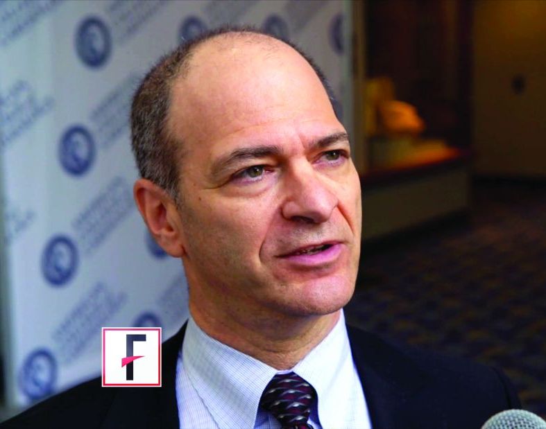
“This was a crisp and clear presentation of a very creative use of observational epidemiology to try to understand the length of therapy that may or may not be appropriate,” commented award session cochair Paul M. Ridker, MD, director of the Center for Cardiovascular Disease Prevention at Brigham and Women’s Hospital and professor of medicine at Harvard Medical School, both in Boston.
Dr. Holt reported on 30,177 patients optimally treated for a first MI in Danish hospitals during 2003-2018, none of whom had a prior indication or contraindication for beta-blocker therapy. “Optimally treated” meant they underwent percutaneous coronary revascularization and were discharged on a statin and aspirin. As a study requirement, all had to be stable 90 days post hospitalization, at which point 24,770 of the patients were on long-term beta-blocker therapy, and 5,407 (18%) were not. The two groups were comparable in terms of age, sex, comorbidities, and baseline medications. All patients were followed through the registries for a maximum of 3 years, the duration of beta-blocker therapy post MI recommended in American Heart Association/American College of Cardiology guidelines. (The Danish Society of Cardiology recommends 2 years.)
At 3 years post MI, there was no between-group difference in a composite outcome comprising cardiovascular death, recurrent MI, heart failure, stroke, angina, or a cardiac procedure, with a rate of 22.9% in the beta-blocker group and 21.6% in patients not on long-term beta-blocker therapy. The rate of recurrent MI was identical at 6.7% in both groups. Cardiovascular death occurred during 3 years of follow-up in 1.4% of patients on beta-blocker therapy and 1.7% who weren’t, a nonsignificant difference.
“We saw no evidence of any cardioprotective effect, but no increased risk of adverse events resulting in hospitalization, either,” Dr. Holt observed. “I would like to acknowledge that no evidence of effect does not necessarily equal evidence of no effect, but even if there was an effect we can with fair certainty say that it’s probably quite minimal.”
He noted that the Danish registry data indicates that each year since 2012 has shown a growing trend for Danish patients to dispense with long-term beta-blocker therapy after an acute MI.
“This might indicate we are nudging toward a change in practice, where more physicians are thinking that long-term beta-blocker therapy might not be indicated for all MI patients in the reperfusion era,” according to Dr. Holt.
Asked by the four-judge award panel about the possibility of unmeasured confounding in this observational study, Dr Holt responded: “I would be very cautious about asking patients to stop beta-blocker therapy after 3 months just based on this observational data. We can’t speak to causality in an observational study.” But he added that “well-designed observational studies provide valuable data regarding this topic and should not be ignored. They should possibly influence the guidelines and the designs for upcoming randomized trials.”
He conducted several supplementary analyses designed to address the possibility of unevenly distributed unmeasured confounding in the registry study. These analyses proved reassuring. A positive exposure control analysis compared 3-year outcomes in patients who remained on long-term statin therapy and those who didn’t. As expected, outcomes were significantly better in those who did: a 3-year composite outcome rate of 22.1%, compared with 32.1% in patients not on a statin; a cardiovascular death rate of 1.3% with and 2.1% without statin therapy; a recurrent MI rate of 6.6%, compared with 10.1% without a statin; and a 2.8% all-cause mortality with and 5.4% without statin therapy.
In contrast, all-cause mortality was unaffected by whether or not patients were on long-term beta-blocker therapy. And in a negative exposure outcome analysis, no association was found between beta-blocker therapy and the risk of hospitalization for pneumonia, as to be expected if the beta-blocker and no-beta-blocker groups were comparable in key respects.
Dr. Holt reported having no financial conflicts regarding his study.
Current American and European guidelines recommending long-term beta-blocker therapy following an acute MI appear to be obsolete in the modern reperfusion era, suggests an analysis of Danish registry data.
Those guidelines are based on old randomized trials of beta-blocker therapy conducted prior to introduction of routine percutaneous coronary intervention and modern multidrug optimal medical therapy for acute MI. There have been no prospective controlled studies in the reperfusion era. And a new Danish national observational study strongly suggests it’s time to reexamine the beta-blocker recommendation, Anders Holt, MD, said at the virtual annual congress of the European Society of Cardiology.
“Stable, optimally treated MI patients do not seem to benefit from beta-blocker treatment exceeding 3 months post hospitalization – bearing in mind this doesn’t apply to patients with other indications for beta-blockers, like heart failure or atrial fibrillation,” said Dr. Holt of Copenhagen University Hospital.
His analysis of Danish national registry data on more than 30,000 patients hospitalized for acute MI during 2003-2018 earned him the annual ESC Young Investigator Award in Population Science.
“This was a crisp and clear presentation of a very creative use of observational epidemiology to try to understand the length of therapy that may or may not be appropriate,” commented award session cochair Paul M. Ridker, MD, director of the Center for Cardiovascular Disease Prevention at Brigham and Women’s Hospital and professor of medicine at Harvard Medical School, both in Boston.
Dr. Holt reported on 30,177 patients optimally treated for a first MI in Danish hospitals during 2003-2018, none of whom had a prior indication or contraindication for beta-blocker therapy. “Optimally treated” meant they underwent percutaneous coronary revascularization and were discharged on a statin and aspirin. As a study requirement, all had to be stable 90 days post hospitalization, at which point 24,770 of the patients were on long-term beta-blocker therapy, and 5,407 (18%) were not. The two groups were comparable in terms of age, sex, comorbidities, and baseline medications. All patients were followed through the registries for a maximum of 3 years, the duration of beta-blocker therapy post MI recommended in American Heart Association/American College of Cardiology guidelines. (The Danish Society of Cardiology recommends 2 years.)
At 3 years post MI, there was no between-group difference in a composite outcome comprising cardiovascular death, recurrent MI, heart failure, stroke, angina, or a cardiac procedure, with a rate of 22.9% in the beta-blocker group and 21.6% in patients not on long-term beta-blocker therapy. The rate of recurrent MI was identical at 6.7% in both groups. Cardiovascular death occurred during 3 years of follow-up in 1.4% of patients on beta-blocker therapy and 1.7% who weren’t, a nonsignificant difference.
“We saw no evidence of any cardioprotective effect, but no increased risk of adverse events resulting in hospitalization, either,” Dr. Holt observed. “I would like to acknowledge that no evidence of effect does not necessarily equal evidence of no effect, but even if there was an effect we can with fair certainty say that it’s probably quite minimal.”
He noted that the Danish registry data indicates that each year since 2012 has shown a growing trend for Danish patients to dispense with long-term beta-blocker therapy after an acute MI.
“This might indicate we are nudging toward a change in practice, where more physicians are thinking that long-term beta-blocker therapy might not be indicated for all MI patients in the reperfusion era,” according to Dr. Holt.
Asked by the four-judge award panel about the possibility of unmeasured confounding in this observational study, Dr Holt responded: “I would be very cautious about asking patients to stop beta-blocker therapy after 3 months just based on this observational data. We can’t speak to causality in an observational study.” But he added that “well-designed observational studies provide valuable data regarding this topic and should not be ignored. They should possibly influence the guidelines and the designs for upcoming randomized trials.”
He conducted several supplementary analyses designed to address the possibility of unevenly distributed unmeasured confounding in the registry study. These analyses proved reassuring. A positive exposure control analysis compared 3-year outcomes in patients who remained on long-term statin therapy and those who didn’t. As expected, outcomes were significantly better in those who did: a 3-year composite outcome rate of 22.1%, compared with 32.1% in patients not on a statin; a cardiovascular death rate of 1.3% with and 2.1% without statin therapy; a recurrent MI rate of 6.6%, compared with 10.1% without a statin; and a 2.8% all-cause mortality with and 5.4% without statin therapy.
In contrast, all-cause mortality was unaffected by whether or not patients were on long-term beta-blocker therapy. And in a negative exposure outcome analysis, no association was found between beta-blocker therapy and the risk of hospitalization for pneumonia, as to be expected if the beta-blocker and no-beta-blocker groups were comparable in key respects.
Dr. Holt reported having no financial conflicts regarding his study.
Current American and European guidelines recommending long-term beta-blocker therapy following an acute MI appear to be obsolete in the modern reperfusion era, suggests an analysis of Danish registry data.
Those guidelines are based on old randomized trials of beta-blocker therapy conducted prior to introduction of routine percutaneous coronary intervention and modern multidrug optimal medical therapy for acute MI. There have been no prospective controlled studies in the reperfusion era. And a new Danish national observational study strongly suggests it’s time to reexamine the beta-blocker recommendation, Anders Holt, MD, said at the virtual annual congress of the European Society of Cardiology.
“Stable, optimally treated MI patients do not seem to benefit from beta-blocker treatment exceeding 3 months post hospitalization – bearing in mind this doesn’t apply to patients with other indications for beta-blockers, like heart failure or atrial fibrillation,” said Dr. Holt of Copenhagen University Hospital.
His analysis of Danish national registry data on more than 30,000 patients hospitalized for acute MI during 2003-2018 earned him the annual ESC Young Investigator Award in Population Science.
“This was a crisp and clear presentation of a very creative use of observational epidemiology to try to understand the length of therapy that may or may not be appropriate,” commented award session cochair Paul M. Ridker, MD, director of the Center for Cardiovascular Disease Prevention at Brigham and Women’s Hospital and professor of medicine at Harvard Medical School, both in Boston.
Dr. Holt reported on 30,177 patients optimally treated for a first MI in Danish hospitals during 2003-2018, none of whom had a prior indication or contraindication for beta-blocker therapy. “Optimally treated” meant they underwent percutaneous coronary revascularization and were discharged on a statin and aspirin. As a study requirement, all had to be stable 90 days post hospitalization, at which point 24,770 of the patients were on long-term beta-blocker therapy, and 5,407 (18%) were not. The two groups were comparable in terms of age, sex, comorbidities, and baseline medications. All patients were followed through the registries for a maximum of 3 years, the duration of beta-blocker therapy post MI recommended in American Heart Association/American College of Cardiology guidelines. (The Danish Society of Cardiology recommends 2 years.)
At 3 years post MI, there was no between-group difference in a composite outcome comprising cardiovascular death, recurrent MI, heart failure, stroke, angina, or a cardiac procedure, with a rate of 22.9% in the beta-blocker group and 21.6% in patients not on long-term beta-blocker therapy. The rate of recurrent MI was identical at 6.7% in both groups. Cardiovascular death occurred during 3 years of follow-up in 1.4% of patients on beta-blocker therapy and 1.7% who weren’t, a nonsignificant difference.
“We saw no evidence of any cardioprotective effect, but no increased risk of adverse events resulting in hospitalization, either,” Dr. Holt observed. “I would like to acknowledge that no evidence of effect does not necessarily equal evidence of no effect, but even if there was an effect we can with fair certainty say that it’s probably quite minimal.”
He noted that the Danish registry data indicates that each year since 2012 has shown a growing trend for Danish patients to dispense with long-term beta-blocker therapy after an acute MI.
“This might indicate we are nudging toward a change in practice, where more physicians are thinking that long-term beta-blocker therapy might not be indicated for all MI patients in the reperfusion era,” according to Dr. Holt.
Asked by the four-judge award panel about the possibility of unmeasured confounding in this observational study, Dr Holt responded: “I would be very cautious about asking patients to stop beta-blocker therapy after 3 months just based on this observational data. We can’t speak to causality in an observational study.” But he added that “well-designed observational studies provide valuable data regarding this topic and should not be ignored. They should possibly influence the guidelines and the designs for upcoming randomized trials.”
He conducted several supplementary analyses designed to address the possibility of unevenly distributed unmeasured confounding in the registry study. These analyses proved reassuring. A positive exposure control analysis compared 3-year outcomes in patients who remained on long-term statin therapy and those who didn’t. As expected, outcomes were significantly better in those who did: a 3-year composite outcome rate of 22.1%, compared with 32.1% in patients not on a statin; a cardiovascular death rate of 1.3% with and 2.1% without statin therapy; a recurrent MI rate of 6.6%, compared with 10.1% without a statin; and a 2.8% all-cause mortality with and 5.4% without statin therapy.
In contrast, all-cause mortality was unaffected by whether or not patients were on long-term beta-blocker therapy. And in a negative exposure outcome analysis, no association was found between beta-blocker therapy and the risk of hospitalization for pneumonia, as to be expected if the beta-blocker and no-beta-blocker groups were comparable in key respects.
Dr. Holt reported having no financial conflicts regarding his study.
FROM ESC CONGRESS 2020
Promising Trends Seen in AIDS-Associated NHL
Prognoses for patients with AIDS-associated, non-Hodgkin lymphomas (AIDS-NHLs) have improved dramatically as HIV/AIDS has become easier to treat, and “we’re actually seeing patients with long-term remissions that are translating to cure,” a hematologist told colleagues at the virtual 2020 annual meeting of the Association of VA Hematology/Oncology (AVAHO).
“Even those with low CD4 counts initially have more chance of survival compared to the historic patients in the pre-HAART [highly active antiretroviral therapy] era,” explained Erin Reid, MD, MS, of the University of California at San Diego Moores Cancer Center. “They’re seeing complete-response rates and overall-survival rates that are nearly matching what we’re seeing in the non-HIV lymphoma cases. And aggressive infection prophylaxis has seemed to mitigate some of the infectious complications.”
Still, Reid said, a severe form of AIDS-NHL continues to have very poor outcomes, although specific regimens appear to be brightening the picture somewhat.
According to Dr. Reid, AIDS-NHLs are the most common malignancy in the HIV-positive population, and patients with these cancers are more likely to have aggressive lymphomas. These patients are also more likely to have lymphomas associated with Epstein-Barr virus—40 to 80%, depending on the subtype of lymphoma—and Kaposi sarcoma-associated herpesvirus (also known as human herpesvirus-8, or HHV8).
“These viruses are driving these cancers, and it begs the question of whether there’s something we can do to target these viruses within these cancer cells in a way that’s therapeutic,” she said.
Compared with the non-HIV population, patients with AIDS-NHL “are much more likely to present with advanced stage, extranodal disease and central nervous system involvement,” she said.
HAART Benefits
It’s become clear that HIV control via HAART has benefits in terms of higher tolerance of chemotherapy doses—“we’re able to use more full or traditional dose regimens”—and perhaps cancer suppression too, she said. A 2013 meta-analysis “favored concurrent therapy with chemotherapy [and HAART]. This has become our recommended standard of care for virtually all cases, except the very rare ones where you cannot find a regimen that is compatible from a PK [pharmacokinetics] standpoint.”
Reid also noted that the HAART era has changed the role of CD4 counts in AIDS-NHLs. “While CD4 count still has some predictive value, its impact on mortality appears attenuated,” she said.
EPOCH Treatment
With regard to treatment, she emphasized the importance of HAART: “We would recommend concurrent HAART whenever possible with chemotherapy, or start it immediately afterward.”
Aggressive infection prophylaxis also is recommended through granulocyte colony-stimulating factor and agents to target threats from pneumocystis jiroveci pneumonia, gram negative rods, and varicella-zoster virus. “I’ve moved away from fungal prophylaxis over the years, only dealing with it if there’s a known fungal infection,” she said.
As for treatment of AIDS-NHL, Reid Suggested that research supports the EPOCH regimen --etoposide, prednisone, vincristine, cyclophosphamide, and doxorubicin. However, “we still need strategies for refractory and relapsed disease,” she said.
Reid noted that she has started to see more plasmablastic cases, although her experience is anecdotal. Plasmablastic lymphoma is much more common in the HIV-positive setting, she said.
Lifespans are poor for these patients, with many failing to live for a year. But research hints that the prognosis in AIDS-NHL patients on HAART may actually be better than in the non-HIV population, she said.
A trial published in September 2020, in fact, reports that 87% of 15 patients with AIDS-associated plasmablastic lymphoma survived for at least one year on the EPOCH regimen. Overall, the study found that “people with a collection of HIV-associated lymphomas were doing well overall with the EPOCH backbone,” Reid explained.
Reid reported no relevant disclosures.
Prognoses for patients with AIDS-associated, non-Hodgkin lymphomas (AIDS-NHLs) have improved dramatically as HIV/AIDS has become easier to treat, and “we’re actually seeing patients with long-term remissions that are translating to cure,” a hematologist told colleagues at the virtual 2020 annual meeting of the Association of VA Hematology/Oncology (AVAHO).
“Even those with low CD4 counts initially have more chance of survival compared to the historic patients in the pre-HAART [highly active antiretroviral therapy] era,” explained Erin Reid, MD, MS, of the University of California at San Diego Moores Cancer Center. “They’re seeing complete-response rates and overall-survival rates that are nearly matching what we’re seeing in the non-HIV lymphoma cases. And aggressive infection prophylaxis has seemed to mitigate some of the infectious complications.”
Still, Reid said, a severe form of AIDS-NHL continues to have very poor outcomes, although specific regimens appear to be brightening the picture somewhat.
According to Dr. Reid, AIDS-NHLs are the most common malignancy in the HIV-positive population, and patients with these cancers are more likely to have aggressive lymphomas. These patients are also more likely to have lymphomas associated with Epstein-Barr virus—40 to 80%, depending on the subtype of lymphoma—and Kaposi sarcoma-associated herpesvirus (also known as human herpesvirus-8, or HHV8).
“These viruses are driving these cancers, and it begs the question of whether there’s something we can do to target these viruses within these cancer cells in a way that’s therapeutic,” she said.
Compared with the non-HIV population, patients with AIDS-NHL “are much more likely to present with advanced stage, extranodal disease and central nervous system involvement,” she said.
HAART Benefits
It’s become clear that HIV control via HAART has benefits in terms of higher tolerance of chemotherapy doses—“we’re able to use more full or traditional dose regimens”—and perhaps cancer suppression too, she said. A 2013 meta-analysis “favored concurrent therapy with chemotherapy [and HAART]. This has become our recommended standard of care for virtually all cases, except the very rare ones where you cannot find a regimen that is compatible from a PK [pharmacokinetics] standpoint.”
Reid also noted that the HAART era has changed the role of CD4 counts in AIDS-NHLs. “While CD4 count still has some predictive value, its impact on mortality appears attenuated,” she said.
EPOCH Treatment
With regard to treatment, she emphasized the importance of HAART: “We would recommend concurrent HAART whenever possible with chemotherapy, or start it immediately afterward.”
Aggressive infection prophylaxis also is recommended through granulocyte colony-stimulating factor and agents to target threats from pneumocystis jiroveci pneumonia, gram negative rods, and varicella-zoster virus. “I’ve moved away from fungal prophylaxis over the years, only dealing with it if there’s a known fungal infection,” she said.
As for treatment of AIDS-NHL, Reid Suggested that research supports the EPOCH regimen --etoposide, prednisone, vincristine, cyclophosphamide, and doxorubicin. However, “we still need strategies for refractory and relapsed disease,” she said.
Reid noted that she has started to see more plasmablastic cases, although her experience is anecdotal. Plasmablastic lymphoma is much more common in the HIV-positive setting, she said.
Lifespans are poor for these patients, with many failing to live for a year. But research hints that the prognosis in AIDS-NHL patients on HAART may actually be better than in the non-HIV population, she said.
A trial published in September 2020, in fact, reports that 87% of 15 patients with AIDS-associated plasmablastic lymphoma survived for at least one year on the EPOCH regimen. Overall, the study found that “people with a collection of HIV-associated lymphomas were doing well overall with the EPOCH backbone,” Reid explained.
Reid reported no relevant disclosures.
Prognoses for patients with AIDS-associated, non-Hodgkin lymphomas (AIDS-NHLs) have improved dramatically as HIV/AIDS has become easier to treat, and “we’re actually seeing patients with long-term remissions that are translating to cure,” a hematologist told colleagues at the virtual 2020 annual meeting of the Association of VA Hematology/Oncology (AVAHO).
“Even those with low CD4 counts initially have more chance of survival compared to the historic patients in the pre-HAART [highly active antiretroviral therapy] era,” explained Erin Reid, MD, MS, of the University of California at San Diego Moores Cancer Center. “They’re seeing complete-response rates and overall-survival rates that are nearly matching what we’re seeing in the non-HIV lymphoma cases. And aggressive infection prophylaxis has seemed to mitigate some of the infectious complications.”
Still, Reid said, a severe form of AIDS-NHL continues to have very poor outcomes, although specific regimens appear to be brightening the picture somewhat.
According to Dr. Reid, AIDS-NHLs are the most common malignancy in the HIV-positive population, and patients with these cancers are more likely to have aggressive lymphomas. These patients are also more likely to have lymphomas associated with Epstein-Barr virus—40 to 80%, depending on the subtype of lymphoma—and Kaposi sarcoma-associated herpesvirus (also known as human herpesvirus-8, or HHV8).
“These viruses are driving these cancers, and it begs the question of whether there’s something we can do to target these viruses within these cancer cells in a way that’s therapeutic,” she said.
Compared with the non-HIV population, patients with AIDS-NHL “are much more likely to present with advanced stage, extranodal disease and central nervous system involvement,” she said.
HAART Benefits
It’s become clear that HIV control via HAART has benefits in terms of higher tolerance of chemotherapy doses—“we’re able to use more full or traditional dose regimens”—and perhaps cancer suppression too, she said. A 2013 meta-analysis “favored concurrent therapy with chemotherapy [and HAART]. This has become our recommended standard of care for virtually all cases, except the very rare ones where you cannot find a regimen that is compatible from a PK [pharmacokinetics] standpoint.”
Reid also noted that the HAART era has changed the role of CD4 counts in AIDS-NHLs. “While CD4 count still has some predictive value, its impact on mortality appears attenuated,” she said.
EPOCH Treatment
With regard to treatment, she emphasized the importance of HAART: “We would recommend concurrent HAART whenever possible with chemotherapy, or start it immediately afterward.”
Aggressive infection prophylaxis also is recommended through granulocyte colony-stimulating factor and agents to target threats from pneumocystis jiroveci pneumonia, gram negative rods, and varicella-zoster virus. “I’ve moved away from fungal prophylaxis over the years, only dealing with it if there’s a known fungal infection,” she said.
As for treatment of AIDS-NHL, Reid Suggested that research supports the EPOCH regimen --etoposide, prednisone, vincristine, cyclophosphamide, and doxorubicin. However, “we still need strategies for refractory and relapsed disease,” she said.
Reid noted that she has started to see more plasmablastic cases, although her experience is anecdotal. Plasmablastic lymphoma is much more common in the HIV-positive setting, she said.
Lifespans are poor for these patients, with many failing to live for a year. But research hints that the prognosis in AIDS-NHL patients on HAART may actually be better than in the non-HIV population, she said.
A trial published in September 2020, in fact, reports that 87% of 15 patients with AIDS-associated plasmablastic lymphoma survived for at least one year on the EPOCH regimen. Overall, the study found that “people with a collection of HIV-associated lymphomas were doing well overall with the EPOCH backbone,” Reid explained.
Reid reported no relevant disclosures.
Bleed Disorders Are Possible in VA Population
Although it may seem that veterans would have a very low risk of bleeding disorders since they were medically cleared for military service, a hematologist/oncologist cautioned that veterans might indeed suffer from both inherited and noninherited forms of these conditions. At the virtual 2020 annual meeting of the Association of VA Hematology/Oncology (AVAHO) Bethany Samuelson Bannow, MD, of Oregon Health & Science University’s Knight Cancer Institute urged colleagues to understand the diagnosis and treatment of bleeding disorders.
“Most importantly, even though these are patients you probably don’t see on a regular basis, you are never alone,” since colleagues are available to help, she said. Samuelson Bannow treats patients at US Department of Veterans Affairs (VA) Portland Health Care System and used her presentation to focus on 4 types of bleeding disorders. A summary of her perspective and recommendations follows.
Acquired hemophilia: Watch for Infections
Acquired hemophilia affects only an estimated 1.3 to 1.5 in 1 million people, but VA physicians may see it more often since it affects an older population (median age is 78 years), Samuelson Bannow said. “I’ve seen about 4 cases in the last 2 years,” she said. “I’m not sure if we’re a magnet, but it does come up.”
The diagnosis is based on laboratory findings, and a lack of personal or family history of coagulopathy is key, she said. Twenty percent or more of patients older than 65 years die from the disorder, but bleeding usually isn’t the cause. Instead, patients tend to die from infections, she said.
Initial treatment must focus on stopping the bleeding, she said. The new drug porcine antihemophilic factor (recombinant)—Obizur—“is very helpful” and is Samuelson Bannow’s first choice, but it may not widely available at all VA medical centers. Recombinant FVIIa (NovoSeven) and activated prothrombin complex concentrate (Feiba) also are options.
“The goal is to overpower the clotting cascade and get that burst of thrombin generation that you need to get the bleeding under control. Titrate to the amount of bleeding the patient is having, and make sure you’re doing local control as well,” Samuelson Bannow said. She added that the 2 agents may not work depending on the patient. Neither is preferred and both may be appropriate. “There’s no real reason to pick one over the other beyond convenience and availability.”
There’s another “equally important component of management,” she said: Inhibitor eradication. “The only way to do this is with immune suppression. You’re going to have to suppress the immune system to get rid of the inhibitor. That’s why we see such high rates of death because we have to use heavy-hitter immunosuppressants.”
Treatment options include steroids and cyclophosphamide (a common first-line option), rituximab, calcineurin inhibitors, and mycophenolate mofetil. “Just be aware that there is an increased risk of infection with these agents,” she said. “You want to see a decrease in the titer of your inhibitor. This can take 3 or more weeks, and it can take longer for it to disappear entirely. Look for normalized factor VIII level and absent inhibitor.”
She added: “I tend to do a slow taper, one agent at a time, over the course of weeks. If you see a return of the inhibitor, you can ramp back up as needed. Continue to monitor for a year or more since patients are at high risk of recurrence.”
Acquired von Willebrand Syndrome: Fix the Associated Conditions
Acquired von Willebrand Syndrome is another rare bleeding disorder that may appear in veterans “since it’s associated with a lot of conditions that we see in the VA,” such as heart disorders, solid tumors, vascular malformations, and lymphoproliferative and myeloproliferative disorders, Samuelson Bannow said.
As with acquired hemophilia, the key is to focus on controlling the bleeding, she said. Then, she advised, focus on the associated condition: “Correct the underlying disorder: Treat the malignancy, address the [cardiac] shear stress, correct hypothyroidism, correct the cardiac defects.”
Inherited Hemophilia: Don’t Rule It Out
It’s possible to “sneak through” military medical screening with undiagnosed inherited bleeding disorders, Dr. Samuelson Bannow said. That’s because service members may have never had an injury that triggered abnormal bleeding, she said. “You can see how someone could slip into the military with a [low clotting factor level]. The classic presentation is going to be joint bleeds and abnormalities. These can be traumatic and spontaneous without any kind of an injury,” she said.
A general rule for these patients is to “replace what’s missing, and 100% is always normal.” The 100% refers to clotting factor level. She recommended reviewing 2013 guidelines for the treatment of the disorder.
Inherited von Willebrand Syndrome: Ask About Tonsillectomies
Inherited von Willebrand Syndrome is more common in women than it is in men. Bleeding “will primarily be mucocutaneous and trauma-induced,” said. Samuelson Bannow. She recommended asking patients if they have had a tonsillectomy and, if so, did they suffer from unusual bleeding. “If they did not have excessive bleeding, it’s a pretty good sign their hemostasis is good.”
She recommends a nasal spray drug called desmopressin (DDAVP, Stimate) for management. “It’s critical to know that you must test for efficacy first,” she said. “One of the most common mistakes that people make is that they may give patients DDAVP before surgery without knowing if it works or not. It doesn’t work for every patient.” She typically avoids this drug in patients aged > 65 years, or even > 60 years, due to increased thrombosis risk.
Recombinant von Willebrand factor (Vonvendi) is a “very helpful drug at patients at increased risk of thrombosis,” she said. However, she noted that the drug, at last check, is not on the VA’s formulary.
At the end of her presentation, Samuelson Bannow urged colleagues to contact specialized Hemophilia Treatment Centers (HTCs) near them: “Follow this link, find an HTC [Hemophilia Treatment Center] near you, and create a partnership.”
Samuelson Bannow reported no relevant disclosures.
Although it may seem that veterans would have a very low risk of bleeding disorders since they were medically cleared for military service, a hematologist/oncologist cautioned that veterans might indeed suffer from both inherited and noninherited forms of these conditions. At the virtual 2020 annual meeting of the Association of VA Hematology/Oncology (AVAHO) Bethany Samuelson Bannow, MD, of Oregon Health & Science University’s Knight Cancer Institute urged colleagues to understand the diagnosis and treatment of bleeding disorders.
“Most importantly, even though these are patients you probably don’t see on a regular basis, you are never alone,” since colleagues are available to help, she said. Samuelson Bannow treats patients at US Department of Veterans Affairs (VA) Portland Health Care System and used her presentation to focus on 4 types of bleeding disorders. A summary of her perspective and recommendations follows.
Acquired hemophilia: Watch for Infections
Acquired hemophilia affects only an estimated 1.3 to 1.5 in 1 million people, but VA physicians may see it more often since it affects an older population (median age is 78 years), Samuelson Bannow said. “I’ve seen about 4 cases in the last 2 years,” she said. “I’m not sure if we’re a magnet, but it does come up.”
The diagnosis is based on laboratory findings, and a lack of personal or family history of coagulopathy is key, she said. Twenty percent or more of patients older than 65 years die from the disorder, but bleeding usually isn’t the cause. Instead, patients tend to die from infections, she said.
Initial treatment must focus on stopping the bleeding, she said. The new drug porcine antihemophilic factor (recombinant)—Obizur—“is very helpful” and is Samuelson Bannow’s first choice, but it may not widely available at all VA medical centers. Recombinant FVIIa (NovoSeven) and activated prothrombin complex concentrate (Feiba) also are options.
“The goal is to overpower the clotting cascade and get that burst of thrombin generation that you need to get the bleeding under control. Titrate to the amount of bleeding the patient is having, and make sure you’re doing local control as well,” Samuelson Bannow said. She added that the 2 agents may not work depending on the patient. Neither is preferred and both may be appropriate. “There’s no real reason to pick one over the other beyond convenience and availability.”
There’s another “equally important component of management,” she said: Inhibitor eradication. “The only way to do this is with immune suppression. You’re going to have to suppress the immune system to get rid of the inhibitor. That’s why we see such high rates of death because we have to use heavy-hitter immunosuppressants.”
Treatment options include steroids and cyclophosphamide (a common first-line option), rituximab, calcineurin inhibitors, and mycophenolate mofetil. “Just be aware that there is an increased risk of infection with these agents,” she said. “You want to see a decrease in the titer of your inhibitor. This can take 3 or more weeks, and it can take longer for it to disappear entirely. Look for normalized factor VIII level and absent inhibitor.”
She added: “I tend to do a slow taper, one agent at a time, over the course of weeks. If you see a return of the inhibitor, you can ramp back up as needed. Continue to monitor for a year or more since patients are at high risk of recurrence.”
Acquired von Willebrand Syndrome: Fix the Associated Conditions
Acquired von Willebrand Syndrome is another rare bleeding disorder that may appear in veterans “since it’s associated with a lot of conditions that we see in the VA,” such as heart disorders, solid tumors, vascular malformations, and lymphoproliferative and myeloproliferative disorders, Samuelson Bannow said.
As with acquired hemophilia, the key is to focus on controlling the bleeding, she said. Then, she advised, focus on the associated condition: “Correct the underlying disorder: Treat the malignancy, address the [cardiac] shear stress, correct hypothyroidism, correct the cardiac defects.”
Inherited Hemophilia: Don’t Rule It Out
It’s possible to “sneak through” military medical screening with undiagnosed inherited bleeding disorders, Dr. Samuelson Bannow said. That’s because service members may have never had an injury that triggered abnormal bleeding, she said. “You can see how someone could slip into the military with a [low clotting factor level]. The classic presentation is going to be joint bleeds and abnormalities. These can be traumatic and spontaneous without any kind of an injury,” she said.
A general rule for these patients is to “replace what’s missing, and 100% is always normal.” The 100% refers to clotting factor level. She recommended reviewing 2013 guidelines for the treatment of the disorder.
Inherited von Willebrand Syndrome: Ask About Tonsillectomies
Inherited von Willebrand Syndrome is more common in women than it is in men. Bleeding “will primarily be mucocutaneous and trauma-induced,” said. Samuelson Bannow. She recommended asking patients if they have had a tonsillectomy and, if so, did they suffer from unusual bleeding. “If they did not have excessive bleeding, it’s a pretty good sign their hemostasis is good.”
She recommends a nasal spray drug called desmopressin (DDAVP, Stimate) for management. “It’s critical to know that you must test for efficacy first,” she said. “One of the most common mistakes that people make is that they may give patients DDAVP before surgery without knowing if it works or not. It doesn’t work for every patient.” She typically avoids this drug in patients aged > 65 years, or even > 60 years, due to increased thrombosis risk.
Recombinant von Willebrand factor (Vonvendi) is a “very helpful drug at patients at increased risk of thrombosis,” she said. However, she noted that the drug, at last check, is not on the VA’s formulary.
At the end of her presentation, Samuelson Bannow urged colleagues to contact specialized Hemophilia Treatment Centers (HTCs) near them: “Follow this link, find an HTC [Hemophilia Treatment Center] near you, and create a partnership.”
Samuelson Bannow reported no relevant disclosures.
Although it may seem that veterans would have a very low risk of bleeding disorders since they were medically cleared for military service, a hematologist/oncologist cautioned that veterans might indeed suffer from both inherited and noninherited forms of these conditions. At the virtual 2020 annual meeting of the Association of VA Hematology/Oncology (AVAHO) Bethany Samuelson Bannow, MD, of Oregon Health & Science University’s Knight Cancer Institute urged colleagues to understand the diagnosis and treatment of bleeding disorders.
“Most importantly, even though these are patients you probably don’t see on a regular basis, you are never alone,” since colleagues are available to help, she said. Samuelson Bannow treats patients at US Department of Veterans Affairs (VA) Portland Health Care System and used her presentation to focus on 4 types of bleeding disorders. A summary of her perspective and recommendations follows.
Acquired hemophilia: Watch for Infections
Acquired hemophilia affects only an estimated 1.3 to 1.5 in 1 million people, but VA physicians may see it more often since it affects an older population (median age is 78 years), Samuelson Bannow said. “I’ve seen about 4 cases in the last 2 years,” she said. “I’m not sure if we’re a magnet, but it does come up.”
The diagnosis is based on laboratory findings, and a lack of personal or family history of coagulopathy is key, she said. Twenty percent or more of patients older than 65 years die from the disorder, but bleeding usually isn’t the cause. Instead, patients tend to die from infections, she said.
Initial treatment must focus on stopping the bleeding, she said. The new drug porcine antihemophilic factor (recombinant)—Obizur—“is very helpful” and is Samuelson Bannow’s first choice, but it may not widely available at all VA medical centers. Recombinant FVIIa (NovoSeven) and activated prothrombin complex concentrate (Feiba) also are options.
“The goal is to overpower the clotting cascade and get that burst of thrombin generation that you need to get the bleeding under control. Titrate to the amount of bleeding the patient is having, and make sure you’re doing local control as well,” Samuelson Bannow said. She added that the 2 agents may not work depending on the patient. Neither is preferred and both may be appropriate. “There’s no real reason to pick one over the other beyond convenience and availability.”
There’s another “equally important component of management,” she said: Inhibitor eradication. “The only way to do this is with immune suppression. You’re going to have to suppress the immune system to get rid of the inhibitor. That’s why we see such high rates of death because we have to use heavy-hitter immunosuppressants.”
Treatment options include steroids and cyclophosphamide (a common first-line option), rituximab, calcineurin inhibitors, and mycophenolate mofetil. “Just be aware that there is an increased risk of infection with these agents,” she said. “You want to see a decrease in the titer of your inhibitor. This can take 3 or more weeks, and it can take longer for it to disappear entirely. Look for normalized factor VIII level and absent inhibitor.”
She added: “I tend to do a slow taper, one agent at a time, over the course of weeks. If you see a return of the inhibitor, you can ramp back up as needed. Continue to monitor for a year or more since patients are at high risk of recurrence.”
Acquired von Willebrand Syndrome: Fix the Associated Conditions
Acquired von Willebrand Syndrome is another rare bleeding disorder that may appear in veterans “since it’s associated with a lot of conditions that we see in the VA,” such as heart disorders, solid tumors, vascular malformations, and lymphoproliferative and myeloproliferative disorders, Samuelson Bannow said.
As with acquired hemophilia, the key is to focus on controlling the bleeding, she said. Then, she advised, focus on the associated condition: “Correct the underlying disorder: Treat the malignancy, address the [cardiac] shear stress, correct hypothyroidism, correct the cardiac defects.”
Inherited Hemophilia: Don’t Rule It Out
It’s possible to “sneak through” military medical screening with undiagnosed inherited bleeding disorders, Dr. Samuelson Bannow said. That’s because service members may have never had an injury that triggered abnormal bleeding, she said. “You can see how someone could slip into the military with a [low clotting factor level]. The classic presentation is going to be joint bleeds and abnormalities. These can be traumatic and spontaneous without any kind of an injury,” she said.
A general rule for these patients is to “replace what’s missing, and 100% is always normal.” The 100% refers to clotting factor level. She recommended reviewing 2013 guidelines for the treatment of the disorder.
Inherited von Willebrand Syndrome: Ask About Tonsillectomies
Inherited von Willebrand Syndrome is more common in women than it is in men. Bleeding “will primarily be mucocutaneous and trauma-induced,” said. Samuelson Bannow. She recommended asking patients if they have had a tonsillectomy and, if so, did they suffer from unusual bleeding. “If they did not have excessive bleeding, it’s a pretty good sign their hemostasis is good.”
She recommends a nasal spray drug called desmopressin (DDAVP, Stimate) for management. “It’s critical to know that you must test for efficacy first,” she said. “One of the most common mistakes that people make is that they may give patients DDAVP before surgery without knowing if it works or not. It doesn’t work for every patient.” She typically avoids this drug in patients aged > 65 years, or even > 60 years, due to increased thrombosis risk.
Recombinant von Willebrand factor (Vonvendi) is a “very helpful drug at patients at increased risk of thrombosis,” she said. However, she noted that the drug, at last check, is not on the VA’s formulary.
At the end of her presentation, Samuelson Bannow urged colleagues to contact specialized Hemophilia Treatment Centers (HTCs) near them: “Follow this link, find an HTC [Hemophilia Treatment Center] near you, and create a partnership.”
Samuelson Bannow reported no relevant disclosures.
Once-weekly insulin data published; could alter treatment
Phase 2 data for the investigational, once-weekly basal insulin analog icodec (Novo Nordisk) showing comparable efficacy and safety to once-daily insulin glargine U100 have been published in the New England Journal of Medicine.
“Insulin icodec could potentially improve acceptance and likely would facilitate management in type 2 diabetes patients needing basal insulin, and I think it will be transformational in the way we manage people with type 2 diabetes requiring insulin,” said lead author Julio Rosenstock, MD, University of Texas Southwestern Medical Center, Dallas, who also presented the data at the virtual annual meeting of the European Association for the Study of Diabetes.
Insulin icodec binds to albumin to create a circulating depot with a 196-hour (8.1 days) half-life, so the once-weekly injection is designed to cover an individual’s basal insulin requirements for a full week, with steady insulin release. Because of its concentrated formulation, its injection volume is equivalent to that of daily glargine U100.
In the 26-week, randomized, phase 2 trial involving 247 insulin-naive patients with type 2 diabetes, once-weekly icodec’s glucose-lowering and safety profiles were similar to those of once-daily insulin glargine U100. These results were previously presented by Dr. Rosenstock in June at the virtual American Diabetes Association conference, as reported by Medscape Medical News.
In addition, new data in a poster at EASD 2020 showed that switching to icodec from other basal insulins is efficacious without causing significant hypoglycemia, as reported by Harpreet Bajaj, MD, MPH, director of the Leadership Sinai Centre for Diabetes, Mount Sinai Hospital, Toronto.
Charles M. Alexander, MD, an endocrinologist and managing director of Alexander Associates, Gwynedd Valley, Pa., said in an interview that “some patients will find once-weekly basal insulin an attractive option, while other patients will be indifferent to its availability.”
Dr. Alexander also pointed out that “payers are not going to be very interested in paying for a once-weekly basal insulin when daily basal insulins have been available for many years, unless the cost is the same or less. Resource-constrained health plans will wait until the price is [similar].”
The phase 2 study: Once weekly is just as good as daily
In the phase 2, randomized, double-blind, double-dummy, parallel-group, treat-to-target trial, the patients had baseline hemoglobin A1c levels of 7.0%-9.5% despite taking metformin, with or without a dipeptidyl peptidase–4 inhibitor.
They were randomized to weekly insulin icodec plus daily placebo (n = 125) or daily insulin glargine U100 plus weekly placebo (n = 122). The primary endpoint, change in A1c from baseline to week 26, dropped 1.33 percentage points with icodec and 1.15 percentage points with glargine, down to 6.7% and 6.9%, respectively. The difference wasn’t significant (P = .08). Fasting plasma glucose levels dropped by 58 mg/dL with icodec and 54 mg/dL with glargine (P = .34).
Time in range (70-140 mg/dL or 3.9-7.8 mmol/L) as assessed by flash glucose monitoring (FreeStyle Libre Pro) was greater with Icodec, by 5.4 percentage points, corresponding to an extra 78 minutes per day in range.
Mild hypoglycemia was more common with icodec than glargine (509 vs. 211 events per 100 patient-years, but rates of moderate/clinically significant hypoglycemia (52.5 vs. 46 per 100 patient-years, respectively) and severe hypoglycemia (1.4 vs. 0 per 100 patient-years) did not differ significantly (P = .85).
And the duration of hypoglycemia wasn’t longer with icodec, compared with glargine, despite its longer duration of action, Dr. Rosenstock emphasized.
Rates of other adverse events were similar between the groups.
Use of a once-weekly basal insulin could reduce the number of annual insulin injections from 365 to just 52, the authors noted in their paper.
New data: Switching to icodec is effective, safe
The new data on switching came from a 16-week, open-label, phase 2 trial of 154 patients with type 2 diabetes with insufficient glycemic control (mean A1c 7.9%) while taking oral medication and basal insulin. They were randomized to once-weekly icodec with or without an initial loading dose, or once-daily glargine U100.
Insulin doses were titrated weekly based on blood glucose levels as measured by continuous glucose monitoring (Dexcom G6).
The primary endpoint, time in range (70-180 mg/dL or 3.9-10.0 mmol/L) during weeks 15-16 was significantly better for icodec plus loading dose, compared with glargine U100 (72.9% vs 65.0%, P = .01) and similar between icodec and glargine U100 (66.0% vs 65.0%, P = .75).
Estimated mean percentage point reductions in A1c were 0.77 for icodec plus loading dose, 0.47 for icodec without the loading dose, and 0.54 for glargine U100.
Rates of moderate to severe hypoglycemia were similar between icodec plus loading dose and glargine U100 (78.0 and 79.4 events per 100 patient-years, respectively), and lower for icodec without the loading dose (14.8/100 patient-years).
There were no unexpected safety findings.
Novo Nordisk’s phase 3 trial for icodec is set to begin in late November.
The company is also developing a coformulation of icodec with its glucagonlike peptide–1 receptor agonist semaglutide, currently in phase 1 testing. Meanwhile, Eli Lilly is also developing a once-weekly basal analog, LY3209590, currently in phase 2 trials.
Dr. Rosenstock reported receiving research support from, being on advisory boards for, and/or receiving consulting honoraria from Merck, Pfizer, Sanofi, Novo Nordisk, Eli Lilly, GlaxoSmithKline, AstraZeneca, Janssen, Genentech, Oramed, Boehringer Ingelheim, Applied Therapeutics, and Intarcia. Dr. Alexander reported no relevant financial relationships.
A version of this article originally appeared on Medscape.com.
Phase 2 data for the investigational, once-weekly basal insulin analog icodec (Novo Nordisk) showing comparable efficacy and safety to once-daily insulin glargine U100 have been published in the New England Journal of Medicine.
“Insulin icodec could potentially improve acceptance and likely would facilitate management in type 2 diabetes patients needing basal insulin, and I think it will be transformational in the way we manage people with type 2 diabetes requiring insulin,” said lead author Julio Rosenstock, MD, University of Texas Southwestern Medical Center, Dallas, who also presented the data at the virtual annual meeting of the European Association for the Study of Diabetes.
Insulin icodec binds to albumin to create a circulating depot with a 196-hour (8.1 days) half-life, so the once-weekly injection is designed to cover an individual’s basal insulin requirements for a full week, with steady insulin release. Because of its concentrated formulation, its injection volume is equivalent to that of daily glargine U100.
In the 26-week, randomized, phase 2 trial involving 247 insulin-naive patients with type 2 diabetes, once-weekly icodec’s glucose-lowering and safety profiles were similar to those of once-daily insulin glargine U100. These results were previously presented by Dr. Rosenstock in June at the virtual American Diabetes Association conference, as reported by Medscape Medical News.
In addition, new data in a poster at EASD 2020 showed that switching to icodec from other basal insulins is efficacious without causing significant hypoglycemia, as reported by Harpreet Bajaj, MD, MPH, director of the Leadership Sinai Centre for Diabetes, Mount Sinai Hospital, Toronto.
Charles M. Alexander, MD, an endocrinologist and managing director of Alexander Associates, Gwynedd Valley, Pa., said in an interview that “some patients will find once-weekly basal insulin an attractive option, while other patients will be indifferent to its availability.”
Dr. Alexander also pointed out that “payers are not going to be very interested in paying for a once-weekly basal insulin when daily basal insulins have been available for many years, unless the cost is the same or less. Resource-constrained health plans will wait until the price is [similar].”
The phase 2 study: Once weekly is just as good as daily
In the phase 2, randomized, double-blind, double-dummy, parallel-group, treat-to-target trial, the patients had baseline hemoglobin A1c levels of 7.0%-9.5% despite taking metformin, with or without a dipeptidyl peptidase–4 inhibitor.
They were randomized to weekly insulin icodec plus daily placebo (n = 125) or daily insulin glargine U100 plus weekly placebo (n = 122). The primary endpoint, change in A1c from baseline to week 26, dropped 1.33 percentage points with icodec and 1.15 percentage points with glargine, down to 6.7% and 6.9%, respectively. The difference wasn’t significant (P = .08). Fasting plasma glucose levels dropped by 58 mg/dL with icodec and 54 mg/dL with glargine (P = .34).
Time in range (70-140 mg/dL or 3.9-7.8 mmol/L) as assessed by flash glucose monitoring (FreeStyle Libre Pro) was greater with Icodec, by 5.4 percentage points, corresponding to an extra 78 minutes per day in range.
Mild hypoglycemia was more common with icodec than glargine (509 vs. 211 events per 100 patient-years, but rates of moderate/clinically significant hypoglycemia (52.5 vs. 46 per 100 patient-years, respectively) and severe hypoglycemia (1.4 vs. 0 per 100 patient-years) did not differ significantly (P = .85).
And the duration of hypoglycemia wasn’t longer with icodec, compared with glargine, despite its longer duration of action, Dr. Rosenstock emphasized.
Rates of other adverse events were similar between the groups.
Use of a once-weekly basal insulin could reduce the number of annual insulin injections from 365 to just 52, the authors noted in their paper.
New data: Switching to icodec is effective, safe
The new data on switching came from a 16-week, open-label, phase 2 trial of 154 patients with type 2 diabetes with insufficient glycemic control (mean A1c 7.9%) while taking oral medication and basal insulin. They were randomized to once-weekly icodec with or without an initial loading dose, or once-daily glargine U100.
Insulin doses were titrated weekly based on blood glucose levels as measured by continuous glucose monitoring (Dexcom G6).
The primary endpoint, time in range (70-180 mg/dL or 3.9-10.0 mmol/L) during weeks 15-16 was significantly better for icodec plus loading dose, compared with glargine U100 (72.9% vs 65.0%, P = .01) and similar between icodec and glargine U100 (66.0% vs 65.0%, P = .75).
Estimated mean percentage point reductions in A1c were 0.77 for icodec plus loading dose, 0.47 for icodec without the loading dose, and 0.54 for glargine U100.
Rates of moderate to severe hypoglycemia were similar between icodec plus loading dose and glargine U100 (78.0 and 79.4 events per 100 patient-years, respectively), and lower for icodec without the loading dose (14.8/100 patient-years).
There were no unexpected safety findings.
Novo Nordisk’s phase 3 trial for icodec is set to begin in late November.
The company is also developing a coformulation of icodec with its glucagonlike peptide–1 receptor agonist semaglutide, currently in phase 1 testing. Meanwhile, Eli Lilly is also developing a once-weekly basal analog, LY3209590, currently in phase 2 trials.
Dr. Rosenstock reported receiving research support from, being on advisory boards for, and/or receiving consulting honoraria from Merck, Pfizer, Sanofi, Novo Nordisk, Eli Lilly, GlaxoSmithKline, AstraZeneca, Janssen, Genentech, Oramed, Boehringer Ingelheim, Applied Therapeutics, and Intarcia. Dr. Alexander reported no relevant financial relationships.
A version of this article originally appeared on Medscape.com.
Phase 2 data for the investigational, once-weekly basal insulin analog icodec (Novo Nordisk) showing comparable efficacy and safety to once-daily insulin glargine U100 have been published in the New England Journal of Medicine.
“Insulin icodec could potentially improve acceptance and likely would facilitate management in type 2 diabetes patients needing basal insulin, and I think it will be transformational in the way we manage people with type 2 diabetes requiring insulin,” said lead author Julio Rosenstock, MD, University of Texas Southwestern Medical Center, Dallas, who also presented the data at the virtual annual meeting of the European Association for the Study of Diabetes.
Insulin icodec binds to albumin to create a circulating depot with a 196-hour (8.1 days) half-life, so the once-weekly injection is designed to cover an individual’s basal insulin requirements for a full week, with steady insulin release. Because of its concentrated formulation, its injection volume is equivalent to that of daily glargine U100.
In the 26-week, randomized, phase 2 trial involving 247 insulin-naive patients with type 2 diabetes, once-weekly icodec’s glucose-lowering and safety profiles were similar to those of once-daily insulin glargine U100. These results were previously presented by Dr. Rosenstock in June at the virtual American Diabetes Association conference, as reported by Medscape Medical News.
In addition, new data in a poster at EASD 2020 showed that switching to icodec from other basal insulins is efficacious without causing significant hypoglycemia, as reported by Harpreet Bajaj, MD, MPH, director of the Leadership Sinai Centre for Diabetes, Mount Sinai Hospital, Toronto.
Charles M. Alexander, MD, an endocrinologist and managing director of Alexander Associates, Gwynedd Valley, Pa., said in an interview that “some patients will find once-weekly basal insulin an attractive option, while other patients will be indifferent to its availability.”
Dr. Alexander also pointed out that “payers are not going to be very interested in paying for a once-weekly basal insulin when daily basal insulins have been available for many years, unless the cost is the same or less. Resource-constrained health plans will wait until the price is [similar].”
The phase 2 study: Once weekly is just as good as daily
In the phase 2, randomized, double-blind, double-dummy, parallel-group, treat-to-target trial, the patients had baseline hemoglobin A1c levels of 7.0%-9.5% despite taking metformin, with or without a dipeptidyl peptidase–4 inhibitor.
They were randomized to weekly insulin icodec plus daily placebo (n = 125) or daily insulin glargine U100 plus weekly placebo (n = 122). The primary endpoint, change in A1c from baseline to week 26, dropped 1.33 percentage points with icodec and 1.15 percentage points with glargine, down to 6.7% and 6.9%, respectively. The difference wasn’t significant (P = .08). Fasting plasma glucose levels dropped by 58 mg/dL with icodec and 54 mg/dL with glargine (P = .34).
Time in range (70-140 mg/dL or 3.9-7.8 mmol/L) as assessed by flash glucose monitoring (FreeStyle Libre Pro) was greater with Icodec, by 5.4 percentage points, corresponding to an extra 78 minutes per day in range.
Mild hypoglycemia was more common with icodec than glargine (509 vs. 211 events per 100 patient-years, but rates of moderate/clinically significant hypoglycemia (52.5 vs. 46 per 100 patient-years, respectively) and severe hypoglycemia (1.4 vs. 0 per 100 patient-years) did not differ significantly (P = .85).
And the duration of hypoglycemia wasn’t longer with icodec, compared with glargine, despite its longer duration of action, Dr. Rosenstock emphasized.
Rates of other adverse events were similar between the groups.
Use of a once-weekly basal insulin could reduce the number of annual insulin injections from 365 to just 52, the authors noted in their paper.
New data: Switching to icodec is effective, safe
The new data on switching came from a 16-week, open-label, phase 2 trial of 154 patients with type 2 diabetes with insufficient glycemic control (mean A1c 7.9%) while taking oral medication and basal insulin. They were randomized to once-weekly icodec with or without an initial loading dose, or once-daily glargine U100.
Insulin doses were titrated weekly based on blood glucose levels as measured by continuous glucose monitoring (Dexcom G6).
The primary endpoint, time in range (70-180 mg/dL or 3.9-10.0 mmol/L) during weeks 15-16 was significantly better for icodec plus loading dose, compared with glargine U100 (72.9% vs 65.0%, P = .01) and similar between icodec and glargine U100 (66.0% vs 65.0%, P = .75).
Estimated mean percentage point reductions in A1c were 0.77 for icodec plus loading dose, 0.47 for icodec without the loading dose, and 0.54 for glargine U100.
Rates of moderate to severe hypoglycemia were similar between icodec plus loading dose and glargine U100 (78.0 and 79.4 events per 100 patient-years, respectively), and lower for icodec without the loading dose (14.8/100 patient-years).
There were no unexpected safety findings.
Novo Nordisk’s phase 3 trial for icodec is set to begin in late November.
The company is also developing a coformulation of icodec with its glucagonlike peptide–1 receptor agonist semaglutide, currently in phase 1 testing. Meanwhile, Eli Lilly is also developing a once-weekly basal analog, LY3209590, currently in phase 2 trials.
Dr. Rosenstock reported receiving research support from, being on advisory boards for, and/or receiving consulting honoraria from Merck, Pfizer, Sanofi, Novo Nordisk, Eli Lilly, GlaxoSmithKline, AstraZeneca, Janssen, Genentech, Oramed, Boehringer Ingelheim, Applied Therapeutics, and Intarcia. Dr. Alexander reported no relevant financial relationships.
A version of this article originally appeared on Medscape.com.
FROM EASD 2020
For better, for worse? Couples’ lifestyles impact diabetes risk
As may be expected, lifestyle risk factors, including physical activity and diet, are found to be more influential in determining type 2 diabetes risk within a married couple than physiologic factors such as glucose tolerance or insulin sensitivity, researchers have shown.
“Essentially, these data suggest that couple-based interventions targeting spouses’ similarities might be [an] efficient way of delivering lifestyle interventions,” said study lead Omar Silverman-Retana, MD, PhD.
“We identified that spousal concordance was strongest for behavioral risk factors, in particular physical activity and diet,” he told Medscape Medical News in an interview.
Silverman-Retana, of Steno Diabetes Center Aarhus, Aarhus University Hospital, Denmark, reported the findings in a poster at this year’s annual meeting of the European Association for the Study of Diabetes (EASD), held online because of the coronavirus pandemic.
Effectively, concordance was found to be weaker in the pathophysiologic markers because these are more biologically determined compared with lifestyle factors.
Janice Kiecolt-Glaser, PhD, is a marital biobehavioral researcher who is interested in spousal concordance for many chronic health conditions.
This “research is part of a growing body of evidence that carries a clear message: Be careful whom you marry, your life may depend on it!” she explained.
“Your partner’s behavior definitely influences your own, and in the case of diabetes, the researchers have found clear behavioral links, and those make sense,” she told Medscape Medical News.
“In addition, data from our lab and others show that the gut microbiomes of cohabiting couples are more similar than those of unrelated pairs,” noted Kiecolt-Glaser, who is professor of psychiatry and behavioral Health at Ohio State University College of Medicine in Columbus.
“Diet and exercise both have substantial influences on the gut microbiome, and there is growing evidence that the gut microbiome contributes to risk for diabetes. This research fits with, and extends, what we know.”
A comprehensive picture of mechanisms leading to diabetes
The research led by Silverman-Retana and colleagues comprised a cross-sectional analysis of couples who participated in The Maastricht Study, an extensive phenotyping trial that focuses on the causes of type 2 diabetes, its classic complications, and its emerging comorbidities.
The researchers measured a comprehensive list of lifestyle and physiologic indicators, and using the social network aspect of the study, identified 172 couples with complete information for the final analysis.
Spousal concordance in lifestyle factors and pathophysiologic mechanisms of type 2 diabetes, including beta cell function and insulin sensitivity, were determined using regression analysis. Risk factors included waist circumference, percentage body fat, physical activity, sedentary time, the Dutch Healthy Diet Index (DHDI), and total energy consumption.
In addition, the researchers assessed glucose metabolism status using fasting and 2-hour plasma glucose, as well as HbA1c, and they also derived beta cell function indices using a seven-time point glucose tolerance test, and insulin sensitivity.
“Most importantly, we measured risk factors and pathophysiologic factors in the same study, and to the same level of detail in both partners, providing a more comprehensive picture of the mechanisms that lead to type 2 diabetes,” Silverman-Retana highlighted.
There have been previous studies addressing disease risk and couples’ concordance. A prior study, also by Silverman-Retana and colleagues at Aarhus University, found a link between the weight of one spouse and the chances of a diagnosis of type 2 diabetes in the other spouse.
Another study, reported by Medscape Medical News in 2018, showed that when one spouse tried to lose weight with a weight management program, the other ‘untreated’ spouse was also likely to drop some weight.
Silverman-Retana noted that other research examining the similarities and differences within couples has investigated physical activity using self-reported questionnaires, but the current study used accelerometer data. “These provide a more precise measure of physical activity,” he said, in pointing out one way in which the new study differs from previous ones.
The findings suggest that for men, the strongest spousal concordance was for the Dutch Healthy Diet Index (DHDI), meaning that a one unit increase in wives’ DHDI was associated with a 0.53 unit difference in the men’s DHDI.
For women, the strongest concordance was for the time spent in high intensity physical activity, such that a one unit increase in husbands’ time spent in high intensity physical activity was associated with a 0.36 unit difference in women’s time spent in high intensity physical activity.
“If we compare the concordance, it weakens as we move downstream to the immediate causal risk factors of type 2 diabetes,” explained Silverman-Retana. “The weakest concordance was found in beta cell function indices and glucose metabolism indicators because these are more biologically determined.”
Concordance is mainly explained by the fact that we tend to choose a partner who has similar characteristics to our own, in terms of social class and/or educational level, smoking status, exercise habits and diet, explained the researcher.
“It would be interesting to know how behavioral similarity depends on the length of marriage or time as a couple. Future studies will need to look into this,” he concluded.
Silverman-Retana and Kiecolt-Glaser have disclosed no relevant financial relationships.
This article first appeared on Medscape.com.
As may be expected, lifestyle risk factors, including physical activity and diet, are found to be more influential in determining type 2 diabetes risk within a married couple than physiologic factors such as glucose tolerance or insulin sensitivity, researchers have shown.
“Essentially, these data suggest that couple-based interventions targeting spouses’ similarities might be [an] efficient way of delivering lifestyle interventions,” said study lead Omar Silverman-Retana, MD, PhD.
“We identified that spousal concordance was strongest for behavioral risk factors, in particular physical activity and diet,” he told Medscape Medical News in an interview.
Silverman-Retana, of Steno Diabetes Center Aarhus, Aarhus University Hospital, Denmark, reported the findings in a poster at this year’s annual meeting of the European Association for the Study of Diabetes (EASD), held online because of the coronavirus pandemic.
Effectively, concordance was found to be weaker in the pathophysiologic markers because these are more biologically determined compared with lifestyle factors.
Janice Kiecolt-Glaser, PhD, is a marital biobehavioral researcher who is interested in spousal concordance for many chronic health conditions.
This “research is part of a growing body of evidence that carries a clear message: Be careful whom you marry, your life may depend on it!” she explained.
“Your partner’s behavior definitely influences your own, and in the case of diabetes, the researchers have found clear behavioral links, and those make sense,” she told Medscape Medical News.
“In addition, data from our lab and others show that the gut microbiomes of cohabiting couples are more similar than those of unrelated pairs,” noted Kiecolt-Glaser, who is professor of psychiatry and behavioral Health at Ohio State University College of Medicine in Columbus.
“Diet and exercise both have substantial influences on the gut microbiome, and there is growing evidence that the gut microbiome contributes to risk for diabetes. This research fits with, and extends, what we know.”
A comprehensive picture of mechanisms leading to diabetes
The research led by Silverman-Retana and colleagues comprised a cross-sectional analysis of couples who participated in The Maastricht Study, an extensive phenotyping trial that focuses on the causes of type 2 diabetes, its classic complications, and its emerging comorbidities.
The researchers measured a comprehensive list of lifestyle and physiologic indicators, and using the social network aspect of the study, identified 172 couples with complete information for the final analysis.
Spousal concordance in lifestyle factors and pathophysiologic mechanisms of type 2 diabetes, including beta cell function and insulin sensitivity, were determined using regression analysis. Risk factors included waist circumference, percentage body fat, physical activity, sedentary time, the Dutch Healthy Diet Index (DHDI), and total energy consumption.
In addition, the researchers assessed glucose metabolism status using fasting and 2-hour plasma glucose, as well as HbA1c, and they also derived beta cell function indices using a seven-time point glucose tolerance test, and insulin sensitivity.
“Most importantly, we measured risk factors and pathophysiologic factors in the same study, and to the same level of detail in both partners, providing a more comprehensive picture of the mechanisms that lead to type 2 diabetes,” Silverman-Retana highlighted.
There have been previous studies addressing disease risk and couples’ concordance. A prior study, also by Silverman-Retana and colleagues at Aarhus University, found a link between the weight of one spouse and the chances of a diagnosis of type 2 diabetes in the other spouse.
Another study, reported by Medscape Medical News in 2018, showed that when one spouse tried to lose weight with a weight management program, the other ‘untreated’ spouse was also likely to drop some weight.
Silverman-Retana noted that other research examining the similarities and differences within couples has investigated physical activity using self-reported questionnaires, but the current study used accelerometer data. “These provide a more precise measure of physical activity,” he said, in pointing out one way in which the new study differs from previous ones.
The findings suggest that for men, the strongest spousal concordance was for the Dutch Healthy Diet Index (DHDI), meaning that a one unit increase in wives’ DHDI was associated with a 0.53 unit difference in the men’s DHDI.
For women, the strongest concordance was for the time spent in high intensity physical activity, such that a one unit increase in husbands’ time spent in high intensity physical activity was associated with a 0.36 unit difference in women’s time spent in high intensity physical activity.
“If we compare the concordance, it weakens as we move downstream to the immediate causal risk factors of type 2 diabetes,” explained Silverman-Retana. “The weakest concordance was found in beta cell function indices and glucose metabolism indicators because these are more biologically determined.”
Concordance is mainly explained by the fact that we tend to choose a partner who has similar characteristics to our own, in terms of social class and/or educational level, smoking status, exercise habits and diet, explained the researcher.
“It would be interesting to know how behavioral similarity depends on the length of marriage or time as a couple. Future studies will need to look into this,” he concluded.
Silverman-Retana and Kiecolt-Glaser have disclosed no relevant financial relationships.
This article first appeared on Medscape.com.
As may be expected, lifestyle risk factors, including physical activity and diet, are found to be more influential in determining type 2 diabetes risk within a married couple than physiologic factors such as glucose tolerance or insulin sensitivity, researchers have shown.
“Essentially, these data suggest that couple-based interventions targeting spouses’ similarities might be [an] efficient way of delivering lifestyle interventions,” said study lead Omar Silverman-Retana, MD, PhD.
“We identified that spousal concordance was strongest for behavioral risk factors, in particular physical activity and diet,” he told Medscape Medical News in an interview.
Silverman-Retana, of Steno Diabetes Center Aarhus, Aarhus University Hospital, Denmark, reported the findings in a poster at this year’s annual meeting of the European Association for the Study of Diabetes (EASD), held online because of the coronavirus pandemic.
Effectively, concordance was found to be weaker in the pathophysiologic markers because these are more biologically determined compared with lifestyle factors.
Janice Kiecolt-Glaser, PhD, is a marital biobehavioral researcher who is interested in spousal concordance for many chronic health conditions.
This “research is part of a growing body of evidence that carries a clear message: Be careful whom you marry, your life may depend on it!” she explained.
“Your partner’s behavior definitely influences your own, and in the case of diabetes, the researchers have found clear behavioral links, and those make sense,” she told Medscape Medical News.
“In addition, data from our lab and others show that the gut microbiomes of cohabiting couples are more similar than those of unrelated pairs,” noted Kiecolt-Glaser, who is professor of psychiatry and behavioral Health at Ohio State University College of Medicine in Columbus.
“Diet and exercise both have substantial influences on the gut microbiome, and there is growing evidence that the gut microbiome contributes to risk for diabetes. This research fits with, and extends, what we know.”
A comprehensive picture of mechanisms leading to diabetes
The research led by Silverman-Retana and colleagues comprised a cross-sectional analysis of couples who participated in The Maastricht Study, an extensive phenotyping trial that focuses on the causes of type 2 diabetes, its classic complications, and its emerging comorbidities.
The researchers measured a comprehensive list of lifestyle and physiologic indicators, and using the social network aspect of the study, identified 172 couples with complete information for the final analysis.
Spousal concordance in lifestyle factors and pathophysiologic mechanisms of type 2 diabetes, including beta cell function and insulin sensitivity, were determined using regression analysis. Risk factors included waist circumference, percentage body fat, physical activity, sedentary time, the Dutch Healthy Diet Index (DHDI), and total energy consumption.
In addition, the researchers assessed glucose metabolism status using fasting and 2-hour plasma glucose, as well as HbA1c, and they also derived beta cell function indices using a seven-time point glucose tolerance test, and insulin sensitivity.
“Most importantly, we measured risk factors and pathophysiologic factors in the same study, and to the same level of detail in both partners, providing a more comprehensive picture of the mechanisms that lead to type 2 diabetes,” Silverman-Retana highlighted.
There have been previous studies addressing disease risk and couples’ concordance. A prior study, also by Silverman-Retana and colleagues at Aarhus University, found a link between the weight of one spouse and the chances of a diagnosis of type 2 diabetes in the other spouse.
Another study, reported by Medscape Medical News in 2018, showed that when one spouse tried to lose weight with a weight management program, the other ‘untreated’ spouse was also likely to drop some weight.
Silverman-Retana noted that other research examining the similarities and differences within couples has investigated physical activity using self-reported questionnaires, but the current study used accelerometer data. “These provide a more precise measure of physical activity,” he said, in pointing out one way in which the new study differs from previous ones.
The findings suggest that for men, the strongest spousal concordance was for the Dutch Healthy Diet Index (DHDI), meaning that a one unit increase in wives’ DHDI was associated with a 0.53 unit difference in the men’s DHDI.
For women, the strongest concordance was for the time spent in high intensity physical activity, such that a one unit increase in husbands’ time spent in high intensity physical activity was associated with a 0.36 unit difference in women’s time spent in high intensity physical activity.
“If we compare the concordance, it weakens as we move downstream to the immediate causal risk factors of type 2 diabetes,” explained Silverman-Retana. “The weakest concordance was found in beta cell function indices and glucose metabolism indicators because these are more biologically determined.”
Concordance is mainly explained by the fact that we tend to choose a partner who has similar characteristics to our own, in terms of social class and/or educational level, smoking status, exercise habits and diet, explained the researcher.
“It would be interesting to know how behavioral similarity depends on the length of marriage or time as a couple. Future studies will need to look into this,” he concluded.
Silverman-Retana and Kiecolt-Glaser have disclosed no relevant financial relationships.
This article first appeared on Medscape.com.
‘Conservative parameters’ key to maximizing cosmetic laser results in skin of color
with special considerations, according to Andrew F. Alexis, MD, MPH.

“With the devices and approaches we have today, we can achieve safe and favorable outcomes, as long as we keep in mind that there is no one-size-fits all approach,” Dr. Alexis, chair of the department of dermatology at Mount Sinai Morningside and Mount Sinai West, New York, said during the virtual annual Masters of Aesthetics Symposium. “Conservative parameters are key.”
According to 2018 data from the American Society for Aesthetic Plastic Surgery, 30% of all aesthetic procedures in the United States are being performed on self-identified non-White racial ethnic groups. “This is projected to continue to increase given demographic changes as well as changes in our technologies and approaches to aesthetic procedures that allow for safer outcomes across a more diverse range of patients,” said Dr. Alexis, professor of dermatology at Icahn School of Medicine at Mount Sinai, New York. “That being said, even though we have many safe and effective options for all skin types today, we still have to consider that on the whole, there are higher risks of pigmentary and scarring complications when we perform most of our aesthetic procedures in darker skin types. The concept of limiting the degree of injury associated with a procedure remains paramount. Even when we pick the correct device for a give patient’s skin type, if our parameters aren’t optimal, or if our technique isn’t optimal, we can still end up with pigmentary and scarring complications.”
He offered key principles for maximining safety and optimal outcomes:
Know your device. Understand the range of parameters that are safe and effective for the given skin types that you see in your practice. “Don’t just rely on what the manufacturer provides in the manual, because you could have safe parameters as directed by the manual but undertreat some patients because the settings are too conservative,” Dr. Alexis said. “On the other hand, there might be scenarios where following recommended settings for a specific skin type might still wind up with a complication. Doing test spots is key in order to master the device that you are using.”
Know your patient. Don’t assume that you know a patient’s skin phototype or ancestry when that person first presents. “When we do that, we can arrive at erroneous conclusions with respect to phototype and with respect to ancestral background, and with respect to risk of pigmentary and scarring complications,” he said. “Treat your patient as an individual; no cookie-cutter responses, no assumptions.” He makes it a point to ask patients about their ancestry and about how their skin responds to sunlight in terms of tanning ability and to injury and inflammation such as insect bites, acne, and minor abrasions. “What happens to their skin when those things happen?” Dr. Alexis said. “Do they have a tendency to hyperpigment or not? You can easily ask for that or look for evidence of that on their skin. Similarly, asking about a personal or family history of keloids or hypertrophic scars is helpful in determining an overall risk assessment for a patient before you proceed with a given procedure.”
Recognize differences in preferred treatment options and parameters. Often, less is more. For example, he said, with laser hair removal, strive for longer wavelengths, lower fluences, longer pulse durations, and increased epidermal cooling. A study from 2002 in the Journal of the American Academy of Dermatology showed that the maximum tolerated fluence of type VI skin with the 1064 Nd: YAG laser was 50 J/cm2.
According to Dr. Alexis, nonablative fractional resurfacing “set the stage for being able to have safe outcomes for all skin types,” he said. “That being said, the higher the skin phototype, the higher the incidence of postinflammatory hyperpigmentation. How can we reduce this? The most important parameter is the treatment density, even though in a retrospective review from my center, high energies were associated with higher PIH rates too. Using conservative treatment densities lowers the risk of hyperpigmentation.”
Prophylactic use of hydroquinone prior to resurfacing with fractional lasers is another way to minimize the risk of postinflammatory hyperpigmentation. With this approach, Dr. Alexis asks patients to apply hydroquinone two weeks before treatment and for at least 4 weeks after. “Sun protection is key,” he said. “But when taking all of this into account, using conservative treatment densities in the range of 11%-20% coverage with a 1,550-nm Erbium-doped fractional laser, you can get favorable outcomes across skin types. But sometimes you can wind up with complications even if you do the right things.” He recalled a patient he treated for acne scarring and atrophic scars. After three treatments with the nonablative fractional 1,550-nm Erbium-doped laser set at level 4 (11% coverage), the patient developed hyperpigmentation of the treatment area. Dr. Alexis chose to continue treatment “with a few tweaks to reduce the risk of further hyperpigmentation,” he said. “I reduced the treatment density and the number of passes by half, so that the total energy delivered was halved. I also increased the concentration of hydroquinone from 4% to 6%. With that, the postinflammatory hyperpigmentation resolved.”
Another tool for resurfacing is the microsecond 1,064-nm Nd:YAG laser. “No anesthesia is required, there’s minimal down time, and you can treat all skin types,” Dr. Alexis said. “No pre- or posttreatment prophylaxis with bleaching agents are necessary, but multiple laser treatment sessions are required in order to achieve clinically meaningful results.” His approach to treating types V and VI skin involves a 1,064-nm Nd:YAG laser with a 5-mm spot size, a 0.3-microsecond pulse duration, a fluence of 12-14 J/cm2, a repetition rate of 5-8 Hz, 1,000-2,000 pulses per cosmetic unit, and avoidance of pulse stacking. He generally performs 4-6 treatment sessions 2-6 weeks apart.
An additional option for resurfacing is the 650-microsecond 1,064-nm Nd:YAG laser. The recommend fluence in skin of color is 14-21 J/cm2. A recent review article in the Journal of Drugs in Dermatology described clinical experience using this device for a wide range of conditions in darker skin types, including acne, hyperpigmentation, and melasma.
A more recent approach is using fractional radiofrequency devices, especially those that feature coated pin tips. These tips “protect the epidermis from heat injury and deliver heat to the deeper dermis where we want it, and minimize the risk to the epidermis,” Dr. Alexis said. In a 2018 study in the Journal of Drugs in Dermatology of 35 patients with skin type VI, participants received three sessions of facial treatments, 4 weeks apart using a fractional RF device with 24-pin coated tip. The researchers found that the regimen was safe and effective, and that it resulted in improved wrinkles, acne scars, and overall skin appearance.
Dr. Alexis disclosed that he has served as an adviser to or has received consulting fees from Leo, Novartis, Menlo, Galderma, Pfizer, Sanofi-Regeneron, Dermavant, Unilever, Celgene, Beiersdorf, Valeant, L’Oreal, BMS, Scientis, Bausch Health, UCB, Foamix, and Cassiopea.
with special considerations, according to Andrew F. Alexis, MD, MPH.
“With the devices and approaches we have today, we can achieve safe and favorable outcomes, as long as we keep in mind that there is no one-size-fits all approach,” Dr. Alexis, chair of the department of dermatology at Mount Sinai Morningside and Mount Sinai West, New York, said during the virtual annual Masters of Aesthetics Symposium. “Conservative parameters are key.”
According to 2018 data from the American Society for Aesthetic Plastic Surgery, 30% of all aesthetic procedures in the United States are being performed on self-identified non-White racial ethnic groups. “This is projected to continue to increase given demographic changes as well as changes in our technologies and approaches to aesthetic procedures that allow for safer outcomes across a more diverse range of patients,” said Dr. Alexis, professor of dermatology at Icahn School of Medicine at Mount Sinai, New York. “That being said, even though we have many safe and effective options for all skin types today, we still have to consider that on the whole, there are higher risks of pigmentary and scarring complications when we perform most of our aesthetic procedures in darker skin types. The concept of limiting the degree of injury associated with a procedure remains paramount. Even when we pick the correct device for a give patient’s skin type, if our parameters aren’t optimal, or if our technique isn’t optimal, we can still end up with pigmentary and scarring complications.”
He offered key principles for maximining safety and optimal outcomes:
Know your device. Understand the range of parameters that are safe and effective for the given skin types that you see in your practice. “Don’t just rely on what the manufacturer provides in the manual, because you could have safe parameters as directed by the manual but undertreat some patients because the settings are too conservative,” Dr. Alexis said. “On the other hand, there might be scenarios where following recommended settings for a specific skin type might still wind up with a complication. Doing test spots is key in order to master the device that you are using.”
Know your patient. Don’t assume that you know a patient’s skin phototype or ancestry when that person first presents. “When we do that, we can arrive at erroneous conclusions with respect to phototype and with respect to ancestral background, and with respect to risk of pigmentary and scarring complications,” he said. “Treat your patient as an individual; no cookie-cutter responses, no assumptions.” He makes it a point to ask patients about their ancestry and about how their skin responds to sunlight in terms of tanning ability and to injury and inflammation such as insect bites, acne, and minor abrasions. “What happens to their skin when those things happen?” Dr. Alexis said. “Do they have a tendency to hyperpigment or not? You can easily ask for that or look for evidence of that on their skin. Similarly, asking about a personal or family history of keloids or hypertrophic scars is helpful in determining an overall risk assessment for a patient before you proceed with a given procedure.”
Recognize differences in preferred treatment options and parameters. Often, less is more. For example, he said, with laser hair removal, strive for longer wavelengths, lower fluences, longer pulse durations, and increased epidermal cooling. A study from 2002 in the Journal of the American Academy of Dermatology showed that the maximum tolerated fluence of type VI skin with the 1064 Nd: YAG laser was 50 J/cm2.
According to Dr. Alexis, nonablative fractional resurfacing “set the stage for being able to have safe outcomes for all skin types,” he said. “That being said, the higher the skin phototype, the higher the incidence of postinflammatory hyperpigmentation. How can we reduce this? The most important parameter is the treatment density, even though in a retrospective review from my center, high energies were associated with higher PIH rates too. Using conservative treatment densities lowers the risk of hyperpigmentation.”
Prophylactic use of hydroquinone prior to resurfacing with fractional lasers is another way to minimize the risk of postinflammatory hyperpigmentation. With this approach, Dr. Alexis asks patients to apply hydroquinone two weeks before treatment and for at least 4 weeks after. “Sun protection is key,” he said. “But when taking all of this into account, using conservative treatment densities in the range of 11%-20% coverage with a 1,550-nm Erbium-doped fractional laser, you can get favorable outcomes across skin types. But sometimes you can wind up with complications even if you do the right things.” He recalled a patient he treated for acne scarring and atrophic scars. After three treatments with the nonablative fractional 1,550-nm Erbium-doped laser set at level 4 (11% coverage), the patient developed hyperpigmentation of the treatment area. Dr. Alexis chose to continue treatment “with a few tweaks to reduce the risk of further hyperpigmentation,” he said. “I reduced the treatment density and the number of passes by half, so that the total energy delivered was halved. I also increased the concentration of hydroquinone from 4% to 6%. With that, the postinflammatory hyperpigmentation resolved.”
Another tool for resurfacing is the microsecond 1,064-nm Nd:YAG laser. “No anesthesia is required, there’s minimal down time, and you can treat all skin types,” Dr. Alexis said. “No pre- or posttreatment prophylaxis with bleaching agents are necessary, but multiple laser treatment sessions are required in order to achieve clinically meaningful results.” His approach to treating types V and VI skin involves a 1,064-nm Nd:YAG laser with a 5-mm spot size, a 0.3-microsecond pulse duration, a fluence of 12-14 J/cm2, a repetition rate of 5-8 Hz, 1,000-2,000 pulses per cosmetic unit, and avoidance of pulse stacking. He generally performs 4-6 treatment sessions 2-6 weeks apart.
An additional option for resurfacing is the 650-microsecond 1,064-nm Nd:YAG laser. The recommend fluence in skin of color is 14-21 J/cm2. A recent review article in the Journal of Drugs in Dermatology described clinical experience using this device for a wide range of conditions in darker skin types, including acne, hyperpigmentation, and melasma.
A more recent approach is using fractional radiofrequency devices, especially those that feature coated pin tips. These tips “protect the epidermis from heat injury and deliver heat to the deeper dermis where we want it, and minimize the risk to the epidermis,” Dr. Alexis said. In a 2018 study in the Journal of Drugs in Dermatology of 35 patients with skin type VI, participants received three sessions of facial treatments, 4 weeks apart using a fractional RF device with 24-pin coated tip. The researchers found that the regimen was safe and effective, and that it resulted in improved wrinkles, acne scars, and overall skin appearance.
Dr. Alexis disclosed that he has served as an adviser to or has received consulting fees from Leo, Novartis, Menlo, Galderma, Pfizer, Sanofi-Regeneron, Dermavant, Unilever, Celgene, Beiersdorf, Valeant, L’Oreal, BMS, Scientis, Bausch Health, UCB, Foamix, and Cassiopea.
with special considerations, according to Andrew F. Alexis, MD, MPH.
“With the devices and approaches we have today, we can achieve safe and favorable outcomes, as long as we keep in mind that there is no one-size-fits all approach,” Dr. Alexis, chair of the department of dermatology at Mount Sinai Morningside and Mount Sinai West, New York, said during the virtual annual Masters of Aesthetics Symposium. “Conservative parameters are key.”
According to 2018 data from the American Society for Aesthetic Plastic Surgery, 30% of all aesthetic procedures in the United States are being performed on self-identified non-White racial ethnic groups. “This is projected to continue to increase given demographic changes as well as changes in our technologies and approaches to aesthetic procedures that allow for safer outcomes across a more diverse range of patients,” said Dr. Alexis, professor of dermatology at Icahn School of Medicine at Mount Sinai, New York. “That being said, even though we have many safe and effective options for all skin types today, we still have to consider that on the whole, there are higher risks of pigmentary and scarring complications when we perform most of our aesthetic procedures in darker skin types. The concept of limiting the degree of injury associated with a procedure remains paramount. Even when we pick the correct device for a give patient’s skin type, if our parameters aren’t optimal, or if our technique isn’t optimal, we can still end up with pigmentary and scarring complications.”
He offered key principles for maximining safety and optimal outcomes:
Know your device. Understand the range of parameters that are safe and effective for the given skin types that you see in your practice. “Don’t just rely on what the manufacturer provides in the manual, because you could have safe parameters as directed by the manual but undertreat some patients because the settings are too conservative,” Dr. Alexis said. “On the other hand, there might be scenarios where following recommended settings for a specific skin type might still wind up with a complication. Doing test spots is key in order to master the device that you are using.”
Know your patient. Don’t assume that you know a patient’s skin phototype or ancestry when that person first presents. “When we do that, we can arrive at erroneous conclusions with respect to phototype and with respect to ancestral background, and with respect to risk of pigmentary and scarring complications,” he said. “Treat your patient as an individual; no cookie-cutter responses, no assumptions.” He makes it a point to ask patients about their ancestry and about how their skin responds to sunlight in terms of tanning ability and to injury and inflammation such as insect bites, acne, and minor abrasions. “What happens to their skin when those things happen?” Dr. Alexis said. “Do they have a tendency to hyperpigment or not? You can easily ask for that or look for evidence of that on their skin. Similarly, asking about a personal or family history of keloids or hypertrophic scars is helpful in determining an overall risk assessment for a patient before you proceed with a given procedure.”
Recognize differences in preferred treatment options and parameters. Often, less is more. For example, he said, with laser hair removal, strive for longer wavelengths, lower fluences, longer pulse durations, and increased epidermal cooling. A study from 2002 in the Journal of the American Academy of Dermatology showed that the maximum tolerated fluence of type VI skin with the 1064 Nd: YAG laser was 50 J/cm2.
According to Dr. Alexis, nonablative fractional resurfacing “set the stage for being able to have safe outcomes for all skin types,” he said. “That being said, the higher the skin phototype, the higher the incidence of postinflammatory hyperpigmentation. How can we reduce this? The most important parameter is the treatment density, even though in a retrospective review from my center, high energies were associated with higher PIH rates too. Using conservative treatment densities lowers the risk of hyperpigmentation.”
Prophylactic use of hydroquinone prior to resurfacing with fractional lasers is another way to minimize the risk of postinflammatory hyperpigmentation. With this approach, Dr. Alexis asks patients to apply hydroquinone two weeks before treatment and for at least 4 weeks after. “Sun protection is key,” he said. “But when taking all of this into account, using conservative treatment densities in the range of 11%-20% coverage with a 1,550-nm Erbium-doped fractional laser, you can get favorable outcomes across skin types. But sometimes you can wind up with complications even if you do the right things.” He recalled a patient he treated for acne scarring and atrophic scars. After three treatments with the nonablative fractional 1,550-nm Erbium-doped laser set at level 4 (11% coverage), the patient developed hyperpigmentation of the treatment area. Dr. Alexis chose to continue treatment “with a few tweaks to reduce the risk of further hyperpigmentation,” he said. “I reduced the treatment density and the number of passes by half, so that the total energy delivered was halved. I also increased the concentration of hydroquinone from 4% to 6%. With that, the postinflammatory hyperpigmentation resolved.”
Another tool for resurfacing is the microsecond 1,064-nm Nd:YAG laser. “No anesthesia is required, there’s minimal down time, and you can treat all skin types,” Dr. Alexis said. “No pre- or posttreatment prophylaxis with bleaching agents are necessary, but multiple laser treatment sessions are required in order to achieve clinically meaningful results.” His approach to treating types V and VI skin involves a 1,064-nm Nd:YAG laser with a 5-mm spot size, a 0.3-microsecond pulse duration, a fluence of 12-14 J/cm2, a repetition rate of 5-8 Hz, 1,000-2,000 pulses per cosmetic unit, and avoidance of pulse stacking. He generally performs 4-6 treatment sessions 2-6 weeks apart.
An additional option for resurfacing is the 650-microsecond 1,064-nm Nd:YAG laser. The recommend fluence in skin of color is 14-21 J/cm2. A recent review article in the Journal of Drugs in Dermatology described clinical experience using this device for a wide range of conditions in darker skin types, including acne, hyperpigmentation, and melasma.
A more recent approach is using fractional radiofrequency devices, especially those that feature coated pin tips. These tips “protect the epidermis from heat injury and deliver heat to the deeper dermis where we want it, and minimize the risk to the epidermis,” Dr. Alexis said. In a 2018 study in the Journal of Drugs in Dermatology of 35 patients with skin type VI, participants received three sessions of facial treatments, 4 weeks apart using a fractional RF device with 24-pin coated tip. The researchers found that the regimen was safe and effective, and that it resulted in improved wrinkles, acne scars, and overall skin appearance.
Dr. Alexis disclosed that he has served as an adviser to or has received consulting fees from Leo, Novartis, Menlo, Galderma, Pfizer, Sanofi-Regeneron, Dermavant, Unilever, Celgene, Beiersdorf, Valeant, L’Oreal, BMS, Scientis, Bausch Health, UCB, Foamix, and Cassiopea.
AT MOA 2020
Lenvatinib combo may offer hope after immunotherapy in melanoma
Patients with advanced melanoma who have progressed on anti–programmed death 1/PD-ligand 1 (PD-L1) immunotherapy could substantially extend their overall survival (OS) with a combination of the tyrosine kinase inhibitor lenvatinib (Lenvima) and pembrolizumab (Keytruda), suggests an open-label, single arm study.
The research was presented Sept. 19 at the European Society for Medical Oncology Virtual Congress 2020.
In LEAP-004 trial, over 100 patients with stage 3 or 4 melanoma who had progressed after immunotherapy were given lenvatinib plus pembrolizumab, which yielded a median progression-free survival (PFS) of more than 4 months and a median OS of more than a year. Median follow-up was 12 months.
Presenting the findings, Ana Maria Arance Fernandez, MD, PhD, Hospital Clínic de Barcelona, Spain, said lenvatinib plus pembrolizumab has “promising” antitumor activity in patients with advanced melanoma with confirmed progression on a PD-1 inhibitor given alone or in combination. “These results are encouraging given the stringent definition of progression on prior anti-PD-1 therapy and the enrollment of poor-risk patients.”
Dr. Arance Fernandez added that “these data support lenvatinib plus pembrolizumab as a potential treatment regimen for this population of high unmet medical need.”
Bartosz Chmielowski, MD, PhD, Jonsson Comprehensive Cancer Center at the University of California, Los Angeles, who was not involved in the study, discussed the findings.
He highlighted that the patients were not randomly assigned in LEAP-004, with all of them receiving the same therapy.
Nevertheless, the response rate was “quite impressive for this patient population.”
He also drew comparison with previous data with nivolumab (Opdivo) alone or in combination with ipilimumab (Yervoy) in a similar population, noting that the overall survival was less than half that seen in the current trial, “which makes these results even more important.”
“It tells us that this combination might be an option with disease progression on anti-PD-1,” Dr. Chmielowski noted.
Dr. Arance Fernandez pointed out that patients with advanced melanoma who progress on standard-of-care treatment with anti-PD-1 therapy or a cytotoxic T-lymphocyte–associated protein 4 (CTLA4) inhibitor plus anti-PD-1 “have very limited therapeutic options available and there is no approved regimen in this indication.”
Response rate, PFS, and OS
Previous studies have indicated that adding an anti-PD-1 drug to lenvatinib achieves superior antitumor activity than either treatment alone, with promising results in phase 1/2b data in pretreated metastatic melanoma.
LEAP-004 therefore enrolled patients with unresectable stage 3 or 4 melanoma, who had disease progression within 12 weeks of their last dose of anti-PD-(L)1 therapy either alone or with a CTLA4 inhibitor. There was no limit on the number of prior treatments.
The patients received pembrolizumab 200 mg IV for up to 35 cycles plus lenvatinib 20 mg daily until progression, unacceptable toxicity, or patient or physician decision.
They were imaged at baseline and every 9 weeks through to week 54, then every 12 weeks until week 102, and then every 24 weeks.
From February to September 2019, 103 patients were enrolled, all of whom received at least one dose of lenvatinib plus pembrolizumab. The median age of the patients was 63 years, and 53.4% were male.
Dr. Arance Fernandez pointed out that this was a high-risk population, with 20.4% having a lactate dehydrogenase level twice the upper limit of normal and 14.6% having brain metastasis, while the median sum of target lesions was 100 mm.
A BRAFv600 mutation was identified in 36.9% of patients, and 64.1% were PD-L1 positive.
Nearly one third (28.2%) had received a prior anti-CTLA4 plus anti-PD-(L)1 combination, and 19.5% had undergone four or more prior lines of therapy.
The overall response rate to lenvatinib plus pembrolizumab was 21.4%, with 1.9% having a complete response and 19.4% a partial response. This was seen across subgroups, including by age and disease stage.
Dr. Arance Fernandez said the overall response rate was even higher in patients who had previously been treated with an anti-CTLA4 plus anti-PD-(L)1 combination, at 31%.
However, Dr. Chmielowski warned that “we must interpret this result with caution since only 29 patients were in this subpopulation.”
The median duration of response (per blinded independent committee review) across the study population was 6.3 months, with 72.6% still responding at 6 months.
The median PFS was 4.2 months with the combination therapy, with 41.7% of patients progression free at 6 months, and 26.2% at 9 months.
Median overall survival was 13.9 months, with 77.3% of patients still alive at 6 months and 65.4% alive at 9 months.
Although 96.1% of patients experienced at least one treatment-related adverse event of any grade, only 44.7% had grade 3 or higher events, and only in 7.8% of cases did that lead to treatment discontinuation.
The most common adverse events were hypertension (56.3%), diarrhea (35.9%), nausea (34%), and hypothyroidism (33%), although, in the vast majority of cases, these events were grade 1 or 2.
LEAP presents challenges
Dr. Chmielowski would like to see treatment in this setting individualized somehow.
“It will be also important to come up with personalized immunotherapy so that, based on the mechanism of resistance in patient populations, we would be able to choose the subsequent treatments,” he commented.
Dr. Arance Fernandez explained that lenvatinib inhibits multiple tyrosine kinases involved in angiogenesis, cell proliferation, and immune modulation, and has demonstrated immunomodulatory activity in the tumor microenvironment.
However, Dr. Arance Fernandez noted that, as resistance to immunotherapy is “multifactorial,” it may be that a combination treatment will be more effective in these patients.
The study was funded by Merck. Dr. Arance Fernandez has financial ties to Merck and multiple other drug companies. Dr. Chmielowski has financial ties to Merck Serono and multiple other companies.
This article first appeared on Medscape.com.
Patients with advanced melanoma who have progressed on anti–programmed death 1/PD-ligand 1 (PD-L1) immunotherapy could substantially extend their overall survival (OS) with a combination of the tyrosine kinase inhibitor lenvatinib (Lenvima) and pembrolizumab (Keytruda), suggests an open-label, single arm study.
The research was presented Sept. 19 at the European Society for Medical Oncology Virtual Congress 2020.
In LEAP-004 trial, over 100 patients with stage 3 or 4 melanoma who had progressed after immunotherapy were given lenvatinib plus pembrolizumab, which yielded a median progression-free survival (PFS) of more than 4 months and a median OS of more than a year. Median follow-up was 12 months.
Presenting the findings, Ana Maria Arance Fernandez, MD, PhD, Hospital Clínic de Barcelona, Spain, said lenvatinib plus pembrolizumab has “promising” antitumor activity in patients with advanced melanoma with confirmed progression on a PD-1 inhibitor given alone or in combination. “These results are encouraging given the stringent definition of progression on prior anti-PD-1 therapy and the enrollment of poor-risk patients.”
Dr. Arance Fernandez added that “these data support lenvatinib plus pembrolizumab as a potential treatment regimen for this population of high unmet medical need.”
Bartosz Chmielowski, MD, PhD, Jonsson Comprehensive Cancer Center at the University of California, Los Angeles, who was not involved in the study, discussed the findings.
He highlighted that the patients were not randomly assigned in LEAP-004, with all of them receiving the same therapy.
Nevertheless, the response rate was “quite impressive for this patient population.”
He also drew comparison with previous data with nivolumab (Opdivo) alone or in combination with ipilimumab (Yervoy) in a similar population, noting that the overall survival was less than half that seen in the current trial, “which makes these results even more important.”
“It tells us that this combination might be an option with disease progression on anti-PD-1,” Dr. Chmielowski noted.
Dr. Arance Fernandez pointed out that patients with advanced melanoma who progress on standard-of-care treatment with anti-PD-1 therapy or a cytotoxic T-lymphocyte–associated protein 4 (CTLA4) inhibitor plus anti-PD-1 “have very limited therapeutic options available and there is no approved regimen in this indication.”
Response rate, PFS, and OS
Previous studies have indicated that adding an anti-PD-1 drug to lenvatinib achieves superior antitumor activity than either treatment alone, with promising results in phase 1/2b data in pretreated metastatic melanoma.
LEAP-004 therefore enrolled patients with unresectable stage 3 or 4 melanoma, who had disease progression within 12 weeks of their last dose of anti-PD-(L)1 therapy either alone or with a CTLA4 inhibitor. There was no limit on the number of prior treatments.
The patients received pembrolizumab 200 mg IV for up to 35 cycles plus lenvatinib 20 mg daily until progression, unacceptable toxicity, or patient or physician decision.
They were imaged at baseline and every 9 weeks through to week 54, then every 12 weeks until week 102, and then every 24 weeks.
From February to September 2019, 103 patients were enrolled, all of whom received at least one dose of lenvatinib plus pembrolizumab. The median age of the patients was 63 years, and 53.4% were male.
Dr. Arance Fernandez pointed out that this was a high-risk population, with 20.4% having a lactate dehydrogenase level twice the upper limit of normal and 14.6% having brain metastasis, while the median sum of target lesions was 100 mm.
A BRAFv600 mutation was identified in 36.9% of patients, and 64.1% were PD-L1 positive.
Nearly one third (28.2%) had received a prior anti-CTLA4 plus anti-PD-(L)1 combination, and 19.5% had undergone four or more prior lines of therapy.
The overall response rate to lenvatinib plus pembrolizumab was 21.4%, with 1.9% having a complete response and 19.4% a partial response. This was seen across subgroups, including by age and disease stage.
Dr. Arance Fernandez said the overall response rate was even higher in patients who had previously been treated with an anti-CTLA4 plus anti-PD-(L)1 combination, at 31%.
However, Dr. Chmielowski warned that “we must interpret this result with caution since only 29 patients were in this subpopulation.”
The median duration of response (per blinded independent committee review) across the study population was 6.3 months, with 72.6% still responding at 6 months.
The median PFS was 4.2 months with the combination therapy, with 41.7% of patients progression free at 6 months, and 26.2% at 9 months.
Median overall survival was 13.9 months, with 77.3% of patients still alive at 6 months and 65.4% alive at 9 months.
Although 96.1% of patients experienced at least one treatment-related adverse event of any grade, only 44.7% had grade 3 or higher events, and only in 7.8% of cases did that lead to treatment discontinuation.
The most common adverse events were hypertension (56.3%), diarrhea (35.9%), nausea (34%), and hypothyroidism (33%), although, in the vast majority of cases, these events were grade 1 or 2.
LEAP presents challenges
Dr. Chmielowski would like to see treatment in this setting individualized somehow.
“It will be also important to come up with personalized immunotherapy so that, based on the mechanism of resistance in patient populations, we would be able to choose the subsequent treatments,” he commented.
Dr. Arance Fernandez explained that lenvatinib inhibits multiple tyrosine kinases involved in angiogenesis, cell proliferation, and immune modulation, and has demonstrated immunomodulatory activity in the tumor microenvironment.
However, Dr. Arance Fernandez noted that, as resistance to immunotherapy is “multifactorial,” it may be that a combination treatment will be more effective in these patients.
The study was funded by Merck. Dr. Arance Fernandez has financial ties to Merck and multiple other drug companies. Dr. Chmielowski has financial ties to Merck Serono and multiple other companies.
This article first appeared on Medscape.com.
Patients with advanced melanoma who have progressed on anti–programmed death 1/PD-ligand 1 (PD-L1) immunotherapy could substantially extend their overall survival (OS) with a combination of the tyrosine kinase inhibitor lenvatinib (Lenvima) and pembrolizumab (Keytruda), suggests an open-label, single arm study.
The research was presented Sept. 19 at the European Society for Medical Oncology Virtual Congress 2020.
In LEAP-004 trial, over 100 patients with stage 3 or 4 melanoma who had progressed after immunotherapy were given lenvatinib plus pembrolizumab, which yielded a median progression-free survival (PFS) of more than 4 months and a median OS of more than a year. Median follow-up was 12 months.
Presenting the findings, Ana Maria Arance Fernandez, MD, PhD, Hospital Clínic de Barcelona, Spain, said lenvatinib plus pembrolizumab has “promising” antitumor activity in patients with advanced melanoma with confirmed progression on a PD-1 inhibitor given alone or in combination. “These results are encouraging given the stringent definition of progression on prior anti-PD-1 therapy and the enrollment of poor-risk patients.”
Dr. Arance Fernandez added that “these data support lenvatinib plus pembrolizumab as a potential treatment regimen for this population of high unmet medical need.”
Bartosz Chmielowski, MD, PhD, Jonsson Comprehensive Cancer Center at the University of California, Los Angeles, who was not involved in the study, discussed the findings.
He highlighted that the patients were not randomly assigned in LEAP-004, with all of them receiving the same therapy.
Nevertheless, the response rate was “quite impressive for this patient population.”
He also drew comparison with previous data with nivolumab (Opdivo) alone or in combination with ipilimumab (Yervoy) in a similar population, noting that the overall survival was less than half that seen in the current trial, “which makes these results even more important.”
“It tells us that this combination might be an option with disease progression on anti-PD-1,” Dr. Chmielowski noted.
Dr. Arance Fernandez pointed out that patients with advanced melanoma who progress on standard-of-care treatment with anti-PD-1 therapy or a cytotoxic T-lymphocyte–associated protein 4 (CTLA4) inhibitor plus anti-PD-1 “have very limited therapeutic options available and there is no approved regimen in this indication.”
Response rate, PFS, and OS
Previous studies have indicated that adding an anti-PD-1 drug to lenvatinib achieves superior antitumor activity than either treatment alone, with promising results in phase 1/2b data in pretreated metastatic melanoma.
LEAP-004 therefore enrolled patients with unresectable stage 3 or 4 melanoma, who had disease progression within 12 weeks of their last dose of anti-PD-(L)1 therapy either alone or with a CTLA4 inhibitor. There was no limit on the number of prior treatments.
The patients received pembrolizumab 200 mg IV for up to 35 cycles plus lenvatinib 20 mg daily until progression, unacceptable toxicity, or patient or physician decision.
They were imaged at baseline and every 9 weeks through to week 54, then every 12 weeks until week 102, and then every 24 weeks.
From February to September 2019, 103 patients were enrolled, all of whom received at least one dose of lenvatinib plus pembrolizumab. The median age of the patients was 63 years, and 53.4% were male.
Dr. Arance Fernandez pointed out that this was a high-risk population, with 20.4% having a lactate dehydrogenase level twice the upper limit of normal and 14.6% having brain metastasis, while the median sum of target lesions was 100 mm.
A BRAFv600 mutation was identified in 36.9% of patients, and 64.1% were PD-L1 positive.
Nearly one third (28.2%) had received a prior anti-CTLA4 plus anti-PD-(L)1 combination, and 19.5% had undergone four or more prior lines of therapy.
The overall response rate to lenvatinib plus pembrolizumab was 21.4%, with 1.9% having a complete response and 19.4% a partial response. This was seen across subgroups, including by age and disease stage.
Dr. Arance Fernandez said the overall response rate was even higher in patients who had previously been treated with an anti-CTLA4 plus anti-PD-(L)1 combination, at 31%.
However, Dr. Chmielowski warned that “we must interpret this result with caution since only 29 patients were in this subpopulation.”
The median duration of response (per blinded independent committee review) across the study population was 6.3 months, with 72.6% still responding at 6 months.
The median PFS was 4.2 months with the combination therapy, with 41.7% of patients progression free at 6 months, and 26.2% at 9 months.
Median overall survival was 13.9 months, with 77.3% of patients still alive at 6 months and 65.4% alive at 9 months.
Although 96.1% of patients experienced at least one treatment-related adverse event of any grade, only 44.7% had grade 3 or higher events, and only in 7.8% of cases did that lead to treatment discontinuation.
The most common adverse events were hypertension (56.3%), diarrhea (35.9%), nausea (34%), and hypothyroidism (33%), although, in the vast majority of cases, these events were grade 1 or 2.
LEAP presents challenges
Dr. Chmielowski would like to see treatment in this setting individualized somehow.
“It will be also important to come up with personalized immunotherapy so that, based on the mechanism of resistance in patient populations, we would be able to choose the subsequent treatments,” he commented.
Dr. Arance Fernandez explained that lenvatinib inhibits multiple tyrosine kinases involved in angiogenesis, cell proliferation, and immune modulation, and has demonstrated immunomodulatory activity in the tumor microenvironment.
However, Dr. Arance Fernandez noted that, as resistance to immunotherapy is “multifactorial,” it may be that a combination treatment will be more effective in these patients.
The study was funded by Merck. Dr. Arance Fernandez has financial ties to Merck and multiple other drug companies. Dr. Chmielowski has financial ties to Merck Serono and multiple other companies.
This article first appeared on Medscape.com.
FROM ESMO 2020
Palbociclib plus letrozole improves PFS in advanced endometrial cancer
Adding palbociclib to letrozole significantly prolonged progression-free survival (PFS) in a phase 2 trial of patients with advanced or recurrent estrogen receptor (ER)–positive endometrial cancer.
This was the first randomized trial to evaluate the efficacy of a CDK4/6 inhibitor in combination with an aromatase inhibitor in patients with advanced or recurrent ER-positive endometrial cancer, noted study investigator Mansoor Raza Mirza, MD, PhD, of Rigshospitalet Copenhagen University Hospital.
Dr. Mirza presented results from this study, ENGOT-EN3-NSGO/PALEO, at the European Society for Medical Oncology Virtual Congress 2020.
Palbociclib is a selective inhibitor of CDK4/, both of which are involved in cell-cycle transitions, Dr. Mirza explained. He observed that endometrial endometrioid adenocarcinomas are hormone dependent, and endocrine treatment with an aromatase inhibitor is well established. Palbociclib has been shown to be superior, when combined with letrozole, to letrozole alone in ER-positive breast cancer.
For the ENGOT-EN3-NSGO/PALEO study, investigators enrolled 77 patients with ER-positive advanced/recurrent endometrial cancer. Patients had received at least one prior systemic therapy, no prior endocrine therapy (except medroxyprogesterone acetate and megestrol acetate), and no prior CDK inhibitor.
Patients were randomized 1:1 to receive oral letrozole (2.5 mg on days 1-28) and either palbociclib (125 mg on days 1-21) or placebo (125 mg on days 1-21) in a 28-day cycle until progression. Baseline characteristics were similar between the treatment arms.
Efficacy
Of the 77 patients enrolled, 73 were evaluable. The primary endpoint was PFS.
The median PFS in the intention-to-treat population was 3.0 months in the placebo arm and 8.3 months in the palbociclib arm (hazard ratio, 0.56; 95% confidence interval, 0.32-0.98; P = .0376).
Looking at stratification factors, PFS was higher in the palbociclib arm among the large majority (about 85%) of patients who had received no prior medroxyprogesterone acetate or megestrol acetate (HR, 0.55; 95% CI, 0.29-01.04; P = .0615) and among patients with relapsed disease (HR, 0.61; 95% CI, 0.34-1.09; P = .0916).
The disease control rate at 24 weeks, a secondary endpoint, was 63.6% in the palbociclib arm and 37.8% in the placebo arm.
Safety
Adverse events were more frequent in the palbociclib arm, with neutropenia being the most common.
Rates of adverse event–related dose reduction (to 100 mg or 75 mg) were 36% in the palbociclib arm and 3% in the placebo arm.
Adverse event–related discontinuation rates were 25% and 19% for palbociclib and letrozole, respectively, in the palbociclib arm and 14% and 11% for placebo and letrozole, respectively, in the placebo arm.
“The toxicity of palbociclib plus letrozole combination therapy was manageable, with most patients remaining on treatment until disease progression,” Dr. Mirza said.
He noted that an analysis of patient-reported outcomes revealed no detrimental effect on quality of life with the combination therapy.
Next steps
“Compared with placebo plus letrozole, the combination of palbociclib plus letrozole demonstrated clinically meaningful improvement in PFS,” Dr. Mirza said. “These results merit a phase 3 validation trial.”
“There is a huge rationale for using this drug in endometrial cancer,” commented study discussant Domenica Lorusso, MD, PhD, of Fondazione Policlinico Universitario Agostino Gemelli IRCCS and Catholic University of Sacred Hearth in Rome.
Dr. Lorusso said hormone receptor expression, which has been identified as a strong predictor of CDK4/6 inhibitor activity, is present in up to 90% of patients with type 1 and in about 65% of patients with type 2 endometrial cancer.
Response rates, in experience with aromatase inhibitors, have been “quite disappointing, in the 10%-20% range,” Dr. Lorusso said, with “dismal prognosis” and guidelines stating that “no standard second-line treatment has been identified.”
This research was sponsored by investigators, but Pfizer provided a study grant. Dr. Mirza disclosed relationships with Pfizer, AstraZeneca, Biocad, Clovis Oncology, Genmab, Karyopharm Therapeutics, Merck, Oncology Adventure, Roche, Seattle Genetics, Sera Prognostics, Sotio, GlaxoSmithKline, Zai Lab, and Boehringer Ingelheim. Dr. Lorusso disclosed relationships with AstraZeneca, Biocad, Clovis Oncology, Genmab, Merck, Roche, Tesaro, Amgen, Immunogen, and Pharma Mar.
SOURCE: Mirza MR et al. ESMO 2020, Abstract LBA28.
Adding palbociclib to letrozole significantly prolonged progression-free survival (PFS) in a phase 2 trial of patients with advanced or recurrent estrogen receptor (ER)–positive endometrial cancer.
This was the first randomized trial to evaluate the efficacy of a CDK4/6 inhibitor in combination with an aromatase inhibitor in patients with advanced or recurrent ER-positive endometrial cancer, noted study investigator Mansoor Raza Mirza, MD, PhD, of Rigshospitalet Copenhagen University Hospital.
Dr. Mirza presented results from this study, ENGOT-EN3-NSGO/PALEO, at the European Society for Medical Oncology Virtual Congress 2020.
Palbociclib is a selective inhibitor of CDK4/, both of which are involved in cell-cycle transitions, Dr. Mirza explained. He observed that endometrial endometrioid adenocarcinomas are hormone dependent, and endocrine treatment with an aromatase inhibitor is well established. Palbociclib has been shown to be superior, when combined with letrozole, to letrozole alone in ER-positive breast cancer.
For the ENGOT-EN3-NSGO/PALEO study, investigators enrolled 77 patients with ER-positive advanced/recurrent endometrial cancer. Patients had received at least one prior systemic therapy, no prior endocrine therapy (except medroxyprogesterone acetate and megestrol acetate), and no prior CDK inhibitor.
Patients were randomized 1:1 to receive oral letrozole (2.5 mg on days 1-28) and either palbociclib (125 mg on days 1-21) or placebo (125 mg on days 1-21) in a 28-day cycle until progression. Baseline characteristics were similar between the treatment arms.
Efficacy
Of the 77 patients enrolled, 73 were evaluable. The primary endpoint was PFS.
The median PFS in the intention-to-treat population was 3.0 months in the placebo arm and 8.3 months in the palbociclib arm (hazard ratio, 0.56; 95% confidence interval, 0.32-0.98; P = .0376).
Looking at stratification factors, PFS was higher in the palbociclib arm among the large majority (about 85%) of patients who had received no prior medroxyprogesterone acetate or megestrol acetate (HR, 0.55; 95% CI, 0.29-01.04; P = .0615) and among patients with relapsed disease (HR, 0.61; 95% CI, 0.34-1.09; P = .0916).
The disease control rate at 24 weeks, a secondary endpoint, was 63.6% in the palbociclib arm and 37.8% in the placebo arm.
Safety
Adverse events were more frequent in the palbociclib arm, with neutropenia being the most common.
Rates of adverse event–related dose reduction (to 100 mg or 75 mg) were 36% in the palbociclib arm and 3% in the placebo arm.
Adverse event–related discontinuation rates were 25% and 19% for palbociclib and letrozole, respectively, in the palbociclib arm and 14% and 11% for placebo and letrozole, respectively, in the placebo arm.
“The toxicity of palbociclib plus letrozole combination therapy was manageable, with most patients remaining on treatment until disease progression,” Dr. Mirza said.
He noted that an analysis of patient-reported outcomes revealed no detrimental effect on quality of life with the combination therapy.
Next steps
“Compared with placebo plus letrozole, the combination of palbociclib plus letrozole demonstrated clinically meaningful improvement in PFS,” Dr. Mirza said. “These results merit a phase 3 validation trial.”
“There is a huge rationale for using this drug in endometrial cancer,” commented study discussant Domenica Lorusso, MD, PhD, of Fondazione Policlinico Universitario Agostino Gemelli IRCCS and Catholic University of Sacred Hearth in Rome.
Dr. Lorusso said hormone receptor expression, which has been identified as a strong predictor of CDK4/6 inhibitor activity, is present in up to 90% of patients with type 1 and in about 65% of patients with type 2 endometrial cancer.
Response rates, in experience with aromatase inhibitors, have been “quite disappointing, in the 10%-20% range,” Dr. Lorusso said, with “dismal prognosis” and guidelines stating that “no standard second-line treatment has been identified.”
This research was sponsored by investigators, but Pfizer provided a study grant. Dr. Mirza disclosed relationships with Pfizer, AstraZeneca, Biocad, Clovis Oncology, Genmab, Karyopharm Therapeutics, Merck, Oncology Adventure, Roche, Seattle Genetics, Sera Prognostics, Sotio, GlaxoSmithKline, Zai Lab, and Boehringer Ingelheim. Dr. Lorusso disclosed relationships with AstraZeneca, Biocad, Clovis Oncology, Genmab, Merck, Roche, Tesaro, Amgen, Immunogen, and Pharma Mar.
SOURCE: Mirza MR et al. ESMO 2020, Abstract LBA28.
Adding palbociclib to letrozole significantly prolonged progression-free survival (PFS) in a phase 2 trial of patients with advanced or recurrent estrogen receptor (ER)–positive endometrial cancer.
This was the first randomized trial to evaluate the efficacy of a CDK4/6 inhibitor in combination with an aromatase inhibitor in patients with advanced or recurrent ER-positive endometrial cancer, noted study investigator Mansoor Raza Mirza, MD, PhD, of Rigshospitalet Copenhagen University Hospital.
Dr. Mirza presented results from this study, ENGOT-EN3-NSGO/PALEO, at the European Society for Medical Oncology Virtual Congress 2020.
Palbociclib is a selective inhibitor of CDK4/, both of which are involved in cell-cycle transitions, Dr. Mirza explained. He observed that endometrial endometrioid adenocarcinomas are hormone dependent, and endocrine treatment with an aromatase inhibitor is well established. Palbociclib has been shown to be superior, when combined with letrozole, to letrozole alone in ER-positive breast cancer.
For the ENGOT-EN3-NSGO/PALEO study, investigators enrolled 77 patients with ER-positive advanced/recurrent endometrial cancer. Patients had received at least one prior systemic therapy, no prior endocrine therapy (except medroxyprogesterone acetate and megestrol acetate), and no prior CDK inhibitor.
Patients were randomized 1:1 to receive oral letrozole (2.5 mg on days 1-28) and either palbociclib (125 mg on days 1-21) or placebo (125 mg on days 1-21) in a 28-day cycle until progression. Baseline characteristics were similar between the treatment arms.
Efficacy
Of the 77 patients enrolled, 73 were evaluable. The primary endpoint was PFS.
The median PFS in the intention-to-treat population was 3.0 months in the placebo arm and 8.3 months in the palbociclib arm (hazard ratio, 0.56; 95% confidence interval, 0.32-0.98; P = .0376).
Looking at stratification factors, PFS was higher in the palbociclib arm among the large majority (about 85%) of patients who had received no prior medroxyprogesterone acetate or megestrol acetate (HR, 0.55; 95% CI, 0.29-01.04; P = .0615) and among patients with relapsed disease (HR, 0.61; 95% CI, 0.34-1.09; P = .0916).
The disease control rate at 24 weeks, a secondary endpoint, was 63.6% in the palbociclib arm and 37.8% in the placebo arm.
Safety
Adverse events were more frequent in the palbociclib arm, with neutropenia being the most common.
Rates of adverse event–related dose reduction (to 100 mg or 75 mg) were 36% in the palbociclib arm and 3% in the placebo arm.
Adverse event–related discontinuation rates were 25% and 19% for palbociclib and letrozole, respectively, in the palbociclib arm and 14% and 11% for placebo and letrozole, respectively, in the placebo arm.
“The toxicity of palbociclib plus letrozole combination therapy was manageable, with most patients remaining on treatment until disease progression,” Dr. Mirza said.
He noted that an analysis of patient-reported outcomes revealed no detrimental effect on quality of life with the combination therapy.
Next steps
“Compared with placebo plus letrozole, the combination of palbociclib plus letrozole demonstrated clinically meaningful improvement in PFS,” Dr. Mirza said. “These results merit a phase 3 validation trial.”
“There is a huge rationale for using this drug in endometrial cancer,” commented study discussant Domenica Lorusso, MD, PhD, of Fondazione Policlinico Universitario Agostino Gemelli IRCCS and Catholic University of Sacred Hearth in Rome.
Dr. Lorusso said hormone receptor expression, which has been identified as a strong predictor of CDK4/6 inhibitor activity, is present in up to 90% of patients with type 1 and in about 65% of patients with type 2 endometrial cancer.
Response rates, in experience with aromatase inhibitors, have been “quite disappointing, in the 10%-20% range,” Dr. Lorusso said, with “dismal prognosis” and guidelines stating that “no standard second-line treatment has been identified.”
This research was sponsored by investigators, but Pfizer provided a study grant. Dr. Mirza disclosed relationships with Pfizer, AstraZeneca, Biocad, Clovis Oncology, Genmab, Karyopharm Therapeutics, Merck, Oncology Adventure, Roche, Seattle Genetics, Sera Prognostics, Sotio, GlaxoSmithKline, Zai Lab, and Boehringer Ingelheim. Dr. Lorusso disclosed relationships with AstraZeneca, Biocad, Clovis Oncology, Genmab, Merck, Roche, Tesaro, Amgen, Immunogen, and Pharma Mar.
SOURCE: Mirza MR et al. ESMO 2020, Abstract LBA28.
FROM ESMO 2020
Vascular dementia risk particularly high in type 2 diabetes
Persons with type 2 diabetes may be at heightened risk for developing vascular dementia than other types of dementia, a team of international researchers has found.

Compared with a nondiabetic control population, those with type 2 diabetes had a statistically significant 35% increased chance of having vascular dementia in a large observational study.
By comparison, the risk for nonvascular dementia was increased by a “more modest” 8%, said the researchers from the University of Glasgow and the University of Gothenburg (Sweden), while the risk for Alzheimer’s dementia appeared to be reduced by 8%.
The link between type 2 diabetes and dementia is not new, observed Carlos Celis-Morales, PhD, who presented the study’s findings at the virtual annual meeting of the European Association for the Study of Diabetes. With people living longer thanks to improved preventative strategies and treatments, there is a risk for developing other chronic conditions, such as dementia.
“A third of all dementia cases may be attributable to modifiable risk factors, among them type 2 diabetes, which accounts for 3.2% of all dementia cases,” said Dr. Celis-Morales, a research fellow at the University of Glasgow’s Institute of Cardiovascular and Medical Sciences.
“Although we know that diabetes is linked to dementia, what we don’t know really well is how much of this association between diabetes and dementia outcomes are explained by modifiable and nonmodifiable risk factors,” Dr. Celis-Morales added.
“Diabetes and dementia share certain risk factors,” commented coinvestigator Naveed Sattar, MD, in a press release issued by the EASD. These include obesity, smoking, and lack of physical activity and might explain part of the association between the two conditions.
Dr. Sattar said that the heightened vascular dementia risk found in the study was “in itself an argument for preventive measures such as healthier lifestyle,” adding that “the importance of prevention is underscored by the fact that, for the majority of dementia diseases, there is no good treatment.”
Using data from the Swedish National Diabetes Register, the research team set out to determine the extent to which type 2 diabetes was associated with dementia and the incidence of different subtypes of dementia. They also looked to see if there were any associations with blood glucose control and what risk factors may be involved.
In total, data on 378,299 individuals with type 2 diabetes were compared with data on 1,886,022 similarly aged (average, 64 years) and gender-matched controls from the general population.
After a mean 7 years of follow-up, 10,143 people with and 46,479 people without type 2 diabetes developed dementia. Nonvascular dementia was the most common type of dementia recorded, followed by Alzheimer’s disease and then vascular dementia.
“Within type 2 diabetes individuals, poor glycemic [control] increased the risk of dementia especially for vascular dementia and nonvascular dementia. However, these associations were not as evident for Alzheimer’s disease,” Dr. Celis-Morales reported.
Comparing those with hemoglobin bA1c of less than 52 mmol/mol (7%) with those whose A1c was above 87 mmol/mol (10.1%), there was 93% increase in the risk for vascular dementia, a 67% increase in the risk for nonvascular dementia, and a 34% higher risk for Alzheimer’s disease–associated dementia.
“We have focused on high levels of HbA1c, but what happens if you have really low limits? It’s something we’re working on right now,” Dr. Celis-Morales said.
Importantly, cardiovascular-related risk factors – some of which, like systolic blood pressure and body weight, were potentially modifiable – accounted for more than 40% of the risk for dementia in type 2 diabetes. This suggests that a large percentage of the dementia risk could perhaps be addressed by identifying high-risk individuals and tailoring interventions accordingly.
“These are observational findings, so we need to be careful before we translate to any sort of recommendation,” Dr. Celis-Morales said.
The study was financed by the Swedish state under the agreement between the government and the county councils, the ALF agreement, as well as grant from the Novo Nordisk Foundation and the Swedish Association of Local Authorities and Regions. Dr. Celis-Morales and Dr. Sattar had no conflicts of interest.
SOURCE: Celis-Morales C et al. EASD 2020, Oral presentation 06.
Persons with type 2 diabetes may be at heightened risk for developing vascular dementia than other types of dementia, a team of international researchers has found.
Compared with a nondiabetic control population, those with type 2 diabetes had a statistically significant 35% increased chance of having vascular dementia in a large observational study.
By comparison, the risk for nonvascular dementia was increased by a “more modest” 8%, said the researchers from the University of Glasgow and the University of Gothenburg (Sweden), while the risk for Alzheimer’s dementia appeared to be reduced by 8%.
The link between type 2 diabetes and dementia is not new, observed Carlos Celis-Morales, PhD, who presented the study’s findings at the virtual annual meeting of the European Association for the Study of Diabetes. With people living longer thanks to improved preventative strategies and treatments, there is a risk for developing other chronic conditions, such as dementia.
“A third of all dementia cases may be attributable to modifiable risk factors, among them type 2 diabetes, which accounts for 3.2% of all dementia cases,” said Dr. Celis-Morales, a research fellow at the University of Glasgow’s Institute of Cardiovascular and Medical Sciences.
“Although we know that diabetes is linked to dementia, what we don’t know really well is how much of this association between diabetes and dementia outcomes are explained by modifiable and nonmodifiable risk factors,” Dr. Celis-Morales added.
“Diabetes and dementia share certain risk factors,” commented coinvestigator Naveed Sattar, MD, in a press release issued by the EASD. These include obesity, smoking, and lack of physical activity and might explain part of the association between the two conditions.
Dr. Sattar said that the heightened vascular dementia risk found in the study was “in itself an argument for preventive measures such as healthier lifestyle,” adding that “the importance of prevention is underscored by the fact that, for the majority of dementia diseases, there is no good treatment.”
Using data from the Swedish National Diabetes Register, the research team set out to determine the extent to which type 2 diabetes was associated with dementia and the incidence of different subtypes of dementia. They also looked to see if there were any associations with blood glucose control and what risk factors may be involved.
In total, data on 378,299 individuals with type 2 diabetes were compared with data on 1,886,022 similarly aged (average, 64 years) and gender-matched controls from the general population.
After a mean 7 years of follow-up, 10,143 people with and 46,479 people without type 2 diabetes developed dementia. Nonvascular dementia was the most common type of dementia recorded, followed by Alzheimer’s disease and then vascular dementia.
“Within type 2 diabetes individuals, poor glycemic [control] increased the risk of dementia especially for vascular dementia and nonvascular dementia. However, these associations were not as evident for Alzheimer’s disease,” Dr. Celis-Morales reported.
Comparing those with hemoglobin bA1c of less than 52 mmol/mol (7%) with those whose A1c was above 87 mmol/mol (10.1%), there was 93% increase in the risk for vascular dementia, a 67% increase in the risk for nonvascular dementia, and a 34% higher risk for Alzheimer’s disease–associated dementia.
“We have focused on high levels of HbA1c, but what happens if you have really low limits? It’s something we’re working on right now,” Dr. Celis-Morales said.
Importantly, cardiovascular-related risk factors – some of which, like systolic blood pressure and body weight, were potentially modifiable – accounted for more than 40% of the risk for dementia in type 2 diabetes. This suggests that a large percentage of the dementia risk could perhaps be addressed by identifying high-risk individuals and tailoring interventions accordingly.
“These are observational findings, so we need to be careful before we translate to any sort of recommendation,” Dr. Celis-Morales said.
The study was financed by the Swedish state under the agreement between the government and the county councils, the ALF agreement, as well as grant from the Novo Nordisk Foundation and the Swedish Association of Local Authorities and Regions. Dr. Celis-Morales and Dr. Sattar had no conflicts of interest.
SOURCE: Celis-Morales C et al. EASD 2020, Oral presentation 06.
Persons with type 2 diabetes may be at heightened risk for developing vascular dementia than other types of dementia, a team of international researchers has found.
Compared with a nondiabetic control population, those with type 2 diabetes had a statistically significant 35% increased chance of having vascular dementia in a large observational study.
By comparison, the risk for nonvascular dementia was increased by a “more modest” 8%, said the researchers from the University of Glasgow and the University of Gothenburg (Sweden), while the risk for Alzheimer’s dementia appeared to be reduced by 8%.
The link between type 2 diabetes and dementia is not new, observed Carlos Celis-Morales, PhD, who presented the study’s findings at the virtual annual meeting of the European Association for the Study of Diabetes. With people living longer thanks to improved preventative strategies and treatments, there is a risk for developing other chronic conditions, such as dementia.
“A third of all dementia cases may be attributable to modifiable risk factors, among them type 2 diabetes, which accounts for 3.2% of all dementia cases,” said Dr. Celis-Morales, a research fellow at the University of Glasgow’s Institute of Cardiovascular and Medical Sciences.
“Although we know that diabetes is linked to dementia, what we don’t know really well is how much of this association between diabetes and dementia outcomes are explained by modifiable and nonmodifiable risk factors,” Dr. Celis-Morales added.
“Diabetes and dementia share certain risk factors,” commented coinvestigator Naveed Sattar, MD, in a press release issued by the EASD. These include obesity, smoking, and lack of physical activity and might explain part of the association between the two conditions.
Dr. Sattar said that the heightened vascular dementia risk found in the study was “in itself an argument for preventive measures such as healthier lifestyle,” adding that “the importance of prevention is underscored by the fact that, for the majority of dementia diseases, there is no good treatment.”
Using data from the Swedish National Diabetes Register, the research team set out to determine the extent to which type 2 diabetes was associated with dementia and the incidence of different subtypes of dementia. They also looked to see if there were any associations with blood glucose control and what risk factors may be involved.
In total, data on 378,299 individuals with type 2 diabetes were compared with data on 1,886,022 similarly aged (average, 64 years) and gender-matched controls from the general population.
After a mean 7 years of follow-up, 10,143 people with and 46,479 people without type 2 diabetes developed dementia. Nonvascular dementia was the most common type of dementia recorded, followed by Alzheimer’s disease and then vascular dementia.
“Within type 2 diabetes individuals, poor glycemic [control] increased the risk of dementia especially for vascular dementia and nonvascular dementia. However, these associations were not as evident for Alzheimer’s disease,” Dr. Celis-Morales reported.
Comparing those with hemoglobin bA1c of less than 52 mmol/mol (7%) with those whose A1c was above 87 mmol/mol (10.1%), there was 93% increase in the risk for vascular dementia, a 67% increase in the risk for nonvascular dementia, and a 34% higher risk for Alzheimer’s disease–associated dementia.
“We have focused on high levels of HbA1c, but what happens if you have really low limits? It’s something we’re working on right now,” Dr. Celis-Morales said.
Importantly, cardiovascular-related risk factors – some of which, like systolic blood pressure and body weight, were potentially modifiable – accounted for more than 40% of the risk for dementia in type 2 diabetes. This suggests that a large percentage of the dementia risk could perhaps be addressed by identifying high-risk individuals and tailoring interventions accordingly.
“These are observational findings, so we need to be careful before we translate to any sort of recommendation,” Dr. Celis-Morales said.
The study was financed by the Swedish state under the agreement between the government and the county councils, the ALF agreement, as well as grant from the Novo Nordisk Foundation and the Swedish Association of Local Authorities and Regions. Dr. Celis-Morales and Dr. Sattar had no conflicts of interest.
SOURCE: Celis-Morales C et al. EASD 2020, Oral presentation 06.
FROM EASD 2020