User login
PCPs Come Home

Hospitalists have been concerned about the entire continuum of care from the early days of the hospital medicine movement. We have tried to look at the system of healthcare from the patients’ viewpoint, where an admission to the hospital is rare in the larger scheme of their healthcare and their interaction with a variety of physicians and health professionals.
Hospitalists will always need to rely on our professional relationships with surgeons and specialists, as well as with the primary care physicians (PCPs) who refer their patients for admission to the hospital and who partner with us to resume patient care at discharge.
With this fundamental backdrop, two interesting trends in the PCP world—the patient-centered medical home (PCMH) and PCPs’ use of performance measurements to drive referrals to hospitals and specialists—will have a profound effect on hospitalists, the patients we treat, and the systems of care in which we work.
PCMH and Hospitalists
For many years, the problems in primary care have been virtually ignored and dismissed by the insurance industry—and even by most of organized medicine. As if a light has suddenly been turned on, everyone now wants to “do something” to save primary care.
One of the latest “solutions’ is the PCMH. As we all know, ideas without funding are just ideas. It’s time to take note of PCMH. Why? Because insurance companies and Medicare are paying for a variety of pilot and demonstration projects to see how PCMH might play out in real-life interactions among patients and physicians.
The main new characteristics in the PCMH (as opposed to the traditional PCP) are a more robust information system and the commitment of personnel and organization within the PCP practice to allow proactive coordination of the patient’s care. The hope on the performance side is that patients will receive better care that will lead to better patient satisfaction and better outcomes. On the payment side, the PCP must receive significant monetary support to pay for the staff and equipment to coordinate and manage their patient’s journey through an increasingly complex series of tests, referrals, and treatments.
Accountability
SHM has tried to raise the issues of how the PCMH would relate to hospitalists at key junctures in the hospitalization continuum. Hopefully this will lead to a dialogue with the PCP community about how PCMH proposals would meet the goals of defining accountability and responsibility for the PCMH and for hospitalists. In turn this would lead to a better, safer system for our patients.
While discussions have specifically focused on the PCMH-hospitalist interface, many of the same tenets of accountability, responsibility, timeliness, and information transfer would apply to the PCMH-specialist interfaces.
Time of Admission
Accurate, timely information is crucial at the time of acute illness. It would be expected that at the time of the patient’s arrival at the hospital the following set of data elements would be available to the hospitalist (and/or emergency department physician or specialist). These elements are lifted directly from the joint SHM-ACP-SGIM Transitions of Care Consensus document from 2007 (available at www.hospitalmedicine.org): principle diagnosis and problem list; medication list (reconciliation), including over-the-counter and herbal products, allergies, and drug interactions; emergency plan and contact number and person; treatment and diagnostic plan; prognosis and goals of care; test results/pending results; clearly identifies medical home and/or transferring coordinating physician/institution; patient’s cognitive status; advance directives, power of attorney, consent; planned interventions, durable medical equipment, wound care; and assessment of caregiver status.
Time of Discharge
While it is clear that the hospitalist is responsible for overseeing the patient’s care while hospitalized, it is essential for patients and their families to know who will be accountable at the time of discharge. SHM proposes that the PCMH:
- Assume the primary role of caring for the patient as of the time of discharge from the hospital;
- Provide a timely first post-discharge office visit consistent with the acute illness as documented in the discharge summary; and
- Ensure a handoff formally confirmed and documented by the hospitalist and PCMH.
The hospitalist should provide to the PCMH:
- An accurate and timely discharge summary; and
- The availability to the PCMH to answer questions about the hospitalization.
Further, discharge summaries should include:
- Primary and secondary diagnoses;
- Pertinent history and physical findings;
- Dates of hospitalization, treatment provided, brief hospital course;
- Results of procedures and abnormal laboratory tests;
- Recommendations of any subspecialty consultants;
- Information given to the patient and family;
- The patient’s condition or functional status at discharge;
- Reconciled discharge medication regimen, with reasons for any changes and indications for newly prescribed medications;
- Details of follow-up arrangements made;
- Specific follow-up needs, including appointments or procedures to be scheduled, and tests pending at the time of discharge; and
- Name and contact information of the responsible hospital physician.1
Other Considerations
Obviously other aspects of the PCMH-hospitalist relationship must be considered, including when the PCMH would like to be informed (or involved) during their patient’s hospitalization, as well as how to manage the handoff of responsibilities and information when the patient leaves the hospital but does not go directly home (e.g., when they are sent to a skilled nursing facility or rehabilitation center). If we continue to look at solutions from the patient’s and family’s points of view, we can come up with a workable solution.
Performance-driven Referral
Part of the result of the growth of the hospital medicine movement is that, increasingly, PCPs are not directly managing their patients for acute illnesses. That said, the PCP still has a significant role in determining whether their patients get the best care available—even if other physicians (e.g., hospitalists, surgeons, subspecialists) deliver the actual care.
We are just at the beginning of performance measurement and reporting. Today, PCPs and their patients can log on to www.hospitalcompare.hhs.gov and look at any hospital’s performance for pneumonia, acute coronary syndrome, and heart failure. The era of disease-specific and institution-specific—even physician-specific—reporting grows on a seemingly monthly basis.
Armed with this information, the PCP’s role shifts from managing the acute illnesses of their patients to understanding the report cards on their local hospitals and specialists and using this information to direct their patients to the hospitals and physicians who have the best outcomes. The expanding role of the PCP as the informed guide for their patients further will drive hospitals and all physicians who rely on referrals to improve their feedback and communication with PCPs.
We will begin to see that best-of-breed hospitals not only will have excellent clinical outcomes but will be pushed to have better patient-satisfaction and PCP-satisfaction scores. This is an opportunity for enlightened PCPs to use their medical background and hands-on understanding of local healthcare to be a vital resource to their patients.
By the same token, this is an opportunity for product differentiation for hospitals and their hospitalists to reshape a healthcare referral world traditionally built more on geography and familiarity than on information and performance, and replace it with strong communication and better outcomes. The best thing about this approach is the patient wins. TH
Dr. Wellikson is the CEO of SHM
Reference
- Kripalani S, LeFevre F, Phillips CO, et al. Deficits in communication and information transfer between hospital-based and primary care physicians: implications for patient safety and continuity of care. JAMA. 2007 Feb 28;297(8):831-841.
Round Up Staff for Better Rounds
There is almost universal agreement that conducting multidisciplinary rounds is a good idea. It’s putting them into practice that has some hospitalists scratching their heads and wondering if the payoff is worth the effort.
Multidisciplinary rounds, in a perfect world, would bring together all care providers every morning to discuss each patient’s condition and the occurrences of the past 24 hours while collaboratively planning for the day ahead. Physicians, nurses, case managers, social workers, respiratory, physical and occupational therapists, pharmacists, and the patient’s family would join in face-to-face communication and share decision-making.
In practice, many hospitals are redefining the term by bringing together only a core group of caregivers or rounding on a selected group of patients each day. Even this seems more likely to happen in hospitals that geographically segregate patients by condition, level of care, or attending physician.
“The term, multidisciplinary rounds, is vague and can even be used to refer to any two types of caregivers talking to each other on a regular basis, which almost all hospitalists regularly do,” says John Nelson, MD, medical director of the hospitalist group at Overlake Hospital Medical Center in Bellevue, Wash., and a consultant to hospitalist practices across the country. “But there are few places that bring together a larger group.”
Real-World Examples
At the minimum, multidisciplinary rounds should include physicians, bedside nurses and case managers, and ideally, everyone involved in a patient’s care, says Joseph Li, MD, director of the hospital medicine program at Beth Israel Deaconess Medical Center in Boston and assistant professor of medicine at the Harvard Medical School. Dr. Li, also a member of SHM’s Board of Directors, has been doing multidisciplinary rounds on his teaching service for more than seven years and on his non-teaching service for almost three years. “I am a huge advocate of multidisciplinary rounds, and I think that all hospitalists should do them,” Dr. Li says. “They are not an option for me because they are one of the ways hospitalists can improve patient care.”
—Ethan Cumbler, MD, director of the acute care for the elderly service, University of Colorado Hospital
Dr. Li’s rounds are done early in the day on weekdays, with hospitalists, nurses, and case managers always involved and other providers when they are available. His team discusses all patients on a 40-bed unit in a little more than half an hour by sticking to a clearly defined script with a checklist he developed. Bedside nurses attend only for the time it takes to discuss their patients. Each floor schedules rounds at different times in the morning so physicians can attend multiple rounds if they have patients on different floors. Dr. Li says the rounds are “a work in progress” because they are continually refined.
At the University of Colorado Hospital in Denver, multidisciplinary rounds are done within the acute care for the elderly service. They are conducted weekdays by all providers caring for patients on the service. This includes four physicians, four nurses plus a charge nurse, case manager, pharmacist, and physical or occupational therapist, with the addition of a pet therapist once a week. A typical meeting covers five to six patients and lasts about 15 minutes, according to Ethan Cumbler, MD, director of the service.
Dr. Cumbler calls the current incarnation of multidisciplinary rounds “2.0.” The previous version didn’t work because elderly patients were spread throughout the hospital. “We scrapped that version and worked on getting most of our patients assigned to the same floor and began again with more success.”
Scheduling staff seems to be the biggest stumbling block for hospitalists who would like to do multidisciplinary rounds but have given up. “Although our intensive care unit does multidisciplinary rounds, we can’t find a way to make it work on our medical floors,” says Matthew Szvetecz, MD, CPE, division of internal medicine director, Kadlec Medical Associates, Richland, Wash. “When you have five geographic units, four rounding physicians, and many nurses and ancillary service providers, you’ve added more levels of scheduling complexity. Try as we might, even in our relatively small hospital, we can’t figure out how to make it work.”
Tricky Logistics
Committing so much of a providers’ time to meetings makes Dr. Nelson skeptical of whether multidisciplinary rounds are worth the effort. “If you have 20 people sitting around in a room for an hour, you’re losing 20 hours of healthcare time. You could take those people and redirect their efforts and get a better result, I suspect,” he says.
Yet, Dr. Nelson agrees multidisciplinary rounds are a good idea and says they have tried to do them at Overlake Hospital. “We’ve done it in fits and starts, but we really don’t have a meaningful model.”
Dr. Li notes that multidisciplinary rounds can be a time-saver, not a time waster. “I view rounds as an investment, and as with any wise investment, it pays off in time savings,” he says. If rounds are effective, hospitalists don’t get as many pages, nor are nurses interrupted by physicians afterward. “Everyone leaves knowing the care plan and is ready to carry it out,” he concludes.
Dr. Nelson believes adapting rounds for patients with common issues may be more effective—for example, rounds on patients age 70 and older with the goal of reducing falls. “Set up multidisciplinary rounds to address the things you know improve care that may be missed during regular caregiver rounds,” he suggests.
Although Dr. Szvetecz believes “nothing is a substitute for face-to-face communication,” he is working on a technological “rounding” system he hopes will come close. He envisions an interactive digital document containing a communication checklist that could be accessed by all caregivers. Information that would have been discussed at multidisciplinary rounds would be entered into the database and each morning caregivers would take action on the items.
“You might still be losing 25% to 50% of the information transfer in face-to-face communication, but if it’s not feasible to do multidisciplinary rounds, this might be the next best thing,” he suggests.
Advocates note that studies have credited multidisciplinary rounds with improving patient care, reducing length of stay, minimizing unneeded services, reducing bounce-back rates, and preventing gaps and delays in care. Some hospitals report that multidisciplinary rounds are a key to developing a culture of collaboration and improvement.
For those considering implementing them, Dr. Cumbler says it’s important to have champions who embrace cultural change and value communication. “People in hospitals aren’t rewarded for communication, so sometimes it’s hard getting everyone to agree to give it a try.”
After his pilot program, nurses reported they were more satisfied with their jobs and saw patient care improve in a direct and immediate way. The hospital awarded a quality improvement grant to track their effect on outcomes such as reduced falls, restraint use, and length of stay. Dr. Cumbler hopes the results will encourage the hospital to implement multidisciplinary rounds hospitalwide.
Dr. Nelson says measurement is critical: “Start with a goal in mind, then go back and measure to make sure multidisciplinary rounds are moving the quality needle on those things.”
Dr. Li says people give up on multidisciplinary rounds when team members fail to show up on time, stray off the topic at hand, and are unprepared to speak. At first, rounds can be too physician-centered, discouraging others to participate.
Dr. Cumbler solves this problem by putting his hand on the shoulder of the presenting physician if he talks for more than 40 seconds. Dr. Li recommends doing rounds standing to encourage people to be quick and to the point.
Dr. Li says caregivers learn to be more effective if they are given a script and encouraged to role play. “We have a checklist of what we need to talk about for each patient,” he says. “It’s like a play. The best way to learn your part is to practice and have a script.” He points out that as staff members change, new ones have to be taught. “It’s always a work in progress,” he notes.
Dr. Li also has found bedside nurses are critical for effective multidisciplinary rounds. “There’s no way a charge nurse can bring the same information as a bedside nurse,” he asserts.
Advocates go on to say that multidisciplinary rounds are the future of hospital medical care because they reflect attitude changes toward more cooperation and teamwork. “Physicians writing orders in isolation breaks down in the light of how sick hospitalized patients are and how complex their treatment has become,” Dr. Cumbler points out.
Perhaps the best argument for multidisciplinary rounds comes with experience. “Once you pilot it, support builds and everyone sees patient care improving,” Dr. Cumbler says. “Then they become self-sustaining.” TH
Barbara Dillard is a medical journalist based in Chicago.
Raajev Alexander, MD
Ed note: This article is the first in a series of interviews with members of Team Hospitalist: 12 hospital medicine experts who are serving a two-year term as special editorial consultants to our magazine.
Raajev Alexander, MD, is one busy hospitalist. For the past three years, he has been the lead hospitalist for the Oregon Medical Group, a group that caters to McKenzie-Willamette Medical Center in Springfield, Ore., and Sacred Heart Medical Center in Eugene, Ore. In addition to seeing about 15 patients a day, Dr. Alexander’s expertise in systems development has made him an attractive local expert. He serves on about five hospital committees (“I’ve lost track.”) and often attends meetings on his days off.
Dr. Alexander graduated from the University of Utah School of Medicine in 1995. After completing an internship and residency at Legacy Portland Hospital’s Internal Medicine program in 1998, he was recruited into the Oregon Medical Group.
He recently spoke with The Hospitalist about what he likes about his job, but why he also feels hospitalists should be compensated for the extra duties they undertake.
What attracted you to hospital medicine?
There is this kind of patient acuity where the sort of problems you’re solving seem important. Patients can have serious illnesses so you’re using your skills as an internist. I also like that there is a discreet arch to the hospitalization: There is the beginning of the hospitalization, the middle, the end, and then you’re sort of done. And I like that there is an interdisciplinary aspect; you work with nurses, care management, speech therapists, physical therapists, and ancillary therapists.
What are the challenges of leading a hospitalist group?
I do more than the full number of shifts per year. In addition to that, I go to meetings and deal with everything from a nurse calls and complaints about a hospitalist, to administration of the group. The CEO [of Oregon Medical Group] and I talk about staffing plans and how we can better serve the two hospitals in our area. I also sit on several hospital committees where I contribute my opinions on how to deploy pharmacists to how to redesign the case management program. My group finally decided to compensate me for certain meetings, but I still don’t get paid for half the meetings I go to.
Is this an issue other groups have?
I’m almost positive this is an ongoing issue for all hospitalist groups—at least I think it ought to be.
Hospitalists provide quality improvement on two different levels. One level is that, because we are in the hospital every day, we get to know the nurses, case managers, unit managers, lead respiratory therapists, and physical therapists. So, we effect change just by standing in the hallway.
The cross-education of pharmacists, nurses, and doctors is getting better every day. This is different from the way it used to be when a doctor had to run to the hospital at noon to see two patients, then run back to the office. Another way we improve quality is through committees. For all of the committees I sit on, the hospital gets get all my knowledge and ideas about systems, medications, and cross-reactions of drugs for free. But there isn’t enough time in my day to see patients, do nurse education and respiratory therapy education, to create protocols, and to sit on committees.
What’s the solution?
There are certain business models in hospital medicine that don’t make it possible to last as a hospitalist for 25 to 30 years. For example, there are some models where you get a bonus if you hit 18 to 20 patient encounters a day—even though those numbers are outside the SHM guidelines. If you’re seeing that many patients, you’re not providing optimal patient care.
A good business model is one where you can have 12 encounters per day and make a good living. Or see eight encounters per day and do administrative work, and still make a good living. The way to get there is for the specialty to better identify its mission and who its constituents are. TH
A Boost for QI Research
In a move that pleased many researchers, the Office of Human Research Protections (OHRP) in mid-February reversed its decision to shut down a Johns Hopkins Quality Improvement study in Michigan.
On the heels of an SHM-led coalition’s efforts, a letter to the Hopkins researchers said the OHRP decided to move on and would immediately lift its ban on data collection by the Michigan hospitals participating in the study.
At first glance the new decision appeared to be a victory for researchers and others who worried the OHRP’s earlier ruling might have a chilling effect on quality improvement (QI) studies. A closer examination of the agency’s response shows that while officials at the OHRP heard and reacted to the loud outcry from the medical community, they haven’t significantly changed their approach to regulating QI research.
In fact, the OHRP’s director explains the apparent about-face wasn’t really a reversal. It simply was a determination that the time for regulation already had passed—that essentially the horse already left the barn.
“Because the five-part intervention (including the checklist) has now been adopted by the Michigan hospitals as a proven effective standard of practice, the intervention no longer represents a research intervention with the patients at the hospitals, and is therefore not research involving human subjects,” says OHRP Director Ivor Pritchard, PhD. “And because Johns Hopkins is not receiving private, identifiable data from the Michigan hospitals, but rather de-identified data about the frequency of infections in the ICUs, this research activity is not research involving human subjects.”
What this means is the OHRP again may decide to step in if it were to receive a complaint about an ongoing QI study, like the Johns Hopkins project.
“Assuming [Health and Human Services] had the authority to regulate the activity, and the regulations had not changed, we would continue to advise institutions that such a QI study would fall under the U.S. Department of Health and Human Services (HHS) protection of human subject regulations,” Dr. Pritchard says. “Whether we would take a compliance action in response to a complaint about such a research activity is a different matter, however, and would depend on the specific facts of the case.”
The most recent letter to Johns Hopkins and Dr. Pritchard’s responses show there really hasn’t been any resolution to the problem, says Mary Ann Baily, PhD, an associate for ethics and health policy at the Hastings Center in Garrison, N.Y.
In fact, the letter to Johns Hopkins suggests a study with the exact design might again run afoul of the OHRP, Dr. Baily says.
Still, Dr. Pritchard’s comments show there have been some changes in the way the OHRP views its role when it comes to QI studies and this may impact the way the agency responds next time, Dr. Baily says.
Although Dr. Pritchard didn’t rule out the possibility a future study might be shut down, the agency appears to have become sensitized to the concerns of the research community. “Our current efforts are directed toward finding better ways to communicate the relationship between quality improvement and research to both the healthcare and research communities,” he explains. “At the same time we are also reviewing the application of these rules to QI activities like the Johns Hopkins project and whether any changes are needed to encourage such work.”
This is a good sign, Dr. Baily says. It shows an openness to outside opinions that hasn’t been obvious in the past, she adds.
QI researchers and healthcare experts also have been heartened by that newfound openness at the OHRP. It’s a solid signal that voices of protest were successful in grabbing the attention of OHRP officials, they say.
“The fact that they rescinded a prior ruling based on pushback from the field is quite important,” says Robert Wachter, MD, professor and chief of the division of hospital medicine at the University of California at San Francisco, a former SHM president, and author of the blog “Wachter’s World” (www.wachtersworld.com). “It says that they have at least heard and responded to pressure from people doing this work.”
Unless there is a clear-cut set of rules that allow researchers to easily figure out when a study might catch the attention of the OHRP, many simply may decide against pursuing QI studies.
Dr. Wachter and others hope the latest communications from the OHRP are a sign officials at the agency are open to outside opinions and ready to start a dialogue.
That would be an important change, says Michael A. Matthay, MD, a professor of medicine and anesthesia at the University of California San Francisco. Up until now, the agency has been unfettered.
Dr. Matthay has had the experience of being second-guessed by the OHRP. In 2003, he was a researcher on a study sponsored and overseen by the National Institutes of Health. The research was brought to a screeching halt when officials at the OHRP decided they didn’t like the study’s design.
Although that study eventually was allowed to resume, the down time wasn’t without its costs, since it delayed results that eventually had a significant effect on patient care, says Dr. Matthay.
The Hopkins case is just another example of what happens when a government agency like the OHRP is allowed to act without oversight of its own actions, experts contend.
It highlights the agency’s ineffectiveness and inability to protect patient interests, Dr. Matthay suggests. “It’s not going to result in a better quality of care, and it’s not protecting patient rights,” he concludes.
“I’m a hopeful guy,” Dr. Wachter says. “If you’d asked me three months ago, when the ruling first came out, whether we would be able to get people an agency that had previously been impervious to public pressure to notice and pay attention, I might not have believed it.
“I think we’ve already gotten somewhere. This is just the first step. And it’s not a trivial first step. Federal agencies tend to turn off the phone and e-mail in response to pressure. We’ve shaken them by the shoulders. They have to realize how much turmoil they’re creating in the field and why this is going to be harmful to quality care of patients.”
In one of the clearest signs that the “pushback” from researchers has had an effect, officials at the OHRP admitted the Hopkins case might have caused confusion among QI researchers. The agency would like to help clear things up, Dr. Pritchard says.
“Our impression is that many institutions are currently grappling with the challenges of determining when QI studies require [internal board review] and when informed consent should be required or waived,” he allows. “You should also be aware that, going forward, HHS officials will make a sincere effort to improve communications with medical providers and researchers so that quality improvement initiatives that pose minimal risks to subjects are not inhibited by the regulations. We’re also encouraging any providers or researchers with questions about these regulations to contact us for guidance. In addition, we’re reviewing the application of these rules to evidence-based quality improvement activities, like the Johns Hopkins project, and whether any changes are needed to encourage such work while safeguarding the rights and welfare of human subjects in research.”
Dr. Wachter and others hope there will be much more communication between researchers and the OHRP.
“My hope is that this is not done, that this is the beginning of a very important conversation,” Dr. Wachter says. “If it is done, then this has simply been a Pyrrhic victory.” TH
Linda Carroll is a medical writer based in New Jersey.
Stay Afloat
How does Martin Izakovic, MD, medical director of the hospitalist program at Mercy Hospital in Iowa City, Iowa, suggest keeping current with medical literature?
“Let your journals pile up in your office, including the free ones you never subscribed to, feel guilty about throwing any away, tell yourself you will get to them one day, and then watch as it almost never happens.”
Dr. Izakovic is kidding, of course, but it’s no joke trying to read the wealth of medical information published daily. In fact, some people call it impossible. So to stay afloat, many hospitalists go electronic or turn to journal clubs.
Electronic Resources to the Rescue
It’s not for lack of trying that you can’t get through all the literature out there. Most hospitalists we queried say they only skim through the major internal medicine-related journals, including the Annals of Internal Medicine, the Journal of the American Medical Association (JAMA), The New England Journal of Medicine, Lancet, the Journal of General Internal Medicine, and the Journal of Hospital Medicine.
What really keeps hospitalists apprised of the latest medical news and research, they say, comes to them by way of the World Wide Web—straight to their inboxes. To start, many register for e-mails of journal tables of contents. Others subscribe to the American College of Physicians Journal Club, which reviews and critiques journal articles, rates the relevance of each article on a five-point scale, offers a customized literature updating service, and bundles mailings with the Annals.
Some physicians, like Leora Horwitz, MD, assistant professor in the division of General Internal Medicine at Yale School of Medicine, New Haven, only wish to receive information pertinent to specific topics. To make this happen, Dr. Horwitz sets up a search through Ovid or PubMed that runs about every two weeks and flags new articles that match her criteria.
“I only do this for absolutely key areas and I make the search criteria very restrictive so I only get one to two hits a month at most,” she says. “Then I set up an alert for one or two major articles in each field I am interested in.”
Dr. Horwitz also sets up alerts for her own published articles.
Hospitalists who work at academic institutions, in particular, are inundated with information via grand rounds, lectures, and formats for topics related to hospital medicine.
Team Hospitalist Weighs In

“I am pretty rigorous about organization now, but it’s taken me several years to get it down,” says R. Neal Axon, MD, assistant professor in the departments of Internal Medicine and Pediatrics, Medical University of South Carolina, Charleston, and member of Team Hospitalist (hospitalist editorial advisors for this publication). “Few things are more frustrating than not being able to find the fact or article you want to reference in a convenient way.”
Dr. Axon subscribes to several journals at his home address (a favorite is the Annals of Internal Medicine, particularly the “Update in …” sections) and to two journals relevant to his research area, hypertension. “Over the years I’ve become much more selective in what I read,” he says. He marks articles of interest and culls through a stack in his office at least twice a month.
Dr. Axon also subscribes to The Hospitalist (“most useful by leaps and bounds, and the ‘In the Literature’ section is better than ever”) as well as Today’s Hospitalist and ACP Hospitalist (“the MKSAP review questions are useful”), which he browses and tends to read at night with the TV on in the background.
In the “Fund of Knowledge” folder on his computer he’s created subfolders organized by topic (“for example, Pulmonary, Renal, GI, Heme/Onc, Peri-op, Research, and Statistics”) and he keeps PDFs of useful articles for teaching, research, and publication. He uses EndNote to organize his bibliography.
Also, “my partners are pretty good about sharing articles of interest with the group by emailing pdf’s, and everyone has their own interest,” says Dr. Axon. “For instance, one of my partners has a particular interest in perioperative medicine, and his institution’s CMO, Patrick Cawley, MD, SHM’s current president, tends to circulate articles on quality improvement and hospital management.”—AS
“We’ll take a list of top conditions relevant to our practice, to review as a working group and then take that to the rest of the group to decide how we’ll standardize care,” says Julia S. Wright, MD, director of hospital medicine and an associate professor of medicine at the University of Wisconsin School of Medicine and Public Health, Madison.
On top of setting up specific searches, many hospitalists use their institutions’ subscriptions to services such as:
- UpToDate, the evidence-based, peer-reviewed electronic resource for doctors;
- InfoPOEMs, Patient-Oriented Evidence that Matters from Essential Evidence Plus;
- Epocrates and Micromedex, for drug-related information;
- JournalWATCH;
- The Medical Letter;
- The Hospitalist’s “In the Literature” department; and
- PubMed.
Physicians each have their favorite subscription services. Bill Stinnette, MD, a hospitalist for the Permanente Medical Group, Inc. at Kaiser Permanente San Rafael Medical Center in northern California, recommends MedPage Today daily headlines online as “an excellent source for breaking news and studies, with subspecialty areas, interactive features, FDA alerts, and CME.”
Kenneth Patrick, MD, hospitalist and ICU director of Chestnut Hill Hospital in Philadelphia, uses Medscape as his main online update method. After having completed a personalized profile of his interests, Dr. Patrick now receives e-mail links and general articles based on his criteria. “There’s no paper, it’s done at a convenient time and location, you don’t have to remember where you put that journal you were reading when you were interrupted, and there’s online CME credits,” he says.
Gatherings Become Informative Discussions
Despite enthusiasm about getting information electronically, many hospitalists continue to benefit from—and enjoy—good old-fashioned journal clubs. For example, the quarterly “Lunch and Learn” at the Hospital of St. Raphael in New Haven, Conn., developed by hospitalist Ilona Figura, MD, “has been a real hit,” says Steven Angelo, MD, director of hospitalist services there.
“On a rotating basis, each hospitalist presents an interesting case and leads our group in a discussion of the differential diagnosis, similar to what is done in the NEJM case presentations,” Dr. Angelo says. “At the end of the meeting, the presenter then provides the relevant points from the literature.”
Valerie J. Lang, MD, and her hospitalist colleagues in the division of Hospital Medicine at the University of Rochester (N.Y.) School of Medicine and Dentistry hold their own journal club twice a month. “We include the General Medicine division [their outpatient counterparts], which adds a nice perspective to our inpatient work,” she says.
Like the physicians at the Hospital of St. Raphael, these doctors also rotate topic selection and presentation. “For example, the last time [it was my turn], I presented a meta-analysis of DVT prophylaxis in medical inpatients along with a review of how to interpret meta-analyses,” Dr. Lang says.
The General Internal Medicine division at the University of Medicine and Dentistry of New Jersey in New Brunswick, where the four-person hospital medicine group (HMG) resides, takes a slightly different approach. The group has a weekly journal club, reviewing a month’s worth of four major journals, one per week, says Gabriela S. Ferreira, MD.
The Waterbury Hospital HMG, Waterbury, Conn., has its journal club once a month—at a restaurant. “One hospitalist presents an article, and then we eat and get drunk and have a generally good time,” says Rachel Lovins, MD, director of the hospitalist program.
When pressed about whether cocktail availability interferes with information retention, Dr. Lovins admits that’s the reason the presentations are made early in the evening. But she also backs down a bit: “We don’t actually get drunk but the social stuff is so important. It’s glue.”
Although the group totals 20 hospitalists, only a core group of six to 10 usually attends the dinners. Dr. Lovins makes sure everyone gets the pertinent information. “When I present an article, I always write up a summary page and hand it out at the meeting and also e-mail to the rest of the group,” she says. “But I’m a dork and no one else really does that.”
It’s All Timing
Sometimes it’s not about the method of receiving information, but about when and where you receive it. For example, when David Pressel, MD, PhD, director of Inpatient Service, General Pediatrics at Nemours Alfred I. duPont Hospital for Children in Wilmington, Del., encounters a patient with a new and different condition, he researches it immediately. “When learning is attached to a patient you see,” he says, “you’re more likely to cement that information in your mind.”
Dr. Wright uses a similar methodology. “I try to look up a couple of articles on every patient every day, with periodic reviews,” she says.
Other physicians, like Benny Gavi, MD, a hospitalist at Stanford Hospital & Clinics in California, print out articles of interest. “I take one or two articles in the pocket of my white coat to read when I have time, for example, when waiting for a meeting to start,” he says. “The pile is also near where I have lunch and I take an article when I eat.”
One hospitalist, who wishes to remain nameless, uses another time to get his literature scoop: at his daily poop, so to speak, during that block of time each day when he sits and reads. “Continuing education is a lifelong process and can happen anytime,” he says, whimsically. TH
Andrea Sattinger is a freelance writer based in North Carolina and a longtime contributor to The Hospitalist.
Reference
- Bennett, HJ. A piece of my mind. Keeping up with the literature. JAMA. 1992;267(7):920.
How does Martin Izakovic, MD, medical director of the hospitalist program at Mercy Hospital in Iowa City, Iowa, suggest keeping current with medical literature?
“Let your journals pile up in your office, including the free ones you never subscribed to, feel guilty about throwing any away, tell yourself you will get to them one day, and then watch as it almost never happens.”
Dr. Izakovic is kidding, of course, but it’s no joke trying to read the wealth of medical information published daily. In fact, some people call it impossible. So to stay afloat, many hospitalists go electronic or turn to journal clubs.
Electronic Resources to the Rescue
It’s not for lack of trying that you can’t get through all the literature out there. Most hospitalists we queried say they only skim through the major internal medicine-related journals, including the Annals of Internal Medicine, the Journal of the American Medical Association (JAMA), The New England Journal of Medicine, Lancet, the Journal of General Internal Medicine, and the Journal of Hospital Medicine.
What really keeps hospitalists apprised of the latest medical news and research, they say, comes to them by way of the World Wide Web—straight to their inboxes. To start, many register for e-mails of journal tables of contents. Others subscribe to the American College of Physicians Journal Club, which reviews and critiques journal articles, rates the relevance of each article on a five-point scale, offers a customized literature updating service, and bundles mailings with the Annals.
Some physicians, like Leora Horwitz, MD, assistant professor in the division of General Internal Medicine at Yale School of Medicine, New Haven, only wish to receive information pertinent to specific topics. To make this happen, Dr. Horwitz sets up a search through Ovid or PubMed that runs about every two weeks and flags new articles that match her criteria.
“I only do this for absolutely key areas and I make the search criteria very restrictive so I only get one to two hits a month at most,” she says. “Then I set up an alert for one or two major articles in each field I am interested in.”
Dr. Horwitz also sets up alerts for her own published articles.
Hospitalists who work at academic institutions, in particular, are inundated with information via grand rounds, lectures, and formats for topics related to hospital medicine.
Team Hospitalist Weighs In
“I am pretty rigorous about organization now, but it’s taken me several years to get it down,” says R. Neal Axon, MD, assistant professor in the departments of Internal Medicine and Pediatrics, Medical University of South Carolina, Charleston, and member of Team Hospitalist (hospitalist editorial advisors for this publication). “Few things are more frustrating than not being able to find the fact or article you want to reference in a convenient way.”
Dr. Axon subscribes to several journals at his home address (a favorite is the Annals of Internal Medicine, particularly the “Update in …” sections) and to two journals relevant to his research area, hypertension. “Over the years I’ve become much more selective in what I read,” he says. He marks articles of interest and culls through a stack in his office at least twice a month.
Dr. Axon also subscribes to The Hospitalist (“most useful by leaps and bounds, and the ‘In the Literature’ section is better than ever”) as well as Today’s Hospitalist and ACP Hospitalist (“the MKSAP review questions are useful”), which he browses and tends to read at night with the TV on in the background.
In the “Fund of Knowledge” folder on his computer he’s created subfolders organized by topic (“for example, Pulmonary, Renal, GI, Heme/Onc, Peri-op, Research, and Statistics”) and he keeps PDFs of useful articles for teaching, research, and publication. He uses EndNote to organize his bibliography.
Also, “my partners are pretty good about sharing articles of interest with the group by emailing pdf’s, and everyone has their own interest,” says Dr. Axon. “For instance, one of my partners has a particular interest in perioperative medicine, and his institution’s CMO, Patrick Cawley, MD, SHM’s current president, tends to circulate articles on quality improvement and hospital management.”—AS
“We’ll take a list of top conditions relevant to our practice, to review as a working group and then take that to the rest of the group to decide how we’ll standardize care,” says Julia S. Wright, MD, director of hospital medicine and an associate professor of medicine at the University of Wisconsin School of Medicine and Public Health, Madison.
On top of setting up specific searches, many hospitalists use their institutions’ subscriptions to services such as:
- UpToDate, the evidence-based, peer-reviewed electronic resource for doctors;
- InfoPOEMs, Patient-Oriented Evidence that Matters from Essential Evidence Plus;
- Epocrates and Micromedex, for drug-related information;
- JournalWATCH;
- The Medical Letter;
- The Hospitalist’s “In the Literature” department; and
- PubMed.
Physicians each have their favorite subscription services. Bill Stinnette, MD, a hospitalist for the Permanente Medical Group, Inc. at Kaiser Permanente San Rafael Medical Center in northern California, recommends MedPage Today daily headlines online as “an excellent source for breaking news and studies, with subspecialty areas, interactive features, FDA alerts, and CME.”
Kenneth Patrick, MD, hospitalist and ICU director of Chestnut Hill Hospital in Philadelphia, uses Medscape as his main online update method. After having completed a personalized profile of his interests, Dr. Patrick now receives e-mail links and general articles based on his criteria. “There’s no paper, it’s done at a convenient time and location, you don’t have to remember where you put that journal you were reading when you were interrupted, and there’s online CME credits,” he says.
Gatherings Become Informative Discussions
Despite enthusiasm about getting information electronically, many hospitalists continue to benefit from—and enjoy—good old-fashioned journal clubs. For example, the quarterly “Lunch and Learn” at the Hospital of St. Raphael in New Haven, Conn., developed by hospitalist Ilona Figura, MD, “has been a real hit,” says Steven Angelo, MD, director of hospitalist services there.
“On a rotating basis, each hospitalist presents an interesting case and leads our group in a discussion of the differential diagnosis, similar to what is done in the NEJM case presentations,” Dr. Angelo says. “At the end of the meeting, the presenter then provides the relevant points from the literature.”
Valerie J. Lang, MD, and her hospitalist colleagues in the division of Hospital Medicine at the University of Rochester (N.Y.) School of Medicine and Dentistry hold their own journal club twice a month. “We include the General Medicine division [their outpatient counterparts], which adds a nice perspective to our inpatient work,” she says.
Like the physicians at the Hospital of St. Raphael, these doctors also rotate topic selection and presentation. “For example, the last time [it was my turn], I presented a meta-analysis of DVT prophylaxis in medical inpatients along with a review of how to interpret meta-analyses,” Dr. Lang says.
The General Internal Medicine division at the University of Medicine and Dentistry of New Jersey in New Brunswick, where the four-person hospital medicine group (HMG) resides, takes a slightly different approach. The group has a weekly journal club, reviewing a month’s worth of four major journals, one per week, says Gabriela S. Ferreira, MD.
The Waterbury Hospital HMG, Waterbury, Conn., has its journal club once a month—at a restaurant. “One hospitalist presents an article, and then we eat and get drunk and have a generally good time,” says Rachel Lovins, MD, director of the hospitalist program.
When pressed about whether cocktail availability interferes with information retention, Dr. Lovins admits that’s the reason the presentations are made early in the evening. But she also backs down a bit: “We don’t actually get drunk but the social stuff is so important. It’s glue.”
Although the group totals 20 hospitalists, only a core group of six to 10 usually attends the dinners. Dr. Lovins makes sure everyone gets the pertinent information. “When I present an article, I always write up a summary page and hand it out at the meeting and also e-mail to the rest of the group,” she says. “But I’m a dork and no one else really does that.”
It’s All Timing
Sometimes it’s not about the method of receiving information, but about when and where you receive it. For example, when David Pressel, MD, PhD, director of Inpatient Service, General Pediatrics at Nemours Alfred I. duPont Hospital for Children in Wilmington, Del., encounters a patient with a new and different condition, he researches it immediately. “When learning is attached to a patient you see,” he says, “you’re more likely to cement that information in your mind.”
Dr. Wright uses a similar methodology. “I try to look up a couple of articles on every patient every day, with periodic reviews,” she says.
Other physicians, like Benny Gavi, MD, a hospitalist at Stanford Hospital & Clinics in California, print out articles of interest. “I take one or two articles in the pocket of my white coat to read when I have time, for example, when waiting for a meeting to start,” he says. “The pile is also near where I have lunch and I take an article when I eat.”
One hospitalist, who wishes to remain nameless, uses another time to get his literature scoop: at his daily poop, so to speak, during that block of time each day when he sits and reads. “Continuing education is a lifelong process and can happen anytime,” he says, whimsically. TH
Andrea Sattinger is a freelance writer based in North Carolina and a longtime contributor to The Hospitalist.
Reference
- Bennett, HJ. A piece of my mind. Keeping up with the literature. JAMA. 1992;267(7):920.
How can a patient with a hip fracture reduce the risk of repeat fractures?
Case
A 66-year-old female with a pack-a-day smoking habit is admitted to orthopedics with a hip fracture following a fall in her home. You are consulted to perform a pre-operative risk assessment and manage her heart failure. The following day, she undergoes an open reduction and internal fixation and does well following the surgery. She is scheduled to be discharged for rehabilitation in two days. She will continue taking her cardiac medications and the narcotics (as needed) for pain. What else can you recommend to reduce her chances of suffering another hip fracture?
Overview
Approximately 300,000 hip fractures occur each year in the United States.¹ The lifetime risk of sustaining a hip fracture is 18% for a woman and 6% for a man.2 One-year mortality after a hip fracture is 20% to 25%, and up to half of patients who live independently prior to their fracture cannot gain independence afterward.
In the late 1990s, inpatient care, nursing home care, and outpatient services associated with hip fractures totaled approximately $14 billion annually. These costs are predicted to reach $50 billion by the year 2040.3 Not surprisingly, second hip fractures are common, with up to 12% of patients suffering another fracture within one year of follow up.1 Risk of morbidity and mortality are even higher after a second hip fracture.
In most experts’ opinions, a fragility fracture indicates osteoporosis and warrants treatment—regardless of bone densitometry findings. Still, multiple studies have shown patients who sustain a hip fracture frequently are not diagnosed, evaluated, or treated for osteoporosis.4 This is analogous to treating an acute coronary syndrome without initiating treatment for a patient’s hypertension and hyperlipidemia prior to discharge. As such, providers clearly are missing an opportunity to begin effective measures at a critical stage in the disease.
Data Review
Physiology of bone strength: Bone minerals—in particular calcium hydroxyapatite—contribute to bone strength by making bone a hard tissue. Collagen adds flexibility and gives bone the ability to absorb energy. The degree of bone mineralization and the number of collagen crosslinks help determine how much stress a bone can tolerate before it breaks. Further, in response to daily stressors, bone accumulates microcracks. Remodeling is then accomplished by bone resorption and formation.5
Estrogen plays an important role in normal remodeling by controlling osteoclast action. Thus, estrogen deficiency leads to prolonged osteoclast activity and increased rates of bone resorption. This explains why bone remodeling typically favors bone resorption later in life and why women are at greatest risk for fracture.5
Vitamin D and calcium: Vitamin D, produced by the skin or ingested, is transported in the circulation by a binding protein to the liver, where it is converted to 25-hydroxyvitamin D. This form is inactive and must be converted by the kidneys to the active form, 1,25-dihydroxyvitamin D. The active form is needed for absorption of renal and intestinal calcium.6
Without vitamin D only 10% to 15% of dietary calcium is absorbed. In one study, serum levels of 25-hydroxyvitamin D directly were related to bone mineral density. When the level was 30 ng/mL or less, there was a significant decrease in intestinal calcium absorption and bone mineral density.6
Diagnostic evaluation: The “gold standard” for diagnosis of osteoporosis is bone mineral density (BMD) testing. The National Osteoporosis Foundation (NOF), the American Association of Clinical Endocrinologists (AACE), and the North American Menopause Society (NAMS) all agree, however, that the history of fragility fracture is diagnostic for osteoporosis, and all recommend initiating pharmacologic therapy in patients with this type of fracture. BMD testing is then used to track a patient’s response to therapy rather than as a diagnostic test.7 An osteoporosis diagnosis should always trigger a history, physical, and evaluation to identify the underlying cause.
Laboratory testing: All patients with osteoporosis should receive laboratory testing. As a baseline obtain chemistry studies, glucose, liver enzymes, albumin, total protein, alkaline phosphatase, and a complete blood count. Also, obtain a 25-hydroxyvitamin D level to help direct the immediate treatment.
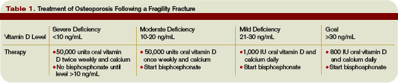
Management
Patients with previous fractures related to osteoporosis require aggressive nonpharmacologic and pharmacologic therapy. Physicians should encourage lifestyle changes that include regular weight-bearing exercise, fall prevention, and discontinuation of tobacco products. Minimizing alcohol ingestion and sedating medications also is recommended. Physical therapy should evaluate gait and balance prior to discharge. Hip protectors may be beneficial, although the data to support this practice is sparse. It also is helpful to arrange a home nurse/therapy visit to assess for hazards in the home that might contribute to falls.
In addition, patients should have adequate calcium and vitamin D intake. The Women’s Health Initiative study showed that calcium with vitamin D use lead to a statistically significant improvement in hip bone density and a 29% reduction in the risk of hip fracture.3 The NOF recommends adults 50 and older have a daily intake of 1,200 mg of calcium and 800 to 1,000 IU of vitamin D. While no definitive data exist to guide the doses of vitamin D and calcium for osteoporosis treatment, it’s reasonable to tailor treatment to the patient’s 25-hydroxyvitamin level.
Specifically, initiate bisphosphonates along with calcium and vitamin D in patients with mild vitamin D deficiency (levels 10 to 30 ng/mL). Patients with severe vitamin D deficiency (<10 ng/mL) should have two to three months of aggressive vitamin D replacement prior to beginning a bisphosphonate. Vitamin D deficiency often is associated with impaired bone mineralization, which potentially could worsen with a bisphosphonate.
Some of the FDA-approved pharmacologic therapies for osteoporosis include antiresorptive bisphosphonates, such as alendronate, risedronate, ibandronate, zoledronic acid, and raloxifene, as well as the human parathyroid hormone teriparatide. Morin et al., performed a population-based, retrospective cohort study using administrative databases to identify patients hospitalized for a hip fracture. They found patients exposed to antiresorptives had a 26% reduction in the rate of recurrent fractures.8
Bisphosphonates are the current first-line treatment of choice unless the clinical situation warrants otherwise. Do not prescribe oral bisphosphonates for patients with hypocalcemia, creatinine clearance lower than 30mL/min, esophageal stricture, or for those who cannot remain upright for 30 minutes.7
Recently, the use of the IV bisphosphonate zolendronic acid within three months of a hip fracture was evaluated. The study randomized approximately 2,100 patients to zolendronic acid 5 mg IV or placebo annually and followed them for a median of 1.9 years. Both groups received vitamin D and calcium supplementation. Those patients using zolendronic acid saw a statistically significant reduction in overall fracture (13.9% vs. 8.6%) and mortality (13.3% vs. 9.6%) rates. While these data support the timely use of bisphosphonate therapy, it is notable that only patients who refused or couldn’t tolerate oral bisphosphonate therapy received the drug, and it was generally not started in the hospital. Still, it’s reasonable to suspect that these beneficial effects would occur even if started in the hospital, as long as the vitamin D and calcium levels did not contraindicate commencement.9
Physicians Don’t Recognize Osteoporosis
In 2000, Kamel et al. retrospectively studied the charts of 170 patients age 65 and older who were hospitalized with a hip fracture, and found that fewer than 5% had been diagnosed with or treated for osteoporosis.7 Follin et al., noted similar results in 2003, reporting that only 14% of the patients were diagnosed with osteoporosis prior to discharge and 75% of patients received no therapy.10
Follin et al., also noted patients who received a diagnosis of osteoporosis prior to discharge were more likely to receive therapy. Sixty-five percent of patients diagnosed with osteoporosis received treatment as opposed to 20% of those not diagnosed. They surmised the lack of treatment may relate to the lack of recognition that a fragility fracture often means osteoporosis.10
Hospitalist Consult, Treatment of Osteoporosis in Hip Fracture Patients
A 2003 retrospective analysis from a university-based academic hospital aimed to determine whether hospitalist consultation during admission for a hip fracture resulted in improved treatment of osteoporosis. The results indicated 29% of patients received treatment for osteoporosis at the time of discharge. Twenty percent received calcium, and only 7% received a bisphosphonate. Those who received hospitalist consultation did not have a significant improvement in osteoporosis treatment, thus representing a huge missed opportunity.11
Back to the Case
You recognize that, because your patient has sustained a fragility fracture, she has osteoporosis and you wish to initiate treatment before she leaves the hospital. Her 25-hydroxyvitamin D level is 18 ng/mL. You commence 50,000 units of vitamin D once weekly and advise that she have her vitamin D level checked again in three months by her primary care provider. She has no contraindications, thus you also initiate a bisphosphonate and remind her to take 1,200 mg of calcium daily.
You encourage smoking cessation, decreased alcohol use, a simplified medication regimen, and weight-bearing exercises in the future. In addition, you ensure she has the proper gait stability items at discharge. You arrange a visiting nurse/therapist to assess her home for fall risks. Lastly, you schedule an outpatient bone mineral density scan and arrange a follow-up with her primary care provider. TH
Dr. Baker is a hospitalist at Ohio State University. Dr McDermott is professor of medicine and clinical pharmacy and endocrinology and diabetes practice director, University of Colorado Denver.
References
- Berry SD, Samelson EJ, Hannan MT, et al. Second hip fracture in older men and women. The Framingham Study. Arch Intern Med. 2007;167(18):1971-1976.
- Juby AG, De Gues-Wenceslau CM. Evaluation of osteoporosis treatment in seniors after hip fracture. Osteoporosis Int. 2002;13:205-210.
- Gardner MJ, Brophy RH, Demetrakopoulos D, et al. Interventions to improve osteoporosis treatment following hip fracture. The Journal of Bone and Joint Surgery. 2005;87-A(1):3-7.
- Gardner MJ, Flik KR, Mooar P, Lane JM.Improve-ment in the undertreatment of osteoporosis following hip fracture. The Journal of Bone and Joint Surgery. 2002;84-A(8):1342-1348.
- Seeman E, Delmas PD. Bone quality-the material and structural basis of bone strength and fragility. N Engl J Med. 2006;354(21):2250-2261.
- Holick, MF. Vitamin D Deficiency. N Engl J Med. 2007;357(3):266-281.
- Glauser T. Practical strategies for managing osteoporosis: An evidence-based approach to risk assessment and treatment. Dialogues in Clinical Practice. 2007.
- Morin S, Rahme E, Behlouli H, Tenenhouse A, Goltzman D, Pilote L. Effectiveness of antiresorptive agents in the prevention of recurrent hip fractures. Osteoporosis Int. 2007;18:1625-1632.
- Lyles KW, Colon-Emeric CS, Magaziner JS, et al. Zolendronic acid and clinical fractures and mortality after hip fracture. N Engl J Med. 2007;357:1799-1809.
- Follin SL, Black JN, McDermott MT. Lack of diagnosis and treatment of osteoporosis in men and women after hip fracture. Pharmacotherapy.2003;23(2):190-198.
- Jachna CM, Whittle J, Lukert B, Graves L, Bhargava T. Effect of hospitalist consultation on treatment of osteoporosis in hip fracture patients. Osteoporosis Int. 2003;14:665-671.
Case
A 66-year-old female with a pack-a-day smoking habit is admitted to orthopedics with a hip fracture following a fall in her home. You are consulted to perform a pre-operative risk assessment and manage her heart failure. The following day, she undergoes an open reduction and internal fixation and does well following the surgery. She is scheduled to be discharged for rehabilitation in two days. She will continue taking her cardiac medications and the narcotics (as needed) for pain. What else can you recommend to reduce her chances of suffering another hip fracture?
Overview
Approximately 300,000 hip fractures occur each year in the United States.¹ The lifetime risk of sustaining a hip fracture is 18% for a woman and 6% for a man.2 One-year mortality after a hip fracture is 20% to 25%, and up to half of patients who live independently prior to their fracture cannot gain independence afterward.
In the late 1990s, inpatient care, nursing home care, and outpatient services associated with hip fractures totaled approximately $14 billion annually. These costs are predicted to reach $50 billion by the year 2040.3 Not surprisingly, second hip fractures are common, with up to 12% of patients suffering another fracture within one year of follow up.1 Risk of morbidity and mortality are even higher after a second hip fracture.
In most experts’ opinions, a fragility fracture indicates osteoporosis and warrants treatment—regardless of bone densitometry findings. Still, multiple studies have shown patients who sustain a hip fracture frequently are not diagnosed, evaluated, or treated for osteoporosis.4 This is analogous to treating an acute coronary syndrome without initiating treatment for a patient’s hypertension and hyperlipidemia prior to discharge. As such, providers clearly are missing an opportunity to begin effective measures at a critical stage in the disease.
Data Review
Physiology of bone strength: Bone minerals—in particular calcium hydroxyapatite—contribute to bone strength by making bone a hard tissue. Collagen adds flexibility and gives bone the ability to absorb energy. The degree of bone mineralization and the number of collagen crosslinks help determine how much stress a bone can tolerate before it breaks. Further, in response to daily stressors, bone accumulates microcracks. Remodeling is then accomplished by bone resorption and formation.5
Estrogen plays an important role in normal remodeling by controlling osteoclast action. Thus, estrogen deficiency leads to prolonged osteoclast activity and increased rates of bone resorption. This explains why bone remodeling typically favors bone resorption later in life and why women are at greatest risk for fracture.5
Vitamin D and calcium: Vitamin D, produced by the skin or ingested, is transported in the circulation by a binding protein to the liver, where it is converted to 25-hydroxyvitamin D. This form is inactive and must be converted by the kidneys to the active form, 1,25-dihydroxyvitamin D. The active form is needed for absorption of renal and intestinal calcium.6
Without vitamin D only 10% to 15% of dietary calcium is absorbed. In one study, serum levels of 25-hydroxyvitamin D directly were related to bone mineral density. When the level was 30 ng/mL or less, there was a significant decrease in intestinal calcium absorption and bone mineral density.6
Diagnostic evaluation: The “gold standard” for diagnosis of osteoporosis is bone mineral density (BMD) testing. The National Osteoporosis Foundation (NOF), the American Association of Clinical Endocrinologists (AACE), and the North American Menopause Society (NAMS) all agree, however, that the history of fragility fracture is diagnostic for osteoporosis, and all recommend initiating pharmacologic therapy in patients with this type of fracture. BMD testing is then used to track a patient’s response to therapy rather than as a diagnostic test.7 An osteoporosis diagnosis should always trigger a history, physical, and evaluation to identify the underlying cause.
Laboratory testing: All patients with osteoporosis should receive laboratory testing. As a baseline obtain chemistry studies, glucose, liver enzymes, albumin, total protein, alkaline phosphatase, and a complete blood count. Also, obtain a 25-hydroxyvitamin D level to help direct the immediate treatment.
Management
Patients with previous fractures related to osteoporosis require aggressive nonpharmacologic and pharmacologic therapy. Physicians should encourage lifestyle changes that include regular weight-bearing exercise, fall prevention, and discontinuation of tobacco products. Minimizing alcohol ingestion and sedating medications also is recommended. Physical therapy should evaluate gait and balance prior to discharge. Hip protectors may be beneficial, although the data to support this practice is sparse. It also is helpful to arrange a home nurse/therapy visit to assess for hazards in the home that might contribute to falls.
In addition, patients should have adequate calcium and vitamin D intake. The Women’s Health Initiative study showed that calcium with vitamin D use lead to a statistically significant improvement in hip bone density and a 29% reduction in the risk of hip fracture.3 The NOF recommends adults 50 and older have a daily intake of 1,200 mg of calcium and 800 to 1,000 IU of vitamin D. While no definitive data exist to guide the doses of vitamin D and calcium for osteoporosis treatment, it’s reasonable to tailor treatment to the patient’s 25-hydroxyvitamin level.
Specifically, initiate bisphosphonates along with calcium and vitamin D in patients with mild vitamin D deficiency (levels 10 to 30 ng/mL). Patients with severe vitamin D deficiency (<10 ng/mL) should have two to three months of aggressive vitamin D replacement prior to beginning a bisphosphonate. Vitamin D deficiency often is associated with impaired bone mineralization, which potentially could worsen with a bisphosphonate.
Some of the FDA-approved pharmacologic therapies for osteoporosis include antiresorptive bisphosphonates, such as alendronate, risedronate, ibandronate, zoledronic acid, and raloxifene, as well as the human parathyroid hormone teriparatide. Morin et al., performed a population-based, retrospective cohort study using administrative databases to identify patients hospitalized for a hip fracture. They found patients exposed to antiresorptives had a 26% reduction in the rate of recurrent fractures.8
Bisphosphonates are the current first-line treatment of choice unless the clinical situation warrants otherwise. Do not prescribe oral bisphosphonates for patients with hypocalcemia, creatinine clearance lower than 30mL/min, esophageal stricture, or for those who cannot remain upright for 30 minutes.7
Recently, the use of the IV bisphosphonate zolendronic acid within three months of a hip fracture was evaluated. The study randomized approximately 2,100 patients to zolendronic acid 5 mg IV or placebo annually and followed them for a median of 1.9 years. Both groups received vitamin D and calcium supplementation. Those patients using zolendronic acid saw a statistically significant reduction in overall fracture (13.9% vs. 8.6%) and mortality (13.3% vs. 9.6%) rates. While these data support the timely use of bisphosphonate therapy, it is notable that only patients who refused or couldn’t tolerate oral bisphosphonate therapy received the drug, and it was generally not started in the hospital. Still, it’s reasonable to suspect that these beneficial effects would occur even if started in the hospital, as long as the vitamin D and calcium levels did not contraindicate commencement.9
Physicians Don’t Recognize Osteoporosis
In 2000, Kamel et al. retrospectively studied the charts of 170 patients age 65 and older who were hospitalized with a hip fracture, and found that fewer than 5% had been diagnosed with or treated for osteoporosis.7 Follin et al., noted similar results in 2003, reporting that only 14% of the patients were diagnosed with osteoporosis prior to discharge and 75% of patients received no therapy.10
Follin et al., also noted patients who received a diagnosis of osteoporosis prior to discharge were more likely to receive therapy. Sixty-five percent of patients diagnosed with osteoporosis received treatment as opposed to 20% of those not diagnosed. They surmised the lack of treatment may relate to the lack of recognition that a fragility fracture often means osteoporosis.10
Hospitalist Consult, Treatment of Osteoporosis in Hip Fracture Patients
A 2003 retrospective analysis from a university-based academic hospital aimed to determine whether hospitalist consultation during admission for a hip fracture resulted in improved treatment of osteoporosis. The results indicated 29% of patients received treatment for osteoporosis at the time of discharge. Twenty percent received calcium, and only 7% received a bisphosphonate. Those who received hospitalist consultation did not have a significant improvement in osteoporosis treatment, thus representing a huge missed opportunity.11
Back to the Case
You recognize that, because your patient has sustained a fragility fracture, she has osteoporosis and you wish to initiate treatment before she leaves the hospital. Her 25-hydroxyvitamin D level is 18 ng/mL. You commence 50,000 units of vitamin D once weekly and advise that she have her vitamin D level checked again in three months by her primary care provider. She has no contraindications, thus you also initiate a bisphosphonate and remind her to take 1,200 mg of calcium daily.
You encourage smoking cessation, decreased alcohol use, a simplified medication regimen, and weight-bearing exercises in the future. In addition, you ensure she has the proper gait stability items at discharge. You arrange a visiting nurse/therapist to assess her home for fall risks. Lastly, you schedule an outpatient bone mineral density scan and arrange a follow-up with her primary care provider. TH
Dr. Baker is a hospitalist at Ohio State University. Dr McDermott is professor of medicine and clinical pharmacy and endocrinology and diabetes practice director, University of Colorado Denver.
References
- Berry SD, Samelson EJ, Hannan MT, et al. Second hip fracture in older men and women. The Framingham Study. Arch Intern Med. 2007;167(18):1971-1976.
- Juby AG, De Gues-Wenceslau CM. Evaluation of osteoporosis treatment in seniors after hip fracture. Osteoporosis Int. 2002;13:205-210.
- Gardner MJ, Brophy RH, Demetrakopoulos D, et al. Interventions to improve osteoporosis treatment following hip fracture. The Journal of Bone and Joint Surgery. 2005;87-A(1):3-7.
- Gardner MJ, Flik KR, Mooar P, Lane JM.Improve-ment in the undertreatment of osteoporosis following hip fracture. The Journal of Bone and Joint Surgery. 2002;84-A(8):1342-1348.
- Seeman E, Delmas PD. Bone quality-the material and structural basis of bone strength and fragility. N Engl J Med. 2006;354(21):2250-2261.
- Holick, MF. Vitamin D Deficiency. N Engl J Med. 2007;357(3):266-281.
- Glauser T. Practical strategies for managing osteoporosis: An evidence-based approach to risk assessment and treatment. Dialogues in Clinical Practice. 2007.
- Morin S, Rahme E, Behlouli H, Tenenhouse A, Goltzman D, Pilote L. Effectiveness of antiresorptive agents in the prevention of recurrent hip fractures. Osteoporosis Int. 2007;18:1625-1632.
- Lyles KW, Colon-Emeric CS, Magaziner JS, et al. Zolendronic acid and clinical fractures and mortality after hip fracture. N Engl J Med. 2007;357:1799-1809.
- Follin SL, Black JN, McDermott MT. Lack of diagnosis and treatment of osteoporosis in men and women after hip fracture. Pharmacotherapy.2003;23(2):190-198.
- Jachna CM, Whittle J, Lukert B, Graves L, Bhargava T. Effect of hospitalist consultation on treatment of osteoporosis in hip fracture patients. Osteoporosis Int. 2003;14:665-671.
The Observation Deck
Observation care provides a mechanism to evaluate and treat patients without the resource utilization and financial responsibility associated with an inpatient admission. Hospitalists may not understand the billing compliance risk and corresponding revenue implications when observation services (OBS) are not captured correctly.
Are OBS best reported with observation care codes (99218-99220, 99234-99236), office visit codes (99201-99215), or initial hospital care codes (99221-99223)? Code selection depends upon the patient’s registered status, the nature of the provided service, and the length of stay. Review the following information before reporting OBS to ensure an accurate claim submission.
Attending Physician Responsibilities
The physician-documented reason for observation substantiates the medical necessity for the OBS admission. Contractors often evaluate medical records to determine the consistency between the physician order (physician intent), the services actually provided (inpatient or outpatient), and the medical necessity of those services, including the medical appropriateness of the inpatient or observation stay.
Certain diagnoses and procedures generally do not support an inpatient admission and fall within the definitions of outpatient observation. Uncomplicated presentations of chest pain (rule out MI), mild asthma/COPD, mild CHF, syncope and decreased responsiveness, atrial arrhythmias, and renal colic all frequently are associated with the expectation of a brief (less than 24-hour) stay unless serious pathology is uncovered.2 Situations that do not meet the criteria for observation care are considered “not medically necessary” and separate payment is not permitted. Examples of circumstances that lack medical necessity include:
- Outpatient blood administration;
- Lack of/delay in patient transportation;
- Provision of a medical exam for patients who do not require skilled support;
- Routine preparation prior to and recovery after diagnostic testing;
- Routine recovery and post-operative care after ambulatory surgery;
- When used for the convenience of the physician, patient or patient’s family;
- While awaiting transfer to another facility;
- Duration of care exceeding 48 hours;
- When an overnight stay is planned prior to diagnostic testing;
- Standing orders following outpatient surgery;
- Services that would normally require inpatient stay;
- No physicians order to admit to observation;
- Observation following an uncomplicated treatment or procedure;
- Services that are not reasonable and necessary for care of the patient;
- Services provided concurrently with chemotherapy; and
- Inpatients discharged to outpatient observation status.3
The attending physician of record assumes responsibility for the patient’s admission to observation and is permitted to report observation care codes. In addition to the reason for admission, a medical record involving the observation stay must include dated and timed physician admitting orders outlining the care plan, physician progress notes, and discharge orders. This documentation must be added to any other record prepared as a result of an emergency department or outpatient clinic encounter. If physicians other than the admitting physician/group (i.e., physicians in different specialties) provide services to the patient during observation, they must use the appropriate outpatient visit (e.g., 99214) or consultation code (e.g., 99244).
Length of Stay4
In general, the duration of observation care services typically does not exceed 24 hours, although in some circumstances patients may require a second day. Observation care for greater than 48 hours without inpatient admission is not considered medically necessary but may be payable after medical review. When the stay spans two calendar days, physician billing is straightforward: Select an initial observation care code (99218-99220) for calendar day one and the observation discharge code (99217) for day two. Only the admitting physician/group may report the discharge service, when applicable. Documentation must demonstrate a face-to-face encounter by the physician for each date of service.
Should the stay only constitute one calendar day, the duration of care becomes a crucial factor in determining the code category. Standard OBS codes (99218-99220) are applicable if the patient stay is less than eight hours on any given date. The OBS discharge code (99217) is not reported in this instance, although the documentation should reflect the attending physician’s written order and appropriate discharge plan. Alternately, same day admit/discharge codes (99234-99236) apply to single-day stays lasting more than eight hours. The OBS discharge code (99217) also is not reported in this instance. Documentation must identify, at a minimum:
- Duration of the stay;
- Presence by the billing physician; and
- Physician performance of each service (i.e., both an admission and discharge note).
Inpatient Admission1,4-5
Sometimes the patient requires inpatient admission after initially being placed in observation. If the inpatient admission occurs on the same day as the OBS admission, only one service is reported (e.g., 99222). The physician need not redocument a complete history and physical (H&P) but merely write the new order for admission and update the OBS assessment with any relevant, new information.
Should the inpatient admission occur on the second calendar day of the OBS stay, the physician is able to report the initial observation care code (e.g., 99219) on day one, and the initial inpatient care code (e.g., 99223) on day two. However, the physician must meet the documentation guidelines for initial hospital care and redocument the H&P associated with the reported visit level. In the case of 99223, the physician must document a comprehensive history (only referring to the previous review of systems and histories, while rewriting the history of present illness) and high complexity decision making. If the physician chooses not to document to this extent, a subsequent hospital care code (99231-99233) is reasonable because the episode of care is a continuation from the observation phase.
Beware that some insurers may change the patient’s status for the entire episode of care. In other words, the conversion to inpatient status occurs on day two of the patient stay, but the insurer may convert the entire stay, including day one, to an inpatient status. Should this happen, the physician is responsible for reporting the visit category that corresponds with the patient’s status. Inpatient services codes are required for claim submission when the patient stay qualifies as an inpatient admission. Because these conversions occur with some frequency, it is advisable to hold claims intended for observation patients until the correct patient status can be confirmed by the utilization review team, and communicated to the physician. TH
Carol Pohlig is a billing and coding expert with the University of Pennsylvania Medical Center, Philadelphia. She also is on the faculty of SHM’s inpatient coding course.
References
- 1. American Medical Association. cpt 2008, Current Procedural Terminology Professional Edition. Chicago, IL: American Medical Association, 2007; 9-16 CPT codes, descriptions and other data only are copyright 2007 American Medical Association (AMA). All Rights Reserved (or such other date of publication of CPT). CPT is a trademark of the AMA.
- 2. Highmark Medicare Services. Local Coverage Determination L27548 Acute Care: Inpatient, Observation and Treatment Room Services. Available at www.highmarkmedicareservices.com/policy/mac-ab/127548.html. Accessed July 14, 2008.
- 3. Cigna. Healthcare Coverage Position: Observation Care. Available at www.cigna.com/customer_care/healthcare_ professional/coverage_positions/medical/mm_0411_coveragepositioncriteria_observation_care.pdf. Accessed July 12, 2008.
- 4. Centers for Medicare and Medicaid Services. Medicare Claims Processing Manual: Chapter 12, Section 30.6.8. Available at www.cms.hhs.gov/manuals/downloads/ clm104c12.pdf. Accessed July 13, 2008.
- 5. Pohlig C. Evaluation & Management Services: An Overview. Coding for Chest Medicine 2008. Northbrook, IL: American College of Chest Physicians, 2008; 57-69.
- 6. Centers for Medicare and Medicaid Services. Medicare Claims Processing Manual: Chapter 1, Section 50.3. Available at www.cms.hhs.gov/manuals/downloads/ clm104c01.pdf. Accessed July 13, 2008.
e-Prescription for Success?
CMS has taken up the e-prescribing torch. In July, the agency announced a preliminary program to promote widespread adoption of electronic prescribing.
E-prescribing is a natural goal for CMS; it has been proven to improve quality of care, reduce medication errors, increase efficiency, and lower administrative costs. Kerry Weems, the acting CMS administrator, says an all-electronic prescribing system could save Medicare as much as $156 million over five years—largely through improved quality care.
Though details on the e-prescribing plan are not yet decided, CMS has revealed that beginning in 2009 (and for the next four years) it will provide incentive payments to physicians who are “successful electronic prescribers.”
Details to Be Determined
The e-prescribing plan will be included in the Physician Quality Reporting Initiative (PQRI), with guidelines included in the 2009 PQRI. (How the new plan will work with the current PQRI e-prescribing measure is one of the unknown details.)
Weems says CMS will use its standard rule-making process to shape the e-prescribing plan. Therefore, details of the incentives program will not be available until this fall, when Medicare releases its final rule on the 2009 physician fee schedule. According to Weems, the 2009 fee schedule and PQRI will clarify some murkiness. “They will be specific about what constitutes e-prescribing, including the extent and reporting of what needs to be done through PQRI,” he says.
Rewards, Then Possible Punishments
Physicians can start reporting on e-prescribing Jan. 1, and those who do will reap the benefits. Patrick Conway, MD, MSc, a hospitalist, an assistant professor at Cincinnati Children’s Hospital Medical Center, and a 2007-2008 White House Fellow working in the Department of Health and Human Services (HHS), says initial discussions about promoting e-prescribing included talk of an incentive-based plan.
“It’s my opinion that, for physicians, it’s beneficial to start with a reward or carrot rather than a punishment,” he says. “And generally, CMS has approached physician programs with this method—like the PQRI.”
The current plan’s outlines indicates that in 2009 and 2010, physicians who successfully report on e-prescribing will receive an incentive payment of up to 2% of their total Medicare allowed charges, matching the maximum bonus they can earn under the regular PQRI. Payment will be additive, so a physician can earn up to 4% (2% for PQRI and 2% for e-prescribing.)
The e-prescribing incentive will drop to 1% in 2011 and 2012, and to 0.5% incentive payment in 2013. After 2013, the carrot is replaced with a stick, and those who do not use e-prescribing will suffer a reduction in payment.
Cost Concerns
CMS estimates the cost of adopting e-prescribing will be approximately $3,000 per individual prescriber. This includes equipment, training, and program maintenance. That can add up to a sizeable expense—particularly for small groups. For that reason, the agency promises a built-in hardship exemption for small practices and others who prove they cannot afford to adopt e-prescribing.
Also, some funding is available: Dr. Conway says CMS has a financial-incentive program for electronic health records, many of which include e-prescribing. “The CMS Electronic Health Records Demonstration is a $150 million program that will provide funds to 1,200 physician practices to adopt this technology,” he says. “They’re currently recruiting practices.” Details on the demonstration are available at www.cms.hhs.gov/DemoProjectsEvalRpts/.
—Patrick Conway, MD, MSc
Will Hospitalists Participate?
Until details of the e-prescribing program are published, no one can say whether the plan will encompass hospitalists. However, Dr. Conway says, “I think this plan is conceptually relevant to hospitalists: It’s possible that hospitalists will be able to participate in the current plan. We don’t know yet. But CMS will continue to push forward on initiatives that increase quality and decrease costs, including e-prescribing. They’ll support electronic health records, whether this particular initiative applies to hospitals or not.”
Even if it turns out hospital medicine groups can’t reap incentive payments from the new plan, Dr. Conway hopes they still will adopt the technology. “Computerized physician order entry (CPOE) and e-prescribing have the potential to decrease errors and increase the quality of care,” he says. “Therefore, I would encourage hospitals and hospitalists to implement electronic health records with computerized order entry and e-prescribing when possible.”
He says the real benefit to hospitals seeking to improve quality and reduce error is not the electronic transmission of prescriptions to the pharmacy, but CPOE. “Most evidence of increased quality is around computerized physician order entry, which includes decision support at the time of the order,” he points out. “One could argue that you could have an incentive for hospitals that utilize CPOE, but I have no idea if CMS will pursue that.”
Next Steps
On Oct. 6-7 CMS will host a conference on the complete e-prescribing plan for pharmacists and physicians in Boston. For details, check the CMS site at www.cms.hhs.gov/eprescribing or www.cms.hhs.gov/pqri.
Dr. Conway thinks the meeting is a good next step for CMS. “I believe it’s very important to engage frontline providers and stakeholders, so the concept of holding a conference to ensure the design of the program is understood, and to get buy-in from the people participating, is a wise choice,” he says.
In the next few months, physicians likely will be inundated with information on e-prescribing processes under the CMS plan. Stay abreast of the latest information through the CMS Web site and, if it turns out, hospitalists can actively participate in the plan, through the SHM Web site at www.hospitalmedicine.org. TH
Jane Jerrard is a medical writer based in Chicago.
Mentoring 101
If you’re in an HM leadership position, don’t be surprised if you’re asked to be a mentor for a less-experienced hospitalist. Why should you voluntarily spend valuable time sharing your guidance and advice? Because to lead is to mentor, and when you dive into the process it rewards all parties involved.
To Lead Is to Mentor
Whether you were just promoted or you’re a leadership veteran approached for the first time by an eager new hospitalist, don’t hesitate to add mentoring to your schedule and responsibilities.
“When you start out as a leader, you get where you want to go by being a mentor,” says Eric E. Howell, MD, director of Collaborative Inpatient Medicine Service, Department of Medicine, Johns Hopkins Bayview Medical Center, Baltimore and faculty for SHM’s Leadership Academy. “You gather disciples, as it were, who will then see you as a leader and support you as a good leadership choice.”
Not only that, but mentoring can add to your skill set as a leader, says Joan C. Faro, MD, FACP, MBA, chief medical officer at John T. Mather Memorial Hospital, Port Jefferson, N.Y. “If you’re interested in developing leadership skills, it’s one of those things you need to do, and do well. If you can’t mentor, then you really can’t lead.”
Plus, when you mentor, you get to feel the reward inherent in helping a young physician whose shoes you once filled. “It’s like raising a kid,” says Dr. Faro. “You want to do a good job because you want to see someone succeed.” This is especially true for mentoring relationships within your HM group.
That means fully flushing out the program and dedicating the time necessary to make it a success. “If you are the de facto leader of a group, you have some obligation to people interested in career development,” says Dr. Howell. “I think it’s part of the job to help advance those people.”
The Ground Rules
Any new mentoring arrangement should start with a discussion of expectations, responsibilities, time frames, and communication. What are the mentee’s expectations for the relationship? How much time can you, the mentor, offer?
Whether the arrangement is formal (a director mentoring a new hire) or casual (an established hospitalist asking a conference speaker for a long-distance mentoring relationship), ground rules are important, Dr. Howell insists. “The mentoring relationship can be established informally, but it’s worthwhile to set some rules on responsibilities: How is the feedback going to come, how frank and honest do you want to be, when should we meet? …Rules will depend on the relationship and on the individuals involved.”
If nothing else, agree to how frequently you will meet or speak. “It could be quarterly or it could be weekly,” says Dr. Howell. “Face time is important, but e-mail and phone calls will work, too, as long as you’ve established some ground rules about this. If the mentee expects a face-to-face meeting and you’re e-mailing your answers, that could be a problem. So you need to establish how you’re going to communicate.”
Those meetings can add up to a sizeable commitment. How much time, exactly, should a new mentor expect to devote to this aspect of leadership? “It varies widely,” Dr. Howell admits. “But I will say that many younger mentees require much more time than older mentors expect. If they’re struggling or haven’t found their stride yet, it can require several hours a week, which is a lot for a busy person’s schedule. But many relationships can be handled weekly or monthly.”
Dr. Faro, who has mentored many hospitalists within her organization, says, “You need to build the relationship; you need enough contact time so that you can understand each other.” For her, that amounts to 40 to 50 hours of getting-to-know-you time, she says. “After that, maybe an hour a week.”
Tailor Mentoring
Dr. Faro tailors her guidance to the personality, capabilities, and level of independence of each person she mentors—hence her lengthy initial time frame.
“You need to start with setting up a clear set of goals and outcomes,” she says. “They really need to know what they’re doing and why they’re doing it. So, set up a plan with specific time frames. It’s your job to determine how independent they are; you may end up giving them goals rather than them stating what they’re going to do.”
—Joan C. Faro, MD, FACP, MBA, chief medical officer, John T. Mather Memorial Hospital, Port Jefferson, N.Y.
For example, she might tell one mentee to develop an order set for patients with syncope, and expect a document by an agreed-upon deadline. She might give another individual the same assignment, but walk that person through each step—within reason. “You can’t do it for them, or they’re not going to learn,” she maintains.
Each completed task is met with constructive criticism. How well was the task done? Did the physician leave out anything? If Dr. Faro senses that more guidance is necessary, she steps up her level of involvement.
What Makes a Successful Mentor?
Dr. Faro understands the mentor/mentee process because she’s been part of it for so long. How can you get to that point? Simply agreeing to be a mentor and having regular meetings with your mentee doesn’t necessarily mean you’re doing a good job.
“Good mentors probably listen more than they talk,” explains Dr. Howell. “For many people, if they can talk it out, they will reach their own conclusions and that’s much more powerful than being told something. That ‘Aha!’ moment is a big career moment.”
He also believes strong mentors can give even non-hospitalists helpful career advice. “Good mentors are able to step out of their own shoes and look at the unique situation of the other person, and give advice tailored to that situation,” he explains. “You have to be altruistic in your mentoring; you can’t do it for your own needs.”
Successful mentors also understand their mentees. For example, mentees in leadership positions should receive advice about how to invest in themselves and their careers. “I always recommend SHM’s Leadership Academy, as well as several books, including Getting to Yes and Good to Great to improve themselves as leaders,” says Dr. Howell.
Finally, a mentor who does the job well understands when the relationship isn’t working. If this is the case, “be up-front and honest, and if possible, point that person to another mentor,” Dr. Howell says. “If you can introduce them and get them started, that’s best. You can also share a mentee with someone else; you can each handle different areas. I have many different mentors in different areas. It’s more productive that way.” TH
Jane Jerrard also writes “Public Policy” for The Hospitalist.
Serotonin Syndrome
A healthy 30-year-old female presented to the urgent care center with confusion, tremor, and a blood pressure of 160/110 mm Hg. She had no history of hypertension, diabetes, dyslipidemia, renal dysfunction, or smoking. A basic metabolic panel revealed no abnormalities.
Her medication history revealed use of paroxetine (20 mg) subsequent to a depressive episode two years prior. A source of the hypertension was not identified, and she was sent home without further follow-up. The next day, she was admitted to the hospital via the emergency department for stroke symptoms, including numbness and weakness on her right side (extremities and face), with confusion and diplopia. She remained hospitalized for four days during which time she continued to experience transient ischemic attacks. The paroxetine eventually was discontinued. She subsequently has recovered without negative sequelae.
Serotonin syndrome is a consequence of a hyperserotonergic state, due to drug-induced serotonin intensification.1 It can be mild or life-threatening and is characterized by a triad of clinical manifestations: mental status changes, autonomic hyperactivity, and neuromuscular abnormalities.2 Clinicians may miss mild symptoms, such as diarrhea, tremor, tachycardia, diaphoresis, or mydriasis. This can result in an increase in the dose of the causative agent or addition of a serotonergic agent, thus yielding a worsening clinical decline.3
Patients with a more severe clinical presentation include those with severe hypertension (as in the case above), tachycardia, muscular rigidity, and shock. Laboratory abnormalities may be present if the patient develops subsequent rhabdomyolysis, seizures, metabolic acidosis, or renal failure. Serotonin syndrome is diagnosed based on the patient’s presentation, history, and physical examination. It should be differentiated from neuroleptic malignant syndrome, which has a similar presentation.4
Serotonergic agents used alone, or in combination, may lead to serotonin syndrome.5 A recent report discussed the appearance of serotonin syndrome in patients receiving only sumatriptan. Other offenders include such antidepressants as monoamine oxidase inhibitors, buspirone, citalopram, clomipramine, escitalopram, fluoxetine, fluvoxamine, nefazodone, paroxetine, sertraline, trazodone, and venlafaxine. Other causative agents include dextromethorphan, fentanyl, granisetron, levodopa, linezolid, lithium, meperidine, metoclopramide, ondansetron, pentazocine, sibutramine, sumatriptan, tramadol, valproate, and drugs of abuse (e.g., amphetamines, cocaine, LSD, ecstasy). Additionally, ginseng, St. John’s Wort, and tryptophan have been implicated.
Many of these agents require an adequate washout period prior to beginning other serotonergic agents. Mild to moderately severe cases usually resolve within 24 to 72 hours, although most resolve within a week depending on the half-life of the medication. Serotonin syndrome carries an 11% mortality rate and is best managed by stopping the offending agent and providing supportive care. TH
Michele B. Kaufman is a freelance medical writer based in New York City.
References
- Sorenson S. Serotonin syndrome. UTox Update 2002;4(4):1-2. A Publication of the Utah Poison Control Center for Health Professionals. Available at http://uuhsc.utah.edu/poison/healthpros/utox/vol4_no4.pdf. Last accessed June 20, 2008.
- Soldin OP, Tonning JM. Serotonin syndrome associated with triptan monotherapy. N Engl J Med. 2008;358(20):2185-2186.
- Boyer EW, Shannon M. The serotonin syndrome. N Engl J Med. 2005;352(11):1112-1120.
- Nolan S, Scoggin JA. Serotonin syndrome: recognition and management. US Pharm. 2002;23(2). www.uspharmacist.com/oldformat.asp?url=newlook/files/feat/acf2fa6.htm. Last accessed June 20, 2008.
- Mayo Clinic.com. Diseases and conditions. www.mayoclinic.com/health/serotonin-syndrome/DS00860. Last accessed June 20, 2008.