User login
Study reveals gap in patient blood management
PHILADELPHIA—Healthcare professionals may not be using blood management interventions in a majority of patients receiving red blood cell (RBC) transfusions, a large study suggests.
The research showed that 72 US hospitals have made strides in reducing the use of RBCs in patients undergoing orthopedic and cardiac surgery.
And smaller reductions have occurred in patients with gastrointestinal bleeding, obstetric patients, and those receiving bone marrow transplants or inpatient chemotherapy.
However, more than 60% of the transfusions studied were given to patients who did not belong to any of the aforementioned groups.
“So when we target our [patient blood management] interventions to these kind of surgical procedures, in fact, we’re looking at only about 40% of the red cell use, and 60% goes to a myriad of other patients,” said Robert L. Thurer, MD, of Haemonetics in Braintree, Massachusetts.
He added that the groups of specific patient populations within that 60% were so small—“2% of patients here and 3% of patients there”—that it was too difficult to examine them individually in this study.
Dr Thurer presented this research at the AABB Annual Meeting 2014 (abstract S65-030K).
He and his colleagues wanted to determine which specific groups of patients have been most affected by patient blood management, understand further opportunities for decreasing transfusion use, and project future blood needs.
So the researchers analyzed data from 3,946,428 inpatients at 72 US hospitals, comparing the use of RBC transfusions in 2009/2010 to use in 2013.
In 2009/2010, there were 1,378,581 patients admitted to the hospitals, the RBC transfusion rate was 11.5.%, and the utilization (total units/total patients) was 0.41. In 2013, there were 861,804 patients, the transfusion rate was 10%, and the utilization was 0.34.
So from 2009/2010 to 2013, there was a 13% reduction in transfusion rate and a 17% reduction in utilization.
The greatest decrease was in orthopedic surgery patients. In those undergoing hip and knee joint replacement surgery, there was a 45% reduction in transfusion rate and a 43% reduction in utilization. In patients with a hip fracture, there was a 20% decrease in transfusion rate and a 20% decrease in utilization.
There was a smaller, though still sizable, reduction in transfusion use among patients undergoing cardiac surgery—a 15% decrease in transfusion rate and an 18% decrease in utilization.
“My personal thoughts about this is that it represents, certainly, lower transfusion triggers, which are becoming more widely adopted, [and] the use of antifibrinolytic drugs, particularly in orthopedic surgeries,” Dr Thurer said.
“We like to think that comprehensive coagulation testing and better matching of coagulation abnormalities contributes to this. Perhaps correction of preoperative anemia [contributes] for elective patients. And, clearly, surgical techniques have evolved, and, as they do, blood loss goes down.”
Reductions in RBC use were also seen in patients with gastrointestinal bleeding, where there was a 3% decrease in transfusion rate and a 13% decrease in utilization.
“For gastrointestinal bleeding, I think the lower transfusion triggers [have made an impact], but there’s also . . . more interest in timely interventions to stop bleeding,” Dr Thurer said.
“So rather than the gastroenterologist saying, ‘Correct the hematocrit and the coagulation factors, and I’ll stop the bleeding in the morning,’ we’re seeing now much more interest in very prompt endoscopy to stop bleeding. And as you know, the way to stop giving transfusions is to stop the patient from bleeding.”
Obstetric patients saw a 5% reduction in transfusion rate and an 8% reduction in utilization. And patients undergoing bone marrow transplant or inpatient chemotherapy saw a 6% decrease in transfusion rate and an 8% decrease in utilization.
Other transplant patients actually saw an increase in RBC transfusions. In liver transplant recipients, there was a 2.2% increase in transfusion rate and a 6% increase in utilization. And in kidney transplant recipients, there was a 0.2% increase in transfusion rate and a 19% increase in utilization.
However, Dr Thurer noted that the majority of RBC transfusions are administered to patients outside of these groups. In 2013, 60.6% of transfusions went to patients who did not fit into any of the aforementioned categories.
“So clearly,” he concluded, “further studies are needed to determine whether these reductions that we’ve seen in some areas can be implemented in a wider variety of patients.” 
PHILADELPHIA—Healthcare professionals may not be using blood management interventions in a majority of patients receiving red blood cell (RBC) transfusions, a large study suggests.
The research showed that 72 US hospitals have made strides in reducing the use of RBCs in patients undergoing orthopedic and cardiac surgery.
And smaller reductions have occurred in patients with gastrointestinal bleeding, obstetric patients, and those receiving bone marrow transplants or inpatient chemotherapy.
However, more than 60% of the transfusions studied were given to patients who did not belong to any of the aforementioned groups.
“So when we target our [patient blood management] interventions to these kind of surgical procedures, in fact, we’re looking at only about 40% of the red cell use, and 60% goes to a myriad of other patients,” said Robert L. Thurer, MD, of Haemonetics in Braintree, Massachusetts.
He added that the groups of specific patient populations within that 60% were so small—“2% of patients here and 3% of patients there”—that it was too difficult to examine them individually in this study.
Dr Thurer presented this research at the AABB Annual Meeting 2014 (abstract S65-030K).
He and his colleagues wanted to determine which specific groups of patients have been most affected by patient blood management, understand further opportunities for decreasing transfusion use, and project future blood needs.
So the researchers analyzed data from 3,946,428 inpatients at 72 US hospitals, comparing the use of RBC transfusions in 2009/2010 to use in 2013.
In 2009/2010, there were 1,378,581 patients admitted to the hospitals, the RBC transfusion rate was 11.5.%, and the utilization (total units/total patients) was 0.41. In 2013, there were 861,804 patients, the transfusion rate was 10%, and the utilization was 0.34.
So from 2009/2010 to 2013, there was a 13% reduction in transfusion rate and a 17% reduction in utilization.
The greatest decrease was in orthopedic surgery patients. In those undergoing hip and knee joint replacement surgery, there was a 45% reduction in transfusion rate and a 43% reduction in utilization. In patients with a hip fracture, there was a 20% decrease in transfusion rate and a 20% decrease in utilization.
There was a smaller, though still sizable, reduction in transfusion use among patients undergoing cardiac surgery—a 15% decrease in transfusion rate and an 18% decrease in utilization.
“My personal thoughts about this is that it represents, certainly, lower transfusion triggers, which are becoming more widely adopted, [and] the use of antifibrinolytic drugs, particularly in orthopedic surgeries,” Dr Thurer said.
“We like to think that comprehensive coagulation testing and better matching of coagulation abnormalities contributes to this. Perhaps correction of preoperative anemia [contributes] for elective patients. And, clearly, surgical techniques have evolved, and, as they do, blood loss goes down.”
Reductions in RBC use were also seen in patients with gastrointestinal bleeding, where there was a 3% decrease in transfusion rate and a 13% decrease in utilization.
“For gastrointestinal bleeding, I think the lower transfusion triggers [have made an impact], but there’s also . . . more interest in timely interventions to stop bleeding,” Dr Thurer said.
“So rather than the gastroenterologist saying, ‘Correct the hematocrit and the coagulation factors, and I’ll stop the bleeding in the morning,’ we’re seeing now much more interest in very prompt endoscopy to stop bleeding. And as you know, the way to stop giving transfusions is to stop the patient from bleeding.”
Obstetric patients saw a 5% reduction in transfusion rate and an 8% reduction in utilization. And patients undergoing bone marrow transplant or inpatient chemotherapy saw a 6% decrease in transfusion rate and an 8% decrease in utilization.
Other transplant patients actually saw an increase in RBC transfusions. In liver transplant recipients, there was a 2.2% increase in transfusion rate and a 6% increase in utilization. And in kidney transplant recipients, there was a 0.2% increase in transfusion rate and a 19% increase in utilization.
However, Dr Thurer noted that the majority of RBC transfusions are administered to patients outside of these groups. In 2013, 60.6% of transfusions went to patients who did not fit into any of the aforementioned categories.
“So clearly,” he concluded, “further studies are needed to determine whether these reductions that we’ve seen in some areas can be implemented in a wider variety of patients.”
PHILADELPHIA—Healthcare professionals may not be using blood management interventions in a majority of patients receiving red blood cell (RBC) transfusions, a large study suggests.
The research showed that 72 US hospitals have made strides in reducing the use of RBCs in patients undergoing orthopedic and cardiac surgery.
And smaller reductions have occurred in patients with gastrointestinal bleeding, obstetric patients, and those receiving bone marrow transplants or inpatient chemotherapy.
However, more than 60% of the transfusions studied were given to patients who did not belong to any of the aforementioned groups.
“So when we target our [patient blood management] interventions to these kind of surgical procedures, in fact, we’re looking at only about 40% of the red cell use, and 60% goes to a myriad of other patients,” said Robert L. Thurer, MD, of Haemonetics in Braintree, Massachusetts.
He added that the groups of specific patient populations within that 60% were so small—“2% of patients here and 3% of patients there”—that it was too difficult to examine them individually in this study.
Dr Thurer presented this research at the AABB Annual Meeting 2014 (abstract S65-030K).
He and his colleagues wanted to determine which specific groups of patients have been most affected by patient blood management, understand further opportunities for decreasing transfusion use, and project future blood needs.
So the researchers analyzed data from 3,946,428 inpatients at 72 US hospitals, comparing the use of RBC transfusions in 2009/2010 to use in 2013.
In 2009/2010, there were 1,378,581 patients admitted to the hospitals, the RBC transfusion rate was 11.5.%, and the utilization (total units/total patients) was 0.41. In 2013, there were 861,804 patients, the transfusion rate was 10%, and the utilization was 0.34.
So from 2009/2010 to 2013, there was a 13% reduction in transfusion rate and a 17% reduction in utilization.
The greatest decrease was in orthopedic surgery patients. In those undergoing hip and knee joint replacement surgery, there was a 45% reduction in transfusion rate and a 43% reduction in utilization. In patients with a hip fracture, there was a 20% decrease in transfusion rate and a 20% decrease in utilization.
There was a smaller, though still sizable, reduction in transfusion use among patients undergoing cardiac surgery—a 15% decrease in transfusion rate and an 18% decrease in utilization.
“My personal thoughts about this is that it represents, certainly, lower transfusion triggers, which are becoming more widely adopted, [and] the use of antifibrinolytic drugs, particularly in orthopedic surgeries,” Dr Thurer said.
“We like to think that comprehensive coagulation testing and better matching of coagulation abnormalities contributes to this. Perhaps correction of preoperative anemia [contributes] for elective patients. And, clearly, surgical techniques have evolved, and, as they do, blood loss goes down.”
Reductions in RBC use were also seen in patients with gastrointestinal bleeding, where there was a 3% decrease in transfusion rate and a 13% decrease in utilization.
“For gastrointestinal bleeding, I think the lower transfusion triggers [have made an impact], but there’s also . . . more interest in timely interventions to stop bleeding,” Dr Thurer said.
“So rather than the gastroenterologist saying, ‘Correct the hematocrit and the coagulation factors, and I’ll stop the bleeding in the morning,’ we’re seeing now much more interest in very prompt endoscopy to stop bleeding. And as you know, the way to stop giving transfusions is to stop the patient from bleeding.”
Obstetric patients saw a 5% reduction in transfusion rate and an 8% reduction in utilization. And patients undergoing bone marrow transplant or inpatient chemotherapy saw a 6% decrease in transfusion rate and an 8% decrease in utilization.
Other transplant patients actually saw an increase in RBC transfusions. In liver transplant recipients, there was a 2.2% increase in transfusion rate and a 6% increase in utilization. And in kidney transplant recipients, there was a 0.2% increase in transfusion rate and a 19% increase in utilization.
However, Dr Thurer noted that the majority of RBC transfusions are administered to patients outside of these groups. In 2013, 60.6% of transfusions went to patients who did not fit into any of the aforementioned categories.
“So clearly,” he concluded, “further studies are needed to determine whether these reductions that we’ve seen in some areas can be implemented in a wider variety of patients.”
Pyrethroid biomarker almost tripled odds of ADHD in boys
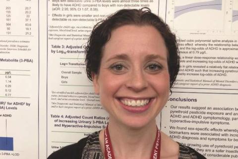
SAN DIEGO – Exposure to common household pesticides called pyrethroids almost tripled the odds of attention-deficit/hyperactivity disorder in boys, but not in girls, authors of a large cross-sectional study reported.
The results resemble findings from prior studies of mice, lead investigator Dr. Melissa L. Wagner-Schuman said in an interview. “Pyrethroids are the most commonly used pesticides for residential pest control and public health,” she and her associates said. “Given the growing use of pyrethroids and the perception that they are a safer insecticide alternative, our results may be of considerable public health import.”

Attention-deficit/hyperactivity disorder is more than twice as prevalent in boys as in girls, according to CDC data. The disorder is “highly heritable,” but environmental factors also play a role, the researchers said at the annual meeting of the American Academy of Child and Adolescent Psychiatry.
For the study, the investigators analyzed National Health and Nutrition Examination Survey data from 2001 to 2002 from 687 children aged 8-18 years. Children were categorized as having ADHD if they met DSM-IV criteria for the disorder on the Diagnostic Interview Schedule for Children caregiver module, had a diagnosis of ADHD reported by their caregivers, or both, the researchers said. Pyrethroid exposure was assessed by testing urine samples for a metabolite of several pyrethroids called 3-phenoxybenzoic acid (3-PBA), they noted.
Boys who had detectable levels of 3-PBA were 2.95 times more likely to have ADHD than were boys who lacked evidence of pyrethroid exposure (95% confidence interval, 1.07-8.08), said Dr. Wagner-Schuman, a pediatrics resident at Cincinnati Children’s Hospital Medical Center. “Effects in girls were smaller and nonsignificant,” she and her associates reported (adjusted odds ratio for girls with detectable biomarker levels, 1.54; 95% CI, 0.32-7.33). The analysis controlled for age; race or ethnicity; income; health insurance status; prenatal tobacco exposure; blood lead levels; urine organophosphate metabolite levels; and urine creatinine level.
Also in boys but not in girls, the odds of ADHD increased linearly with rising 3-PBA levels and did not plateau, the researchers reported.
In the mouse studies, pyrethroid exposure was found to trigger abnormalities in the dopamine system, which produced an “ADHD phenotype,” the investigators said. “Male animals appear to have a heightened vulnerability to exposure,” they added. Other studies have shown that prenatal pyrethroid exposure in humans can increase the risk of neurodevelopmental problems, the investigators noted.
The analysis was limited by its cross-sectional design, Dr. Wagner-Schuman said. Future studies should serially quantify pyrethroid exposure over time, she added.
The National Institute of Environmental Health Sciences funded the research. One coauthor reported having served as a consultant and expert witness for the California Attorney General’s Office, and having consulted for the California Department of Toxic Substances Control and the U.S. Environmental Protection Agency. The other investigators declared no conflicts of interest.
SAN DIEGO – Exposure to common household pesticides called pyrethroids almost tripled the odds of attention-deficit/hyperactivity disorder in boys, but not in girls, authors of a large cross-sectional study reported.
The results resemble findings from prior studies of mice, lead investigator Dr. Melissa L. Wagner-Schuman said in an interview. “Pyrethroids are the most commonly used pesticides for residential pest control and public health,” she and her associates said. “Given the growing use of pyrethroids and the perception that they are a safer insecticide alternative, our results may be of considerable public health import.”
Attention-deficit/hyperactivity disorder is more than twice as prevalent in boys as in girls, according to CDC data. The disorder is “highly heritable,” but environmental factors also play a role, the researchers said at the annual meeting of the American Academy of Child and Adolescent Psychiatry.
For the study, the investigators analyzed National Health and Nutrition Examination Survey data from 2001 to 2002 from 687 children aged 8-18 years. Children were categorized as having ADHD if they met DSM-IV criteria for the disorder on the Diagnostic Interview Schedule for Children caregiver module, had a diagnosis of ADHD reported by their caregivers, or both, the researchers said. Pyrethroid exposure was assessed by testing urine samples for a metabolite of several pyrethroids called 3-phenoxybenzoic acid (3-PBA), they noted.
Boys who had detectable levels of 3-PBA were 2.95 times more likely to have ADHD than were boys who lacked evidence of pyrethroid exposure (95% confidence interval, 1.07-8.08), said Dr. Wagner-Schuman, a pediatrics resident at Cincinnati Children’s Hospital Medical Center. “Effects in girls were smaller and nonsignificant,” she and her associates reported (adjusted odds ratio for girls with detectable biomarker levels, 1.54; 95% CI, 0.32-7.33). The analysis controlled for age; race or ethnicity; income; health insurance status; prenatal tobacco exposure; blood lead levels; urine organophosphate metabolite levels; and urine creatinine level.
Also in boys but not in girls, the odds of ADHD increased linearly with rising 3-PBA levels and did not plateau, the researchers reported.
In the mouse studies, pyrethroid exposure was found to trigger abnormalities in the dopamine system, which produced an “ADHD phenotype,” the investigators said. “Male animals appear to have a heightened vulnerability to exposure,” they added. Other studies have shown that prenatal pyrethroid exposure in humans can increase the risk of neurodevelopmental problems, the investigators noted.
The analysis was limited by its cross-sectional design, Dr. Wagner-Schuman said. Future studies should serially quantify pyrethroid exposure over time, she added.
The National Institute of Environmental Health Sciences funded the research. One coauthor reported having served as a consultant and expert witness for the California Attorney General’s Office, and having consulted for the California Department of Toxic Substances Control and the U.S. Environmental Protection Agency. The other investigators declared no conflicts of interest.
SAN DIEGO – Exposure to common household pesticides called pyrethroids almost tripled the odds of attention-deficit/hyperactivity disorder in boys, but not in girls, authors of a large cross-sectional study reported.
The results resemble findings from prior studies of mice, lead investigator Dr. Melissa L. Wagner-Schuman said in an interview. “Pyrethroids are the most commonly used pesticides for residential pest control and public health,” she and her associates said. “Given the growing use of pyrethroids and the perception that they are a safer insecticide alternative, our results may be of considerable public health import.”
Attention-deficit/hyperactivity disorder is more than twice as prevalent in boys as in girls, according to CDC data. The disorder is “highly heritable,” but environmental factors also play a role, the researchers said at the annual meeting of the American Academy of Child and Adolescent Psychiatry.
For the study, the investigators analyzed National Health and Nutrition Examination Survey data from 2001 to 2002 from 687 children aged 8-18 years. Children were categorized as having ADHD if they met DSM-IV criteria for the disorder on the Diagnostic Interview Schedule for Children caregiver module, had a diagnosis of ADHD reported by their caregivers, or both, the researchers said. Pyrethroid exposure was assessed by testing urine samples for a metabolite of several pyrethroids called 3-phenoxybenzoic acid (3-PBA), they noted.
Boys who had detectable levels of 3-PBA were 2.95 times more likely to have ADHD than were boys who lacked evidence of pyrethroid exposure (95% confidence interval, 1.07-8.08), said Dr. Wagner-Schuman, a pediatrics resident at Cincinnati Children’s Hospital Medical Center. “Effects in girls were smaller and nonsignificant,” she and her associates reported (adjusted odds ratio for girls with detectable biomarker levels, 1.54; 95% CI, 0.32-7.33). The analysis controlled for age; race or ethnicity; income; health insurance status; prenatal tobacco exposure; blood lead levels; urine organophosphate metabolite levels; and urine creatinine level.
Also in boys but not in girls, the odds of ADHD increased linearly with rising 3-PBA levels and did not plateau, the researchers reported.
In the mouse studies, pyrethroid exposure was found to trigger abnormalities in the dopamine system, which produced an “ADHD phenotype,” the investigators said. “Male animals appear to have a heightened vulnerability to exposure,” they added. Other studies have shown that prenatal pyrethroid exposure in humans can increase the risk of neurodevelopmental problems, the investigators noted.
The analysis was limited by its cross-sectional design, Dr. Wagner-Schuman said. Future studies should serially quantify pyrethroid exposure over time, she added.
The National Institute of Environmental Health Sciences funded the research. One coauthor reported having served as a consultant and expert witness for the California Attorney General’s Office, and having consulted for the California Department of Toxic Substances Control and the U.S. Environmental Protection Agency. The other investigators declared no conflicts of interest.
Key clinical point: Consider environmental factors such as exposure to pyrethroid pesticides when assessing boys for attention-deficit/hyperactivity disorder.
Major finding: Boys who had a biomarker for pyrethroids were 2.95 times more likely to have ADHD, compared with boys who lacked the metabolite.
Data source: Cross-sectional analysis of National Health and Nutrition Examination Survey data, and urine tests for 687 children and adolescents.
Disclosures: The National Institute of Environmental Health Sciences funded the research. One coauthor reported having served as a consultant and expert witness for the California Attorney General’s Office, and having consulted for the California Department of Toxic Substances Control and the U.S. Environmental Protection Agency. The other investigators declared no conflicts of interest.
Under My Skin: Neglect
Two disturbing patients came by last week.
The first was a frail old man. His daughter brought him. She said he’d been living in Florida and “shown up” on her doorstep.
As a dermatologist, I’m not often thrown by what I see, but this unfortunate man’s face was hard to look at, with a gaping hole where his left nasolabial fold should have been.
How long had the cancer been there to gouge that hole? How could he neglect it so long? What kind of relationship (or nonrelationship) with his child did it take for this to happen?
I didn’t pursue these questions. Instead, I referred him and his daughter to a skin oncology center where, I hoped, therapy could manage a situation whose severity could surely have been prevented.
Two days later, a Russian woman came in. Remarkably hale at the age of 95 years, she spoke no English. The man who accompanied her, a relative youngster in his mid-70’s, was not a relative, just a stranger who took pity on a fellow visitor to a Russian senior center. “She has two sons,” he explained, “but they live in Minnesota and Texas.”
Her problem was also a basal cell, but this one was on the back of her right ear, large but manageable. I arranged to remove the lesion and offered to speak with her sons. Neither ever called.
Disease is a physical problem in a social context. Patients often present with problems they ignored until other people insisted they take care of them. Parents bring their children. Women drag their husbands. Patients tolerate their itch until their coworkers get annoyed “at seeing me scratch like a monkey.” In situations like these – you can come up with many others – the problem is not just with the patients, but with the people in their vicinity. Sometimes there are people in patients’ lives who notice and care, who demand, “Have that looked at!” But what if nobody cares? Or what if there is no one around at all?
Factors like mental, family, and social dysfunction often underlie whether and to what extent the diseases we diagnose get treated. As practicing physicians, we have little control over such factors. We just try to manage what presents in our offices.
So we make assumptions– that patients can afford to see us, that they have the common sense to come, that they have family or friends who encourage them to come and make doing that possible.
In cases like the ones I’ve just described, these assumptions were wrong. The old man from Florida probably rarely left his apartment, and when he did people just looked away in disgust. He wasn’t their problem. In both cases family was nowhere to be found. How many such lonely and neglected people are there with no support systems, who don’t show up on our office doorstep until it is hard or impossible to take care of them?
I sometimes think back to a case that has haunted me since my early years, when I worked in several Boston-area health centers and sometimes made house calls in gritty neighborhoods. One day I was called to see a patient on the first floor of a rundown example of one of Boston’s wood-frame triple-deckers.
The front door was open. No one was around. I wandered past the parlor into a bedroom. There lay the patient: A woman in late middle age, lying on her back in a dirty nightgown, staring at the ceiling. That image has haunted me for 30 years.
I no longer remember what her skin problem was, just the pitiful sight of her and all the questions it raised: Where was everybody? Who looked after this woman? Who cooked for her, shopped for her? If I prescribed something, who would see that she got it and used it?
I didn’t know. Even if I did, there was nothing I could do about it. Doctors in practice can’t make families stay together, or weave a social safety net that neglected people don’t slip through.
When something lies beyond the scope of what you take to be your responsibility, it’s easier to look away. Now and then a neglected patient forces us to face our own limitations and pay attention to what we have been not looking at.
Dr. Rockoff practices dermatology in Brookline, Mass., and is a longtime contributor to Skin & Allergy News. He serves on the clinical faculty at Tufts University, Boston, and has taught senior medical students and other trainees for 30 years.
Two disturbing patients came by last week.
The first was a frail old man. His daughter brought him. She said he’d been living in Florida and “shown up” on her doorstep.
As a dermatologist, I’m not often thrown by what I see, but this unfortunate man’s face was hard to look at, with a gaping hole where his left nasolabial fold should have been.
How long had the cancer been there to gouge that hole? How could he neglect it so long? What kind of relationship (or nonrelationship) with his child did it take for this to happen?
I didn’t pursue these questions. Instead, I referred him and his daughter to a skin oncology center where, I hoped, therapy could manage a situation whose severity could surely have been prevented.
Two days later, a Russian woman came in. Remarkably hale at the age of 95 years, she spoke no English. The man who accompanied her, a relative youngster in his mid-70’s, was not a relative, just a stranger who took pity on a fellow visitor to a Russian senior center. “She has two sons,” he explained, “but they live in Minnesota and Texas.”
Her problem was also a basal cell, but this one was on the back of her right ear, large but manageable. I arranged to remove the lesion and offered to speak with her sons. Neither ever called.
Disease is a physical problem in a social context. Patients often present with problems they ignored until other people insisted they take care of them. Parents bring their children. Women drag their husbands. Patients tolerate their itch until their coworkers get annoyed “at seeing me scratch like a monkey.” In situations like these – you can come up with many others – the problem is not just with the patients, but with the people in their vicinity. Sometimes there are people in patients’ lives who notice and care, who demand, “Have that looked at!” But what if nobody cares? Or what if there is no one around at all?
Factors like mental, family, and social dysfunction often underlie whether and to what extent the diseases we diagnose get treated. As practicing physicians, we have little control over such factors. We just try to manage what presents in our offices.
So we make assumptions– that patients can afford to see us, that they have the common sense to come, that they have family or friends who encourage them to come and make doing that possible.
In cases like the ones I’ve just described, these assumptions were wrong. The old man from Florida probably rarely left his apartment, and when he did people just looked away in disgust. He wasn’t their problem. In both cases family was nowhere to be found. How many such lonely and neglected people are there with no support systems, who don’t show up on our office doorstep until it is hard or impossible to take care of them?
I sometimes think back to a case that has haunted me since my early years, when I worked in several Boston-area health centers and sometimes made house calls in gritty neighborhoods. One day I was called to see a patient on the first floor of a rundown example of one of Boston’s wood-frame triple-deckers.
The front door was open. No one was around. I wandered past the parlor into a bedroom. There lay the patient: A woman in late middle age, lying on her back in a dirty nightgown, staring at the ceiling. That image has haunted me for 30 years.
I no longer remember what her skin problem was, just the pitiful sight of her and all the questions it raised: Where was everybody? Who looked after this woman? Who cooked for her, shopped for her? If I prescribed something, who would see that she got it and used it?
I didn’t know. Even if I did, there was nothing I could do about it. Doctors in practice can’t make families stay together, or weave a social safety net that neglected people don’t slip through.
When something lies beyond the scope of what you take to be your responsibility, it’s easier to look away. Now and then a neglected patient forces us to face our own limitations and pay attention to what we have been not looking at.
Dr. Rockoff practices dermatology in Brookline, Mass., and is a longtime contributor to Skin & Allergy News. He serves on the clinical faculty at Tufts University, Boston, and has taught senior medical students and other trainees for 30 years.
Two disturbing patients came by last week.
The first was a frail old man. His daughter brought him. She said he’d been living in Florida and “shown up” on her doorstep.
As a dermatologist, I’m not often thrown by what I see, but this unfortunate man’s face was hard to look at, with a gaping hole where his left nasolabial fold should have been.
How long had the cancer been there to gouge that hole? How could he neglect it so long? What kind of relationship (or nonrelationship) with his child did it take for this to happen?
I didn’t pursue these questions. Instead, I referred him and his daughter to a skin oncology center where, I hoped, therapy could manage a situation whose severity could surely have been prevented.
Two days later, a Russian woman came in. Remarkably hale at the age of 95 years, she spoke no English. The man who accompanied her, a relative youngster in his mid-70’s, was not a relative, just a stranger who took pity on a fellow visitor to a Russian senior center. “She has two sons,” he explained, “but they live in Minnesota and Texas.”
Her problem was also a basal cell, but this one was on the back of her right ear, large but manageable. I arranged to remove the lesion and offered to speak with her sons. Neither ever called.
Disease is a physical problem in a social context. Patients often present with problems they ignored until other people insisted they take care of them. Parents bring their children. Women drag their husbands. Patients tolerate their itch until their coworkers get annoyed “at seeing me scratch like a monkey.” In situations like these – you can come up with many others – the problem is not just with the patients, but with the people in their vicinity. Sometimes there are people in patients’ lives who notice and care, who demand, “Have that looked at!” But what if nobody cares? Or what if there is no one around at all?
Factors like mental, family, and social dysfunction often underlie whether and to what extent the diseases we diagnose get treated. As practicing physicians, we have little control over such factors. We just try to manage what presents in our offices.
So we make assumptions– that patients can afford to see us, that they have the common sense to come, that they have family or friends who encourage them to come and make doing that possible.
In cases like the ones I’ve just described, these assumptions were wrong. The old man from Florida probably rarely left his apartment, and when he did people just looked away in disgust. He wasn’t their problem. In both cases family was nowhere to be found. How many such lonely and neglected people are there with no support systems, who don’t show up on our office doorstep until it is hard or impossible to take care of them?
I sometimes think back to a case that has haunted me since my early years, when I worked in several Boston-area health centers and sometimes made house calls in gritty neighborhoods. One day I was called to see a patient on the first floor of a rundown example of one of Boston’s wood-frame triple-deckers.
The front door was open. No one was around. I wandered past the parlor into a bedroom. There lay the patient: A woman in late middle age, lying on her back in a dirty nightgown, staring at the ceiling. That image has haunted me for 30 years.
I no longer remember what her skin problem was, just the pitiful sight of her and all the questions it raised: Where was everybody? Who looked after this woman? Who cooked for her, shopped for her? If I prescribed something, who would see that she got it and used it?
I didn’t know. Even if I did, there was nothing I could do about it. Doctors in practice can’t make families stay together, or weave a social safety net that neglected people don’t slip through.
When something lies beyond the scope of what you take to be your responsibility, it’s easier to look away. Now and then a neglected patient forces us to face our own limitations and pay attention to what we have been not looking at.
Dr. Rockoff practices dermatology in Brookline, Mass., and is a longtime contributor to Skin & Allergy News. He serves on the clinical faculty at Tufts University, Boston, and has taught senior medical students and other trainees for 30 years.

Shrink Rap News: California hospital system offers new resource for treating violence
For a clinician working in a forensic setting, the assessment and management of violence is a core clinical skill. Unfortunately, evidence-based practices are often lacking for those patients with serious, treatment-resistant psychoses who defy current guidelines, or for those with severe personality disorders.
This is why I was pleased to see a new resource put out by the California Department of State Hospitals entitled, appropriately enough, the California State Hospital Violence Assessment and Treatment (Cal-VAT) guidelines (CNS Spectrums 2014;19:449-65). The authors, led by Dr. Stephen M. Stahl, are respected experts in forensic psychiatry, psychopharmacology, and psychology. The document is a thorough and thoughtfully organized work that can be employed in both forensic and nonforensic settings.
Too often, the general public will view violence as the sine qua non of mental illness. Rather, violence is often attributable to an amalgam of risk factors, and even the best psychiatrist can be hard-pressed to tease out which aspect is the predominant driving force behind aggressive behavior. Dr. Stahl and his colleagues do an excellent job laying out the most common etiologies: psychosis and disorders associated with impulsivity, psychopathy, medical conditions, and adverse reactions to medication. They provide a structured and logical approach to pharmacologic management, complete with flowcharts and a discussion of general treatment principles relevant to each etiology. They suggest both primary or acute pharmacologic interventions as well as adjunctive therapies and long-term treatment options.
Compared with other papers I’ve read related to the treatment of violence, the Cal-VAT provides a more comprehensive approach by also considering environmental and staffing factors that contribute to violence, and by outlining nonpharmacologic strategies. I found these additional considerations to be just as important as the prescribing recommendations, so much so that I wanted to emphasize them in this column. Understaffing, high staff turnover, inexperience, and inadequate staff training will affect both morale and unit safety. The Cal-VAT guidelines recommend peer support for colleagues as well as postincident opportunities for staff to process violent incidents by patients. Training in de-escalation techniques and violence prevention, as well as good communication between team members, also can minimize the level of tension on a ward.
I was somewhat more skeptical regarding recommendations for the management of predatory aggression, but again, the Cal-VAT cautions that treatment gains in this domain might be modest or nonexistent. I strongly agree with the need for treatment in a highly structured environment for patients high in sociopathy, along with consistent strong boundaries and close external supervision. Good communication between security and clinical staff is also essential.
The guidelines were developed for hospitals, and in a hospital setting, security measures like management of contraband or adequate security staff are just as important as in the correctional setting. However, some of the recommendations in the Cal-VAT may not be practical for a correctional setting, where formularies may be limited and there may not be a means for providing PRN medication. For example, the use of clozapine in a correctional setting may not be practical when weekly blood draws can’t be guaranteed. Nevertheless, the guidelines provide a useful framework for risk assessment and violence risk management.
The Cal-VAT assumes that there are options for moving a patient to a higher level of care within a facility when the violence risk becomes too great. Given that many public hospitals now have a high proportion of forensic patients, a change in ward may not be sufficient to manage this risk. As a profession, we can move violence management forward by addressing this last remaining barrier – the resistance some systems put up against accepting the difficult-to-treat patient. Correctional facilities should be willing to provide the treatment resources needed for mentally ill prisoners high in sociopathy, and hospitals must be equally willing to accept severely ill offenders who cannot be medicated within a jail or prison. For a full spectrum of care, violence management requires a system-level commitment by both public mental health and corrections.
Dr. Hanson is a forensic psychiatrist and coauthor of “Shrink Rap: Three Psychiatrists Explain Their Work.” The opinions expressed are those of the author only, and do not represent those of any of Dr. Hanson’s employers or consultees, including the Maryland Department of Health and Mental Hygiene or the Maryland Division of Correction.
For a clinician working in a forensic setting, the assessment and management of violence is a core clinical skill. Unfortunately, evidence-based practices are often lacking for those patients with serious, treatment-resistant psychoses who defy current guidelines, or for those with severe personality disorders.
This is why I was pleased to see a new resource put out by the California Department of State Hospitals entitled, appropriately enough, the California State Hospital Violence Assessment and Treatment (Cal-VAT) guidelines (CNS Spectrums 2014;19:449-65). The authors, led by Dr. Stephen M. Stahl, are respected experts in forensic psychiatry, psychopharmacology, and psychology. The document is a thorough and thoughtfully organized work that can be employed in both forensic and nonforensic settings.
Too often, the general public will view violence as the sine qua non of mental illness. Rather, violence is often attributable to an amalgam of risk factors, and even the best psychiatrist can be hard-pressed to tease out which aspect is the predominant driving force behind aggressive behavior. Dr. Stahl and his colleagues do an excellent job laying out the most common etiologies: psychosis and disorders associated with impulsivity, psychopathy, medical conditions, and adverse reactions to medication. They provide a structured and logical approach to pharmacologic management, complete with flowcharts and a discussion of general treatment principles relevant to each etiology. They suggest both primary or acute pharmacologic interventions as well as adjunctive therapies and long-term treatment options.
Compared with other papers I’ve read related to the treatment of violence, the Cal-VAT provides a more comprehensive approach by also considering environmental and staffing factors that contribute to violence, and by outlining nonpharmacologic strategies. I found these additional considerations to be just as important as the prescribing recommendations, so much so that I wanted to emphasize them in this column. Understaffing, high staff turnover, inexperience, and inadequate staff training will affect both morale and unit safety. The Cal-VAT guidelines recommend peer support for colleagues as well as postincident opportunities for staff to process violent incidents by patients. Training in de-escalation techniques and violence prevention, as well as good communication between team members, also can minimize the level of tension on a ward.
I was somewhat more skeptical regarding recommendations for the management of predatory aggression, but again, the Cal-VAT cautions that treatment gains in this domain might be modest or nonexistent. I strongly agree with the need for treatment in a highly structured environment for patients high in sociopathy, along with consistent strong boundaries and close external supervision. Good communication between security and clinical staff is also essential.
The guidelines were developed for hospitals, and in a hospital setting, security measures like management of contraband or adequate security staff are just as important as in the correctional setting. However, some of the recommendations in the Cal-VAT may not be practical for a correctional setting, where formularies may be limited and there may not be a means for providing PRN medication. For example, the use of clozapine in a correctional setting may not be practical when weekly blood draws can’t be guaranteed. Nevertheless, the guidelines provide a useful framework for risk assessment and violence risk management.
The Cal-VAT assumes that there are options for moving a patient to a higher level of care within a facility when the violence risk becomes too great. Given that many public hospitals now have a high proportion of forensic patients, a change in ward may not be sufficient to manage this risk. As a profession, we can move violence management forward by addressing this last remaining barrier – the resistance some systems put up against accepting the difficult-to-treat patient. Correctional facilities should be willing to provide the treatment resources needed for mentally ill prisoners high in sociopathy, and hospitals must be equally willing to accept severely ill offenders who cannot be medicated within a jail or prison. For a full spectrum of care, violence management requires a system-level commitment by both public mental health and corrections.
Dr. Hanson is a forensic psychiatrist and coauthor of “Shrink Rap: Three Psychiatrists Explain Their Work.” The opinions expressed are those of the author only, and do not represent those of any of Dr. Hanson’s employers or consultees, including the Maryland Department of Health and Mental Hygiene or the Maryland Division of Correction.
For a clinician working in a forensic setting, the assessment and management of violence is a core clinical skill. Unfortunately, evidence-based practices are often lacking for those patients with serious, treatment-resistant psychoses who defy current guidelines, or for those with severe personality disorders.
This is why I was pleased to see a new resource put out by the California Department of State Hospitals entitled, appropriately enough, the California State Hospital Violence Assessment and Treatment (Cal-VAT) guidelines (CNS Spectrums 2014;19:449-65). The authors, led by Dr. Stephen M. Stahl, are respected experts in forensic psychiatry, psychopharmacology, and psychology. The document is a thorough and thoughtfully organized work that can be employed in both forensic and nonforensic settings.
Too often, the general public will view violence as the sine qua non of mental illness. Rather, violence is often attributable to an amalgam of risk factors, and even the best psychiatrist can be hard-pressed to tease out which aspect is the predominant driving force behind aggressive behavior. Dr. Stahl and his colleagues do an excellent job laying out the most common etiologies: psychosis and disorders associated with impulsivity, psychopathy, medical conditions, and adverse reactions to medication. They provide a structured and logical approach to pharmacologic management, complete with flowcharts and a discussion of general treatment principles relevant to each etiology. They suggest both primary or acute pharmacologic interventions as well as adjunctive therapies and long-term treatment options.
Compared with other papers I’ve read related to the treatment of violence, the Cal-VAT provides a more comprehensive approach by also considering environmental and staffing factors that contribute to violence, and by outlining nonpharmacologic strategies. I found these additional considerations to be just as important as the prescribing recommendations, so much so that I wanted to emphasize them in this column. Understaffing, high staff turnover, inexperience, and inadequate staff training will affect both morale and unit safety. The Cal-VAT guidelines recommend peer support for colleagues as well as postincident opportunities for staff to process violent incidents by patients. Training in de-escalation techniques and violence prevention, as well as good communication between team members, also can minimize the level of tension on a ward.
I was somewhat more skeptical regarding recommendations for the management of predatory aggression, but again, the Cal-VAT cautions that treatment gains in this domain might be modest or nonexistent. I strongly agree with the need for treatment in a highly structured environment for patients high in sociopathy, along with consistent strong boundaries and close external supervision. Good communication between security and clinical staff is also essential.
The guidelines were developed for hospitals, and in a hospital setting, security measures like management of contraband or adequate security staff are just as important as in the correctional setting. However, some of the recommendations in the Cal-VAT may not be practical for a correctional setting, where formularies may be limited and there may not be a means for providing PRN medication. For example, the use of clozapine in a correctional setting may not be practical when weekly blood draws can’t be guaranteed. Nevertheless, the guidelines provide a useful framework for risk assessment and violence risk management.
The Cal-VAT assumes that there are options for moving a patient to a higher level of care within a facility when the violence risk becomes too great. Given that many public hospitals now have a high proportion of forensic patients, a change in ward may not be sufficient to manage this risk. As a profession, we can move violence management forward by addressing this last remaining barrier – the resistance some systems put up against accepting the difficult-to-treat patient. Correctional facilities should be willing to provide the treatment resources needed for mentally ill prisoners high in sociopathy, and hospitals must be equally willing to accept severely ill offenders who cannot be medicated within a jail or prison. For a full spectrum of care, violence management requires a system-level commitment by both public mental health and corrections.
Dr. Hanson is a forensic psychiatrist and coauthor of “Shrink Rap: Three Psychiatrists Explain Their Work.” The opinions expressed are those of the author only, and do not represent those of any of Dr. Hanson’s employers or consultees, including the Maryland Department of Health and Mental Hygiene or the Maryland Division of Correction.

LISTEN NOW: Highlights of the November 2014 issue of The Hospitalist newsmagazine


The Right Choice? Paternalism, Autonomy, and the Incidental Finding
The case had been straightforward. My patient had primary hyperparathyroidism and her localization studies had shown a single parathyroid adenoma. In the operating room, with her under general anesthesia, I had found and removed the abnormal parathyroid gland. The intraoperative parathyroid hormone levels were being run outside the OR door. I was getting ready to close with my fellow when I happened to palpate the thyroid isthmus. There was a firm nodule right in the center of the isthmus. The thyroid looked fine, but the nodule was unmistakable.
This was a surprise. The patient had undergone an ultrasound in radiology the week before, and the study was notable for there being no thyroid nodules. We had performed our own ultrasound in the OR. We had confirmed the location of the parathyroid adenoma and saw no thyroid nodules. I was faced with the initial question of what to do with this incidental finding. Although I could not see the nodule, certainly by feel, it was suspicious, but it was also very small – several millimeters at most. One option was to simply ignore the finding – certainly a bad choice. I knew that the patient had come to the hospital with her sister and a close friend. They were both in the waiting room expecting my update as soon as we were finished. I could have discussed this unexpected finding with the sister and friend, but I felt certain that no one would object to me removing a small piece of thyroid when this added little or no risk. It seemed unnecessary to seek permission to do this small additional procedure.
We proceeded to resect the nodule within the thyroid gland, taking enough adjacent thyroid tissue that I never actually saw the nodule. Once it was removed, I faced another question: Should I send it for frozen section? This seemed to be an easy one to answer. If I was suspicious enough to remove it, I should also know what it is.
The frozen section report was called in a short time later. It was a 4-mm papillary thyroid cancer (PTC) that was within normal thyroid tissue. I had been expecting this possible result. Now I had more choices. I could talk with the family/friend in the waiting room and seek advice on what to do. Alternatively, I could simply say that the presence of PTC was enough of a reason to just take out the thyroid gland since I had not expected this finding and the negative preoperative ultrasound had certainly missed this small tumor (and there might even be others). Finally, I could simply close the patient based on the fact that a 4-mm PTC is of no real clinical significance. Certainly, if this small PTC had been removed with a thyroid lobe for other reasons, we would never go back to take out the rest of the thyroid gland.
As I considered these options, it seemed clear to me that if I went to talk with the family/friend with an unexpected diagnosis of cancer, it was very likely that the patient would wind up with a bigger operation than might be necessary. Ten or fifteen years ago, most surgeons would have removed the thyroid gland for almost any diagnosis of PTC so that patients could go on to receive radioactive iodine. However, today many patients with small incidental PTCs found on lobectomy are simply followed with surveillance ultrasounds because the risks of recurrence or spread are very low. It was clear that I had no basis to take out the whole thyroid gland for a small PTC that was already out. It also seemed unwise to ask what to do, when I felt certain that I knew what was best for the patient. Of course, the suggestion that “I knew what was best for the patient” is a very paternalistic thing to say. It suggests that the medical issues trump all others. It is also quite contrary to the movement of medical ethics in the last several decades that has emphasized shared decision making yet doing what is best for the patient is what surgical patients expect of their surgeons.
I decided to close the patient and then explain what I did and why I did it. She might have been angry with me that I had found a cancer and had not taken out her thyroid gland. However, I felt that the medical evidence supported a less-aggressive surgical approach. In addition, I could always take out her thyroid if she was too worried by the concept of surveillance but I could never put it back if I had removed it!
The patient was understandably surprised and concerned when talked to her. Her first response was one of concern about recurrence. She wanted to know how I knew that there was no more cancer in her thyroid gland. I explained that I actually could not know that, but based on the ultrasound, there was no evidence of any clinically significant thyroid cancer. Fortunately, she was ultimately relieved that the thyroid cancer had been found even though it raised concerns for the future that she had never considered previously.
Whenever surgeons operate on patients under general anesthesia, we are faced with the potential need to make decisions for our patients without the patient’s input. Sometimes it is appropriate to seek input from family when there are multiple good options. However, surgery requires surgeons to make many decisions on their patient’s behalf with no input from the patient – that is, surgeons are expected to act paternalistically in the OR. Rather than being detrimental to the ethical care of patients, such limited paternalism is sometimes the best that we can offer our patients and critical to our role as surgeon.
Dr. Angelos is an ACS Fellow; the Linda Kohler Anderson Professor of Surgery and Surgical Ethics; chief, endocrine surgery; and associate director of the MacLean Center for Clinical Medical Ethics at the University of Chicago.
The case had been straightforward. My patient had primary hyperparathyroidism and her localization studies had shown a single parathyroid adenoma. In the operating room, with her under general anesthesia, I had found and removed the abnormal parathyroid gland. The intraoperative parathyroid hormone levels were being run outside the OR door. I was getting ready to close with my fellow when I happened to palpate the thyroid isthmus. There was a firm nodule right in the center of the isthmus. The thyroid looked fine, but the nodule was unmistakable.
This was a surprise. The patient had undergone an ultrasound in radiology the week before, and the study was notable for there being no thyroid nodules. We had performed our own ultrasound in the OR. We had confirmed the location of the parathyroid adenoma and saw no thyroid nodules. I was faced with the initial question of what to do with this incidental finding. Although I could not see the nodule, certainly by feel, it was suspicious, but it was also very small – several millimeters at most. One option was to simply ignore the finding – certainly a bad choice. I knew that the patient had come to the hospital with her sister and a close friend. They were both in the waiting room expecting my update as soon as we were finished. I could have discussed this unexpected finding with the sister and friend, but I felt certain that no one would object to me removing a small piece of thyroid when this added little or no risk. It seemed unnecessary to seek permission to do this small additional procedure.
We proceeded to resect the nodule within the thyroid gland, taking enough adjacent thyroid tissue that I never actually saw the nodule. Once it was removed, I faced another question: Should I send it for frozen section? This seemed to be an easy one to answer. If I was suspicious enough to remove it, I should also know what it is.
The frozen section report was called in a short time later. It was a 4-mm papillary thyroid cancer (PTC) that was within normal thyroid tissue. I had been expecting this possible result. Now I had more choices. I could talk with the family/friend in the waiting room and seek advice on what to do. Alternatively, I could simply say that the presence of PTC was enough of a reason to just take out the thyroid gland since I had not expected this finding and the negative preoperative ultrasound had certainly missed this small tumor (and there might even be others). Finally, I could simply close the patient based on the fact that a 4-mm PTC is of no real clinical significance. Certainly, if this small PTC had been removed with a thyroid lobe for other reasons, we would never go back to take out the rest of the thyroid gland.
As I considered these options, it seemed clear to me that if I went to talk with the family/friend with an unexpected diagnosis of cancer, it was very likely that the patient would wind up with a bigger operation than might be necessary. Ten or fifteen years ago, most surgeons would have removed the thyroid gland for almost any diagnosis of PTC so that patients could go on to receive radioactive iodine. However, today many patients with small incidental PTCs found on lobectomy are simply followed with surveillance ultrasounds because the risks of recurrence or spread are very low. It was clear that I had no basis to take out the whole thyroid gland for a small PTC that was already out. It also seemed unwise to ask what to do, when I felt certain that I knew what was best for the patient. Of course, the suggestion that “I knew what was best for the patient” is a very paternalistic thing to say. It suggests that the medical issues trump all others. It is also quite contrary to the movement of medical ethics in the last several decades that has emphasized shared decision making yet doing what is best for the patient is what surgical patients expect of their surgeons.
I decided to close the patient and then explain what I did and why I did it. She might have been angry with me that I had found a cancer and had not taken out her thyroid gland. However, I felt that the medical evidence supported a less-aggressive surgical approach. In addition, I could always take out her thyroid if she was too worried by the concept of surveillance but I could never put it back if I had removed it!
The patient was understandably surprised and concerned when talked to her. Her first response was one of concern about recurrence. She wanted to know how I knew that there was no more cancer in her thyroid gland. I explained that I actually could not know that, but based on the ultrasound, there was no evidence of any clinically significant thyroid cancer. Fortunately, she was ultimately relieved that the thyroid cancer had been found even though it raised concerns for the future that she had never considered previously.
Whenever surgeons operate on patients under general anesthesia, we are faced with the potential need to make decisions for our patients without the patient’s input. Sometimes it is appropriate to seek input from family when there are multiple good options. However, surgery requires surgeons to make many decisions on their patient’s behalf with no input from the patient – that is, surgeons are expected to act paternalistically in the OR. Rather than being detrimental to the ethical care of patients, such limited paternalism is sometimes the best that we can offer our patients and critical to our role as surgeon.
Dr. Angelos is an ACS Fellow; the Linda Kohler Anderson Professor of Surgery and Surgical Ethics; chief, endocrine surgery; and associate director of the MacLean Center for Clinical Medical Ethics at the University of Chicago.
The case had been straightforward. My patient had primary hyperparathyroidism and her localization studies had shown a single parathyroid adenoma. In the operating room, with her under general anesthesia, I had found and removed the abnormal parathyroid gland. The intraoperative parathyroid hormone levels were being run outside the OR door. I was getting ready to close with my fellow when I happened to palpate the thyroid isthmus. There was a firm nodule right in the center of the isthmus. The thyroid looked fine, but the nodule was unmistakable.
This was a surprise. The patient had undergone an ultrasound in radiology the week before, and the study was notable for there being no thyroid nodules. We had performed our own ultrasound in the OR. We had confirmed the location of the parathyroid adenoma and saw no thyroid nodules. I was faced with the initial question of what to do with this incidental finding. Although I could not see the nodule, certainly by feel, it was suspicious, but it was also very small – several millimeters at most. One option was to simply ignore the finding – certainly a bad choice. I knew that the patient had come to the hospital with her sister and a close friend. They were both in the waiting room expecting my update as soon as we were finished. I could have discussed this unexpected finding with the sister and friend, but I felt certain that no one would object to me removing a small piece of thyroid when this added little or no risk. It seemed unnecessary to seek permission to do this small additional procedure.
We proceeded to resect the nodule within the thyroid gland, taking enough adjacent thyroid tissue that I never actually saw the nodule. Once it was removed, I faced another question: Should I send it for frozen section? This seemed to be an easy one to answer. If I was suspicious enough to remove it, I should also know what it is.
The frozen section report was called in a short time later. It was a 4-mm papillary thyroid cancer (PTC) that was within normal thyroid tissue. I had been expecting this possible result. Now I had more choices. I could talk with the family/friend in the waiting room and seek advice on what to do. Alternatively, I could simply say that the presence of PTC was enough of a reason to just take out the thyroid gland since I had not expected this finding and the negative preoperative ultrasound had certainly missed this small tumor (and there might even be others). Finally, I could simply close the patient based on the fact that a 4-mm PTC is of no real clinical significance. Certainly, if this small PTC had been removed with a thyroid lobe for other reasons, we would never go back to take out the rest of the thyroid gland.
As I considered these options, it seemed clear to me that if I went to talk with the family/friend with an unexpected diagnosis of cancer, it was very likely that the patient would wind up with a bigger operation than might be necessary. Ten or fifteen years ago, most surgeons would have removed the thyroid gland for almost any diagnosis of PTC so that patients could go on to receive radioactive iodine. However, today many patients with small incidental PTCs found on lobectomy are simply followed with surveillance ultrasounds because the risks of recurrence or spread are very low. It was clear that I had no basis to take out the whole thyroid gland for a small PTC that was already out. It also seemed unwise to ask what to do, when I felt certain that I knew what was best for the patient. Of course, the suggestion that “I knew what was best for the patient” is a very paternalistic thing to say. It suggests that the medical issues trump all others. It is also quite contrary to the movement of medical ethics in the last several decades that has emphasized shared decision making yet doing what is best for the patient is what surgical patients expect of their surgeons.
I decided to close the patient and then explain what I did and why I did it. She might have been angry with me that I had found a cancer and had not taken out her thyroid gland. However, I felt that the medical evidence supported a less-aggressive surgical approach. In addition, I could always take out her thyroid if she was too worried by the concept of surveillance but I could never put it back if I had removed it!
The patient was understandably surprised and concerned when talked to her. Her first response was one of concern about recurrence. She wanted to know how I knew that there was no more cancer in her thyroid gland. I explained that I actually could not know that, but based on the ultrasound, there was no evidence of any clinically significant thyroid cancer. Fortunately, she was ultimately relieved that the thyroid cancer had been found even though it raised concerns for the future that she had never considered previously.
Whenever surgeons operate on patients under general anesthesia, we are faced with the potential need to make decisions for our patients without the patient’s input. Sometimes it is appropriate to seek input from family when there are multiple good options. However, surgery requires surgeons to make many decisions on their patient’s behalf with no input from the patient – that is, surgeons are expected to act paternalistically in the OR. Rather than being detrimental to the ethical care of patients, such limited paternalism is sometimes the best that we can offer our patients and critical to our role as surgeon.
Dr. Angelos is an ACS Fellow; the Linda Kohler Anderson Professor of Surgery and Surgical Ethics; chief, endocrine surgery; and associate director of the MacLean Center for Clinical Medical Ethics at the University of Chicago.
FDA approves simeprevir-sofosbuvir combo for hepatitis C
The Food and Drug Administration has approved the use of sofosbuvir in combination with simeprevir for treatment of patients with chronic hepatitis C virus. It is a ribavirin- and interferon-free regimen.
The approval is reflected in changes to the label of simeprevir (Olysio), an HCV NS3/4A protease inhibitor, which was approved in 2013 for chronic hepatitis C genotype 1 infection “as a component of a combination antiviral treatment regimen.”
The new label summarizes the results of COSMOS, an open label, randomized phase II trial of HCV genotype 1–infected prior null responders with a METAVIR fibrosis score of F0 F2 or treatment naive subjects and prior null responders with a METAVIR fibrosis score of F3 F4 and compensated liver disease. The sustained virologic response rates 12 weeks after planned end of treatment was 93% among those treated with the combination for 12 weeks, and 97% among those treated for 24 weeks. The study was published online (Lancet 2014 July [doi:10.1016/S0140-6736(14)61036-9]).
In COSMOS, the most common adverse reactions reported by more than 10% of treated patients during 12 weeks of combination treatment were fatigue in 25%, headache (21%), nausea (21%), insomnia (14%), pruritus (11%), rash (11%), and photosensitivity (7%). Among those treated for 24 weeks, dizziness (16%) and diarrhea (16%) were reported, according to the revised label.
When combined with sofosbuvir, treatment of treatment-naive and treatment-experienced patients is recommended for 12 weeks (for patients without cirrhosis) or 24 weeks (for patients with cirrhosis).
Simeprevir is marketed by Janssen Therapeutics. Sofosbuvir, an HCV nucleotide analog NS5B polymerase inhibitor approved in December 2013 for treatment of chronic hepatitis C as a component of a combination antiviral treatment regimen, is marketed as Sovaldi by Gilead Sciences. The combination was approved Nov. 5.
The Food and Drug Administration has approved the use of sofosbuvir in combination with simeprevir for treatment of patients with chronic hepatitis C virus. It is a ribavirin- and interferon-free regimen.
The approval is reflected in changes to the label of simeprevir (Olysio), an HCV NS3/4A protease inhibitor, which was approved in 2013 for chronic hepatitis C genotype 1 infection “as a component of a combination antiviral treatment regimen.”
The new label summarizes the results of COSMOS, an open label, randomized phase II trial of HCV genotype 1–infected prior null responders with a METAVIR fibrosis score of F0 F2 or treatment naive subjects and prior null responders with a METAVIR fibrosis score of F3 F4 and compensated liver disease. The sustained virologic response rates 12 weeks after planned end of treatment was 93% among those treated with the combination for 12 weeks, and 97% among those treated for 24 weeks. The study was published online (Lancet 2014 July [doi:10.1016/S0140-6736(14)61036-9]).
In COSMOS, the most common adverse reactions reported by more than 10% of treated patients during 12 weeks of combination treatment were fatigue in 25%, headache (21%), nausea (21%), insomnia (14%), pruritus (11%), rash (11%), and photosensitivity (7%). Among those treated for 24 weeks, dizziness (16%) and diarrhea (16%) were reported, according to the revised label.
When combined with sofosbuvir, treatment of treatment-naive and treatment-experienced patients is recommended for 12 weeks (for patients without cirrhosis) or 24 weeks (for patients with cirrhosis).
Simeprevir is marketed by Janssen Therapeutics. Sofosbuvir, an HCV nucleotide analog NS5B polymerase inhibitor approved in December 2013 for treatment of chronic hepatitis C as a component of a combination antiviral treatment regimen, is marketed as Sovaldi by Gilead Sciences. The combination was approved Nov. 5.
The Food and Drug Administration has approved the use of sofosbuvir in combination with simeprevir for treatment of patients with chronic hepatitis C virus. It is a ribavirin- and interferon-free regimen.
The approval is reflected in changes to the label of simeprevir (Olysio), an HCV NS3/4A protease inhibitor, which was approved in 2013 for chronic hepatitis C genotype 1 infection “as a component of a combination antiviral treatment regimen.”
The new label summarizes the results of COSMOS, an open label, randomized phase II trial of HCV genotype 1–infected prior null responders with a METAVIR fibrosis score of F0 F2 or treatment naive subjects and prior null responders with a METAVIR fibrosis score of F3 F4 and compensated liver disease. The sustained virologic response rates 12 weeks after planned end of treatment was 93% among those treated with the combination for 12 weeks, and 97% among those treated for 24 weeks. The study was published online (Lancet 2014 July [doi:10.1016/S0140-6736(14)61036-9]).
In COSMOS, the most common adverse reactions reported by more than 10% of treated patients during 12 weeks of combination treatment were fatigue in 25%, headache (21%), nausea (21%), insomnia (14%), pruritus (11%), rash (11%), and photosensitivity (7%). Among those treated for 24 weeks, dizziness (16%) and diarrhea (16%) were reported, according to the revised label.
When combined with sofosbuvir, treatment of treatment-naive and treatment-experienced patients is recommended for 12 weeks (for patients without cirrhosis) or 24 weeks (for patients with cirrhosis).
Simeprevir is marketed by Janssen Therapeutics. Sofosbuvir, an HCV nucleotide analog NS5B polymerase inhibitor approved in December 2013 for treatment of chronic hepatitis C as a component of a combination antiviral treatment regimen, is marketed as Sovaldi by Gilead Sciences. The combination was approved Nov. 5.
Results support transfusing with caution in TTP, HIT
PHILADELPHIA—Results of a large study support the recommendation that patients with platelet consumptive disorders only receive platelet transfusions if they exhibit severe or life-threatening bleeding that is refractory to other therapies.
The research indicated that platelet transfusions may increase the risk of arterial thrombosis and mortality among hospitalized patients with thrombotic thrombocytopenic purpura (TTP) and those with heparin-induced thrombocytopenia (HIT).
Platelet transfusions were also associated with a greater risk of acute myocardial infarction in TTP patients.
However, transfused patients with immune thrombocytopenia (ITP) did not have an increased risk of such complications.
The study did not establish a causal link between transfusions and complications, as it was retrospective and the researchers did not know the exact timing of events.
However, the complications and the transfusions did occur during the same hospital admission, noted Ruchika Goel, MD, of Johns Hopkins University in Baltimore, Maryland. She presented these findings at the AABB Annual Meeting 2014 (abstract S41-030G).
Dr Goel and her colleagues conducted this study to assess current platelet transfusion practices in the US in hospitalized patients with TTP, HIT, and ITP. The team wanted to explore any associations between transfusions and bleeding, venous and arterial thrombotic events, acute myocardial infarction, stroke, and in-hospital mortality in these patients.
“Currently, very little data are available on the risks and benefits associated with platelet transfusions in various platelet consumptive or disruptive disorders,” Dr Goel said. “Thus, evidence-based platelet transfusion guidelines in these disorders are either non-existent or they’re based on consensus statements, with not much supportive data.”
With this in mind, the researchers analyzed data from the Nationwide Inpatient Sample, a stratified probability sample of 20% of all discharges at community hospitals in the US, which covers more than 1100 hospitals across 47 states. The team looked at 5 years of data spanning the period from 2007 through 2011.
They included patients in whom TTP and ITP were the primary admitting diagnoses and patients in whom HIT was 1 of the top 3 diagnoses. Hospitalizations in which patients had a prior history of thrombosis were excluded, as were hospitalizations with any thrombosis/thromboembolism listed as the primary admitting diagnosis (implying that it was already present at admission).
So the analysis included 10,624 patients with TTP, 6332 with HIT, and 79,980 with ITP. The median ages were 47.4, 61.8, and 47.5, respectively. And platelet transfusions were given to 10.1%, 7.1%, and 25.8% of patients, respectively.
When the researchers adjusted their analysis for age and gender, they discovered a significantly increased risk of bleeding among all transfused patients. The odds ratios (ORs) were 2.3 for TTP, 5.5 for HIT, and 5.1 for ITP patients.
“The odds of platelet transfusion were significantly higher in patients who had bleeding, thus implying that . . . that was the indication for the transfusion—an actual bleeding complication,” Dr Goel said.
The results also showed that none of the transfused patients had a significantly increased risk of venous thrombosis or stroke. The ORs for venous thrombosis were 1.1 for TTP, 0.8 for HIT, and 1.3 for ITP patients. And the ORs for stroke were 1.6, 0.5, and 1.3, respectively.
However, both TTP patients and HIT patients had a significantly increased risk of arterial thrombosis. The ORs were 5.8 for TTP, 3.4 for HIT, and 0.3 for ITP patients.
TTP patients also had a significantly increased risk of acute myocardial infarction. The ORs were 2.0 for TTP, 1.9 for HIT, and 1.3 for ITP patients.
And patients with TTP and HIT had a significantly increased risk of in-hospital mortality. The ORs were 2.0 for TTP, 5.2 for HIT, and 1.1 for ITP patients.
Dr Goel noted that this study had several limitations. The temporality of events was not reported, there was no information on platelet thresholds for transfusion or disease severity and the effect on outcomes, and accuracy was limited by the precision of discharge coding.
Therefore, further studies are needed to assess whether platelet transfusions are directly responsible for complications or if they serve as a surrogate marker for the severity of illness.
“We propose that, until such studies or trials are indeed available, which are very hard [to conduct in] these rare disorders, platelets should continue to be considered relatively contraindicated and used only for severe or life-threatening bleeding which is refractory to other therapies,” Dr Goel concluded.
PHILADELPHIA—Results of a large study support the recommendation that patients with platelet consumptive disorders only receive platelet transfusions if they exhibit severe or life-threatening bleeding that is refractory to other therapies.
The research indicated that platelet transfusions may increase the risk of arterial thrombosis and mortality among hospitalized patients with thrombotic thrombocytopenic purpura (TTP) and those with heparin-induced thrombocytopenia (HIT).
Platelet transfusions were also associated with a greater risk of acute myocardial infarction in TTP patients.
However, transfused patients with immune thrombocytopenia (ITP) did not have an increased risk of such complications.
The study did not establish a causal link between transfusions and complications, as it was retrospective and the researchers did not know the exact timing of events.
However, the complications and the transfusions did occur during the same hospital admission, noted Ruchika Goel, MD, of Johns Hopkins University in Baltimore, Maryland. She presented these findings at the AABB Annual Meeting 2014 (abstract S41-030G).
Dr Goel and her colleagues conducted this study to assess current platelet transfusion practices in the US in hospitalized patients with TTP, HIT, and ITP. The team wanted to explore any associations between transfusions and bleeding, venous and arterial thrombotic events, acute myocardial infarction, stroke, and in-hospital mortality in these patients.
“Currently, very little data are available on the risks and benefits associated with platelet transfusions in various platelet consumptive or disruptive disorders,” Dr Goel said. “Thus, evidence-based platelet transfusion guidelines in these disorders are either non-existent or they’re based on consensus statements, with not much supportive data.”
With this in mind, the researchers analyzed data from the Nationwide Inpatient Sample, a stratified probability sample of 20% of all discharges at community hospitals in the US, which covers more than 1100 hospitals across 47 states. The team looked at 5 years of data spanning the period from 2007 through 2011.
They included patients in whom TTP and ITP were the primary admitting diagnoses and patients in whom HIT was 1 of the top 3 diagnoses. Hospitalizations in which patients had a prior history of thrombosis were excluded, as were hospitalizations with any thrombosis/thromboembolism listed as the primary admitting diagnosis (implying that it was already present at admission).
So the analysis included 10,624 patients with TTP, 6332 with HIT, and 79,980 with ITP. The median ages were 47.4, 61.8, and 47.5, respectively. And platelet transfusions were given to 10.1%, 7.1%, and 25.8% of patients, respectively.
When the researchers adjusted their analysis for age and gender, they discovered a significantly increased risk of bleeding among all transfused patients. The odds ratios (ORs) were 2.3 for TTP, 5.5 for HIT, and 5.1 for ITP patients.
“The odds of platelet transfusion were significantly higher in patients who had bleeding, thus implying that . . . that was the indication for the transfusion—an actual bleeding complication,” Dr Goel said.
The results also showed that none of the transfused patients had a significantly increased risk of venous thrombosis or stroke. The ORs for venous thrombosis were 1.1 for TTP, 0.8 for HIT, and 1.3 for ITP patients. And the ORs for stroke were 1.6, 0.5, and 1.3, respectively.
However, both TTP patients and HIT patients had a significantly increased risk of arterial thrombosis. The ORs were 5.8 for TTP, 3.4 for HIT, and 0.3 for ITP patients.
TTP patients also had a significantly increased risk of acute myocardial infarction. The ORs were 2.0 for TTP, 1.9 for HIT, and 1.3 for ITP patients.
And patients with TTP and HIT had a significantly increased risk of in-hospital mortality. The ORs were 2.0 for TTP, 5.2 for HIT, and 1.1 for ITP patients.
Dr Goel noted that this study had several limitations. The temporality of events was not reported, there was no information on platelet thresholds for transfusion or disease severity and the effect on outcomes, and accuracy was limited by the precision of discharge coding.
Therefore, further studies are needed to assess whether platelet transfusions are directly responsible for complications or if they serve as a surrogate marker for the severity of illness.
“We propose that, until such studies or trials are indeed available, which are very hard [to conduct in] these rare disorders, platelets should continue to be considered relatively contraindicated and used only for severe or life-threatening bleeding which is refractory to other therapies,” Dr Goel concluded.
PHILADELPHIA—Results of a large study support the recommendation that patients with platelet consumptive disorders only receive platelet transfusions if they exhibit severe or life-threatening bleeding that is refractory to other therapies.
The research indicated that platelet transfusions may increase the risk of arterial thrombosis and mortality among hospitalized patients with thrombotic thrombocytopenic purpura (TTP) and those with heparin-induced thrombocytopenia (HIT).
Platelet transfusions were also associated with a greater risk of acute myocardial infarction in TTP patients.
However, transfused patients with immune thrombocytopenia (ITP) did not have an increased risk of such complications.
The study did not establish a causal link between transfusions and complications, as it was retrospective and the researchers did not know the exact timing of events.
However, the complications and the transfusions did occur during the same hospital admission, noted Ruchika Goel, MD, of Johns Hopkins University in Baltimore, Maryland. She presented these findings at the AABB Annual Meeting 2014 (abstract S41-030G).
Dr Goel and her colleagues conducted this study to assess current platelet transfusion practices in the US in hospitalized patients with TTP, HIT, and ITP. The team wanted to explore any associations between transfusions and bleeding, venous and arterial thrombotic events, acute myocardial infarction, stroke, and in-hospital mortality in these patients.
“Currently, very little data are available on the risks and benefits associated with platelet transfusions in various platelet consumptive or disruptive disorders,” Dr Goel said. “Thus, evidence-based platelet transfusion guidelines in these disorders are either non-existent or they’re based on consensus statements, with not much supportive data.”
With this in mind, the researchers analyzed data from the Nationwide Inpatient Sample, a stratified probability sample of 20% of all discharges at community hospitals in the US, which covers more than 1100 hospitals across 47 states. The team looked at 5 years of data spanning the period from 2007 through 2011.
They included patients in whom TTP and ITP were the primary admitting diagnoses and patients in whom HIT was 1 of the top 3 diagnoses. Hospitalizations in which patients had a prior history of thrombosis were excluded, as were hospitalizations with any thrombosis/thromboembolism listed as the primary admitting diagnosis (implying that it was already present at admission).
So the analysis included 10,624 patients with TTP, 6332 with HIT, and 79,980 with ITP. The median ages were 47.4, 61.8, and 47.5, respectively. And platelet transfusions were given to 10.1%, 7.1%, and 25.8% of patients, respectively.
When the researchers adjusted their analysis for age and gender, they discovered a significantly increased risk of bleeding among all transfused patients. The odds ratios (ORs) were 2.3 for TTP, 5.5 for HIT, and 5.1 for ITP patients.
“The odds of platelet transfusion were significantly higher in patients who had bleeding, thus implying that . . . that was the indication for the transfusion—an actual bleeding complication,” Dr Goel said.
The results also showed that none of the transfused patients had a significantly increased risk of venous thrombosis or stroke. The ORs for venous thrombosis were 1.1 for TTP, 0.8 for HIT, and 1.3 for ITP patients. And the ORs for stroke were 1.6, 0.5, and 1.3, respectively.
However, both TTP patients and HIT patients had a significantly increased risk of arterial thrombosis. The ORs were 5.8 for TTP, 3.4 for HIT, and 0.3 for ITP patients.
TTP patients also had a significantly increased risk of acute myocardial infarction. The ORs were 2.0 for TTP, 1.9 for HIT, and 1.3 for ITP patients.
And patients with TTP and HIT had a significantly increased risk of in-hospital mortality. The ORs were 2.0 for TTP, 5.2 for HIT, and 1.1 for ITP patients.
Dr Goel noted that this study had several limitations. The temporality of events was not reported, there was no information on platelet thresholds for transfusion or disease severity and the effect on outcomes, and accuracy was limited by the precision of discharge coding.
Therefore, further studies are needed to assess whether platelet transfusions are directly responsible for complications or if they serve as a surrogate marker for the severity of illness.
“We propose that, until such studies or trials are indeed available, which are very hard [to conduct in] these rare disorders, platelets should continue to be considered relatively contraindicated and used only for severe or life-threatening bleeding which is refractory to other therapies,” Dr Goel concluded.
RNAi therapy may eliminate resistance in B-ALL
CD22ΔE12-siRNA nanoparticle
Credit: Fatih Uckun
Researchers have identified a molecular target for RNA interference (RNAi) therapy, and they believe that, by targeting this molecular lesion, they may be able to eliminate drug resistance in B-lineage acute lymphoblastic leukemia (ALL).
“We knew that we could kill chemotherapy-resistant leukemia cells if we only knew what made them so resistant,” said study author Fatih Uckun, MD, PhD, of the Children’s Hospital Los Angeles in California.
“Once we determined the mechanism, the next step was obvious—to rationally design a drug that would take out that specific target.”
The target is a defective gene that results in the production of abnormal CD22—CD22ΔE12. The researchers found that pediatric and adult B-lineage lymphoid malignancies are characterized by a high incidence of CD22ΔE12.
The group’s experiments revealed a causal link between CD22ΔE12 and the aggressive biology of B-ALL cells. When the researchers knocked down CD22ΔE12 in primary B-ALL cells using small interfering RNA (siRNA), they observed inhibition of clonogenicity.
The team therefore theorized that the siRNA-mediated depletion of CD22ΔE12 might be useful for treating B-ALL.
So they designed a nanoparticle formulation of CD22ΔE12-siRNA in which a polypeptide functions as the delivery system. The particle has a diameter of 100 nanometers.
Experiments showed that the nanoparticle could deliver its siRNA cargo into the cytosol of ALL-1 cells, thereby depleting CD22ΔE12 and inhibiting leukemic cell growth in vitro.
The researchers said that further development of this nanoparticle or other nanoformulation platforms for CD22ΔE12-siRNA could facilitate an effective therapeutic RNAi strategy to treat aggressive or chemotherapy-resistant B-lineage lymphoid malignancies.
“The goal is to translate our recent research discoveries in nanotechnology and biotherapy into effective patient-tailored treatment programs for the most common form of childhood cancer,” Dr Uckun said.
He and his colleagues described this research in EBioMedicine.
CD22ΔE12-siRNA nanoparticle
Credit: Fatih Uckun
Researchers have identified a molecular target for RNA interference (RNAi) therapy, and they believe that, by targeting this molecular lesion, they may be able to eliminate drug resistance in B-lineage acute lymphoblastic leukemia (ALL).
“We knew that we could kill chemotherapy-resistant leukemia cells if we only knew what made them so resistant,” said study author Fatih Uckun, MD, PhD, of the Children’s Hospital Los Angeles in California.
“Once we determined the mechanism, the next step was obvious—to rationally design a drug that would take out that specific target.”
The target is a defective gene that results in the production of abnormal CD22—CD22ΔE12. The researchers found that pediatric and adult B-lineage lymphoid malignancies are characterized by a high incidence of CD22ΔE12.
The group’s experiments revealed a causal link between CD22ΔE12 and the aggressive biology of B-ALL cells. When the researchers knocked down CD22ΔE12 in primary B-ALL cells using small interfering RNA (siRNA), they observed inhibition of clonogenicity.
The team therefore theorized that the siRNA-mediated depletion of CD22ΔE12 might be useful for treating B-ALL.
So they designed a nanoparticle formulation of CD22ΔE12-siRNA in which a polypeptide functions as the delivery system. The particle has a diameter of 100 nanometers.
Experiments showed that the nanoparticle could deliver its siRNA cargo into the cytosol of ALL-1 cells, thereby depleting CD22ΔE12 and inhibiting leukemic cell growth in vitro.
The researchers said that further development of this nanoparticle or other nanoformulation platforms for CD22ΔE12-siRNA could facilitate an effective therapeutic RNAi strategy to treat aggressive or chemotherapy-resistant B-lineage lymphoid malignancies.
“The goal is to translate our recent research discoveries in nanotechnology and biotherapy into effective patient-tailored treatment programs for the most common form of childhood cancer,” Dr Uckun said.
He and his colleagues described this research in EBioMedicine.
CD22ΔE12-siRNA nanoparticle
Credit: Fatih Uckun
Researchers have identified a molecular target for RNA interference (RNAi) therapy, and they believe that, by targeting this molecular lesion, they may be able to eliminate drug resistance in B-lineage acute lymphoblastic leukemia (ALL).
“We knew that we could kill chemotherapy-resistant leukemia cells if we only knew what made them so resistant,” said study author Fatih Uckun, MD, PhD, of the Children’s Hospital Los Angeles in California.
“Once we determined the mechanism, the next step was obvious—to rationally design a drug that would take out that specific target.”
The target is a defective gene that results in the production of abnormal CD22—CD22ΔE12. The researchers found that pediatric and adult B-lineage lymphoid malignancies are characterized by a high incidence of CD22ΔE12.
The group’s experiments revealed a causal link between CD22ΔE12 and the aggressive biology of B-ALL cells. When the researchers knocked down CD22ΔE12 in primary B-ALL cells using small interfering RNA (siRNA), they observed inhibition of clonogenicity.
The team therefore theorized that the siRNA-mediated depletion of CD22ΔE12 might be useful for treating B-ALL.
So they designed a nanoparticle formulation of CD22ΔE12-siRNA in which a polypeptide functions as the delivery system. The particle has a diameter of 100 nanometers.
Experiments showed that the nanoparticle could deliver its siRNA cargo into the cytosol of ALL-1 cells, thereby depleting CD22ΔE12 and inhibiting leukemic cell growth in vitro.
The researchers said that further development of this nanoparticle or other nanoformulation platforms for CD22ΔE12-siRNA could facilitate an effective therapeutic RNAi strategy to treat aggressive or chemotherapy-resistant B-lineage lymphoid malignancies.
“The goal is to translate our recent research discoveries in nanotechnology and biotherapy into effective patient-tailored treatment programs for the most common form of childhood cancer,” Dr Uckun said.
He and his colleagues described this research in EBioMedicine.
Dabigatran increases major, GI bleeding in AF

Dabigatran poses a greater risk of major bleeding and gastrointestinal (GI) bleeding compared to warfarin, according to a study of patients with atrial fibrillation.
The incidence of GI and major bleeding with dabigatran was particularly high in African Americans, patients with chronic kidney disease (CKD), patients age 75
and older, and those with 7 or more comorbidities.
On the other hand, the risk of intracranial hemorrhage was higher in most warfarin-treated patients. African Americans were the exception to this rule.
Researchers reported these results in JAMA Internal Medicine alongside a related editorial.
“Dabigatran was introduced in 2010 and, at the time of approval, it was the only available alternative to warfarin,” noted study author Yuting Zhang, PhD, of the University of Pittsburgh in Pennsylvania.
“Warfarin dosing can be tricky, and regular monitoring with blood tests is required, so doctors and patients were glad to have a drug that was easier to manage. But some recent studies suggest that dabigatran is associated with a higher risk of bleeding.”
To investigate that possibility, Dr Zhang and her colleagues reviewed pharmacy and medical claims data from 2010 and 2011 of a random national sample of Medicare beneficiaries. The team tracked 1302 dabigatran users and 8102 warfarin users to see whether they experienced bleeding episodes.
The researchers classified bleeding events as major, such as intracranial bleeding or GI bleeding requiring a hospital or emergency room stay, or minor, such as GI bleeding that was treated on an outpatient basis or nose bleeds.
The incidence of any bleeding was significantly higher in dabigatran-treated patients than in warfarin-treated patients (32.7% vs 26.5%, P<0.001). The same was true of major bleeding (9% vs 5.9%, P<0.001) and GI bleeding (17.4% vs 10%, P<0.001).
However, the rate of intracranial bleeding was higher with warfarin than with dabigatran (1.8% vs 0.6%, P<0.001), as was the rate of epistaxis (3.1% vs 2%, P=0.002) and not-otherwise-specified hemorrhage (5.9% vs 4.4%, P=0.003).
All other types of bleeding were more frequent in the dabigatran group than the warfarin group. This included hematuria (12% vs 8.8%, P<0.001), vaginal bleeding (0.7% vs 0.3%, P=0.003), hemarthrosis (0.5% vs 0.2%, P=0.007), and hemoptysis (2% vs 1.4%, P=0.03).
The researchers also looked at bleeding episodes in 4 high-risk subgroups: patients who were 75 years of age and older, African Americans, patients with CKD, and those with 7 or more co-existing medical problems.
The risk of major bleeding was higher among dabigatran-treated patients belonging to these subgroups. The hazard ratios (HRs) were 1.60 for patients 75 and older, 2.12 for African Americans, 2.07 for patients with CKD, and 1.88 for patients with 7 or more comorbidities.
Likewise, the risk of GI bleeding was higher among dabigatran-treated patients in the subgroups. The HRs were 1.85 for patients 75 and older, 2.38 for African Americans, 1.81 for patients with CKD, and 1.81 for patients with 7 or more comorbidities.
“These findings indicate that physicians should be cautious when prescribing dabigatran, particularly to African Americans and patients with kidney impairments,” said study author Inmaculada Hernandez, PharmD, of the University of Pittsburgh.
“Also, the incidence of gastrointestinal bleeding was high in all the subgroups, so we recommend doctors explain to patients how to detect it so that it can be treated promptly.”
The researchers also noted that the risk of intracranial bleeding varied among the subgroups. The HRs for dabigatran vs warfarin were 0.10 for patients 75 and older, 2.29 for African Americans, 0.71 for patients with CKD, and 0.59 for patients with 7 or more comorbidities.
“We plan to examine 2012 data to monitor the risk of stroke for patients on dabigatran, which is the primary indication for taking the blood thinner,” Dr Zhang said. “It’s possible that, for some patients, a greater reduction in the risk of stroke will outweigh the higher risk of bleeding with dabigatran compared to warfarin.”
Dabigatran poses a greater risk of major bleeding and gastrointestinal (GI) bleeding compared to warfarin, according to a study of patients with atrial fibrillation.
The incidence of GI and major bleeding with dabigatran was particularly high in African Americans, patients with chronic kidney disease (CKD), patients age 75
and older, and those with 7 or more comorbidities.
On the other hand, the risk of intracranial hemorrhage was higher in most warfarin-treated patients. African Americans were the exception to this rule.
Researchers reported these results in JAMA Internal Medicine alongside a related editorial.
“Dabigatran was introduced in 2010 and, at the time of approval, it was the only available alternative to warfarin,” noted study author Yuting Zhang, PhD, of the University of Pittsburgh in Pennsylvania.
“Warfarin dosing can be tricky, and regular monitoring with blood tests is required, so doctors and patients were glad to have a drug that was easier to manage. But some recent studies suggest that dabigatran is associated with a higher risk of bleeding.”
To investigate that possibility, Dr Zhang and her colleagues reviewed pharmacy and medical claims data from 2010 and 2011 of a random national sample of Medicare beneficiaries. The team tracked 1302 dabigatran users and 8102 warfarin users to see whether they experienced bleeding episodes.
The researchers classified bleeding events as major, such as intracranial bleeding or GI bleeding requiring a hospital or emergency room stay, or minor, such as GI bleeding that was treated on an outpatient basis or nose bleeds.
The incidence of any bleeding was significantly higher in dabigatran-treated patients than in warfarin-treated patients (32.7% vs 26.5%, P<0.001). The same was true of major bleeding (9% vs 5.9%, P<0.001) and GI bleeding (17.4% vs 10%, P<0.001).
However, the rate of intracranial bleeding was higher with warfarin than with dabigatran (1.8% vs 0.6%, P<0.001), as was the rate of epistaxis (3.1% vs 2%, P=0.002) and not-otherwise-specified hemorrhage (5.9% vs 4.4%, P=0.003).
All other types of bleeding were more frequent in the dabigatran group than the warfarin group. This included hematuria (12% vs 8.8%, P<0.001), vaginal bleeding (0.7% vs 0.3%, P=0.003), hemarthrosis (0.5% vs 0.2%, P=0.007), and hemoptysis (2% vs 1.4%, P=0.03).
The researchers also looked at bleeding episodes in 4 high-risk subgroups: patients who were 75 years of age and older, African Americans, patients with CKD, and those with 7 or more co-existing medical problems.
The risk of major bleeding was higher among dabigatran-treated patients belonging to these subgroups. The hazard ratios (HRs) were 1.60 for patients 75 and older, 2.12 for African Americans, 2.07 for patients with CKD, and 1.88 for patients with 7 or more comorbidities.
Likewise, the risk of GI bleeding was higher among dabigatran-treated patients in the subgroups. The HRs were 1.85 for patients 75 and older, 2.38 for African Americans, 1.81 for patients with CKD, and 1.81 for patients with 7 or more comorbidities.
“These findings indicate that physicians should be cautious when prescribing dabigatran, particularly to African Americans and patients with kidney impairments,” said study author Inmaculada Hernandez, PharmD, of the University of Pittsburgh.
“Also, the incidence of gastrointestinal bleeding was high in all the subgroups, so we recommend doctors explain to patients how to detect it so that it can be treated promptly.”
The researchers also noted that the risk of intracranial bleeding varied among the subgroups. The HRs for dabigatran vs warfarin were 0.10 for patients 75 and older, 2.29 for African Americans, 0.71 for patients with CKD, and 0.59 for patients with 7 or more comorbidities.
“We plan to examine 2012 data to monitor the risk of stroke for patients on dabigatran, which is the primary indication for taking the blood thinner,” Dr Zhang said. “It’s possible that, for some patients, a greater reduction in the risk of stroke will outweigh the higher risk of bleeding with dabigatran compared to warfarin.”
Dabigatran poses a greater risk of major bleeding and gastrointestinal (GI) bleeding compared to warfarin, according to a study of patients with atrial fibrillation.
The incidence of GI and major bleeding with dabigatran was particularly high in African Americans, patients with chronic kidney disease (CKD), patients age 75
and older, and those with 7 or more comorbidities.
On the other hand, the risk of intracranial hemorrhage was higher in most warfarin-treated patients. African Americans were the exception to this rule.
Researchers reported these results in JAMA Internal Medicine alongside a related editorial.
“Dabigatran was introduced in 2010 and, at the time of approval, it was the only available alternative to warfarin,” noted study author Yuting Zhang, PhD, of the University of Pittsburgh in Pennsylvania.
“Warfarin dosing can be tricky, and regular monitoring with blood tests is required, so doctors and patients were glad to have a drug that was easier to manage. But some recent studies suggest that dabigatran is associated with a higher risk of bleeding.”
To investigate that possibility, Dr Zhang and her colleagues reviewed pharmacy and medical claims data from 2010 and 2011 of a random national sample of Medicare beneficiaries. The team tracked 1302 dabigatran users and 8102 warfarin users to see whether they experienced bleeding episodes.
The researchers classified bleeding events as major, such as intracranial bleeding or GI bleeding requiring a hospital or emergency room stay, or minor, such as GI bleeding that was treated on an outpatient basis or nose bleeds.
The incidence of any bleeding was significantly higher in dabigatran-treated patients than in warfarin-treated patients (32.7% vs 26.5%, P<0.001). The same was true of major bleeding (9% vs 5.9%, P<0.001) and GI bleeding (17.4% vs 10%, P<0.001).
However, the rate of intracranial bleeding was higher with warfarin than with dabigatran (1.8% vs 0.6%, P<0.001), as was the rate of epistaxis (3.1% vs 2%, P=0.002) and not-otherwise-specified hemorrhage (5.9% vs 4.4%, P=0.003).
All other types of bleeding were more frequent in the dabigatran group than the warfarin group. This included hematuria (12% vs 8.8%, P<0.001), vaginal bleeding (0.7% vs 0.3%, P=0.003), hemarthrosis (0.5% vs 0.2%, P=0.007), and hemoptysis (2% vs 1.4%, P=0.03).
The researchers also looked at bleeding episodes in 4 high-risk subgroups: patients who were 75 years of age and older, African Americans, patients with CKD, and those with 7 or more co-existing medical problems.
The risk of major bleeding was higher among dabigatran-treated patients belonging to these subgroups. The hazard ratios (HRs) were 1.60 for patients 75 and older, 2.12 for African Americans, 2.07 for patients with CKD, and 1.88 for patients with 7 or more comorbidities.
Likewise, the risk of GI bleeding was higher among dabigatran-treated patients in the subgroups. The HRs were 1.85 for patients 75 and older, 2.38 for African Americans, 1.81 for patients with CKD, and 1.81 for patients with 7 or more comorbidities.
“These findings indicate that physicians should be cautious when prescribing dabigatran, particularly to African Americans and patients with kidney impairments,” said study author Inmaculada Hernandez, PharmD, of the University of Pittsburgh.
“Also, the incidence of gastrointestinal bleeding was high in all the subgroups, so we recommend doctors explain to patients how to detect it so that it can be treated promptly.”
The researchers also noted that the risk of intracranial bleeding varied among the subgroups. The HRs for dabigatran vs warfarin were 0.10 for patients 75 and older, 2.29 for African Americans, 0.71 for patients with CKD, and 0.59 for patients with 7 or more comorbidities.
“We plan to examine 2012 data to monitor the risk of stroke for patients on dabigatran, which is the primary indication for taking the blood thinner,” Dr Zhang said. “It’s possible that, for some patients, a greater reduction in the risk of stroke will outweigh the higher risk of bleeding with dabigatran compared to warfarin.”