User login
Balanced transfusion strategy may be better

Credit: UAB Hospital
A new study indicates that transfusing a balanced ratio of plasma, platelets, and red blood cells (RBCs) can decrease bleeding better than blood products with a higher ratio of RBCs, but this does not seem to affect mortality rates.
Patients who received blood products with a plasma-platelet-RBC ratio of 1:1:1 were more likely to achieve hemostasis and less likely to experience exsanguination than patients who received blood products with a ratio of 1:1:2.
However, there was no significant difference between the two transfusion strategies in overall death rates at 24 hours or at 30 days.
John B. Holcomb, MD, of the University of Texas Health Science Center at Houston, and his colleagues reported these results in JAMA.
The team conducted a study of 680 severely injured patients who arrived at 1 of 12 Level 1 trauma centers. The patients were predicted to require massive transfusion and were randomly assigned to receive blood products with ratios of 1:1:1 or 1:1:2 during active resuscitation, in addition to all local standard-of-care interventions.
The researchers found no significant differences for the primary outcomes of the study: mortality at 24 hours—12.7% in the 1:1:1 group and 17.0% in the 1:1:2 group (P=0.12)—or at 30 days—22.4% and 26.1%, respectively (P=0.26).
However, the incidence of exsanguination, which was the predominant cause of death within the first 24 hours, was significantly lower in the 1:1:1 group than in the 1:1:2 group—9.2% and 14.6%, respectively (P=0.03).
And more patients in the 1:1:1 group achieved hemostasis than in the 1:1:2 group—86% and 78%, respectively (P=0.006).
On the other hand, there were no significant differences between the two groups for rates of multiple inflammatory-mediated complications, such as acute respiratory distress syndrome, multiple organ failure, infection, sepsis, venous thromboembolism, and transfusion-related complications.
Based on these results, Dr Holcomb and his colleagues recommended that clinicians consider using a 1:1:1 transfusion protocol, starting with the initial units transfused while patients are actively bleeding, and then transitioning to laboratory-guided treatment once they’ve achieved hemorrhage control.
The team added that future studies should concentrate on the physiologically relevant period of active bleeding after injury and use acute complications and later deaths as safety endpoints. 
Credit: UAB Hospital
A new study indicates that transfusing a balanced ratio of plasma, platelets, and red blood cells (RBCs) can decrease bleeding better than blood products with a higher ratio of RBCs, but this does not seem to affect mortality rates.
Patients who received blood products with a plasma-platelet-RBC ratio of 1:1:1 were more likely to achieve hemostasis and less likely to experience exsanguination than patients who received blood products with a ratio of 1:1:2.
However, there was no significant difference between the two transfusion strategies in overall death rates at 24 hours or at 30 days.
John B. Holcomb, MD, of the University of Texas Health Science Center at Houston, and his colleagues reported these results in JAMA.
The team conducted a study of 680 severely injured patients who arrived at 1 of 12 Level 1 trauma centers. The patients were predicted to require massive transfusion and were randomly assigned to receive blood products with ratios of 1:1:1 or 1:1:2 during active resuscitation, in addition to all local standard-of-care interventions.
The researchers found no significant differences for the primary outcomes of the study: mortality at 24 hours—12.7% in the 1:1:1 group and 17.0% in the 1:1:2 group (P=0.12)—or at 30 days—22.4% and 26.1%, respectively (P=0.26).
However, the incidence of exsanguination, which was the predominant cause of death within the first 24 hours, was significantly lower in the 1:1:1 group than in the 1:1:2 group—9.2% and 14.6%, respectively (P=0.03).
And more patients in the 1:1:1 group achieved hemostasis than in the 1:1:2 group—86% and 78%, respectively (P=0.006).
On the other hand, there were no significant differences between the two groups for rates of multiple inflammatory-mediated complications, such as acute respiratory distress syndrome, multiple organ failure, infection, sepsis, venous thromboembolism, and transfusion-related complications.
Based on these results, Dr Holcomb and his colleagues recommended that clinicians consider using a 1:1:1 transfusion protocol, starting with the initial units transfused while patients are actively bleeding, and then transitioning to laboratory-guided treatment once they’ve achieved hemorrhage control.
The team added that future studies should concentrate on the physiologically relevant period of active bleeding after injury and use acute complications and later deaths as safety endpoints.
Credit: UAB Hospital
A new study indicates that transfusing a balanced ratio of plasma, platelets, and red blood cells (RBCs) can decrease bleeding better than blood products with a higher ratio of RBCs, but this does not seem to affect mortality rates.
Patients who received blood products with a plasma-platelet-RBC ratio of 1:1:1 were more likely to achieve hemostasis and less likely to experience exsanguination than patients who received blood products with a ratio of 1:1:2.
However, there was no significant difference between the two transfusion strategies in overall death rates at 24 hours or at 30 days.
John B. Holcomb, MD, of the University of Texas Health Science Center at Houston, and his colleagues reported these results in JAMA.
The team conducted a study of 680 severely injured patients who arrived at 1 of 12 Level 1 trauma centers. The patients were predicted to require massive transfusion and were randomly assigned to receive blood products with ratios of 1:1:1 or 1:1:2 during active resuscitation, in addition to all local standard-of-care interventions.
The researchers found no significant differences for the primary outcomes of the study: mortality at 24 hours—12.7% in the 1:1:1 group and 17.0% in the 1:1:2 group (P=0.12)—or at 30 days—22.4% and 26.1%, respectively (P=0.26).
However, the incidence of exsanguination, which was the predominant cause of death within the first 24 hours, was significantly lower in the 1:1:1 group than in the 1:1:2 group—9.2% and 14.6%, respectively (P=0.03).
And more patients in the 1:1:1 group achieved hemostasis than in the 1:1:2 group—86% and 78%, respectively (P=0.006).
On the other hand, there were no significant differences between the two groups for rates of multiple inflammatory-mediated complications, such as acute respiratory distress syndrome, multiple organ failure, infection, sepsis, venous thromboembolism, and transfusion-related complications.
Based on these results, Dr Holcomb and his colleagues recommended that clinicians consider using a 1:1:1 transfusion protocol, starting with the initial units transfused while patients are actively bleeding, and then transitioning to laboratory-guided treatment once they’ve achieved hemorrhage control.
The team added that future studies should concentrate on the physiologically relevant period of active bleeding after injury and use acute complications and later deaths as safety endpoints.
mAb prompts responses in pretreated MM

Credit: Linda Bartlett
Single-agent daratumumab can elicit responses in patients with multiple myeloma (MM) who have failed treatment with proteasome inhibitors and immunomodulatory agents (IMiDs), preliminary results of a phase 2 study suggest.
The study enrolled MM patients who have received at least 3 different lines of therapy, including both a proteasome inhibitor and an IMiD, and patients who are double-refractory to a proteasome inhibitor and an IMiD.
Daratumumab, an anti-CD38 monoclonal antibody, has breakthrough therapy designation from the US Food and Drug Administration to treat this patient population.
Genmab A/S, the company that discovered daratumumab and licensed it to Janssen Biotech, Inc. for development, announced results from the phase 2 trial (Sirius MMY2002) yesterday.
This 2-part study enrolled 124 MM patients. The goal of part 1 was to define an optimal daratumumab regimen going forward, and part 2 was an expansion based on the optimal regimen.
Patients were randomized to receive daratumumab at 8 mg/kg every 4 weeks continuously by intravenous infusion or at 16 mg/kg administered at weekly intervals for 8 weeks, then every 2 weeks for an additional 16 weeks, and every 4 weeks thereafter by intravenous infusion.
Genmab reported that the overall response rate was 29.2% in the 16 mg/kg dosing group. And the median duration of response was 7.4 months, as determined by an independent review committee.
Daratumumab also showed a manageable safety profile, according to Genmab. The company said the data will be discussed with health authorities at upcoming meetings, pending their agreement.
Credit: Linda Bartlett
Single-agent daratumumab can elicit responses in patients with multiple myeloma (MM) who have failed treatment with proteasome inhibitors and immunomodulatory agents (IMiDs), preliminary results of a phase 2 study suggest.
The study enrolled MM patients who have received at least 3 different lines of therapy, including both a proteasome inhibitor and an IMiD, and patients who are double-refractory to a proteasome inhibitor and an IMiD.
Daratumumab, an anti-CD38 monoclonal antibody, has breakthrough therapy designation from the US Food and Drug Administration to treat this patient population.
Genmab A/S, the company that discovered daratumumab and licensed it to Janssen Biotech, Inc. for development, announced results from the phase 2 trial (Sirius MMY2002) yesterday.
This 2-part study enrolled 124 MM patients. The goal of part 1 was to define an optimal daratumumab regimen going forward, and part 2 was an expansion based on the optimal regimen.
Patients were randomized to receive daratumumab at 8 mg/kg every 4 weeks continuously by intravenous infusion or at 16 mg/kg administered at weekly intervals for 8 weeks, then every 2 weeks for an additional 16 weeks, and every 4 weeks thereafter by intravenous infusion.
Genmab reported that the overall response rate was 29.2% in the 16 mg/kg dosing group. And the median duration of response was 7.4 months, as determined by an independent review committee.
Daratumumab also showed a manageable safety profile, according to Genmab. The company said the data will be discussed with health authorities at upcoming meetings, pending their agreement.
Credit: Linda Bartlett
Single-agent daratumumab can elicit responses in patients with multiple myeloma (MM) who have failed treatment with proteasome inhibitors and immunomodulatory agents (IMiDs), preliminary results of a phase 2 study suggest.
The study enrolled MM patients who have received at least 3 different lines of therapy, including both a proteasome inhibitor and an IMiD, and patients who are double-refractory to a proteasome inhibitor and an IMiD.
Daratumumab, an anti-CD38 monoclonal antibody, has breakthrough therapy designation from the US Food and Drug Administration to treat this patient population.
Genmab A/S, the company that discovered daratumumab and licensed it to Janssen Biotech, Inc. for development, announced results from the phase 2 trial (Sirius MMY2002) yesterday.
This 2-part study enrolled 124 MM patients. The goal of part 1 was to define an optimal daratumumab regimen going forward, and part 2 was an expansion based on the optimal regimen.
Patients were randomized to receive daratumumab at 8 mg/kg every 4 weeks continuously by intravenous infusion or at 16 mg/kg administered at weekly intervals for 8 weeks, then every 2 weeks for an additional 16 weeks, and every 4 weeks thereafter by intravenous infusion.
Genmab reported that the overall response rate was 29.2% in the 16 mg/kg dosing group. And the median duration of response was 7.4 months, as determined by an independent review committee.
Daratumumab also showed a manageable safety profile, according to Genmab. The company said the data will be discussed with health authorities at upcoming meetings, pending their agreement.
Product deemed breakthrough for beta-thalassemia major

The US Food and Drug Administration (FDA) has granted breakthrough therapy designation to LentiGlobin® BB305 for the treatment of transfusion-dependent patients with beta-thalassemia major.
The product is created by inserting a functional human beta-globin gene into a patient’s hematopoietic stem cells ex vivo. The cells are returned to the patient via transplant.
The product is intended to treat sickle cell disease as well as beta-thalassemia major.
“The FDA’s breakthrough designation of LentiGlobin highlights that new therapies are needed for the treatment of patients with beta-thalassemia major, especially treatments with the potential to meaningfully reduce or liberate patients from transfusion dependence,” said David Davidson, MD, chief medical officer of bluebird bio, the company developing LentiGlobin.
“Our early clinical data investigating the use of LentiGlobin in patients with multiple genotypes of beta-thalassemia major . . . are very encouraging, and we remain on track to complete enrollment in the Northstar and HGB-205 studies in 2015.”
Early study results
The breakthrough designation is supported by data from the ongoing phase 1/2 Northstar (HGB-204) and HGB-205 studies. Findings in 8 subjects from both studies were presented at the 2014 ASH Annual Meeting.
As of December 1, five subjects with beta-thalassemia major had received LentiGlobin in the Northstar Study. The first 2 subjects were producing steadily increasing amounts of beta-T87Q-globin and were free of the need for transfusion for 5 months and 3 months, respectively.
Three additional subjects have received LentiGlobin as well, but researchers said it is too early to draw any meaningful conclusions on clinical efficacy.
As of December 1, two subjects with beta-thalassemia major received LentiGlobin as part of the HGB-205 study. Both achieved rapid transfusion independence with near-normal hemoglobin levels. And they were free from the need for transfusions for 12 months and 9 months, respectively.
The third treated subject, the first individual with sickle cell disease ever to be treated with gene therapy, has achieved neutrophil engraftment. But researchers said it is too early to draw any meaningful conclusions on clinical efficacy.
About breakthrough designation
The FDA’s breakthrough therapy designation is intended to expedite the development and review of a drug candidate intended to treat a serious or life-threatening condition.
For a drug to gain the designation, preliminary clinical evidence must suggest the drug could offer substantial improvement over existing therapies on one or more clinically significant endpoints.
The benefits of breakthrough designation include the same benefits as fast track designation (priority review, rolling review, etc.), plus an organizational commitment involving the FDA’s senior managers with more intensive guidance from the FDA.
Breakthrough therapy designation does not change the standards for approval.
The US Food and Drug Administration (FDA) has granted breakthrough therapy designation to LentiGlobin® BB305 for the treatment of transfusion-dependent patients with beta-thalassemia major.
The product is created by inserting a functional human beta-globin gene into a patient’s hematopoietic stem cells ex vivo. The cells are returned to the patient via transplant.
The product is intended to treat sickle cell disease as well as beta-thalassemia major.
“The FDA’s breakthrough designation of LentiGlobin highlights that new therapies are needed for the treatment of patients with beta-thalassemia major, especially treatments with the potential to meaningfully reduce or liberate patients from transfusion dependence,” said David Davidson, MD, chief medical officer of bluebird bio, the company developing LentiGlobin.
“Our early clinical data investigating the use of LentiGlobin in patients with multiple genotypes of beta-thalassemia major . . . are very encouraging, and we remain on track to complete enrollment in the Northstar and HGB-205 studies in 2015.”
Early study results
The breakthrough designation is supported by data from the ongoing phase 1/2 Northstar (HGB-204) and HGB-205 studies. Findings in 8 subjects from both studies were presented at the 2014 ASH Annual Meeting.
As of December 1, five subjects with beta-thalassemia major had received LentiGlobin in the Northstar Study. The first 2 subjects were producing steadily increasing amounts of beta-T87Q-globin and were free of the need for transfusion for 5 months and 3 months, respectively.
Three additional subjects have received LentiGlobin as well, but researchers said it is too early to draw any meaningful conclusions on clinical efficacy.
As of December 1, two subjects with beta-thalassemia major received LentiGlobin as part of the HGB-205 study. Both achieved rapid transfusion independence with near-normal hemoglobin levels. And they were free from the need for transfusions for 12 months and 9 months, respectively.
The third treated subject, the first individual with sickle cell disease ever to be treated with gene therapy, has achieved neutrophil engraftment. But researchers said it is too early to draw any meaningful conclusions on clinical efficacy.
About breakthrough designation
The FDA’s breakthrough therapy designation is intended to expedite the development and review of a drug candidate intended to treat a serious or life-threatening condition.
For a drug to gain the designation, preliminary clinical evidence must suggest the drug could offer substantial improvement over existing therapies on one or more clinically significant endpoints.
The benefits of breakthrough designation include the same benefits as fast track designation (priority review, rolling review, etc.), plus an organizational commitment involving the FDA’s senior managers with more intensive guidance from the FDA.
Breakthrough therapy designation does not change the standards for approval.
The US Food and Drug Administration (FDA) has granted breakthrough therapy designation to LentiGlobin® BB305 for the treatment of transfusion-dependent patients with beta-thalassemia major.
The product is created by inserting a functional human beta-globin gene into a patient’s hematopoietic stem cells ex vivo. The cells are returned to the patient via transplant.
The product is intended to treat sickle cell disease as well as beta-thalassemia major.
“The FDA’s breakthrough designation of LentiGlobin highlights that new therapies are needed for the treatment of patients with beta-thalassemia major, especially treatments with the potential to meaningfully reduce or liberate patients from transfusion dependence,” said David Davidson, MD, chief medical officer of bluebird bio, the company developing LentiGlobin.
“Our early clinical data investigating the use of LentiGlobin in patients with multiple genotypes of beta-thalassemia major . . . are very encouraging, and we remain on track to complete enrollment in the Northstar and HGB-205 studies in 2015.”
Early study results
The breakthrough designation is supported by data from the ongoing phase 1/2 Northstar (HGB-204) and HGB-205 studies. Findings in 8 subjects from both studies were presented at the 2014 ASH Annual Meeting.
As of December 1, five subjects with beta-thalassemia major had received LentiGlobin in the Northstar Study. The first 2 subjects were producing steadily increasing amounts of beta-T87Q-globin and were free of the need for transfusion for 5 months and 3 months, respectively.
Three additional subjects have received LentiGlobin as well, but researchers said it is too early to draw any meaningful conclusions on clinical efficacy.
As of December 1, two subjects with beta-thalassemia major received LentiGlobin as part of the HGB-205 study. Both achieved rapid transfusion independence with near-normal hemoglobin levels. And they were free from the need for transfusions for 12 months and 9 months, respectively.
The third treated subject, the first individual with sickle cell disease ever to be treated with gene therapy, has achieved neutrophil engraftment. But researchers said it is too early to draw any meaningful conclusions on clinical efficacy.
About breakthrough designation
The FDA’s breakthrough therapy designation is intended to expedite the development and review of a drug candidate intended to treat a serious or life-threatening condition.
For a drug to gain the designation, preliminary clinical evidence must suggest the drug could offer substantial improvement over existing therapies on one or more clinically significant endpoints.
The benefits of breakthrough designation include the same benefits as fast track designation (priority review, rolling review, etc.), plus an organizational commitment involving the FDA’s senior managers with more intensive guidance from the FDA.
Breakthrough therapy designation does not change the standards for approval.
The high price of desonide
As we all know, professional life gets more complicated every passing year. Meaningful use regulations grow more burdensome, even as the medical records we get fill up with electronic medical records boilerplate. To renew my medical license, my state board has me take a course in opioid management and end-of-life issues. Fill in your own examples.
But recently I’ve become aware of a new wrinkle that complicates daily practice life for both doctors and patients in a significant way. I can’t make any sense if it.
I mean the high price of desonide.
When I was student many years ago, my teachers told me that I should prescribe generic drugs whenever possible. This would help hold down medical costs. It was the right thing to do.
Because I am a good person who tries to do the right thing, I prescribe generics because it’s the right thing to do. I also do it for Pavlovian reasons: prescribing brand-name drugs means more prior authorization forms – we have enough of those anyway – and more calls from patients unhappy with high copays or other out-of-pocket costs. Also, fewer threats of sanctions from insurers or hospital purchasing groups over my pricey prescribing habits.
Besides, most patients I prescribe generics for do just fine.
Of course, some of the anomalous realities of generic prescribing filter through at times. Generic terbinafine and finasteride, for instance, may have higher profit margins, but don’t save patients much money.
But lately I’ve been getting complaints from patients about the high cost of desonide. My first reaction to these was, “How on earth is that possible?”
One patient a few months ago insisted that I contact his mail order pharmacy in Nevada to find a cheaper alternative. I considered this an unreasonable demand – I obviously can’t do a cost comparison for every patient, but this time I went along. The pharmacist came up with another nonfluorinated steroid that was much less expensive under that patient’s particular contract.
Then this week it happened again. I prescribed hydrocortisone valerate 0.2% for a groin rash. The patient left a message asking me for an over-the-counter suggestion, since the prescription was going to cost him $52.70 out of pocket.
I asked my secretary to call the pharmacy to get a price for other generic steroid creams. Triamcinolone would cost $14.70. Alclometasone would cost $35.20.
And desonide – generic desonide – would cost $111.70. For a 15-g tube. $111.70 for 15 g of a generic cream that’s been on the market forever! Does that make any sense?
I’ve gotten similar calls, by the way, from patients unhappy with the cost of generic doxycycline.
There are no doubt economic reasons for such pricing anomalies. Maybe generic manufacturers have dropped out of making certain drugs because they don’t make enough money on them, leaving the ones who remain in a position to charge whatever they can get away with. Maybe insurers or pharmacies cut deals with the makers of some drugs at the expense of others.
I don’t know. And that’s the point.
Because I have no way of knowing any more which of the plain-vanilla generic drugs I’ve prescribed forever are going to be fine, and which are going to cost my patients an arm and a leg and encourage them to call back and yell at me – or else not bother to pick up the medication at all – I don’t even know half the time what to recommend anymore. I certainly don’t have the time to go shopping for every prescription I order. There are just too many drugs, too many prescriptions, too many patients, too many pharmacies, too many insurance contracts, each with its own formulary quirks.
If anyone out there has any explanations or suggestions, I’m all ears.
In the meantime, I may try to simplify my life by sending all my patients to the local Russian deli and prescribing topical caviar. It’s likely to be cheaper than desonide.
Dr. Rockoff practices dermatology in Brookline, Mass., and is a longtime contributor to Dermatology News. He serves on the clinical faculty at Tufts University, Boston, and has taught senior medical students and other trainees for 30 years.
As we all know, professional life gets more complicated every passing year. Meaningful use regulations grow more burdensome, even as the medical records we get fill up with electronic medical records boilerplate. To renew my medical license, my state board has me take a course in opioid management and end-of-life issues. Fill in your own examples.
But recently I’ve become aware of a new wrinkle that complicates daily practice life for both doctors and patients in a significant way. I can’t make any sense if it.
I mean the high price of desonide.
When I was student many years ago, my teachers told me that I should prescribe generic drugs whenever possible. This would help hold down medical costs. It was the right thing to do.
Because I am a good person who tries to do the right thing, I prescribe generics because it’s the right thing to do. I also do it for Pavlovian reasons: prescribing brand-name drugs means more prior authorization forms – we have enough of those anyway – and more calls from patients unhappy with high copays or other out-of-pocket costs. Also, fewer threats of sanctions from insurers or hospital purchasing groups over my pricey prescribing habits.
Besides, most patients I prescribe generics for do just fine.
Of course, some of the anomalous realities of generic prescribing filter through at times. Generic terbinafine and finasteride, for instance, may have higher profit margins, but don’t save patients much money.
But lately I’ve been getting complaints from patients about the high cost of desonide. My first reaction to these was, “How on earth is that possible?”
One patient a few months ago insisted that I contact his mail order pharmacy in Nevada to find a cheaper alternative. I considered this an unreasonable demand – I obviously can’t do a cost comparison for every patient, but this time I went along. The pharmacist came up with another nonfluorinated steroid that was much less expensive under that patient’s particular contract.
Then this week it happened again. I prescribed hydrocortisone valerate 0.2% for a groin rash. The patient left a message asking me for an over-the-counter suggestion, since the prescription was going to cost him $52.70 out of pocket.
I asked my secretary to call the pharmacy to get a price for other generic steroid creams. Triamcinolone would cost $14.70. Alclometasone would cost $35.20.
And desonide – generic desonide – would cost $111.70. For a 15-g tube. $111.70 for 15 g of a generic cream that’s been on the market forever! Does that make any sense?
I’ve gotten similar calls, by the way, from patients unhappy with the cost of generic doxycycline.
There are no doubt economic reasons for such pricing anomalies. Maybe generic manufacturers have dropped out of making certain drugs because they don’t make enough money on them, leaving the ones who remain in a position to charge whatever they can get away with. Maybe insurers or pharmacies cut deals with the makers of some drugs at the expense of others.
I don’t know. And that’s the point.
Because I have no way of knowing any more which of the plain-vanilla generic drugs I’ve prescribed forever are going to be fine, and which are going to cost my patients an arm and a leg and encourage them to call back and yell at me – or else not bother to pick up the medication at all – I don’t even know half the time what to recommend anymore. I certainly don’t have the time to go shopping for every prescription I order. There are just too many drugs, too many prescriptions, too many patients, too many pharmacies, too many insurance contracts, each with its own formulary quirks.
If anyone out there has any explanations or suggestions, I’m all ears.
In the meantime, I may try to simplify my life by sending all my patients to the local Russian deli and prescribing topical caviar. It’s likely to be cheaper than desonide.
Dr. Rockoff practices dermatology in Brookline, Mass., and is a longtime contributor to Dermatology News. He serves on the clinical faculty at Tufts University, Boston, and has taught senior medical students and other trainees for 30 years.
As we all know, professional life gets more complicated every passing year. Meaningful use regulations grow more burdensome, even as the medical records we get fill up with electronic medical records boilerplate. To renew my medical license, my state board has me take a course in opioid management and end-of-life issues. Fill in your own examples.
But recently I’ve become aware of a new wrinkle that complicates daily practice life for both doctors and patients in a significant way. I can’t make any sense if it.
I mean the high price of desonide.
When I was student many years ago, my teachers told me that I should prescribe generic drugs whenever possible. This would help hold down medical costs. It was the right thing to do.
Because I am a good person who tries to do the right thing, I prescribe generics because it’s the right thing to do. I also do it for Pavlovian reasons: prescribing brand-name drugs means more prior authorization forms – we have enough of those anyway – and more calls from patients unhappy with high copays or other out-of-pocket costs. Also, fewer threats of sanctions from insurers or hospital purchasing groups over my pricey prescribing habits.
Besides, most patients I prescribe generics for do just fine.
Of course, some of the anomalous realities of generic prescribing filter through at times. Generic terbinafine and finasteride, for instance, may have higher profit margins, but don’t save patients much money.
But lately I’ve been getting complaints from patients about the high cost of desonide. My first reaction to these was, “How on earth is that possible?”
One patient a few months ago insisted that I contact his mail order pharmacy in Nevada to find a cheaper alternative. I considered this an unreasonable demand – I obviously can’t do a cost comparison for every patient, but this time I went along. The pharmacist came up with another nonfluorinated steroid that was much less expensive under that patient’s particular contract.
Then this week it happened again. I prescribed hydrocortisone valerate 0.2% for a groin rash. The patient left a message asking me for an over-the-counter suggestion, since the prescription was going to cost him $52.70 out of pocket.
I asked my secretary to call the pharmacy to get a price for other generic steroid creams. Triamcinolone would cost $14.70. Alclometasone would cost $35.20.
And desonide – generic desonide – would cost $111.70. For a 15-g tube. $111.70 for 15 g of a generic cream that’s been on the market forever! Does that make any sense?
I’ve gotten similar calls, by the way, from patients unhappy with the cost of generic doxycycline.
There are no doubt economic reasons for such pricing anomalies. Maybe generic manufacturers have dropped out of making certain drugs because they don’t make enough money on them, leaving the ones who remain in a position to charge whatever they can get away with. Maybe insurers or pharmacies cut deals with the makers of some drugs at the expense of others.
I don’t know. And that’s the point.
Because I have no way of knowing any more which of the plain-vanilla generic drugs I’ve prescribed forever are going to be fine, and which are going to cost my patients an arm and a leg and encourage them to call back and yell at me – or else not bother to pick up the medication at all – I don’t even know half the time what to recommend anymore. I certainly don’t have the time to go shopping for every prescription I order. There are just too many drugs, too many prescriptions, too many patients, too many pharmacies, too many insurance contracts, each with its own formulary quirks.
If anyone out there has any explanations or suggestions, I’m all ears.
In the meantime, I may try to simplify my life by sending all my patients to the local Russian deli and prescribing topical caviar. It’s likely to be cheaper than desonide.
Dr. Rockoff practices dermatology in Brookline, Mass., and is a longtime contributor to Dermatology News. He serves on the clinical faculty at Tufts University, Boston, and has taught senior medical students and other trainees for 30 years.

Consent and the mature minor
Question: C, age 17 years, was diagnosed with Hodgkin’s lymphoma. According to her oncologists, chemotherapy would offer an 80% chance of cure, whereas death within 2 years was almost a certainty without treatment. C refused treatment, a decision supported by her mother. Under these facts, which of the following is best?
A. C has a constitutional right to forgo medical treatment.
B. C is a minor and needs parental consent regarding all medical intervention.
C. The mother’s refusal to give consent means the doctors have no recourse but to accept the joint decision of mother and daughter.
D. If C can show she is a mature minor or an emancipated minor, she can then decide for herself.
E. In life-and-death matters, the state has the authority to compel lifesaving beneficial treatment without prior judicial approval.
Answer: D. The above scenario raises the issue of doing what’s best when a minor refuses clearly beneficial medical treatment. It is taken from a very recent case wherein the Connecticut Supreme Court held that the state could force treatment in a minor with Hodgkin’s lymphoma. The patient, a 17-year-old teenager, knowingly and with full information, objected to receiving treatment, a decision supported by her mother. There was no finding of mental incompetence or deficiency in either mother or daughter. Can the daughter, against her will, be forced to receive treatment?
The teenager, known as Cassandra, was diagnosed with Hodgkin’s lymphoma, which her oncologists believed had an 80% chance of remission if treated with chemotherapy. Cassandra refused, a decision supported by her mother with whom she lives. Placed under the jurisdiction of Connecticut’s Department of Children and Families (DCF), she initially agreed to a trial of medication if the state allowed her to stay at home with her mother, but she ran away. The case is reminiscent of Billy Best, a 16-year-old diagnosed with Hodgkin’s disease in August 1994, who ran away from his Massachusetts home after 2½ months of chemotherapy because he felt the medicine was killing him instead of helping him.1
In order to compel treatment, DCF had to obtain judicial approval, which the lower court granted notwithstanding arguments that forcing Cassandra to undergo unwanted medical treatment against her will violated her constitutional substantive due process rights. Also, as there was no competency hearing, so the litigants asserted that this was also a violation of her procedural due process rights. On appeal, the Supreme Court of Connecticut found that Cassandra was not a mature minor, and thus had no constitutional claim to the right of medical decision-making autonomy.2 The Court unanimously ruled that the state could compel treatment. Its final opinion is yet to be published, but in its preliminary order, the Court wrote: “… Cassandra is suffering from Hodgkin’s lymphoma, a cancer that has a high rate of cure if treated and that will certainly kill Cassandra if not treated … Cassandra represented, under oath … that she would undergo treatment for her cancer if she were allowed to return home, and then was allowed to do so, she ran away from home and stopped treatment. Thus, … Cassandra either intentionally misrepresented her intentions to the trial court or she changed her mind on this issue of life and death. In either case, her conduct amply supports Judge Quinn’s finding that the respondents have failed to prove that Cassandra was a mature minor under any standard.”
Whereas all adults of sound mind are presumed to be competent to give informed consent, the doctor typically needs permission from the parents or guardian for a minor unless emancipated, i.e., conducting himself or herself as an adult and is no longer under the support or control of the parents. Another exception is the “mature minor,” a term used to describe the minor who is able to understand the nature and consequences of treatment. In 1987, the Tennessee Supreme Court was the first to recognize a mature minor exception to the common law rule that requires a physician to obtain parental consent.3 The issue there was whether Sandra, an alleged malpractice victim 5 months shy of her 18th birthday, was capable of giving consent for an osteopathic treatment. The court acknowledged that for well over a century, the common law recognized that minors achieved varying degrees of maturity and responsibility, and referenced the common law rule of capacity, known as “The Rule of Sevens.” The Rule presumes differing levels of capacity depending on whether the individual is less than 7 years old, between the ages of 7 and 14, or older than 14 years. The youngest group lacks capacity, but a rebuttable presumption of lack of capacity exists for children between the ages of 7 and 14, and one presuming capacity exists for children between the ages of 14 and 21. In Commonwealth jurisdictions, the term “Gillick competence” is used to describe a minor under the age of 16 years who is deemed to have legal capacity to consent to medical treatment if there is sufficient intelligence and maturity to understand the nature, implications, and consequences of treatment.
Two years later in a landmark case,4 the Illinois Supreme Court specifically recognized the right of some minors to refuse medical treatment. The litigant was a 17-year-old girl with leukemia who needed life-sustaining blood transfusions. Both the minor and her mother were Jehovah’s Witnesses, and withheld consent to the blood transfusion because of their religious beliefs. The Court opined that the age of majority, “is not an impenetrable barrier that magically precludes a minor from possessing and exercising certain rights normally associated with adulthood,” and that if clear and convincing evidence was presented regarding maturity, then the “mature minor doctrine affords [the minor] the common law right to consent to or refuse medical treatment.” The following year, the Maine Supreme Judicial Court held that a minor’s clear and convincing decision not to be maintained by life-sustaining procedures must be respected.5 In that instance, Chad, a minor just under 18, had lapsed into a persistent vegetative state following an auto accident.
Other court decisions favoring the minor abound.6 For example, a court held that a 14-year-old boy could decide whether he wanted his cleft palate and harelip repaired, regardless of his father’s objections to the operation. In another case, the court ordered treatment for a 12-year-old arthritis victim whose parents relied on faith healing, where there was uncontested medical testimony in favor of treatment. In yet another, a 13-year-old boy was placed under state supervision for purposes of receiving chemotherapy and surgery that was estimated to have a 65% of curing his cancer. The court ruled that the information given by his father regarding the preference for and effectiveness of herbal therapy was wrong, and the minor’s refusal of consent was not an informed one. On the other hand, even a mature minor does not have an unfettered right to refuse treatment, especially where such refusal is against medical advice. For example, a 16-year-old was forced, against her will, to accept tube feedings to treat her condition of anorexia nervosa.
In addressing the right of mature minors to refuse life-sustaining treatment, the 1985 New York Task Force on Life and the Law acknowledged the need to balance “the developing rights of the minor and parental rights,” and society’s interest “in promoting the autonomy and well-being of minors.” The Task Force recognized that some minors have the maturity and capacity to participate in medical decisions, and recommended that the treating physician assess the minor’s maturity, conceptual ability and life’s experience in order for the minor to assume a substantial, though not exclusive, role in decisions to refuse life-sustaining treatment.
Notes
1. Joan-Margaret Kun, Rejecting the Adage Children Should Be Seen and Not Heard – The Mature Minor Doctrine, 16 Pace L. Rev. 423 (1996). Available at http://digitalcommons.pace.edu/plr/vol16/iss2/13.
2. In re Cassandra C., Supreme Court, State of Connecticut, No. 19426, Jan. 8, 2015.
3. Cardwell v. Bechtol, 724 S.W. 2d 739 (Tenn. 1987)
4. In re E.G., 549 N.E. 2d 322 (Ill. 1990).
5. In re Swan, 569 A. 2d 1202 (Me. 1990).
6. Weir RF and Peters C. Affirming the Decisions Adolescents Make about Life and Death. Hastings Center Report 1997; 27:29-40.
Dr. Tan is emeritus professor of medicine and former adjunct professor of law at the University of Hawaii, Honolulu, and currently directs the St. Francis International Center for Healthcare Ethics, also in Honolulu. This article is meant to be educational and does not constitute medical, ethical, or legal advice. Some of the articles in this series are adapted from the author’s 2006 book, “Medical Malpractice: Understanding the Law, Managing the Risk,” and his 2012 Halsbury treatise, “Medical Negligence and Professional Misconduct.” For additional information, readers may contact the author at siang@hawaii.edu.
Question: C, age 17 years, was diagnosed with Hodgkin’s lymphoma. According to her oncologists, chemotherapy would offer an 80% chance of cure, whereas death within 2 years was almost a certainty without treatment. C refused treatment, a decision supported by her mother. Under these facts, which of the following is best?
A. C has a constitutional right to forgo medical treatment.
B. C is a minor and needs parental consent regarding all medical intervention.
C. The mother’s refusal to give consent means the doctors have no recourse but to accept the joint decision of mother and daughter.
D. If C can show she is a mature minor or an emancipated minor, she can then decide for herself.
E. In life-and-death matters, the state has the authority to compel lifesaving beneficial treatment without prior judicial approval.
Answer: D. The above scenario raises the issue of doing what’s best when a minor refuses clearly beneficial medical treatment. It is taken from a very recent case wherein the Connecticut Supreme Court held that the state could force treatment in a minor with Hodgkin’s lymphoma. The patient, a 17-year-old teenager, knowingly and with full information, objected to receiving treatment, a decision supported by her mother. There was no finding of mental incompetence or deficiency in either mother or daughter. Can the daughter, against her will, be forced to receive treatment?
The teenager, known as Cassandra, was diagnosed with Hodgkin’s lymphoma, which her oncologists believed had an 80% chance of remission if treated with chemotherapy. Cassandra refused, a decision supported by her mother with whom she lives. Placed under the jurisdiction of Connecticut’s Department of Children and Families (DCF), she initially agreed to a trial of medication if the state allowed her to stay at home with her mother, but she ran away. The case is reminiscent of Billy Best, a 16-year-old diagnosed with Hodgkin’s disease in August 1994, who ran away from his Massachusetts home after 2½ months of chemotherapy because he felt the medicine was killing him instead of helping him.1
In order to compel treatment, DCF had to obtain judicial approval, which the lower court granted notwithstanding arguments that forcing Cassandra to undergo unwanted medical treatment against her will violated her constitutional substantive due process rights. Also, as there was no competency hearing, so the litigants asserted that this was also a violation of her procedural due process rights. On appeal, the Supreme Court of Connecticut found that Cassandra was not a mature minor, and thus had no constitutional claim to the right of medical decision-making autonomy.2 The Court unanimously ruled that the state could compel treatment. Its final opinion is yet to be published, but in its preliminary order, the Court wrote: “… Cassandra is suffering from Hodgkin’s lymphoma, a cancer that has a high rate of cure if treated and that will certainly kill Cassandra if not treated … Cassandra represented, under oath … that she would undergo treatment for her cancer if she were allowed to return home, and then was allowed to do so, she ran away from home and stopped treatment. Thus, … Cassandra either intentionally misrepresented her intentions to the trial court or she changed her mind on this issue of life and death. In either case, her conduct amply supports Judge Quinn’s finding that the respondents have failed to prove that Cassandra was a mature minor under any standard.”
Whereas all adults of sound mind are presumed to be competent to give informed consent, the doctor typically needs permission from the parents or guardian for a minor unless emancipated, i.e., conducting himself or herself as an adult and is no longer under the support or control of the parents. Another exception is the “mature minor,” a term used to describe the minor who is able to understand the nature and consequences of treatment. In 1987, the Tennessee Supreme Court was the first to recognize a mature minor exception to the common law rule that requires a physician to obtain parental consent.3 The issue there was whether Sandra, an alleged malpractice victim 5 months shy of her 18th birthday, was capable of giving consent for an osteopathic treatment. The court acknowledged that for well over a century, the common law recognized that minors achieved varying degrees of maturity and responsibility, and referenced the common law rule of capacity, known as “The Rule of Sevens.” The Rule presumes differing levels of capacity depending on whether the individual is less than 7 years old, between the ages of 7 and 14, or older than 14 years. The youngest group lacks capacity, but a rebuttable presumption of lack of capacity exists for children between the ages of 7 and 14, and one presuming capacity exists for children between the ages of 14 and 21. In Commonwealth jurisdictions, the term “Gillick competence” is used to describe a minor under the age of 16 years who is deemed to have legal capacity to consent to medical treatment if there is sufficient intelligence and maturity to understand the nature, implications, and consequences of treatment.
Two years later in a landmark case,4 the Illinois Supreme Court specifically recognized the right of some minors to refuse medical treatment. The litigant was a 17-year-old girl with leukemia who needed life-sustaining blood transfusions. Both the minor and her mother were Jehovah’s Witnesses, and withheld consent to the blood transfusion because of their religious beliefs. The Court opined that the age of majority, “is not an impenetrable barrier that magically precludes a minor from possessing and exercising certain rights normally associated with adulthood,” and that if clear and convincing evidence was presented regarding maturity, then the “mature minor doctrine affords [the minor] the common law right to consent to or refuse medical treatment.” The following year, the Maine Supreme Judicial Court held that a minor’s clear and convincing decision not to be maintained by life-sustaining procedures must be respected.5 In that instance, Chad, a minor just under 18, had lapsed into a persistent vegetative state following an auto accident.
Other court decisions favoring the minor abound.6 For example, a court held that a 14-year-old boy could decide whether he wanted his cleft palate and harelip repaired, regardless of his father’s objections to the operation. In another case, the court ordered treatment for a 12-year-old arthritis victim whose parents relied on faith healing, where there was uncontested medical testimony in favor of treatment. In yet another, a 13-year-old boy was placed under state supervision for purposes of receiving chemotherapy and surgery that was estimated to have a 65% of curing his cancer. The court ruled that the information given by his father regarding the preference for and effectiveness of herbal therapy was wrong, and the minor’s refusal of consent was not an informed one. On the other hand, even a mature minor does not have an unfettered right to refuse treatment, especially where such refusal is against medical advice. For example, a 16-year-old was forced, against her will, to accept tube feedings to treat her condition of anorexia nervosa.
In addressing the right of mature minors to refuse life-sustaining treatment, the 1985 New York Task Force on Life and the Law acknowledged the need to balance “the developing rights of the minor and parental rights,” and society’s interest “in promoting the autonomy and well-being of minors.” The Task Force recognized that some minors have the maturity and capacity to participate in medical decisions, and recommended that the treating physician assess the minor’s maturity, conceptual ability and life’s experience in order for the minor to assume a substantial, though not exclusive, role in decisions to refuse life-sustaining treatment.
Notes
1. Joan-Margaret Kun, Rejecting the Adage Children Should Be Seen and Not Heard – The Mature Minor Doctrine, 16 Pace L. Rev. 423 (1996). Available at http://digitalcommons.pace.edu/plr/vol16/iss2/13.
2. In re Cassandra C., Supreme Court, State of Connecticut, No. 19426, Jan. 8, 2015.
3. Cardwell v. Bechtol, 724 S.W. 2d 739 (Tenn. 1987)
4. In re E.G., 549 N.E. 2d 322 (Ill. 1990).
5. In re Swan, 569 A. 2d 1202 (Me. 1990).
6. Weir RF and Peters C. Affirming the Decisions Adolescents Make about Life and Death. Hastings Center Report 1997; 27:29-40.
Dr. Tan is emeritus professor of medicine and former adjunct professor of law at the University of Hawaii, Honolulu, and currently directs the St. Francis International Center for Healthcare Ethics, also in Honolulu. This article is meant to be educational and does not constitute medical, ethical, or legal advice. Some of the articles in this series are adapted from the author’s 2006 book, “Medical Malpractice: Understanding the Law, Managing the Risk,” and his 2012 Halsbury treatise, “Medical Negligence and Professional Misconduct.” For additional information, readers may contact the author at siang@hawaii.edu.
Question: C, age 17 years, was diagnosed with Hodgkin’s lymphoma. According to her oncologists, chemotherapy would offer an 80% chance of cure, whereas death within 2 years was almost a certainty without treatment. C refused treatment, a decision supported by her mother. Under these facts, which of the following is best?
A. C has a constitutional right to forgo medical treatment.
B. C is a minor and needs parental consent regarding all medical intervention.
C. The mother’s refusal to give consent means the doctors have no recourse but to accept the joint decision of mother and daughter.
D. If C can show she is a mature minor or an emancipated minor, she can then decide for herself.
E. In life-and-death matters, the state has the authority to compel lifesaving beneficial treatment without prior judicial approval.
Answer: D. The above scenario raises the issue of doing what’s best when a minor refuses clearly beneficial medical treatment. It is taken from a very recent case wherein the Connecticut Supreme Court held that the state could force treatment in a minor with Hodgkin’s lymphoma. The patient, a 17-year-old teenager, knowingly and with full information, objected to receiving treatment, a decision supported by her mother. There was no finding of mental incompetence or deficiency in either mother or daughter. Can the daughter, against her will, be forced to receive treatment?
The teenager, known as Cassandra, was diagnosed with Hodgkin’s lymphoma, which her oncologists believed had an 80% chance of remission if treated with chemotherapy. Cassandra refused, a decision supported by her mother with whom she lives. Placed under the jurisdiction of Connecticut’s Department of Children and Families (DCF), she initially agreed to a trial of medication if the state allowed her to stay at home with her mother, but she ran away. The case is reminiscent of Billy Best, a 16-year-old diagnosed with Hodgkin’s disease in August 1994, who ran away from his Massachusetts home after 2½ months of chemotherapy because he felt the medicine was killing him instead of helping him.1
In order to compel treatment, DCF had to obtain judicial approval, which the lower court granted notwithstanding arguments that forcing Cassandra to undergo unwanted medical treatment against her will violated her constitutional substantive due process rights. Also, as there was no competency hearing, so the litigants asserted that this was also a violation of her procedural due process rights. On appeal, the Supreme Court of Connecticut found that Cassandra was not a mature minor, and thus had no constitutional claim to the right of medical decision-making autonomy.2 The Court unanimously ruled that the state could compel treatment. Its final opinion is yet to be published, but in its preliminary order, the Court wrote: “… Cassandra is suffering from Hodgkin’s lymphoma, a cancer that has a high rate of cure if treated and that will certainly kill Cassandra if not treated … Cassandra represented, under oath … that she would undergo treatment for her cancer if she were allowed to return home, and then was allowed to do so, she ran away from home and stopped treatment. Thus, … Cassandra either intentionally misrepresented her intentions to the trial court or she changed her mind on this issue of life and death. In either case, her conduct amply supports Judge Quinn’s finding that the respondents have failed to prove that Cassandra was a mature minor under any standard.”
Whereas all adults of sound mind are presumed to be competent to give informed consent, the doctor typically needs permission from the parents or guardian for a minor unless emancipated, i.e., conducting himself or herself as an adult and is no longer under the support or control of the parents. Another exception is the “mature minor,” a term used to describe the minor who is able to understand the nature and consequences of treatment. In 1987, the Tennessee Supreme Court was the first to recognize a mature minor exception to the common law rule that requires a physician to obtain parental consent.3 The issue there was whether Sandra, an alleged malpractice victim 5 months shy of her 18th birthday, was capable of giving consent for an osteopathic treatment. The court acknowledged that for well over a century, the common law recognized that minors achieved varying degrees of maturity and responsibility, and referenced the common law rule of capacity, known as “The Rule of Sevens.” The Rule presumes differing levels of capacity depending on whether the individual is less than 7 years old, between the ages of 7 and 14, or older than 14 years. The youngest group lacks capacity, but a rebuttable presumption of lack of capacity exists for children between the ages of 7 and 14, and one presuming capacity exists for children between the ages of 14 and 21. In Commonwealth jurisdictions, the term “Gillick competence” is used to describe a minor under the age of 16 years who is deemed to have legal capacity to consent to medical treatment if there is sufficient intelligence and maturity to understand the nature, implications, and consequences of treatment.
Two years later in a landmark case,4 the Illinois Supreme Court specifically recognized the right of some minors to refuse medical treatment. The litigant was a 17-year-old girl with leukemia who needed life-sustaining blood transfusions. Both the minor and her mother were Jehovah’s Witnesses, and withheld consent to the blood transfusion because of their religious beliefs. The Court opined that the age of majority, “is not an impenetrable barrier that magically precludes a minor from possessing and exercising certain rights normally associated with adulthood,” and that if clear and convincing evidence was presented regarding maturity, then the “mature minor doctrine affords [the minor] the common law right to consent to or refuse medical treatment.” The following year, the Maine Supreme Judicial Court held that a minor’s clear and convincing decision not to be maintained by life-sustaining procedures must be respected.5 In that instance, Chad, a minor just under 18, had lapsed into a persistent vegetative state following an auto accident.
Other court decisions favoring the minor abound.6 For example, a court held that a 14-year-old boy could decide whether he wanted his cleft palate and harelip repaired, regardless of his father’s objections to the operation. In another case, the court ordered treatment for a 12-year-old arthritis victim whose parents relied on faith healing, where there was uncontested medical testimony in favor of treatment. In yet another, a 13-year-old boy was placed under state supervision for purposes of receiving chemotherapy and surgery that was estimated to have a 65% of curing his cancer. The court ruled that the information given by his father regarding the preference for and effectiveness of herbal therapy was wrong, and the minor’s refusal of consent was not an informed one. On the other hand, even a mature minor does not have an unfettered right to refuse treatment, especially where such refusal is against medical advice. For example, a 16-year-old was forced, against her will, to accept tube feedings to treat her condition of anorexia nervosa.
In addressing the right of mature minors to refuse life-sustaining treatment, the 1985 New York Task Force on Life and the Law acknowledged the need to balance “the developing rights of the minor and parental rights,” and society’s interest “in promoting the autonomy and well-being of minors.” The Task Force recognized that some minors have the maturity and capacity to participate in medical decisions, and recommended that the treating physician assess the minor’s maturity, conceptual ability and life’s experience in order for the minor to assume a substantial, though not exclusive, role in decisions to refuse life-sustaining treatment.
Notes
1. Joan-Margaret Kun, Rejecting the Adage Children Should Be Seen and Not Heard – The Mature Minor Doctrine, 16 Pace L. Rev. 423 (1996). Available at http://digitalcommons.pace.edu/plr/vol16/iss2/13.
2. In re Cassandra C., Supreme Court, State of Connecticut, No. 19426, Jan. 8, 2015.
3. Cardwell v. Bechtol, 724 S.W. 2d 739 (Tenn. 1987)
4. In re E.G., 549 N.E. 2d 322 (Ill. 1990).
5. In re Swan, 569 A. 2d 1202 (Me. 1990).
6. Weir RF and Peters C. Affirming the Decisions Adolescents Make about Life and Death. Hastings Center Report 1997; 27:29-40.
Dr. Tan is emeritus professor of medicine and former adjunct professor of law at the University of Hawaii, Honolulu, and currently directs the St. Francis International Center for Healthcare Ethics, also in Honolulu. This article is meant to be educational and does not constitute medical, ethical, or legal advice. Some of the articles in this series are adapted from the author’s 2006 book, “Medical Malpractice: Understanding the Law, Managing the Risk,” and his 2012 Halsbury treatise, “Medical Negligence and Professional Misconduct.” For additional information, readers may contact the author at siang@hawaii.edu.

Improved version of gastric stimulator for gastroparesis approved as humanitarian device
An improved version of a neurostimulator device for treating gastroparesis has been approved by the Food and Drug Administration, according to the manufacturer.
In a Jan. 23 press release, Medtronic said that the FDA had approved a new version of the Enterra neurostimulator device, which has been available since 2000. It was approved as a treatment for chronic, intractable (drug refractory) nausea and vomiting associated with gastroparesis related to diabetes or of idiopathic etiology in patients aged 18-70 years, under a Humanitarian Device Exemption.

The programmable device – a battery-powered neurostimulator implanted under the skin in the lower abdominal region – “uses mild electrical pulses to stimulate the antrum portion of the stomach muscle wall,” which “may reduce the symptoms of nausea and the number of vomiting episodes related to gastroparesis,” according to the Medtronic website. The improvements of the new version – the Enterra II system – include new programming software and an indicator that alerts the clinician and patient when the device needs to be replaced; the battery lasts from 5 to 10 years. In addition, it does not have a magnetic activation switch, so it is more compatible with electromagnetic interference, according to the website.
A humanitarian use device “is a device that is intended to benefit patients by treating or diagnosing a disease or condition that affects or is manifested in fewer than 4,000 individuals in the United States per year,” according to the FDA. For approval, the company submits a humanitarian device exemption application, which does not require submission of effectiveness data, but “must contain sufficient information for the FDA to determine that the device does not pose an unreasonable or significant risk of illness or injury, and that the probable benefit to health outweighs the risk of injury or illness from its use, taking into account the probable risks and benefits of currently available devices or alternative forms of treatment.”
The Medtronic release points out that use of the gastric electrical stimulation therapy at a medical center requires approval from the center’s Institutional Review Board.
An improved version of a neurostimulator device for treating gastroparesis has been approved by the Food and Drug Administration, according to the manufacturer.
In a Jan. 23 press release, Medtronic said that the FDA had approved a new version of the Enterra neurostimulator device, which has been available since 2000. It was approved as a treatment for chronic, intractable (drug refractory) nausea and vomiting associated with gastroparesis related to diabetes or of idiopathic etiology in patients aged 18-70 years, under a Humanitarian Device Exemption.
The programmable device – a battery-powered neurostimulator implanted under the skin in the lower abdominal region – “uses mild electrical pulses to stimulate the antrum portion of the stomach muscle wall,” which “may reduce the symptoms of nausea and the number of vomiting episodes related to gastroparesis,” according to the Medtronic website. The improvements of the new version – the Enterra II system – include new programming software and an indicator that alerts the clinician and patient when the device needs to be replaced; the battery lasts from 5 to 10 years. In addition, it does not have a magnetic activation switch, so it is more compatible with electromagnetic interference, according to the website.
A humanitarian use device “is a device that is intended to benefit patients by treating or diagnosing a disease or condition that affects or is manifested in fewer than 4,000 individuals in the United States per year,” according to the FDA. For approval, the company submits a humanitarian device exemption application, which does not require submission of effectiveness data, but “must contain sufficient information for the FDA to determine that the device does not pose an unreasonable or significant risk of illness or injury, and that the probable benefit to health outweighs the risk of injury or illness from its use, taking into account the probable risks and benefits of currently available devices or alternative forms of treatment.”
The Medtronic release points out that use of the gastric electrical stimulation therapy at a medical center requires approval from the center’s Institutional Review Board.
An improved version of a neurostimulator device for treating gastroparesis has been approved by the Food and Drug Administration, according to the manufacturer.
In a Jan. 23 press release, Medtronic said that the FDA had approved a new version of the Enterra neurostimulator device, which has been available since 2000. It was approved as a treatment for chronic, intractable (drug refractory) nausea and vomiting associated with gastroparesis related to diabetes or of idiopathic etiology in patients aged 18-70 years, under a Humanitarian Device Exemption.
The programmable device – a battery-powered neurostimulator implanted under the skin in the lower abdominal region – “uses mild electrical pulses to stimulate the antrum portion of the stomach muscle wall,” which “may reduce the symptoms of nausea and the number of vomiting episodes related to gastroparesis,” according to the Medtronic website. The improvements of the new version – the Enterra II system – include new programming software and an indicator that alerts the clinician and patient when the device needs to be replaced; the battery lasts from 5 to 10 years. In addition, it does not have a magnetic activation switch, so it is more compatible with electromagnetic interference, according to the website.
A humanitarian use device “is a device that is intended to benefit patients by treating or diagnosing a disease or condition that affects or is manifested in fewer than 4,000 individuals in the United States per year,” according to the FDA. For approval, the company submits a humanitarian device exemption application, which does not require submission of effectiveness data, but “must contain sufficient information for the FDA to determine that the device does not pose an unreasonable or significant risk of illness or injury, and that the probable benefit to health outweighs the risk of injury or illness from its use, taking into account the probable risks and benefits of currently available devices or alternative forms of treatment.”
The Medtronic release points out that use of the gastric electrical stimulation therapy at a medical center requires approval from the center’s Institutional Review Board.

Guideline: Diagnosis and treatment of interstitial cystitis/bladder pain syndrome
Updates have been issued to guidelines on diagnosing and treating interstitial cystitis and bladder pain by the American Urological Association.
The basic assessment should include a careful history, physical examination, and laboratory examination to rule in symptoms that characterize interstitial cystitis/bladder pain syndrome (and rule out other disorders). Baseline voiding symptoms and pain levels should be obtained in order to measure subsequent treatment effects.
Overall management should initially focus on conservative therapies including patient education, behavior modification, and stress management. Treatment, including pain management, is determined by symptom severity, clinician judgment, and patient preferences.
Second-line treatments include manual physical therapy techniques, oral medications (amitriptyline, cimetidine, hydroxyzine, or pentosan polysulfate), and intravesical treatments (dimethyl sulfoxide, heparin, or lidocaine). Third-line treatments are cystoscopy under anesthesia with short-duration, low-pressure hydrodistension, fulguration, and injection of triamcinolone, the guidelines state.
Find the full guideline sat the Agency for Healthcare Research and Quality website.
Updates have been issued to guidelines on diagnosing and treating interstitial cystitis and bladder pain by the American Urological Association.
The basic assessment should include a careful history, physical examination, and laboratory examination to rule in symptoms that characterize interstitial cystitis/bladder pain syndrome (and rule out other disorders). Baseline voiding symptoms and pain levels should be obtained in order to measure subsequent treatment effects.
Overall management should initially focus on conservative therapies including patient education, behavior modification, and stress management. Treatment, including pain management, is determined by symptom severity, clinician judgment, and patient preferences.
Second-line treatments include manual physical therapy techniques, oral medications (amitriptyline, cimetidine, hydroxyzine, or pentosan polysulfate), and intravesical treatments (dimethyl sulfoxide, heparin, or lidocaine). Third-line treatments are cystoscopy under anesthesia with short-duration, low-pressure hydrodistension, fulguration, and injection of triamcinolone, the guidelines state.
Find the full guideline sat the Agency for Healthcare Research and Quality website.
Updates have been issued to guidelines on diagnosing and treating interstitial cystitis and bladder pain by the American Urological Association.
The basic assessment should include a careful history, physical examination, and laboratory examination to rule in symptoms that characterize interstitial cystitis/bladder pain syndrome (and rule out other disorders). Baseline voiding symptoms and pain levels should be obtained in order to measure subsequent treatment effects.
Overall management should initially focus on conservative therapies including patient education, behavior modification, and stress management. Treatment, including pain management, is determined by symptom severity, clinician judgment, and patient preferences.
Second-line treatments include manual physical therapy techniques, oral medications (amitriptyline, cimetidine, hydroxyzine, or pentosan polysulfate), and intravesical treatments (dimethyl sulfoxide, heparin, or lidocaine). Third-line treatments are cystoscopy under anesthesia with short-duration, low-pressure hydrodistension, fulguration, and injection of triamcinolone, the guidelines state.
Find the full guideline sat the Agency for Healthcare Research and Quality website.
The private-academic surgeon salary gap: Would you pick academia if you stood to lose $1.3 million?
LAKE BUENA VISTA, FLA. – Academic surgeons earn an average of 10% or $1.3 million less in gross income across their lifetime than surgeons in private practice, an analysis shows.
Some surgical specialties fare better than others, with academic neurosurgeons having the largest reduction in gross income at $4.2 million (-24.2%), while academic pediatric surgeons earn $238,376 more (1.53%) than their private practice counterparts. They were the only ones to do so.
Several academic surgical specialties did not make the 10% average including trauma surgeons whose lifetime earnings were down 12% or $2.4 million, vascular surgeons at 13.8% or $1.7 million, and surgical oncologists at 12.2% or $1.3 million.

“The concern that we have is that the academic surgeons are where the education of the future lies,” lead study author Dr. Joseph Martin Lopez said at the annual scientific assembly of the Eastern Association for the Surgery of Trauma (EAST).
Every year a new class of surgeons is faced with the question of academic practice or private practice, but they are also struggling with increasing student loan debt and longer training as more surgical residents elect to enter fellowship rather than general practice. This growing financial liability coupled with declining physician reimbursement could rapidly shift physician practices and thus threaten the fiscal viability of certain surgical fields or academic surgical careers.
“The more financially irresponsible you make it to become an academic surgeon, the more we put at risk our current mode of training,” Dr. Lopez of Wake Forest University in Winston-Salem, N.C., said.
To account for additional factors outside gross income, the investigators ran the numbers using a second analysis, a net present value calculation, however, and came up with roughly the same salary gap to contend with.
Net present value (NPV) calculations are commonly used in business to calculate the profitability of an investment and also have been used in the medical field to gauge return on investment for various careers. The NPV calculation accounts for positive and negative cash flows over the entire length of a career, using in this case, a 5% discount rate and adjusting for inflation, Dr. Lopez explained.
Both the lifetime gross income and 5% NPV calculation used data from the Medical Group Management Association’s 2012 physician salary report, the 2012 Association of American Medical Colleges physician salary report, and the AAMC database for residency and fellow salary.
The NPV assumed a career length of 37-39 years, based on a retirement age of 65 years for all specialties. Positive cash flows included annual salary less federal income tax. Negative cash flows included the average principal for student loans, according to the AAMC, and interest at 5%, the average for the three largest student loan lenders in 2014, he said. Student loan repayment was calculated for a fixed-rate loan to be paid over 25 years beginning after residency or any required fellowship.
The average reduction in 5% NPV across surgical specialties for an academic surgeon versus a privately employed surgeon was 12.8% or $246,499, Dr. Lopez said.
Once again, academic neurosurgeons had the largest reduction in 5% NPV at 25.5% or a loss of $619,681, followed closely by trauma surgeons (23% or $381,179) and surgical oncologists (16.3% or $256,373). Academic pediatric surgeons had the smallest reduction in 5% NPV at 4.2% or $88,827.
During a discussion of the provocative poster, attendees questioned whether it was fair to say that private surgeons make more money without acknowledging the risk they face, compared with surgeons employed in an academic setting.
Dr. Lopez countered that increasingly, even private surgeons are no longer truly private surgeons.
“More and more surgical groups are being bought up by hospitals, and even the private surgical groups are being bought up by hospitals, which does stabilize your income to some extent,” he said. “We all still have RVU goals to meet and RVU incentives that make it so you can get paid a little more, but it’s something that’s a consideration. It is a risk-reward to be a private surgeon. Depending on how your contract is structured or how your group decides to pay the partners, it may be that if you don’t take very much call or take that many cases, you’ll end up on the short end of the stick.”
Dr. Ben L. Zarzaur, a general surgeon at Indiana University in Indianapolis who comoderated the poster discussion, pointed out that market pressures unaccounted for in the model can dramatically influence a surgeon’s salary over a lifetime.
Dr. Lopez agreed, citing how the increasing number of stent placements by cardiologists, for example, has impacted the bottom line of cardiothoracic surgeons. The NPV calculation was specifically used, however, because it gets at market forces such as inflation and return on investment, not addressed by gross income figures alone.
Finally, Dr. Zarzaur turned and asked the relatively young crowd what they would do if offered $600,000 a year, but had to work 110 hours a week or could get $250,000 and work only 40 hours a week. Most responded that they’d choose the former to repay their student loans and then switch to the lower-paying position. Responders made much of job satisfaction, work-life balance, and the ability of surgeons in academic practice to take time away from clinical work to conduct research, their ready access to continuing medical education, and their ability to educate the next generation of surgeons.
“Any time we see this academic-private disparity, you have to think about these secondary gains,” Dr. Zarzaur said. “This is really interesting work. It gets into why we choose what we do, why we’d take $600,000, work 110 hours a week, and get our rear ends kicked. The flip side is, if I saw this, why would you ever go into academics? But people still choose to do it. I’m in academics so there’s a bias, but we choose to do it anyway up to a point. I don’t know where that point is, but up to a point we do.”
LAKE BUENA VISTA, FLA. – Academic surgeons earn an average of 10% or $1.3 million less in gross income across their lifetime than surgeons in private practice, an analysis shows.
Some surgical specialties fare better than others, with academic neurosurgeons having the largest reduction in gross income at $4.2 million (-24.2%), while academic pediatric surgeons earn $238,376 more (1.53%) than their private practice counterparts. They were the only ones to do so.
Several academic surgical specialties did not make the 10% average including trauma surgeons whose lifetime earnings were down 12% or $2.4 million, vascular surgeons at 13.8% or $1.7 million, and surgical oncologists at 12.2% or $1.3 million.
“The concern that we have is that the academic surgeons are where the education of the future lies,” lead study author Dr. Joseph Martin Lopez said at the annual scientific assembly of the Eastern Association for the Surgery of Trauma (EAST).
Every year a new class of surgeons is faced with the question of academic practice or private practice, but they are also struggling with increasing student loan debt and longer training as more surgical residents elect to enter fellowship rather than general practice. This growing financial liability coupled with declining physician reimbursement could rapidly shift physician practices and thus threaten the fiscal viability of certain surgical fields or academic surgical careers.
“The more financially irresponsible you make it to become an academic surgeon, the more we put at risk our current mode of training,” Dr. Lopez of Wake Forest University in Winston-Salem, N.C., said.
To account for additional factors outside gross income, the investigators ran the numbers using a second analysis, a net present value calculation, however, and came up with roughly the same salary gap to contend with.
Net present value (NPV) calculations are commonly used in business to calculate the profitability of an investment and also have been used in the medical field to gauge return on investment for various careers. The NPV calculation accounts for positive and negative cash flows over the entire length of a career, using in this case, a 5% discount rate and adjusting for inflation, Dr. Lopez explained.
Both the lifetime gross income and 5% NPV calculation used data from the Medical Group Management Association’s 2012 physician salary report, the 2012 Association of American Medical Colleges physician salary report, and the AAMC database for residency and fellow salary.
The NPV assumed a career length of 37-39 years, based on a retirement age of 65 years for all specialties. Positive cash flows included annual salary less federal income tax. Negative cash flows included the average principal for student loans, according to the AAMC, and interest at 5%, the average for the three largest student loan lenders in 2014, he said. Student loan repayment was calculated for a fixed-rate loan to be paid over 25 years beginning after residency or any required fellowship.
The average reduction in 5% NPV across surgical specialties for an academic surgeon versus a privately employed surgeon was 12.8% or $246,499, Dr. Lopez said.
Once again, academic neurosurgeons had the largest reduction in 5% NPV at 25.5% or a loss of $619,681, followed closely by trauma surgeons (23% or $381,179) and surgical oncologists (16.3% or $256,373). Academic pediatric surgeons had the smallest reduction in 5% NPV at 4.2% or $88,827.
During a discussion of the provocative poster, attendees questioned whether it was fair to say that private surgeons make more money without acknowledging the risk they face, compared with surgeons employed in an academic setting.
Dr. Lopez countered that increasingly, even private surgeons are no longer truly private surgeons.
“More and more surgical groups are being bought up by hospitals, and even the private surgical groups are being bought up by hospitals, which does stabilize your income to some extent,” he said. “We all still have RVU goals to meet and RVU incentives that make it so you can get paid a little more, but it’s something that’s a consideration. It is a risk-reward to be a private surgeon. Depending on how your contract is structured or how your group decides to pay the partners, it may be that if you don’t take very much call or take that many cases, you’ll end up on the short end of the stick.”
Dr. Ben L. Zarzaur, a general surgeon at Indiana University in Indianapolis who comoderated the poster discussion, pointed out that market pressures unaccounted for in the model can dramatically influence a surgeon’s salary over a lifetime.
Dr. Lopez agreed, citing how the increasing number of stent placements by cardiologists, for example, has impacted the bottom line of cardiothoracic surgeons. The NPV calculation was specifically used, however, because it gets at market forces such as inflation and return on investment, not addressed by gross income figures alone.
Finally, Dr. Zarzaur turned and asked the relatively young crowd what they would do if offered $600,000 a year, but had to work 110 hours a week or could get $250,000 and work only 40 hours a week. Most responded that they’d choose the former to repay their student loans and then switch to the lower-paying position. Responders made much of job satisfaction, work-life balance, and the ability of surgeons in academic practice to take time away from clinical work to conduct research, their ready access to continuing medical education, and their ability to educate the next generation of surgeons.
“Any time we see this academic-private disparity, you have to think about these secondary gains,” Dr. Zarzaur said. “This is really interesting work. It gets into why we choose what we do, why we’d take $600,000, work 110 hours a week, and get our rear ends kicked. The flip side is, if I saw this, why would you ever go into academics? But people still choose to do it. I’m in academics so there’s a bias, but we choose to do it anyway up to a point. I don’t know where that point is, but up to a point we do.”
LAKE BUENA VISTA, FLA. – Academic surgeons earn an average of 10% or $1.3 million less in gross income across their lifetime than surgeons in private practice, an analysis shows.
Some surgical specialties fare better than others, with academic neurosurgeons having the largest reduction in gross income at $4.2 million (-24.2%), while academic pediatric surgeons earn $238,376 more (1.53%) than their private practice counterparts. They were the only ones to do so.
Several academic surgical specialties did not make the 10% average including trauma surgeons whose lifetime earnings were down 12% or $2.4 million, vascular surgeons at 13.8% or $1.7 million, and surgical oncologists at 12.2% or $1.3 million.
“The concern that we have is that the academic surgeons are where the education of the future lies,” lead study author Dr. Joseph Martin Lopez said at the annual scientific assembly of the Eastern Association for the Surgery of Trauma (EAST).
Every year a new class of surgeons is faced with the question of academic practice or private practice, but they are also struggling with increasing student loan debt and longer training as more surgical residents elect to enter fellowship rather than general practice. This growing financial liability coupled with declining physician reimbursement could rapidly shift physician practices and thus threaten the fiscal viability of certain surgical fields or academic surgical careers.
“The more financially irresponsible you make it to become an academic surgeon, the more we put at risk our current mode of training,” Dr. Lopez of Wake Forest University in Winston-Salem, N.C., said.
To account for additional factors outside gross income, the investigators ran the numbers using a second analysis, a net present value calculation, however, and came up with roughly the same salary gap to contend with.
Net present value (NPV) calculations are commonly used in business to calculate the profitability of an investment and also have been used in the medical field to gauge return on investment for various careers. The NPV calculation accounts for positive and negative cash flows over the entire length of a career, using in this case, a 5% discount rate and adjusting for inflation, Dr. Lopez explained.
Both the lifetime gross income and 5% NPV calculation used data from the Medical Group Management Association’s 2012 physician salary report, the 2012 Association of American Medical Colleges physician salary report, and the AAMC database for residency and fellow salary.
The NPV assumed a career length of 37-39 years, based on a retirement age of 65 years for all specialties. Positive cash flows included annual salary less federal income tax. Negative cash flows included the average principal for student loans, according to the AAMC, and interest at 5%, the average for the three largest student loan lenders in 2014, he said. Student loan repayment was calculated for a fixed-rate loan to be paid over 25 years beginning after residency or any required fellowship.
The average reduction in 5% NPV across surgical specialties for an academic surgeon versus a privately employed surgeon was 12.8% or $246,499, Dr. Lopez said.
Once again, academic neurosurgeons had the largest reduction in 5% NPV at 25.5% or a loss of $619,681, followed closely by trauma surgeons (23% or $381,179) and surgical oncologists (16.3% or $256,373). Academic pediatric surgeons had the smallest reduction in 5% NPV at 4.2% or $88,827.
During a discussion of the provocative poster, attendees questioned whether it was fair to say that private surgeons make more money without acknowledging the risk they face, compared with surgeons employed in an academic setting.
Dr. Lopez countered that increasingly, even private surgeons are no longer truly private surgeons.
“More and more surgical groups are being bought up by hospitals, and even the private surgical groups are being bought up by hospitals, which does stabilize your income to some extent,” he said. “We all still have RVU goals to meet and RVU incentives that make it so you can get paid a little more, but it’s something that’s a consideration. It is a risk-reward to be a private surgeon. Depending on how your contract is structured or how your group decides to pay the partners, it may be that if you don’t take very much call or take that many cases, you’ll end up on the short end of the stick.”
Dr. Ben L. Zarzaur, a general surgeon at Indiana University in Indianapolis who comoderated the poster discussion, pointed out that market pressures unaccounted for in the model can dramatically influence a surgeon’s salary over a lifetime.
Dr. Lopez agreed, citing how the increasing number of stent placements by cardiologists, for example, has impacted the bottom line of cardiothoracic surgeons. The NPV calculation was specifically used, however, because it gets at market forces such as inflation and return on investment, not addressed by gross income figures alone.
Finally, Dr. Zarzaur turned and asked the relatively young crowd what they would do if offered $600,000 a year, but had to work 110 hours a week or could get $250,000 and work only 40 hours a week. Most responded that they’d choose the former to repay their student loans and then switch to the lower-paying position. Responders made much of job satisfaction, work-life balance, and the ability of surgeons in academic practice to take time away from clinical work to conduct research, their ready access to continuing medical education, and their ability to educate the next generation of surgeons.
“Any time we see this academic-private disparity, you have to think about these secondary gains,” Dr. Zarzaur said. “This is really interesting work. It gets into why we choose what we do, why we’d take $600,000, work 110 hours a week, and get our rear ends kicked. The flip side is, if I saw this, why would you ever go into academics? But people still choose to do it. I’m in academics so there’s a bias, but we choose to do it anyway up to a point. I don’t know where that point is, but up to a point we do.”
AT THE EAST SCIENTIFIC ASSEMBLY
Key clinical point: Whether calculated as gross lifetime income or 5% net present value, a salary disparity exists between academic and private practice surgeons.
Major finding: Academic surgeons earn an average of 10% or $1.3 million less in gross lifetime income than surgeons in private practice.
Data source: Salary analysis and net present value calculation.
Disclosures: Dr. Lopez and his coauthors reported having no financial disclosures. Dr. Zarzaur disclosed honorarium from and serving as an advisor for Merck.
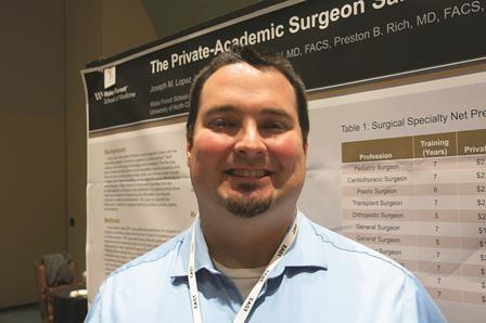
Methotrexate coverage woes
Some things make my job much harder than it should be. One of them is prior authorizations.
At the start of every year, insurers release a new formulary, so prior authorizations are often required, even drugs that patients have been on for a long time. Everyday I spend anywhere from 10 to 30 minutes filling out prior authorization paperwork.
Sometimes, it’s not too difficult. If someone is getting a biologic for the first time, it usually means they’ve had an inadequate response to methotrexate. The form makes sure we’ve checked the patient’s TB status. Sometimes, it asks about a history of heart failure. These are reasonable questions designed to protect the patient.
Frequently, though, the questionnaires are more onerous, and for less altruistic motives. A prior authorization for celecoxib (Celebrex) requires that I list at least two other prescription NSAIDs that the patient has been on. I can guarantee that I do not know that information off the top of my head, so this requires some digging into the chart. If it’s a patient whose care I’ve assumed from someone else, or whose chart is several volumes thick, I won’t even know where to begin. Even worse is the pregabalin (Lyrica) prior authorization that asks about previous use of tricyclic antidepressants, cyclobenzaprine, SSRIs, gabapentin. When were they on these agents and for how long? What was the outcome of each failed medication?
These two examples do not ensure patient safety. They simply go directly to what the insurer’s bottom line is. They benefit no one but the insurer, and create lots of problems for everyone who has to abide by the insurer’s rules.
The worst of it, this year, has to be prior authorization for injectable methotrexate. I have recently run into a lot of problems with this one. For one patient who had been on weekly adalimumab (Humira) and subcutaneous methotrexate, I was asked to justify the combination with a journal article or two, because mechanistically the insurers reject methotrexate as having any role in therapy for patients already on weekly adalimumab, antidrug antibodies notwithstanding. I had inherited this patient from another rheumatologist, and she’d been on this regimen for over a decade; it was infuriating and frustrating to be asked to justify a regimen that the patient had not had problems with for the previous 10 years and that was working fine for her.
For another patient, I was asked to fax a letter to the pharmacy stating why the drug was necessary. Then I was asked to fax another letter to the insurer’s benefits manager. A day later, I was informed that there was no process of appeal for medications that were not on formulary. None whatsoever. The letters that they had asked me to write had no bearing and meant nothing to them. This was the very definition of a complete waste of time.
If I wanted the patient to be on subcutaneous methotrexate, the patient would have to pay out of pocket for the drug. I got on the phone with someone whose job was to repeatedly tell me that I had no options. She couldn’t direct me to anyone higher up than her. I understand that this automaton would have no intelligent answers for me, but I asked rhetorically if etanercept would be covered. She said yes. I then asked rhetorically if she knew how much more the biologic agent would cost the insurance company. I cut her off as she was about to look up the price of the drug.
I can think of hundreds of ways in which I could make better use of my time instead of playing a game I am forced to play against my will, a game that is shrouded in layers of bureaucracy over which I have very little control and has no benefit to my patients. There are days when the game is enough to make me want to throw in the towel.
Dr. Chan practices rheumatology in Pawtucket, R.I.
Some things make my job much harder than it should be. One of them is prior authorizations.
At the start of every year, insurers release a new formulary, so prior authorizations are often required, even drugs that patients have been on for a long time. Everyday I spend anywhere from 10 to 30 minutes filling out prior authorization paperwork.
Sometimes, it’s not too difficult. If someone is getting a biologic for the first time, it usually means they’ve had an inadequate response to methotrexate. The form makes sure we’ve checked the patient’s TB status. Sometimes, it asks about a history of heart failure. These are reasonable questions designed to protect the patient.
Frequently, though, the questionnaires are more onerous, and for less altruistic motives. A prior authorization for celecoxib (Celebrex) requires that I list at least two other prescription NSAIDs that the patient has been on. I can guarantee that I do not know that information off the top of my head, so this requires some digging into the chart. If it’s a patient whose care I’ve assumed from someone else, or whose chart is several volumes thick, I won’t even know where to begin. Even worse is the pregabalin (Lyrica) prior authorization that asks about previous use of tricyclic antidepressants, cyclobenzaprine, SSRIs, gabapentin. When were they on these agents and for how long? What was the outcome of each failed medication?
These two examples do not ensure patient safety. They simply go directly to what the insurer’s bottom line is. They benefit no one but the insurer, and create lots of problems for everyone who has to abide by the insurer’s rules.
The worst of it, this year, has to be prior authorization for injectable methotrexate. I have recently run into a lot of problems with this one. For one patient who had been on weekly adalimumab (Humira) and subcutaneous methotrexate, I was asked to justify the combination with a journal article or two, because mechanistically the insurers reject methotrexate as having any role in therapy for patients already on weekly adalimumab, antidrug antibodies notwithstanding. I had inherited this patient from another rheumatologist, and she’d been on this regimen for over a decade; it was infuriating and frustrating to be asked to justify a regimen that the patient had not had problems with for the previous 10 years and that was working fine for her.
For another patient, I was asked to fax a letter to the pharmacy stating why the drug was necessary. Then I was asked to fax another letter to the insurer’s benefits manager. A day later, I was informed that there was no process of appeal for medications that were not on formulary. None whatsoever. The letters that they had asked me to write had no bearing and meant nothing to them. This was the very definition of a complete waste of time.
If I wanted the patient to be on subcutaneous methotrexate, the patient would have to pay out of pocket for the drug. I got on the phone with someone whose job was to repeatedly tell me that I had no options. She couldn’t direct me to anyone higher up than her. I understand that this automaton would have no intelligent answers for me, but I asked rhetorically if etanercept would be covered. She said yes. I then asked rhetorically if she knew how much more the biologic agent would cost the insurance company. I cut her off as she was about to look up the price of the drug.
I can think of hundreds of ways in which I could make better use of my time instead of playing a game I am forced to play against my will, a game that is shrouded in layers of bureaucracy over which I have very little control and has no benefit to my patients. There are days when the game is enough to make me want to throw in the towel.
Dr. Chan practices rheumatology in Pawtucket, R.I.
Some things make my job much harder than it should be. One of them is prior authorizations.
At the start of every year, insurers release a new formulary, so prior authorizations are often required, even drugs that patients have been on for a long time. Everyday I spend anywhere from 10 to 30 minutes filling out prior authorization paperwork.
Sometimes, it’s not too difficult. If someone is getting a biologic for the first time, it usually means they’ve had an inadequate response to methotrexate. The form makes sure we’ve checked the patient’s TB status. Sometimes, it asks about a history of heart failure. These are reasonable questions designed to protect the patient.
Frequently, though, the questionnaires are more onerous, and for less altruistic motives. A prior authorization for celecoxib (Celebrex) requires that I list at least two other prescription NSAIDs that the patient has been on. I can guarantee that I do not know that information off the top of my head, so this requires some digging into the chart. If it’s a patient whose care I’ve assumed from someone else, or whose chart is several volumes thick, I won’t even know where to begin. Even worse is the pregabalin (Lyrica) prior authorization that asks about previous use of tricyclic antidepressants, cyclobenzaprine, SSRIs, gabapentin. When were they on these agents and for how long? What was the outcome of each failed medication?
These two examples do not ensure patient safety. They simply go directly to what the insurer’s bottom line is. They benefit no one but the insurer, and create lots of problems for everyone who has to abide by the insurer’s rules.
The worst of it, this year, has to be prior authorization for injectable methotrexate. I have recently run into a lot of problems with this one. For one patient who had been on weekly adalimumab (Humira) and subcutaneous methotrexate, I was asked to justify the combination with a journal article or two, because mechanistically the insurers reject methotrexate as having any role in therapy for patients already on weekly adalimumab, antidrug antibodies notwithstanding. I had inherited this patient from another rheumatologist, and she’d been on this regimen for over a decade; it was infuriating and frustrating to be asked to justify a regimen that the patient had not had problems with for the previous 10 years and that was working fine for her.
For another patient, I was asked to fax a letter to the pharmacy stating why the drug was necessary. Then I was asked to fax another letter to the insurer’s benefits manager. A day later, I was informed that there was no process of appeal for medications that were not on formulary. None whatsoever. The letters that they had asked me to write had no bearing and meant nothing to them. This was the very definition of a complete waste of time.
If I wanted the patient to be on subcutaneous methotrexate, the patient would have to pay out of pocket for the drug. I got on the phone with someone whose job was to repeatedly tell me that I had no options. She couldn’t direct me to anyone higher up than her. I understand that this automaton would have no intelligent answers for me, but I asked rhetorically if etanercept would be covered. She said yes. I then asked rhetorically if she knew how much more the biologic agent would cost the insurance company. I cut her off as she was about to look up the price of the drug.
I can think of hundreds of ways in which I could make better use of my time instead of playing a game I am forced to play against my will, a game that is shrouded in layers of bureaucracy over which I have very little control and has no benefit to my patients. There are days when the game is enough to make me want to throw in the towel.
Dr. Chan practices rheumatology in Pawtucket, R.I.

Guideline: Screening, assessment, and care of anxiety and depression in adults with cancer
A Canadian guideline on screening, assessment, and care of anxiety and depressive symptoms in adults with cancer has been adapted for the United States by the American Society of Clinical Oncology.
It is recommended that all patients with cancer be evaluated for symptoms of depression and anxiety at periodic times across the trajectory of care. Assessment should be performed using validated, published measures and procedures. Depending on levels of symptoms and supplementary information, differing treatment pathways are recommended. Failure to identify and treat anxiety and depression increases the risk for poor quality of life and potential disease-related morbidity and mortality, according to ASCO.
The full guideline can be found on the Agency for Healthcare Research and Quality website.
A Canadian guideline on screening, assessment, and care of anxiety and depressive symptoms in adults with cancer has been adapted for the United States by the American Society of Clinical Oncology.
It is recommended that all patients with cancer be evaluated for symptoms of depression and anxiety at periodic times across the trajectory of care. Assessment should be performed using validated, published measures and procedures. Depending on levels of symptoms and supplementary information, differing treatment pathways are recommended. Failure to identify and treat anxiety and depression increases the risk for poor quality of life and potential disease-related morbidity and mortality, according to ASCO.
The full guideline can be found on the Agency for Healthcare Research and Quality website.
A Canadian guideline on screening, assessment, and care of anxiety and depressive symptoms in adults with cancer has been adapted for the United States by the American Society of Clinical Oncology.
It is recommended that all patients with cancer be evaluated for symptoms of depression and anxiety at periodic times across the trajectory of care. Assessment should be performed using validated, published measures and procedures. Depending on levels of symptoms and supplementary information, differing treatment pathways are recommended. Failure to identify and treat anxiety and depression increases the risk for poor quality of life and potential disease-related morbidity and mortality, according to ASCO.
The full guideline can be found on the Agency for Healthcare Research and Quality website.