User login
What’s on tap at ASH 2015
FROM A TELECONFERENCE – The American Society of Hematology’s (ASH) 57th annual meeting in Orlando is chock-full of much-anticipated results in cancer immunotherapies such as CAR T cell therapies and checkpoint inhibitors, advances in sickle cell disease, and practical advice on managing the latest drugs in the clinic, ASH officials said in a teleconference. Here are some of the day-by-day picks selected by ASH president Dr. David Williams and ASH secretary Dr. Stephanie J. Lee, who gave their recommendations during a conference call for the press. Meeting abstracts are now available online.
Saturday, Dec. 5
Clinical applications of newly approved drugs
The popular special education session on clinical applications of newly approved drugs returns on Saturday, Dec. 5 at 9:30 a.m., with didactic presentations that address issues clinicians may face such as drug-drug interactions, side effects, and adverse events. The three drugs to be discussed this year are: idarucizumab (Praxbind), the first specific reversal agent approved for dabigatran reversal; blinatumomab (Blincyto), approved for second-line treatment of Philadelphia chromosomenegative acute lymphoblastic leukemia; and the histone deacetylase (HDAC) inhibitor panobinostat (Farydak), approved for the treatment of multiple myeloma.
Adoptive immunotherapy
One presentation to look out for next month is abstract 99at 12:30 p.m. on Saturday, Dec. 5 in the adoptive immunotherapy session, Dr. Williams told reporters. The chimeric antigen receptor (CAR)-T-cell approach has relied on genetically engineering the patient’s own T cells to rev up the immune system. This group’s approach is to treat B-cell malignancies after allogeneic hematopoietic stem cell transplantation using a single infusion of anti-CD19 CAR-T cells from the patient’s transplant donor.
Eight of 20 patients treated with this strategy achieved remission, including six complete remissions and two partial remissions. Importantly, none of these patients developed acute graft-versus-host disease, a potential consequence of using allogeneic rather than autologous T cells, he said. The authors also noted that patients who responded and went into remission were marked by higher numbers of these infused CAR-T cells in their circulation, suggesting a biomarker of response.
Checkpoint, please?
Immunotherapy is a “very hot area,” so ASH has put together a special session at 4 p.m. Saturday called “Checkpoint, Please?” Dr. Williams said. Topics include the role of programmed death (PD)-1 and PD-ligand 1 in acute and chronic graft-versus-host disease, checkpoint blockade with neoantigen cancer vaccines, and insights into the mechanisms of action of anti-CTLA-4 (cytotoxic T-lymphocyte–associated protein 4) antibody therapy.
Sunday, Dec. 6
Precision medicine
Sunday’s plenary scientific session will include several noteworthy personalized medicine abstracts featuring emerging therapies targeted to specific genetic subtypes, Dr. Lee, from the University of Washington, Seattle, said.
Plenary abstract 6 is a large, multinational study looking at whether adding the multikinase inhibitor midostaurin to standard induction therapy and carried through 1 year of maintenance would improve outcomes in newly diagnosed acute myeloid leukemia with FLT3 mutations. Patients with these deleterious mutations do enter remission with chemotherapy, but often relapse.
Overall and event-free survival were better at 5 years by about 7% to 8% in the experimental arm using midostaurin, she said. Caveats are that complete response rates were similar in both arms and lower than reported in other trials.
“Because we know that patients with this FLT3 mutation have a very poor prognosis with standard chemotherapy, more than half of the patients in this trial received an allogeneic transplant,” Dr. Lee noted. “But the abstract does say that the results are similar if you censor at the time of the transplant.”
In this same vein of precision medicine is plenary abstract 1, testing whether adding rituximab to standard chemotherapy improves outcomes in adults with CD-20–positive, Philadelphia chromosome–negative, B-cell precursor acute lymphoblastic leukemia (ALL). Rituximab (Rituxan) binds to CD-20, which is found in about 30% to 50% of adult B-cell ALL, she said.
At 2 years, patients treated with rituximab had longer event-free survival than controls (65% vs. 52%; P = .038), but similar overall survival (71% vs. 64%; P = .09), according to the abstract. The rituximab arm also received more allogeneic transplants, but again, after censoring the data, the abstract states that both event-free and overall survival were longer with rituximab, Dr. Lee said.
Sickle cell anemia
Sunday’s plenary session will also feature the very important TWiTCH (TCD with Transfusions Changing to Hydroxyurea) study evaluating hydroxyurea therapy as an alternative to chronic blood transfusions to prevent stroke. Stroke is one of the most dreaded complications of sickle cell disease, occurring in up to 10% of children, Dr. Williams said. Though transfusions are effective, they have to be continued indefinitely and lead to iron overload. Hydroxyurea increases the amount of fetal hemoglobin and fetal red blood cells and has become a standard therapy to attenuate the complications of sickle cell.
The phase III noninferiority study, which used Transcranial Doppler (TCD) screening to identify children at elevated risk for stroke, showed that hydroxyurea “was as good as current therapy with red cell transfusions and there was some indication, although not significant, that it might even be superior in lowering the TCD levels,” Dr. Williams said. An added benefit of the hydroxyurea was that it improved the patients’ iron overload status. There were no strokes in either group.
Sunday’s abstract 202 is another presentation “that I’m sure will get a lot of attention,” Dr. Williams said. It offers updated details on outcomes from patients with sickle cell disease (SCD) treated with a novel gene therapy transduced with the LentiGlobin BB305 (Bluebird Bio) lentiviral vector. Patients with beta thalassemia major have remained transfusion-independent for more than a year after this treatment, with results now available from four patients with SCD. One patient with a severe phenotype has had no sickle cell complications and has been able to stop his transfusion therapy, while two of the other four patients are also transfusion-independent.
“This is an early study showing what appears to be efficacy of the gene therapy approach not in thalassemia, but in sickle cell disease,” Dr. Williams said, noting that abstract 3233 will also feature results using LentiGlobin gene therapy in severe SCD.
ASH/EHA joint symposium
Also noteworthy is a special joint ASH/European Hematology Association symposium looking at how well genomic data are being incorporated into practice in the U.S. and Europe.
Monday, Dec. 7
ASH/FDA joint symposium
A joint ASH/FDA symposium on late-breaking drug approvals is new this year and features drugs that gained approval in November 2015. FDA product-reviewers will discuss safety and efficacy issues in the clinical approval trials and toxicity studies, while clinicians will share their experiences in the real-world use of these drugs.
“This is information that is really going to be very hot off the press and presented in conjunction with the FDA,” Dr. Lee said.
Dr. Williams reported research funding from Bluebird Bio. Dr. Lee reported having no conflicts of interest.
Starting ACS patients on varenicline in the hospital boosted smoking quit rates
ORLANDO – Starting varenicline in smokers while hospitalized for an acute coronary syndrome resulted in substantially higher smoking abstinence rates than with placebo at all time points through 6 months of follow-up in the double-blind, randomized EVITA trial.
“The ACS population is typically older, they’re long-term smokers, and they come into the hospital with a life-threatening condition. Their family is all around them. They’ve had angioplasty or CABG [coronary artery bypass graft] surgery. So they have pressure on them to stop smoking. This is a teachable moment, a window of opportunity. The public health benefit for smoking cessation in this population is huge. You can cut their risk of death and significant morbidity in half if you can get them to stop,” Dr. Mark J. Eisenberg said in presenting the EVITA results at the American Heart Association scientific sessions.

“To our knowledge, this is the highest-risk population that’s been exposed to varenicline,” said Dr. Eisenberg, professor of medicine at McGill University and director of the cardiovascular health services research program at Jewish General Hospital in Montreal.
The primary study endpoint was continuous self-reported abstinence since baseline backed by biochemical confirmation in the form of an exhaled carbon monoxide level of 10 ppm or less at week 24 as well as at all the earlier follow-up visits. The rate was 47.3% in the varenicline group, compared with 32.5% in placebo-treated controls. That placebo response rate is in line with numerous prior studies that have shown that less than one-third of smokers with ACS remain abstinent after leaving the hospital.
“Most cardiologists would say, ‘All my patients stop smoking.’ But in the clinic if you look in the patients’ pockets, you find a pack of cigarettes. They stop smoking while in hospital, but as soon as they’re discharged, the relapse rate is almost immediate. Most patients are smoking the day they get out of hospital,” according to Dr. Eisenberg.
In EVITA, the number-needed-to-treat with varenicline for 12 weeks in order to produce 1 extra nonsmoker at 6 months was 6.8 patients.
The secondary endpoint of at least a 50% reduction in the number of cigarettes per day from baseline to 6 months was met by 67.4% of the varenicline group and 55.6% of controls, with a number-needed-to-treat of 8.5, he continued.
No safety issues emerged in the study, although as Dr. Eisenberg noted, EVITA wasn’t sufficiently powered to look at safety. The only side effect more common in varenicline-treated patients was abnormal dreams, with a 12-week incidence of 15%, threefold higher than in controls, a phenomenon seen in other, larger varenicline studies as well.
The EVITA investigators plan to follow participants out to 12 months. “If we see someone who at 1 year post MI is still smoking, maybe it’s time to go after them again, perhaps with another medication or behavioral therapy,” he said.
There are no randomized clinical trials demonstrating that starting nicotine patches or bupropion in the hospital is effective for smoking cessation in the ACS population, according to the cardiologist.
Dr. Eisenberg predicted this study will change clinical practice. In much the same way physicians now routinely start ACS patients on a statin, beta-blocker, and aspirin before they leave the hospital, physicians will capitalize on this opportunity to help ACS patients quit smoking as well, he said.
“The use of varenicline in ACS patients before they leave the hospital is a very important step forward. Cardiologists are increasingly comfortable with the idea of starting secondary prevention medications in the hospital, and there’s very little more important for a person with heart disease who smokes cigarettes than to help them quit smoking. It’s probably the No. 1 priority. So evidence that we can start a medication in the hospital and get more people who smoke cigarettes to quit smoking is definitely game changing, I think,” said Dr. Goff, professor of epidemiology and dean of the Colorado School of Public Health in Aurora.
Dr. Eisenberg reported receiving funding from Pfizer to perform the EVITA trial. He has also gotten honoraria from the company for providing continuing medical education talks on smoking cessation.
ORLANDO – Starting varenicline in smokers while hospitalized for an acute coronary syndrome resulted in substantially higher smoking abstinence rates than with placebo at all time points through 6 months of follow-up in the double-blind, randomized EVITA trial.
“The ACS population is typically older, they’re long-term smokers, and they come into the hospital with a life-threatening condition. Their family is all around them. They’ve had angioplasty or CABG [coronary artery bypass graft] surgery. So they have pressure on them to stop smoking. This is a teachable moment, a window of opportunity. The public health benefit for smoking cessation in this population is huge. You can cut their risk of death and significant morbidity in half if you can get them to stop,” Dr. Mark J. Eisenberg said in presenting the EVITA results at the American Heart Association scientific sessions.
“To our knowledge, this is the highest-risk population that’s been exposed to varenicline,” said Dr. Eisenberg, professor of medicine at McGill University and director of the cardiovascular health services research program at Jewish General Hospital in Montreal.
The primary study endpoint was continuous self-reported abstinence since baseline backed by biochemical confirmation in the form of an exhaled carbon monoxide level of 10 ppm or less at week 24 as well as at all the earlier follow-up visits. The rate was 47.3% in the varenicline group, compared with 32.5% in placebo-treated controls. That placebo response rate is in line with numerous prior studies that have shown that less than one-third of smokers with ACS remain abstinent after leaving the hospital.
“Most cardiologists would say, ‘All my patients stop smoking.’ But in the clinic if you look in the patients’ pockets, you find a pack of cigarettes. They stop smoking while in hospital, but as soon as they’re discharged, the relapse rate is almost immediate. Most patients are smoking the day they get out of hospital,” according to Dr. Eisenberg.
In EVITA, the number-needed-to-treat with varenicline for 12 weeks in order to produce 1 extra nonsmoker at 6 months was 6.8 patients.
The secondary endpoint of at least a 50% reduction in the number of cigarettes per day from baseline to 6 months was met by 67.4% of the varenicline group and 55.6% of controls, with a number-needed-to-treat of 8.5, he continued.
No safety issues emerged in the study, although as Dr. Eisenberg noted, EVITA wasn’t sufficiently powered to look at safety. The only side effect more common in varenicline-treated patients was abnormal dreams, with a 12-week incidence of 15%, threefold higher than in controls, a phenomenon seen in other, larger varenicline studies as well.
The EVITA investigators plan to follow participants out to 12 months. “If we see someone who at 1 year post MI is still smoking, maybe it’s time to go after them again, perhaps with another medication or behavioral therapy,” he said.
There are no randomized clinical trials demonstrating that starting nicotine patches or bupropion in the hospital is effective for smoking cessation in the ACS population, according to the cardiologist.
Dr. Eisenberg predicted this study will change clinical practice. In much the same way physicians now routinely start ACS patients on a statin, beta-blocker, and aspirin before they leave the hospital, physicians will capitalize on this opportunity to help ACS patients quit smoking as well, he said.
“The use of varenicline in ACS patients before they leave the hospital is a very important step forward. Cardiologists are increasingly comfortable with the idea of starting secondary prevention medications in the hospital, and there’s very little more important for a person with heart disease who smokes cigarettes than to help them quit smoking. It’s probably the No. 1 priority. So evidence that we can start a medication in the hospital and get more people who smoke cigarettes to quit smoking is definitely game changing, I think,” said Dr. Goff, professor of epidemiology and dean of the Colorado School of Public Health in Aurora.
Dr. Eisenberg reported receiving funding from Pfizer to perform the EVITA trial. He has also gotten honoraria from the company for providing continuing medical education talks on smoking cessation.
AT THE AHA SCIENTIFIC SESSIONS
Key clinical point:
Major finding: The number of smokers who need to be started on varenicline while hospitalized for an acute coronary syndrome in order to produce one extra nonsmoker at 6 months is 6.8.
Data source: EVITA, a double-blind, randomized multicenter trial in 302 smokers hospitalized for ACS who were prospectively followed for 6 months.
Disclosures: The EVITA study was funded by Pfizer. The presenter reported receiving a grant from the company to conduct the trial, as well as honoraria for giving continuing medical education talks on smoking cessation.
Younger Type 2 Diabetics Face Greater Mortality Risks
NEW YORK - People with type 2 diabetes are 15 percent more likely to die from any cause and 14 percent more likely to die from a cardiovascular cause than non-diabetics at any given time, according to data from several Swedish registries.
The rates are significantly lower than previous estimates. Fifteen years ago, research was suggesting that having diabetes doubled the risk of premature death.
But the new study also found that the risk was dramatically elevated among people whose type 2 diabetes appeared by age 54. The worse their glycemic control and the more evidence of renal problems, the higher the risk.
In contrast, by age 75, type 2 diabetes posed little additional risk for people with good control and no kidney issues, according to the results.
"The overall increased risk of 15 percent among type 2 diabetics in general is a very low figure that has not been found in earlier type 2 diabetes studies," coauthor Dr. Marcus Lind of Uddevalla Hospital said in a telephone interview.
"The other thing that was interesting is that when we looked at patients with good glycemic control and no renal complications, if they were 75 years age, they had a lower risk than those in the general population. That hasn't been shown before," he said.
"What we are seeing is, if you are younger, aggressive management makes a difference," said Dr. Robert Ratner, chief scientific and medical officer of the American Diabetes Association, who was not involved in the research. At age 75, "you don't have to worry about it as much."
The study, published in the October 29 New England Journal of Medicine, is the largest to date to look at premature death in general - and death from cardiovascular causes in particular - among people with type 2 diabetes.
It compared more than 435,000 diabetics who were followed for a mean of 4.6 years with more than 2 million matched controls who were tracked for a mean of 4.8 years. The diabetics had had glucose problems for an average of 5.7 years.
In terms of actual death rates, cardiovascular mortality during the study period was 7.9 percent for diabetics versus 6.1 percent for controls (adjusted hazard ratio, 1.14; 95 percent confidence interval: 1.13-1.15). The respective rates for death from any cause were 17.7 percent and 14.5 percent (aHR, 1.15; 95 percent CI: 1.14-1.16).
For patients under age 55 with glycated hemoglobin levels below 7.0 percent, the risk of death from any cause nearly doubled (aHR, 1.92; 95 percent CI: 1.75-2.11).
"Those who are younger than 55, those who have target glycemic control and no signs of any renal complications, they had a clearly-elevated risk," said Dr. Lind.
But for people over 75, the hazard was actually 5 percent lower than it was for people without diabetes (aHR, 0.95; 95 percent CI: 0.94-0.96).
When the research team factored in people with normoalbuminuria, the risks were slightly mitigated.
Heart attack was the most common cause of death among diabetics.
When glycated hemoglobin levels were at 9.7 percent and higher for people below age 55, the hazard of death from any cause more than quadrupled. The hazard of death from cardiovascular causes rose more than five-fold.
Once again, the danger was far less extreme for people over 75, the researchers found.
"Excess mortality in type 2 diabetes was substantially higher with worsening glycemic control, severe renal complications, impaired renal function, and younger age," they concluded.
Renal function is a key element, Dr. Ratner said.
The study "reinforces the importance of early aggressive management of diabetes in order to prevent premature death and the fact is that the prevention of renal disease is probably the most potent thing we can do to reduce cardiovascular events," he said.
Dr. Lind said the risk may appear lower in the elderly because older people with diabetes are more likely to be getting aggressive treatment for their high blood pressure and high lipid levels, therapy that other people who also have hypertension and high cholesterol levels might not be receiving.
"I think that's the reason the rates are a bit lower" for seniors, he said.
Dr. Ratner said he believes the data for older diabetics simply reflects the fact that "you're seeing the survival cohort. They've made it past the difficult time."
We're all going to die, he said. "The issue is when does it happen? With diabetes, it's happening years ahead of time - in their 50s and early 60s, more so than when they reach 75. The younger you are, the greater that risk. That's when aggressive therapy should be given."
Timing of irradiation affects RCCs
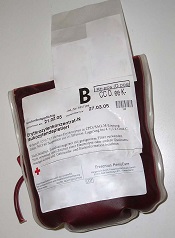
ANAHEIM, CA—The timing of gamma irradiation influences in vitro characteristics of red cell concentrates (RCCs), according to a new study.
The research showed that RCCs sustain more damage the longer they are stored prior to gamma irradiation and the longer they are stored after irradiation.
However, RCCs from female donors appeared to be less susceptible to irradiation injury, and the additive solution used seemed to affect the level of injury as well.
Dirk de Korte, PhD, of Sanquin Blood Bank in Amsterdam, Netherlands, presented these results at the 2015 AABB Annual Meeting (abstract S72-040A).
The study included 7 centers, each of which used its standard RCCs. Five centers used SAGM as additive solution, 1 used AS-3, and 1 used PAGGSM. Two centers used whole blood filtration to prepare leukoreduced RCCs, and 5 centers used buffy coat removal and RCC filtration.
Each center produced 4 pools of 7 RCCs, 2 male and 2 female pools. The units were stored for 43 days, and 1 pool was gamma-irradiated every week.
The researchers also performed weekly sampling to assess in vitro quality parameters. They took an extra sample 24 hours after irradiation and 72 hours after irradiation.
The team found that the age of RCCs at the time of irradiation influenced the rate of increase of hemolysis and the absolute level of hemolysis (P<0.0001).
Hemolysis was higher in units irradiated early and then stored. And the rate of change of hemolysis increased if RCCs were stored for longer before irradiation.
The researchers also found that the age of RCCs at the time of irradiation influenced the rate of increase of potassium and the absolute level of potassium (P<0.0001).
The rate of change of potassium decreased if RCCs were stored longer before irradiation, as potassium was already partly released if the cells were stored longer. Within 7 days of irradiation, potassium levels exceeded those observed in control cells stored for 43 days.
Hemolysis and potassium levels also appeared to be affected by donor sex and the additive solution used.
Hemolysis was lower in RCCs from female donors (P=0.045) and in cells exposed to AS-3 or PAGGSM rather than SAGM (P=0.0597).
Potassium release was lower in cells from female donors (P=0.0032) and in cells exposed to AS-3 rather than PAGGSM or SAGM (P=0.0391).
“This study shows or confirms interesting differences between red cells from males and females, and, of course, we are interested in the underlying mechanism,” Dr de Korte said.
He also said the results of this study will be used to formulate guidance on the maximal pre- and post-irradiation storage time for RCCs with respect to either acceptable hemolysis or potassium release.
Dr de Korte said that, if hemolysis is used as guidance, irradiation should be performed within the first 28 days of storage, and the cells should be used within these 28 days.
If potassium is used as guidance, cells should be used within 7 days of irradiation if the irradiation occurs during the first 10 to 14 days of storage, or the cells should be used immediately after irradiation if irradiation takes place later during storage. ![]()
CCSs have increased risk of autoimmune diseases

Photo by Bill Branson
Childhood cancer survivors (CCSs) have an increased risk of developing autoimmune diseases, according to research published in the Annals of the Rheumatic Diseases.
CCSs had a significantly increased risk for 11 of 33 autoimmune diseases studied, and the highest risk was observed for autoimmune hemolytic anemia.
Survivors of leukemia and Hodgkin lymphoma were among those CCSs at the greatest risk of developing
an autoimmune disease.
Anna Sällfors Holmqvist, MD, of Lund University in Sweden, and her colleagues conducted this research.
They used national cancer registry data from Denmark, Iceland, and Sweden, spanning the period from the 1940s to 2008, to identify subjects who had cancer as a child.
The researchers identified 20,361 adults who had cancer before the age of 20 and survived for at least a year. These subjects were matched (for age, gender, and country of birth) to 125,794 individuals who had not had cancer as children.
The health of all participants was tracked for an average of 15 to 19 years. The researchers used hospital records to determine the difference between the expected and excess number of autoimmune diseases, expressed as a standardized hospitalization rate ratio (SHRR).
In all, 724 (3.6%) CCSs had at least 1 episode of hospital treatment for any autoimmune condition, but only 516 would have been expected.
So CCSs had an SHRR for autoimmune diseases of 1.4. This corresponds to an absolute excess risk of 67 per 100,000 person-years.
SHRRs were significantly higher for 11 autoimmune diseases, including autoimmune hemolytic anemia (16.3), Addison’s disease (13.9), polyarteritis nodosa (5.8), chronic rheumatic heart disease (4.5), localized scleroderma (3.6), idiopathic thrombocytopenia (3.4), Hashimoto’s thyroiditis (3.1), pernicious anemia (2.7), sarcoidosis (2.2), Sjögren’s syndrome (2.0), and insulin-dependent diabetes mellitus (1.6).
SHRRs for any autoimmune disease were significantly increased for survivors of leukemia (1.6), Hodgkin lymphoma (1.6), renal tumors (1.6), and central nervous system neoplasms (1.4).
The excess risk for all autoimmune diseases combined peaked in the first 5 years after a cancer diagnosis. However, the risk persisted for up to 30 years later for most conditions and up to 50 years later for some conditions.
The researchers said the peak observed in the first 5 years may be a consequence of closer medical monitoring during this time period.
They added that a possible explanation for these findings is that persistent immune abnormalities after chemotherapy predispose CCSs to develop autoantibodies, which are central to the pathogenesis of many autoimmune diseases.
The team said the cancer itself, immunosuppressive treatment, and the increased number and types of infections during cancer treatment could alter the immune system as a whole and result in immunologically different antigens, leading to the production of autoantibodies. ![]()
Relapses ‘critical’ for sustained malaria transmission
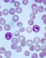
Plasmodium vivax
Image by Mae Melvin
New research indicates that most childhood malaria infections in Papua New Guinea (PNG) are the result of relapsed, not new, infections.
The data suggest that, among children ages 5 to 10 living in a malaria-hyperendemic region of PNG, relapses cause about 4 of every 5 Plasmodium vivax infections and 3 of every 5 Plasmodium ovale infections.
Investigators therefore concluded that relapses are important for sustaining malaria transmission in PNG.
The team reported their findings in PLOS Medicine.
“Our research has shown that one of the biggest problems in realizing malaria eradication is relapsing P vivax infections, which are critical for sustained transmission in the region,” said study author Leanne Robinson, PhD, of the Walter and Eliza Hall Institute of Medical Research in Parkville, Victoria, Australia.
“P vivax parasites are able to hide in the liver for long periods of time before ‘reawakening’ to cause disease and continue the transmission cycle. Mass drug administration that includes a drug that kills parasites in the liver is likely to be a highly effective strategy for eliminating malaria in PNG.”
To investigate this possibility, Dr Robinson and her colleagues analyzed 524 children, ages 5 to 10, living in a region of PNG where Plasmodium falciparum and P vivax are hyperendemic.
Roughly half the children (n=261) received an antimalarial treatment regimen that targets both blood-stage and liver-stage parasites (3 days of chloroquine, 3 days of artemether-lumefantrine, and 20 days of primaquine).
The other half (n=263) received antimalarial treatment targeting only blood-stage parasites (3 days of chloroquine, 3 days of artemether-lumefantrine, and 20 days of placebo).
The subjects were followed for 8 months. Compared to children in the placebo arm, those in the primaquine arm had a reduced risk of having at least 1 P vivax or P ovale infection during the follow-up period. The hazard ratio was 0.18 for P vivax (P<0.001) and 0.31 for P ovale (P=0.011).
“Children treated with drugs that targeted the liver and blood stages of infection had 80% fewer malaria infections than those treated with drugs that only targeted the blood stage of infection,” Dr Robinson said.
Children in the primaquine arm also had a reduced risk of having at least 1 clinical P vivax episode. The hazard ratio was 0.25 (P=0.002).
In addition, primaquine reduced the molecular force of P vivax blood-stage infection in the first 3 months of follow-up. The incidence rate ratio was 0.21 (P<0.001).
And children who received primaquine were less likely to carry P vivax gametocytes than children who received placebo, with an incidence rate ratio of 0.27 (P<0.001).
To build upon these findings, the investigators fed the trial data into a mathematical transmission model.
The model predicted that a mass drug administration program using blood-stage treatment alone would have only a transient effect on P vivax transmission levels. But a mass drug administration program that includes blood- and liver-stage treatment would be an effective strategy for P vivax elimination.
“We need a better way of identifying children who are chronically infected with malaria so that they can be treated,” said study author Ivo Mueller, PhD, of the Walter and Eliza Hall Institute.
“It is the only way to stop the malaria transmission cycle in PNG and is likely to be the case for eliminating malaria in other parts of the Asia-Pacific and Americas.”
Dr Mueller and an international team of collaborators are currently developing a test that identifies people with dormant malaria parasites in their liver. ![]()
Predicting risk of VTE in patients with leg casts

Photo by Nikki McLeod
Researchers say they have developed prediction models and a scoring system that can help identify a high risk of venous thromboembolism (VTE) in a patient who has a plaster leg cast.
The group utilized data from 3 large studies to develop and validate 3 prediction models and a risk score, known as the L-TRiP (cast) score, which are intended to help doctors decide whether patients with casts require thromboprophylaxis.
The models and the scoring system all predicted VTE with fairly high accuracy, but the researchers said this study had 2 major limitations.
First, due to the retrospective nature of the study, the team was unable to determine which of the patients may have received thromboprophylaxis. And second, blood samples were taken 3 months after VTE.
Banne Nemeth, MD, of Leiden University Medical Center in Leiden, Netherlands, and colleagues conducted this research and reported the results in PLOS Medicine.
Developing prediction tools
The researchers first analyzed data from the MEGA study, a population-based, case-control study in the Netherlands that included 4446 patients with VTE and 6118 controls.
From these data, the team attempted to identify biomarkers, genetic factors, and environmental factors predictive of VTE and build 3 different prediction models. Their full prediction model consists of 32 predictors for VTE, including 3 genetic factors and 6 biomarkers.
Their restricted model consists of 11 predictors for VTE, including 2 genetic factors and 1 biomarker. And their clinical model consists of 14 environmental predictors that can all be determined without drawing blood or performing any laboratory assays.
The predictors in the clinical model were given numerical values that, when summed, produce the L-TRiP (cast) score (Leiden–Thrombosis Risk Prediction for patients with cast immobilization score) to stratify patients into high- or low-risk groups for VTE.
To determine the discriminatory power of the models and the score in patients with casts, the researchers calculated the area under the curve (AUC) for each tool.
The AUC was 0.85 for the full model, 0.84 for the restricted model, 0.77 for the clinical model, and 0.76 for the L-TRiP (cast) score.
Validating the tools
The researchers validated the prediction models and risk score in 2 independent cohorts, THE-VTE study (784 VTE cases and 523 controls from the Netherlands and the UK) and the Milan study (2117 cases and 2088 controls from Italy).
In THE-VTE study, the AUC was 0.75 for the clinical model and 0.77 for the L-TRiP (cast) score. (There were no data provided for the full model or the restricted model in this cohort.)
In the Milan study, the AUC was 0.93 for the full model, 0.92 for the restricted model, 0.96 for the clinical model, and 0.95 for the L-TRiP (cast) score.
The researchers said these results suggest that genetic and biomarker information may provide added value in predicting VTE.
However, their clinical model (which consists solely of environmental factors) proved only slightly inferior to the full model in the derivation cohort and performed as well or better than the full model in the validation cohorts.
So it is unclear whether genetic and biomarker information will lead to higher accuracy in the prediction algorithm. ![]()
Survivors of childhood cancers at increased risk for autoimmune diseases
Analysis of cancer registry data from Denmark, Iceland, and Sweden over more than 60 years found that survivors of childhood cancer had a 1.4-fold higher risk of autoimmune disease compared with matched controls.
Results showed significantly increased rates of hospital visits for 11 of 33 autoimmune diseases investigated. The most prominent excesses were for insulin-dependent diabetes mellitus, Addison’s disease, and Hashimoto’s thyroiditis, accounting for more than half the total number of excess autoimmune cases (Ann Rheum Dis 2015 Nov 6. doi: 10.1136/annrheumdis-2015-207659). The investigators could not rule out the influence of surveillance bias, given that hospitalization rate ratios were highest in the first 5 years after cancer diagnosis, potentially a consequence of closer surveillance during that period. For most autoimmune diseases, however, the excess risk persisted through the second and third decades after cancer diagnosis.
The Nordic childhood cancer survivor cohort comprised 25,635 individuals diagnosed with cancer before the age of 20 years, from the 1940s-1950s (start of cancer registries in Denmark, Iceland, and Sweden) to 2008. Expected autoimmune disease rates were based on rates of hospital visits for the comparison cohort, which included 128,023 individuals matched for age, sex, and country of the corresponding survivor. The standardized hospitalization rate ratio is the observed number of autoimmune diseases among survivors divided by the expected rate. Childhood malignancies with the most pronounced risk increases were leukemia (standardized hospitalization rate ratio, 1.6), Hodgkin lymphoma (1.6), renal tumors (1.6), and central nervous system neoplasms (1.4).
“Cure is no longer a sufficient goal in childhood cancer care. As the vast majority of these patients survive, attention must be to paid to their long-term quality of life and health challenges,” wrote Dr. Anna Salifors Holmqvist of the department of clinical sciences, pediatric oncology and hematology, Skane University Hospital, Lund University, Sweden.
Increased risks were noted for a wide range of relatively rare autoimmune diseases after treatment for several types of childhood malignancies, and underlying mechanisms should be addressed in future studies, noted Dr. Holmqvist and her colleagues.
FROM ANNALS OF RHEUMATIC DISEASES
Key clinical point: Survivors of childhood cancers, especially leukemia, Hodgkin lymphoma, and renal tumors, had a significantly increased risk of autoimmune disease.
Major finding: Compared with matched controls, survivors had a 1.4-fold increased risk for hospital contact concerning an autoimmune disease.
Data source: The Nordic childhood cancer survivor cohort comprised 25,635 individuals diagnosed with cancer before age 20 years, from the 1940s-1950s (start of cancer registries in Denmark, Iceland, and Sweden) to 2008.
Disclosures: Dr. Holmqvist and coauthors reported having no disclosures.
Preauthorization for medications: Who oversees placement of the hoops?
The letter from the insurance company was addressed to my patient. The two pages of information boiled down to one simple sentence: “After a thorough review, our decision to not cover the medication Provigil (modafinil) is unchanged.” The letter went on to explain that there was no further recourse, and that the medication would not be approved because it was not Food and Drug Administration–approved for the condition my patient had: major depression. If she chose to take it, there would be no reimbursement. In many psychiatric conditions, the FDA-approved options are very limited; for some disorders, there simply are no approved medications, despite the fact that research has shown medications to be helpful. For a medication that now has a generic, there is no reason for the pharmaceutical agency to incur the cost of getting a medication approved by the FDA for a specific use.
Psychiatrists are all familiar with the process of medication preauthorization. Insurance companies require the physician to make an extra effort in order to prescribe certain medications. Often, as in the case of modafinil, the medications are expensive, and one might wonder why a generic medication costs $25 a pill. The name-brand medication, Provigil, costs more than $33 for a single dose. Sometimes, however, a preauthorization process is put in place for very inexpensive medications that might cost only a few dollars a month. Preauthorization requirements waste enormous amounts of physician time and don’t necessarily save money for insurers.
What many physicians don’t realize is that there is no national oversight to this process. In some arenas, the insurers can set the hoops as high as they want and in perpetual motion, thereby making the process a long and miserable adventure – all while the patient suffers.
Nothing about my patient’s depression has been easy to treat. We started working together 8 years ago; her suffering at that time was extreme and her presentation was unusual. I arranged for her admission to a specialty unit in a psychiatric hospital, and she remained under their care for several months – an astounding period of time in an era where the average length of stay is 7 to 10 days. She was discharged – very much improved – on an unusual cocktail of medications.
Over time, we tapered some of the medications because of side effects. Five years ago, her insurance changed and after I answered a 13-question prior authorization form, the medication was denied because she did not have narcolepsy or any of the sleep disorders for which the FDA does approve the use of Provigil. An appeal was denied, and I prescribed stimulants instead. It wasn’t until recently, when the patient’s depression returned full-force, and every medication option I could think of had been tried, that I decided to try modafinil again, especially since I have found it helpful in another patient with a treatment-resistant depression.
I wrote a prescription and heard from the pharmacy that Provigil required preauthorization. I prescribed Nuvigil instead, but that too required preauthorization. A 20-minute phone call resulted in directions on how to find a form and fax a request for the medication. It contained many of the same questions from years ago, and the request was denied.
Several more phone calls led me to a reviewer with the pharmacy benefits company in Nevada, who told me I could find a form on the website, send in an appeal to the insurance company in Iowa, and I would hear within 15 business days if my patient could get the medication. When I said that was not acceptable – the patient was suffering and needed the medication now – I was told I would receive a call from a peer reviewer within 48 hours. A week later, a pharmacist called and asked the exact same questions that I had already answered. When I said that no, I had not ordered polysomnography or multiple sleep latency tests because they were not medically indicated, she denied approval of the medication and said she had no leeway to approve its use if the questions were not answered correctly. I found it curious that this step was called a “peer-to-peer” review. By this point, I had spent about an hour in a two-step process where the second step was exactly the same as the first step. It’s okay; the insurance company doesn’t mind the inefficient use of everyone’s time.
I was told that to file an appeal, I would need more forms off the website, and I was given (at my request) directions on where to find those forms. But first, I needed to get the patient to sign a form to authorize to release information on her behalf. The patient lives a 90-minute drive from my office and does not have a fax machine, and this delayed the process. It remained unclear to me why the patient’s permission was needed when it had not been needed for the first two steps of the process. The authorization was to be mailed or faxed to one location, while the appeal letter was to be mailed (fax or electronic submissions were not options) to another address.
In the appeal letter, I discussed her past history of a lengthy hospitalization, a long list of other medications that had been tried, and a study that documented the efficacy of modafinil as an adjunct agent in the treatment of depression.
As the weeks ticked by, I contacted the chief medical officer of the insurance company and complained; I had spent hours on an arbitrary, repetitive, and inefficient process that was yielding no results. I asked how he sleeps at night.
The response was infuriating: “I know that the appeal is being set up in the queue. I am sorry that this process has been drawn out for you and your patient.” In the queue? Had he ever sat with a sobbing, depressed patient who couldn’t get out of bed all day? A family member was taking off work to stay with her, and her life was on hold. The chief medical officer didn’t give any indication as to how long this queue might be. The family was hurting both emotionally and financially.
I tried one of my U.S. senators. His office replied that preauthorization oversight was not a legislative issue. I spoke with the insurance commission in Maryland where I practice, and I filed a formal complaint with the insurance commissioner in Iowa. I was told it could take up to 45 days for a response, but the next day, I heard back: The plan is funded through a family member’s employer and is not under the jurisdiction of the insurance commissioner. Simply put, the insurance company can set up as many hurdles as they like and take as long as they want to respond, regardless of the patient’s need.
Eight weeks after I wrote the prescription, a copy of the denial letter arrived in my mail. A physician in Iowa has determined that the medication is not medically necessary for a patient he or she had never seen in Maryland. I’ve turned to the American Psychiatric Association. With so many psychiatrists complaining about the burdens of preauthorization, this case would be a good example; one of its attorneys has agreed to write a letter to the Department of Labor. Perhaps that will help, but as of this writing, I have not yet seen that letter. All in all, it has been hours of my time, and lots of waiting, all without regard to the person who could possibly find some relief with a medication that is available to some, but not to her.
Dr. Miller is a coauthor of “Shrink Rap: Three Psychiatrists Explain Their Work” (Baltimore: Johns Hopkins University Press, 2011).
Experimental LOXO-101 induces regression in several hard-to-treat cancers
BOSTON – An experimental agent that targets the byproducts of gene fusions has shown surprising clinical activity against notoriously treatment-refractory cancers in early results from a phase I trial.
“I think that’s what remarkable about this is that we have all these different histologies and many different fusions, but all seem to be having some kind of [response] to this molecule,” said lead investigator Dr. David S. Hong, deputy chair and associate professor in the department of investigational cancer therapeutics at the University of Texas MD Anderson Cancer Center, Houston.
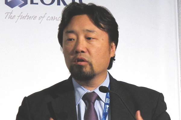
The molecule, LOXO-101 is a selective inhibitor of abnormal TRKA, TRKAB, and TRKC kinases that arise from gene fusions. TRK fusions have been implicated in tumor development in preclinical studies, he reported at the AACR–NCI–EORTC International Conference on Molecular Targets and Cancer Therapeutics.
One patient, a 41-year-old woman with undifferentiated soft tissue sarcoma of the groin that had metastasized to her lungs had rapid and “substantial” tumor and lung-nodule regression that radiographically fell just shy of a complete response, Dr. Hong said.
The drug also induced robust partial responses in a 55-year-old man with a gastrointestinal stromal tumor (GIST) for whom five prior targeted agents had failed, and a 33-year-old man with mammary analogue secretory carcinoma of the salivary gland (MASC) that had progressed on chemotherapy with docetaxel, carboplatin, and 5-fluorauracil.
A fourth patient, a man (age unspecified) who had papillary thyroid cancer with palpable lymphadenopathy, had no palpable nodes at 1-month follow-up, and appears to be responding to the drug, Dr. Hong added.
“This is really a dramatic example, I think, of a targeted therapy that is not histology specific, and this is another area in oncology where it’s new, and we’re just feeling our way,” commented Dr. Lee C. Helman, a sarcoma specialist at the National Cancer Institute in Bethesda, Md., who moderated the briefing where Dr. Hong presented the data.
Tumor promoters
The TRK family of proteins consists of separate tyrosine kinases that, when functioning normally, are important for the development of peripheral central nervous system processes such as pain and thermoregulation, Dr. Hong explained.
“The NTRK 1, 2, and 3 genes which encode for the TRK proteins are subject to these gene rearrangements and fusions, and the resulting TRK gene fusions may become erroneously expressed, and the kinase domain constitutively activated,” he said.
Gene fusions resulting in constitutively active TRKA, B, and C kinases can occur in the range of about 2%-25% in a wide variety of tumors, including adenocarcinomas of the lung, gliomas, thyroid tumors, head and neck cancers, sarcomas, and other types. These fusions have also been detected in more than 75% of MASC tumors, secretory breast carcinomas, and infantile (congenital) fibrosarcoma.
LOXO-101 is an orally delivered small molecule that was rationally designed to target the TRK fusions while largely leaving other kinases alone. In preclinical studies, the compound showed potent tumor growth inhibition and regression in mice bearing NTRK fusions.
The phase I trial is a dose-finding study including a total of 24 patients with various treatment-refractory advanced or metastatic solid tumors. Patients were enrolled regardless of fusional status.
Well tolerated
At the selected dose of 100 mg twice daily, patients appeared to tolerate the drug very well, with few off-target adverse events, Dr. Hong said. The most common side effect was mild dizziness. Grade 3 or 4 adverse events occurring at all doses levels include fatigue (one patient), anemia (two), abdominal pain (one), increased alkaline phosphatase (one), increased aspartate aminotransferase, delirium, and syncope (two each).
A total of six of the 24 patients had TRK fusions in their tumors, and three of these patients were available for assessment as of Oct. 20, 2015.
Dr. Hong and colleagues previously reportedon the case of the 41-year old woman with sarcoma. She was found to have a fusion of LMNA (a gene that encodes for nuclear membrane proteins) with NTRK1. She was started on the 100-mg b.i.d. dose of LOX-101 and had rapid resolution of dyspnea and hypoxemia. She had a confirmed partial response, and a CT scan showed that her multiple pulmonary nodules had regressed, with just a single, small disease site remaining at most recent follow-up. This patient continues on treatment and has been followed for more than 8 months.
The man with GIST had experienced disease progression while on therapy with imatinib, sunitinib, sorafenib, nilotinib, and regorafenib. He was treated in the 150-mg b.i.d. dose cohort, had a confirmed partial response, and remains on study after 4-plus months.
The third patient, the 33-year-old man with MASC, was assigned to the 100-mg b.i.d. dose. He too had a partial response, with radiologic evidence of substantial tumor shrinkage, and a persistent, tumor-related cough that disappeared after about 10 days of treatment, and remains on study after more than 3 months of follow-up.
The investigators have begun accruing patients for a phase II “basket” trial in adult patients with advanced or metastatic solid tumors displaying TRK fusions, including non–small cell lung cancers, thyroid tumors, sarcomas, colorectal cancer, salivary gland cancers, primary central nervous system cancers, and all other solid tumors.
Dr. Hong said that the results indicate the importance of tumor profiling for the majority of patients, especially those whose disease is refractory to standard therapies.
The study was funded by Loxo Oncology. Dr. Hong disclosed receiving travel expenses from the company. Three coauthors are employees and shareholders of the company.
BOSTON – An experimental agent that targets the byproducts of gene fusions has shown surprising clinical activity against notoriously treatment-refractory cancers in early results from a phase I trial.
“I think that’s what remarkable about this is that we have all these different histologies and many different fusions, but all seem to be having some kind of [response] to this molecule,” said lead investigator Dr. David S. Hong, deputy chair and associate professor in the department of investigational cancer therapeutics at the University of Texas MD Anderson Cancer Center, Houston.
The molecule, LOXO-101 is a selective inhibitor of abnormal TRKA, TRKAB, and TRKC kinases that arise from gene fusions. TRK fusions have been implicated in tumor development in preclinical studies, he reported at the AACR–NCI–EORTC International Conference on Molecular Targets and Cancer Therapeutics.
One patient, a 41-year-old woman with undifferentiated soft tissue sarcoma of the groin that had metastasized to her lungs had rapid and “substantial” tumor and lung-nodule regression that radiographically fell just shy of a complete response, Dr. Hong said.
The drug also induced robust partial responses in a 55-year-old man with a gastrointestinal stromal tumor (GIST) for whom five prior targeted agents had failed, and a 33-year-old man with mammary analogue secretory carcinoma of the salivary gland (MASC) that had progressed on chemotherapy with docetaxel, carboplatin, and 5-fluorauracil.
A fourth patient, a man (age unspecified) who had papillary thyroid cancer with palpable lymphadenopathy, had no palpable nodes at 1-month follow-up, and appears to be responding to the drug, Dr. Hong added.
“This is really a dramatic example, I think, of a targeted therapy that is not histology specific, and this is another area in oncology where it’s new, and we’re just feeling our way,” commented Dr. Lee C. Helman, a sarcoma specialist at the National Cancer Institute in Bethesda, Md., who moderated the briefing where Dr. Hong presented the data.
Tumor promoters
The TRK family of proteins consists of separate tyrosine kinases that, when functioning normally, are important for the development of peripheral central nervous system processes such as pain and thermoregulation, Dr. Hong explained.
“The NTRK 1, 2, and 3 genes which encode for the TRK proteins are subject to these gene rearrangements and fusions, and the resulting TRK gene fusions may become erroneously expressed, and the kinase domain constitutively activated,” he said.
Gene fusions resulting in constitutively active TRKA, B, and C kinases can occur in the range of about 2%-25% in a wide variety of tumors, including adenocarcinomas of the lung, gliomas, thyroid tumors, head and neck cancers, sarcomas, and other types. These fusions have also been detected in more than 75% of MASC tumors, secretory breast carcinomas, and infantile (congenital) fibrosarcoma.
LOXO-101 is an orally delivered small molecule that was rationally designed to target the TRK fusions while largely leaving other kinases alone. In preclinical studies, the compound showed potent tumor growth inhibition and regression in mice bearing NTRK fusions.
The phase I trial is a dose-finding study including a total of 24 patients with various treatment-refractory advanced or metastatic solid tumors. Patients were enrolled regardless of fusional status.
Well tolerated
At the selected dose of 100 mg twice daily, patients appeared to tolerate the drug very well, with few off-target adverse events, Dr. Hong said. The most common side effect was mild dizziness. Grade 3 or 4 adverse events occurring at all doses levels include fatigue (one patient), anemia (two), abdominal pain (one), increased alkaline phosphatase (one), increased aspartate aminotransferase, delirium, and syncope (two each).
A total of six of the 24 patients had TRK fusions in their tumors, and three of these patients were available for assessment as of Oct. 20, 2015.
Dr. Hong and colleagues previously reportedon the case of the 41-year old woman with sarcoma. She was found to have a fusion of LMNA (a gene that encodes for nuclear membrane proteins) with NTRK1. She was started on the 100-mg b.i.d. dose of LOX-101 and had rapid resolution of dyspnea and hypoxemia. She had a confirmed partial response, and a CT scan showed that her multiple pulmonary nodules had regressed, with just a single, small disease site remaining at most recent follow-up. This patient continues on treatment and has been followed for more than 8 months.
The man with GIST had experienced disease progression while on therapy with imatinib, sunitinib, sorafenib, nilotinib, and regorafenib. He was treated in the 150-mg b.i.d. dose cohort, had a confirmed partial response, and remains on study after 4-plus months.
The third patient, the 33-year-old man with MASC, was assigned to the 100-mg b.i.d. dose. He too had a partial response, with radiologic evidence of substantial tumor shrinkage, and a persistent, tumor-related cough that disappeared after about 10 days of treatment, and remains on study after more than 3 months of follow-up.
The investigators have begun accruing patients for a phase II “basket” trial in adult patients with advanced or metastatic solid tumors displaying TRK fusions, including non–small cell lung cancers, thyroid tumors, sarcomas, colorectal cancer, salivary gland cancers, primary central nervous system cancers, and all other solid tumors.
Dr. Hong said that the results indicate the importance of tumor profiling for the majority of patients, especially those whose disease is refractory to standard therapies.
The study was funded by Loxo Oncology. Dr. Hong disclosed receiving travel expenses from the company. Three coauthors are employees and shareholders of the company.
Key clinical point:Inhibition of TRK gene fusion products is a novel strategy for treating cancer.
Major finding: Three of three patients evaluable for response had near-complete responses.
Data source: Phase I dose-finding study in 24 patients, with and without TRK gene fusions.
Disclosures: The study was funded by Loxo Oncology. Dr. Hong disclosed receiving travel expenses from the company. Three coauthors are employees and shareholders of the company.
