User login
Dr. Alexa will see you now: Is Amazon primed to come to your rescue?
Now that it’s upending the way you play music, cook, shop, hear the news, and check the weather, the friendly voice emanating from your Amazon Alexa–enabled smart speaker is poised to wriggle its way into all things health care.

Amazon has big ambitions for its devices. It thinks Alexa, the virtual assistant inside them, could help doctors diagnose mental illness, autism, concussion, and Parkinson’s disease. It even hopes Alexa will detect when you’re having a heart attack.
At present, Alexa can perform a handful of health care–related tasks: “She” can track blood glucose levels, describe symptoms, access postsurgical care instructions, monitor home prescription deliveries, and make same-day appointments at the nearest urgent care center.
Amazon has partnered with numerous health care companies, including several in California, to let consumers and employees use Alexa for health care purposes. Workers at Cigna Corp. can manage their health improvement goals and earn wellness incentives with Alexa. And Alexa helps people who use Omron Healthcare’s blood pressure monitor, HeartGuide, track their readings.
But a flood of new opportunities is emerging since Alexa won permission to use protected patient health records controlled under the U.S. privacy law known as HIPAA.
Before, Alexa had been limited to providing generic responses about medical conditions. Now that it can transmit private patient information, Amazon has extended its Alexa Skills Kit, the software development tools used to add functions. Soon, the virtual assistant will be able to send and receive individualized patient records, allowing health care companies to create services for consumers to use at home.
Amazon’s efforts in this domain are important because, with its 100 million smart devices in use worldwide, it could radically change the way consumers get health information and even treatment – and not just tech-savvy consumers. Analysts expect that 55% of U.S. households will have smart speakers by 2022.
Some of Alexa’s new skills depend on a little-understood feature of the devices: They listen to every sound around them. They have to in order to be ready to respond to a request, like “Alexa, how many tablespoons in a half-pint?” or “Put carrots on the shopping list.”
University of Washington researchers recently published a study in which they taught Alexa and two other devices – an iPhone 5s and a Samsung Galaxy S4 – to listen for so-called agonal breathing, the distinct gasping sounds that are an early-warning sign in about half of all cardiac arrests. These devices correctly identified agonal breathing in 97% of instances, while registering a false positive only 0.2% of the time.
Earlier research had shown that a machine learning system could recognize cardiac arrest during 911 emergency calls more accurately and far faster than human dispatchers could.
Amazon, which declined to comment for this article, holds a patent on an acoustic technology that recognizes and could act on significant audio interruptions. Combined with patented technology from the University of Washington that differentiates coughs and sneezes from other background noises, for example, Alexa could discern when someone is ill and suggest solutions.
Because Amazon also holds patents on monitoring blood flow and heart rate through an Alexa-enabled camera, Alexa could send vitals to a doctor’s office before you head to your appointment and continue to monitor your condition after you get home.
“It opens possibilities to deliver care at a distance,” said Sandhya Pruthi, MD, lead investigator for several breast cancer prevention trials at the Mayo Clinic, which has been on the front lines of using voice assistants in health care. “Think about people living in small towns who aren’t always getting access to care and knowing when to get health care,” she said. “Could this be an opportunity, if someone had symptoms, to say, ‘It’s time for this to get checked out’?”
A growing number of clinics, hospitals, home health care providers and insurers have begun experimenting with products using Alexa:
- Livongo, a Mountain View, Calif.–based startup focused on managing chronic diseases, sells an Alexa-connected blood glucose monitor that can help diabetes patients track their condition.
- Home health care provider Libertana Home Health, based in Sherman Oaks, Calif., created an Alexa skill that lets elderly or frail residents connect with caregivers, sets up reminders about medications, reports their weight and blood pressure, and schedules appointments.
- Cedars-Sinai Medical Center in Los Angeles put Amazon devices loaded with a plug-in called Aiva into more than 100 rooms to connect patients with staff and to provide hands-free television controls. Unlike a static call button, the voice-controlled device can tell nurses why a patient needs help and can then tell the patient the status of their request.
- Boston Children’s Hospital, which offered the first Alexa health care software with an educational tool called Kids MD, now uses Alexa to share postsurgical recovery data between a patient’s home and the hospital.
Many medical technology companies are tantalized by the possibilities offered by Alexa and similar technologies for an aging population. A wearable device could transmit information about falls or an uneven gait. Alexa could potentially combat loneliness. It is learning how to make conversation.
“Alexa can couple a practical interaction around health care with an interaction that can engage the patient, even delight the patient,” said elder care advocate Laurie M. Orlov.
It and other voice assistants also might help bring some relief to doctors and other medical practitioners who commonly complain that entering medical information into EHRs is too time consuming and detracts from effective interactions with patients.
This technology could work in the background to take notes on doctor-patient meetings, even suggesting possible treatments. Several startup companies are working on such applications.
One such company is Suki, based in Redwood City, Calif., which bills itself as “Alexa for doctors.” Its artificial intelligence software listens in on interactions between doctors and patients to write up medical notes automatically.
Amazon devices will need to excel at conversational artificial intelligence, capable of relating an earlier phrase to a subsequent one, if it is to remain dominant in homes.
In a 2018 interview on Amazon’s corporate blog, Rohit Prasad, a company vice president who is head scientist for Amazon Alexa, described Alexa’s anticipated evolution using “federated learning” that lets algorithms make themselves smarter by incorporating input from a wide variety of sources.
“With these advances, we will see Alexa become more contextually aware in how she recognizes, understands and responds to requests from users,” Prasad said.
This Kaiser Health News story first published on California Healthline, a service of the California Health Care Foundation. KHN is a nonprofit national health policy news service. It is an editorially independent program of the Henry J. Kaiser Family Foundation that is not affiliated with Kaiser Permanente.
Now that it’s upending the way you play music, cook, shop, hear the news, and check the weather, the friendly voice emanating from your Amazon Alexa–enabled smart speaker is poised to wriggle its way into all things health care.
Amazon has big ambitions for its devices. It thinks Alexa, the virtual assistant inside them, could help doctors diagnose mental illness, autism, concussion, and Parkinson’s disease. It even hopes Alexa will detect when you’re having a heart attack.
At present, Alexa can perform a handful of health care–related tasks: “She” can track blood glucose levels, describe symptoms, access postsurgical care instructions, monitor home prescription deliveries, and make same-day appointments at the nearest urgent care center.
Amazon has partnered with numerous health care companies, including several in California, to let consumers and employees use Alexa for health care purposes. Workers at Cigna Corp. can manage their health improvement goals and earn wellness incentives with Alexa. And Alexa helps people who use Omron Healthcare’s blood pressure monitor, HeartGuide, track their readings.
But a flood of new opportunities is emerging since Alexa won permission to use protected patient health records controlled under the U.S. privacy law known as HIPAA.
Before, Alexa had been limited to providing generic responses about medical conditions. Now that it can transmit private patient information, Amazon has extended its Alexa Skills Kit, the software development tools used to add functions. Soon, the virtual assistant will be able to send and receive individualized patient records, allowing health care companies to create services for consumers to use at home.
Amazon’s efforts in this domain are important because, with its 100 million smart devices in use worldwide, it could radically change the way consumers get health information and even treatment – and not just tech-savvy consumers. Analysts expect that 55% of U.S. households will have smart speakers by 2022.
Some of Alexa’s new skills depend on a little-understood feature of the devices: They listen to every sound around them. They have to in order to be ready to respond to a request, like “Alexa, how many tablespoons in a half-pint?” or “Put carrots on the shopping list.”
University of Washington researchers recently published a study in which they taught Alexa and two other devices – an iPhone 5s and a Samsung Galaxy S4 – to listen for so-called agonal breathing, the distinct gasping sounds that are an early-warning sign in about half of all cardiac arrests. These devices correctly identified agonal breathing in 97% of instances, while registering a false positive only 0.2% of the time.
Earlier research had shown that a machine learning system could recognize cardiac arrest during 911 emergency calls more accurately and far faster than human dispatchers could.
Amazon, which declined to comment for this article, holds a patent on an acoustic technology that recognizes and could act on significant audio interruptions. Combined with patented technology from the University of Washington that differentiates coughs and sneezes from other background noises, for example, Alexa could discern when someone is ill and suggest solutions.
Because Amazon also holds patents on monitoring blood flow and heart rate through an Alexa-enabled camera, Alexa could send vitals to a doctor’s office before you head to your appointment and continue to monitor your condition after you get home.
“It opens possibilities to deliver care at a distance,” said Sandhya Pruthi, MD, lead investigator for several breast cancer prevention trials at the Mayo Clinic, which has been on the front lines of using voice assistants in health care. “Think about people living in small towns who aren’t always getting access to care and knowing when to get health care,” she said. “Could this be an opportunity, if someone had symptoms, to say, ‘It’s time for this to get checked out’?”
A growing number of clinics, hospitals, home health care providers and insurers have begun experimenting with products using Alexa:
- Livongo, a Mountain View, Calif.–based startup focused on managing chronic diseases, sells an Alexa-connected blood glucose monitor that can help diabetes patients track their condition.
- Home health care provider Libertana Home Health, based in Sherman Oaks, Calif., created an Alexa skill that lets elderly or frail residents connect with caregivers, sets up reminders about medications, reports their weight and blood pressure, and schedules appointments.
- Cedars-Sinai Medical Center in Los Angeles put Amazon devices loaded with a plug-in called Aiva into more than 100 rooms to connect patients with staff and to provide hands-free television controls. Unlike a static call button, the voice-controlled device can tell nurses why a patient needs help and can then tell the patient the status of their request.
- Boston Children’s Hospital, which offered the first Alexa health care software with an educational tool called Kids MD, now uses Alexa to share postsurgical recovery data between a patient’s home and the hospital.
Many medical technology companies are tantalized by the possibilities offered by Alexa and similar technologies for an aging population. A wearable device could transmit information about falls or an uneven gait. Alexa could potentially combat loneliness. It is learning how to make conversation.
“Alexa can couple a practical interaction around health care with an interaction that can engage the patient, even delight the patient,” said elder care advocate Laurie M. Orlov.
It and other voice assistants also might help bring some relief to doctors and other medical practitioners who commonly complain that entering medical information into EHRs is too time consuming and detracts from effective interactions with patients.
This technology could work in the background to take notes on doctor-patient meetings, even suggesting possible treatments. Several startup companies are working on such applications.
One such company is Suki, based in Redwood City, Calif., which bills itself as “Alexa for doctors.” Its artificial intelligence software listens in on interactions between doctors and patients to write up medical notes automatically.
Amazon devices will need to excel at conversational artificial intelligence, capable of relating an earlier phrase to a subsequent one, if it is to remain dominant in homes.
In a 2018 interview on Amazon’s corporate blog, Rohit Prasad, a company vice president who is head scientist for Amazon Alexa, described Alexa’s anticipated evolution using “federated learning” that lets algorithms make themselves smarter by incorporating input from a wide variety of sources.
“With these advances, we will see Alexa become more contextually aware in how she recognizes, understands and responds to requests from users,” Prasad said.
This Kaiser Health News story first published on California Healthline, a service of the California Health Care Foundation. KHN is a nonprofit national health policy news service. It is an editorially independent program of the Henry J. Kaiser Family Foundation that is not affiliated with Kaiser Permanente.
Now that it’s upending the way you play music, cook, shop, hear the news, and check the weather, the friendly voice emanating from your Amazon Alexa–enabled smart speaker is poised to wriggle its way into all things health care.
Amazon has big ambitions for its devices. It thinks Alexa, the virtual assistant inside them, could help doctors diagnose mental illness, autism, concussion, and Parkinson’s disease. It even hopes Alexa will detect when you’re having a heart attack.
At present, Alexa can perform a handful of health care–related tasks: “She” can track blood glucose levels, describe symptoms, access postsurgical care instructions, monitor home prescription deliveries, and make same-day appointments at the nearest urgent care center.
Amazon has partnered with numerous health care companies, including several in California, to let consumers and employees use Alexa for health care purposes. Workers at Cigna Corp. can manage their health improvement goals and earn wellness incentives with Alexa. And Alexa helps people who use Omron Healthcare’s blood pressure monitor, HeartGuide, track their readings.
But a flood of new opportunities is emerging since Alexa won permission to use protected patient health records controlled under the U.S. privacy law known as HIPAA.
Before, Alexa had been limited to providing generic responses about medical conditions. Now that it can transmit private patient information, Amazon has extended its Alexa Skills Kit, the software development tools used to add functions. Soon, the virtual assistant will be able to send and receive individualized patient records, allowing health care companies to create services for consumers to use at home.
Amazon’s efforts in this domain are important because, with its 100 million smart devices in use worldwide, it could radically change the way consumers get health information and even treatment – and not just tech-savvy consumers. Analysts expect that 55% of U.S. households will have smart speakers by 2022.
Some of Alexa’s new skills depend on a little-understood feature of the devices: They listen to every sound around them. They have to in order to be ready to respond to a request, like “Alexa, how many tablespoons in a half-pint?” or “Put carrots on the shopping list.”
University of Washington researchers recently published a study in which they taught Alexa and two other devices – an iPhone 5s and a Samsung Galaxy S4 – to listen for so-called agonal breathing, the distinct gasping sounds that are an early-warning sign in about half of all cardiac arrests. These devices correctly identified agonal breathing in 97% of instances, while registering a false positive only 0.2% of the time.
Earlier research had shown that a machine learning system could recognize cardiac arrest during 911 emergency calls more accurately and far faster than human dispatchers could.
Amazon, which declined to comment for this article, holds a patent on an acoustic technology that recognizes and could act on significant audio interruptions. Combined with patented technology from the University of Washington that differentiates coughs and sneezes from other background noises, for example, Alexa could discern when someone is ill and suggest solutions.
Because Amazon also holds patents on monitoring blood flow and heart rate through an Alexa-enabled camera, Alexa could send vitals to a doctor’s office before you head to your appointment and continue to monitor your condition after you get home.
“It opens possibilities to deliver care at a distance,” said Sandhya Pruthi, MD, lead investigator for several breast cancer prevention trials at the Mayo Clinic, which has been on the front lines of using voice assistants in health care. “Think about people living in small towns who aren’t always getting access to care and knowing when to get health care,” she said. “Could this be an opportunity, if someone had symptoms, to say, ‘It’s time for this to get checked out’?”
A growing number of clinics, hospitals, home health care providers and insurers have begun experimenting with products using Alexa:
- Livongo, a Mountain View, Calif.–based startup focused on managing chronic diseases, sells an Alexa-connected blood glucose monitor that can help diabetes patients track their condition.
- Home health care provider Libertana Home Health, based in Sherman Oaks, Calif., created an Alexa skill that lets elderly or frail residents connect with caregivers, sets up reminders about medications, reports their weight and blood pressure, and schedules appointments.
- Cedars-Sinai Medical Center in Los Angeles put Amazon devices loaded with a plug-in called Aiva into more than 100 rooms to connect patients with staff and to provide hands-free television controls. Unlike a static call button, the voice-controlled device can tell nurses why a patient needs help and can then tell the patient the status of their request.
- Boston Children’s Hospital, which offered the first Alexa health care software with an educational tool called Kids MD, now uses Alexa to share postsurgical recovery data between a patient’s home and the hospital.
Many medical technology companies are tantalized by the possibilities offered by Alexa and similar technologies for an aging population. A wearable device could transmit information about falls or an uneven gait. Alexa could potentially combat loneliness. It is learning how to make conversation.
“Alexa can couple a practical interaction around health care with an interaction that can engage the patient, even delight the patient,” said elder care advocate Laurie M. Orlov.
It and other voice assistants also might help bring some relief to doctors and other medical practitioners who commonly complain that entering medical information into EHRs is too time consuming and detracts from effective interactions with patients.
This technology could work in the background to take notes on doctor-patient meetings, even suggesting possible treatments. Several startup companies are working on such applications.
One such company is Suki, based in Redwood City, Calif., which bills itself as “Alexa for doctors.” Its artificial intelligence software listens in on interactions between doctors and patients to write up medical notes automatically.
Amazon devices will need to excel at conversational artificial intelligence, capable of relating an earlier phrase to a subsequent one, if it is to remain dominant in homes.
In a 2018 interview on Amazon’s corporate blog, Rohit Prasad, a company vice president who is head scientist for Amazon Alexa, described Alexa’s anticipated evolution using “federated learning” that lets algorithms make themselves smarter by incorporating input from a wide variety of sources.
“With these advances, we will see Alexa become more contextually aware in how she recognizes, understands and responds to requests from users,” Prasad said.
This Kaiser Health News story first published on California Healthline, a service of the California Health Care Foundation. KHN is a nonprofit national health policy news service. It is an editorially independent program of the Henry J. Kaiser Family Foundation that is not affiliated with Kaiser Permanente.
ASTRO announces new president-elect, board members
The American Society for Radiation Oncology (ASTRO) has elected a new president-elect and three new officers to its board of directors.

The incoming president-elect is Laura Dawson, MD, of Princess Margaret Cancer Centre and University of Toronto. Dr. Dawson is a professor and radiation oncologist specializing in gastrointestinal malignancies with a focus on hepatobiliary carcinoma and liver metastases, stereotactic body radiation therapy, image-guided radiation therapy, and normal tissue radiation toxicity.
Dr. Dawson will begin her term in September 2019 during ASTRO’s 61st annual meeting. She will serve a 1-year term as president-elect, a 1-year term as president, and a 1-year term as chair of the ASTRO board. During her tenure, Dr. Dawson plans to address issues such as physician burnout, restrictive prior authorization practices, and diversity in the workforce.
Neha Vapiwala, MD, is ASTRO’s new secretary/treasurer-elect. Dr. Vapiwala is an associate professor, vice-chair of education in the department of radiation oncology, and dean of admissions at the Perelman School of Medicine at the University of Pennsylvania in Philadelphia.

She specializes in the management of patients with genitourinary cancers, and her research is focused on improving the delivery of photon- and proton-based radiation.
Dr. Vapiwala will serve a 1-year term as ASTRO’s secretary/treasurer-elect followed by a 3-year term as secretary/treasurer. In these roles, she plans to address challenges in health care economics and assess the role of artificial intelligence and other new technologies in radiation oncology.
Constantine Mantz, MD, is ASTRO’s new Health Policy Council vice-chair. Dr. Mantz is chief policy officer and a radiation oncologist at 21st Century Oncology in Ft. Meyers, Fla.

He specializes in head and neck, prostate, breast, lung, and colorectal cancers.
Dr. Mantz will serve a 2-year term as vice-chair of the Health Policy Council followed by a 2-year term as chair. Dr. Mantz plans to address payment reform issues, including the implementation of an alternative payment model for radiation oncology.
Brian Marples, PhD, is ASTRO’s new Science Council vice-chair. Dr. Marples is a research professor and director of radiobiology in the department of radiation oncology at the University of Miami Miller School of Medicine. His research is focused on maximizing a tumor’s response to radiation while minimizing damage to normal tissue.

Dr. Marples will serve a 2-year term as vice-chair of the Science Council, followed by a 2-year term as chair. He plans to engage the medical community and the public in radiation oncology innovations, such as new technologies that combine radiation and immunotherapy.
Movers in Medicine highlights career moves and personal achievements by hematologists and oncologists. Did you switch jobs, take on a new role, climb a mountain? Tell us all about it at hematologynews@mdedge.com, and you could be featured in Movers in Medicine.
The American Society for Radiation Oncology (ASTRO) has elected a new president-elect and three new officers to its board of directors.
The incoming president-elect is Laura Dawson, MD, of Princess Margaret Cancer Centre and University of Toronto. Dr. Dawson is a professor and radiation oncologist specializing in gastrointestinal malignancies with a focus on hepatobiliary carcinoma and liver metastases, stereotactic body radiation therapy, image-guided radiation therapy, and normal tissue radiation toxicity.
Dr. Dawson will begin her term in September 2019 during ASTRO’s 61st annual meeting. She will serve a 1-year term as president-elect, a 1-year term as president, and a 1-year term as chair of the ASTRO board. During her tenure, Dr. Dawson plans to address issues such as physician burnout, restrictive prior authorization practices, and diversity in the workforce.
Neha Vapiwala, MD, is ASTRO’s new secretary/treasurer-elect. Dr. Vapiwala is an associate professor, vice-chair of education in the department of radiation oncology, and dean of admissions at the Perelman School of Medicine at the University of Pennsylvania in Philadelphia.
She specializes in the management of patients with genitourinary cancers, and her research is focused on improving the delivery of photon- and proton-based radiation.
Dr. Vapiwala will serve a 1-year term as ASTRO’s secretary/treasurer-elect followed by a 3-year term as secretary/treasurer. In these roles, she plans to address challenges in health care economics and assess the role of artificial intelligence and other new technologies in radiation oncology.
Constantine Mantz, MD, is ASTRO’s new Health Policy Council vice-chair. Dr. Mantz is chief policy officer and a radiation oncologist at 21st Century Oncology in Ft. Meyers, Fla.
He specializes in head and neck, prostate, breast, lung, and colorectal cancers.
Dr. Mantz will serve a 2-year term as vice-chair of the Health Policy Council followed by a 2-year term as chair. Dr. Mantz plans to address payment reform issues, including the implementation of an alternative payment model for radiation oncology.
Brian Marples, PhD, is ASTRO’s new Science Council vice-chair. Dr. Marples is a research professor and director of radiobiology in the department of radiation oncology at the University of Miami Miller School of Medicine. His research is focused on maximizing a tumor’s response to radiation while minimizing damage to normal tissue.
Dr. Marples will serve a 2-year term as vice-chair of the Science Council, followed by a 2-year term as chair. He plans to engage the medical community and the public in radiation oncology innovations, such as new technologies that combine radiation and immunotherapy.
Movers in Medicine highlights career moves and personal achievements by hematologists and oncologists. Did you switch jobs, take on a new role, climb a mountain? Tell us all about it at hematologynews@mdedge.com, and you could be featured in Movers in Medicine.
The American Society for Radiation Oncology (ASTRO) has elected a new president-elect and three new officers to its board of directors.
The incoming president-elect is Laura Dawson, MD, of Princess Margaret Cancer Centre and University of Toronto. Dr. Dawson is a professor and radiation oncologist specializing in gastrointestinal malignancies with a focus on hepatobiliary carcinoma and liver metastases, stereotactic body radiation therapy, image-guided radiation therapy, and normal tissue radiation toxicity.
Dr. Dawson will begin her term in September 2019 during ASTRO’s 61st annual meeting. She will serve a 1-year term as president-elect, a 1-year term as president, and a 1-year term as chair of the ASTRO board. During her tenure, Dr. Dawson plans to address issues such as physician burnout, restrictive prior authorization practices, and diversity in the workforce.
Neha Vapiwala, MD, is ASTRO’s new secretary/treasurer-elect. Dr. Vapiwala is an associate professor, vice-chair of education in the department of radiation oncology, and dean of admissions at the Perelman School of Medicine at the University of Pennsylvania in Philadelphia.
She specializes in the management of patients with genitourinary cancers, and her research is focused on improving the delivery of photon- and proton-based radiation.
Dr. Vapiwala will serve a 1-year term as ASTRO’s secretary/treasurer-elect followed by a 3-year term as secretary/treasurer. In these roles, she plans to address challenges in health care economics and assess the role of artificial intelligence and other new technologies in radiation oncology.
Constantine Mantz, MD, is ASTRO’s new Health Policy Council vice-chair. Dr. Mantz is chief policy officer and a radiation oncologist at 21st Century Oncology in Ft. Meyers, Fla.
He specializes in head and neck, prostate, breast, lung, and colorectal cancers.
Dr. Mantz will serve a 2-year term as vice-chair of the Health Policy Council followed by a 2-year term as chair. Dr. Mantz plans to address payment reform issues, including the implementation of an alternative payment model for radiation oncology.
Brian Marples, PhD, is ASTRO’s new Science Council vice-chair. Dr. Marples is a research professor and director of radiobiology in the department of radiation oncology at the University of Miami Miller School of Medicine. His research is focused on maximizing a tumor’s response to radiation while minimizing damage to normal tissue.
Dr. Marples will serve a 2-year term as vice-chair of the Science Council, followed by a 2-year term as chair. He plans to engage the medical community and the public in radiation oncology innovations, such as new technologies that combine radiation and immunotherapy.
Movers in Medicine highlights career moves and personal achievements by hematologists and oncologists. Did you switch jobs, take on a new role, climb a mountain? Tell us all about it at hematologynews@mdedge.com, and you could be featured in Movers in Medicine.

Most patients with new hypertension under revised BP guidelines won’t need pharmacotherapy
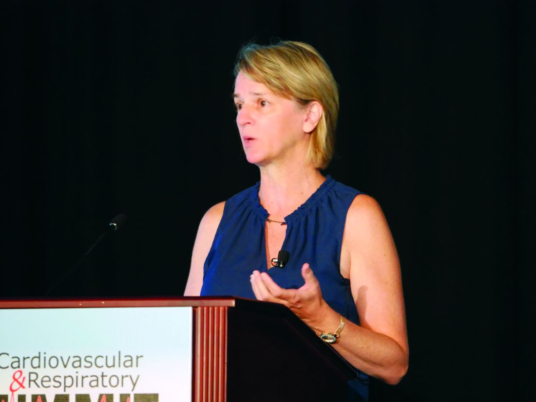
ORLANDO – An additional 14% of Americans have been reclassified as having hypertension – an increase from 32% to 46% of adults – under the latest guidelines on management of high blood pressure released by the American College of Cardiology and American Heart Association in 2017. However, this does not mean these patients are mandated for pharmacologic therapy, since most of them have been reclassified as having stage 1 hypertension, said Leslie L. Davis, PhD, RN, ANP-BC, FPCNA, FAANP, FAHA, associate professor of nursing at the University of North Carolina, Greensboro.
According to the new guidelines, normal BP is classified as less than 120 mm Hg systolic and less than 80 mm Hg diastolic. Elevated BP is 120-129 mm Hg systolic and under 80 mm Hg diastolic, while patients are classified as having stage 1 hypertension if their systolic BP is 130-139 mm Hg or diastolic BP is 80-90 mm Hg. Patients now have stage 2 hypertension if their systolic BP is higher than 140 mm Hg or diastolic BP is above 90 mm Hg.
This raises the importance of getting an accurate BP measurement from patients. At least two readings over two or more visits should be used before categorizing a patient. To take an ideal reading, patients should be sitting at rest with their back supported, feet positioned on a flat surface (no crossing legs), and their arm at heart level for at least 5 minutes. Patients should also refrain from tobacco or caffeine use 30 minutes before the reading. “These numbers need to be correct, because we’ve got new [BP] categories, and also for managing blood pressure, you’re making decisions based on these numbers,” said Dr. Davis at the Cardiovascular & Respiratory Summit by Global Academy for Medical Education.
When taking a patient’s BP, neither the patient nor the provider should talk, and constricting clothes on the upper extremity should be removed instead of pushed up. Using a incorrectly sized cuff can artificially raise or lower a patient’s BP level, so clinicians should use one that is 80% of the length and 40% of the width of the patient’s arm circumference. If the BP reading is elevated, confirm the reading in the other arm and use the arm with the higher reading for future measurements. BP measurements taken in different settings, such as ambulatory BP monitoring or home BP monitoring, can help give context to the in-office reading and whether the patient has white-coat hypertension or masked hypertension.
After a patient has their accurate BP reading, they can calculate their atherosclerotic cardiovascular disease (ASCVD) risk using the ACC’s ASCVD Risk Estimator, said Dr. Davis. If a patient has confirmed cardiovascular disease or has a 10% or greater 10-year ASCVD risk, the target to lower BP is 130 based on high-quality evidence, but expert opinion recommends targeting 130/80 for patients with confirmed hypertension as well.
“If somebody is overweight or obese, 10 pounds is 10 points,” said Dr. Davis. “Even if you don’t get them to the appropriate body mass index over 3 months’ time, that’s as much as a low or medium dose of antihypertensive therapy. For you to be able to get double-digit reduction, that’s what a med does.”
Other nonpharmacologic interventions for these patients include a heart-healthy diet such as the DASH diet, lowering sodium and increasing potassium, structuring exercise and physical activity, lowering use of or avoiding alcohol, and smoking cessation. The goal of nonpharmacologic therapy is not only to lower BP, but make the medication work better, said Dr. Davis.
Pharmacologic therapy should be initiated when patients exceed or are above the cutoff values for the new BP categories and if a patient has already had a cardiovascular event. “Basically, if you’re above that line in the sand of your goal, that’s when to start medications,” she said.
For stage 1 hypertension, first-line therapy is lifestyle change plus thiazides, calcium-channel blockers, or ACE inhibitors, with stage 1 hypertension therapy consisting of a combination of two first-line therapy therapies to reduce systolic BP by about 20 mm Hg and diastolic BP by 10 mm Hg. Beta-blockers are not first-line antihypertension therapy, but can be considered in patients with coronary artery disease and heart failure with reduced left ventricular ejection fraction.
With regard to follow-up, patients with low ASCVD risk and stage 1 hypertension can be monitored in 3-6 months after lifestyle changes, while patients with high ASCVD risk and stage 1 hypertension should be followed up in 1 month. Patients with stage 2 hypertension should follow up with their primary care provider 1 month after beginning their therapy, and those with very high BP should promptly be started on drug treatment with lifestyle changes, with upward dose adjustments as needed.
In adults aged 65 years or older, the ACC/AHA guidelines also focused on how to prevent cognitive decline and dementia, said Dr. Davis. The goal for ambulatory, community-dwelling adults is still to have a systolic BP of less than 130 mm Hg, but clinical judgment should prevail because of comorbid conditions and limited life expectancy in these patients. Patient preference should also be considered, and clinicians should use a team-based approach with shared decision making to determine goals for each patient.
Dr. Davis reported no relevant financial disclosures. Global Academy for Medical Education and this news organization are owned by the same parent company.
ORLANDO – An additional 14% of Americans have been reclassified as having hypertension – an increase from 32% to 46% of adults – under the latest guidelines on management of high blood pressure released by the American College of Cardiology and American Heart Association in 2017. However, this does not mean these patients are mandated for pharmacologic therapy, since most of them have been reclassified as having stage 1 hypertension, said Leslie L. Davis, PhD, RN, ANP-BC, FPCNA, FAANP, FAHA, associate professor of nursing at the University of North Carolina, Greensboro.
According to the new guidelines, normal BP is classified as less than 120 mm Hg systolic and less than 80 mm Hg diastolic. Elevated BP is 120-129 mm Hg systolic and under 80 mm Hg diastolic, while patients are classified as having stage 1 hypertension if their systolic BP is 130-139 mm Hg or diastolic BP is 80-90 mm Hg. Patients now have stage 2 hypertension if their systolic BP is higher than 140 mm Hg or diastolic BP is above 90 mm Hg.
This raises the importance of getting an accurate BP measurement from patients. At least two readings over two or more visits should be used before categorizing a patient. To take an ideal reading, patients should be sitting at rest with their back supported, feet positioned on a flat surface (no crossing legs), and their arm at heart level for at least 5 minutes. Patients should also refrain from tobacco or caffeine use 30 minutes before the reading. “These numbers need to be correct, because we’ve got new [BP] categories, and also for managing blood pressure, you’re making decisions based on these numbers,” said Dr. Davis at the Cardiovascular & Respiratory Summit by Global Academy for Medical Education.
When taking a patient’s BP, neither the patient nor the provider should talk, and constricting clothes on the upper extremity should be removed instead of pushed up. Using a incorrectly sized cuff can artificially raise or lower a patient’s BP level, so clinicians should use one that is 80% of the length and 40% of the width of the patient’s arm circumference. If the BP reading is elevated, confirm the reading in the other arm and use the arm with the higher reading for future measurements. BP measurements taken in different settings, such as ambulatory BP monitoring or home BP monitoring, can help give context to the in-office reading and whether the patient has white-coat hypertension or masked hypertension.
After a patient has their accurate BP reading, they can calculate their atherosclerotic cardiovascular disease (ASCVD) risk using the ACC’s ASCVD Risk Estimator, said Dr. Davis. If a patient has confirmed cardiovascular disease or has a 10% or greater 10-year ASCVD risk, the target to lower BP is 130 based on high-quality evidence, but expert opinion recommends targeting 130/80 for patients with confirmed hypertension as well.
“If somebody is overweight or obese, 10 pounds is 10 points,” said Dr. Davis. “Even if you don’t get them to the appropriate body mass index over 3 months’ time, that’s as much as a low or medium dose of antihypertensive therapy. For you to be able to get double-digit reduction, that’s what a med does.”
Other nonpharmacologic interventions for these patients include a heart-healthy diet such as the DASH diet, lowering sodium and increasing potassium, structuring exercise and physical activity, lowering use of or avoiding alcohol, and smoking cessation. The goal of nonpharmacologic therapy is not only to lower BP, but make the medication work better, said Dr. Davis.
Pharmacologic therapy should be initiated when patients exceed or are above the cutoff values for the new BP categories and if a patient has already had a cardiovascular event. “Basically, if you’re above that line in the sand of your goal, that’s when to start medications,” she said.
For stage 1 hypertension, first-line therapy is lifestyle change plus thiazides, calcium-channel blockers, or ACE inhibitors, with stage 1 hypertension therapy consisting of a combination of two first-line therapy therapies to reduce systolic BP by about 20 mm Hg and diastolic BP by 10 mm Hg. Beta-blockers are not first-line antihypertension therapy, but can be considered in patients with coronary artery disease and heart failure with reduced left ventricular ejection fraction.
With regard to follow-up, patients with low ASCVD risk and stage 1 hypertension can be monitored in 3-6 months after lifestyle changes, while patients with high ASCVD risk and stage 1 hypertension should be followed up in 1 month. Patients with stage 2 hypertension should follow up with their primary care provider 1 month after beginning their therapy, and those with very high BP should promptly be started on drug treatment with lifestyle changes, with upward dose adjustments as needed.
In adults aged 65 years or older, the ACC/AHA guidelines also focused on how to prevent cognitive decline and dementia, said Dr. Davis. The goal for ambulatory, community-dwelling adults is still to have a systolic BP of less than 130 mm Hg, but clinical judgment should prevail because of comorbid conditions and limited life expectancy in these patients. Patient preference should also be considered, and clinicians should use a team-based approach with shared decision making to determine goals for each patient.
Dr. Davis reported no relevant financial disclosures. Global Academy for Medical Education and this news organization are owned by the same parent company.
ORLANDO – An additional 14% of Americans have been reclassified as having hypertension – an increase from 32% to 46% of adults – under the latest guidelines on management of high blood pressure released by the American College of Cardiology and American Heart Association in 2017. However, this does not mean these patients are mandated for pharmacologic therapy, since most of them have been reclassified as having stage 1 hypertension, said Leslie L. Davis, PhD, RN, ANP-BC, FPCNA, FAANP, FAHA, associate professor of nursing at the University of North Carolina, Greensboro.
According to the new guidelines, normal BP is classified as less than 120 mm Hg systolic and less than 80 mm Hg diastolic. Elevated BP is 120-129 mm Hg systolic and under 80 mm Hg diastolic, while patients are classified as having stage 1 hypertension if their systolic BP is 130-139 mm Hg or diastolic BP is 80-90 mm Hg. Patients now have stage 2 hypertension if their systolic BP is higher than 140 mm Hg or diastolic BP is above 90 mm Hg.
This raises the importance of getting an accurate BP measurement from patients. At least two readings over two or more visits should be used before categorizing a patient. To take an ideal reading, patients should be sitting at rest with their back supported, feet positioned on a flat surface (no crossing legs), and their arm at heart level for at least 5 minutes. Patients should also refrain from tobacco or caffeine use 30 minutes before the reading. “These numbers need to be correct, because we’ve got new [BP] categories, and also for managing blood pressure, you’re making decisions based on these numbers,” said Dr. Davis at the Cardiovascular & Respiratory Summit by Global Academy for Medical Education.
When taking a patient’s BP, neither the patient nor the provider should talk, and constricting clothes on the upper extremity should be removed instead of pushed up. Using a incorrectly sized cuff can artificially raise or lower a patient’s BP level, so clinicians should use one that is 80% of the length and 40% of the width of the patient’s arm circumference. If the BP reading is elevated, confirm the reading in the other arm and use the arm with the higher reading for future measurements. BP measurements taken in different settings, such as ambulatory BP monitoring or home BP monitoring, can help give context to the in-office reading and whether the patient has white-coat hypertension or masked hypertension.
After a patient has their accurate BP reading, they can calculate their atherosclerotic cardiovascular disease (ASCVD) risk using the ACC’s ASCVD Risk Estimator, said Dr. Davis. If a patient has confirmed cardiovascular disease or has a 10% or greater 10-year ASCVD risk, the target to lower BP is 130 based on high-quality evidence, but expert opinion recommends targeting 130/80 for patients with confirmed hypertension as well.
“If somebody is overweight or obese, 10 pounds is 10 points,” said Dr. Davis. “Even if you don’t get them to the appropriate body mass index over 3 months’ time, that’s as much as a low or medium dose of antihypertensive therapy. For you to be able to get double-digit reduction, that’s what a med does.”
Other nonpharmacologic interventions for these patients include a heart-healthy diet such as the DASH diet, lowering sodium and increasing potassium, structuring exercise and physical activity, lowering use of or avoiding alcohol, and smoking cessation. The goal of nonpharmacologic therapy is not only to lower BP, but make the medication work better, said Dr. Davis.
Pharmacologic therapy should be initiated when patients exceed or are above the cutoff values for the new BP categories and if a patient has already had a cardiovascular event. “Basically, if you’re above that line in the sand of your goal, that’s when to start medications,” she said.
For stage 1 hypertension, first-line therapy is lifestyle change plus thiazides, calcium-channel blockers, or ACE inhibitors, with stage 1 hypertension therapy consisting of a combination of two first-line therapy therapies to reduce systolic BP by about 20 mm Hg and diastolic BP by 10 mm Hg. Beta-blockers are not first-line antihypertension therapy, but can be considered in patients with coronary artery disease and heart failure with reduced left ventricular ejection fraction.
With regard to follow-up, patients with low ASCVD risk and stage 1 hypertension can be monitored in 3-6 months after lifestyle changes, while patients with high ASCVD risk and stage 1 hypertension should be followed up in 1 month. Patients with stage 2 hypertension should follow up with their primary care provider 1 month after beginning their therapy, and those with very high BP should promptly be started on drug treatment with lifestyle changes, with upward dose adjustments as needed.
In adults aged 65 years or older, the ACC/AHA guidelines also focused on how to prevent cognitive decline and dementia, said Dr. Davis. The goal for ambulatory, community-dwelling adults is still to have a systolic BP of less than 130 mm Hg, but clinical judgment should prevail because of comorbid conditions and limited life expectancy in these patients. Patient preference should also be considered, and clinicians should use a team-based approach with shared decision making to determine goals for each patient.
Dr. Davis reported no relevant financial disclosures. Global Academy for Medical Education and this news organization are owned by the same parent company.
EXPERT ANALYSIS FROM CARPS 2019
AGA remembers Dr. Henry T. Lynch
Henry T. Lynch, MD, came from a humble background, growing up in a rough neighborhood in New York City. He enlisted in the Navy and served in the South Pacific during World War II. Afterward, Dr. Lynch focused his efforts on completing his education, which eventually lead him to the medical field.
After obtaining his high-school equivalency, and completing his undergraduate degree at the University of Oklahoma and his master’s degree in clinical psychology at the University of Denver, his path turned toward the field in which he would make his thrilling and unprecedented discoveries. He studied for a PhD in human genetics at the University of Texas at Austin and received his medical degree from the University of Texas Medical Branch in Galveston. He completed his internship at St. Mary’s Hospital in Evansville, Indiana, and his residency in internal medicine at the University of Nebraska College of Medicine. His first faculty appointment was at The University of Texas MD Anderson Cancer Center.
In 1967, he accepted a position at Creighton, in Omaha, Neb., where he would spend the rest of his storied career. Dr. Lynch was a professor at Creighton University School of Medicine, and the founder and director of the Hereditary Cancer Center at Creighton, established in 1984. He served as chair of the institution’s Department of Preventive Medicine and Public Health, and was named the inaugural holder of the Charles F. and Mary C. Heider Endowed Chair in Cancer Research at Creighton.
A patient he encountered in 1962 – an alcoholic that drank because he believed he would die of colon cancer since everyone in his family had – was the catalyst for his groundbreaking work into the possibility of a hereditary component to some forms of cancer. During this time, it was understood that carcinogenic chemicals and viruses were the primary cause of cancer.
Dr. Lynch provided the first complete description of hereditary nonpolyposis colorectal cancer, a form of colon cancer eventually renamed Lynch syndrome. He continued his research, eventually identifying a hereditary form of breast and ovarian cancers, melanoma, and prostate and pancreatic cancers. His efforts also resulted in one of the world’s largest databases of family cancer histories.
Dr. Lynch passed away on June 2, 2019, at the age of 91. AGA members are sharing their stories and the impact Dr. Lynch had on their work in the AGA Community.
Lucas Franki contributed to this report.
Henry T. Lynch, MD, came from a humble background, growing up in a rough neighborhood in New York City. He enlisted in the Navy and served in the South Pacific during World War II. Afterward, Dr. Lynch focused his efforts on completing his education, which eventually lead him to the medical field.
After obtaining his high-school equivalency, and completing his undergraduate degree at the University of Oklahoma and his master’s degree in clinical psychology at the University of Denver, his path turned toward the field in which he would make his thrilling and unprecedented discoveries. He studied for a PhD in human genetics at the University of Texas at Austin and received his medical degree from the University of Texas Medical Branch in Galveston. He completed his internship at St. Mary’s Hospital in Evansville, Indiana, and his residency in internal medicine at the University of Nebraska College of Medicine. His first faculty appointment was at The University of Texas MD Anderson Cancer Center.
In 1967, he accepted a position at Creighton, in Omaha, Neb., where he would spend the rest of his storied career. Dr. Lynch was a professor at Creighton University School of Medicine, and the founder and director of the Hereditary Cancer Center at Creighton, established in 1984. He served as chair of the institution’s Department of Preventive Medicine and Public Health, and was named the inaugural holder of the Charles F. and Mary C. Heider Endowed Chair in Cancer Research at Creighton.
A patient he encountered in 1962 – an alcoholic that drank because he believed he would die of colon cancer since everyone in his family had – was the catalyst for his groundbreaking work into the possibility of a hereditary component to some forms of cancer. During this time, it was understood that carcinogenic chemicals and viruses were the primary cause of cancer.
Dr. Lynch provided the first complete description of hereditary nonpolyposis colorectal cancer, a form of colon cancer eventually renamed Lynch syndrome. He continued his research, eventually identifying a hereditary form of breast and ovarian cancers, melanoma, and prostate and pancreatic cancers. His efforts also resulted in one of the world’s largest databases of family cancer histories.
Dr. Lynch passed away on June 2, 2019, at the age of 91. AGA members are sharing their stories and the impact Dr. Lynch had on their work in the AGA Community.
Lucas Franki contributed to this report.
Henry T. Lynch, MD, came from a humble background, growing up in a rough neighborhood in New York City. He enlisted in the Navy and served in the South Pacific during World War II. Afterward, Dr. Lynch focused his efforts on completing his education, which eventually lead him to the medical field.
After obtaining his high-school equivalency, and completing his undergraduate degree at the University of Oklahoma and his master’s degree in clinical psychology at the University of Denver, his path turned toward the field in which he would make his thrilling and unprecedented discoveries. He studied for a PhD in human genetics at the University of Texas at Austin and received his medical degree from the University of Texas Medical Branch in Galveston. He completed his internship at St. Mary’s Hospital in Evansville, Indiana, and his residency in internal medicine at the University of Nebraska College of Medicine. His first faculty appointment was at The University of Texas MD Anderson Cancer Center.
In 1967, he accepted a position at Creighton, in Omaha, Neb., where he would spend the rest of his storied career. Dr. Lynch was a professor at Creighton University School of Medicine, and the founder and director of the Hereditary Cancer Center at Creighton, established in 1984. He served as chair of the institution’s Department of Preventive Medicine and Public Health, and was named the inaugural holder of the Charles F. and Mary C. Heider Endowed Chair in Cancer Research at Creighton.
A patient he encountered in 1962 – an alcoholic that drank because he believed he would die of colon cancer since everyone in his family had – was the catalyst for his groundbreaking work into the possibility of a hereditary component to some forms of cancer. During this time, it was understood that carcinogenic chemicals and viruses were the primary cause of cancer.
Dr. Lynch provided the first complete description of hereditary nonpolyposis colorectal cancer, a form of colon cancer eventually renamed Lynch syndrome. He continued his research, eventually identifying a hereditary form of breast and ovarian cancers, melanoma, and prostate and pancreatic cancers. His efforts also resulted in one of the world’s largest databases of family cancer histories.
Dr. Lynch passed away on June 2, 2019, at the age of 91. AGA members are sharing their stories and the impact Dr. Lynch had on their work in the AGA Community.
Lucas Franki contributed to this report.
Top AGA Community patient cases
Physicians with difficult patient scenarios bring their questions to the AGA Community (https://community.gastro.org) to seek advice from colleagues about therapy and disease management options, best practices, and diagnoses. In case you missed it, here are the most popular clinical discussions shared in the forum recently:
1. Crohn’s disease, Infliximab and liver abscess
(http://ow.ly/MehK30p2UZr)
2. Positive Cologuard testing in patient on blood thinners
(http://ow.ly/lJXF30p2V12)
3. Recombinant zoster vaccine in IBD patients on biologics
(http://ow.ly/FWGA30p2V1F)
4. Hair loss and Crohn’s disease
(http://ow.ly/C6Sa30p2V2h)
Access these clinical cases and more discussions at https://community.gastro.org/discussions.
Physicians with difficult patient scenarios bring their questions to the AGA Community (https://community.gastro.org) to seek advice from colleagues about therapy and disease management options, best practices, and diagnoses. In case you missed it, here are the most popular clinical discussions shared in the forum recently:
1. Crohn’s disease, Infliximab and liver abscess
(http://ow.ly/MehK30p2UZr)
2. Positive Cologuard testing in patient on blood thinners
(http://ow.ly/lJXF30p2V12)
3. Recombinant zoster vaccine in IBD patients on biologics
(http://ow.ly/FWGA30p2V1F)
4. Hair loss and Crohn’s disease
(http://ow.ly/C6Sa30p2V2h)
Access these clinical cases and more discussions at https://community.gastro.org/discussions.
Physicians with difficult patient scenarios bring their questions to the AGA Community (https://community.gastro.org) to seek advice from colleagues about therapy and disease management options, best practices, and diagnoses. In case you missed it, here are the most popular clinical discussions shared in the forum recently:
1. Crohn’s disease, Infliximab and liver abscess
(http://ow.ly/MehK30p2UZr)
2. Positive Cologuard testing in patient on blood thinners
(http://ow.ly/lJXF30p2V12)
3. Recombinant zoster vaccine in IBD patients on biologics
(http://ow.ly/FWGA30p2V1F)
4. Hair loss and Crohn’s disease
(http://ow.ly/C6Sa30p2V2h)
Access these clinical cases and more discussions at https://community.gastro.org/discussions.
Remember the AGA Research Foundation in your will or living trust
What if all you had to do to ensure that the AGA Research Foundation can have an impact for years to come is to write a simple sentence? Sound impossible?
The AGA Research Foundation provides a key source of funding at a critical juncture in young investigators’ career. Securing the future of the talented investigators we serve really is as simple as one sentence. By including a gift to the AGA Research Foundation in your will, you can support our mission tomorrow without giving away any of your assets today.
Including the AGA Research Foundation in your will is a popular gift to give because it is:
- Affordable. The actual giving of your gift occurs after your lifetime, so your current income is not affected.
- Flexible. Until your will goes into effect, you are free to alter your plans or change your mind.
- Versatile. You can give a specific item, a set amount of money or a percentage of your estate. You can also make your gift contingent upon certain events.
We hope you’ll consider including a gift to the AGA Research Foundation in your will or living trust. It’s simple – just a few sentences in your will or trust are all that is needed. The official bequest language for the AGA Research Foundation is: “I, [name], of [city, state, ZIP], give, devise and bequeath to the AGA Research Foundation [written amount or percentage of the estate or description of property] for its unrestricted use and purpose.”
Join others in donating to the AGA Research Foundation and help fill the funding gap and protect the next generation of investigators.
Please contact us for more information at foundation@gastro.org or visit http://gastro.planmylegacy.org/.
What if all you had to do to ensure that the AGA Research Foundation can have an impact for years to come is to write a simple sentence? Sound impossible?
The AGA Research Foundation provides a key source of funding at a critical juncture in young investigators’ career. Securing the future of the talented investigators we serve really is as simple as one sentence. By including a gift to the AGA Research Foundation in your will, you can support our mission tomorrow without giving away any of your assets today.
Including the AGA Research Foundation in your will is a popular gift to give because it is:
- Affordable. The actual giving of your gift occurs after your lifetime, so your current income is not affected.
- Flexible. Until your will goes into effect, you are free to alter your plans or change your mind.
- Versatile. You can give a specific item, a set amount of money or a percentage of your estate. You can also make your gift contingent upon certain events.
We hope you’ll consider including a gift to the AGA Research Foundation in your will or living trust. It’s simple – just a few sentences in your will or trust are all that is needed. The official bequest language for the AGA Research Foundation is: “I, [name], of [city, state, ZIP], give, devise and bequeath to the AGA Research Foundation [written amount or percentage of the estate or description of property] for its unrestricted use and purpose.”
Join others in donating to the AGA Research Foundation and help fill the funding gap and protect the next generation of investigators.
Please contact us for more information at foundation@gastro.org or visit http://gastro.planmylegacy.org/.
What if all you had to do to ensure that the AGA Research Foundation can have an impact for years to come is to write a simple sentence? Sound impossible?
The AGA Research Foundation provides a key source of funding at a critical juncture in young investigators’ career. Securing the future of the talented investigators we serve really is as simple as one sentence. By including a gift to the AGA Research Foundation in your will, you can support our mission tomorrow without giving away any of your assets today.
Including the AGA Research Foundation in your will is a popular gift to give because it is:
- Affordable. The actual giving of your gift occurs after your lifetime, so your current income is not affected.
- Flexible. Until your will goes into effect, you are free to alter your plans or change your mind.
- Versatile. You can give a specific item, a set amount of money or a percentage of your estate. You can also make your gift contingent upon certain events.
We hope you’ll consider including a gift to the AGA Research Foundation in your will or living trust. It’s simple – just a few sentences in your will or trust are all that is needed. The official bequest language for the AGA Research Foundation is: “I, [name], of [city, state, ZIP], give, devise and bequeath to the AGA Research Foundation [written amount or percentage of the estate or description of property] for its unrestricted use and purpose.”
Join others in donating to the AGA Research Foundation and help fill the funding gap and protect the next generation of investigators.
Please contact us for more information at foundation@gastro.org or visit http://gastro.planmylegacy.org/.
Consider adding chemical peels for your acne patients
MILAN – according to Dee Anna Glaser, MD.
Speaking at the World Congress of Dermatology, Dr. Glaser, director of clinical research and interim chair of the department of dermatology at Saint Louis University, said that in her practice, acne and actinic keratosis are the most common medical indications for chemical peels and that “acne is just a winner all the way around.”
For acne, she added: “Chemical peels can help both the comedonal and the inflammatory component. It should probably be combined with other therapies, and it does produce both an exfoliative and an anti-inflammatory benefit,.”
A variety of chemical peel formulations can be considered for acne, Dr. Glaser noted. “Typically, you’re going to use a light chemical peel,” such as glycolic or salicylic acid. Other options include Jessner’s solution or a light trichloroacetic acid formulation, she said, adding that tretinoin alone can also be considered.
In choosing between glycolic and salicylic acid, Dr. Glaser said, “salicylic acid should theoretically be the best agent because it is lipophilic and the glycolic acid is hydrophilic.” The reality of how these agents perform clinically, though, may sort out differently.
Dr. Glaser pointed to a double-blind, randomized controlled trial of the two agents in 20 women with facial acne. The severity of participants’ inflammatory acne was mild to moderate, with an average of 27 inflammatory lesions, and they had been on a stable prescription or over-the-counter acne regimen for at least 2 months (Dermatol Surg. 2008 Jan;34[1]:45-50).
Patients received six peels – one every 2 weeks – with 30% glycolic acid (an alpha-hydroxy acid) and 30% salicylic acid (a beta-hydroxy acid) in the split-face study.
All participants started at 4 minutes of exposure and increased up to 5 minutes as tolerated, although timing is only really important for glycolic acid, the same duration of exposure was maintained for each agent for the sake of consistency between arms, said Dr. Glaser, one of the investigators.
Sharing photographs of study participants, she observed that after six peels, “there really isn’t a significant difference.” Therefore, she added, “even though salicylic acid should be better, you can see that glycolic acid really held its own in this study.”
Dr. Glaser pointed out that a trend was seen for slightly better results with salicylic acid and results with this agent were more durable than those seen with glycolic acid. Patients reported fewer side effects on the beta-hydroxy–treated side as well.
She referred to another study, conducted in Japan, that used a double-blind, split-face design to compare 40% glycolic acid with a placebo that had a similarly low pH of 2.0. The 26 patients with moderate acne received five peels on a biweekly schedule, with glycolic acid significantly outperforming placebo. Among acne subtypes, noninflammatory acne improved more than inflammatory acne with glycolic acid (Dermatol Surg. 2014 Mar;40[3]:314-22).
Dr. Glaser said that in her own practice, she still tries to use salicylic acid for her acne patients,” though some patients prefer the experience of a glycolic acid peel, with which there’s likely to be less pain. “So if you have a preference, or your patient has a preference, you will probably be able to use the acid that works best for you,” she said.
Whatever peel is chosen, it should be considered an adjuvant to other topical and systemic acne therapies, Dr. Glaser stressed. “To maintain the results, you really do need to maintain the patient on some sort of standard acne therapy that you would normally do.”
Peels can also be an effective part of a multipronged approach that includes laser therapy and intralesional steroids, she said. However, peels can be considered for monotherapy in patients who don’t tolerate other acne therapies, and they can be used safely in pregnancy, she said.
As with all such treatments, dermatologists should remember to consider and counsel about herpes simplex virus prophylaxis and sun protection.
Dr. Glaser reported financial relationships with Galderma, Ulthera, Ortho, Allergan, Cellgene, and other pharmaceutical companies.
MILAN – according to Dee Anna Glaser, MD.
Speaking at the World Congress of Dermatology, Dr. Glaser, director of clinical research and interim chair of the department of dermatology at Saint Louis University, said that in her practice, acne and actinic keratosis are the most common medical indications for chemical peels and that “acne is just a winner all the way around.”
For acne, she added: “Chemical peels can help both the comedonal and the inflammatory component. It should probably be combined with other therapies, and it does produce both an exfoliative and an anti-inflammatory benefit,.”
A variety of chemical peel formulations can be considered for acne, Dr. Glaser noted. “Typically, you’re going to use a light chemical peel,” such as glycolic or salicylic acid. Other options include Jessner’s solution or a light trichloroacetic acid formulation, she said, adding that tretinoin alone can also be considered.
In choosing between glycolic and salicylic acid, Dr. Glaser said, “salicylic acid should theoretically be the best agent because it is lipophilic and the glycolic acid is hydrophilic.” The reality of how these agents perform clinically, though, may sort out differently.
Dr. Glaser pointed to a double-blind, randomized controlled trial of the two agents in 20 women with facial acne. The severity of participants’ inflammatory acne was mild to moderate, with an average of 27 inflammatory lesions, and they had been on a stable prescription or over-the-counter acne regimen for at least 2 months (Dermatol Surg. 2008 Jan;34[1]:45-50).
Patients received six peels – one every 2 weeks – with 30% glycolic acid (an alpha-hydroxy acid) and 30% salicylic acid (a beta-hydroxy acid) in the split-face study.
All participants started at 4 minutes of exposure and increased up to 5 minutes as tolerated, although timing is only really important for glycolic acid, the same duration of exposure was maintained for each agent for the sake of consistency between arms, said Dr. Glaser, one of the investigators.
Sharing photographs of study participants, she observed that after six peels, “there really isn’t a significant difference.” Therefore, she added, “even though salicylic acid should be better, you can see that glycolic acid really held its own in this study.”
Dr. Glaser pointed out that a trend was seen for slightly better results with salicylic acid and results with this agent were more durable than those seen with glycolic acid. Patients reported fewer side effects on the beta-hydroxy–treated side as well.
She referred to another study, conducted in Japan, that used a double-blind, split-face design to compare 40% glycolic acid with a placebo that had a similarly low pH of 2.0. The 26 patients with moderate acne received five peels on a biweekly schedule, with glycolic acid significantly outperforming placebo. Among acne subtypes, noninflammatory acne improved more than inflammatory acne with glycolic acid (Dermatol Surg. 2014 Mar;40[3]:314-22).
Dr. Glaser said that in her own practice, she still tries to use salicylic acid for her acne patients,” though some patients prefer the experience of a glycolic acid peel, with which there’s likely to be less pain. “So if you have a preference, or your patient has a preference, you will probably be able to use the acid that works best for you,” she said.
Whatever peel is chosen, it should be considered an adjuvant to other topical and systemic acne therapies, Dr. Glaser stressed. “To maintain the results, you really do need to maintain the patient on some sort of standard acne therapy that you would normally do.”
Peels can also be an effective part of a multipronged approach that includes laser therapy and intralesional steroids, she said. However, peels can be considered for monotherapy in patients who don’t tolerate other acne therapies, and they can be used safely in pregnancy, she said.
As with all such treatments, dermatologists should remember to consider and counsel about herpes simplex virus prophylaxis and sun protection.
Dr. Glaser reported financial relationships with Galderma, Ulthera, Ortho, Allergan, Cellgene, and other pharmaceutical companies.
MILAN – according to Dee Anna Glaser, MD.
Speaking at the World Congress of Dermatology, Dr. Glaser, director of clinical research and interim chair of the department of dermatology at Saint Louis University, said that in her practice, acne and actinic keratosis are the most common medical indications for chemical peels and that “acne is just a winner all the way around.”
For acne, she added: “Chemical peels can help both the comedonal and the inflammatory component. It should probably be combined with other therapies, and it does produce both an exfoliative and an anti-inflammatory benefit,.”
A variety of chemical peel formulations can be considered for acne, Dr. Glaser noted. “Typically, you’re going to use a light chemical peel,” such as glycolic or salicylic acid. Other options include Jessner’s solution or a light trichloroacetic acid formulation, she said, adding that tretinoin alone can also be considered.
In choosing between glycolic and salicylic acid, Dr. Glaser said, “salicylic acid should theoretically be the best agent because it is lipophilic and the glycolic acid is hydrophilic.” The reality of how these agents perform clinically, though, may sort out differently.
Dr. Glaser pointed to a double-blind, randomized controlled trial of the two agents in 20 women with facial acne. The severity of participants’ inflammatory acne was mild to moderate, with an average of 27 inflammatory lesions, and they had been on a stable prescription or over-the-counter acne regimen for at least 2 months (Dermatol Surg. 2008 Jan;34[1]:45-50).
Patients received six peels – one every 2 weeks – with 30% glycolic acid (an alpha-hydroxy acid) and 30% salicylic acid (a beta-hydroxy acid) in the split-face study.
All participants started at 4 minutes of exposure and increased up to 5 minutes as tolerated, although timing is only really important for glycolic acid, the same duration of exposure was maintained for each agent for the sake of consistency between arms, said Dr. Glaser, one of the investigators.
Sharing photographs of study participants, she observed that after six peels, “there really isn’t a significant difference.” Therefore, she added, “even though salicylic acid should be better, you can see that glycolic acid really held its own in this study.”
Dr. Glaser pointed out that a trend was seen for slightly better results with salicylic acid and results with this agent were more durable than those seen with glycolic acid. Patients reported fewer side effects on the beta-hydroxy–treated side as well.
She referred to another study, conducted in Japan, that used a double-blind, split-face design to compare 40% glycolic acid with a placebo that had a similarly low pH of 2.0. The 26 patients with moderate acne received five peels on a biweekly schedule, with glycolic acid significantly outperforming placebo. Among acne subtypes, noninflammatory acne improved more than inflammatory acne with glycolic acid (Dermatol Surg. 2014 Mar;40[3]:314-22).
Dr. Glaser said that in her own practice, she still tries to use salicylic acid for her acne patients,” though some patients prefer the experience of a glycolic acid peel, with which there’s likely to be less pain. “So if you have a preference, or your patient has a preference, you will probably be able to use the acid that works best for you,” she said.
Whatever peel is chosen, it should be considered an adjuvant to other topical and systemic acne therapies, Dr. Glaser stressed. “To maintain the results, you really do need to maintain the patient on some sort of standard acne therapy that you would normally do.”
Peels can also be an effective part of a multipronged approach that includes laser therapy and intralesional steroids, she said. However, peels can be considered for monotherapy in patients who don’t tolerate other acne therapies, and they can be used safely in pregnancy, she said.
As with all such treatments, dermatologists should remember to consider and counsel about herpes simplex virus prophylaxis and sun protection.
Dr. Glaser reported financial relationships with Galderma, Ulthera, Ortho, Allergan, Cellgene, and other pharmaceutical companies.
EXPERT ANALYSIS FROM WCD2019
Washington State removes exemption for MMR vaccine
Washington state parents may no longer cite personal or philosophical objections to refuse the MMR vaccine for their children, effective July 28, according to the state’s department of health.

“In Washington state we believe in our doctors. We believe in our nurses. We believe in our educators. We believe in science and we love our children,” Gov. Jay Inslee (D) said when he signed the bill into law on May 10. “And that is why in Washington State, we are against measles.”
The new law applies only to the MMR vaccine and “does not change religious and medical exemption laws. Children who have one of these types of exemptions on file are not affected by the new law,” the health department said.
Washington is one of 45 states that allows religious exemptions from school immunization requirements, according to the National Conference of State Legislatures, which also reported that 15 of those states allow personal-belief exemptions.
The five states that do not allow any form of nonmedical exemption are California, Maine, Mississippi, New York, and West Virginia.
Washington state parents may no longer cite personal or philosophical objections to refuse the MMR vaccine for their children, effective July 28, according to the state’s department of health.
“In Washington state we believe in our doctors. We believe in our nurses. We believe in our educators. We believe in science and we love our children,” Gov. Jay Inslee (D) said when he signed the bill into law on May 10. “And that is why in Washington State, we are against measles.”
The new law applies only to the MMR vaccine and “does not change religious and medical exemption laws. Children who have one of these types of exemptions on file are not affected by the new law,” the health department said.
Washington is one of 45 states that allows religious exemptions from school immunization requirements, according to the National Conference of State Legislatures, which also reported that 15 of those states allow personal-belief exemptions.
The five states that do not allow any form of nonmedical exemption are California, Maine, Mississippi, New York, and West Virginia.
Washington state parents may no longer cite personal or philosophical objections to refuse the MMR vaccine for their children, effective July 28, according to the state’s department of health.
“In Washington state we believe in our doctors. We believe in our nurses. We believe in our educators. We believe in science and we love our children,” Gov. Jay Inslee (D) said when he signed the bill into law on May 10. “And that is why in Washington State, we are against measles.”
The new law applies only to the MMR vaccine and “does not change religious and medical exemption laws. Children who have one of these types of exemptions on file are not affected by the new law,” the health department said.
Washington is one of 45 states that allows religious exemptions from school immunization requirements, according to the National Conference of State Legislatures, which also reported that 15 of those states allow personal-belief exemptions.
The five states that do not allow any form of nonmedical exemption are California, Maine, Mississippi, New York, and West Virginia.

Inflammation diminishes quality of life in NAFLD, not fibrosis
A variety of demographic and disease-related factors contribute to poorer quality of life in patients with nonalcoholic fatty liver disease (NAFLD), based on questionnaires involving 304 European patients.
In contrast with previous research, lobular inflammation, but not hepatic fibrosis, was associated with worse quality of life, reported to lead author Yvonne Huber, MD, of Johannes Gutenberg University in Mainz, Germany, and colleagues. Women and those with advanced disease or comorbidities had the lowest health-related quality of life (HRQL) scores. The investigators suggested that these findings could be used for treatment planning at a population and patient level.
“With the emergence of medical therapy for [nonalcoholic steatohepatitis (NASH)], it will be of importance to identify patients with the highest unmet need for treatment,” the investigators wrote in Clinical Gastroenterology and Hepatology, emphasizing that therapies targeting inflammation could provide the greatest relief.
To determine which patients with NAFLD were most affected by their condition, the investigators used the Chronic Liver Disease Questionnaire (CLDQ), which assesses physical, mental, social, and emotional function, with lower scores indicating poorer health-related quality of life. “[The CLDQ] more specifically addresses symptoms of patients with chronic liver disease, including extrahepatic manifestations, compared with traditional HRQL measures such as the [Short Form–36 (SF-36)] Health Survey Questionnaire,” the investigators explained. Recent research has used the CLDQ to reveal a variety of findings, the investigators noted, such as a 2016 study by Alt and colleagues outlining the most common symptoms in noninfectious chronic liver disease (abdominal discomfort, fatigue, and anxiety), and two studies by Younossi and colleagues describing quality of life improvements after curing hepatitis C virus, and negative impacts of viremia and hepatic inflammation in patients with hepatitis B.
The current study involved 304 patients with histologically confirmed NAFLD who were prospectively entered into the European NAFLD registry via centers in Germany (n = 133), the United Kingdom (n = 154), and Spain (n = 17). Patient data included demographic factors, laboratory findings, and histologic features. Within 6 months of liver biopsy, patients completed the CLDQ.
The mean patient age was 52.3 years, with slightly more men than women (53.3% vs. 46.7%). Most patients (75%) were obese, leading to a median body mass index of 33.3 kg/m2. More than two-thirds of patients (69.1%) had NASH, while approximately half of the population (51.4%) had moderate steatosis, no or low-grade fibrosis (F0-2, 58.2%), and no or low-grade lobular inflammation (grade 0 or 1, 54.7%). The three countries had significantly different population profiles; for example, the United Kingdom had an approximately 10% higher prevalence of type 2 diabetes and obesity compared with the entire cohort, but a decreased arterial hypertension rate of a similar magnitude. The United Kingdom also had a significantly lower mean CLDQ score than that of the study population as a whole (4.73 vs. 4.99).
Analysis of the entire cohort revealed that a variety of demographic and disease-related factors negatively impacted health-related quality of life. Women had a significantly lower mean CLDQ score than that of men (5.31 vs. 4.62; P less than .001), more often reporting abdominal symptoms, fatigue, systemic symptoms, reduced activity, diminished emotional functioning, and worry. CLDQ overall score was negatively influenced by obesity (4.83 vs. 5.46), type 2 diabetes (4.74 vs. 5.25), and hyperlipidemia (4.84 vs. 5.24), but not hypertension. Laboratory findings that negatively correlated with CLDQ included aspartate transaminase (AST) and HbA1c, whereas ferritin was positively correlated.
Generally, patients with NASH reported worse quality of life than that of those with just NAFLD (4.85 vs. 5.31). Factors contributing most to this disparity were fatigue, systemic symptoms, activity, and worry. On a histologic level, hepatic steatosis, ballooning, and lobular inflammation predicted poorer quality of life; although advanced fibrosis and compensated cirrhosis were associated with a trend toward reduced quality of life, this pattern lacked statistical significance. Multivariate analysis, which accounted for age, sex, body mass index, country, and type 2 diabetes, revealed independent associations between reduced quality of life and type 2 diabetes, sex, age, body mass index, and hepatic inflammation, but not fibrosis.
“The striking finding of the current analysis in this well-characterized European cohort was that, in contrast to the published data on predictors of overall and liver-specific mortality, lobular inflammation correlated independently with HRQL,” the investigators wrote. “These results differ from the NASH [Clinical Research Network] cohort, which found lower HRQL using the generic [SF-36 Health Survey Questionnaire] in NASH compared with a healthy U.S. population and a significant effect in cirrhosis only.” The investigators suggested that mechanistic differences in disease progression could explain this discordance.
Although hepatic fibrosis has been tied with quality of life by some studies, the investigators pointed out that patients with chronic hepatitis B or C have reported improved quality of life after viral elimination or suppression, which reduce inflammation, but not fibrosis. “On the basis of the current analysis, it can be expected that improvement of steatohepatitis, and in particular lobular inflammation, will have measurable influence on HRQL even independently of fibrosis improvement,” the investigators concluded.
The study was funded by H2020. The investigators reported no conflicts of interest.
SOURCE: Huber Y et al. CGH. 2018 Dec 20. doi: 10.1016/j.cgh.2018.12.016.
A variety of demographic and disease-related factors contribute to poorer quality of life in patients with nonalcoholic fatty liver disease (NAFLD), based on questionnaires involving 304 European patients.
In contrast with previous research, lobular inflammation, but not hepatic fibrosis, was associated with worse quality of life, reported to lead author Yvonne Huber, MD, of Johannes Gutenberg University in Mainz, Germany, and colleagues. Women and those with advanced disease or comorbidities had the lowest health-related quality of life (HRQL) scores. The investigators suggested that these findings could be used for treatment planning at a population and patient level.
“With the emergence of medical therapy for [nonalcoholic steatohepatitis (NASH)], it will be of importance to identify patients with the highest unmet need for treatment,” the investigators wrote in Clinical Gastroenterology and Hepatology, emphasizing that therapies targeting inflammation could provide the greatest relief.
To determine which patients with NAFLD were most affected by their condition, the investigators used the Chronic Liver Disease Questionnaire (CLDQ), which assesses physical, mental, social, and emotional function, with lower scores indicating poorer health-related quality of life. “[The CLDQ] more specifically addresses symptoms of patients with chronic liver disease, including extrahepatic manifestations, compared with traditional HRQL measures such as the [Short Form–36 (SF-36)] Health Survey Questionnaire,” the investigators explained. Recent research has used the CLDQ to reveal a variety of findings, the investigators noted, such as a 2016 study by Alt and colleagues outlining the most common symptoms in noninfectious chronic liver disease (abdominal discomfort, fatigue, and anxiety), and two studies by Younossi and colleagues describing quality of life improvements after curing hepatitis C virus, and negative impacts of viremia and hepatic inflammation in patients with hepatitis B.
The current study involved 304 patients with histologically confirmed NAFLD who were prospectively entered into the European NAFLD registry via centers in Germany (n = 133), the United Kingdom (n = 154), and Spain (n = 17). Patient data included demographic factors, laboratory findings, and histologic features. Within 6 months of liver biopsy, patients completed the CLDQ.
The mean patient age was 52.3 years, with slightly more men than women (53.3% vs. 46.7%). Most patients (75%) were obese, leading to a median body mass index of 33.3 kg/m2. More than two-thirds of patients (69.1%) had NASH, while approximately half of the population (51.4%) had moderate steatosis, no or low-grade fibrosis (F0-2, 58.2%), and no or low-grade lobular inflammation (grade 0 or 1, 54.7%). The three countries had significantly different population profiles; for example, the United Kingdom had an approximately 10% higher prevalence of type 2 diabetes and obesity compared with the entire cohort, but a decreased arterial hypertension rate of a similar magnitude. The United Kingdom also had a significantly lower mean CLDQ score than that of the study population as a whole (4.73 vs. 4.99).
Analysis of the entire cohort revealed that a variety of demographic and disease-related factors negatively impacted health-related quality of life. Women had a significantly lower mean CLDQ score than that of men (5.31 vs. 4.62; P less than .001), more often reporting abdominal symptoms, fatigue, systemic symptoms, reduced activity, diminished emotional functioning, and worry. CLDQ overall score was negatively influenced by obesity (4.83 vs. 5.46), type 2 diabetes (4.74 vs. 5.25), and hyperlipidemia (4.84 vs. 5.24), but not hypertension. Laboratory findings that negatively correlated with CLDQ included aspartate transaminase (AST) and HbA1c, whereas ferritin was positively correlated.
Generally, patients with NASH reported worse quality of life than that of those with just NAFLD (4.85 vs. 5.31). Factors contributing most to this disparity were fatigue, systemic symptoms, activity, and worry. On a histologic level, hepatic steatosis, ballooning, and lobular inflammation predicted poorer quality of life; although advanced fibrosis and compensated cirrhosis were associated with a trend toward reduced quality of life, this pattern lacked statistical significance. Multivariate analysis, which accounted for age, sex, body mass index, country, and type 2 diabetes, revealed independent associations between reduced quality of life and type 2 diabetes, sex, age, body mass index, and hepatic inflammation, but not fibrosis.
“The striking finding of the current analysis in this well-characterized European cohort was that, in contrast to the published data on predictors of overall and liver-specific mortality, lobular inflammation correlated independently with HRQL,” the investigators wrote. “These results differ from the NASH [Clinical Research Network] cohort, which found lower HRQL using the generic [SF-36 Health Survey Questionnaire] in NASH compared with a healthy U.S. population and a significant effect in cirrhosis only.” The investigators suggested that mechanistic differences in disease progression could explain this discordance.
Although hepatic fibrosis has been tied with quality of life by some studies, the investigators pointed out that patients with chronic hepatitis B or C have reported improved quality of life after viral elimination or suppression, which reduce inflammation, but not fibrosis. “On the basis of the current analysis, it can be expected that improvement of steatohepatitis, and in particular lobular inflammation, will have measurable influence on HRQL even independently of fibrosis improvement,” the investigators concluded.
The study was funded by H2020. The investigators reported no conflicts of interest.
SOURCE: Huber Y et al. CGH. 2018 Dec 20. doi: 10.1016/j.cgh.2018.12.016.
A variety of demographic and disease-related factors contribute to poorer quality of life in patients with nonalcoholic fatty liver disease (NAFLD), based on questionnaires involving 304 European patients.
In contrast with previous research, lobular inflammation, but not hepatic fibrosis, was associated with worse quality of life, reported to lead author Yvonne Huber, MD, of Johannes Gutenberg University in Mainz, Germany, and colleagues. Women and those with advanced disease or comorbidities had the lowest health-related quality of life (HRQL) scores. The investigators suggested that these findings could be used for treatment planning at a population and patient level.
“With the emergence of medical therapy for [nonalcoholic steatohepatitis (NASH)], it will be of importance to identify patients with the highest unmet need for treatment,” the investigators wrote in Clinical Gastroenterology and Hepatology, emphasizing that therapies targeting inflammation could provide the greatest relief.
To determine which patients with NAFLD were most affected by their condition, the investigators used the Chronic Liver Disease Questionnaire (CLDQ), which assesses physical, mental, social, and emotional function, with lower scores indicating poorer health-related quality of life. “[The CLDQ] more specifically addresses symptoms of patients with chronic liver disease, including extrahepatic manifestations, compared with traditional HRQL measures such as the [Short Form–36 (SF-36)] Health Survey Questionnaire,” the investigators explained. Recent research has used the CLDQ to reveal a variety of findings, the investigators noted, such as a 2016 study by Alt and colleagues outlining the most common symptoms in noninfectious chronic liver disease (abdominal discomfort, fatigue, and anxiety), and two studies by Younossi and colleagues describing quality of life improvements after curing hepatitis C virus, and negative impacts of viremia and hepatic inflammation in patients with hepatitis B.
The current study involved 304 patients with histologically confirmed NAFLD who were prospectively entered into the European NAFLD registry via centers in Germany (n = 133), the United Kingdom (n = 154), and Spain (n = 17). Patient data included demographic factors, laboratory findings, and histologic features. Within 6 months of liver biopsy, patients completed the CLDQ.
The mean patient age was 52.3 years, with slightly more men than women (53.3% vs. 46.7%). Most patients (75%) were obese, leading to a median body mass index of 33.3 kg/m2. More than two-thirds of patients (69.1%) had NASH, while approximately half of the population (51.4%) had moderate steatosis, no or low-grade fibrosis (F0-2, 58.2%), and no or low-grade lobular inflammation (grade 0 or 1, 54.7%). The three countries had significantly different population profiles; for example, the United Kingdom had an approximately 10% higher prevalence of type 2 diabetes and obesity compared with the entire cohort, but a decreased arterial hypertension rate of a similar magnitude. The United Kingdom also had a significantly lower mean CLDQ score than that of the study population as a whole (4.73 vs. 4.99).
Analysis of the entire cohort revealed that a variety of demographic and disease-related factors negatively impacted health-related quality of life. Women had a significantly lower mean CLDQ score than that of men (5.31 vs. 4.62; P less than .001), more often reporting abdominal symptoms, fatigue, systemic symptoms, reduced activity, diminished emotional functioning, and worry. CLDQ overall score was negatively influenced by obesity (4.83 vs. 5.46), type 2 diabetes (4.74 vs. 5.25), and hyperlipidemia (4.84 vs. 5.24), but not hypertension. Laboratory findings that negatively correlated with CLDQ included aspartate transaminase (AST) and HbA1c, whereas ferritin was positively correlated.
Generally, patients with NASH reported worse quality of life than that of those with just NAFLD (4.85 vs. 5.31). Factors contributing most to this disparity were fatigue, systemic symptoms, activity, and worry. On a histologic level, hepatic steatosis, ballooning, and lobular inflammation predicted poorer quality of life; although advanced fibrosis and compensated cirrhosis were associated with a trend toward reduced quality of life, this pattern lacked statistical significance. Multivariate analysis, which accounted for age, sex, body mass index, country, and type 2 diabetes, revealed independent associations between reduced quality of life and type 2 diabetes, sex, age, body mass index, and hepatic inflammation, but not fibrosis.
“The striking finding of the current analysis in this well-characterized European cohort was that, in contrast to the published data on predictors of overall and liver-specific mortality, lobular inflammation correlated independently with HRQL,” the investigators wrote. “These results differ from the NASH [Clinical Research Network] cohort, which found lower HRQL using the generic [SF-36 Health Survey Questionnaire] in NASH compared with a healthy U.S. population and a significant effect in cirrhosis only.” The investigators suggested that mechanistic differences in disease progression could explain this discordance.
Although hepatic fibrosis has been tied with quality of life by some studies, the investigators pointed out that patients with chronic hepatitis B or C have reported improved quality of life after viral elimination or suppression, which reduce inflammation, but not fibrosis. “On the basis of the current analysis, it can be expected that improvement of steatohepatitis, and in particular lobular inflammation, will have measurable influence on HRQL even independently of fibrosis improvement,” the investigators concluded.
The study was funded by H2020. The investigators reported no conflicts of interest.
SOURCE: Huber Y et al. CGH. 2018 Dec 20. doi: 10.1016/j.cgh.2018.12.016.
FROM CLINICAL GASTROENTEROLOGY AND HEPATOLOGY
RNA interference drug fitusiran looks effective in both hemophilia A and B
MELBOURNE – An investigational RNA interference therapeutic that suppresses the production of antithrombin has shown significant reductions in bleeding rates with no major safety events, according to findings presented at the International Society on Thrombosis and Haemostasis congress.
Fitusiran is a once-monthly, fixed-dose subcutaneous therapy that uses RNA interference to silence the gene for the endogenous anticoagulant antithrombin.
“The therapeutic hypothesis is based on the fact that hemophilia A and B are essentially thrombin-deficiency disorders, so if we lack factor VIII or factor IX, we can’t generate enough thrombin and we can’t produce a significant and substantial blood clot,” John Pasi, MBChB, PhD, of the Royal London Haemophilia Centre, Barts Health NHS Trust. “If we, however, administer fitusiran, which will suppress antithrombin production, we can rebalance coagulation, generate more thrombin and form a much more substantial clot.”
Dr. Pasi presented results of an interim analysis of safety and efficacy data from an open-label, phase 2 extension study in 34 individuals with hemophilia A or B, with or without inhibitors, who were treated either with 50-mg or 80-mg doses of fitusiran for a median of at least 2 years.
Researchers saw significant declines in annualized bleeding rates in patients with hemophilia A and B, with and without inhibitors. Among those without inhibitors, the median annualized bleeding rate declined from 2.00 in patients already on hemophilia prophylaxis and 12.00 in those using on-demand treatment to 1.08 overall. In patients with inhibitors, the median annualized bleeding rate dropped from 42.00 to 1.04.
The treatment was also associated with substantial reductions in antithrombin production and increases in thrombin generation.
One patient in the phase 1 study experienced a fatal cerebral venous sinus thrombosis, which subsequently led to introduction of a bleed management protocol.
“Following that last case, we revised and reviewed the bleed management guidelines in view of the fact that there might potentially be an interaction between the amount of replacement therapy and thrombin generation,” Dr. Pasi said. Since introduction of that protocol, there have been no related thrombotic events.
The majority of adverse events reported were mild and deemed not related to the study drug, Dr. Pasi said. These included headache, injection site erythema, and arthralgia. A total of 14 subjects – all of whom were positive for hepatitis C at baseline – experienced rises in ALT levels but these were asymptomatic and resolved spontaneously.
One patient with chronic active hepatitis C infection also showed significant ALT/AST elevation which led to discontinuation of treatment.
In an interview, Dr. Pasi said one of the biggest advantages of fitusiran was that it could be used in patients with hemophilia A and B. “You’ve got patients with hemophilia B who’ve got no options at the moment. That would be an obvious specific group that would gain from this.”
Another advantage was fitusiran’s stability and dosing, he said, pointing out that the treatment was fixed dosing and stable at room temperature. Fitusiran is now undergoing phase 3 trials.
The study was funded by Sanofi Genzyme and Alnylam Pharmaceuticals, and six authors were employees of Sanofi Genzyme. Dr. Pasi reported financial relationships with pharmaceutical companies, including Alnylam.
SOURCE: Pasi J et al. 2019 ISTH Congress, Abstract OC 11.3.
MELBOURNE – An investigational RNA interference therapeutic that suppresses the production of antithrombin has shown significant reductions in bleeding rates with no major safety events, according to findings presented at the International Society on Thrombosis and Haemostasis congress.
Fitusiran is a once-monthly, fixed-dose subcutaneous therapy that uses RNA interference to silence the gene for the endogenous anticoagulant antithrombin.
“The therapeutic hypothesis is based on the fact that hemophilia A and B are essentially thrombin-deficiency disorders, so if we lack factor VIII or factor IX, we can’t generate enough thrombin and we can’t produce a significant and substantial blood clot,” John Pasi, MBChB, PhD, of the Royal London Haemophilia Centre, Barts Health NHS Trust. “If we, however, administer fitusiran, which will suppress antithrombin production, we can rebalance coagulation, generate more thrombin and form a much more substantial clot.”
Dr. Pasi presented results of an interim analysis of safety and efficacy data from an open-label, phase 2 extension study in 34 individuals with hemophilia A or B, with or without inhibitors, who were treated either with 50-mg or 80-mg doses of fitusiran for a median of at least 2 years.
Researchers saw significant declines in annualized bleeding rates in patients with hemophilia A and B, with and without inhibitors. Among those without inhibitors, the median annualized bleeding rate declined from 2.00 in patients already on hemophilia prophylaxis and 12.00 in those using on-demand treatment to 1.08 overall. In patients with inhibitors, the median annualized bleeding rate dropped from 42.00 to 1.04.
The treatment was also associated with substantial reductions in antithrombin production and increases in thrombin generation.
One patient in the phase 1 study experienced a fatal cerebral venous sinus thrombosis, which subsequently led to introduction of a bleed management protocol.
“Following that last case, we revised and reviewed the bleed management guidelines in view of the fact that there might potentially be an interaction between the amount of replacement therapy and thrombin generation,” Dr. Pasi said. Since introduction of that protocol, there have been no related thrombotic events.
The majority of adverse events reported were mild and deemed not related to the study drug, Dr. Pasi said. These included headache, injection site erythema, and arthralgia. A total of 14 subjects – all of whom were positive for hepatitis C at baseline – experienced rises in ALT levels but these were asymptomatic and resolved spontaneously.
One patient with chronic active hepatitis C infection also showed significant ALT/AST elevation which led to discontinuation of treatment.
In an interview, Dr. Pasi said one of the biggest advantages of fitusiran was that it could be used in patients with hemophilia A and B. “You’ve got patients with hemophilia B who’ve got no options at the moment. That would be an obvious specific group that would gain from this.”
Another advantage was fitusiran’s stability and dosing, he said, pointing out that the treatment was fixed dosing and stable at room temperature. Fitusiran is now undergoing phase 3 trials.
The study was funded by Sanofi Genzyme and Alnylam Pharmaceuticals, and six authors were employees of Sanofi Genzyme. Dr. Pasi reported financial relationships with pharmaceutical companies, including Alnylam.
SOURCE: Pasi J et al. 2019 ISTH Congress, Abstract OC 11.3.
MELBOURNE – An investigational RNA interference therapeutic that suppresses the production of antithrombin has shown significant reductions in bleeding rates with no major safety events, according to findings presented at the International Society on Thrombosis and Haemostasis congress.
Fitusiran is a once-monthly, fixed-dose subcutaneous therapy that uses RNA interference to silence the gene for the endogenous anticoagulant antithrombin.
“The therapeutic hypothesis is based on the fact that hemophilia A and B are essentially thrombin-deficiency disorders, so if we lack factor VIII or factor IX, we can’t generate enough thrombin and we can’t produce a significant and substantial blood clot,” John Pasi, MBChB, PhD, of the Royal London Haemophilia Centre, Barts Health NHS Trust. “If we, however, administer fitusiran, which will suppress antithrombin production, we can rebalance coagulation, generate more thrombin and form a much more substantial clot.”
Dr. Pasi presented results of an interim analysis of safety and efficacy data from an open-label, phase 2 extension study in 34 individuals with hemophilia A or B, with or without inhibitors, who were treated either with 50-mg or 80-mg doses of fitusiran for a median of at least 2 years.
Researchers saw significant declines in annualized bleeding rates in patients with hemophilia A and B, with and without inhibitors. Among those without inhibitors, the median annualized bleeding rate declined from 2.00 in patients already on hemophilia prophylaxis and 12.00 in those using on-demand treatment to 1.08 overall. In patients with inhibitors, the median annualized bleeding rate dropped from 42.00 to 1.04.
The treatment was also associated with substantial reductions in antithrombin production and increases in thrombin generation.
One patient in the phase 1 study experienced a fatal cerebral venous sinus thrombosis, which subsequently led to introduction of a bleed management protocol.
“Following that last case, we revised and reviewed the bleed management guidelines in view of the fact that there might potentially be an interaction between the amount of replacement therapy and thrombin generation,” Dr. Pasi said. Since introduction of that protocol, there have been no related thrombotic events.
The majority of adverse events reported were mild and deemed not related to the study drug, Dr. Pasi said. These included headache, injection site erythema, and arthralgia. A total of 14 subjects – all of whom were positive for hepatitis C at baseline – experienced rises in ALT levels but these were asymptomatic and resolved spontaneously.
One patient with chronic active hepatitis C infection also showed significant ALT/AST elevation which led to discontinuation of treatment.
In an interview, Dr. Pasi said one of the biggest advantages of fitusiran was that it could be used in patients with hemophilia A and B. “You’ve got patients with hemophilia B who’ve got no options at the moment. That would be an obvious specific group that would gain from this.”
Another advantage was fitusiran’s stability and dosing, he said, pointing out that the treatment was fixed dosing and stable at room temperature. Fitusiran is now undergoing phase 3 trials.
The study was funded by Sanofi Genzyme and Alnylam Pharmaceuticals, and six authors were employees of Sanofi Genzyme. Dr. Pasi reported financial relationships with pharmaceutical companies, including Alnylam.
SOURCE: Pasi J et al. 2019 ISTH Congress, Abstract OC 11.3.
REPORTING FROM 2019 ISTH CONGRESS