User login
American Heart Association (AHA): International Stroke Conference
Manual aspiration removes blood clots in more than half of ischemic stroke patients
SAN DIEGO – Manual, endoscopic clot aspiration restored cerebral blood flow in 66 (58.9%) of 112 acute ischemic stroke patients at the University of Pittsburgh Medical Center, according to a retrospective review.
When it failed, trying again with a stentriever ultimately restored flow on the thrombolysis in cerebral infarction scale (TICI 2b/3) in 97 (86.6%) patients, about the same rate of success as if a stentriever had been used in the first place. Patients needed a median of two passes.
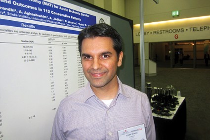
Stentrievers are "the new technology that everyone is using, and they work very well, but they’re expensive," in the range of $5,000-$7,000, said investigator and interventional neurologist Dr. Ashutosh P. Jadhav.
"Our local practice is to use manual aspiration thrombectomy as our first pass. We are [often] able to do the procedure with just the catheter alone, which is a fifth of the price. We like it because it’s cost effective, and we are comfortable doing it. If the clot is refractory, then we move onto stentrievers," he said at the International Stroke Conference, sponsored by the American Heart Association (Tech. Vasc. Interv. Radiol. 2012;15:68-77).
The median time from symptom onset to groin puncture was 267 minutes. When manual aspiration was used alone, the mean procedural time was 63 minutes. When the team had to try again with a stentriever, the mean procedural time was 97 minutes (P less than .0001).
"Maybe if we did a trial against stentriever" on first pass, "we might find" trying manual aspiration first "takes a little bit longer, but we don’t have evidence of that right now," Dr. Jadhav of the UMPC Stroke Institute said.
Large-bore catheters – 0.07 inches and above – were used in about a quarter of the cases, and medium-bore catheters – 0.054-0.058 inches – in the rest. "Primary manual aspiration thrombectomy was carried out with a preference for the largest catheter considered to be trackable into the target lesion." Catheter make and size were not associated with higher or faster recanalization rates, Dr. Jadhav and his team noted.
"When we get the catheter into the clot, we pull back [on the attached 20 cc syringe] in one movement, so we don’t see a high rate of embolism. We did get a 9.8% risk of parenchymal hematoma, and a 6.2% risk of symptomatic hemorrhage," but the rates are "comparable to what people have seen with other therapies," he said. Four patients (3.6%) had intracranial distal wire perforations.
The median age in the series was 67 years, and median NIH stroke scale score was 17. Seventy patients (62.5%) were occluded in the first branch of the middle cerebral artery, and nine (8.0%) were occluded in the second branch. Twenty-one (18.8%) were occluded in the terminus of the internal carotid artery, and 12 (10.7%) had vertebrobasilar occlusions.
Almost half of the patients had 90-day modified Rankin Scale scores of 2 or less, but 35 (31.3%) had died at 3 months. Manual aspiration alone was not associated with better outcomes.
The investigators did not report outside funding. Dr. Jadhav said he has no disclosures.
AT THE INTERNATIONAL STROKE CONFERENCE
Major finding: Manual aspiration – without the aid of a stentriever – restores TICI 2b/3 blood flow in 58.9% of ischemic stroke patients.
Data source: A retrospective review of 112 acute ischemic stroke patients.
Disclosures: The investigators did not report outside funding. The presenter has no disclosures.
Systolic variability after intracerebral hemorrhage raises odds of death, disability
SAN DIEGO – Patients with greater variability in systolic blood pressure in the first day or week after an acute intracerebral hemorrhage were more likely to die within 90 days or have a major disability, a secondary analysis of data on 2,839 patients found.
The study compared patient outcomes with data from 5 blood pressure measurements taken within 24 hours of an acute intracerebral hemorrhage and 12 measurements taken in days 2-7 after the hemorrhage. The investigators described blood pressure variability as a standard deviation of systolic pressure, divided into quintiles.
They found a significant linear association between systolic pressure variability and death or major disability at 90 days that was independent of mean systolic blood pressure. The risk for death or disability was 41% higher in patients in the highest quintile of systolic variability within 24 hours of an acute intracerebral hemorrhage, compared with patients in the lowest quintile. The risk was 57% higher in the highest quintile of systolic variability in days 2-7 after the hemorrhage, compared with the lowest quintile, Dr. Lisa Manning reported at the International Stroke Conference.
The greatest predictors of death or major disability were maximum systolic blood pressure in the first 24 hours after a hemorrhage and standard deviation in systolic pressure in days 2-7, said Dr. Manning of the University of Leicester (England).
Similar significant associations were seen for the secondary outcome, which was an ordinal shift in modified Rankin Scale scores at 90 days. The likelihood of this shift was 43% higher in patients in the highest quintile of systolic variability in the first day after a hemorrhage and 46% higher in patients in the highest quintile of variability in days 2-7, compared with the lowest-quartile patients, she said at the meeting, sponsored by the American Heart Association.
The results were published online in Lancet Neurology (2014 Feb. 13 [doi:10.1016/S1474-4422(14)70018-3]).
Data for the analysis came from the INTERACT2 trial (The Second Intensive Blood Pressure in Acute Haemorrhage trial). The prospective, international multicenter study randomized patients with spontaneous intracerebral hemorrhage and high systolic blood pressure to early intensive lowering of blood pressure (within 1 hour of randomization) or to conventional treatment as recommended in guidelines. Results showed no significant reduction in the risk of death or disability with early intensive treatment (N. Engl. J. Med. 2013;368:2355-65).
For the current analysis, investigators studied data on 2,645 patients with blood pressure measurement in the first day after a hemorrhage (93% of the INTERACT2 cohort) and 2,347 patients with measurements in days 2-7 (83% of the total cohort).
The findings present several clinical implications, Dr. Manning suggested. Consistent, sustained efforts to control blood pressure in the first few days after an acute intracerebral hemorrhage may benefit patients, and clinicians may want to adjust the frequency and intensity of blood pressure monitoring to ensure that systolic blood pressure consistently falls and stabilizes throughout hospitalization in these patients.
The increased risk for death or disability with greater systolic variability was seen after investigators adjusted for the effects of other factors such as age, sex, the randomized treatment group, geographical region, hematoma volume at baseline, and high scores on the National Institutes of Health Stroke Scale.
The study was funded by the National Health and Medical Research Council of Australia. Two of Dr. Manning’s associates in the study reported financial associations with Osaka Pharmaceuticals, Novartis, Omron Healthcare, Pfizer, Takeda, A&D Pharma, and/or Servier.
On Twitter @sherryboschert
AT THE INTERNATIONAL STROKE CONFERENCE
Major finding: Death or major disability were 41% more likely in patients in the highest quintile of systolic variability 1 day after acute intracerebral hemorrhage and 57% higher in those in the highest quintile 2-7 days after hemorrhage, compared with the lowest quintile.
Data source: A secondary analysis of data on 2,839 patients in the prospective, randomized INTERACT2 trial of early intensive blood pressure treatment after acute intracerebral hemorrhage.
Disclosures: The study was funded by the National Health and Medical Research Council of Australia. Two of Dr. Manning’s associates in the study reported financial associations with Osaka Pharmaceuticals, Novartis, Omron Healthcare, Pfizer, Takeda, A&D Pharma, and/or Servier.
Recent cocaine use quadrupled stroke risk
SAN DIEGO – Cocaine use within the past 24 hours was reported by 2.4% of 1,101 patients aged 15-49 years who developed ischemic stroke, compared with 0.4% of 1,154 age-matched controls without stroke.
The recent cocaine use was associated with a fourfold increase in the odds of ischemic stroke after adjustment for the effects of age, current smoking status, sex, and ethnicity. The difference in cocaine use between groups and the increased risk with cocaine were statistically significant, Yu-Ching Cheng, Ph.D., reported.
Females seemed to be at particular risk from recent cocaine use, with an adjusted odds ratio for ischemic stroke of 11 in those who had used cocaine within the previous 24 hours, compared with controls. Males had an odds ratio of 2 for ischemic stroke after recent cocaine use, compared with controls, she reported in a poster presentation at the International Stroke Conference.
"With few exceptions, we believe every young stroke patient should be screened for drug abuse at the time of hospital admission, especially in the case where there’s no clear etiology" for the stroke, she said in an interview at the poster. Knowing about acute cocaine use probably wouldn’t change management of the stroke but might help optimize recovery if the patient is encouraged to seek drug abuse counseling. That also may help prevent secondary strokes from continued cocaine use, she added.
Other factors increased the risk of stroke in the case-control study, but none did as much as cocaine use within the past 24 hours. Diabetes was significantly more common in the stroke patients (17%), compared with controls (5%), and was associated with approximately 3.5-fold higher odds for stroke after adjustment for confounding factors. Stroke patients were significantly more likely than controls to have hypertension (42% vs. 18%, respectively), which was associated with a threefold greater likelihood of stroke. Significantly more stroke patients were current smokers (45% vs. 29%, respectively), which doubled the likelihood of stroke, reported Dr. Cheng of the University of Maryland, Baltimore.
The Stroke Prevention in Young Adults Study was a population-based, case-control study of people in the Baltimore-Washington area aged 15-49 years who either had a first ischemic stroke between 1992 and 2008 or were age-matched controls without stroke who were contacted by random-digit dialing. Investigators collected data through a standardized interview.
The stroke risk from recent cocaine use did not differ by race, with an adjusted odds ratio of nearly 5 for both blacks and whites after cocaine use in the previous 24 hours, she reported at the meeting, sponsored by the American Heart Association.
Ever having used cocaine or having used cocaine in the last 1-30 days was not significantly associated with increased risk for stroke.
Males comprised 53% of the stroke patients and 46% of controls.
Approximately 1.9 million U.S. residents have used cocaine in the past month, previous data have suggested, with adults aged 18-25 years most likely to use cocaine, she said. Cocaine is known to constrict blood vessels; increase heart rate, body temperature, and blood pressure; and decrease oxygen supply to the brain.
Dr. Cheng reported having no relevant financial disclosures. The study was funded by the National Institute of Neurological Disorders and Stroke, the Department of Veterans Affairs, and the Centers for Disease Control and Prevention.
On Twitter @sherryboschert
AT THE INTERNATIONAL STROKE CONFERENCE
Major finding: Cocaine use in the past 24 hours was reported by 2.4% of young adults with ischemic stroke and 0.4% of age-matched controls without stroke, and was associated with a fourfold increased odds of stroke.
Data source: A case-control comparison of data on 1,101 ischemic stroke patients aged 15-49 years and 1,154 controls reached by random-digit dialing for phone questionnaires.
Disclosures: Dr. Cheng reported having no relevant financial disclosures. The study was funded by the National Institute of Neurological Disorders and Stroke, the Department of Veterans Affairs, and the Centers for Disease Control and Prevention.
Half of stroke survivors returned to driving
SAN DIEGO – Only 6% of 162 stroke survivors underwent a formal driving evaluation, but 51% returned to driving within a year, a survey found.
Among the 83 who returned to driving, 9 (11%) did so despite reporting that their stroke greatly affected their ability to "engage in valued life activities," Dr. Shelly D. Ozark reported in a poster presentation at the International Stroke Conference.
Twenty-six (31%) of the 83 drivers said their stroke had some effect on their ability to engage in valued life activities, and 48 drivers (58%) said the stroke did not affect that part of their lives.
Formal driving evaluations were completed by 2 of the 9 stroke survivors who returned to driving despite great effects from the stroke, 3 of 26 drivers who reported some effects, and 4 of 48 drivers who reported no effects of the stroke on their abilities to engage in valued life activities, reported Dr. Ozark of the Medical University of South Carolina, Charleston.
Forty-nine (59%) of the 83 drivers returned to driving less than a month after their stroke, 21 (25%) resumed driving 1-3 months after their stroke, and 13 (16%) resumed driving more than 3 months post stroke, the investigators reported at the meeting, sponsored by the American Heart Association.
Thirty-eight (46%) of the 83 drivers said they had self-imposed limits on their driving. This included 17 (35%) of the 48 drivers who reported no effects of the stroke on valued life activities. It is unclear whether a formal driving evaluation would have supported their decision to limit driving, Dr. Ozark noted. Self-imposed driving limits also were reported by 16 of 26 drivers who experienced some effects of the stroke and 5 of 9 drivers who reported great effects of the stroke on their ability to engage in valued life activities.
The study was a secondary analysis of a subset of data from the longitudinal surveys of the Stroke Education and Prevention - South Carolina Project.
Dr. Ozark reported having no relevant financial disclosures.
On Twitter @sherryboschert
AT THE INTERNATIONAL STROKE CONFERENCE
Major finding: Six percent of stroke survivors underwent formal driving evaluations, but 51% of survivors returned to driving within 1 year.
Data source: A secondary analysis of survey responses from 162 stroke survivors, a subset of the Stroke Prevention and Education–South Carolina Project data
Disclosures: Dr. Ozark reported having no relevant financial disclosures.
Physicians, nurses beat clinical scores for ICH outcome prediction
SAN DIEGO – Neurologists and neurology nurses predict 3-month functional outcomes following intracerebral hemorrhagic strokes better than do commonly used clinical scores, according to a prospective, observational study.
At each of the five centers participating in the study, the investigators asked one physician – usually a neurologist – and one neurology nurse on the treatment team to predict 3-month modified Rankin Scale (mRS) scores for primary intracerebral hemorrhagic (ICH) stroke patients under their care within 24 hours of admission.
The team then calculated the admission ICH score and FUNC score themselves based on imaging and other records. The results for all centers then were compared with actual 3-month mRS scores for the 100 patients included in the study.
With 1 representing a perfect correlation with actual 3-month mRS scores, the Spearman’s rank correlation for attending physicians was 0.81, for neurology nurses 0.72, for the ICH scale 0.55, and for the FUNC score –0.46 (negative because higher scores mean better outcomes).
To control for self-fulfilling prophecies among providers, the team ran the analysis again, excluding the 18 patients for whom comfort care was recommended early on. The results were similar: r = 0.78 for attendings, 0.66 for nurses, 0.48 for the ICH score, and –0.37 for the FUNC score. Results in both analyses were statistically significant. The accuracy advantage for physicians and nurses remained when predictions were examined for only the 35 patients who were alive at 3 months.
"Existing formal scales such as the ICH score and FUNC score are useful for providing general probabilities for outcomes among ICH [patient populations], but it is appropriate for trained neurologists and neuroscience nurses to use their best subjective judgment ... when counseling any individual ICH patient and their family," said lead investigator Dr. David Hwang, a neurointensivist in the neurology department at Yale University, New Haven, Conn.
The study findings matter because early predictions guide treatment. It’s the first time that stroke scores have been pitted against clinical judgment, he said at the International Stroke Conference, sponsored by the American Heart Association.
Seventy-five of the physicians were attendings, and 25% were trainees. Although attendings and neurology nurses significantly beat clinical scores, trainees did not. "The vast majority of physicians and nurses were specialists in neuroscience," Dr. Hwang noted.
Patents were, on average, 67 years old. The ICH involved a deep brain bleed in 53% and had a volume of less than 30 mL in 71%. Glasgow Coma Scale scores of 13-15 occurred in 64%.
"Our cohort contained a majority of well-appearing patients, with good exams and small bleeds," a potential limitation. But overall mortality at 3 months was 35%, which is comparable to results from large, published cohorts, Dr. Hwang said.
Even when the 18 comfort-care patients were excluded, "the self-fulfilling prophecy [may have had an] effect," but the team was trying to mimic real life in its work, where they "are the ones to figure out and give prognostic advice to patients and families," he said.
Dr. Hwang said he had no relevant disclosures. The study was funded by the American Heart Association and the National Institute of Neurological Disorders and Stroke.
AT THE INTERNATIONAL STROKE CONFERENCE
Major finding: With 1 representing a perfect correlation with actual 3-month mRS scores, the Spearman’s rank correlation for ICH functional outcomes predicted by attending physicians was 0.81, for predictions by neurology nurses 0.72, for ICH scale score predictions 0.55, and for the FUNC score –0.46.
Data Source: A prospective, observational study in 100 ICH patients.
Disclosures: Dr. Hwang said he had no relevant disclosures. The study was funded by the American Heart Association and the National Institute of Neurological Disorders and Stroke.
Transcranial ultrasound method outdoes echocardiography for finding PFO
SAN DIEGO – Transcranial Doppler saline studies were superior to transesophageal echocardiography in diagnosing a hole in the heart, according to a prospective study of patients who had a cryptogenic stroke.
Researchers analyzed data from 340 patients with cryptogenic stroke suspected of paradoxical embolism who had had their right-left shunt confirmed with transcranial Doppler saline study (TCDSS). Transesophageal echocardiography (TEE) failed to show a shunt such as patent foramen ovale (PFO) in 15% of the cases.
The video associated with this article is no longer available on this site. Please view all of our videos on the MDedge YouTube channel
Courtesy Dr. J. David Spence, Robarts Research Institute
"I was surprised by the number," said Dr. J. David Spence, senior researcher of the study, who presented the findings at the International Stroke Conference, sponsored by the American Heart Association. "I had realized that some of the patients with shunts were missed by TEE, ... but I was surprised by how often that happened."
Nearly a quarter of the population has a PFO, and 4.0%-5.5% of strokes are due to paradoxical embolism through a right-left shunt. But patching the shunts isn’t the simple answer, because the procedure is not without complications, and so far, the studies haven’t been able to make a strong case for the procedure.
TCDSS is an ultrasound method in which 1 mL of tiny bubbles is injected into the vein and if detected in the brain, could give clues to a right-left shunt. The method is safer, the equipment is cheaper, and the training is faster than for TEE, Dr. Spence said.
He added that one reason that TCDSS may be more sensitive than TEE is that sedation for TEE may prevent an adequate Valsalva maneuver. He also showed in a video that detecting the bubbles in TCDSS is rather straightforward.
Given the findings of his study, Dr. Spence said that there’s a need to identify which PFO patients are more likely to have paradoxical embolism and are more likely to respond to patching.
One solution is paying attention to the clinical clues of paradoxical embolism (J. Neurol. Sci. 2008;275:121-7). And Dr. Spence’s recent findings may add another tool to help with this prediction.
Aside from the superiority of TCDSS to TEE in identifying PFO, Dr. Spence and his colleagues found that TCDSS was also better for risk stratification of PFOs. The analysis found that 25% of the shunts that were missed on TEE were high-grade shunts (Spencer grade 3 or higher). Patients with a shunt grade of 3 or higher were significantly more likely to have a stroke or transient ischemic attack in the 4-year follow-up period (P = .028), said Dr. Spence, director of the Stroke Prevention & Atherosclerosis Research Centre at the University of Western Ontario, London. This was not predicted by the presence of shunt on TEE, nor was it predicted by mobile atrial septum or septal aneurysm. TEE missed nearly 46% of grade 1 shunts, 32% of grade 2, 13% of grade 3, 7% of grade 4, and almost 5% of grade 5.
"This doesn’t mean that everyone should go pack up their TEE machines, because we still need it for diagnosis of other cardioembolism," said Dr. Spence. "But these techniques can be complementary."
He added that it’s too soon to use these findings as grounds for change in clinical practice, and there’s a need for further studies such as randomized trials.
The majority (62%) of the study’s 340 patients were female. The patients had a mean age of 53 years and underwent follow-up for a median of 420 days. All the patients, who visited the center between 2000 and 2013, had cryptogenic stroke and were suspected of having paradoxical embolism. A total of 280 cases had TEE data available.
Dr. Spence said he had no disclosures relevant to the study.
On Twitter @naseemsmiller
AT THE INTERNATIONAL STROKE CONFERENCE
Major finding: Echocardiography (TEE) failed to show right-to-left shunts, such as patent foramen ovale, which were identified by TCDSS, in 15% of the cases.
Data source: Prospective study of 340 patients who had a cryptogenic stroke.
Disclosures: Dr. Spence had no disclosures relevant to the study.
VIDEO: Traumatic injury ups stroke risk in people under 50
SAN DIEGO – Eleven of every 100,000 patients younger than 50 years who were seen for traumatic injury to the head or neck developed a stroke within a month, a study of 1.3 million found. Trauma can tear blood vessels that lead to the brain and cause blood clots resulting in ischemic stroke. A total of 10% of patients in the study who developed a stroke were diagnosed with tears in blood vessels leading to the brain, but some of these arterial dissections were diagnosed after the stroke occurred.
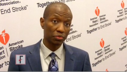
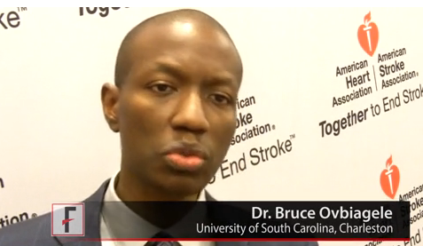
Dr. Bruce Ovbiagele spoke with us about the significance of the findings for both young adults and children who experience physical trauma.
The video associated with this article is no longer available on this site. Please view all of our videos on the MDedge YouTube channel
AT THE INTERNATIONAL STROKE CONFERENCE
Mobilize subarachnoid hemorrhage patients as soon as possible
SAN DIEGO – Aneurysmal subarachnoid hemorrhage patients left the ICU a mean of 3 days earlier when they were mobilized quickly after their stroke, according to a retrospective study from the Capital Institute for Neurosciences in Trenton, N.J.
Investigators there compared functional outcomes for 38 historical subarachnoid hemorrhage (SAH) controls, and 55 SAH patients after an early mobilization program was started in the neuro-ICU about 5 years ago.
The groups were matched for demographics, clip vs. coil ligation, and Hunt and Hess severity grade, among other things. Patients were generally in their 40s and 50s.
The early mobilization group got out of bed quicker (mean 4.2 days vs. 6.4 days), walked 50 feet sooner (mean 6.4 days vs. 10.5 days), and left the ICU earlier (mean 12.8 days vs. 15.7 days). The findings were all statistically significant; there was also a possible trend towards more discharges to the community (60% vs. 50%; P = .481).
"What we focused on was getting patients sitting in a chair. We were very aggressive; some of these patients were post-op day 1. Even with patients who were almost comatose, we did something, even just help them sit on the edge of the bed; the upright position facilitates arousal. We at least give it a shot to see if it helped," said investigator and physical therapist Melissa Arcaro, who presented the findings at the International Stroke Conference, sponsored by the American Heart Association.
Patients were assessed daily by physicians to see if they were hemodynamically and neurologically stable enough to participate. They had to be able to open their eyes and move one extremity on command. Transcranial Doppler ultrasound was performed before each session, and patients were excused for the day if their Lindegaard ratios were greater than 3.
Tachycardia and orthostatic issues were the main problems. "We didn’t have any patients who started to rebleed or had vasospasms that resulted in infarcts," Ms. Arcaro said.
Early mobilization is now standard practice at her ICU. "Patients are stuck in bed all the time, so they look forward to us coming in and helping them get up and brush their teeth or use the toilet. The biggest comment I get is, ‘Oh my God, I feel like a normal person again,’ " she said.
Prolonged bed rest and immobility are known to be bad for hospital patients, but "mobilization in the ICU, and the neuro-ICU in particular, is a new area. There’s not a whole lot of information out there," she said.
The investigators have no disclosures, and did not report outside funding.
AT THE INTERNATIONAL STROKE CONFERENCE
Major finding: Aneurysmal subarachnoid hemorrhage patients mobilized as early as post-op day 1 left the ICU sooner than did those who were left in bed (mean 12.8 vs. 15.7 days).
Data source: A retrospective functional outcomes review of 55 patients mobilized early in the neuro-ICU, and 38 controls.
Disclosures: The investigators have no disclosures and did not report their funding source.
Stroke risk jumps after head, neck trauma
SAN DIEGO – Eleven of every 100,000 patients younger than 50 years who were seen for traumatic injury developed an ischemic stroke within 4 weeks, a study of data on 1.3 million people found.
Patients with head or neck trauma were three times more likely overall to have a stroke than were those who had other forms of trauma, although the risk varied by age, Dr. Heather Fullerton said in a press briefing at the International Stroke Conference, sponsored by the American Heart Association. Among those with head or neck trauma, ischemic strokes occurred in 0.01% of the children and in 0.05% of the adults within a month of being seen.
Rates in both those age groups were significantly higher than rates reported in the literature for similar ages in the general population, she added.
"Strokes are something that affect young people, not just older people, particularly after a traumatic event," the findings showed, so people should apply the FAST criteria for recognizing the warning signs of stroke to potential stroke victims of any age, Dr. Fullerton said. The FAST acronym refers to the face (does the smile droop?), the arms (if arms are raised, does one drift downward?), speech (slurred or strange?), and time (get emergency help if these signs are present).
"People should seek emergency care for those symptoms regardless of their age, but especially if they happen to have a recent traumatic event," said Dr. Fullerton, professor of neurology at the University of California, San Francisco, and director of the pediatric stroke and cerebrovascular disease center there. Emergency departments see more than 2 million people under age 50 every month for nonfatal traumatic injuries in the United States.
The investigators analyzed data on patients aged 50 years or younger who were insured by Kaiser Permanente and seen in emergency departments or admitted for trauma at either Kaiser or non-Kaiser hospitals in 1997-2011. A neurologist reviewed the records and excluded cases inconsistent with ischemic stroke.
Combining the findings with other national data, she estimated that 214 people younger than 50 years develop an ischemic stroke after a traumatic injury every month in the United States.
Among patients with head or neck trauma, 48 of every 100,000 adults in the study developed a stroke within a month, compared with stroke rates in the general population of young adults of approximately 10/100,000 adults per year, not per month, she said.
In children with head or neck trauma, 11/100,000 in the study developed stroke within a month, compared with general population rates of approximately 2.5 strokes per 100,000 children per year, not per month. Previous research by Dr. Fullerton and her associates suggests that the highest risk in children is within the first week after trauma, she added.
Trauma can tear blood vessels that lead to the brain and cause blood clots resulting in ischemic stroke. Ten percent of patients in the study who developed a stroke were diagnosed with tears in blood vessels leading to the brain, but some of these arterial dissections were diagnosed after the stroke occurred. Most of the strokes in the study probably were due to arterial dissection but were possibly mild enough that an obvious tear didn’t show up on imaging, Dr. Fullerton suggested.
The researchers next are planning a nested case-control study to help identify trauma patients at the highest risk of stroke and to examine the possibilities for stroke prevention.
The American Heart Association funded the current study. The investigators reported having no financial disclosures.
On Twitter @sherryboschert
AT THE INTERNATIONAL STROKE CONFERENCE
Major finding: Eleven of every 100,000 patients developed a stroke within 4 weeks of traumatic injury.
Data source: A retrospective analysis of data on 1.3 million Kaiser patients younger than 50 years who were seen in emergency departments or hospitals for trauma in 1997-2011.
Disclosures: The American Heart Association funded the study. The investigators reported having no financial disclosures.
VIDEO: In-ambulance magnesium treatment for stroke fails
SAN DIEGO – Dr. Jeffrey L. Saver explains how his trial of in-ambulance magnesium treatment for stroke was both a success and a failure. Magnesium therapy didn’t improve stroke outcomes, but successful administration of treatment within the first "golden hour" after stroke symptoms started paved the way for future trials, giving other potentially neuroprotective agents in the field.
Dr. Bruce Ovbiagele also gives his perspective on the importance of this study in our video interview.
The video associated with this article is no longer available on this site. Please view all of our videos on the MDedge YouTube channel
SAN DIEGO – Dr. Jeffrey L. Saver explains how his trial of in-ambulance magnesium treatment for stroke was both a success and a failure. Magnesium therapy didn’t improve stroke outcomes, but successful administration of treatment within the first "golden hour" after stroke symptoms started paved the way for future trials, giving other potentially neuroprotective agents in the field.
Dr. Bruce Ovbiagele also gives his perspective on the importance of this study in our video interview.

