User login
Risk-reducing salpingectomy during surgery for benign indications
Ovarian cancer is the most deadly gynecologic malignancy in the United States, with 14,270 deaths expected in 2014 (CA Cancer J. Clin. 2014;64:9-29 ). The 5-year overall survival remains less than 50%. Difficulties in treatment arise due to its aggressive nature, coupled with vague symptomatology and no effective screening test. Advanced-stage disease at the time of diagnosis is an unfortunate hallmark.
Traditional teaching about the pathogenesis of ovarian cancers has been that a metaplastic change in the mesothelial ovarian surface leads to their de novo development. Under this paradigm, 70% of serous tumors were ovarian, 17% peritoneal, and 13% tubal in origin. However, a major change occurred when BRCA carriers began having risk-reducing bilateral salpingo-oophorectomies (BSO). Sequential histologic sections of the adnexa found occult malignancy in 30% of fallopian tubes, but similar lesions were not present within the ovary (Am. J. Surg. Pathol. 1020;34:1407-16). Could these cancers in the fallopian tube be the precursor lesion and then seed or spread to the ovarian surface?
Molecular and genetic analysis of these serous tubal intraepithelial carcinoma (STIC) cells found p53 mutations that were identical to those in concurrent “ovarian” tumors. STICs have been identified in 70% of nonhereditary forms of serous ovarian cancer. Under this paradigm of STIC as tubal in origin, only 28% of serous tumors were ovarian and 64% were of tubal origin (Am. J. Surg. Pathol. 2007;31:161-9).
This paradigm shift elicited new hypotheses about the etiology of endometrioid and clear cell types of ovarian cancer. Rather than metaplasia of the surface epithelium, endometrioid and clear cell tumors may result from retrograde menstruation, with the fallopian tube acting as a conduit for cells to gain access to the peritoneal cavity and the ovarian surface. This hypothesis provides a mechanism for the 34% reduction in ovarian cancer with bilateral tubal ligation (Hum. Reprod. Update 2011;17:55-67). Interestingly, in a large collaborative pooled analysis, the risk reduction of tubal ligation was greatest for endometrioid and clear cell subtypes (Int. J. Epidemiol. 2013;42:579-89).
Given the mounting evidence of fallopian tube involvement in the development of ovarian cancers, there are new primary prevention considerations. After 5 or more years of oral contraceptive use, a 50% reduction in the relative risk of ovarian cancer has been reported (Ann. Epidemiol. 2011;21:188-96). Bilateral salpingo-oophorectomy provides a greater than 50% risk reduction even in the highest-risk BRCA-positive population. However, results from the Nurses’ Health Study suggest that while there are fewer cancers in a low-risk population following BSO, it comes with an increase in all-cause mortality, predominately due to negative cardiovascular effects. With these issues in mind, is it time to consider incorporating prophylactic bilateral salpingectomy in benign gynecologic surgery (Obstet. Gynecol. 2013;121:709-16)?
While salpingectomy at the time of hysterectomy for benign conditions or for sterilization is becoming more common, there are concerns about premature loss of ovarian function secondary to compromise of ovarian blood supply. However, amassing data demonstrates preserved ovarian function. A retrospective study comparing total laparoscopic hysterectomy (TLH) to TLH with bilateral salpingectomy found no difference in markers of ovarian function (anti-Müllerian hormone, FSH, antral follicle count, mean ovarian diameters) up to 3 months postoperatively (Gynecol. Oncol. 2013;129:448-51). In a randomized controlled trial, 30 women were 1:1 randomized to TLH vs. TLH with salpingectomy. There was no change in anti-Müllerian hormone levels (at 3 months), operative time, or estimated blood loss (Fertil. Steril. 2013;100:1704-8). While there are concerns about the paucity of long-term follow-up data, these initial studies are encouraging. Additionally, a large retrospective study of 540 BRCA-negative patients found no difference in surgical outcomes with salpingectomy (estimated blood loss, hospital stay), and furthermore, the study found that removal of the tubes significantly reduces the risk of developing subsequent benign adnexal lesions by nearly 50% (J. Cancer Res. Clin. Oncol. 2014;140:859-65).
Though salpingectomy removes tubal re-anastomosis as an option in cases of “tubal ligation regret,” wisely choosing candidates can minimize this risk. Women less than 30 years old are at highest risk for regret, and the decision for salpingectomy in these patients should be made with caution and extensive counseling. Yet recently, emerging thought leaders in family planning have called for removal to be routinely considered (Obstet. Gynecol. 2014;124:596-9).
Surgical technique involves electrosurgery or suture ligation just inferior to the fallopian tube, ligating the fallopian branches of the ovarian and utero-ovarian arteries while avoiding unnecessary involvement of ovarian branches within the mesosalpinx. Since the fimbria are thought to be the site of origin for many serous carcinomas, removing the fimbrial portion of the tube is crucial.
Ovarian cancer remains the most deadly gynecologic malignancy. Efforts to find effective screening methods have not yet delivered. Pathologic data confirms that over half of “ovarian” cancers are actually of tubal origin, and we should consider risk-reducing salpingectomy in the low-risk population. The Society of Gynecologic Oncology in their November 2013 Clinical Practice Statement stated, “For women at average risk of ovarian cancer, risk-reducing salpingectomy should also be discussed and considered with patients at the time of abdominal or pelvic surgery, hysterectomy or in lieu of tubal ligation [once childbearing is complete].”
Dr. Pierce is a third-year resident in the department of obstetrics and gynecology at the University of North Carolina at Chapel Hill. Dr. Clarke-Pearson is the chair and the Robert A. Ross Distinguished Professor of Obstetrics and Gynecology, and a professor in the division of gynecologic oncology at the university. Dr. Pierce and Dr. Clarke-Pearson said that they had no relevant financial disclosures.

LOY linked to higher risk of cancer, mortality in men

Credit: William Weinert
SAN DIEGO—Age-related loss of the Y chromosome (LOY) from blood cells is associated with an elevated risk of cancer and mortality, a new study indicates.
This finding could help explain why men tend to have a shorter life span and higher rates of non-sex-specific cancers than women, said Lars Forsberg, PhD, of Uppsala University in Sweden.
He and his colleagues presented this research at the American Society of Human Genetics 2014 Annual Meeting and described it
in a letter to Nature Genetics.
LOY, which occurs occasionally as a man’s blood cells replicate, was first reported nearly 50 years ago and remains largely unexplained in both its causes and effects. Recent advances have allowed researchers to use a blood test to detect when only a small fraction of a man’s blood cells have undergone LOY.
Dr Forsberg and his colleagues studied the relationship between LOY, cancer, and mortality using blood samples from 1153 men aged 70 to 84 years who were followed for up to 40 years.
In survival analyses, the investigators studied 982 participants who did not have cancer prior to sampling. The team adjusted their analyses for age, hypertension, exercise, smoking, diabetes, body mass index, low-density lipoprotein cholesterol, high-density lipoprotein cholesterol, and ancestry.
The researchers calculated the degree of LOY for each subject from the median log R ratio values for approximately 2560 probes in the male-specific region of chromosome Y (mLRR-Y).
In a primary analysis, the investigators found that men with a higher degree of LOY had an increased risk of all-cause mortality (hazard ratio [HR]=2.13, P=0.029).
And LOY was a key risk factor for cancer-related mortality (HR=3.76, P=0.022), but it was not significantly associated with non-cancer-related mortality (P=0.245).
The researchers then scored participants on the basis of a defined threshold of mLRR-Y. Men with an mLRR-Y of –0.4 or less were scored as 1, and other subjects were scored as 0.
This analysis confirmed the effect of LOY on the risk of all-cause mortality (HR=1.91, P=0.010). It also showed that median survival times in men with LOY were 5.5 years shorter than for the other subjects, representing half the survival time.
In addition, the analysis confirmed the effect of LOY on the risk of cancer-related mortality (HR=3.29, P=0.003) and death from non-hematologic cancers (HR=3.62, P=0.003).
The investigators could not test the effect of LOY on mortality related to hematologic malignancies, as only one man with an mLRR-Y of –0.4 or less died from a hematologic malignancy.
However, the team did find that the risk of any cancer diagnosis was higher in men with an mLRR-Y of −0.4 or less (HR=2.47, P=0.014). And the same was true for the risk of developing a non-hematologic cancer (HR=2.68, P=0.008).
“Many people think the Y chromosome only contains genes involved in sex determination and sperm production,” said Jan Dumanski, MD, PhD, also of Uppsala University. “In fact, these genes have other important functions, such as possibly playing a role in preventing tumors.”
The researchers noted that LOY in blood cells is associated with many different cancers, and this may be because Y chromosome genes enable blood cells to assist with immunosurveillance.
“Our hypothesis is that LOY disrupts the immunosurveillance normally conducted by blood cells, allowing tumors to grow unchecked and develop into cancer,” Dr Forsberg said.
These findings suggest a new approach to early detection of cancer risk in men: a blood test to assess LOY.
“LOY is not very dangerous in a small fraction of blood cells but becomes increasingly predictive of cancer as more cells lose their Y chromosome,” Dr Forsberg explained. “This takes years, so you’d have a window of time to do something to reduce your risk.”
The investigators are currently exploring LOY in more detail, including the effects of various lifestyle factors and other health conditions on LOY. They are also examining the frequency and consequences of LOY in different types of cells and throughout life. ![]()
Hospital acquisitions lead to increased patient costs

Credit: NCI
The trend of hospitals consolidating medical groups and physician practices is increasing the cost of patient care, new research suggests.
The study was conducted using 4 years of data from nearly 160 medical groups in California.
Results showed that hospital-owned physician organizations incurred higher expenditures than physician-owned organizations for services to patients covered by commercial health maintenance organization (HMO) insurance.
The findings, published in JAMA, come as a growing number of local hospitals and large, multihospital systems in the US are acquiring physician groups and medical practices.
“This consolidation is meant to better coordinate care and to have a stronger bargaining position with insurance plans,” said James Robinson, PhD, of the University of California, Berkeley School of Public Health.
“The movement also aligns with the goals of the Affordable Care Act, since physicians and hospitals working together in ‘accountable care organizations’ can provide care better than the traditional fee-for-service and solo-practice models. The intent of consolidation is to reduce costs and improve quality, but the problem with all this is that hospitals are very expensive and complex organizations, and they are not known for their efficiency and low prices.”
Dr Robinson teamed up with Kelly Miller, of Integrated Healthcare Association in Oakland, California, to analyze data on 158 major medical groups. The data spanned the period from 2009 to 2012.
Of the 158 organizations, 118 (75%) were physician-owned and provided care for 3,065,551 patients, 19 (12%) were owned by local hospitals and provided care for 728,608 patients, and 21 (13%) were owned by multihospital systems and provided care for 693,254 patients.
The patients were covered by commercial HMO insurance. The data did not include patients covered by commercial preferred provider organization (PPO) insurance, Medicare, or Medicaid.
The researchers assessed costs related to physician visits, inpatient hospital admissions, outpatient surgery and diagnostic procedures, drugs, and all other forms of medical care except mental health services.
Results revealed that the average expenditure per patient across all physician organizations increased by 16.5% between 2009 and 2012, from $2954 to $3443.
By 2012, expenditures per patient had increased to an average of $3066 in physician-owned organizations, $4312 in local hospital-owned organizations, and $4776 in multihospital system-owned organizations.
This represents a 40.6% relative difference in expenditures per patient associated with hospital ownership and a 55.8% relative difference associated with ownership by a multihospital system compared with ownership by member physicians.
After adjusting for patient severity and other factors over the period, local hospital-owned physician organizations incurred expenditures per patient 10.3% higher than physician-owned organizations. And organizations owned by multihospital systems incurred expenditures 19.8% higher than physician-owned organizations.
Dr Robinson said these findings might be explained by the fact that, once a medical group has been acquired by a multihospital system, physicians in those groups are expected to admit their patients to the high-priced hospital.
“Hospital-owned medical groups usually are expected to conduct ambulatory surgery and diagnostic procedures in the outpatient departments of their parent hospital, but hospital outpatient departments are much more costly and charge much higher prices than freestanding, non-hospital ambulatory centers,” he said.
Dr Robinson added that public policy should not encourage mergers and acquisitions as a means of promoting collaboration. Instead, policymakers should consider supporting the use of bundled payments for hospitals and physicians to improve the coordination of care.
“Hospitals are an essential part of the healthcare system, but they should not be the center of the delivery system,” he said. “Rather, physician-led organizations based in ambulatory and community settings are likely to be more efficient and provide cheaper care.”
The researchers noted that their findings are limited to California, and further studies should be conducted using data from other states.
“Nevertheless, these findings are important,” Dr Robinson said, “since California is the nation’s leader in terms of having physicians participate in large medical groups that already perform the functions ascribed to ‘accountable care organizations’ by the Obama administration.” ![]()
Less competition equals higher healthcare costs

Credit: Petr Kratochvil
Medical practices in less competitive markets charge more for their services, according to a study published in JAMA.
The study, based on US healthcare data from 2010, provides new information about the effects of competition on prices for office visits paid by preferred provider organizations (PPOs).
“The research comes out of trying to understand some dramatic changes that have occurred in the healthcare system over a couple of decades,” said Laurence Baker, PhD, of the Stanford University School of Medicine in California.
One such change is the shift from practices with one or two doctors toward larger, more complex organizations with many physicians. An impact of this can be reduced competition among physician practices.
Dr Baker and his colleagues sought to understand how variation in the amount of competition within a region affects the amounts doctors are paid.
The researchers assessed the relationship between competition and prices paid by PPOs for the most commonly billed services within 10 prominent physician specialties—internal medicine, family practice, cardiology, dermatology, gastroenterology, neurology, general surgery, orthopedics, urology, and otolaryngology.
To establish what prices various medical practices were paid for services, the team used Truven Analytics MarketScan Commercial Claims and Encounters database, which contains the prices paid to physicians for more than 49 million privately insured people from all over the US. They obtained the number of claims and the mean price paid for each service in 1058 counties representing all 50 states.
To measure competitiveness, the researchers drew inspiration from the business world. Using Medicare data, they adapted a standard economic competition measure to track physician practice competition for different US regions.
The Hirschman-Herfindahl Index (HHI) uses the relative sizes of practices to measure market concentration. A higher HHI indicates a less competitive market, and a lower HHI indicates higher competition.
Less competition, higher prices
The researchers found that less competition among physician practices was associated with higher prices paid by private PPOs for office visits.
Across 10 types of office visits, the difference in the HHI was associated with average prices for office visits 8.3% to 16.1% higher. In a more conservative model, the difference in the HHI was associated with 3.5% to 5.4% higher average prices.
The researchers pointed out that, in 2011, privately insured individuals in the US spent nearly $250 billion on physician services. In that context, these small percentage increases could translate to tens of billions of dollars in extra spending.
The team also found that, between 2003 and 2010, prices increased more rapidly in areas that were less competitive. Even when there is no change in HHI, practices in less competitive areas could continue to drive up prices.
“These larger organizations might have better processes in place to optimize care,” said Kate Bundorf, PhD, also of the Stanford University School of Medicine.
“But our research also points out [that we] have to think about the effect on prices and try to balance those two things when we think about how to form policy about these organizations.”
Dr Baker echoed that sentiment.
“Sometimes, it can be tempting to say our goals for the healthcare system should be only about taking care of patients and doing it as well as possible,” he said. “We don’t want to worry about the economics. But the truth is, we do have to worry about the prices because the bill does come, even if you wish it wouldn’t.” ![]()
Docs often don’t know about patients’ CVCs

A multicenter study showed that roughly 1 in 5 physicians did not know when a hospitalized patient had a central venous catheter (CVC) in place.
Hospitalists were the least likely to know of a CVC’s presence, followed by general medicine teaching attendings, interns, and residents.
These findings raise questions about patient safety, as CVCs pose a risk of bloodstream infection and thrombosis, both of which can be prevented by removing catheters that are no longer necessary.
“We know that central venous catheters are invaluable for the safe and comprehensive care of some hospitalized patients, but just as they are helpful, they can be harmful,” said study author Vineet Chopra, MD, of the University of Michigan Health System in Ann Arbor.
“The key to preventing complications is to remove devices when they are no longer necessary, but that requires knowing they are there in the first place. Our findings suggest that patient safety may be jeopardized if medical providers don’t improve their practices regarding device awareness.”
Dr Chopra and his colleagues reported their findings in the Annals of Internal Medicine.
The study included 990 patients and 1881 clinical assessments at 3 academic medical centers in the US. Responses from interns (n=454), residents and physician extenders (n=513), general medicine teaching attendings (n=245), subspecialty attendings (n=176), intensivists (n=95), and hospitalists (n=398) were included.
The overall prevalence of CVCs was 21.1%, 60.3% of which were peripherally inserted central catheters (PICCs). The remaining CVCs were triple-lumen catheters inserted in the neck (19.6%), chest (11.5%), or groin (8.6%).
In all, 21.2% of clinicians interviewed did not know when a patient had a CVC in place. And 5.6% of clinicians said a patient had a CVC when there was no device in place.
Hospitalists were most likely to be unaware of a CVC (30.5%), followed by general medicine teaching attendings (25.8%), interns (19.1%), and residents (13.8%).
When assessed by service, critical care physicians were the least likely to be unaware of a CVC (12.6%), compared to general medicine teaching attendings/hospitalists (26.2%) and clinicians in other subspecialties (22.5%). Awareness was best among specialties that used CVCs often, such as cardiology and oncology.
Lack of awareness was greatest for PICCs; 25.1% of clinicians were unaware of a PICC’s presence, and 15.6% were unaware of a triple lumen catheter’s presence.
“These findings have important patient safety and policy implications for health systems nationwide,” Dr Chopra said, “because they suggest that removal of clinically unnecessary catheters may be limited by lack of awareness by providers, especially in non-intensive care settings.” ![]()
ID CONSULT: Influenza virus and pneumococci dance together
Most practitioners know that the flu vaccine has been proven to reduce the frequency of middle ear infections, sinusitis, and pneumonia. However, how that happens is not as clear. My group has been studying the details of the interaction between flu virus and pneumococci to unravel the steps in the dance between the flu virus and the pneumococcus in the nasopharynx that results in significant respiratory diseases. Pneumococci live in the posterior part of the nose and upper pharynx as commensal bacteria in all of us, harmlessly present in relatively low numbers. The bacteria are so common that studies to detect pneumococci in the nasopharynx discover their presence in up to 80% of infants and young children, and about 20% of adults at any one time. The bacteria are harmless in patients that have a competent immune system unless an intercurrent viral upper respiratory infection (URI) occurs.
The trigger in pathogenesis of pneumococcal infections is a viral URI, and particularly influenza infection. The combination of pneumococci and flu in the nose can cause compromise in all four aspects of host defense: 1) structural change, 2) physiologic change, 3) innate immunity change, and 4) adaptive immunity change. Structural change is swelling of the nasal passageways, Eustachian tube, osteomeatal sinus pathway, and tracheobronchial tree. Physiologic change is increased mucus production and reduced cilia beat, resulting in stasis of thickened mucus in the respiratory tree. Thus the stage is set for compromise in the immune response.

Innate immunity basically translates to the response of neutrophils, macrophages, and lymphocytes that are resident in the respiratory pathways or migrate there in response to signals from the site of infection that a problem is brewing. To start the process of innate immunity, chemicals are released from resident epithelial cells, lymphocytes, and neutrophils/macrophages. The chemicals are called cytokines and chemokines. The viruses enter the epithelial cells of the nasopharynx and tracheobronchial tree, and leave a change on the surface of the epithelial cells that alerts lymphocytes to kill and destroy those cells harboring virus. Neutrophils and macrophages ingest the bacteria by recognizing surface proteins on the bacteria that are foreign. Sometimes that is all that is needed, and the host clears the infection. But sometimes the innate response is not enough.
The innate response is good and bad. The bad part is that the release of the cytokines and chemokines and the migration of immune cells to the site of infection results in the release of even more cytokines and chemokines that cause increased inflammation. Microbes love inflammation. The inflammation caused by the virus, such as flu virus, creates a very favorable environment for the pneumococci. So the pneumococci start to reproduce in abundance. Then when the secretions of the nose are swept into the Eustachian tube and middle ear or the sinus drainage pathways and then to the sinuses or into the trachea and bronchi and then the lungs, we see the clinical manifestations of acute otitis media, sinusitis, or pneumonia. The innate response failed.
The adaptive response – as the word implies – is when the immune cells recognize and adapt to the presence of foreign microbes by recognizing their presence, migrating to lymph nodes and spleen, communicating with each other, and consequently multiplying into great numbers. The interaction between the immune cells – T cells and B cells – in the lymph node and migration back to the site of infection takes a few days to occur (3-5 days) if the host has prior immunity from prior infections or vaccination. If there is no prior immunity and no vaccination, then it takes 10-14 days for the adaptive immunity response to kick in and clear the infection. During that extra time, the pneumococci are gaining in numbers, causing more inflammation, and we see those clinical signs of fever, redness, and swelling at the site of infection, and pain.
So influenza can cause all of the events above by itself, but when the virus dances with the pneumococci, and the pneumococci benefit from the partnership, that is the most frequent cause of acute otitis media, sinusitis, and pneumonia. And all of that could have been prevented in most of our patients if they only got their annual flu vaccine.
Dr. Pichichero, a specialist in pediatric infectious diseases, is director of the Research Institute, Rochester (N.Y.) General Hospital. He is also a pediatrician at Legacy Pediatrics in Rochester. The study was supported by a National Institutes of Health grant. Dr. Pichichero said he had no relevant financial disclosures. Email him at pdnews@frontlinemedcom.com.
Most practitioners know that the flu vaccine has been proven to reduce the frequency of middle ear infections, sinusitis, and pneumonia. However, how that happens is not as clear. My group has been studying the details of the interaction between flu virus and pneumococci to unravel the steps in the dance between the flu virus and the pneumococcus in the nasopharynx that results in significant respiratory diseases. Pneumococci live in the posterior part of the nose and upper pharynx as commensal bacteria in all of us, harmlessly present in relatively low numbers. The bacteria are so common that studies to detect pneumococci in the nasopharynx discover their presence in up to 80% of infants and young children, and about 20% of adults at any one time. The bacteria are harmless in patients that have a competent immune system unless an intercurrent viral upper respiratory infection (URI) occurs.
The trigger in pathogenesis of pneumococcal infections is a viral URI, and particularly influenza infection. The combination of pneumococci and flu in the nose can cause compromise in all four aspects of host defense: 1) structural change, 2) physiologic change, 3) innate immunity change, and 4) adaptive immunity change. Structural change is swelling of the nasal passageways, Eustachian tube, osteomeatal sinus pathway, and tracheobronchial tree. Physiologic change is increased mucus production and reduced cilia beat, resulting in stasis of thickened mucus in the respiratory tree. Thus the stage is set for compromise in the immune response.
Innate immunity basically translates to the response of neutrophils, macrophages, and lymphocytes that are resident in the respiratory pathways or migrate there in response to signals from the site of infection that a problem is brewing. To start the process of innate immunity, chemicals are released from resident epithelial cells, lymphocytes, and neutrophils/macrophages. The chemicals are called cytokines and chemokines. The viruses enter the epithelial cells of the nasopharynx and tracheobronchial tree, and leave a change on the surface of the epithelial cells that alerts lymphocytes to kill and destroy those cells harboring virus. Neutrophils and macrophages ingest the bacteria by recognizing surface proteins on the bacteria that are foreign. Sometimes that is all that is needed, and the host clears the infection. But sometimes the innate response is not enough.
The innate response is good and bad. The bad part is that the release of the cytokines and chemokines and the migration of immune cells to the site of infection results in the release of even more cytokines and chemokines that cause increased inflammation. Microbes love inflammation. The inflammation caused by the virus, such as flu virus, creates a very favorable environment for the pneumococci. So the pneumococci start to reproduce in abundance. Then when the secretions of the nose are swept into the Eustachian tube and middle ear or the sinus drainage pathways and then to the sinuses or into the trachea and bronchi and then the lungs, we see the clinical manifestations of acute otitis media, sinusitis, or pneumonia. The innate response failed.
The adaptive response – as the word implies – is when the immune cells recognize and adapt to the presence of foreign microbes by recognizing their presence, migrating to lymph nodes and spleen, communicating with each other, and consequently multiplying into great numbers. The interaction between the immune cells – T cells and B cells – in the lymph node and migration back to the site of infection takes a few days to occur (3-5 days) if the host has prior immunity from prior infections or vaccination. If there is no prior immunity and no vaccination, then it takes 10-14 days for the adaptive immunity response to kick in and clear the infection. During that extra time, the pneumococci are gaining in numbers, causing more inflammation, and we see those clinical signs of fever, redness, and swelling at the site of infection, and pain.
So influenza can cause all of the events above by itself, but when the virus dances with the pneumococci, and the pneumococci benefit from the partnership, that is the most frequent cause of acute otitis media, sinusitis, and pneumonia. And all of that could have been prevented in most of our patients if they only got their annual flu vaccine.
Dr. Pichichero, a specialist in pediatric infectious diseases, is director of the Research Institute, Rochester (N.Y.) General Hospital. He is also a pediatrician at Legacy Pediatrics in Rochester. The study was supported by a National Institutes of Health grant. Dr. Pichichero said he had no relevant financial disclosures. Email him at pdnews@frontlinemedcom.com.

Gun ownership in patients with dementia
The video associated with this article is no longer available on this site. Please view all of our videos on the MDedge YouTube channel
The video associated with this article is no longer available on this site. Please view all of our videos on the MDedge YouTube channel
The video associated with this article is no longer available on this site. Please view all of our videos on the MDedge YouTube channel

Acting strange after trying to ‘get numb’
CASE Numb and confused
Mr. L, age 17, is admitted to the hospital after ingesting 24 diphenhydramine 25-mg tablets in 3 hours as a possible suicide attempt. His parents witnessed him behaving strangely and brought him to the hospital. They state that their son was visibly agitated and acting inappropriately. He was seen talking to birds, trees, and the walls of the house.
Mr. L says he is upset because he broke up with his girlfriend a week earlier after she asked if they could “take a break.” He says that he took the diphenhydramine because he wanted to “get numb” to deal with the emotional stress caused by the break-up.
After the break-up, Mr. L experienced middle-to-late insomnia and was unable to get more than 3 or 4 hours of sleep a night. He reports significant fatigue, depressed mood, anhedonia, impaired concentration, and psychomotor retardation. He denies homicidal ideation or auditory and visual hallucinations.
As an aside, Mr. L reports that, for the past year, he had difficulties with gender identity, sometimes thinking that he might be better off if he had been born a girl and that he felt uncomfortable in a male body.
Which treatment option would you choose for Mr. L’s substance abuse?
a) refer him to a 12-step program
b) begin supportive measures
c) administer activated charcoal
d) prescribe a benzodiazepine to control agitation
The authors’ observations
As youths gain increasing access to medical and pharmaceutical knowledge through the Internet and other sources, it appears that adolescent drug abuse has, in part, shifted toward more easily attainable over-the-counter (OTC) medications. Diphenhydramine, a first-generation antihistamine, can be abused for its effects on the CNS, such as disturbed coordination, irritability, paresthesia, blurred vision, and depression. Effects of diphenhydramine are increased by the presence of alcohol, monoamine oxidase inhibitors, diazepam, hypnotics, sedatives, tranquilizers, and other CNS depressants. In 2011, diphenhydramine abuse was involved in 19,012 emergency room visits, of which 9,301 were for drug-related suicide attempts.1
Diphenhydramine is an inverse agonist of the histamine H1 receptor.2 It is a member of the ethanolamine subclass of antihistaminergic agents.3 By reversing the effects of histamine on capillaries, diphenhydramine can reduce the intensity of allergic symptoms. Diphenhydramine also crosses the blood–brain barrier and antagonizes H1 receptors centrally.
Used as a common sleep aid and allergy medication, the drug works primarily as an H1 receptor partial agonist, but also is a strong competitive antagonist at muscarinic acetylcholine receptors.4 It is abused for its sedative effects and its capacity to cause delirium and hallucinations.5 Diphenhydramine can have a stimulatory effect in children and young adults, instead of the sedating properties seen in adults.6 Such misuse is concerning because diphenhydramine overdose can lead to delirium, confusion, and hallucinations, tachycardia, seizures, mydriasis, xerostomia, urinary retention, ileus, anhidrosis, and hyperthermia. In severe cases it has been associated with cardiac arrhythmias, rhabdomyolysis, status epilepticus, and death.4,6 Neurologic symptoms of diphenhydramine overdose are listed in Table 1.
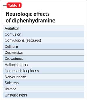
HISTORY Polysubstance abuse
Mr. L has a 2-year history of major depressive disorder and a history of Cannabis abuse with physiological dependence; Robitussin (base active ingredient, guaifenesin) and hydrocodone abuse with physiological dependence; 3,4-methylenedioxymethamphetamine (MDMA) abuse; and diphenhydramine abuse. He also has a history of gender dysphoria, although he reports that these feelings have become less severe over the past year.
Mr. L attends bi-weekly appointments with an outpatient psychiatrist and reportedly adheres to his medication regimen: fluoxetine, 40 mg/d, and risperidone, 1 mg at bedtime. He denies previous suicidal ideation, suicide attempts, homicidal ideation, or homicidal attempts. He reports no history of physical, sexual, or emotional abuse. He gets good grades in school and has no outstanding academic problems.
Mr. L began using Cannabis at age 14; his last use was 3 weeks before admission. He is guarded about his use of Robitussin, hydrocodone, and MDMA. However, Mr. L reports that he has researched diphenhydramine on the internet and believes that he can safely take up to 1,200 mg without overdosing. He reports normally taking 450 mg of diphenhydramine daily. Mr. L reports difficulty urinating after using diphenhydramine but no other physical complaints.
Mr. L lives with his father and stepmother and has a history of one psychiatric hospitalization at a different facility 2 months ago, followed by outpatient therapy. He obtained his Graduate Equivalency Diploma (GED) and plans to attend college.
At age 5, Mr. L emigrated from Turkey to the United States with his parents. His mother returned to Turkey when he was age 6 and has had no contact with her son since. Whenever Mr. L visits Turkey with his father, the patient refuses to see her, as per collaterals. He gets along well with his stepmother, who is his maternal aunt. Mr. L has been bullied at school and reportedly has few friends.
On mental status examination, Mr. L has an appropriate appearance and appears to be his stated age. He shows good eye contact and is cooperative. Muscle tone and gait are within normal limits. He has no abnormal movements. Speech, thought processes, and associations are normal. He denies auditory hallucinations, visual hallucinations, suicidal ideation (although he presented with a probable suicide attempt), or homicidal ideation. No delusions are elicited.
Mr. L shows poor judgment about his drug use and situation. He demonstrates limited insight, because he says his only goal is to get out of the hospital. He is alert, awake, and oriented to person, place, and time. He shows no memory or knowledge impairment. He appears euthymic with an inappropriate and constricted affect. On neurologic exam, he had mild tremors in his hands. The authors’ observationsTreatment for diphenhydramine overdose should begin quickly to prevent life-threatening effects and reduce the risk for mortality. The toxin can be removed from the patient’s GI tract with activated charcoal or gastric lavage if the patient presents within 1 hour of ingesting the substance. Administering IV fluids will prevent dehydration. Cardiac functioning is monitored and benzodiazepines could be administered to manage seizures.
Key elements of a toxicologic physical examination include:
• eyes: pupillary size, symmetry, and response to light (vertical or horizontal nystagmus)
• oropharynx: moist or dry mucous membranes, presence or absence of the gag reflex, distinctive odors
• abdomen: presence or absence and quality of bowel sounds
• skin: warm and dry, warm and sweaty, or cool
• neurologic: level of consciousness and mental status, presence of tremors, seizures, or other movement disorders, presence or absence and quality of deep tendon reflexes.7
If a child or adolescent patient cannot communicate how much of a drug he (she) has ingested, questions to ask parents or other informants include:
• Was the medication purchased recently, and if so was the bottle or box full before the patient took the pills?
• If the medication was not new, how many pills were in the bottle before the patient got to it?
• If the medication was prescribed, how many pills were originally prescribed, when was the medication prescribed, and how many pills were already taken prior to the patient getting to the bottle?
• How many pills were left in the bottle?
• How many pills were seen around the area where the patient was found?
• How many pills were found in the patient’s mouth?7
Recommendations
It is well known that OTC medication abuse is a growing medical problem (Table 2). Antihistamines, including diphenhydramine, are readily available to minors and adults. Because of the powerful sedating effects of antihistamines, many adolescent health practitioners give them to patients who have insomnia as a sleep aid.8 As in our case, antihistamines are used recreationally for their hallucinogenic effects, at dosages of 300 to 700 mg.9 Severe symptoms of toxicity, such as delirium and psychosis, seizures, and coma, occur at dosages ≥1,000 mg.9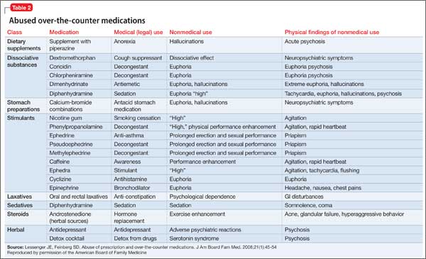
With growing abuse of these medications, we aim to encourage detailed history taking about abuse of OTC drugs, especially diphenhydramine in adolescent patients.
Outcome Improvement, discharge
Mr. L is given a dual diagnosis of diphenhydramine-induced psychotic disorder with
hallucinations and diphenhydramine-induced depressive disorder, both with onset during intoxication. He also is given a provisional diagnosis of psychotic disorder not otherwise specified and major depressive disorder. Last, he is given a diagnosis of Cannabis dependence with physiological dependence, MDMA abuse, hydrocodone abuse, and Robitussin abuse.
Mr. L is maintained on fluoxetine, 40 mg/d, and risperidone, 1 mg at bedtime and 0.5 mg in the morning. He receives milieu, individual, group, recreational, and medical therapy while in the hospital. Symptoms abate and he is discharged with a plan to follow up with outpatient providers.
Bottom Line
Abuse of over-the-counter (OTC) drugs, such as diphenhydramine, among youths is a growing problem. Remember to question adolescents who appear intoxicated or to have overdosed not only about abuse of alcohol and illicit substances but also of common—and easily and legally accessible—OTC drugs.
Related Resources
• Carr BC. Efficacy, abuse, and toxicity of over-the-counter cough and cold medicines in the pediatric population. Curr Opin Pediatr. 2006;18(2):184-188.
• Thomas A, Nallur DG, Jones N, et al. Diphenhydramine abuse and detoxification: a brief review and case report. J Psychopharmacol. 2009;23(1):101-105.
Drug Brand Names
Diazepam • Valium Hydrocodone • Vicodin
Diphenhydramine • Benadryl Risperidone • Risperdal
Fluoxetine • Prozac
Disclosures
The authors report no financial relationships with any company whose products are mentioned in this article or with manufacturers of competing products.
1. U.S. Department of Health and Human Services. Drug Abuse Warning Network, 2011: National estimates of drug-related emergency department visits. http://www.samhsa. gov/data/2k13/DAWN2k11ED/DAWN2k11ED.htm. Published May 2013. Accessed on September 29, 2014.
2. Yamashiro K, Kiryu J, Tsujikawa A, et al. Suppressive effects of histamine H1 receptor antagonist diphenhydramine on the leukocyte infiltration during endotoxin-induced uveitis. Exp Eye Res. 2001;73(1):69-80.
3. Skidgel RA, Kaplan AP, Erdos EG. Histamine, bradykinin, and their antagonists. In: Brunton L, Chabner B, Knollman B, eds. Goodman & Gilman’s the pharmacological basis of therapeutics. 12th ed. New York, NY: McGraw Hill; 2011: 911-935.
4. Vearrier D, Curtis JA. Case files of the medical toxicology fellowship at Drexel University. Rhabdomyolysis and compartment syndrome following acute diphenhydramine overdose. J Med Toxicol. 2011;7(3):213-219.
5. Ho M, Tsai K, Liu C. Diphenhydramine overdose related delirium: a case report. Journal of Emergency and Critical Care Medicine. 2006;17(2):77-79.
6. Krenzelok EP, Anderson GM, Mirick M. Massive diphenhydramine overdose resulting in death. Ann Emerg Med. 1982;11(4):212-213.
7. Inaba AS. Toxicologic teasers: Testing your knowledge of clinical toxicology. Hawaii Med J. 1998;57(4):471-473.
8. Kaplan SL. Busner J. The use of prn and stat medication in three child psychiatric inpatient settings. Psychopharmacol Bull. 1997;33(1):161-164.
9. Radovanovic D, Meier PJ, Guirguis M, et al. Dose-dependent toxicity of diphenhydramine overdose. Hum Exp Toxicol. 2000;19(9):489-495.
CASE Numb and confused
Mr. L, age 17, is admitted to the hospital after ingesting 24 diphenhydramine 25-mg tablets in 3 hours as a possible suicide attempt. His parents witnessed him behaving strangely and brought him to the hospital. They state that their son was visibly agitated and acting inappropriately. He was seen talking to birds, trees, and the walls of the house.
Mr. L says he is upset because he broke up with his girlfriend a week earlier after she asked if they could “take a break.” He says that he took the diphenhydramine because he wanted to “get numb” to deal with the emotional stress caused by the break-up.
After the break-up, Mr. L experienced middle-to-late insomnia and was unable to get more than 3 or 4 hours of sleep a night. He reports significant fatigue, depressed mood, anhedonia, impaired concentration, and psychomotor retardation. He denies homicidal ideation or auditory and visual hallucinations.
As an aside, Mr. L reports that, for the past year, he had difficulties with gender identity, sometimes thinking that he might be better off if he had been born a girl and that he felt uncomfortable in a male body.
Which treatment option would you choose for Mr. L’s substance abuse?
a) refer him to a 12-step program
b) begin supportive measures
c) administer activated charcoal
d) prescribe a benzodiazepine to control agitation
The authors’ observations
As youths gain increasing access to medical and pharmaceutical knowledge through the Internet and other sources, it appears that adolescent drug abuse has, in part, shifted toward more easily attainable over-the-counter (OTC) medications. Diphenhydramine, a first-generation antihistamine, can be abused for its effects on the CNS, such as disturbed coordination, irritability, paresthesia, blurred vision, and depression. Effects of diphenhydramine are increased by the presence of alcohol, monoamine oxidase inhibitors, diazepam, hypnotics, sedatives, tranquilizers, and other CNS depressants. In 2011, diphenhydramine abuse was involved in 19,012 emergency room visits, of which 9,301 were for drug-related suicide attempts.1
Diphenhydramine is an inverse agonist of the histamine H1 receptor.2 It is a member of the ethanolamine subclass of antihistaminergic agents.3 By reversing the effects of histamine on capillaries, diphenhydramine can reduce the intensity of allergic symptoms. Diphenhydramine also crosses the blood–brain barrier and antagonizes H1 receptors centrally.
Used as a common sleep aid and allergy medication, the drug works primarily as an H1 receptor partial agonist, but also is a strong competitive antagonist at muscarinic acetylcholine receptors.4 It is abused for its sedative effects and its capacity to cause delirium and hallucinations.5 Diphenhydramine can have a stimulatory effect in children and young adults, instead of the sedating properties seen in adults.6 Such misuse is concerning because diphenhydramine overdose can lead to delirium, confusion, and hallucinations, tachycardia, seizures, mydriasis, xerostomia, urinary retention, ileus, anhidrosis, and hyperthermia. In severe cases it has been associated with cardiac arrhythmias, rhabdomyolysis, status epilepticus, and death.4,6 Neurologic symptoms of diphenhydramine overdose are listed in Table 1.
HISTORY Polysubstance abuse
Mr. L has a 2-year history of major depressive disorder and a history of Cannabis abuse with physiological dependence; Robitussin (base active ingredient, guaifenesin) and hydrocodone abuse with physiological dependence; 3,4-methylenedioxymethamphetamine (MDMA) abuse; and diphenhydramine abuse. He also has a history of gender dysphoria, although he reports that these feelings have become less severe over the past year.
Mr. L attends bi-weekly appointments with an outpatient psychiatrist and reportedly adheres to his medication regimen: fluoxetine, 40 mg/d, and risperidone, 1 mg at bedtime. He denies previous suicidal ideation, suicide attempts, homicidal ideation, or homicidal attempts. He reports no history of physical, sexual, or emotional abuse. He gets good grades in school and has no outstanding academic problems.
Mr. L began using Cannabis at age 14; his last use was 3 weeks before admission. He is guarded about his use of Robitussin, hydrocodone, and MDMA. However, Mr. L reports that he has researched diphenhydramine on the internet and believes that he can safely take up to 1,200 mg without overdosing. He reports normally taking 450 mg of diphenhydramine daily. Mr. L reports difficulty urinating after using diphenhydramine but no other physical complaints.
Mr. L lives with his father and stepmother and has a history of one psychiatric hospitalization at a different facility 2 months ago, followed by outpatient therapy. He obtained his Graduate Equivalency Diploma (GED) and plans to attend college.
At age 5, Mr. L emigrated from Turkey to the United States with his parents. His mother returned to Turkey when he was age 6 and has had no contact with her son since. Whenever Mr. L visits Turkey with his father, the patient refuses to see her, as per collaterals. He gets along well with his stepmother, who is his maternal aunt. Mr. L has been bullied at school and reportedly has few friends.
On mental status examination, Mr. L has an appropriate appearance and appears to be his stated age. He shows good eye contact and is cooperative. Muscle tone and gait are within normal limits. He has no abnormal movements. Speech, thought processes, and associations are normal. He denies auditory hallucinations, visual hallucinations, suicidal ideation (although he presented with a probable suicide attempt), or homicidal ideation. No delusions are elicited.
Mr. L shows poor judgment about his drug use and situation. He demonstrates limited insight, because he says his only goal is to get out of the hospital. He is alert, awake, and oriented to person, place, and time. He shows no memory or knowledge impairment. He appears euthymic with an inappropriate and constricted affect. On neurologic exam, he had mild tremors in his hands. The authors’ observationsTreatment for diphenhydramine overdose should begin quickly to prevent life-threatening effects and reduce the risk for mortality. The toxin can be removed from the patient’s GI tract with activated charcoal or gastric lavage if the patient presents within 1 hour of ingesting the substance. Administering IV fluids will prevent dehydration. Cardiac functioning is monitored and benzodiazepines could be administered to manage seizures.
Key elements of a toxicologic physical examination include:
• eyes: pupillary size, symmetry, and response to light (vertical or horizontal nystagmus)
• oropharynx: moist or dry mucous membranes, presence or absence of the gag reflex, distinctive odors
• abdomen: presence or absence and quality of bowel sounds
• skin: warm and dry, warm and sweaty, or cool
• neurologic: level of consciousness and mental status, presence of tremors, seizures, or other movement disorders, presence or absence and quality of deep tendon reflexes.7
If a child or adolescent patient cannot communicate how much of a drug he (she) has ingested, questions to ask parents or other informants include:
• Was the medication purchased recently, and if so was the bottle or box full before the patient took the pills?
• If the medication was not new, how many pills were in the bottle before the patient got to it?
• If the medication was prescribed, how many pills were originally prescribed, when was the medication prescribed, and how many pills were already taken prior to the patient getting to the bottle?
• How many pills were left in the bottle?
• How many pills were seen around the area where the patient was found?
• How many pills were found in the patient’s mouth?7
Recommendations
It is well known that OTC medication abuse is a growing medical problem (Table 2). Antihistamines, including diphenhydramine, are readily available to minors and adults. Because of the powerful sedating effects of antihistamines, many adolescent health practitioners give them to patients who have insomnia as a sleep aid.8 As in our case, antihistamines are used recreationally for their hallucinogenic effects, at dosages of 300 to 700 mg.9 Severe symptoms of toxicity, such as delirium and psychosis, seizures, and coma, occur at dosages ≥1,000 mg.9
With growing abuse of these medications, we aim to encourage detailed history taking about abuse of OTC drugs, especially diphenhydramine in adolescent patients.
Outcome Improvement, discharge
Mr. L is given a dual diagnosis of diphenhydramine-induced psychotic disorder with
hallucinations and diphenhydramine-induced depressive disorder, both with onset during intoxication. He also is given a provisional diagnosis of psychotic disorder not otherwise specified and major depressive disorder. Last, he is given a diagnosis of Cannabis dependence with physiological dependence, MDMA abuse, hydrocodone abuse, and Robitussin abuse.
Mr. L is maintained on fluoxetine, 40 mg/d, and risperidone, 1 mg at bedtime and 0.5 mg in the morning. He receives milieu, individual, group, recreational, and medical therapy while in the hospital. Symptoms abate and he is discharged with a plan to follow up with outpatient providers.
Bottom Line
Abuse of over-the-counter (OTC) drugs, such as diphenhydramine, among youths is a growing problem. Remember to question adolescents who appear intoxicated or to have overdosed not only about abuse of alcohol and illicit substances but also of common—and easily and legally accessible—OTC drugs.
Related Resources
• Carr BC. Efficacy, abuse, and toxicity of over-the-counter cough and cold medicines in the pediatric population. Curr Opin Pediatr. 2006;18(2):184-188.
• Thomas A, Nallur DG, Jones N, et al. Diphenhydramine abuse and detoxification: a brief review and case report. J Psychopharmacol. 2009;23(1):101-105.
Drug Brand Names
Diazepam • Valium Hydrocodone • Vicodin
Diphenhydramine • Benadryl Risperidone • Risperdal
Fluoxetine • Prozac
Disclosures
The authors report no financial relationships with any company whose products are mentioned in this article or with manufacturers of competing products.
1. U.S. Department of Health and Human Services. Drug Abuse Warning Network, 2011: National estimates of drug-related emergency department visits. http://www.samhsa. gov/data/2k13/DAWN2k11ED/DAWN2k11ED.htm. Published May 2013. Accessed on September 29, 2014.
2. Yamashiro K, Kiryu J, Tsujikawa A, et al. Suppressive effects of histamine H1 receptor antagonist diphenhydramine on the leukocyte infiltration during endotoxin-induced uveitis. Exp Eye Res. 2001;73(1):69-80.
3. Skidgel RA, Kaplan AP, Erdos EG. Histamine, bradykinin, and their antagonists. In: Brunton L, Chabner B, Knollman B, eds. Goodman & Gilman’s the pharmacological basis of therapeutics. 12th ed. New York, NY: McGraw Hill; 2011: 911-935.
4. Vearrier D, Curtis JA. Case files of the medical toxicology fellowship at Drexel University. Rhabdomyolysis and compartment syndrome following acute diphenhydramine overdose. J Med Toxicol. 2011;7(3):213-219.
5. Ho M, Tsai K, Liu C. Diphenhydramine overdose related delirium: a case report. Journal of Emergency and Critical Care Medicine. 2006;17(2):77-79.
6. Krenzelok EP, Anderson GM, Mirick M. Massive diphenhydramine overdose resulting in death. Ann Emerg Med. 1982;11(4):212-213.
7. Inaba AS. Toxicologic teasers: Testing your knowledge of clinical toxicology. Hawaii Med J. 1998;57(4):471-473.
8. Kaplan SL. Busner J. The use of prn and stat medication in three child psychiatric inpatient settings. Psychopharmacol Bull. 1997;33(1):161-164.
9. Radovanovic D, Meier PJ, Guirguis M, et al. Dose-dependent toxicity of diphenhydramine overdose. Hum Exp Toxicol. 2000;19(9):489-495.
How to assess the merits of psychological and neuropsychological test evaluations
Psychological and neuropsychological test evaluations, like all consultative diagnostic services, can vary in quality and clinical utility. Many of these examinations provide valuable insights and helpful recommendations; regrettably, some assessments are only marginally beneficial and can contribute to diagnostic confusion and uncertainty.
When weighing the pros and cons of evaluations, consider these best practices.
Gold-standard tests ought to be in-cluded in the assessment. These include (but are not limited to) the Wechsler Adult Intelligence Scale-Fourth Edition (WAIS-IV); Wechsler Memory Scale-Fourth Edition (WMS-IV); Delis-Kaplan Executive Function System (D-KEFS); Wechsler Individual Achievement Test-Third Edition (WIAT-III); and the Minnesota Multiphasic Personality Inventory-2 (MMPI-2). These tests have a strong evidence base that:
• demonstrates good reliability (ie, produce consistent and accurate scores across examiners and time intervals and are relatively free of measurement error)
• demonstrates good validity (ie, have been shown to measure aspects of psychological and neuropsychological functioning that they claim to measure).
Many gold-standard tests are normed on national samples and are stratified by age, sex, ethnicity or race, educational level, and geographic region. They also include normative data based on the performance of patients who have neuropsychiatric syndromes often seen by psychiatrists in practice.1
The test battery ought to comprise cognitive and neuropsychological measures as well as affective and behavioral measures. When feasible, these tests should be supplemented by informant-based measures of neuropsychiatric functioning to obtain a comprehensive assessment of the patient’s capacities and skills.
An estimated premorbid baseline should be established. This is done by taking a relevant history and administering tests, such as the National Adult Reading Test (NART), that can be used to compare against current test performance. This testing-in-context approach helps differentiate long-term limitations in information processing, which might be attributed to a DSM-5 intellectual disability, specific learning disorder, or other neurodevelopmental disorder, from a known or suspected recent neurobehavioral change.
Tests in the assessment should tap a broad set of neurobehavioral functions. Doing so ensures that, when a patient is referred with a change in cognition or other aspects of mental status, it will be easier to determine whether clinically significant score discrepancies exist across different ability and skill domains. Such dissociations in performance can have important implications for the differential diagnosis and everyday functioning.
Tests that are sensitive to a patient’s over-reporting of symptoms should be used as part of the evaluation in cases of suspected malingering—especially subtle simulation that might elude identification with brief screening-level measures.2 These tests can include the Test of Memory Malingering (TOMM) and the Structured Interview of Reported Symptoms, 2nd edition (SIRS-2).
Test recommendations ought to be grounded in findings; practical; and relatively easy to implement. They also should be consistent with the treatment setting and the patient’s lifestyle, values, and treatment preferences.3
Disclosure
Dr. Pollak reports no financial relationships with any company whose products are mentioned in this article or with manufacturers of competing products.
1. Geisinger KF, Bracken BA, Carlson JF, et al, eds. APA handbook of testing and assessment in psychology. Washington, DC: American Psychological Association Press; 2013.
2. Brady MC, Scher LM, Newman W. “I just saw Big Bird. He was 100 feet tall!” Malingering in the emergency department. Current Psychiatry. 2013;12(10):33-38,40.
3. McHugh RK, Whitton SW, Peckham AD, et al. Patient p for psychological vs pharmacologic treatment of psychiatric disorders: a meta-analytic review. J Clin Psychiatry. 2013;74(6):595-602.
Psychological and neuropsychological test evaluations, like all consultative diagnostic services, can vary in quality and clinical utility. Many of these examinations provide valuable insights and helpful recommendations; regrettably, some assessments are only marginally beneficial and can contribute to diagnostic confusion and uncertainty.
When weighing the pros and cons of evaluations, consider these best practices.
Gold-standard tests ought to be in-cluded in the assessment. These include (but are not limited to) the Wechsler Adult Intelligence Scale-Fourth Edition (WAIS-IV); Wechsler Memory Scale-Fourth Edition (WMS-IV); Delis-Kaplan Executive Function System (D-KEFS); Wechsler Individual Achievement Test-Third Edition (WIAT-III); and the Minnesota Multiphasic Personality Inventory-2 (MMPI-2). These tests have a strong evidence base that:
• demonstrates good reliability (ie, produce consistent and accurate scores across examiners and time intervals and are relatively free of measurement error)
• demonstrates good validity (ie, have been shown to measure aspects of psychological and neuropsychological functioning that they claim to measure).
Many gold-standard tests are normed on national samples and are stratified by age, sex, ethnicity or race, educational level, and geographic region. They also include normative data based on the performance of patients who have neuropsychiatric syndromes often seen by psychiatrists in practice.1
The test battery ought to comprise cognitive and neuropsychological measures as well as affective and behavioral measures. When feasible, these tests should be supplemented by informant-based measures of neuropsychiatric functioning to obtain a comprehensive assessment of the patient’s capacities and skills.
An estimated premorbid baseline should be established. This is done by taking a relevant history and administering tests, such as the National Adult Reading Test (NART), that can be used to compare against current test performance. This testing-in-context approach helps differentiate long-term limitations in information processing, which might be attributed to a DSM-5 intellectual disability, specific learning disorder, or other neurodevelopmental disorder, from a known or suspected recent neurobehavioral change.
Tests in the assessment should tap a broad set of neurobehavioral functions. Doing so ensures that, when a patient is referred with a change in cognition or other aspects of mental status, it will be easier to determine whether clinically significant score discrepancies exist across different ability and skill domains. Such dissociations in performance can have important implications for the differential diagnosis and everyday functioning.
Tests that are sensitive to a patient’s over-reporting of symptoms should be used as part of the evaluation in cases of suspected malingering—especially subtle simulation that might elude identification with brief screening-level measures.2 These tests can include the Test of Memory Malingering (TOMM) and the Structured Interview of Reported Symptoms, 2nd edition (SIRS-2).
Test recommendations ought to be grounded in findings; practical; and relatively easy to implement. They also should be consistent with the treatment setting and the patient’s lifestyle, values, and treatment preferences.3
Disclosure
Dr. Pollak reports no financial relationships with any company whose products are mentioned in this article or with manufacturers of competing products.
1. Geisinger KF, Bracken BA, Carlson JF, et al, eds. APA handbook of testing and assessment in psychology. Washington, DC: American Psychological Association Press; 2013.
2. Brady MC, Scher LM, Newman W. “I just saw Big Bird. He was 100 feet tall!” Malingering in the emergency department. Current Psychiatry. 2013;12(10):33-38,40.
3. McHugh RK, Whitton SW, Peckham AD, et al. Patient p for psychological vs pharmacologic treatment of psychiatric disorders: a meta-analytic review. J Clin Psychiatry. 2013;74(6):595-602.
Physician groups: Fix interoperability before advancing with meaningful use
Physician groups are growing increasingly frustrated with the focus on meaningful use of electronic health records at the expense of creating an interoperable health information technology infrastructure and are calling on the Department of Health & Human Services to step up on interoperability.
In an Oct. 15 letter to HHS Secretary Sylvia Burwell, a number of groups cited the HHS Office of the National Coordinator for Health Information Technology’s finding that less than 14% of physicians are able to electronically transmit health information outside of their organization because of a lack of EHR interoperability and other issues.

“These barriers to data exchange proliferated as a result of a variety of factors [including] strict MU [meaningful use] requirements and deadlines that do not provide sufficient time to focus on achieving interoperability. This dynamic is also in part due to the strict EHR certification requirements that have forced all stakeholders involved to focus on meeting MU measures as opposed to developing more innovative technological solutions that will enhance patient care and safety while growing the marketplace.”
Groups signing the letter include the American Academy of Family Physicians, American Medical Association, Medical Group Management Association, National Rural Health Association and a number of health care systems.
The letter also notes that in addition to interoperability, usability remains an issue that causes disruption in provider workflow and diverts resources away from patient care, noting that “vendors are limited from addressing these concerns as they focus on meeting increasingly complex certification requirements.”
Among its recommendations, the groups asked for HHS to recognize that the vendor community needs time to develop, test, and implement updates to meet new criteria and should be afforded that time “before continuing on with subsequent stages of the MU program. Testing and achievement of specific performance benchmarks should occur before providers are held accountable for any MU requirements.”
The letter comes as an advisory committee to the Office of the National Coordinator (ONC) is making a same-day recommendation that it delays or staggers meaningful use stage 3 to shift focus on achieving meaningful interoperability and addressing other infrastructure issues.
In its October 2014 report to Congress, the ONC acknowledged issues related to interoperability and other issues that are presenting a barrier for health IT to achieve potential.
“Despite progress in establishing standards and services to support health information exchange and interoperability, practice patterns have not changed to the point that health care providers share patient health information electronically across organizational, vendor, and geographic boundaries,” the report states. “Patient electronic health information needs to be available for appropriate use in solving major challenges, such as providing more effective care and informing and accelerating scientific research.”
To that end, ONC released during an Oct. 15 advisory committee meeting some top-level aspects of its 10-year framework on how it will improve interoperability, which is scheduled to be formalized in March 2015.
According to draft materials, the roadmap calls for health care providers to be able to send, receive, find, and use a basic set of essential health information. By 2020, more granular information should be accessible across systems, which would lead to improved quality and reduced costs. By 2024, the interoperability vision, with systems communicating in full, will lead to a learning health system and facilitate ubiquitous precision medicine.
Separately, AMA in an Oct. 14 letter to CMS and ONC criticized the meaningful use program and offered a series of recommendations to fix it before movement to stage 3 of the program. The group wants to see more flexibility in requirements physicians need to meet requirements, expanding hardship exemptions for all stages, improving quality reporting, and addressing physician EHR usability challenges.
“Many of the MU requirements were designed to increase patient choice and quality of care,” the AMA writes. “Unfortunately, many of these requirements, especially those in the latter phases of the MU program, are having the opposite effect. Oftentimes the requirements decrease the efficiency of patient visits.”
AMA also called on CMS and ONC to “study the total cost of compliance with MU to understand the impact this program is having on practice.”
Physician groups are growing increasingly frustrated with the focus on meaningful use of electronic health records at the expense of creating an interoperable health information technology infrastructure and are calling on the Department of Health & Human Services to step up on interoperability.
In an Oct. 15 letter to HHS Secretary Sylvia Burwell, a number of groups cited the HHS Office of the National Coordinator for Health Information Technology’s finding that less than 14% of physicians are able to electronically transmit health information outside of their organization because of a lack of EHR interoperability and other issues.
“These barriers to data exchange proliferated as a result of a variety of factors [including] strict MU [meaningful use] requirements and deadlines that do not provide sufficient time to focus on achieving interoperability. This dynamic is also in part due to the strict EHR certification requirements that have forced all stakeholders involved to focus on meeting MU measures as opposed to developing more innovative technological solutions that will enhance patient care and safety while growing the marketplace.”
Groups signing the letter include the American Academy of Family Physicians, American Medical Association, Medical Group Management Association, National Rural Health Association and a number of health care systems.
The letter also notes that in addition to interoperability, usability remains an issue that causes disruption in provider workflow and diverts resources away from patient care, noting that “vendors are limited from addressing these concerns as they focus on meeting increasingly complex certification requirements.”
Among its recommendations, the groups asked for HHS to recognize that the vendor community needs time to develop, test, and implement updates to meet new criteria and should be afforded that time “before continuing on with subsequent stages of the MU program. Testing and achievement of specific performance benchmarks should occur before providers are held accountable for any MU requirements.”
The letter comes as an advisory committee to the Office of the National Coordinator (ONC) is making a same-day recommendation that it delays or staggers meaningful use stage 3 to shift focus on achieving meaningful interoperability and addressing other infrastructure issues.
In its October 2014 report to Congress, the ONC acknowledged issues related to interoperability and other issues that are presenting a barrier for health IT to achieve potential.
“Despite progress in establishing standards and services to support health information exchange and interoperability, practice patterns have not changed to the point that health care providers share patient health information electronically across organizational, vendor, and geographic boundaries,” the report states. “Patient electronic health information needs to be available for appropriate use in solving major challenges, such as providing more effective care and informing and accelerating scientific research.”
To that end, ONC released during an Oct. 15 advisory committee meeting some top-level aspects of its 10-year framework on how it will improve interoperability, which is scheduled to be formalized in March 2015.
According to draft materials, the roadmap calls for health care providers to be able to send, receive, find, and use a basic set of essential health information. By 2020, more granular information should be accessible across systems, which would lead to improved quality and reduced costs. By 2024, the interoperability vision, with systems communicating in full, will lead to a learning health system and facilitate ubiquitous precision medicine.
Separately, AMA in an Oct. 14 letter to CMS and ONC criticized the meaningful use program and offered a series of recommendations to fix it before movement to stage 3 of the program. The group wants to see more flexibility in requirements physicians need to meet requirements, expanding hardship exemptions for all stages, improving quality reporting, and addressing physician EHR usability challenges.
“Many of the MU requirements were designed to increase patient choice and quality of care,” the AMA writes. “Unfortunately, many of these requirements, especially those in the latter phases of the MU program, are having the opposite effect. Oftentimes the requirements decrease the efficiency of patient visits.”
AMA also called on CMS and ONC to “study the total cost of compliance with MU to understand the impact this program is having on practice.”