User login
Predictive Factors for CKD
Q) Quite a few of my teenage patients are overweight. I know they are at risk for diabetes, but does their weight also affect their kidneys? Isn’t diabetes the main cause of kidney failure?
The number one cause of chronic kidney disease (CKD) in the United States and worldwide is diabetes, but it is certainly not the only risk factor. Studies have shown a link between obesity and CKD; even in the absence of kidney disease, obesity may cause glomerular dysfunction and an increase in glomerular size.1
Obesity during adolescence has been identified as a strong predictor of CKD in adulthood. Other diseases and conditions that, if present in adolescence, indicate future risk for kidney disease include diabetes, hypertension, inflammation, and proteinuria.
A recent Swedish study followed patients from adolescence to adulthood to identify markers that would predict later kidney disease. In this study, the most predictive factor of kidney failure in adulthood was proteinuria in adolescence (odds ratio, 7.72). These results may be limited by the homogeneity of the predominantly white, male study population, but the extensive follow-up period, which “highlights the long natural history” of kidney disease, is one strength of this study.2
Based on these and other findings, you know that if your teenage patients have proteinuria, they are much more likely to develop kidney failure as an adult. Yet, in the US, the American Academy of Pediatrics and the US Preventive Services Task Force do not recommend urine screening for asymptomatic children.3
Interestingly, however, a survey of pediatric practices revealed that 58% of pediatricians screen adolescents with urinalysis, even if they are asymptomatic.4 In other words, they ignore the guidelines. If they did not, we would likely miss what is possibly the most important predictive factor for kidney failure in adults. —TAH
Tia Austin Hayes, FNP-C
UMMC/JMM Outpatient Dialysis/Renal Clinic, Jackson, Mississippi
REFERENCES
1. Rocchini A. Childhood obesity and a diabetes epidemic. N Engl J Med. 2002;346(11):854-855.
2. Sundin PO, Udumyan R, Sjöström P, Montgomery S. Predictors in adolescence of ESRD in middle-aged men. Am J Kidney Dis. 2014;64(5):723-729.
3. Kaplan RE, Springate JE, Feld LG. Screening dipstick urinalysis: a time to change. Pediatrics. 1997;100(6):919-921.
4. Sox CM, Christakis DA. Pediatricians’ screening urinalysis practices. J Pediatr. 2005; 147(3):362-365.
The author would like to thank Eric Judd, MD, of the University of Alabama at Birmingham, for his advice on the preparation of this response.
| Clinician Reviews in partnership with |
Renal Consult is edited by Jane S. Davis, CRNP, DNP, a member of the Clinician Reviews editorial board, who is a nurse practitioner in the Division of Nephrology at the University of Alabama at Birmingham and is the communications chairperson for the National Kidney Foundation’s Council of Advanced Practitioners (NKF-CAP); and Kim Zuber, PA-C, MSPS, DFAAPA, who is a physician assistant with Metropolitan Nephrology in Alexandria, Virginia, and Clinton, Maryland; she is also past chair of the NKF-CAP. This month’s responses were authored by Tia Austin Hayes, FNP-C, who practices at UMMC/JMM Outpatient Dialysis/Renal Clinic in Jackson, Mississippi.
Q) Quite a few of my teenage patients are overweight. I know they are at risk for diabetes, but does their weight also affect their kidneys? Isn’t diabetes the main cause of kidney failure?
The number one cause of chronic kidney disease (CKD) in the United States and worldwide is diabetes, but it is certainly not the only risk factor. Studies have shown a link between obesity and CKD; even in the absence of kidney disease, obesity may cause glomerular dysfunction and an increase in glomerular size.1
Obesity during adolescence has been identified as a strong predictor of CKD in adulthood. Other diseases and conditions that, if present in adolescence, indicate future risk for kidney disease include diabetes, hypertension, inflammation, and proteinuria.
A recent Swedish study followed patients from adolescence to adulthood to identify markers that would predict later kidney disease. In this study, the most predictive factor of kidney failure in adulthood was proteinuria in adolescence (odds ratio, 7.72). These results may be limited by the homogeneity of the predominantly white, male study population, but the extensive follow-up period, which “highlights the long natural history” of kidney disease, is one strength of this study.2
Based on these and other findings, you know that if your teenage patients have proteinuria, they are much more likely to develop kidney failure as an adult. Yet, in the US, the American Academy of Pediatrics and the US Preventive Services Task Force do not recommend urine screening for asymptomatic children.3
Interestingly, however, a survey of pediatric practices revealed that 58% of pediatricians screen adolescents with urinalysis, even if they are asymptomatic.4 In other words, they ignore the guidelines. If they did not, we would likely miss what is possibly the most important predictive factor for kidney failure in adults. —TAH
Tia Austin Hayes, FNP-C
UMMC/JMM Outpatient Dialysis/Renal Clinic, Jackson, Mississippi
REFERENCES
1. Rocchini A. Childhood obesity and a diabetes epidemic. N Engl J Med. 2002;346(11):854-855.
2. Sundin PO, Udumyan R, Sjöström P, Montgomery S. Predictors in adolescence of ESRD in middle-aged men. Am J Kidney Dis. 2014;64(5):723-729.
3. Kaplan RE, Springate JE, Feld LG. Screening dipstick urinalysis: a time to change. Pediatrics. 1997;100(6):919-921.
4. Sox CM, Christakis DA. Pediatricians’ screening urinalysis practices. J Pediatr. 2005; 147(3):362-365.
The author would like to thank Eric Judd, MD, of the University of Alabama at Birmingham, for his advice on the preparation of this response.
“Spry” Woman Reports Rapid Heart Rate
ANSWER
The correct interpretation includes atrial fibrillation with a rapid ventricular response and aberrantly conducted complexes, left axis deviation, and a left bundle branch block.
Atrial fibrillation is evidenced by the irregularly irregular heart rhythm without a measurable PR interval, and the rapid ventricular response is indicated by a ventricular rate > 100 beats/min.
Aberrant conduction, caused by conduction delay down the His-Purkinje system, is evidenced by the wide QRS complexes with a normally conducted beat (see first beat in leads V1-V3). Criteria for left axis deviation include an R axis between –30° and –90°, and left bundle branch block criteria include a QRS duration > 120 ms, a dominant S wave in V1, and broad monophasic R waves in leads I, aVL, and V5-V6.
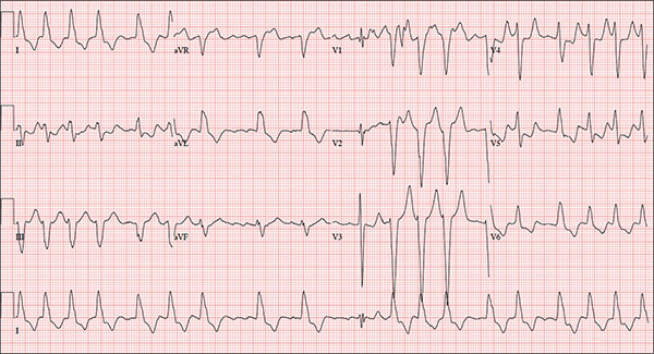
An 84-year-old woman who recently relocated to be closer to her children presents to your practice as a new patient. She is a resident of an assisted living facility near your office, and although she has no specific complaints, she does report that her home health nurse observed a rapid heart rate and recommended she get it checked. A comprehensive medical history—provided by the patient, her daughter, and the aforementioned nurse—includes hypertension, paroxysmal atrial fibrillation, hypothyroidism, and type 2 diabetes. She has taken medication for these diagnoses for more than 30 years. Surgical history is remarkable for cholecystectomy, appendectomy, and abdominal hysterectomy and oophorectomy, all of which were performed in the 1970s. Her current medication list—confirmed by the assisted living facility—includes furosemide, glyburide, metoprolol, potassium, and levothyroxine. She has not missed any doses. She is allergic to sulfa. The patient, a retired teacher, has never smoked, but she does “enjoy” one martini at dinner on a regular basis. She is widowed; her two daughters and four sons are all alive and well. The review of systems is remarkable for corrective lenses, bilateral hearing aids, and chronic joint pain. The patient does not routinely weigh herself but thinks, based on the fit of her clothes, that she may have gained some weight. She denies constitutional symptoms and shortness of breath. She thinks she may have a urinary tract infection, as she’s had burning with urination for several days, but says this is beginning to improve. Physical exam reveals a blood pressure of 168/90 mm Hg; pulse, 106 beats/min; temperature, 98.4° F; and O2 saturation, 94% on room air. Her weight is 132 lb and her height, 60 in. She is alert and quite spry, with a lot of energy. She wears glasses and bilateral hearing aids. Jugular distention is present to the angle of the jaw. There is no thyromegaly. The pulmonary exam is remarkable for crackles in both lung bases. The heart rhythm is irregularly irregular at a rate of 110 beats/min, and a grade II/VI murmur of mitral regurgitation is heard at the left lower sternal border. The abdomen is soft and nontender, with multiple surgical scars. The lower extremities are remarkable for 2+ pitting edema bilaterally to the level of the mid-calf. Osteoarthritic changes are present in both hands. The neurologic exam is grossly intact. An ECG reveals a ventricular rate of 110 beats/min; PR interval, not measured; QRS duration, 144 ms; QT/QTc interval, 298/403 ms; no P axis; R axis, –36°; and T axis, 169°. What is your interpretation of this ECG?

Sterile or non-sterile gloves for minor skin excisions?
Consider using non-sterile gloves during minor skin excisions (even those that require sutures) because the infection rate is not increased compared to using sterile gloves.1
Strength of recommendation
B: Based on a randomized controlled trial done in a primary care practice.
Heal C, Sriharan S, Buttner PG, et al. Comparing non-sterile to sterile gloves for minor surgery: a prospective randomized controlled noninferiority trial. Med J Aust. 2015;202:27-31.
Illustrative case
A 50-year-old man comes to your office to have a mole removed from his arm. You decide to excise the lesion in your office today. Do you need to use sterile gloves for this procedure, or can you use gloves from the clean non-sterile box in the exam room?
Non-sterile gloves are readily available during a typical office visit and cost up to a dollar less per pair than sterile gloves.1-3 Studies conducted in settings other than primary care offices have shown that non-sterile gloves do not increase the risk of infection during several types of minor skin procedures.
A partially blinded, randomized controlled trial (RCT) in an emergency department found no significant difference in infection rates between the use of sterile (6.1%) vs non-sterile (4.4%) gloves during laceration repairs.2 Similarly, a small RCT in an outpatient dermatology clinic and a larger prospective trial by a Mohs dermatologist showed that infection rates were not increased after Mohs surgery using non-sterile (0.49%) vs sterile (0.50%) gloves.3,4
Guidelines on the use of sterile vs non-sterile gloves for minor skin excisions in outpatient primary care are difficult to come by. Current guidelines from the Centers for Disease Control and Prevention (CDC) and other agencies regarding surgical site infections are broad and focus on the operating room environment.5-7
The American Academy of Dermatology is working on a guideline for treatment of non-melanoma skin cancer that’s due out this winter, and this may provide additional guidance.8 A 2003 review instructed primary care physicians to use sterile gloves for excisional skin biopsies that require sutures.9
The 2015 study by Heal et al1 appears to be the first RCT to address the question of sterile vs non-sterile glove use for minor skin excisions in a primary care outpatient practice.
STUDY SUMMARY: Non-sterile gloves are not inferior to sterile gloves
Heal et al1 conducted a prospective, randomized, controlled, noninferiority trial to compare the incidence of infection after minor skin surgery performed by 6 physicians from a single general practice in Australia using sterile vs non-sterile clean gloves. They evaluated 576 consecutive patients who presented for skin excision between June 2012 and March 2013. Eighty-three patients were excluded because they had a latex allergy, were using oral antibiotics or immunosuppressive drugs, or required a skin flap procedure or excision of a sebaceous cyst. The physicians followed a standard process for performing the procedures and did not use topical antibiotics or antiseptic cleansing after the procedure.
The primary outcome was surgical site infection within 30 days of the excision, defined as purulent discharge, pain or tenderness, localized swelling or redness or heat at the site, or a diagnosis of skin or soft tissue infection by a general practitioner. The clinicians who assessed for infection were blinded to the patient’s assignment to the sterile or non-sterile glove group, and a stitch abscess was not counted as an infection.
The patients’ mean age was 65 years and 59% were men. At baseline, there were no large differences between patients in the sterile and non-sterile glove groups in terms of smoking status, anticoagulant or steroid use, diabetes, excision site, size of excision, and median days until removal of sutures. The lesions were identified histologically as nevus or seborrheic keratosis, skin cancer and precursor, or other.
The incidence of infection in the non-sterile gloves group was 21/241 (8.7%; 95% confidence interval [CI], 4.9%-12.6%) vs 22/237 in the control group (9.3%; 95% CI, 7.4%-11.1%). The CI (95%) for the difference in infection rate (-0.6%) was -4.0% to 2.9%. This was significantly below the predetermined noninferiority margin of 7%. In a sensitivity analysis of patients lost to follow-up (15 patients, 3%) that assumed all of these patients were without infection, or with infection, the CI was still below the noninferiority margin of 7%. The per-protocol analysis showed similar results.
WHAT'S NEW: New evidence questions the need for sterile gloves for in-office excisions
Heal et al1 demonstrated that in a primary care setting, non-sterile gloves are not inferior to sterile gloves for performing excisional procedures that require sutures. While standard practice has many family physicians using sterile gloves for these procedures, this study promotes changing this behavior.
CAVEATS: A high infection rate, other factors might limit generalizability
The overall rate of infection in this study (9%) was higher than that found in the studies from emergency medicine and dermatology literature cited earlier.2-4 A similarly high infection rate has been found in other studies of minor surgery by Heal et al, including a 2006 study that showed a wound infection rate of 8.6%.10 The significance of the higher infection rate is unknown, but there is no clear reason why non-sterile gloves might be less effective in preventing infection in environments with lower infection rates.
This was not a double-blinded study, and physicians might change their behavior during a procedure depending on the type of gloves they are wearing. The sterile gloves used in this study contained powder, while the non-sterile gloves were powderless, but this variable is not known to affect infection rates. A study of Mohs surgery avoided this variable by only using powderless gloves, and had similar outcomes in terms of the difference in infection rate between sterile and non-sterile gloves.4
CHALLENGES TO IMPLEMENTATION: Ingrained habits can be hard to change
Tradition and training die hard. While multiple studies in several settings have found non-sterile gloves are non-inferior to sterile gloves in preventing surgical site infection after minor skin surgeries, this single study in the primary care office setting may not be enough to sway family physicians from ingrained habits.
ACKNOWLEDGEMENT
The PURLs Surveillance System was supported in part by Grant Number UL1RR024999 from the National Center For Research Resources, a Clinical Translational Science Award to the University of Chicago. The content is solely the responsibility of the authors and does not necessarily represent the official views of the National Center For Research Resources or the National Institutes of Health.
1. Heal C, Sriharan S, Buttner PG, et al. Comparing non-sterile to sterile gloves for minor surgery: a prospective randomized controlled non-inferiority trial. Med J Aust. 2015;202:27-31.
2. Perelman VS, Francis GJ, Rutledge T, et al. Sterile versus nonsterile gloves for repair of uncomplicated lacerations in the emergency department: a randomized controlled trial. Ann Emerg Med. 2004;43:362-370.
3. Mehta D, Chambers N, Adams B, et al. Comparison of the prevalence of surgical site infection with use of sterile versus nonsterile gloves for resection and reconstruction during Mohs surgery. Dermatol Surg. 2014;40:234-239.
4. Xia Y, Cho S, Greenway HT, et al. Infection rates of wound repairs during Mohs micrographic surgery using sterile versus nonsterile gloves: a prospective randomized pilot study. Dermatol Surg. 2011;37:651-656.
5. Mangram AJ, Horan TC, Pearson ML, et al. Guideline for prevention of surgical site infection, 1999. Centers for Disease Control and Prevention (CDC) Hospital Infection Control Practices Advisory Committee. Am J Infect Control. 1999;27:97-132.
6. National Institute for Health and Care Excellence. Surgical site infections: prevention and treatment. October 2008. Available at: https://www.nice.org.uk/guidance/cg74. Accessed July 28, 2015.
7. National Health and Medical Research Council. Australian Guidelines for the Prevention and Control of Infection in Healthcare (2010). Updated August 28, 2013. Available at: http://www.nhmrc.gov.au/book/html-australian-guideline-sprevention-and-control-infection-healthcare-2010. Accessed July 31, 2015.
8. American Academy of Dermatology. Clinical Guidelines. American Academy of Dermatology Web site. Available at: https://www.aad.org/education/clinical-guidelines. Accessed July 28, 2015.
9. Zuber TJ. Fusiform excision. Am Fam Physician. 2003;67:1539-1544.
10. Heal C, Buettner P, Browning S. Risk factors for wound infection after minor surgery in general practice. Med J Aust. 2006;18:255-258.
Consider using non-sterile gloves during minor skin excisions (even those that require sutures) because the infection rate is not increased compared to using sterile gloves.1
Strength of recommendation
B: Based on a randomized controlled trial done in a primary care practice.
Heal C, Sriharan S, Buttner PG, et al. Comparing non-sterile to sterile gloves for minor surgery: a prospective randomized controlled noninferiority trial. Med J Aust. 2015;202:27-31.
Illustrative case
A 50-year-old man comes to your office to have a mole removed from his arm. You decide to excise the lesion in your office today. Do you need to use sterile gloves for this procedure, or can you use gloves from the clean non-sterile box in the exam room?
Non-sterile gloves are readily available during a typical office visit and cost up to a dollar less per pair than sterile gloves.1-3 Studies conducted in settings other than primary care offices have shown that non-sterile gloves do not increase the risk of infection during several types of minor skin procedures.
A partially blinded, randomized controlled trial (RCT) in an emergency department found no significant difference in infection rates between the use of sterile (6.1%) vs non-sterile (4.4%) gloves during laceration repairs.2 Similarly, a small RCT in an outpatient dermatology clinic and a larger prospective trial by a Mohs dermatologist showed that infection rates were not increased after Mohs surgery using non-sterile (0.49%) vs sterile (0.50%) gloves.3,4
Guidelines on the use of sterile vs non-sterile gloves for minor skin excisions in outpatient primary care are difficult to come by. Current guidelines from the Centers for Disease Control and Prevention (CDC) and other agencies regarding surgical site infections are broad and focus on the operating room environment.5-7
The American Academy of Dermatology is working on a guideline for treatment of non-melanoma skin cancer that’s due out this winter, and this may provide additional guidance.8 A 2003 review instructed primary care physicians to use sterile gloves for excisional skin biopsies that require sutures.9
The 2015 study by Heal et al1 appears to be the first RCT to address the question of sterile vs non-sterile glove use for minor skin excisions in a primary care outpatient practice.
STUDY SUMMARY: Non-sterile gloves are not inferior to sterile gloves
Heal et al1 conducted a prospective, randomized, controlled, noninferiority trial to compare the incidence of infection after minor skin surgery performed by 6 physicians from a single general practice in Australia using sterile vs non-sterile clean gloves. They evaluated 576 consecutive patients who presented for skin excision between June 2012 and March 2013. Eighty-three patients were excluded because they had a latex allergy, were using oral antibiotics or immunosuppressive drugs, or required a skin flap procedure or excision of a sebaceous cyst. The physicians followed a standard process for performing the procedures and did not use topical antibiotics or antiseptic cleansing after the procedure.
The primary outcome was surgical site infection within 30 days of the excision, defined as purulent discharge, pain or tenderness, localized swelling or redness or heat at the site, or a diagnosis of skin or soft tissue infection by a general practitioner. The clinicians who assessed for infection were blinded to the patient’s assignment to the sterile or non-sterile glove group, and a stitch abscess was not counted as an infection.
The patients’ mean age was 65 years and 59% were men. At baseline, there were no large differences between patients in the sterile and non-sterile glove groups in terms of smoking status, anticoagulant or steroid use, diabetes, excision site, size of excision, and median days until removal of sutures. The lesions were identified histologically as nevus or seborrheic keratosis, skin cancer and precursor, or other.
The incidence of infection in the non-sterile gloves group was 21/241 (8.7%; 95% confidence interval [CI], 4.9%-12.6%) vs 22/237 in the control group (9.3%; 95% CI, 7.4%-11.1%). The CI (95%) for the difference in infection rate (-0.6%) was -4.0% to 2.9%. This was significantly below the predetermined noninferiority margin of 7%. In a sensitivity analysis of patients lost to follow-up (15 patients, 3%) that assumed all of these patients were without infection, or with infection, the CI was still below the noninferiority margin of 7%. The per-protocol analysis showed similar results.
WHAT'S NEW: New evidence questions the need for sterile gloves for in-office excisions
Heal et al1 demonstrated that in a primary care setting, non-sterile gloves are not inferior to sterile gloves for performing excisional procedures that require sutures. While standard practice has many family physicians using sterile gloves for these procedures, this study promotes changing this behavior.
CAVEATS: A high infection rate, other factors might limit generalizability
The overall rate of infection in this study (9%) was higher than that found in the studies from emergency medicine and dermatology literature cited earlier.2-4 A similarly high infection rate has been found in other studies of minor surgery by Heal et al, including a 2006 study that showed a wound infection rate of 8.6%.10 The significance of the higher infection rate is unknown, but there is no clear reason why non-sterile gloves might be less effective in preventing infection in environments with lower infection rates.
This was not a double-blinded study, and physicians might change their behavior during a procedure depending on the type of gloves they are wearing. The sterile gloves used in this study contained powder, while the non-sterile gloves were powderless, but this variable is not known to affect infection rates. A study of Mohs surgery avoided this variable by only using powderless gloves, and had similar outcomes in terms of the difference in infection rate between sterile and non-sterile gloves.4
CHALLENGES TO IMPLEMENTATION: Ingrained habits can be hard to change
Tradition and training die hard. While multiple studies in several settings have found non-sterile gloves are non-inferior to sterile gloves in preventing surgical site infection after minor skin surgeries, this single study in the primary care office setting may not be enough to sway family physicians from ingrained habits.
ACKNOWLEDGEMENT
The PURLs Surveillance System was supported in part by Grant Number UL1RR024999 from the National Center For Research Resources, a Clinical Translational Science Award to the University of Chicago. The content is solely the responsibility of the authors and does not necessarily represent the official views of the National Center For Research Resources or the National Institutes of Health.
1. Heal C, Sriharan S, Buttner PG, et al. Comparing non-sterile to sterile gloves for minor surgery: a prospective randomized controlled non-inferiority trial. Med J Aust. 2015;202:27-31.
2. Perelman VS, Francis GJ, Rutledge T, et al. Sterile versus nonsterile gloves for repair of uncomplicated lacerations in the emergency department: a randomized controlled trial. Ann Emerg Med. 2004;43:362-370.
3. Mehta D, Chambers N, Adams B, et al. Comparison of the prevalence of surgical site infection with use of sterile versus nonsterile gloves for resection and reconstruction during Mohs surgery. Dermatol Surg. 2014;40:234-239.
4. Xia Y, Cho S, Greenway HT, et al. Infection rates of wound repairs during Mohs micrographic surgery using sterile versus nonsterile gloves: a prospective randomized pilot study. Dermatol Surg. 2011;37:651-656.
5. Mangram AJ, Horan TC, Pearson ML, et al. Guideline for prevention of surgical site infection, 1999. Centers for Disease Control and Prevention (CDC) Hospital Infection Control Practices Advisory Committee. Am J Infect Control. 1999;27:97-132.
6. National Institute for Health and Care Excellence. Surgical site infections: prevention and treatment. October 2008. Available at: https://www.nice.org.uk/guidance/cg74. Accessed July 28, 2015.
7. National Health and Medical Research Council. Australian Guidelines for the Prevention and Control of Infection in Healthcare (2010). Updated August 28, 2013. Available at: http://www.nhmrc.gov.au/book/html-australian-guideline-sprevention-and-control-infection-healthcare-2010. Accessed July 31, 2015.
8. American Academy of Dermatology. Clinical Guidelines. American Academy of Dermatology Web site. Available at: https://www.aad.org/education/clinical-guidelines. Accessed July 28, 2015.
9. Zuber TJ. Fusiform excision. Am Fam Physician. 2003;67:1539-1544.
10. Heal C, Buettner P, Browning S. Risk factors for wound infection after minor surgery in general practice. Med J Aust. 2006;18:255-258.
Copyright © 2015. The Family Physicians Inquiries Network. All rights reserved.

Sizing up EMRs and patient care from the other side of the bed rail
Dr. Unger’s guest editorial, “Med students: Look up from your EMRs” (J Fam Pract. 2015;64:517-518), vividly describes what those who have been paying attention see quite clearly: Not only has the widespread implementation of electronic medical records (EMRs) failed to deliver all it has promised, but it has made patient care worse. Many students and members of the health care team spend as little time as possible talking and listening to patients. Instead, the goal is to complete every box in our EMRs to qualify for meaningful use payments and whatever “quality” incentives are available in our local environment.
That said, I believe EMRs are very good at doing the things computers do well, and I hope I never again have to rifle through a paper chart the size of a phone book to find a critical piece of information. The problem lies in the myriad inappropriate ways the EMR is used in place of accurately telling the patient’s story, and the resulting diversion of the entire health care team away from caring for the patients we are supposedly here to serve.
I am tired of complaining to my patients, partners, family, friends, and anyone else who will listen. It is time for family medicine to reclaim its role as “counterculture” and lead the charge for comprehensive, continuous, compassionate care—whose centerpiece is actually talking to, listening to, and examining patients.
David A. Silverstein, MD
Buffalo, NY
While I agree with Dr. Unger about EMRs, I respectfully disagree with his approach when he suspected he had appendicitis. When he initially ordered his own computed tomography scan, rather than seeing his own doctor or going to the emergency department, he (inadvertently) “assigned” himself as his own doctor. He then should have at least offered his history in the hospital, rather than making it a test for the student and the hospital. It sounds like an adversarial situation developed, which did not help matters. Good that he’s doing OK!
Michael Kelly, MD
Minneapolis, Minn
Advances in Colorectal Cancer Screening
Colorectal cancer (CRC) screening has been shown to save lives. Screening can prevent CRC by detecting and removing precancerous adenomatous polyps, which are the precursors of most cancers.1 Screening also can detect cancer at an early, asymptomatic stage while it is still localized and amenable to treatment; 5-year survival rates are 80% to 90% for patients with localized, early stage I/II CRC.2
Colorectal cancer (CRC) screening has been shown to save lives. Screening can prevent CRC by detecting and removing precancerous adenomatous polyps, which are the precursors of most cancers.1 Screening also can detect cancer at an early, asymptomatic stage while it is still localized and amenable to treatment; 5-year survival rates are 80% to 90% for patients with localized, early stage I/II CRC.2
Colorectal cancer (CRC) screening has been shown to save lives. Screening can prevent CRC by detecting and removing precancerous adenomatous polyps, which are the precursors of most cancers.1 Screening also can detect cancer at an early, asymptomatic stage while it is still localized and amenable to treatment; 5-year survival rates are 80% to 90% for patients with localized, early stage I/II CRC.2
Seeing eye to eye

It seems like every time I ask a family physician how things are going, the electronic medical record (EMR) inevitably rears its ugly face. At the annual Illinois Academy of Family Physicians business meeting last month, one of the physicians lamented the evenings he spends finishing his charting. A family physician I consider a master user of EMRs e-mailed me recently, saying he is fed up with documentation expectations for coding, billing, meaningful use, and quality measures. He wrote, “We are challenged by good intentions but crushingly poor execution … and it is taking its toll.”
At the 2015 American Academy of Family Physicians Family Medicine Expo, keynote speaker, general internist, and bestselling author Abraham Verghese, MD, talked about the “iPatient.” He said, “The patient in the bed has become a mere icon for the ‘real patient’ who is in the computer. The iPatient is getting wonderful care all across America. The real patient is wondering where the heck is everyone and when are they going to tell me what is going on.”
He had received this comment from a patient: “When I go to my doctor’s office, I have to remind him that I am hard of hearing and need him to look at me when I talk. But it only lasts about 30 seconds until he needs to shift back to the competing screen.”
Patients don’t like us attending to the screen instead of to them. The observational study of 126 primary care encounters by Farber et al in this issue supports this assertion. Although Farber et al found that patients’ satisfaction with their primary care physician or nurse practitioner was high overall, patients were even more satisfied with their office visit when the clinician spent more time looking at them. Patients want to engage in a face-to-face conversation, not face-to-back or face-to-side-of-head.
Until clever innovators figure out a much better way to document patient visits, there are ways to overcome this patient-physician-computer screen triangle. Take my optometrist, for example. He opens my EMR at the beginning of the visit to take a quick look, but doesn’t return to the computer until the end of the visit. When he does the charting, he excuses himself and says, “I need to enter some information in the computer. It will take me a few minutes.” I pull out my cell phone to check e-mails while he types.
I follow his example, and patients regularly thank me for truly listening to them.
Make the Diagnosis - November 2015
Diagnosis: Basal cell carcinoma
Basal cell carcinoma (BCC) is the most common skin cancer diagnosed in the United States, with approximately 2.8 million cases diagnosed annually, according to the Skin Cancer Foundation. While a minority of cases are linked to genetic syndromes (e.g., basal cell nevus syndrome), most cases result from ultraviolet sun exposure. A power law model was recently described linking ultraviolet exposure and incidence of basal cell carcinoma.
Many diagnosed BCC cases are small (< 1cm in size) and easily treated in the clinic. In cases where treatment is delayed for years due to financial, psychological, or psychiatric reasons, tumors can cause significant local tissue destruction and grow to alarming sizes.

The differential diagnosis of BCC may vary depending on the clinical sub-type (e.g., superficial, nodular, infiltrative, etc). Nodular BCC may mimic adnexal neoplasms, intradermal melanocytic nevi, Merkel cell carcinoma, or even amelanotic melanoma. Superficial BCC may mimic a lichenoid keratosis, Bowen’s disease, or other inflammatory conditions such as psoriasis and dermatitis. A larger lesion may mimic chronic infections such as mycetoma, or distant metastases from another primary carcinoma (breast or renal). Location and history can assist with teasing out the probable cause.
While diagnosis of this lesion can be made based on history and clinical appearance, this is best assisted with a biopsy, preferably a punch or incisional biopsy as chronic scarring and pseudoepitheliomatous hyperplasia may affect the pathologic diagnosis. Bacteria often colonize these large tumors and can lead to secondary infections. In this patient, maggots were noted in the skin.
While there are multiple modalities available for treating small tumors (e.g., topical imiquimod, 5-fluorouracil, electrodessication and curettage, excision, Mohs), larger tumors are often handled differently. While some might consider radiation therapy in this case, typically Mohs surgery is the treatment of choice. Oral vismodegib, a smoothened inhibitor, has been marketed for locally advanced basal cell carcinoma and has been used as a primary or adjunctive therapy with Mohs in tumors of this size. One important factor determining the choice of treatment is whether the primary tumor has spread. In tumors who have been left untreated for a long time, it is reasonable to image the patient to evaluate for metastases.
We present this case as an example where Mohs provided immediate definitive treatment. This tumor was cleared in 1 stage, with 67 slides read to evaluate the entire peripheral and deep margin. The Mohs surgery was performed under local anesthesia with no additional sedatives and 3-0 nylon sutures were used to approximate the wound. A two month follow-up photo is shown showing an almost healed surgical wound with an acceptable cosmetic result.
Diagnosis: Basal cell carcinoma
Basal cell carcinoma (BCC) is the most common skin cancer diagnosed in the United States, with approximately 2.8 million cases diagnosed annually, according to the Skin Cancer Foundation. While a minority of cases are linked to genetic syndromes (e.g., basal cell nevus syndrome), most cases result from ultraviolet sun exposure. A power law model was recently described linking ultraviolet exposure and incidence of basal cell carcinoma.
Many diagnosed BCC cases are small (< 1cm in size) and easily treated in the clinic. In cases where treatment is delayed for years due to financial, psychological, or psychiatric reasons, tumors can cause significant local tissue destruction and grow to alarming sizes.
The differential diagnosis of BCC may vary depending on the clinical sub-type (e.g., superficial, nodular, infiltrative, etc). Nodular BCC may mimic adnexal neoplasms, intradermal melanocytic nevi, Merkel cell carcinoma, or even amelanotic melanoma. Superficial BCC may mimic a lichenoid keratosis, Bowen’s disease, or other inflammatory conditions such as psoriasis and dermatitis. A larger lesion may mimic chronic infections such as mycetoma, or distant metastases from another primary carcinoma (breast or renal). Location and history can assist with teasing out the probable cause.
While diagnosis of this lesion can be made based on history and clinical appearance, this is best assisted with a biopsy, preferably a punch or incisional biopsy as chronic scarring and pseudoepitheliomatous hyperplasia may affect the pathologic diagnosis. Bacteria often colonize these large tumors and can lead to secondary infections. In this patient, maggots were noted in the skin.
While there are multiple modalities available for treating small tumors (e.g., topical imiquimod, 5-fluorouracil, electrodessication and curettage, excision, Mohs), larger tumors are often handled differently. While some might consider radiation therapy in this case, typically Mohs surgery is the treatment of choice. Oral vismodegib, a smoothened inhibitor, has been marketed for locally advanced basal cell carcinoma and has been used as a primary or adjunctive therapy with Mohs in tumors of this size. One important factor determining the choice of treatment is whether the primary tumor has spread. In tumors who have been left untreated for a long time, it is reasonable to image the patient to evaluate for metastases.
We present this case as an example where Mohs provided immediate definitive treatment. This tumor was cleared in 1 stage, with 67 slides read to evaluate the entire peripheral and deep margin. The Mohs surgery was performed under local anesthesia with no additional sedatives and 3-0 nylon sutures were used to approximate the wound. A two month follow-up photo is shown showing an almost healed surgical wound with an acceptable cosmetic result.
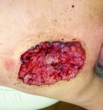
Case and photo courtesy of: Andrew R. Styperek MD; Houston Methodist Hospital and DermSurgery Associates, Houston TX Arash Kimyai-Asadi MD; DermSurgery Associates, Houston TX Dr. Bilu Martin is in private practice at Premier Dermatology, MD in Aventura, Fla. To submit your case for possible publication, send an e-mail to dermnews@frontlinemedcom.com. A 65 year old Caucasian male arrived with a decades-long history of a lesion on his back measuring 25 x 21 centimeters. In the last few years he has noticed discharge and an unpleasant smell. He denied any fatigue, shortness of breath, lymph node enlargement, or any other systemic symptoms. He had a history of hyperlipidemia and hypertension, which were controlled with daily oral medications. The patient was not taking aspirin or any other anticoagulant therapy.

Adjuvant Systemic Therapy for Early-Stage Breast Cancer
Over the past 20 years, substantial progress has been achieved in our understanding of breast cancer and in breast cancer treatment, with mortality from breast cancer declining by more than 25% over this time. This progress has been characterized by a greater understanding of the molecular biology of breast cancer, rational drug design, development of agents with specific cellular targets and pathways, development of better prognostic and predictive multigene assays, and marked improvements in supportive care.
To read the full article in PDF:
Perspectives in Pediatrics: From Theory to Practice

Supplement Editor:
Camille Sabella, MD
Contents
ADHD and behavioral disorders: Assessment, management, and an update from DSM-5
Joseph Austerman, DO
Use of long-acting reversible contraceptives to reduce the rate of teen pregnancy
Ellen Rome, MD, MPH
RSV infections: State of the art
Giovanni Piedimonte, MD
Dermatology for the pediatrician: Advances in diagnosis and treatment of common and not-so-common skin conditions
Joan Tamburro, DO
Learning disorders: How pediatricians can help
Elain E. Schulte, MD, MPH
Developmental delays and autism: Screening and surveillance
Carol Delahunty, MD
ADHD and behavioral disorders: Assessment, management, and an update from DSM-5
Behavioral disorders in pediatric patients—primarily attention deficit hyperactivity disorder (ADHD)—pose a clinical challenge for health care providers to accurately assess, diagnose, and treat. In 2013, the criteria for several disruptive behavioral disorders were updated in the fifth edition of the Diagnostic and Statistical Manual of Mental Disorders (DSM-5),1 their first major revisions since 1994. Among the most clinically relevant changes were revisions to the diagnosis of ADHD and the creation of a new diagnostic entity: disruptive mood dysregulation disorder (DMDD).
This article focuses on the updated diagnostic criteria published in the DSM-5 for behavioral disorders, describes the assessment of ADHD, and summarizes management strategies.
UPDATED DIAGNOSTIC CRITERIA
ADHD
This disorder is a chronic, neurologically based illness characterized by a persistent pattern of inattention and/or hyperactivity and impulsivity that are more inappropriate or disruptive than those in other children of a comparable age resulting in functional impairment in multiple settings, and these behaviors have been present for at least 6 months. Revised diagnostic criteria in DSM-5 used the same two categories for ADHD symptoms—inattention and hyperactivity-impulsive behaviors—but modified several diagnostic requirements.
Revised criteria
Impairment before age 12 instead of age 6. As a neurodevelopmental disorder, ADHD usually starts at a young age; teenagers presenting with newly developed ADHD-type symptoms probably do not have ADHD and efforts should be made to rule out other illnesses or social dynamics. The DSM-5 raised the age limit for onset of qualifying symptoms to before 12 years (previously by age 6) primarily to capture a cohort of pediatric patients, typically female, who present solely with inattention symptoms and may not display overt functional impairment early on.
Symptoms required in at least two settings. Symptoms must be present in at least two settings to qualify for a diagnosis of ADHD. This ensures that the behaviors occur globally; they do not occur just at school or at home but occur in both places.
Fewer symptoms required for diagnosis in adolescents. Although the diagnostic criteria retain the same symptoms as those in DSM-IV for different age groups, individuals aged 17 and older are now required to display only five or more inattentive or hyperactive-impulsive symptoms. Previously, at least six were required.
Partial remission criteria
The concept of partial remission was introduced in DSM-5. This acknowledges that two-thirds of children diagnosed with ADHD do not have symptoms that functionally impact activities of daily living beyond age 18.
Oppositional defiant disorder
In DSM-5, oppositional defiant disorder (ODD) is defined by emotional and behavioral symptoms grouped into three categories:
- Constant anger or irritability
- Argumentative or defiant behavior (arguing with authority figures)
- Vindictiveness.
Because defiant behavior may represent difficulty with self-control, ODD is associated with executive functioning deficits that are present in ADHD. Children with ODD tend to perform best in situations in which they can dominate or exert authority. To qualify as ODD, the pattern of behavior must be consistent for longer than 6 months. A severity rating was added based on pervasiveness of ODD symptoms. Otherwise the diagnosis did not change.
Conduct disorder: Purposeful aggression
The hallmarks of conduct disorder are purposeful aggression (eg, bullying), destruction of property, deceitfulness or theft, and serious violation of rules (eg, running away from home, repeat truancy). Some consider conduct disorder to be a separate illness from ODD, whereas others consider it a continuum of the same disorder. Conduct disorder can manifest as violence, as in initiating physical fights, or it can manifest in behaviors such as truancy, stealing, lying, and running away from home without the physical-aggression aspect.
Intermittent explosive disorder
Failure to control aggressive impulses defines intermittent explosive disorder (IED). The aggressive outbursts can be verbal or behavioral and tend to be impulsive. A small subset of children display isolated aggression out of proportion to provocation. The disorder tends to manifest at ages 3 or 4, and a diagnosis requires a stable environment with no significant early childhood trauma. Most often these symptoms are seen in children with intellectual disabilities or an autism spectrum disorder.
Disruptive mood dysregulation disorder
A new diagnostic category in DSM-5 is termed disruptive mood dysregulation disorder (DMDD). This captures many children who previously would have been diagnosed with pediatric bipolar disorder, even though most of them do not fulfill criteria for bipolar disorder as adults. The presence of baseline irritability separates this disorder from IED, which requires intermittent rapid and severe outbursts. The severe temper outbursts of DMDD must be recurrent, with an average of three occurrences per week, and have background irritability. The symptoms must have a duration of at least 12 months and be present in two settings. A diagnosis of DMDD cannot be made earlier than age 6, with onset before age 10.
ASSESSMENT OF ADHD
The clinical interview in conjunction with objective scales is the primary tool for diagnosing ADHD. The most frequent source of information is from the parents followed by the child’s schoolteachers. Patient interview, although unreliable in young children, should also be part of the assessment. Comparing the patient’s functional impairment against children of a similar age is necessary for an ADHD diagnosis.
The medical history can help rule out children with asthma or allergy being treated with corticosteroids and those with hypothyroidism and hyperthyroidism whose symptoms often fulfill the diagnostic criteria for ADHD.2,3 Symptoms of ADHD also may appear suddenly after a traumatic brain injury or other neurologic event.4 Other psychiatric illnesses, especially learning disorders, mood disorders, anxiety, other disruptive behavior disorders, or substance abuse, can mimic ADHD.
Ruling out other factors from a social history (eg, family conflict, bullying, sleep deprivation, being overscheduled with activities) adds to the reliability of an ADHD diagnosis. For example, repetitive uprooting and frequent changes in schools can cause academic problems that may be mistaken for ADHD, and use of stimulants may have failed to improve symptoms in these children.
Assessment scales
Pediatric assessment scales that can be performed in an office are more practical than standardized clinical assessments (Table 1). The Vanderbilt ADHD Diagnostic Teacher Rating Scale correlates highly with a diagnosis of ADHD. We use the Vanderbilt ADHD Diagnostic Parent Rating Scale for children up to age 1 year. Other scales track symptoms and functional impairment over time and can be administered before the patient’s appointment. The Conners Third Edition scale can be used to establish a baseline before initiating therapy and to help monitor changes over time.
Standardized tests to bolster the utility of the clinical interview include the Diagnostic Interview Schedule for Children and Adolescents and the Schedule for Affective Disorders and Schizophrenia in School-Age Children–Present and Lifetime Version. Free training is available regarding use of some of these standardized tests.
Developmental course, risk factors
The clinical course of ADHD is chronic. The onset of hyperactivity usually occurs at age 3 or 4, with combined hyperactivity and inattention usually appearing from ages 5 to 8.5,6 The evolution of symptoms is progressive and constant. Between 50% and 80% have symptoms that continue into adolescence, and in about 40%, symptoms continue into adulthood.7,8 Some children with ADHD have a temperament-neuropsychological profile characterized by aggressiveness, irritability, and mood lability. Deficits in planning, delayed aversion, and temporal processing are present.
Risk factors include prematurity, prenatal complications, an anoxic event, nutritional deficits (specifically iron and zinc), and lack of appropriate socialization.9–11 The disorder is heritable, which is usually clear from the clinical interview. Rates of delinquency and peer rejection are high. This may result in secondary comorbidity such as emotional, disruptive, or substance abuse problems.
MANAGEMENT STRATEGIES
Stimulants
The first-line pharmacologic treatment of ADHD is stimulants: methylphenidate, dexmethylphenidate, mixed amphetamine salts, dextroamphetamine, and lisdexamfetamine. Head-to-head trials of medications versus behavioral management favor medication use, even over the long term.12–14
Methylphenidate and amphetamines are equally effective and have similar adverse effect profiles. Insomnia and anorexia are the most common side effects of stimulants. Cardiac effects include tachycardia, chest pain, and hypertension. Very rarely, stimulants have been associated with sudden cardiac death syndrome in patients with underlying cardiac problems. The consensus is that stimulants are safe in the general population. The need to obtain an electrocardiogram before initiating a stimulant was removed by the US Food and Drug Administration (FDA) unless it is otherwise indicated.15
The response rate to stimulants is in the range of 70%. About one-third of patients have side effects, and approximately 15% have side effects severe enough to requiring changing or withdrawing the medication.16,17
Stimulants are available in several delivery systems. For the best effect, medications should be combined with behavioral management.
Alternatives to stimulants
If stimulants are ineffective, atomoxetine can be used to treat patients with inattention; however, its effect on hyperactivity and impulsivity is less pronounced than that of stimulants. Bupropion is another option for inattention. Both agents are well tolerated. Irritability and insomnia are side effects of atomoxetine, and liver damage is possible, so liver function tests must be ordered if the patient complains of upper-right-quadrant pain.
The evidence to support the use of modafinil is equivocal.18,19 Unlike stimulants, modafinil is associated with a slight increase in motivation.
Alpha-2 agonists are effective for treating aggression in the setting of ADHD, especially in younger children, and are well tolerated.20 Extended-release forms are available.
Combination therapy
Polypharmacy is sometimes indicated in the treatment of ADHD. A stimulant used in combination with atomoxetine was shown to be superior to either treatment alone in improving symptoms of hyperactivity and inattention.21 The combination, however, markedly increased the incidence of appetite loss, insomnia, and irritability.
A more promising combination is a stimulant with an alpha agonist. Symptoms of hyperactivity and inattention were improved more with this combination than with a stimulant plus placebo, with no difference in side effects.22
ADHD with comorbidities
Patients with ADHD, both adults and children, often have comorbid externalizing disorders and other emotional disorders, such as depression and anxiety, occurring in up to half of cases (Table 2).23,24 These comorbidities are important to consider when developing a treatment strategy. The following describes treatment options for the most common ADHD comorbidities (Table 3).
ODD or conduct disorder. The first-line therapy for these patients is a stimulant plus behavioral therapy. Adding an alpha agonist to this combination may be indicated if the comorbidity is severe. Second-generation antipsychotics also have been used as add-ons to stimulants with behavioral therapy, but weight gain and hormonal side effects are common.
Behavioral interventions are effective in targeting disruptive behavioral disorders, specifically multisystemic therapy. Multisystemic therapy is intensive therapy that involves working with the patient’s peer group or school, but most children must enter the legal system to receive this intervention. Multisystemic therapy is the only intervention shown to improve symptoms associated with comorbid ADHD and conduct disorder.25
Mood disorders. For these patients, the mood disorder is treated first. In doing so, symptoms of ADHD may disappear. For those with bipolar disease, a second-generation antipsychotic agent is superior to lithium in efficacy, maintenance of remission, and side effects in patients with a clear bipolar affective disorder, after which a stimulant can be added with less risk of developing manic symptoms. Using a stimulant first for this indication risks mood destabilization.
For patients with a major depressive disorder, bupropion can be used, although this indication is not FDA-approved, followed by the addition of a stimulant. One alternative is a selective serotonin reuptake inhibitor plus a stimulant; another is cognitive behavioral therapy plus atomoxetine and an alpha agonist.
Substance abuse. Patients with ADHD have high rates of substance abuse.26,27 Whether treatment of ADHD with stimulants reduces the risk of substance abuse is controversial. Because abuse of stimulants is common, start treatment with atomoxetine, bupropion, an alpha agonist, or a stimulant that is difficult to abuse (eg, lisdexamfetamine). Refer patients who are abusing substances to a specialist in substance abuse for behavioral management.
Anxiety. Atomoxetine is recommended for the treatment of anxiety that coexists with ADHD. A selective serotonin reuptake inhibitor in combination with a stimulant or alpha agonist, plus cognitive behavioral therapy, is another option for treating anxiety and ADHD. Tricyclic antidepressants have shown benefit in pediatric anxiety. Bupropion should not be used to target anxiety as it has been shown to have a limited effect on anxiety.
Tics. Stimulants may transiently exacerbate underlying tic disorders, but no longstanding difference in the course of tics has been observed with stimulant use.28 Alpha-2 antagonists target both tics and ADHD, so their use is preferred.29 Atomoxetine does not exacerbate tics but may reduce their frequency and severity.30
Dietary factors
Although challenging to accomplish, management of diet, specifically removal of artificial food coloring and sodium benzoate preservatives, has been more efficacious than behavioral management in the long-term reduction of core symptoms of ADHD.31,32 No herbal remedy has demonstrated efficacy in improving ADHD symptoms. The use of omega-3 fatty acids as a complement to stimulants has demonstrated efficacy in reducing core symptoms in ADHD.33
Behavioral therapy
Several forms of behavioral therapy have shown utility in improving symptoms in ADHD. Evidence supports that ADHD responds to cognitive behavioral therapy.34 In-school neurofeedback training for ADHD was shown to be better than cognitive training in improving inattention and hyperactivity-impulsivity at 6 months of follow-up.35
Parental training has the most evidence to support its use in children with ADHD. The two most common forms are Pathways Triple P (Positive Parenting Program) and The Incredible Years. Triple P is an early intervention designed to promote positive parent-child relationships to reduce behavior problems.36 The Incredible Years is a multicomponent program that emphasizes creating opportunities for active involvement, reinforcement of positive behavior, teaching skills, and setting clear limits, all of which are central to the social development strategy.37
Many children with ADHD respond to in-school interventions, at least an evaluation to rule out learning disorders, which typically have high morbidity. Children may qualify for Individualized Education Program (IEP) services, such as peer tutoring,38 computer-assisted instruction,39,40 and task-modification instruction.41 All of these have evidence to support their use.
- American Psychiatric Association. Diagnostic and Statistical Manual of Mental Disorders. 5th ed. Arlington, VA: American Psychiatric Association; 2013.
- Hak E, de Vries TW, Hoekstra PJ, Jick SS. Association of childhood attention-deficit/hyperactivity disorder with atopic diseases and skin infections? A matched case-control study using the General Practice Research Database. Ann Allergy Asthma Immunol 2013; 111:102–106.
- Pretorius E. Corticosteroids, depression and the role of serotonin. Rev Neurosci 2004; 15:109–116.
- Keenan HT, Hall GC, Marshall SW. Early head injury and attention deficit hyperactivity disorder: retrospective cohort study. BMJ 2008; 337:a1984.
- Applegate B, Lahey BB, Hart EL, et al. Validity of the age-of-onset criterion for ADHD: a report of the DSM-IV field trials. J Am Acad Child Adolesc Psychiatry 1997; 36:1211–1221.
- Loeber R, Green SM, Lahey BB, Christ MAG, Frick PJ. Developmental sequences in the age of onset of disruptive child behaviors. J Child Fam Studies 1992; 1:21–41.
- Barkley RA, Murphy KR, Fischer M. Adult ADHD: What the Science Says. New York, NY: Guilford Publications; 2008.
- Rasmussen P, Gillberg C. Natural outcome of ADHD with developmental coordination disorder at age 22 years: a controlled, longitudinal, community-based study. J Am Acad Child Adolesc Psychiatry 2000; 39:1424–1431.
- Silva D, Colvin L, Hagemann E, Bower C. Environmental risk factors by gender associated with attention-deficit/hyperactivity disorder. Pediatrics 2014; 133:e14–e22.
- Lindström K, Lindblad F, Hjern A. Preterm birth and attention-deficit/hyperactivity disorder in schoolchildren. Pediatrics 2011; 127:858–865.
- Banerjee TD, Middleton F, Faraone SV. Environmental risk factors for attention-deficit hyperactivity disorder. Acta Paediatr 2007; 96:1269–1274.
- Brown RT, Amler RW, Freeman WS, et al; for the American Academy of Pediatrics Committee on Quality Improvement; American Academy of Pediatrics Subcommittee on Attention-Deficit/Hyperactivity Disorder. Treatment of attention-deficit/hyperactivity disorder: overview of the evidence. Pediatrics 2005; 115:e749–e757.
- The MTA Cooperative Group. A 14-month randomized clinical trial of treatment strategies for attention-deficit/hyperactivity disorder. Arch Gen Psychiatry 1999; 56:1073–1086.
- MTA Cooperative Group. National Institute of Mental Health Multimodal Treatment Study of ADHD follow-up: 24-month outcomes of treatment strategies for attention-deficit/hyperactivity disorder. Pediatrics 2004; 113:754–761.
- Martinez-Raga J, Knecht C, Szerman N, Martinez MI. Risk of serious cardiovascular problems with medications for attention-deficit hyperactivity disorder. CNS Drugs 2013; 27:15–30.
- Barbaresi WJ, Katusic SK, Colligan RC, Weaver AL, Leibson CL, Jacobsen SJ. Long-term stimulant medication treatment of attention-deficit/hyperactivity disorder: results from a population-based study. J Dev Behav Pediatr 2006; 27:1–10.
- Schachter HM, Pham B, King J, Langford S, Moher D. How efficacious and safe is short-acting methylphenidate for the treatment of attention-deficit disorder in children and adolescents? A meta-analysis. CMAJ 2001; 165:1475–1488.
- Biederman J, Pliszka SR. Modafinil improves symptoms of attention-deficit/hyperactivity disorder across subtypes in children and adolescents. J Pediatr 2008; 152:394–399.
- Taylor FB, Russo J. Efficacy of modafinil compared to dextroamphetamine for the treatment of attention deficit hyperactivity disorder in adults. J Child Adolesc Psychopharmacol 2000; 10:311–320.
- Hirota T, Schwartz S, Correll CU. Alpha-2 agonists for attention-deficit/hyperactivity disorder in youth: a systematic review and meta-analysis of monotherapy and add-on trials to stimulant therapy. J Am Acad Child Adolesc Psychiatry 2014; 53:153–173.
- Wilens TE. Combined pharmacotherapy in pediatric psychopharmacology: friend or foe? J Child Adolesc Psychopharmacol 2009; 19:483–484.
- Wilens TE, Bukstein O, Brams M, et al. A controlled trial of extended-release guanfacine and psychostimulants for attention-deficit/hyperactivity disorder. J Am Acad Child Adolesc Psychiatry 2012; 51:74–85.e2.
- Michielsen M, Comijs HC, Semeijn EJ, Beekman ATF, Deeg DJH, Kooij JJS. The comorbidity of anxiety and depressive symptoms in older adults with attention-deficit/hyperactivity disorder: a longitudinal study. J Affect Disord 2013; 148:220–227.
- Jensen PS, Hinshaw SP, Kraemer HC, et al. ADHD comorbidity findings from the MTA study: comparing comorbid subgroups. J Am Acad Child Adolesc Psychiatry 2001; 40:147–158.
- Hazell P. Review of attention-deficit/hyperactivity disorder comorbid with oppositional defiant disorder. Australas Psychiatry 2010; 18:556–559.
- Wilens TE, Martelon M, Joshi G, et al. Does ADHD predict substance-use disorders? A 10-year follow-up study of young adults with ADHD. J Am Acad Child Adolesc Psychiatry 2011; 50:543–553.
- Charach A, Yeung E, Climans T, Lillie E. Childhood attention-deficit/hyperactivity disorder and future substance use disorders: comparative meta-analyses. J Am Acad Child Adolesc Psychiatry 2011; 50:9–21.
- Spencer T, Biederman J, Wilens T, et al. Efficacy of a mixed amphetamine salts compound in adults with attention-deficit/hyperactivity disorder. Arch Gen Psychiatry 2001; 58:775–782.
- Weisman H, Qureshi IA, Leckman JF, Scahill L, Bloch MH. Systematic review: pharmacological treatment of tic disorders—efficacy of antipsychotic and alpha-2 adrenergic agonist agents. Neurosci Biobehav Rev 2013; 37:1162–1171.
- Allen AJ, Kurlan RM, Gilbert DL, et al. Atomoxetine treatment in children and adolescents with ADHD and comorbid tic disorders. Neurology 2005; 65:1941–1949.
- McCann D, Barrett A, Cooper A, et al. Food additives and hyperactive behaviour in 3-year-old and 8/9-year-old children in the community: a randomised, double-blinded, placebo-controlled trial. Lancet 2007; 370:1560–1567.
- Schab DW, Trinh NH. Do artificial food colors promote hyperactivity in children with hyperactive syndromes? A meta-analysis of double-blind placebo-controlled trials. J Dev Behav Pediatr 2004; 25:423–434.
- Freeman MP, Hibbeln JR, Wisner KL, et al. Omega-3 fatty acids: evidence basis for treatment and future research in psychiatry. J Clin Psychiatry 2006; 67:1954–1967.
- Muñoz-Solomando A, Kendall T, Whittington CJ. Cognitive behavioural therapy for children and adolescents. Curr Opin Psychiatry 2008; 21:332–337.
- Steiner NJ, Frenette EC, Rene KM, Brennan RT, Perrin EC. In-school neurofeedback training for ADHD: sustained improvements from a randomized control trial. Pediatrics 2014; 133:483–492.
- Wiggins TL, Sofronoff K, Sanders MR. Pathways Triple P-positive parenting program: effects on parent-child relationships and child behavior problems. Fam Process 2009; 48:517–530.
- Haggerty KP, McGlynn-Wright A, Klima T. Promising parenting programs for reducing adolescent problem behaviors. J Child Serv 2013; 8(4).
- Greenwood CR. Longitudinal analysis of time, engagement, and achievement in at-risk versus non-risk students. Except Child 1991; 57:521–535.
- Mautone JA, DuPaul GJ, Jitendra AK. The effects of computer-assisted instruction on the mathematics performance and classroom behavior of children with ADHD. J Atten Disord 2005; 9:301–312.
- Rabiner DL, Murray DW, Skinner AT, Malone PS. A randomized trial of two promising computer-based interventions for students with attention difficulties. J Abnorm Child Psychol 2010; 38:131–142.
- Dunlap G, dePerczel M, Clarke S, et al. Choice making to promote adaptive behavior for students with emotional and behavioral challenges. J Appl Behav Anal 1994; 27:505–518.
Behavioral disorders in pediatric patients—primarily attention deficit hyperactivity disorder (ADHD)—pose a clinical challenge for health care providers to accurately assess, diagnose, and treat. In 2013, the criteria for several disruptive behavioral disorders were updated in the fifth edition of the Diagnostic and Statistical Manual of Mental Disorders (DSM-5),1 their first major revisions since 1994. Among the most clinically relevant changes were revisions to the diagnosis of ADHD and the creation of a new diagnostic entity: disruptive mood dysregulation disorder (DMDD).
This article focuses on the updated diagnostic criteria published in the DSM-5 for behavioral disorders, describes the assessment of ADHD, and summarizes management strategies.
UPDATED DIAGNOSTIC CRITERIA
ADHD
This disorder is a chronic, neurologically based illness characterized by a persistent pattern of inattention and/or hyperactivity and impulsivity that are more inappropriate or disruptive than those in other children of a comparable age resulting in functional impairment in multiple settings, and these behaviors have been present for at least 6 months. Revised diagnostic criteria in DSM-5 used the same two categories for ADHD symptoms—inattention and hyperactivity-impulsive behaviors—but modified several diagnostic requirements.
Revised criteria
Impairment before age 12 instead of age 6. As a neurodevelopmental disorder, ADHD usually starts at a young age; teenagers presenting with newly developed ADHD-type symptoms probably do not have ADHD and efforts should be made to rule out other illnesses or social dynamics. The DSM-5 raised the age limit for onset of qualifying symptoms to before 12 years (previously by age 6) primarily to capture a cohort of pediatric patients, typically female, who present solely with inattention symptoms and may not display overt functional impairment early on.
Symptoms required in at least two settings. Symptoms must be present in at least two settings to qualify for a diagnosis of ADHD. This ensures that the behaviors occur globally; they do not occur just at school or at home but occur in both places.
Fewer symptoms required for diagnosis in adolescents. Although the diagnostic criteria retain the same symptoms as those in DSM-IV for different age groups, individuals aged 17 and older are now required to display only five or more inattentive or hyperactive-impulsive symptoms. Previously, at least six were required.
Partial remission criteria
The concept of partial remission was introduced in DSM-5. This acknowledges that two-thirds of children diagnosed with ADHD do not have symptoms that functionally impact activities of daily living beyond age 18.
Oppositional defiant disorder
In DSM-5, oppositional defiant disorder (ODD) is defined by emotional and behavioral symptoms grouped into three categories:
- Constant anger or irritability
- Argumentative or defiant behavior (arguing with authority figures)
- Vindictiveness.
Because defiant behavior may represent difficulty with self-control, ODD is associated with executive functioning deficits that are present in ADHD. Children with ODD tend to perform best in situations in which they can dominate or exert authority. To qualify as ODD, the pattern of behavior must be consistent for longer than 6 months. A severity rating was added based on pervasiveness of ODD symptoms. Otherwise the diagnosis did not change.
Conduct disorder: Purposeful aggression
The hallmarks of conduct disorder are purposeful aggression (eg, bullying), destruction of property, deceitfulness or theft, and serious violation of rules (eg, running away from home, repeat truancy). Some consider conduct disorder to be a separate illness from ODD, whereas others consider it a continuum of the same disorder. Conduct disorder can manifest as violence, as in initiating physical fights, or it can manifest in behaviors such as truancy, stealing, lying, and running away from home without the physical-aggression aspect.
Intermittent explosive disorder
Failure to control aggressive impulses defines intermittent explosive disorder (IED). The aggressive outbursts can be verbal or behavioral and tend to be impulsive. A small subset of children display isolated aggression out of proportion to provocation. The disorder tends to manifest at ages 3 or 4, and a diagnosis requires a stable environment with no significant early childhood trauma. Most often these symptoms are seen in children with intellectual disabilities or an autism spectrum disorder.
Disruptive mood dysregulation disorder
A new diagnostic category in DSM-5 is termed disruptive mood dysregulation disorder (DMDD). This captures many children who previously would have been diagnosed with pediatric bipolar disorder, even though most of them do not fulfill criteria for bipolar disorder as adults. The presence of baseline irritability separates this disorder from IED, which requires intermittent rapid and severe outbursts. The severe temper outbursts of DMDD must be recurrent, with an average of three occurrences per week, and have background irritability. The symptoms must have a duration of at least 12 months and be present in two settings. A diagnosis of DMDD cannot be made earlier than age 6, with onset before age 10.
ASSESSMENT OF ADHD
The clinical interview in conjunction with objective scales is the primary tool for diagnosing ADHD. The most frequent source of information is from the parents followed by the child’s schoolteachers. Patient interview, although unreliable in young children, should also be part of the assessment. Comparing the patient’s functional impairment against children of a similar age is necessary for an ADHD diagnosis.
The medical history can help rule out children with asthma or allergy being treated with corticosteroids and those with hypothyroidism and hyperthyroidism whose symptoms often fulfill the diagnostic criteria for ADHD.2,3 Symptoms of ADHD also may appear suddenly after a traumatic brain injury or other neurologic event.4 Other psychiatric illnesses, especially learning disorders, mood disorders, anxiety, other disruptive behavior disorders, or substance abuse, can mimic ADHD.
Ruling out other factors from a social history (eg, family conflict, bullying, sleep deprivation, being overscheduled with activities) adds to the reliability of an ADHD diagnosis. For example, repetitive uprooting and frequent changes in schools can cause academic problems that may be mistaken for ADHD, and use of stimulants may have failed to improve symptoms in these children.
Assessment scales
Pediatric assessment scales that can be performed in an office are more practical than standardized clinical assessments (Table 1). The Vanderbilt ADHD Diagnostic Teacher Rating Scale correlates highly with a diagnosis of ADHD. We use the Vanderbilt ADHD Diagnostic Parent Rating Scale for children up to age 1 year. Other scales track symptoms and functional impairment over time and can be administered before the patient’s appointment. The Conners Third Edition scale can be used to establish a baseline before initiating therapy and to help monitor changes over time.
Standardized tests to bolster the utility of the clinical interview include the Diagnostic Interview Schedule for Children and Adolescents and the Schedule for Affective Disorders and Schizophrenia in School-Age Children–Present and Lifetime Version. Free training is available regarding use of some of these standardized tests.
Developmental course, risk factors
The clinical course of ADHD is chronic. The onset of hyperactivity usually occurs at age 3 or 4, with combined hyperactivity and inattention usually appearing from ages 5 to 8.5,6 The evolution of symptoms is progressive and constant. Between 50% and 80% have symptoms that continue into adolescence, and in about 40%, symptoms continue into adulthood.7,8 Some children with ADHD have a temperament-neuropsychological profile characterized by aggressiveness, irritability, and mood lability. Deficits in planning, delayed aversion, and temporal processing are present.
Risk factors include prematurity, prenatal complications, an anoxic event, nutritional deficits (specifically iron and zinc), and lack of appropriate socialization.9–11 The disorder is heritable, which is usually clear from the clinical interview. Rates of delinquency and peer rejection are high. This may result in secondary comorbidity such as emotional, disruptive, or substance abuse problems.
MANAGEMENT STRATEGIES
Stimulants
The first-line pharmacologic treatment of ADHD is stimulants: methylphenidate, dexmethylphenidate, mixed amphetamine salts, dextroamphetamine, and lisdexamfetamine. Head-to-head trials of medications versus behavioral management favor medication use, even over the long term.12–14
Methylphenidate and amphetamines are equally effective and have similar adverse effect profiles. Insomnia and anorexia are the most common side effects of stimulants. Cardiac effects include tachycardia, chest pain, and hypertension. Very rarely, stimulants have been associated with sudden cardiac death syndrome in patients with underlying cardiac problems. The consensus is that stimulants are safe in the general population. The need to obtain an electrocardiogram before initiating a stimulant was removed by the US Food and Drug Administration (FDA) unless it is otherwise indicated.15
The response rate to stimulants is in the range of 70%. About one-third of patients have side effects, and approximately 15% have side effects severe enough to requiring changing or withdrawing the medication.16,17
Stimulants are available in several delivery systems. For the best effect, medications should be combined with behavioral management.
Alternatives to stimulants
If stimulants are ineffective, atomoxetine can be used to treat patients with inattention; however, its effect on hyperactivity and impulsivity is less pronounced than that of stimulants. Bupropion is another option for inattention. Both agents are well tolerated. Irritability and insomnia are side effects of atomoxetine, and liver damage is possible, so liver function tests must be ordered if the patient complains of upper-right-quadrant pain.
The evidence to support the use of modafinil is equivocal.18,19 Unlike stimulants, modafinil is associated with a slight increase in motivation.
Alpha-2 agonists are effective for treating aggression in the setting of ADHD, especially in younger children, and are well tolerated.20 Extended-release forms are available.
Combination therapy
Polypharmacy is sometimes indicated in the treatment of ADHD. A stimulant used in combination with atomoxetine was shown to be superior to either treatment alone in improving symptoms of hyperactivity and inattention.21 The combination, however, markedly increased the incidence of appetite loss, insomnia, and irritability.
A more promising combination is a stimulant with an alpha agonist. Symptoms of hyperactivity and inattention were improved more with this combination than with a stimulant plus placebo, with no difference in side effects.22
ADHD with comorbidities
Patients with ADHD, both adults and children, often have comorbid externalizing disorders and other emotional disorders, such as depression and anxiety, occurring in up to half of cases (Table 2).23,24 These comorbidities are important to consider when developing a treatment strategy. The following describes treatment options for the most common ADHD comorbidities (Table 3).
ODD or conduct disorder. The first-line therapy for these patients is a stimulant plus behavioral therapy. Adding an alpha agonist to this combination may be indicated if the comorbidity is severe. Second-generation antipsychotics also have been used as add-ons to stimulants with behavioral therapy, but weight gain and hormonal side effects are common.
Behavioral interventions are effective in targeting disruptive behavioral disorders, specifically multisystemic therapy. Multisystemic therapy is intensive therapy that involves working with the patient’s peer group or school, but most children must enter the legal system to receive this intervention. Multisystemic therapy is the only intervention shown to improve symptoms associated with comorbid ADHD and conduct disorder.25
Mood disorders. For these patients, the mood disorder is treated first. In doing so, symptoms of ADHD may disappear. For those with bipolar disease, a second-generation antipsychotic agent is superior to lithium in efficacy, maintenance of remission, and side effects in patients with a clear bipolar affective disorder, after which a stimulant can be added with less risk of developing manic symptoms. Using a stimulant first for this indication risks mood destabilization.
For patients with a major depressive disorder, bupropion can be used, although this indication is not FDA-approved, followed by the addition of a stimulant. One alternative is a selective serotonin reuptake inhibitor plus a stimulant; another is cognitive behavioral therapy plus atomoxetine and an alpha agonist.
Substance abuse. Patients with ADHD have high rates of substance abuse.26,27 Whether treatment of ADHD with stimulants reduces the risk of substance abuse is controversial. Because abuse of stimulants is common, start treatment with atomoxetine, bupropion, an alpha agonist, or a stimulant that is difficult to abuse (eg, lisdexamfetamine). Refer patients who are abusing substances to a specialist in substance abuse for behavioral management.
Anxiety. Atomoxetine is recommended for the treatment of anxiety that coexists with ADHD. A selective serotonin reuptake inhibitor in combination with a stimulant or alpha agonist, plus cognitive behavioral therapy, is another option for treating anxiety and ADHD. Tricyclic antidepressants have shown benefit in pediatric anxiety. Bupropion should not be used to target anxiety as it has been shown to have a limited effect on anxiety.
Tics. Stimulants may transiently exacerbate underlying tic disorders, but no longstanding difference in the course of tics has been observed with stimulant use.28 Alpha-2 antagonists target both tics and ADHD, so their use is preferred.29 Atomoxetine does not exacerbate tics but may reduce their frequency and severity.30
Dietary factors
Although challenging to accomplish, management of diet, specifically removal of artificial food coloring and sodium benzoate preservatives, has been more efficacious than behavioral management in the long-term reduction of core symptoms of ADHD.31,32 No herbal remedy has demonstrated efficacy in improving ADHD symptoms. The use of omega-3 fatty acids as a complement to stimulants has demonstrated efficacy in reducing core symptoms in ADHD.33
Behavioral therapy
Several forms of behavioral therapy have shown utility in improving symptoms in ADHD. Evidence supports that ADHD responds to cognitive behavioral therapy.34 In-school neurofeedback training for ADHD was shown to be better than cognitive training in improving inattention and hyperactivity-impulsivity at 6 months of follow-up.35
Parental training has the most evidence to support its use in children with ADHD. The two most common forms are Pathways Triple P (Positive Parenting Program) and The Incredible Years. Triple P is an early intervention designed to promote positive parent-child relationships to reduce behavior problems.36 The Incredible Years is a multicomponent program that emphasizes creating opportunities for active involvement, reinforcement of positive behavior, teaching skills, and setting clear limits, all of which are central to the social development strategy.37
Many children with ADHD respond to in-school interventions, at least an evaluation to rule out learning disorders, which typically have high morbidity. Children may qualify for Individualized Education Program (IEP) services, such as peer tutoring,38 computer-assisted instruction,39,40 and task-modification instruction.41 All of these have evidence to support their use.
- American Psychiatric Association. Diagnostic and Statistical Manual of Mental Disorders. 5th ed. Arlington, VA: American Psychiatric Association; 2013.
- Hak E, de Vries TW, Hoekstra PJ, Jick SS. Association of childhood attention-deficit/hyperactivity disorder with atopic diseases and skin infections? A matched case-control study using the General Practice Research Database. Ann Allergy Asthma Immunol 2013; 111:102–106.
- Pretorius E. Corticosteroids, depression and the role of serotonin. Rev Neurosci 2004; 15:109–116.
- Keenan HT, Hall GC, Marshall SW. Early head injury and attention deficit hyperactivity disorder: retrospective cohort study. BMJ 2008; 337:a1984.
- Applegate B, Lahey BB, Hart EL, et al. Validity of the age-of-onset criterion for ADHD: a report of the DSM-IV field trials. J Am Acad Child Adolesc Psychiatry 1997; 36:1211–1221.
- Loeber R, Green SM, Lahey BB, Christ MAG, Frick PJ. Developmental sequences in the age of onset of disruptive child behaviors. J Child Fam Studies 1992; 1:21–41.
- Barkley RA, Murphy KR, Fischer M. Adult ADHD: What the Science Says. New York, NY: Guilford Publications; 2008.
- Rasmussen P, Gillberg C. Natural outcome of ADHD with developmental coordination disorder at age 22 years: a controlled, longitudinal, community-based study. J Am Acad Child Adolesc Psychiatry 2000; 39:1424–1431.
- Silva D, Colvin L, Hagemann E, Bower C. Environmental risk factors by gender associated with attention-deficit/hyperactivity disorder. Pediatrics 2014; 133:e14–e22.
- Lindström K, Lindblad F, Hjern A. Preterm birth and attention-deficit/hyperactivity disorder in schoolchildren. Pediatrics 2011; 127:858–865.
- Banerjee TD, Middleton F, Faraone SV. Environmental risk factors for attention-deficit hyperactivity disorder. Acta Paediatr 2007; 96:1269–1274.
- Brown RT, Amler RW, Freeman WS, et al; for the American Academy of Pediatrics Committee on Quality Improvement; American Academy of Pediatrics Subcommittee on Attention-Deficit/Hyperactivity Disorder. Treatment of attention-deficit/hyperactivity disorder: overview of the evidence. Pediatrics 2005; 115:e749–e757.
- The MTA Cooperative Group. A 14-month randomized clinical trial of treatment strategies for attention-deficit/hyperactivity disorder. Arch Gen Psychiatry 1999; 56:1073–1086.
- MTA Cooperative Group. National Institute of Mental Health Multimodal Treatment Study of ADHD follow-up: 24-month outcomes of treatment strategies for attention-deficit/hyperactivity disorder. Pediatrics 2004; 113:754–761.
- Martinez-Raga J, Knecht C, Szerman N, Martinez MI. Risk of serious cardiovascular problems with medications for attention-deficit hyperactivity disorder. CNS Drugs 2013; 27:15–30.
- Barbaresi WJ, Katusic SK, Colligan RC, Weaver AL, Leibson CL, Jacobsen SJ. Long-term stimulant medication treatment of attention-deficit/hyperactivity disorder: results from a population-based study. J Dev Behav Pediatr 2006; 27:1–10.
- Schachter HM, Pham B, King J, Langford S, Moher D. How efficacious and safe is short-acting methylphenidate for the treatment of attention-deficit disorder in children and adolescents? A meta-analysis. CMAJ 2001; 165:1475–1488.
- Biederman J, Pliszka SR. Modafinil improves symptoms of attention-deficit/hyperactivity disorder across subtypes in children and adolescents. J Pediatr 2008; 152:394–399.
- Taylor FB, Russo J. Efficacy of modafinil compared to dextroamphetamine for the treatment of attention deficit hyperactivity disorder in adults. J Child Adolesc Psychopharmacol 2000; 10:311–320.
- Hirota T, Schwartz S, Correll CU. Alpha-2 agonists for attention-deficit/hyperactivity disorder in youth: a systematic review and meta-analysis of monotherapy and add-on trials to stimulant therapy. J Am Acad Child Adolesc Psychiatry 2014; 53:153–173.
- Wilens TE. Combined pharmacotherapy in pediatric psychopharmacology: friend or foe? J Child Adolesc Psychopharmacol 2009; 19:483–484.
- Wilens TE, Bukstein O, Brams M, et al. A controlled trial of extended-release guanfacine and psychostimulants for attention-deficit/hyperactivity disorder. J Am Acad Child Adolesc Psychiatry 2012; 51:74–85.e2.
- Michielsen M, Comijs HC, Semeijn EJ, Beekman ATF, Deeg DJH, Kooij JJS. The comorbidity of anxiety and depressive symptoms in older adults with attention-deficit/hyperactivity disorder: a longitudinal study. J Affect Disord 2013; 148:220–227.
- Jensen PS, Hinshaw SP, Kraemer HC, et al. ADHD comorbidity findings from the MTA study: comparing comorbid subgroups. J Am Acad Child Adolesc Psychiatry 2001; 40:147–158.
- Hazell P. Review of attention-deficit/hyperactivity disorder comorbid with oppositional defiant disorder. Australas Psychiatry 2010; 18:556–559.
- Wilens TE, Martelon M, Joshi G, et al. Does ADHD predict substance-use disorders? A 10-year follow-up study of young adults with ADHD. J Am Acad Child Adolesc Psychiatry 2011; 50:543–553.
- Charach A, Yeung E, Climans T, Lillie E. Childhood attention-deficit/hyperactivity disorder and future substance use disorders: comparative meta-analyses. J Am Acad Child Adolesc Psychiatry 2011; 50:9–21.
- Spencer T, Biederman J, Wilens T, et al. Efficacy of a mixed amphetamine salts compound in adults with attention-deficit/hyperactivity disorder. Arch Gen Psychiatry 2001; 58:775–782.
- Weisman H, Qureshi IA, Leckman JF, Scahill L, Bloch MH. Systematic review: pharmacological treatment of tic disorders—efficacy of antipsychotic and alpha-2 adrenergic agonist agents. Neurosci Biobehav Rev 2013; 37:1162–1171.
- Allen AJ, Kurlan RM, Gilbert DL, et al. Atomoxetine treatment in children and adolescents with ADHD and comorbid tic disorders. Neurology 2005; 65:1941–1949.
- McCann D, Barrett A, Cooper A, et al. Food additives and hyperactive behaviour in 3-year-old and 8/9-year-old children in the community: a randomised, double-blinded, placebo-controlled trial. Lancet 2007; 370:1560–1567.
- Schab DW, Trinh NH. Do artificial food colors promote hyperactivity in children with hyperactive syndromes? A meta-analysis of double-blind placebo-controlled trials. J Dev Behav Pediatr 2004; 25:423–434.
- Freeman MP, Hibbeln JR, Wisner KL, et al. Omega-3 fatty acids: evidence basis for treatment and future research in psychiatry. J Clin Psychiatry 2006; 67:1954–1967.
- Muñoz-Solomando A, Kendall T, Whittington CJ. Cognitive behavioural therapy for children and adolescents. Curr Opin Psychiatry 2008; 21:332–337.
- Steiner NJ, Frenette EC, Rene KM, Brennan RT, Perrin EC. In-school neurofeedback training for ADHD: sustained improvements from a randomized control trial. Pediatrics 2014; 133:483–492.
- Wiggins TL, Sofronoff K, Sanders MR. Pathways Triple P-positive parenting program: effects on parent-child relationships and child behavior problems. Fam Process 2009; 48:517–530.
- Haggerty KP, McGlynn-Wright A, Klima T. Promising parenting programs for reducing adolescent problem behaviors. J Child Serv 2013; 8(4).
- Greenwood CR. Longitudinal analysis of time, engagement, and achievement in at-risk versus non-risk students. Except Child 1991; 57:521–535.
- Mautone JA, DuPaul GJ, Jitendra AK. The effects of computer-assisted instruction on the mathematics performance and classroom behavior of children with ADHD. J Atten Disord 2005; 9:301–312.
- Rabiner DL, Murray DW, Skinner AT, Malone PS. A randomized trial of two promising computer-based interventions for students with attention difficulties. J Abnorm Child Psychol 2010; 38:131–142.
- Dunlap G, dePerczel M, Clarke S, et al. Choice making to promote adaptive behavior for students with emotional and behavioral challenges. J Appl Behav Anal 1994; 27:505–518.