User login
CDC to celebrate best blood clot prevention strategies
The Centers for Disease Control and Prevention has launched a program to honor hospitals, health systems, and managed care organizations that have implemented effective strategies to prevent health care–associated blood clots.
The HA-VTE Prevention Challenge invites provider organizations around the world to submit evidence of demonstrated successful use of venous thromboembolism (VTE) prevention strategies and interventions. VTE leads to approximately 100,000 premature deaths in the United States every year, according to the CDC, yet as many as 70% of HA-VTEs are preventable, although fewer than half of hospital patients receive appropriate prevention. Indeed, about half of all blood clots happen after a recent hospital stay or surgery.
“Doctors and nurses in hospitals and other health care settings can save lives by implementing the best practices discovered through this challenge,” Dr. Tom Frieden, CDC director, said in a statement. “Tell us about what you are doing and what’s helping prevent blood clots, so we can advance science and save lives together.”
The purpose of the challenge is to highlight the systems, processes, and staffing that contribute to exceptional VTE prevention, according to the CDC. Processes may include the implementation of protocols, risk assessments, and the use of health information technology and clinical decision support tools. Seven of the highest scoring U.S. non-federal hospitals, multihospital systems, hospital networks, and managed care organizations will be recognized as HA-VTE Prevention Champions and will receive a cash award of $10,000 each. Winning submissions from U.S. federal and international entities will be eligible for nonmonetary recognition.
The CDC will accept submissions from Nov. 2, 2015, until Jan. 10, 2016. Winners will be announced in March 2016.
For more information, visit the HA-VTE Prevention Challenge website.
On Twitter: @richpizzi
FROM THE SVS: Society responds to recent PAD study
A written statement from the SVS:
The Society for Vascular Surgery, with more than 5,000 vascular surgeon and allied health members, reviewed with interest the study of Thomas T. Tsai, et al entitled “The Contemporary Safety and Effectiveness of Lower Extremity Bypass Surgery and Peripheral Endovascular Interventions in the Treatment of Symptomatic Peripheral Arterial Disease.”
Over the last decade there has been a dramatic evolution in the treatment of peripheral arterial disease with less invasive endovascular therapy far surpassing conventional open surgery as the first line therapy in the majority of cases. According to Dr. Bruce A. Perler, President of the Society for Vascular Surgery, “Vascular surgeons are truly comprehensive vascular specialists who perform both endovascular and open vascular surgical procedures, as well as medical management, and therefore have a unique perspective on the relative risks and benefits of treatment options for peripheral arterial disease.” Utilizing a community-based registry including more than 1,800 patients, Tsai, et al. found that while endovascular therapy was associated with fewer complications in the 30 day peri-procedural period, the rate of target lesion revascularization was significantly higher at one and three years following the procedure.
According to Dr. Perler, “These results are not unexpected, and are certainly consistent with the information we’ve been developing from our Vascular Quality Initiative which now includes more than 300,000 cases. While the less invasive approach to treating arterial occlusive disease would be expected to be associated with a lower rate of complications, and this is obviously appealing to patients and practitioners, the implications of the higher rate of subsequent reinterventions should not be ignored and will become an increasingly important consideration as we enter this era of value- based reimbursement with an emphasis on longitudinal care and bundled payments in the American health care system. In other words, will the lower rate of early complications be more than counterbalanced by a need for repeat interventions and greater costs mid- and long-term.”
The Society for Vascular Surgery has had a long-standing interest in cost efficacy analyses of treatment options. “As we continue to develop and publish clinical practice guidelines, in the future we will be including the relative costs of alternate treatment options in assessing and determining the most appropriate treatment option for our patients. In that regard, this study raises important questions that our research will address,” Dr. Perler said.
Survey: 3 in 10 MSMs don’t comply with UK blood donor policy
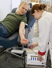
Photo by Marja Helander
ANAHEIM—A survey of UK blood donors suggests that as many as 30% of donors who are men who have sex with men (MSM) may not be compliant with the MSM blood donor policy.
The UK’s policy requires that MSMs do not donate blood if they have engaged in sexual activity with another male in the last 12 months.
But the survey indicates that as many as 3 in 10 MSMs are disregarding this policy.
The research also suggests that MSMs who do not comply with the policy engage in riskier sexual behavior than non-MSM male blood donors.
However, the researchers found no increase in the number of sexually transmitted infections present in the blood supply since the donation policy for MSMs changed from a lifetime ban to a 12-month deferral period.
The infections evaluated include human immunodeficiency virus (HIV), hepatitis C virus (HCV), hepatitis B virus (HBV), and syphilis.
The researchers also emphasized that the prevalence of HIV-positive blood donations in the UK remains low.
Katie Davidson, of Public Health England in London, presented these findings at the 2015 AABB Annual Meeting (abstract S36-030E*).
She noted that, in 2011, the blood services of England, Wales, and Scotland changed the blood donor policy for MSMs from a lifetime ban to a 12-month deferral since last male-to-male sex.
Before this policy change took effect, the blood services estimated that the change would mean 2679 MSMs would be newly eligible to donate blood (0.7% of male donors), and 8 of these donors would have HIV. So there would be a 0.5% increase in HIV risk.
“But what was clear was that these predictions in terms of HIV risk would be very dependent upon compliance,” Davidson said. “And what we mean by compliance is that a donor understands the rule, applies it correctly, and discloses any relevant information when they’re asked.”
To investigate donor behavior and compliance, Davidson and her colleagues conducted a large-scale, anonymous, web-based survey of blood donors.
Each month for 1 year (2013), all eligible new blood donors and at least an equal number of repeat blood donors in the UK were invited, via email, to complete an online questionnaire asking about their sexual history and compliance with the 12-month deferral policy for MSM (if applicable).
The researchers also looked at UK surveillance data on infections (HIV, HBV, HCV, and syphilis) in new and repeat blood donors over 6 years, comparing the incidence of infections before and after the policy change took effect (3 years pre- and post-change).
Donation and compliance
Among 65,439 survey respondents, 22,776 (35%) were male, and 242 (1%) were MSMs. Among the MSMs, 73 reported male-to-male sex within the last 12 months (non-compliance), and 181 said it had been more than 12 months since their last male-to-male sexual encounter.
The researchers adjusted these proportions for differences among the respondents and the donor population and extrapolated the data to the whole UK donor population.
The team estimated that, among 488,523 UK donors, there would be 5471 MSMs. Of the MSM donors, 3713 would be eligible under the new policy, and 1759 would be non-compliant.
So MSM compliance with the 12-month deferral policy would be 99.7% among all male donors but 70.4% of the MSM population.
“So 3 in every 10 MSMs donating blood in the UK shouldn’t be, [according to the estimates],” Davidson said.
The survey asked non-compliant MSM donors to provide their reasons for non-compliance, and many gave more than 1 reason.
“The reasons seemed to be associated, mostly, with self-assessment of their own risk [of transmitting infection] to be low,” Davidson said. “So that was based on the fact that they were in a monogamous relationship, they used condoms, practiced safe sex, or had regular [sexual health] screenings.”
However, there were some donors who regarded the policy as unimportant or said they didn’t agree with it. And there were some donors who didn’t declare their sexual behavior because they knew they wouldn’t be allowed to donate if they did.
Sexual behavior
Among all male respondents who reported having sex within the last 12 months, MSMs were more likely than men who had only female sexual partners to report having sex with more than 1 partner. Fifty percent of MSMs had more than 1 sexual partner in the last 12 months, as did 9.1% of male donors with only female sexual partners.
Ten percent of MSMs reported paying for sex, as did 0.3% of non-MSMs. None of the MSMs reported having a partner who was HIV-positive, and less than 0.1% of non-MSMs said they had an HIV-positive partner.
Eleven percent of MSMs said they had a history of sexually transmitted infection, as did less than 0.1% of non-MSMs.
“So among the responders, there was very low numbers who reported a high-risk partner in the last 12 months,” Davidson noted. “But there was some suggestion, among these low numbers, that this was more common in the MSMs than the non-MSMs.”
She also acknowledged that some donors were unsure about whether they had a high-risk partner in the last 12 months.
Infections
The UK surveillance data on infections encompassed HIV, HBV, HCV, and syphilis.
In all, 3,667,408 blood donations from males were tested for infection in the 3 years prior to the MSM donor policy change, and 3,066,076 donations were tested in the 3 years after the change was implemented.
There were 428 donors who reported having an infection risk before the change and 268 who did so after. There were 577 donors who actually had an infection before the change and 434 who did after. And there were 32 infected MSM donors before the change and 34 after.
“So the number of male donors fell post-change by approximately 20%, [and] the total number of infected donors . . . fell by almost 30%,” Davidson noted.
“However, the number of MSM infected donors marginally increased, [and] the proportion of male infected donors who were MSMs, among all of those who reported a risk, increased from 7% [32/428] to 13% [34/268]. So there seems to be some impact [on infection] from MSMs, but the numbers are very small, and these differences are not significant.”
Predictions and HIV infection
Finally, the researchers compared their predictions from before the MSM blood donor policy change to the actual data after the change. This comparison assumed that the absolute number of compliant MSMs did not change after the policy changed.
In 2007, the group predicted there would be about 2 million blood donations, including 2679 from MSMs. In reality, in 2014, there were 1.9 million blood donations, including 3126 from MSMs.
The researchers predicted the number of HIV-positive donations would be 30, including 8 from MSMs. In reality, in 2014, there were 13 HIV-positive donations, including 1 from an MSM.
So the predicted HIV prevalence per 100,000 donations was 1.4, and the actual HIV prevalence was 0.7. The predicted HIV incidence per 100,000 person-years was 0.9, and the actual HIV incidence was 0.7.
The predicted HIV risk was 0.022 per 100,000, and the actual HIV risk was 0.016 per 100,000.
“So the estimated risk of HIV post-change remains very low,” Davidson noted, adding that she and her colleagues will continue to monitor the impact of the policy change. ![]()
*Data in the abstract differ from data presented at the meeting.
Duo solves mystery regarding FVIII
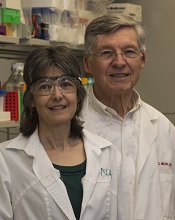
Turner and Joel Moake
Photo from Rice University
A pair of researchers solved a long-standing mystery about where factor VIII (FVIII) is made, stored, and deployed in the body, according to an article published in PLOS ONE.
The duo found that FVIII is synthesized in glomerular microvascular endothelial cells (GMVECs) and human umbilical vein endothelial cells (HUVECs).
FVIII and von Willebrand factor (VWF) are stored in Weibel-Palade bodies (WPBs) in both of these cell types. And stimulated GMVECs and HUVECs secrete cell-anchored, ultra-large VWF strings with FVIII bound to them.
The researchers said these findings may lead to improved therapy for hemophilia A.
“Great clinical advances have been made over the past 50 years, in spite of our lack of understanding of where FVIII was made and stored,” said study author Joel Moake, MD, of Rice University in Houston, Texas.
“Understanding how the body makes, stores, and deploys the protein will be increasingly important in the future as physicians look to develop gene therapies that could free patients from a reliance on regular injections of FVIII.”
The FVIII research in Dr Moake’s lab began in early 2014 and was based on a hunch by lead author Nancy Turner, who specializes in the study of endothelial cells.
“Endothelial cells are the gate-controllers for the blood system, and, to me, they make all the exciting proteins,” she said. “They’re right on the surface next to the blood, and they constantly interact with the plasma. Similar to circulating cells, they produce their own defensive proteins to fight infections.”
Though Turner had not previously studied FVIII, she had done extensive experiments on VWF, which is made in endothelial cells and stored in WPBs. And VWF and FVIII are often bound together.
Based on her prior work, Turner was intrigued by a pair of 2014 studies that examined FVIII in mouse endothelial cells and showed that FVIII was produced in the endothelial cells within the liver, and not in the liver cells.
“They stopped short of saying that FVIII was stored in endothelial cells, but they suggested the possibility,” she said. “One of the papers was very elegant, and I liked it a lot, but it made a statement that really bothered me.”
The paper explained that FVIII had been found in a half-dozen types of endothelial cell types, but never HUVECs, the type of endothelial cell found in large veins.
“HUVECs are the generic human endothelial cells that [researchers] use the first time they do anything,” Turner said. “They’re cheap. They’re easy to work with, and they’ve been the model for endothelial cells for, I don’t know, at least 50 years.”
“So far, everything I’ve ever looked at in endothelial cells has been consistent. The different types might have different amounts of something, but they’re very similar. There hasn’t been anything that was wholly different from one type to another. Not yet. So I thought, ‘OK, fine. I’m already doing gene-expression experiments. I might as well just throw FVIII in there and see.”
The experiments she was conducting involved both HUVECs and GMVECs, which are found in the smallest capillaries of the kidney. Turner was conducting an extensive analysis to see how protein production differed in the 2 varieties.
She examined the cells for FVIII messenger RNA and found that it was present, which meant it was possible the cells were making the protein. She then ordered a fluorescent antibody designed to detect FVIII.
“I did the experiment and looked, and not only was it there, but it was bright, easy to see, and it worked perfectly the first time,” Turner said. “I thought, ‘What have people been doing for 20 or 30 years? Why couldn’t they see this?’ And then I thought, ‘This was too easy. No one is going to believe me.’”
So Turner set about running controls to rule out mistakes. Were the antibodies interfering with one another? Was there any contamination? Was the microscope working correctly? Could the reading be a false-positive, an inadvertent result of another reaction she hadn’t expected?
“No. 1, I had to convince myself,” she said. “I am always skeptical if something is too easy. I have reviewed many, many papers, and I can always find what people do wrong.”
Once she had convinced herself, she and Dr Moake had to convince the paper’s referees. As she’d expected, they were skeptical. She said the bulk of the work over the past year involved doing a number of controls to remove any doubts about the findings.
In the end, the research confirmed that FVIII was made in both HUVECs and GMVECs. Moreover, Turner found that FVIII, like VWF, is both stored and secreted from WPBs.
Dr Moake said the discovery has clear implications for any future treatments that aim to repair genetic defects in patients with hemophilia A.
“Now that we recognize that FVIII is normally synthesized in endothelial cells and stored in Weibel-Palade bodies,” Dr Moake said, “those become the precise, most effective physiological targets for gene delivery.” ![]()
A new category of cytopenias

New research suggests that clonality may underlie the cytopenias observed in patients with idiopathic cytopenias of undetermined significance (ICUS).
So researchers say these patients should be classified as having clonal cytopenias of undetermined significance (CCUS).
The team believes that recognition of CCUS as a clinical entity would help identify it as a formally defined diagnostic group and could aid future studies.
“We don’t know to what extent patients who have low blood counts and mutations are at increased risk of developing an overt malignancy,” said Rafael Bejar, MD, PhD, of the UCSD Moores Cancer Center in La Jolla, California.
“We hope that by defining CCUS, future studies will follow these patients to learn what these mutations mean for their future, as their genetically abnormal cells may represent early stages of subsequent blood cancers.”
Dr Bejar and his colleagues defined CCUS in Blood. Their research was funded, in part, by Genoptix Medical Laboratory, and several study authors are employees of Genoptix.
The researchers prospectively examined bone marrow samples from 144 patients with unexplained cytopenias. Seventeen percent of these patients were diagnosed with myelodysplastic syndromes (MDS), 15% with ICUS and some evidence of dysplasia, and 69% with ICUS and no dysplasia.
The team then sequenced patient samples looking for mutations in 22 frequently mutated myeloid malignancy genes. They identified somatic mutations in 71% of MDS patients, 62% of patients with ICUS and some dysplasia, and 20% of ICUS patients with no dysplasia.
Overall, 35% of ICUS patients carried a somatic mutation or chromosomal abnormality indicative of clonal hematopoiesis.
To build upon these findings, the researchers performed a retrospective analysis of data from 91 patients with lower-risk MDS and 249 patients with ICUS.
The team identified mutations in 79% of patients with MDS, 45% of patients with ICUS and dysplasia, and 17% of patients with ICUS and no dysplasia.
The researchers said the spectrum of mutated genes was similar among the 3 groups. The exception was SF3B1, which was rarely mutated in patients without dysplasia.
The team added that variant allele fractions were comparable between clonal ICUS (CCUS) and MDS, and the same was true for the patients’ mean age and blood counts. But CCUS was a more frequent diagnosis than MDS.
Dr Bejar and his colleagues said larger, carefully controlled studies will be needed to confirm the findings of this research. ![]()
Autophagy works in nucleus to ward off cancer

Image by Sarah Pfau
Autophagy works in the cell nucleus to guard against the start of cancer, according to research published in Nature.
The study showed, for the first time, that autophagy is used to digest nuclear material in mammalian cells.
“We found that the molecular machinery of autophagy guides the degradation of components of the nuclear lamina in mammals,” said study author Shelley Berger, PhD, of the University of Pennsylvania in Philadelphia.
The nuclear lamina is a network of protein filaments lining the inside of the membrane of the nucleus. It provides mechanical support to the nucleus and regulates gene expression by making some areas of the genome less or more available to be transcribed into messenger RNA.
Previous studies showed that the autophagy protein LC3 can be found in the nucleus, but it was not clear why, as the protein was thought to be functional in the cytoplasm. Study author Zhixun Dou, PhD, came to the Berger lab with this in mind.
At the same time, Peter Adams, PhD, from the University of Glasgow in Scotland, published a study on the breakdown of the nuclear lamina in which he observed a peculiar protrusion, or blebbing, of the nuclear envelope into the cytoplasm. These blebs contained DNA, nuclear lamina proteins, and chromatin.
This evidence led the Berger and Adams labs to work together to find out what was going on.
Using biochemical and sequencing methods, Dr Dou found that laminB1, a key component of the nuclear lamina, and LC3 were contacting each other in the same places on chromatin.
In fact, LC3 and laminB1 are physically bound to each other. LC3 directly interacts with lamin B1 and binds to lamin-associated domains on chromatin.
Autophagy in cancer and aging
The investigators found that, in response to cellular stress that can cause cancer, LC3, chromatin, and laminB1 migrate from the nucleus—via the nuclear blebs—into the cytoplasm and are eventually targeted for disposal.
This breakdown of laminB1 and other nuclear material leads to senescence. The Berger and Adams labs have been studying senescence in conjunction with cancer for quite some time. One way human cells protect themselves from becoming cancerous is to accelerate aging via senescence so the cells can no longer replicate.
The team showed that when a cell’s DNA is damaged or an oncogene is activated (both of which can cause cancer), a normal cell triggers the digestion of nuclear lamina by autophagy, which promotes senescence. Inhibiting this digestion of nuclear material weakens the senescence program and leads to cancerous growth of cells.
“The nucleus is the headquarters of a cell,” Dr Dou said. “When a cell receives a danger alarm, amazingly, it deliberately messes up its headquarters, with the consequence that many functions are completely stopped for the cell. Our study suggests this new function of autophagy is a guarding mechanism that protects cells from becoming cancerous.”
The investigators noted that senescence is associated with normal aging, and senescent cells accumulate in aged tissues, which impair the normal functions of the tissue and contribute to age-related diseases. While autophagy in the nucleus is able to restrain cancer, this machinery is improperly turned on during normal aging.
“There is a short-term ‘tactical’ advantage, but a long-term ‘strategic’ defeat,” Dr Berger explained. “This mechanism makes a normal cell, even without cancer stress, get old much faster, and in a detrimental way.”
In support of this notion, the investigators found that, in late-middle-aged normal cells, blocking the autophagy-driven breakdown of the nuclear lamina can make cells live 60% longer. In fact, the team said the age extension is equivalent to a 70-year-old person living to over 110 years old.
Looking toward the future, the investigators reason that specific manipulation of the nuclear digestion by autophagy holds promise to intervene in age-related diseases.
The team showed that blocking a peptide, which inhibits LC3-laminB1 interaction, is able to slow cell aging. The implications are that a small molecule could be made to stop the long-term “strategic defeat” of the senescence pathway, and to treat age-related diseases, especially those related to chronic inflammation.
Such a molecule might also be able to ameliorate the side effects of chemotherapy or radiation therapy in cancer patients. Drs Dou and Berger are actively investigating this possibility. ![]()
iPad Apps Help Inpatients Bridge Information Gaps
A study in the Journal of the American Medical Informatics Association found that giving hospitalized patients iPads with basic information about their care helped them engage more with physicians.
The patient portal application helped inpatients remember their doctors’ names, the study noted, but had no statistically significant impact on patients’ ability to remember scheduled procedures or active medications.
Hospitalist and lead author Kevin O’Leary, MD, MS, SFHM, of Northwestern University Feinberg School of Medicine in Chicago says the portals can be a way to improve the discharge process.
“We know that patients have a fairly poor comprehension of their care during hospitalization, which doesn’t set them up to do well after discharge,” says Dr. O’Leary, who has studied the potential of portals. “This patient portal has the opportunity to fill in the gaps.”
Although the study didn’t address qualitative issues around why patients didn’t have better recall of information about tests, procedures, or active medications, Dr. O’Leary says future work will address how to tweak portals to improve their efficacy.
“Our study had limited information purposely to just test the concept,” Dr. O’Leary says. “I predict that 10 years from now, this is going to be commonplace. Even five years from now, many hospitals will be offering portal access to inpatients.”
Dr. O’Leary says he believes that using the increasingly ubiquitous technology of smartphones and tablet computers to engage patients creates greater transparency, which could result in better retention of care instructions.
“You’re giving ownership to the patient over information, and it’s much earlier than right at discharge and then a phone call after discharge,” Dr. O’Leary adds. “Why not begin to educate the patient about their condition and their medications from the very first minute of their admission? And give them access to that information and have them identify their own needs and knowledge deficits?”
Visit our website for more information about iPad technology in medical practice.
Joint Commission Unveils Online Resource to Promote High-Reliability Healthcare
Zero preventable harm is the goal of every healthcare organization, and a new online application from the Joint Commission Center for Transforming Healthcare can help groups reach that goal. Oro 2.0 can assist hospital leaders with determining their organization’s status across multiple components of high reliability that will let them achieve zero preventable harm.
Hospitalists are integral to this process, says Coleen Smith, RN, BSN, MBA, CPHQ, director of high reliability initiatives for the Joint Commission. “Hospitalists are a crucial piece of improving the quality of an organization. Physicians routinely getting involved in high reliability and leading those activities are how we want high-reliability hospitals to look,” says Smith.
The Oro 2.0 application has two major elements. The first is an assessment tool for senior hospital leadership that covers 14 different performance areas, covering components such as safety culture and leadership. After an assessment is complete, the application issues a report that identifies strengths and opportunities for improvement and directs the user to specific resources, which are the application’s second element: a library of published materials about specific areas of high reliability offering more than 125 references and tools. These materials will help organizations educate themselves about the 14 components included in Oro 2.0’s high-reliability model.
This is an idea long overdue in healthcare, which lags behind other industries, according to Smith. “We know high-reliability industries like nuclear power and commercial aviation do very complex daily work but have far fewer bad things happening,” she adds. “Healthcare is nowhere near that state of reliability, but it needs to get there.”
Participation by hospitalists is required to make such a change, Smith says. She hopes they will access Oro 2.0’s resource library to understand the solutions the Joint Commission is proposing. “Then they can go to the senior leadership of their hospital and ask them to consider committing to zero patient harm,” she says. “They can support their senior leadership in this work and be vocal about their interest in pursuing high reliability and zero patient harm.”
Visit our website for more information on hospitalists and healthcare safety.
Acute Generalized Exanthematous Pustulosis Associated With Ranolazine
Acute generalized exanthematous pustulosis (AGEP) is a potentially widespread, pustular, cutaneous eruption. In 90% of cases, AGEP results from drug administration.1,2 It manifests as numerous subcorneal, nonfollicular, sterile pustules of rapid onset on an erythematous base,2 often in conjunction with fever, peripheral leukocytosis, and neutrophilia.3 Numerous drug therapies have been implicated in the etiology of AGEP, most commonly the β-lactam antibiotics, such as the penicillin derivatives and cephalosporins.2 Typically, AGEP occurs soon after drug ingestion and resolves spontaneously, shortly after the causative drug is discontinued.
Ranolazine is an antianginal, anti-ischemic medication with an undetermined mechanism of action. Its antianginal and anti-ischemic effects do not depend on reduced heart rate or blood pressure. At therapeutic levels, it inhibits the cardiac late sodium current (INa), reducing the sodium-induced calcium overload in ischemic cardiac myocytes. Severe adverse reactions include angioedema; paresthesia; pancytopenia; and, in animal studies, tumorigenicity.4 Herein we report a case of AGEP associated with the use of ranolazine.
Case Report
An 83-year-old man presented with a generalized rash of approximately 12 days’ duration. The patient reported that the small “pimple-like” bumps initially erupted on the back of the neck but gradually spread to the chest, back, and extremities. The lesions were asymptomatic at the outset and became pruritic over time. For the last several years, the patient had been taking tamsulosin for benign prostatic hypertrophy and rosuvastatin for hyperlipidemia. Twelve days prior to the exanthem, he had started taking ranolazine for symptomatic ischemia until coronary angiography could be performed. He reported having no associated fevers, chills, or malaise and had no personal history of psoriasis, though he had a maternal history of the disorder.
Examination revealed numerous nonfollicular-based pustules on diffuse erythematous patches (Figure 1). There was no mucosal involvement and the skin was negative for the Nikolsky sign. Spongiform intracorneal collections of neutrophils were visible on punch biopsy (Figures 2 and 3). Periodic acid–Schiff stains for fungi were negative.

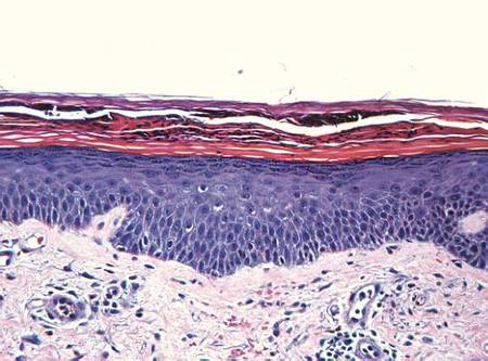
| 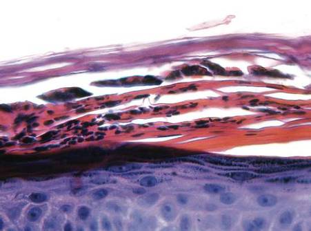
| |
Figure 2. A punch biopsy showed spongiform intracorneal collections of neutrophils (H&E, original magnification ×200). | Figure 3. A cornified layer of epidermis with neutrophils, as visible on punch biopsy (H&E, original magnification ×630). |
The patient’s primary care physician had initiated a course of oral prednisone 5 mg daily, 3 days before he presented to our outpatient dermatology clinic, but it had little effect on the rash. Upon dermatologic evaluation, we discontinued ranolazine therapy and prescribed the following tapered course of oral prednisone: 60 mg daily for 4 days; 40 mg daily for 3 days; 30 mg daily for 3 days; 20 mg daily for 3 days; 10 mg daily for 3 days; and 5 mg daily for 3 days). Within a week after this regimen was initiated, the rash showed improvement with eventual resolution and desquamation (Figure 4). Subsequently, the patient underwent successful angioplasty and multiple stent placement, which ultimately alleviated his angina.
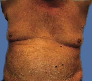
Comment
Since its original description in 1968,5 AGEP has been misdiagnosed and underreported. Due to its rarity and clinical resemblance to more common pustular eruptions, such as exanthematous pustular psoriasis, the typical characteristics of AGEP were not clearly delineated until Beylot et al3 coined the term AGEP in 1980. Since that time, formalized criteria for the diagnosis and characterization of AGEP have been published.1,2,6-8
Numerous drug therapies have been implicated in the etiology of AGEP, most commonly antimicrobial agents, such as β-lactam antibiotics. Many other drugs, however, also have been identified as potential causative agents,8 including but not limited to antifungal, anticonvulsant, and antihypertensive agents. Other less common etiologies include viral infections,6,9-11 UV radiation, contrast media, heavy metal exposure (eg, to mercury), ingestion of urushiol (eg, in lacquered chicken), and spider bites.2,8,12-16 Nevertheless, more than 90% of AGEP cases are attributed to drug exposure, with 80% of drug-induced cases believed to be caused by antibiotics.1,8
The incidence of AGEP is estimated to be between 1 and 5 cases per million per year, using inclusion criteria from the EuroSCAR study, a multinational, case-controlled, pharmacoepidemiologic study of severe cutaneous adverse reactions.8,16 The condition seems to affect males and females equally.1,4 There are no reports of age or racial predilection.1,6,17 It has been suggested that those with AGEP may have some form of psoriatic background.1 Our patient had no personal history of inflammatory skin disease, although his mother had psoriasis.
The dermatitis presents as the sudden onset of a diffuse exanthematous eruption, which typically produces dozens to hundreds of sterile, nonfollicular, superficial pustules on an erythematous and possibly edematous base. Atypical presentations include target lesions, purpura, and vesicles. The reaction usually begins on the face or intertriginous areas of flexural surfaces and quickly disseminates. Patients may experience burning or pruritus. Acute generalized exanthematous pustulosis may involve mucous membranes but is usually limited to 1 location, most often the oral mucosa.1,8,16,18 Systemic signs and symptoms include fever, lymphadenopathy, pharyngitis, and hepatosplenomegaly. Unlike most drug allergies that demonstrate eosinophilia, AGEP is associated with leukocytosis and neutrophilic predominance. Only 25% of affected patients exhibit eosinophilia.1 Approximately 30% of patients in a retrospective analysis demonstrated abnormal renal function,2 and there have been reports of mildly elevated transaminases.8,19
In the EuroSCAR study, for reasons that were not apparent, symptoms developed within 24 hours of exposure to triggering antibiotics, whereas the median time to rash onset in response to non–anti-infective agents was 11 days.8 This finding is consistent with the delayed onset of symptoms experienced by our patient after initiating ranolazine therapy.
The differential diagnosis of AGEP primarily includes pustular psoriasis, subcorneal pustulosis, pustular folliculitis, DRESS (drug reaction with eosinophilia and systemic symptoms) syndrome, bullous impetigo, and occasionally erythema multiforme and toxic epidermal necrolysis, with the latter typically characterized by more mucous membrane involvement.20 Biopsy does not always support a definitive diagnosis; clinical correlation is often necessary. Because of the EuroSCAR study, Sidoroff et al8 devised a clinical validation score based on morphology (presence of pustules and erythema, distribution, and eventual desquamation), histopathology (presence of intraepidermal pustules, spongiosis, and papillary edema), and disease course (duration of symptoms, neutrophilia, fever, acute onset, and time to resolution). A definitive score is 8 to 12 (out of 12), and our patient’s score was 10; the score may have been higher had blood work been performed, but by the time the diagnosis was made the patient’s condition had improved enough to make laboratory workup unnecessary.
Several theories have been proposed to explain the pathophysiology of AGEP. Some hold that the causative agent induces the formation of antigen-antibody complexes, thereby activating the complement system, which in turn produces neutrophil chemotaxis.3,21 A more recent theory suggests that drug exposure causes drug-specific CD4 and CD8 cells to migrate into dermal and epidermal layers of the skin.17 Both T cells and keratinocytes express IL-8, which attracts polymorphonuclear leukocytes, causing them to accumulate in the dermis and then the epidermis. The different clinical presentations of AGEP may be attributed to other cytokines and interleukins that T cells express during this process. In the epidermis, CD8 cells kill keratinocytes, causing focal necrosis and prompting the formation of subcorneal vesicles filled primarily with CD4 cells. CD4 and CD8 cells are then localized to the dermis where neutrophils enter the vesicles, transforming them into sterile pustules.6,16,17
Acute generalized exanthematous pustulosis has been characterized as a type IV delayed hypersensitivity reaction, with affected patients often demonstrating positive patch testing or a history of prior sensitization to the perpetrating agent.18,19,21 Although there have been reports of positive patch testing for certain drugs, the unknown sensitivity and specificity of such testing as well as preparation-dependent variables may limit the diagnostic utility of this approach.21 The additional risk for inducing AGEP by patch testing the suspected drug also is a consideration. Due to our patient’s definitive clinical validation score, we did not perform this test.21
The AGEP eruption is typically self-limited and tends to resolve within 4 to 10 days after cessation of the triggering agent. Postpustular desquamation often occurs upon resolution of the primary lesions. Treatment usually involves discontinuation of the suspected causative agent and the use of antihistamines, antipyretics, topical corticosteroids, and emollients. Although there are reports of AGEP responsiveness to oral and intravenous steroids, such treatment rarely is required.8,16,22 We prescribed a tapered course of oral prednisone due to our patient’s imminent need for angioplasty.
Conclusion
This case of AGEP induced by ranolazine is notable. Given the potential widespread use of this antianginal medication and the severity of this potential adverse reaction, it is important for clinicians to recognize AGEP, discontinue ranolazine if determined to be a causative agent, and then initiate an appropriate alternative antianginal therapy.
1. Roujeau JC, Bioulac-Sage P, Bourseau C, et al. Acute generalized exanthematous pustulosis. analysis of 63 cases. Arch Dermatol. 1991;127:1333-1338.
2. Sidoroff A, Halevy S, Bavnick JN, et al. Acute generalized exanthematous pustulosis (AGEP)–a clinical reaction pattern. J Cutan Pathol. 2001;28:113-119.
3. Beylot C, Bioulac P, Doutre MS. Acute generalized exanthematic pustuloses (four cases) [in French]. Ann Dermatol Venereol. 1980;107:37-48.
4. Ranexa [package insert]. Foster City, CA: Gilead Sciences, Inc; December 2013.
5. Baker H, Ryan TJ. Generalized pustular psoriasis. a clinical and epidemiological study of 104 cases. Br J Dermatol. 1968;80:771-793.
6. Guevara-Gutierrez E, Uribe-Jimenez E, Diaz-Canchola M, et al. Acute generalized exanthematous pustulosis: report of 12 cases and literature review. Int J Dermatol. 2009;48:253-258.
7. Chang SL, Huang YH, Yang CH, et al. Clinical manifestations and characteristics of patients with acute generalized exanthematous pustulosis in Asia. Acta Derm Venereol. 2008;88:363-365.
8. Sidoroff A, Dunant A, Viboud C, et al. Risk factors for acute generalized exanthematous pustulosis (AGEP)-results of a multinational case-control study (EuroSCAR) [published online ahead of print September 13, 2007]. Br J Dermatol. 2007;157:989-996.
9. Rouchouse B, Bonnefoy M, Pallot B, et al. Acute generalized exanthematous pustular dermatitis and viral infection. Dermatologica. 1986;173:180-184.
10. Naides SJ, Piette W, Veach LA, et al. Human parvovirus B19-induced vesiculopustular skin eruption. Am J Med. 1988;84:968-972.
11. Feio AB, Apetato M, Costa MM, et al. Acute generalized exanthematous pustulosis due to Coxsackie B4 virus [in Portuguese]. Acta Med Port. 1997;10:487-491.
12. Goh TK, Pang SM, Thirumoorthy T, et al. Acute generalised exanthematous pustulosis and toxic epidermal necrolysis induced by carbamazepine. Singapore Med J. 2008;49:507-510.
13. Ofuji S, Yamamoto O. Acute generalized exanthematous pustulosis associated with a human parvovirus B19 infection. J Dermatol. 2007;34:121-123.
14. Davidovici BB, Pavel D, Cagnano E, et al. Acute generalized exanthematous pustulosis following a spider bite: report of 3 cases. J Am Acad Dermatol. 2006;55:525-529.
15. Park YM, Park JG, Kang H, et al. Acute generalized exanthematous pustulosis induced by ingestion of lacquer chicken. Br J Dermatol. 2000;143:230-232.
16. Hammerbeck AA, Daniels NH, Callen JP. Ioversol-induced acute generalized exanthematous pustulosis: a case report. Arch Dermatol. 2009;145:683-687.17. Halevy S. Acute generalized exanthematous pustulosis. Curr Opin Allergy Clin Immunol. 2009;9:322-328.
18. Kim HJ, Jung KD, Lee KT, et al. Acute generalized exanthematous pustulosis caused by diltiazem [published online ahead of print February 28, 2011]. Ann Dermatol. 2011;23:108-110.
19. Speck LM, Wilkerson MG, Perri AJ, et al. Acute generalized exanthematous pustulosis caused by terazosin hydrochloride. J Drugs Dermatol. 2008;7:395-397.
20. Sidoroff A. Acute generalized exanthematous pustulosis (AGEP). UpToDate Web site. http://www.uptodate.com /contents/acute-generalized-exanthematous-pustulosis -agep?source=search_result&search=agep&selected Title=1~85. Updated March 18, 2015. Accessed October 6, 2015.
21. Mashiah J, Brenner S. A systemic reaction to patch testing for the evaluation of acute generalized exanthematous pustulosis. Arch Dermatol. 2003;139:1181-1183.
22. Ibrahimi O, Gunawardane N, Sepehr A, et al. Terbinafine-induced acute generalized exanthematous pustulosis (AGEP) responsive to high dose intravenous corticosteroid. Dermatol Online J. 2009;15:8.
Acute generalized exanthematous pustulosis (AGEP) is a potentially widespread, pustular, cutaneous eruption. In 90% of cases, AGEP results from drug administration.1,2 It manifests as numerous subcorneal, nonfollicular, sterile pustules of rapid onset on an erythematous base,2 often in conjunction with fever, peripheral leukocytosis, and neutrophilia.3 Numerous drug therapies have been implicated in the etiology of AGEP, most commonly the β-lactam antibiotics, such as the penicillin derivatives and cephalosporins.2 Typically, AGEP occurs soon after drug ingestion and resolves spontaneously, shortly after the causative drug is discontinued.
Ranolazine is an antianginal, anti-ischemic medication with an undetermined mechanism of action. Its antianginal and anti-ischemic effects do not depend on reduced heart rate or blood pressure. At therapeutic levels, it inhibits the cardiac late sodium current (INa), reducing the sodium-induced calcium overload in ischemic cardiac myocytes. Severe adverse reactions include angioedema; paresthesia; pancytopenia; and, in animal studies, tumorigenicity.4 Herein we report a case of AGEP associated with the use of ranolazine.
Case Report
An 83-year-old man presented with a generalized rash of approximately 12 days’ duration. The patient reported that the small “pimple-like” bumps initially erupted on the back of the neck but gradually spread to the chest, back, and extremities. The lesions were asymptomatic at the outset and became pruritic over time. For the last several years, the patient had been taking tamsulosin for benign prostatic hypertrophy and rosuvastatin for hyperlipidemia. Twelve days prior to the exanthem, he had started taking ranolazine for symptomatic ischemia until coronary angiography could be performed. He reported having no associated fevers, chills, or malaise and had no personal history of psoriasis, though he had a maternal history of the disorder.
Examination revealed numerous nonfollicular-based pustules on diffuse erythematous patches (Figure 1). There was no mucosal involvement and the skin was negative for the Nikolsky sign. Spongiform intracorneal collections of neutrophils were visible on punch biopsy (Figures 2 and 3). Periodic acid–Schiff stains for fungi were negative.
|
|
| |
Figure 2. A punch biopsy showed spongiform intracorneal collections of neutrophils (H&E, original magnification ×200). | Figure 3. A cornified layer of epidermis with neutrophils, as visible on punch biopsy (H&E, original magnification ×630). |
The patient’s primary care physician had initiated a course of oral prednisone 5 mg daily, 3 days before he presented to our outpatient dermatology clinic, but it had little effect on the rash. Upon dermatologic evaluation, we discontinued ranolazine therapy and prescribed the following tapered course of oral prednisone: 60 mg daily for 4 days; 40 mg daily for 3 days; 30 mg daily for 3 days; 20 mg daily for 3 days; 10 mg daily for 3 days; and 5 mg daily for 3 days). Within a week after this regimen was initiated, the rash showed improvement with eventual resolution and desquamation (Figure 4). Subsequently, the patient underwent successful angioplasty and multiple stent placement, which ultimately alleviated his angina.
Comment
Since its original description in 1968,5 AGEP has been misdiagnosed and underreported. Due to its rarity and clinical resemblance to more common pustular eruptions, such as exanthematous pustular psoriasis, the typical characteristics of AGEP were not clearly delineated until Beylot et al3 coined the term AGEP in 1980. Since that time, formalized criteria for the diagnosis and characterization of AGEP have been published.1,2,6-8
Numerous drug therapies have been implicated in the etiology of AGEP, most commonly antimicrobial agents, such as β-lactam antibiotics. Many other drugs, however, also have been identified as potential causative agents,8 including but not limited to antifungal, anticonvulsant, and antihypertensive agents. Other less common etiologies include viral infections,6,9-11 UV radiation, contrast media, heavy metal exposure (eg, to mercury), ingestion of urushiol (eg, in lacquered chicken), and spider bites.2,8,12-16 Nevertheless, more than 90% of AGEP cases are attributed to drug exposure, with 80% of drug-induced cases believed to be caused by antibiotics.1,8
The incidence of AGEP is estimated to be between 1 and 5 cases per million per year, using inclusion criteria from the EuroSCAR study, a multinational, case-controlled, pharmacoepidemiologic study of severe cutaneous adverse reactions.8,16 The condition seems to affect males and females equally.1,4 There are no reports of age or racial predilection.1,6,17 It has been suggested that those with AGEP may have some form of psoriatic background.1 Our patient had no personal history of inflammatory skin disease, although his mother had psoriasis.
The dermatitis presents as the sudden onset of a diffuse exanthematous eruption, which typically produces dozens to hundreds of sterile, nonfollicular, superficial pustules on an erythematous and possibly edematous base. Atypical presentations include target lesions, purpura, and vesicles. The reaction usually begins on the face or intertriginous areas of flexural surfaces and quickly disseminates. Patients may experience burning or pruritus. Acute generalized exanthematous pustulosis may involve mucous membranes but is usually limited to 1 location, most often the oral mucosa.1,8,16,18 Systemic signs and symptoms include fever, lymphadenopathy, pharyngitis, and hepatosplenomegaly. Unlike most drug allergies that demonstrate eosinophilia, AGEP is associated with leukocytosis and neutrophilic predominance. Only 25% of affected patients exhibit eosinophilia.1 Approximately 30% of patients in a retrospective analysis demonstrated abnormal renal function,2 and there have been reports of mildly elevated transaminases.8,19
In the EuroSCAR study, for reasons that were not apparent, symptoms developed within 24 hours of exposure to triggering antibiotics, whereas the median time to rash onset in response to non–anti-infective agents was 11 days.8 This finding is consistent with the delayed onset of symptoms experienced by our patient after initiating ranolazine therapy.
The differential diagnosis of AGEP primarily includes pustular psoriasis, subcorneal pustulosis, pustular folliculitis, DRESS (drug reaction with eosinophilia and systemic symptoms) syndrome, bullous impetigo, and occasionally erythema multiforme and toxic epidermal necrolysis, with the latter typically characterized by more mucous membrane involvement.20 Biopsy does not always support a definitive diagnosis; clinical correlation is often necessary. Because of the EuroSCAR study, Sidoroff et al8 devised a clinical validation score based on morphology (presence of pustules and erythema, distribution, and eventual desquamation), histopathology (presence of intraepidermal pustules, spongiosis, and papillary edema), and disease course (duration of symptoms, neutrophilia, fever, acute onset, and time to resolution). A definitive score is 8 to 12 (out of 12), and our patient’s score was 10; the score may have been higher had blood work been performed, but by the time the diagnosis was made the patient’s condition had improved enough to make laboratory workup unnecessary.
Several theories have been proposed to explain the pathophysiology of AGEP. Some hold that the causative agent induces the formation of antigen-antibody complexes, thereby activating the complement system, which in turn produces neutrophil chemotaxis.3,21 A more recent theory suggests that drug exposure causes drug-specific CD4 and CD8 cells to migrate into dermal and epidermal layers of the skin.17 Both T cells and keratinocytes express IL-8, which attracts polymorphonuclear leukocytes, causing them to accumulate in the dermis and then the epidermis. The different clinical presentations of AGEP may be attributed to other cytokines and interleukins that T cells express during this process. In the epidermis, CD8 cells kill keratinocytes, causing focal necrosis and prompting the formation of subcorneal vesicles filled primarily with CD4 cells. CD4 and CD8 cells are then localized to the dermis where neutrophils enter the vesicles, transforming them into sterile pustules.6,16,17
Acute generalized exanthematous pustulosis has been characterized as a type IV delayed hypersensitivity reaction, with affected patients often demonstrating positive patch testing or a history of prior sensitization to the perpetrating agent.18,19,21 Although there have been reports of positive patch testing for certain drugs, the unknown sensitivity and specificity of such testing as well as preparation-dependent variables may limit the diagnostic utility of this approach.21 The additional risk for inducing AGEP by patch testing the suspected drug also is a consideration. Due to our patient’s definitive clinical validation score, we did not perform this test.21
The AGEP eruption is typically self-limited and tends to resolve within 4 to 10 days after cessation of the triggering agent. Postpustular desquamation often occurs upon resolution of the primary lesions. Treatment usually involves discontinuation of the suspected causative agent and the use of antihistamines, antipyretics, topical corticosteroids, and emollients. Although there are reports of AGEP responsiveness to oral and intravenous steroids, such treatment rarely is required.8,16,22 We prescribed a tapered course of oral prednisone due to our patient’s imminent need for angioplasty.
Conclusion
This case of AGEP induced by ranolazine is notable. Given the potential widespread use of this antianginal medication and the severity of this potential adverse reaction, it is important for clinicians to recognize AGEP, discontinue ranolazine if determined to be a causative agent, and then initiate an appropriate alternative antianginal therapy.
1. Roujeau JC, Bioulac-Sage P, Bourseau C, et al. Acute generalized exanthematous pustulosis. analysis of 63 cases. Arch Dermatol. 1991;127:1333-1338.
2. Sidoroff A, Halevy S, Bavnick JN, et al. Acute generalized exanthematous pustulosis (AGEP)–a clinical reaction pattern. J Cutan Pathol. 2001;28:113-119.
3. Beylot C, Bioulac P, Doutre MS. Acute generalized exanthematic pustuloses (four cases) [in French]. Ann Dermatol Venereol. 1980;107:37-48.
4. Ranexa [package insert]. Foster City, CA: Gilead Sciences, Inc; December 2013.
5. Baker H, Ryan TJ. Generalized pustular psoriasis. a clinical and epidemiological study of 104 cases. Br J Dermatol. 1968;80:771-793.
6. Guevara-Gutierrez E, Uribe-Jimenez E, Diaz-Canchola M, et al. Acute generalized exanthematous pustulosis: report of 12 cases and literature review. Int J Dermatol. 2009;48:253-258.
7. Chang SL, Huang YH, Yang CH, et al. Clinical manifestations and characteristics of patients with acute generalized exanthematous pustulosis in Asia. Acta Derm Venereol. 2008;88:363-365.
8. Sidoroff A, Dunant A, Viboud C, et al. Risk factors for acute generalized exanthematous pustulosis (AGEP)-results of a multinational case-control study (EuroSCAR) [published online ahead of print September 13, 2007]. Br J Dermatol. 2007;157:989-996.
9. Rouchouse B, Bonnefoy M, Pallot B, et al. Acute generalized exanthematous pustular dermatitis and viral infection. Dermatologica. 1986;173:180-184.
10. Naides SJ, Piette W, Veach LA, et al. Human parvovirus B19-induced vesiculopustular skin eruption. Am J Med. 1988;84:968-972.
11. Feio AB, Apetato M, Costa MM, et al. Acute generalized exanthematous pustulosis due to Coxsackie B4 virus [in Portuguese]. Acta Med Port. 1997;10:487-491.
12. Goh TK, Pang SM, Thirumoorthy T, et al. Acute generalised exanthematous pustulosis and toxic epidermal necrolysis induced by carbamazepine. Singapore Med J. 2008;49:507-510.
13. Ofuji S, Yamamoto O. Acute generalized exanthematous pustulosis associated with a human parvovirus B19 infection. J Dermatol. 2007;34:121-123.
14. Davidovici BB, Pavel D, Cagnano E, et al. Acute generalized exanthematous pustulosis following a spider bite: report of 3 cases. J Am Acad Dermatol. 2006;55:525-529.
15. Park YM, Park JG, Kang H, et al. Acute generalized exanthematous pustulosis induced by ingestion of lacquer chicken. Br J Dermatol. 2000;143:230-232.
16. Hammerbeck AA, Daniels NH, Callen JP. Ioversol-induced acute generalized exanthematous pustulosis: a case report. Arch Dermatol. 2009;145:683-687.17. Halevy S. Acute generalized exanthematous pustulosis. Curr Opin Allergy Clin Immunol. 2009;9:322-328.
18. Kim HJ, Jung KD, Lee KT, et al. Acute generalized exanthematous pustulosis caused by diltiazem [published online ahead of print February 28, 2011]. Ann Dermatol. 2011;23:108-110.
19. Speck LM, Wilkerson MG, Perri AJ, et al. Acute generalized exanthematous pustulosis caused by terazosin hydrochloride. J Drugs Dermatol. 2008;7:395-397.
20. Sidoroff A. Acute generalized exanthematous pustulosis (AGEP). UpToDate Web site. http://www.uptodate.com /contents/acute-generalized-exanthematous-pustulosis -agep?source=search_result&search=agep&selected Title=1~85. Updated March 18, 2015. Accessed October 6, 2015.
21. Mashiah J, Brenner S. A systemic reaction to patch testing for the evaluation of acute generalized exanthematous pustulosis. Arch Dermatol. 2003;139:1181-1183.
22. Ibrahimi O, Gunawardane N, Sepehr A, et al. Terbinafine-induced acute generalized exanthematous pustulosis (AGEP) responsive to high dose intravenous corticosteroid. Dermatol Online J. 2009;15:8.
Practice Points
- Encountering an acute pustular reaction pattern should trigger the clinician to rule out acute generalized exanthematous pustulosis (AGEP).
- Ranolazine, a new antianginal therapy, has been associated with AGEP.
- Upon confirmation of AGEP, the patient’s recent medication history should be reviewed so the potential causative agent can be identified and withdrawn.
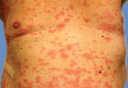
Burden of psychiatric comorbidity higher in MS patients
The burden of psychiatric comorbidity is greater in patients with multiple sclerosis (MS), compared with the general population, reported Dr. Ruth Ann Marrie and coauthors from the departments of psychiatry and medicine at the University of Manitoba, Winnipeg.
A study of 44,452 MS patients and 220,849 controls in four Canadian provinces from 1995 to 2005 found that the incidence of depression in the MS group was 0.98% (95% CI; 0.81%-1.15%), compared with 0.72% (95% CI; 0.67%-0.76%) in the control group. The prevalence of depression was 20.1% in MS patients (19.5%-20.6%), compared with 11.9% (11.8%-12.1%) in the matched population, the authors noted.

Also, the incidence and prevalence of anxiety disorder in the MS population was 0.64% (0.54%-0.73%) and 8.7% (8.4%-9.1%), respectively, compared with 0.42% (0.39%-0.45%) and 5.1% (4.9%-5.2%) in controls .
For bipolar disorder, the MS group had an incidence of 0.33% (0.26%-0.39%), compared with 0.16% (0.14%-0.18%) in controls. Prevalence was 4.7% (4.4%-4.9%) in the MS group and 2.3% (2.2%-2.3%) in controls .
Lastly, in schizophrenia, MS patients had an incidence of 0.060% (0.031%-0.080%), compared with 0.018% (0.011%-0.024%) in controls. Prevalence was 1.28% (1.15%-1.41%), in the MS group and 1.03% (0.99%-1.08%) in controls, the investigators said.
The findings suggest a “nonspecific effect of MS on psychiatric comorbidity,” Dr. Marrie and colleagues said in the report.
“From a policy perspective, this implies the need for general psychiatric support rather than illness-specific strategies,” they concluded.
Read the study in Neurology.
The burden of psychiatric comorbidity is greater in patients with multiple sclerosis (MS), compared with the general population, reported Dr. Ruth Ann Marrie and coauthors from the departments of psychiatry and medicine at the University of Manitoba, Winnipeg.
A study of 44,452 MS patients and 220,849 controls in four Canadian provinces from 1995 to 2005 found that the incidence of depression in the MS group was 0.98% (95% CI; 0.81%-1.15%), compared with 0.72% (95% CI; 0.67%-0.76%) in the control group. The prevalence of depression was 20.1% in MS patients (19.5%-20.6%), compared with 11.9% (11.8%-12.1%) in the matched population, the authors noted.
Also, the incidence and prevalence of anxiety disorder in the MS population was 0.64% (0.54%-0.73%) and 8.7% (8.4%-9.1%), respectively, compared with 0.42% (0.39%-0.45%) and 5.1% (4.9%-5.2%) in controls .
For bipolar disorder, the MS group had an incidence of 0.33% (0.26%-0.39%), compared with 0.16% (0.14%-0.18%) in controls. Prevalence was 4.7% (4.4%-4.9%) in the MS group and 2.3% (2.2%-2.3%) in controls .
Lastly, in schizophrenia, MS patients had an incidence of 0.060% (0.031%-0.080%), compared with 0.018% (0.011%-0.024%) in controls. Prevalence was 1.28% (1.15%-1.41%), in the MS group and 1.03% (0.99%-1.08%) in controls, the investigators said.
The findings suggest a “nonspecific effect of MS on psychiatric comorbidity,” Dr. Marrie and colleagues said in the report.
“From a policy perspective, this implies the need for general psychiatric support rather than illness-specific strategies,” they concluded.
Read the study in Neurology.