User login
Surgeons dinged by incomplete risk adjustment for emergency cases
SAN ANTONIO – Perioperative risk factors affect postoperative morbidity and mortality differently in emergency and nonemergency surgery, according to an analysis of the ACS National Surgical Quality Improvement Program (ACS NSQIP) database by investigators from Massachusetts General Hospital, Boston.
“Instead of using the same risk-adjustment model for both ... as is currently being done, our findings strongly suggest the need to benchmark emergent and elective surgeries separately,” they concluded.

“Most risk-adjustment models simply have an on/off switch for whether or not the patient underwent emergency surgery and treat comorbidities and other perioperative variables the same.” Those variables, however, “don’t behave the same way in emergency surgery,” said investigator Dr. Jordan Bohnen, a surgical research resident at Mass General.
Because risk adjustment doesn’t take into account variables that have a particularly strong negative impact in emergency settings, acute care surgeons are getting “unnecessarily dinged for having higher complication rates,” he said at the Eastern Association for the Surgery of Trauma scientific assembly.
For example, the team found that preop transfusions and white blood counts (WBC) at or below 4.5 carry a significantly higher risk of 30-day major morbidity or mortality (MMM) in emergency versus nonemergency surgery. Conversely, ascites, preop anemia, and leukocytosis carry a greater MMM risk in nonemergent cases.
The findings come from a comparison of 110,182 nonemergent surgeries to 59,949 emergency cases – generally meaning surgery within 12 hours of emergency department (ED) admission – from the NSQIP database for 2011-2012.
As expected, the overall risk of MMM was significantly higher for emergency cases (16.75% vs. 9.73%; P less than .001), and four procedures – laparoscopic cholecystectomy, exploratory laparotomy, and umbilical and incisional hernia repairs – were relatively riskier when done emergently.
“As surgical quality improvement efforts mature, it’s increasingly important to apply accurate risk-adjustment models to benchmark quality improvement for surgeons, hospitals, and health care systems.” The current “assumption that perioperative variables have an equal impact on outcomes in emergent and nonemergent settings” is incorrect. “Risk factors for bad outcomes change depending on the setting,” Dr. Bohnen said.
Dr. Bohnen has no disclosures.
SAN ANTONIO – Perioperative risk factors affect postoperative morbidity and mortality differently in emergency and nonemergency surgery, according to an analysis of the ACS National Surgical Quality Improvement Program (ACS NSQIP) database by investigators from Massachusetts General Hospital, Boston.
“Instead of using the same risk-adjustment model for both ... as is currently being done, our findings strongly suggest the need to benchmark emergent and elective surgeries separately,” they concluded.
“Most risk-adjustment models simply have an on/off switch for whether or not the patient underwent emergency surgery and treat comorbidities and other perioperative variables the same.” Those variables, however, “don’t behave the same way in emergency surgery,” said investigator Dr. Jordan Bohnen, a surgical research resident at Mass General.
Because risk adjustment doesn’t take into account variables that have a particularly strong negative impact in emergency settings, acute care surgeons are getting “unnecessarily dinged for having higher complication rates,” he said at the Eastern Association for the Surgery of Trauma scientific assembly.
For example, the team found that preop transfusions and white blood counts (WBC) at or below 4.5 carry a significantly higher risk of 30-day major morbidity or mortality (MMM) in emergency versus nonemergency surgery. Conversely, ascites, preop anemia, and leukocytosis carry a greater MMM risk in nonemergent cases.
The findings come from a comparison of 110,182 nonemergent surgeries to 59,949 emergency cases – generally meaning surgery within 12 hours of emergency department (ED) admission – from the NSQIP database for 2011-2012.
As expected, the overall risk of MMM was significantly higher for emergency cases (16.75% vs. 9.73%; P less than .001), and four procedures – laparoscopic cholecystectomy, exploratory laparotomy, and umbilical and incisional hernia repairs – were relatively riskier when done emergently.
“As surgical quality improvement efforts mature, it’s increasingly important to apply accurate risk-adjustment models to benchmark quality improvement for surgeons, hospitals, and health care systems.” The current “assumption that perioperative variables have an equal impact on outcomes in emergent and nonemergent settings” is incorrect. “Risk factors for bad outcomes change depending on the setting,” Dr. Bohnen said.
Dr. Bohnen has no disclosures.
AT THE EAST SCIENTIFIC ASSEMBLY
Key clinical point: Emergent and elective surgeries need to be benchmarked separately.
Major finding: Preop transfusions and WBC counts at or below 4.5 carry a significantly higher risk of 30-day major morbidity or mortality (MMM) in emergency surgery. Conversely, ascites, preop anemia, and leukocytosis carry a greater MMM risk in nonemergent cases.
Data source: More than 170,000 cases in the NSQIP database
Disclosures: The presenter has no disclosures.

Lenalidomide shows promise for treating ATLL
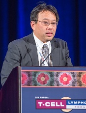
Photo by Larry Young
SAN FRANCISCO—Results of a phase 2 trial suggest lenalidomide may be a treatment option for patients with relapsed adult T-cell leukemia-lymphoma (ATLL).
Lenalidomide produced a 42% overall response rate (ORR) in this trial, and patients had a “favorable” median overall survival, according to Kisato Nosaka, MD, PhD, of the National Center for Global Health and Medicine in Tokyo, Japan.
He noted, however, that the survival data are still immature and may have been confounded by subsequent therapies.
Dr Nosaka presented these results at the 8th Annual T-cell Lymphoma Forum. The study was sponsored by Celgene K.K.
The trial included 26 Japanese patients with relapsed ATLL. Fifty-eight percent of patients had the acute subtype, 27% had the lymphoma subtype, and 15% had the unfavorable chronic subtype.
The patients’ median age was 68.5 (range, 53-81), and 65% of patients were 65 or older. Fifty percent of patients had an ECOG performance status of 0, 35% had a status of 1, and 15% had a status of 2.
Most patients had a low-risk simplified ATL-PI score (65%), but 35% had an intermediate-risk score. Fifteen percent had bone marrow involvement.
The median number of prior treatment regimens was 2 (range, 1-4). Forty-two percent of patients had prior mogamulizumab, and 35% had received LSG15 or modified LSG15.
Patients received lenalidomide at 25 mg per day, given continuously until disease progression or intolerability.
Safety
The median duration of treatment was 3.7 months (range, 0.4 to 18.3 months). There were no deaths during treatment or for 28 days after.
Nine patients (35%) experienced serious adverse events (AEs), but only 1 serious AE occurred in more than 1 patient. Two patients had serious thrombocytopenia.
The most frequent AEs were thrombocytopenia (77%), neutropenia (73%), lymphopenia (69%), and increased C-reactive protein (42%). The most frequent grade 3 or higher AEs were neutropenia (65%), leukopenia (38%), and lymphopenia (38%).
Response and survival
The median follow-up was 3.9 months. The ORR was 42%, including 5 complete responses/unconfirmed complete responses and 6 partial responses. Eight patients (31%) had stable disease, and 7 (27%) progressed.
Dr Nosaka noted that responses occurred in all disease subtypes and at all disease sites. The ORR was 33% (5/15) for the acute subtype, 50% (2/4) for the unfavorable chronic subtype, and 57% (4/7) for the lymphoma subtype.
The ORR was 31% (5/16) at the target lesion, 60% (6/10) in the peripheral blood, and 75% (6/8) for PGA (Physician’s Global Assessment of Clinical Condition, used to assess skin lesions).
Dr Nosaka also pointed out that the ORR was higher in patients who did not receive prior mogamulizumab (60%) than in patients who did (18%). However, he said the number of patients was too small for a definitive conclusion to be reached.
Similarly, the ORR was higher for patients in the low-risk simplified ATL-PI risk group than for those in the intermediate-risk group—53% and 22%, respectively.
Among the 11 responders, the median duration of response was not reached (range, 0.5 months to not reached). The mean duration of response was 5.2 months (range, 0 to 16.6 months).
The median progression-free survival was 3.8 months (range, 1.9 months to not reached). The median overall survival was 20.3 months (range, 9.1 months to not reached).
The overall survival was longer for patients in the low-risk ATL-PI group than the intermediate-risk group—not reached and 10.1 months, respectively (P=0.03).
In closing, Dr Nosaka said these results support lenalidomide as a possible treatment option for patients with relapsed/recurrent ATLL. ![]()
EHA creates ‘roadmap’ for hematology research

Photo by Daniel Sone
The European Hematology Association (EHA) has created a “roadmap” for hematology research in Europe.
This guidance document summarizes the current status of basic, translational, and clinical hematology research and identifies areas of unmet scientific and medical need in Europe.
It is intended to help European and national policy makers, funding agencies, charities, research institutes, and researchers make decisions on initiating, funding, or developing research.
The guidance, “The European Hematology Association Roadmap for European Hematology Research: A Consensus Document,” is published in this month’s issue of haematologica.
“For the first time, hematologists in Europe came together to develop a roadmap to guide hematology research in Europe” said Andreas Engert, MD, chair of the EHA Research Roadmap Task Force.
“Hematology in Europe has achieved a lot, but the discipline must focus and collaborate to be efficient and remain successful in improving patient outcomes. The roadmap does just that and will determine the research agenda in Europe in the coming years.”
Roughly 300 experts from more than 20 countries—including clinicians, basic researchers, and patients—contributed to the roadmap. Stakeholders such as national hematology societies, patient organizations, hematology trial groups, and other European organizations were consulted to comment on the final draft version.
The final roadmap has 9 sections: normal hematopoiesis, malignant lymphoid and myeloid diseases, anemias and related diseases, platelet disorders, blood coagulation and hemostatic disorders, transfusion medicine, infections in hematology, and hematopoietic stem cell transplantation.
The roadmap lists priorities and needs in these areas, including the need for targeted therapies based on genomic profiling and chemical biology, the need to eradicate minimal residual disease, and the need for treatments that are better tolerated by elderly patients.
“Now’s the time for Europe to pay attention,” said Ulrich Jäger, MD, chair of the EHA European Affairs Committee.
“With an aging population, the slow recovery from the financial and Euro crises, costly medical breakthroughs and innovations—quite a few of which involve hematology researchers—Europe faces increased health expenditures while budgets are limited.”
“Policy makers are rightfully cautious when spending the taxpayers’ money. So it is our responsibility to provide the policy makers with the information and evidence they need to decide where their support impacts knowledge and health most efficiently, to the benefit of patients and society. The Research Roadmap delivers on that. Now, it is up to the policy makers in the EU to deliver too.” ![]()
Olfactory receptor could be target for AML therapy

Investigators have discovered that an olfactory receptor in white blood cells responds to Sandalore, a synthetic odorant with a sandalwood note.
The team identified 7 olfactory receptors in a chronic myeloid leukemia (CML) cell line that were also present in white blood cells from patients with acute myeloid leukemia (AML).
One of the highest expressed receptors, OR2AT4, responded to Sandalore by fighting off the leukemia.
The investigators believe this finding could aid the development of new treatment for AML.
Hanns Hatt, PhD, DrMed, of the Ruhr-Universität Bochum in Germany, and his colleagues described this work in Cell Death Discovery.
The team found 7 olfactory receptors in the CML cell line K562—OR51B4, OR51B5, OR52D1, OR2W3, OR2B6, OR2AT4, and OR51I2. These receptors were also expressed in samples from AML patients.
The investigators then found that OR2AT4, one of the highest expressed olfactory receptors, is activated by Sandalore.
If Sandalore was used to activate the receptor, it inhibited leukemia cell proliferation and induced apoptosis in the leukemia cells. It also induced erythroid differentiation.
In 2014, Dr Hatt and his colleagues discovered that OR2AT4 is present in skin cells and that, by activating it with sandalwood aroma, wound healing is promoted. Through a series of tests, the team identified the signaling pathways underlying the observed effects.
With the current study, the investigators found that if Sandalore activates OR2AT4 in the context of CML or AML, processes similar to those in the olfactory cells in the nose start in blood cells.
The concentration of calcium ions in the cells increases. This, in turn, activates signaling pathways in which phosphate groups are transmitted to MAP kinases.
“This could be a new starting point for the development of leukemia treatment,” Dr Hatt said. “Acute myeloid leukemia, in particular, is a disease for which specific medication is not, as yet, available.” ![]()
Reducing the risk of Zika transmission via transfusion
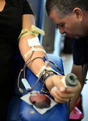
The continued spread of the Zika virus has raised concerns about transmission via blood transfusion.
So organizations in the US are asking people who have visited Zika outbreak zones to defer their plans to donate blood.
The US Food and Drug Administration is currently reviewing its blood donation policy with regard to the virus, but the American Red Cross and AABB are recommending donor self-deferral.
Both organizations said people should refrain from donating blood for 28 days if they have visited Mexico, the Caribbean, Central America, or South America in the past 4 weeks.
AABB said blood collection facilities should implement self-deferral, but blood-center-documented donor deferral is not required.
AABB has also recommended that donors who don’t defer call the blood collection facility if they travelled to Zika outbreak areas or other tropical areas and develop an unexplained illness that includes 2 or more symptoms common to Zika, dengue, and chikungunya virus infection in the 14 days after they donate blood.
In addition, blood collection facilities should recall nontransfused products if an infected donor reports experiencing 2 or more such symptoms.
And if a blood collection facility receives a post-donation report of a confirmed case of Zika, the facility should recall any in-date products collected in the 14 days before the onset of symptoms and defer the donor for 28 days after the symptoms are resolved.
About the virus
Zika is a flavivirus transmitted by Aedes mosquitoes. The virus was first described in Africa, but it began to cause epidemics in the Pacific in 2007. In 2015, Zika was found in Brazil, and local transmission has since been reported in more than 20 countries and territories in the Western Hemisphere.
When symptomatic, Zika infection typically causes a mild illness characterized by fever, myalgia, rash, retro-orbital pain, and prostration. However, asymptomatic infection occurs in about 80% of Zika-infected individuals.
Microcephaly has been linked to the ongoing Zika epidemic in Brazil, although the connection has not been confirmed. It has been suggested that microcephaly may be a result of maternal transmission of the Zika virus to the fetus.
During the French Polynesian outbreak of Zika virus that occurred in 2013, there was a 20-fold increase in the number of individuals diagnosed with Guillain-Barré syndrome.
It is not clear what risk the Zika virus poses to the blood supply, but the potential for transfusion transmission was suggested during the French Polynesian outbreak.
The maximum duration of viremia is thought to be less than 28 days, which is why AABB and American Red Cross are recommending a 28-day deferral period for blood donors who may have the virus. ![]()
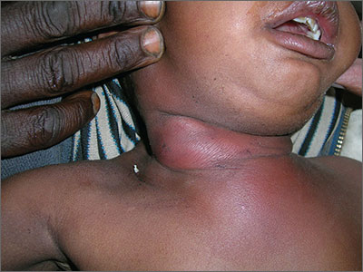
Red, swollen neck

The FP diagnosed cellulitis with a probable abscess over the neck. He explained that the pus over the neck needed to be drained and the grandmother (the child’s sole caretaker) gave her consent for the procedure. The area of maximal fluctuance was identified by palpation and the FP injected 1% lidocaine with epinephrine to numb the area. He then proceeded with the incision and drainage of the abscess, being careful to stay away from vital structures in the neck.
A significant amount of pus was drained and the finger of a sterile glove was cut off to place in the drainage site to allow for continued drainage. The patient was given ceftriaxone intravenously and the child remained in the community health center overnight with her grandmother and the nursing staff on duty.
The following day, the child was doing much better and her appetite started to return. Two days later, the child was afebrile and was sent home with her grandmother. The FP and the medical student mission team continued to make house calls daily and administer antibiotics until the child's infection was fully resolved.
The importance of incision and drainage cannot be overstated in this case. While cutting over the neck of a non-anesthetized child has its risks, allowing a neck abscess to remain surgically untreated can be life-threatening.
Photos and text for Photo Rounds Friday courtesy of Richard P. Usatine, MD. This case was adapted from: Usatine R. Cellulitis. In: Usatine R, Smith M, Mayeaux EJ, et al, eds. Color Atlas of Family Medicine. 2nd ed. New York, NY: McGraw-Hill; 2013:693-697.
To learn more about the Color Atlas of Family Medicine, see: www.amazon.com/Color-Family-Medicine-Richard-Usatine/dp/0071769641/
You can now get the second edition of the Color Atlas of Family Medicine as an app by clicking on this link: usatinemedia.com
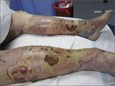
Violaceous bullae on legs

The FP suspected a Vibrio vulnificus infection secondary to ingesting the raw oysters, especially because the patient had a history of liver disease. V vulnificus grew out of the patient’s blood cultures, confirming the diagnosis.
V vulnificus is a free-living bacterium that is found in warm saltwater, such as in the Gulf of Mexico. This patient had been visiting the Gulf Coast when he ate the raw oysters. V vulnificus becomes concentrated in filter-feeding shellfish such as oysters.
Eating raw oysters can lead to overwhelming infections from V vulnificus, especially in those with liver disease, lymphoma, leukemia, and diabetes. The mortality rate in people with primary V vulnificus sepsis exceeds 40%.1
Unfortunately, the patient’s liver disease predisposed him to a more serious infection. Despite appropriate use of systemic antibiotics and supportive care, the patient died from sepsis.
1. Falcon LM, Pham L. Images in clinical medicine. Hemorrhagic cellulitis after consumption of raw oysters. N Engl J Med. 2005;353:1604.
Photo courtesy of Donna Nguyen, MD. Text for Photo Rounds Friday courtesy of Richard P. Usatine, MD. This case was adapted from: Usatine R. Cellulitis. In: Usatine R, Smith M, Mayeaux EJ, et al, eds. Color Atlas of Family Medicine. 2nd ed. New York, NY: McGraw-Hill; 2013:693-697.
To learn more about the Color Atlas of Family Medicine, see: www.amazon.com/Color-Family-Medicine-Richard-Usatine/dp/0071769641/
You can now get the second edition of the Color Atlas of Family Medicine as an app by clicking on this link: usatinemedia.com
Cosmeceuticals for managing acne: more useful than you might think
ORLANDO – The increasingly popular role of cosmeceuticals in treating acne has created some confusion among both dermatologists and their patients as to what’s really effective and worth recommending.
This was the focus of a presentation at the Orlando Dermatology Aesthetic and Clinical Conference by Dr. Hilary E. Baldwin, who reviewed the cosmeceuticals most likely to make a clinical impact on patients with acne.

While there are no definitive data that prove that cosmeceuticals are the most effective means of managing acne, “sometimes cosmeceuticals may actually be helpful as adjunctive therapy,” said Dr. Baldwin, vice chair of dermatology at the State University of New York at Brooklyn. “Compared to prescription medications, I think these are just a drop in the bucket, but they’re a drop in the right direction.”
The main benefit of using cosmeceuticals for acne is to improve the barrier function of the skin. With increasing evidence that acne either causes or is caused by barrier defects, cosmeceuticals can be used, at the very least, as “extraordinarily well-made moisturizers,” according to Dr. Baldwin. In addition, because moisturizers are anti-inflammatory, they can improve the tolerability of other topical treatments dermatologists recommend to their patients, both prescription and over-the-counter.
For reducing Propionibacterium acnes, consider tea tree oil and lily leaf oil, both of which have a small but promising amount of clinical data behind them. For tea tree oil, Dr. Baldwin referred to a randomized study of 124 patients, which compared 5% tea tree oil gel with 5% benzoyl peroxide for treatment of mild to moderate acne (Med J Aust. 1990 Oct 15;153[8]:455-8). The study found that although the onset of action was slower for tea tree oil, overall it had a significant effect in improving acne in the patients, by reducing the number of inflamed and non-inflamed lesions.
“Both of them worked, but benzoyl peroxide was statistically better,” Dr. Baldwin said. “There were fewer side effects in the tea tree oil group, with less people complaining about skin discomfort.”
There are less data regarding lily leaf extract, however, with only one study she said was worth mentioning: a 4-week trial comparing lily leaf extract and 5% benzoyl peroxide that was “so complicated, and had so many arms, that they ended up having only 4-5 patients in each arm, so I don’t think they can conclude anything,” she remarked.
For management of acne-related inflammation, there is good evidence to suggest botanicals are an effective treatment. A double-blind, randomized, 12-week study coauthored by Dr. Baldwin found that in a cohort of 80 patients, benzoyl peroxide and salicylic acid were more effective when combined with botanical extracts than when used on their own (Semin Cutan Med Surg. 2015 Sep;34[5S]:S82-S85).
Furthermore, explained Dr. Baldwin, “evidence suggests that patients were also using [the botanical extract treatment] more because there was a preference for that,” indicating the increasing desire for more natural, cosmeceutical approaches to treating skin ailments by the general public.
“[Cosmeceuticals] appeal to this increasingly mature and demanding acne patient population,” she said. “[Patients] have a preference for a natural approach to skin disease, they believe that strengthening the host is more important than killing a pathogen, they think [cosmeceuticals] have less of a potential for side effects, and it also gives [patients] a sense of control, which attenuates some of the psychological sequelae of acne.
Dr. Baldwin also recommended oatmeal-based cosmeceuticals for their potential benefit in barrier repair, licorice-based cosmeceuticals for their ability to reduce both postinflammatory hyperpigmentation and post inflammatory erythema, and niacinamide. Niacinamide has been shown to reduce postinflammatory hyperpigmentation when used with other treatment options.
Dr. Baldwin emphasized, however, that cosmeceuticals should always be considered as a supplement to other, ongoing treatments, not the main treatment for acne.
She did not report any relevant financial disclosures.
ORLANDO – The increasingly popular role of cosmeceuticals in treating acne has created some confusion among both dermatologists and their patients as to what’s really effective and worth recommending.
This was the focus of a presentation at the Orlando Dermatology Aesthetic and Clinical Conference by Dr. Hilary E. Baldwin, who reviewed the cosmeceuticals most likely to make a clinical impact on patients with acne.
While there are no definitive data that prove that cosmeceuticals are the most effective means of managing acne, “sometimes cosmeceuticals may actually be helpful as adjunctive therapy,” said Dr. Baldwin, vice chair of dermatology at the State University of New York at Brooklyn. “Compared to prescription medications, I think these are just a drop in the bucket, but they’re a drop in the right direction.”
The main benefit of using cosmeceuticals for acne is to improve the barrier function of the skin. With increasing evidence that acne either causes or is caused by barrier defects, cosmeceuticals can be used, at the very least, as “extraordinarily well-made moisturizers,” according to Dr. Baldwin. In addition, because moisturizers are anti-inflammatory, they can improve the tolerability of other topical treatments dermatologists recommend to their patients, both prescription and over-the-counter.
For reducing Propionibacterium acnes, consider tea tree oil and lily leaf oil, both of which have a small but promising amount of clinical data behind them. For tea tree oil, Dr. Baldwin referred to a randomized study of 124 patients, which compared 5% tea tree oil gel with 5% benzoyl peroxide for treatment of mild to moderate acne (Med J Aust. 1990 Oct 15;153[8]:455-8). The study found that although the onset of action was slower for tea tree oil, overall it had a significant effect in improving acne in the patients, by reducing the number of inflamed and non-inflamed lesions.
“Both of them worked, but benzoyl peroxide was statistically better,” Dr. Baldwin said. “There were fewer side effects in the tea tree oil group, with less people complaining about skin discomfort.”
There are less data regarding lily leaf extract, however, with only one study she said was worth mentioning: a 4-week trial comparing lily leaf extract and 5% benzoyl peroxide that was “so complicated, and had so many arms, that they ended up having only 4-5 patients in each arm, so I don’t think they can conclude anything,” she remarked.
For management of acne-related inflammation, there is good evidence to suggest botanicals are an effective treatment. A double-blind, randomized, 12-week study coauthored by Dr. Baldwin found that in a cohort of 80 patients, benzoyl peroxide and salicylic acid were more effective when combined with botanical extracts than when used on their own (Semin Cutan Med Surg. 2015 Sep;34[5S]:S82-S85).
Furthermore, explained Dr. Baldwin, “evidence suggests that patients were also using [the botanical extract treatment] more because there was a preference for that,” indicating the increasing desire for more natural, cosmeceutical approaches to treating skin ailments by the general public.
“[Cosmeceuticals] appeal to this increasingly mature and demanding acne patient population,” she said. “[Patients] have a preference for a natural approach to skin disease, they believe that strengthening the host is more important than killing a pathogen, they think [cosmeceuticals] have less of a potential for side effects, and it also gives [patients] a sense of control, which attenuates some of the psychological sequelae of acne.
Dr. Baldwin also recommended oatmeal-based cosmeceuticals for their potential benefit in barrier repair, licorice-based cosmeceuticals for their ability to reduce both postinflammatory hyperpigmentation and post inflammatory erythema, and niacinamide. Niacinamide has been shown to reduce postinflammatory hyperpigmentation when used with other treatment options.
Dr. Baldwin emphasized, however, that cosmeceuticals should always be considered as a supplement to other, ongoing treatments, not the main treatment for acne.
She did not report any relevant financial disclosures.
AT THE ODAC CONFERENCE

Heightened emphasis on sex-specific cardiovascular risk factors
SNOWMASS, COLO. – Achieving continued reductions in cardiovascular deaths in U.S. women will require that physicians make greater use of sex-specific risk factors that aren’t incorporated in the ACC/AHA atherosclerotic cardiovascular disease risk score, Dr. Jennifer H. Mieres asserted at the Annual Cardiovascular Conference at Snowmass.
In the 13-year period beginning in 2000, with the launch of a national initiative to boost the research focus on cardiovascular disease in women, the annual number of women dying from cardiovascular disease has dropped by roughly 30%. That’s a steeper decline than in men. One of the keys to further reductions in women is more widespread physician evaluation of sex-specific risk factors – such as a history of elevated blood pressure in pregnancy, polycystic ovarian syndrome, or radiation therapy for breast cancer – as part of routine cardiovascular risk assessment in women, said Dr. Mieres, senior vice president office of community and public health at Hofstra Northwell in Hempstead, N.Y.

Hypertension in pregnancy as a harbinger of premature cardiovascular disease and other chronic diseases has been a topic of particularly fruitful research in the past few years.
“The ongoing hypothesis is that pregnancy is a sort of stress test. Pregnancy-related complications indicate an inability to adequately adapt to the physiologic stress of pregnancy and thus reveal the presence of underlying susceptibility to ischemic heart disease,” according to the cardiologist.
She cited a landmark prospective study of 10,314 women born in Northern Finland in 1966 and followed for an average of more than 39 years after a singleton pregnancy. The investigators showed that any elevation in blood pressure during pregnancy, including isolated systolic or diastolic hypertension that resolved during or shortly after pregnancy, was associated with increased future risks of various forms of cardiovascular disease.
For example, de novo gestational hypertension without proteinuria was associated with significantly increased risks of subsequent ischemic cerebrovascular disease, chronic kidney disease, diabetes, ischemic heart disease, acute MI, chronic hypertension, and heart failure. The MIs that occurred in Finns with a history of gestational hypertension were more serious, too, with an associated threefold greater risk of being fatal than MIs in women who had been normotensive in pregnancy (Circulation. 2013 Feb 12;127[6]:681-90).

New-onset isolated systolic or diastolic hypertension emerged during pregnancy in about 17% of the Finnish women. Roughly 30% of them had a cardiovascular event before their late 60s. This translated to a 14%-18% greater risk than in women who remained normotensive in pregnancy.
The highest risk of all in the Finnish study was seen in women with preeclampsia/eclampsia superimposed on a background of chronic hypertension. They had a 3.18-fold greater risk of subsequent MI than did women who were normotensive in pregnancy, a 3.32-fold increased risk of heart failure, and a 2.22-fold greater risk of developing diabetes.
In addition to the growing appreciation that it’s important to consider sex-specific cardiovascular risk factors, recent evidence shows that many of the traditional risk factors are stronger predictors of ischemic heart disease in women than men. These include diabetes, smoking, obesity, and hypertension, Dr. Mieres observed.
For example, a recent meta-analysis of 26 studies including more than 214,000 subjects concluded that women with type 1 diabetes had a 2.5-fold greater risk of incident coronary heart disease than did men with type 1 diabetes. The women with type 1 diabetes also had an 86% greater risk of fatal cardiovascular diseases, a 44% increase in the risk of fatal kidney disease, a 37% greater risk of stroke, and a 37% increase in all-cause mortality relative to type 1 diabetic men (Lancet Diabetes Endocrinol. 2015 Mar;3[3]:198-206).
A wealth of accumulating data indicates that type 2 diabetes, too, is a much stronger risk factor for cardiovascular diseases in women than in men. The evidence prompted a recent formal scientific statement to that effect by the American Heart Association (Circulation. 2015 Dec 22;132[25]:2424-47).
Dr. Mieres reported having no financial conflicts of interest regarding her presentation.
SNOWMASS, COLO. – Achieving continued reductions in cardiovascular deaths in U.S. women will require that physicians make greater use of sex-specific risk factors that aren’t incorporated in the ACC/AHA atherosclerotic cardiovascular disease risk score, Dr. Jennifer H. Mieres asserted at the Annual Cardiovascular Conference at Snowmass.
In the 13-year period beginning in 2000, with the launch of a national initiative to boost the research focus on cardiovascular disease in women, the annual number of women dying from cardiovascular disease has dropped by roughly 30%. That’s a steeper decline than in men. One of the keys to further reductions in women is more widespread physician evaluation of sex-specific risk factors – such as a history of elevated blood pressure in pregnancy, polycystic ovarian syndrome, or radiation therapy for breast cancer – as part of routine cardiovascular risk assessment in women, said Dr. Mieres, senior vice president office of community and public health at Hofstra Northwell in Hempstead, N.Y.
Hypertension in pregnancy as a harbinger of premature cardiovascular disease and other chronic diseases has been a topic of particularly fruitful research in the past few years.
“The ongoing hypothesis is that pregnancy is a sort of stress test. Pregnancy-related complications indicate an inability to adequately adapt to the physiologic stress of pregnancy and thus reveal the presence of underlying susceptibility to ischemic heart disease,” according to the cardiologist.
She cited a landmark prospective study of 10,314 women born in Northern Finland in 1966 and followed for an average of more than 39 years after a singleton pregnancy. The investigators showed that any elevation in blood pressure during pregnancy, including isolated systolic or diastolic hypertension that resolved during or shortly after pregnancy, was associated with increased future risks of various forms of cardiovascular disease.
For example, de novo gestational hypertension without proteinuria was associated with significantly increased risks of subsequent ischemic cerebrovascular disease, chronic kidney disease, diabetes, ischemic heart disease, acute MI, chronic hypertension, and heart failure. The MIs that occurred in Finns with a history of gestational hypertension were more serious, too, with an associated threefold greater risk of being fatal than MIs in women who had been normotensive in pregnancy (Circulation. 2013 Feb 12;127[6]:681-90).
New-onset isolated systolic or diastolic hypertension emerged during pregnancy in about 17% of the Finnish women. Roughly 30% of them had a cardiovascular event before their late 60s. This translated to a 14%-18% greater risk than in women who remained normotensive in pregnancy.
The highest risk of all in the Finnish study was seen in women with preeclampsia/eclampsia superimposed on a background of chronic hypertension. They had a 3.18-fold greater risk of subsequent MI than did women who were normotensive in pregnancy, a 3.32-fold increased risk of heart failure, and a 2.22-fold greater risk of developing diabetes.
In addition to the growing appreciation that it’s important to consider sex-specific cardiovascular risk factors, recent evidence shows that many of the traditional risk factors are stronger predictors of ischemic heart disease in women than men. These include diabetes, smoking, obesity, and hypertension, Dr. Mieres observed.
For example, a recent meta-analysis of 26 studies including more than 214,000 subjects concluded that women with type 1 diabetes had a 2.5-fold greater risk of incident coronary heart disease than did men with type 1 diabetes. The women with type 1 diabetes also had an 86% greater risk of fatal cardiovascular diseases, a 44% increase in the risk of fatal kidney disease, a 37% greater risk of stroke, and a 37% increase in all-cause mortality relative to type 1 diabetic men (Lancet Diabetes Endocrinol. 2015 Mar;3[3]:198-206).
A wealth of accumulating data indicates that type 2 diabetes, too, is a much stronger risk factor for cardiovascular diseases in women than in men. The evidence prompted a recent formal scientific statement to that effect by the American Heart Association (Circulation. 2015 Dec 22;132[25]:2424-47).
Dr. Mieres reported having no financial conflicts of interest regarding her presentation.
EXPERT ANALYSIS FROM THE CARDIOVASCULAR CONFERENCE AT SNOWMASS

PICCs Increase Risk for Upper- and Lower-Extremity DVT
Clinical question: Do peripherally inserted central catheters increase the risk for upper- and lower-extremity deep venous thromboses?
Bottom line: Although the association between peripherally inserted central catheters (PICCs) and upper-extremity deep venous thromboses (DVTs) was already known, this study shows that PICCs are also associated with a greater risk of lower-extremity DVTs, suggesting that PICC insertion in itself may be a trigger for thrombosis. (LOE = 2b)
Reference: Greene MT, Flander SA, Woller SC, Bernstein SJ, Chopra V. The association between PICC use and venous thromboembolism in upper and lower extremities. Am J Med 2015;128(9):986–993.
Study design: Cohort (retrospective)
Funding source: Industry
Setting: Inpatient (any location) with outpatient follow-up
Synopsis
Using a statewide registry as well as individual medical records, these investigators collected data for 76,242 hospitalized patients to examine the association between PICC placement and venous thromboembolism (VTE). Patients with a history of VTE, those undergoing surgery, those admitted to an intensive care unit, and those under observation were excluded. Patients were followed up for 90 days after index hospitalization to identify the development of symptomatic pulmonary emboli or upper- or lower-extremity proximal DVTs.
Overall, 5% of the cohort had PICCs present on admission or placed during the hospitalization. As compared with those without PICCs, patients with PICCs were more likely to be older than 70 years; have recent surgery or history of VTE; and have diabetes, inflammatory bowel disease, sepsis, or pneumonia. After adjusting for other risk factors for VTE, the presence of a PICC was not only associated with risk of upper-extremity DVT (hazard ratio [HR] = 10.49; 95% CI 7.79-14.11; P < .001), but also modestly associated with risk of lower-extremity DVT (HR = 1.48; 1.02-2.15; P = .038). The authors hypothesize that PICC line insertion may trigger a systemic thrombosis leading to DVTs in different locations, including the lower extremities. There was no significant association with pulmonary embolism.
Dr. Kulkarni is an assistant professor of hospital medicine at Northwestern University in Chicago.