User login
One strikeout, one hit against low-grade serous carcinomas
Two MEK inhibitors were tested against recurrent low-grade serous carcinomas of the ovaries, fallopian tubes, or peritoneum, but only one inhibitor offered clinical benefit over standard care, investigators from two randomized trials reported.
Trametinib improved progression-free survival (PFS) when compared with standard care, while binimetinib conferred no PFS benefit.
In a phase 2/3 trial, the median PFS was 13 months for patients treated with trametinib and 7.2 months for patients who received an aromatase inhibitor or chemotherapy (P < .0001).
“Our findings suggest that trametinib represents a new standard-of-care treatment option for women with recurrent low-grade serous carcinoma,” said investigator David M. Gershenson, MD, of the University of Texas MD Anderson Cancer Center in Houston.
In contrast, in the phase 3 MILO/ENGOT-ov11 trial, there was no significant difference in PFS between patients treated with binimetinib and those who received physician’s choice of chemotherapy. The median PFS was 11.2 months with binimetinib and 14.1 months with chemotherapy (P = .752).
“Although this study did not meet its primary endpoint, binimetinib showed activity in low-grade serous ovarian cancer across the efficacy endpoints evaluated, with a response rate of 24% and a median PFS of 11.2 months on updated analysis. Chemotherapy responses were better than predicted, based on historical retrospective data,” said investigator Rachel N. Grisham, MD, of Memorial Sloan Kettering Cancer Center in New York.
The binimetinib trial and the trametinib trial were both discussed during a webinar on rare tumors covering research slated for presentation at the Society of Gynecologic Oncology’s Annual Meeting on Women’s Cancer. The meeting was canceled because of the COVID-19 pandemic.
Chemoresistant cancers
Low-grade serous ovarian or peritoneal cancers are rare, accounting for only 5% to 10% of all serous cancers, Dr. Gershenson noted.
“[Low-grade serous cancers are] characterized by alterations in the MAP kinase pathway, as well as relative chemoresistance, and prolonged overall survival compared to high-grade serous cancers. Because of this subtype’s relative chemoresistance, the search for novel therapeutics has predominated over the last decade or so,” he said.
MEK inhibitors interfere with the MEK1 and MEK2 enzymes in the MAPK pathway. Alterations in MAPK, especially in KRAS and BRAF proteins, are found in 30%-60% of low-grade serous carcinomas, providing the rationale for MEK inhibitors in these rare malignancies.
Trametinib study
In a phase 2/3 study, Dr. Gershenson and colleagues enrolled 260 patients with recurrent, low-grade serous carcinoma of the ovary or peritoneum. Patients were randomized to receive either trametinib at 2 mg daily continuously until progression (n = 130) or standard care (n = 130).
Standard care consisted of one of the following: letrozole at 2.5 mg daily; pegylated liposomal doxorubicin at 40-50 mg IV every 28 days; weekly paclitaxel at 80 mg/m2 for 3 out of 4 weeks; tamoxifen at 20 mg twice daily; or topotecan at 4 mg/m2 on days 1, 8, and 15 every 28 days. Patients randomized to standard care could be crossed over to trametinib at progression.
All patients had at least one prior line of platinum-based chemotherapy, and nearly half had three or more prior lines of therapy. The median age was 56.6 years in the trametinib arm and 55.3 years in the control arm.
At a median follow-up of 31.4 months, median PFS, the primary endpoint, was 13 months with trametinib vs. 7.2 months with standard care. The hazard ratio (HR) for progression on trametinib was 0.48 (P < .0001).
The overall response rates were 26.2% in the trametinib arm and 6.2% in the standard care arm. The odds ratio for response on trametinib was 5.4 (P < .0001).
For 88 patients who crossed over to trametinib, the median PFS was 10.8 months, and the overall response rate was 15%.
Trametinib was also associated with a significantly longer response duration, at a median of 13.6 months, compared with 5.9 months for standard care (P value not shown).
The median overall survival was 37 months with trametinib and 29.2 months with standard care, with an HR favoring trametinib of 0.75, although this just missed statistical significance (P = .054). Dr. Gershenson pointed out that the overall survival in the standard care arm included patients who had been crossed over to trametinib.
Grade 3 or greater adverse events included hematologic toxicities in 13.4% of patients on trametinib and 9.4% on standard care; gastrointestinal toxicity in 27.6% and 29%, respectively; skin toxicities in 15% and 3.9%, respectively; and vascular toxicities in 18.9% and 8.6%, respectively.
Binimetinib study
The phase 3 MILO/ENGOT-ov11 study enrolled 341 patients with low-grade serous carcinomas of the ovaries, fallopian tubes, or peritoneum. Patients were randomized on a 2:1 basis to receive either binimetinib at 45 mg twice daily (n = 228) or physician’s choice of chemotherapy (n = 113). Chemotherapy consisted of pegylated liposomal doxorubicin at 40 mg/m2 on day 1 of each 28-day cycle; paclitaxel at 80 mg/m2 on days 1, 8, and 15 of each 28-day cycle; or topotecan at 1.25 mg/m2 IV on days 1-5 of each 21-day cycle.
The efficacy analysis included 227 patients assigned to binimetinib and 106 assigned to chemotherapy.
A planned interim analysis was performed in 2016 after the first 303 patients were enrolled. At that time, the median PFS by blinded central review was 9.1 months in the binimetinib arm and 10.6 months in the physician’s choice arm (HR, 1.21; P = .807), so the trial was halted early for futility. Patients on active treatment at the time could continue until progression and were followed by local radiology.
At the interim analysis, secondary endpoints were also similar between the arms. The overall response rate was 16% in the binimetinib arm and 13% in the chemotherapy arm.
The most common grade 3 or greater adverse events with binimetinib were blood creatinine phosphokinase increase (26%) and vomiting (10%).
Dr. Grisham also reported updated follow-up results through January 2019.
The median PFS in the updated analysis was 11.2 months with the MEK inhibitor and 14.1 months with chemotherapy, a difference that was not statistically significant (HR, 1.12; P = .752). Updated overall response rates were the same in both arms, at 24%.
A post hoc molecular analysis of 215 patients suggested a possible association between KRAS mutation and response to binimetinib.
Two MEKs, one ‘meh’
Discussant Jubilee Brown, MD, of the Levine Cancer Institute at Atrium Health in Charlotte, N.C., said that “with a 2% to 5% chance of response in patients with low-grade serous ovarian cancer, there is a low bar for any compound to demonstrate success.”
Regarding the MILO/ENGOT-ov11 trial, she noted that “this study did not meet its primary endpoint, but perhaps the endpoint is not reflective of the importance of the study.”
A different outcome might have occurred had investigators stratified patients by KRAS status upfront, comparing patients with KRAS mutations treated with binimetinib to KRAS wild-type patients treated with either a MEK inhibitor or physician’s choice of care, Dr. Brown said.
She agreed with the assertion by Dr. Gershenson and colleagues that improved PFS qualifies trametinib to be considered a new option for standard care, ”especially in a rare tumor setting with limited options. This is a huge win for patients.”
The trametinib study was sponsored by NRG Oncology and the UK National Cancer Research Institute. Dr. Gershenson disclosed relationships with NRG Oncology, Genentech, Novartis, Elsevier, and UpToDate, as well as stock in several companies.
The binimetinib study was sponsored by Pfizer. Dr. Grisham disclosed relationships with Clovis, Regeneron, Mateon, Amgen, Abbvie, OncLive, PRIME, MCM, and Medscape. MDedge News and Medscape are owned by the same parent organization.
Dr. Brown disclosed consulting for Biodesix, Caris, Clovis, Genentech, Invitae, Janssen, Olympus, OncLive, and Tempus.
SOURCE: Gershenson DM et al. SGO 2020, Abstract 42; Grisham RN et al. SGO 2020, Abstract 41.
Two MEK inhibitors were tested against recurrent low-grade serous carcinomas of the ovaries, fallopian tubes, or peritoneum, but only one inhibitor offered clinical benefit over standard care, investigators from two randomized trials reported.
Trametinib improved progression-free survival (PFS) when compared with standard care, while binimetinib conferred no PFS benefit.
In a phase 2/3 trial, the median PFS was 13 months for patients treated with trametinib and 7.2 months for patients who received an aromatase inhibitor or chemotherapy (P < .0001).
“Our findings suggest that trametinib represents a new standard-of-care treatment option for women with recurrent low-grade serous carcinoma,” said investigator David M. Gershenson, MD, of the University of Texas MD Anderson Cancer Center in Houston.
In contrast, in the phase 3 MILO/ENGOT-ov11 trial, there was no significant difference in PFS between patients treated with binimetinib and those who received physician’s choice of chemotherapy. The median PFS was 11.2 months with binimetinib and 14.1 months with chemotherapy (P = .752).
“Although this study did not meet its primary endpoint, binimetinib showed activity in low-grade serous ovarian cancer across the efficacy endpoints evaluated, with a response rate of 24% and a median PFS of 11.2 months on updated analysis. Chemotherapy responses were better than predicted, based on historical retrospective data,” said investigator Rachel N. Grisham, MD, of Memorial Sloan Kettering Cancer Center in New York.
The binimetinib trial and the trametinib trial were both discussed during a webinar on rare tumors covering research slated for presentation at the Society of Gynecologic Oncology’s Annual Meeting on Women’s Cancer. The meeting was canceled because of the COVID-19 pandemic.
Chemoresistant cancers
Low-grade serous ovarian or peritoneal cancers are rare, accounting for only 5% to 10% of all serous cancers, Dr. Gershenson noted.
“[Low-grade serous cancers are] characterized by alterations in the MAP kinase pathway, as well as relative chemoresistance, and prolonged overall survival compared to high-grade serous cancers. Because of this subtype’s relative chemoresistance, the search for novel therapeutics has predominated over the last decade or so,” he said.
MEK inhibitors interfere with the MEK1 and MEK2 enzymes in the MAPK pathway. Alterations in MAPK, especially in KRAS and BRAF proteins, are found in 30%-60% of low-grade serous carcinomas, providing the rationale for MEK inhibitors in these rare malignancies.
Trametinib study
In a phase 2/3 study, Dr. Gershenson and colleagues enrolled 260 patients with recurrent, low-grade serous carcinoma of the ovary or peritoneum. Patients were randomized to receive either trametinib at 2 mg daily continuously until progression (n = 130) or standard care (n = 130).
Standard care consisted of one of the following: letrozole at 2.5 mg daily; pegylated liposomal doxorubicin at 40-50 mg IV every 28 days; weekly paclitaxel at 80 mg/m2 for 3 out of 4 weeks; tamoxifen at 20 mg twice daily; or topotecan at 4 mg/m2 on days 1, 8, and 15 every 28 days. Patients randomized to standard care could be crossed over to trametinib at progression.
All patients had at least one prior line of platinum-based chemotherapy, and nearly half had three or more prior lines of therapy. The median age was 56.6 years in the trametinib arm and 55.3 years in the control arm.
At a median follow-up of 31.4 months, median PFS, the primary endpoint, was 13 months with trametinib vs. 7.2 months with standard care. The hazard ratio (HR) for progression on trametinib was 0.48 (P < .0001).
The overall response rates were 26.2% in the trametinib arm and 6.2% in the standard care arm. The odds ratio for response on trametinib was 5.4 (P < .0001).
For 88 patients who crossed over to trametinib, the median PFS was 10.8 months, and the overall response rate was 15%.
Trametinib was also associated with a significantly longer response duration, at a median of 13.6 months, compared with 5.9 months for standard care (P value not shown).
The median overall survival was 37 months with trametinib and 29.2 months with standard care, with an HR favoring trametinib of 0.75, although this just missed statistical significance (P = .054). Dr. Gershenson pointed out that the overall survival in the standard care arm included patients who had been crossed over to trametinib.
Grade 3 or greater adverse events included hematologic toxicities in 13.4% of patients on trametinib and 9.4% on standard care; gastrointestinal toxicity in 27.6% and 29%, respectively; skin toxicities in 15% and 3.9%, respectively; and vascular toxicities in 18.9% and 8.6%, respectively.
Binimetinib study
The phase 3 MILO/ENGOT-ov11 study enrolled 341 patients with low-grade serous carcinomas of the ovaries, fallopian tubes, or peritoneum. Patients were randomized on a 2:1 basis to receive either binimetinib at 45 mg twice daily (n = 228) or physician’s choice of chemotherapy (n = 113). Chemotherapy consisted of pegylated liposomal doxorubicin at 40 mg/m2 on day 1 of each 28-day cycle; paclitaxel at 80 mg/m2 on days 1, 8, and 15 of each 28-day cycle; or topotecan at 1.25 mg/m2 IV on days 1-5 of each 21-day cycle.
The efficacy analysis included 227 patients assigned to binimetinib and 106 assigned to chemotherapy.
A planned interim analysis was performed in 2016 after the first 303 patients were enrolled. At that time, the median PFS by blinded central review was 9.1 months in the binimetinib arm and 10.6 months in the physician’s choice arm (HR, 1.21; P = .807), so the trial was halted early for futility. Patients on active treatment at the time could continue until progression and were followed by local radiology.
At the interim analysis, secondary endpoints were also similar between the arms. The overall response rate was 16% in the binimetinib arm and 13% in the chemotherapy arm.
The most common grade 3 or greater adverse events with binimetinib were blood creatinine phosphokinase increase (26%) and vomiting (10%).
Dr. Grisham also reported updated follow-up results through January 2019.
The median PFS in the updated analysis was 11.2 months with the MEK inhibitor and 14.1 months with chemotherapy, a difference that was not statistically significant (HR, 1.12; P = .752). Updated overall response rates were the same in both arms, at 24%.
A post hoc molecular analysis of 215 patients suggested a possible association between KRAS mutation and response to binimetinib.
Two MEKs, one ‘meh’
Discussant Jubilee Brown, MD, of the Levine Cancer Institute at Atrium Health in Charlotte, N.C., said that “with a 2% to 5% chance of response in patients with low-grade serous ovarian cancer, there is a low bar for any compound to demonstrate success.”
Regarding the MILO/ENGOT-ov11 trial, she noted that “this study did not meet its primary endpoint, but perhaps the endpoint is not reflective of the importance of the study.”
A different outcome might have occurred had investigators stratified patients by KRAS status upfront, comparing patients with KRAS mutations treated with binimetinib to KRAS wild-type patients treated with either a MEK inhibitor or physician’s choice of care, Dr. Brown said.
She agreed with the assertion by Dr. Gershenson and colleagues that improved PFS qualifies trametinib to be considered a new option for standard care, ”especially in a rare tumor setting with limited options. This is a huge win for patients.”
The trametinib study was sponsored by NRG Oncology and the UK National Cancer Research Institute. Dr. Gershenson disclosed relationships with NRG Oncology, Genentech, Novartis, Elsevier, and UpToDate, as well as stock in several companies.
The binimetinib study was sponsored by Pfizer. Dr. Grisham disclosed relationships with Clovis, Regeneron, Mateon, Amgen, Abbvie, OncLive, PRIME, MCM, and Medscape. MDedge News and Medscape are owned by the same parent organization.
Dr. Brown disclosed consulting for Biodesix, Caris, Clovis, Genentech, Invitae, Janssen, Olympus, OncLive, and Tempus.
SOURCE: Gershenson DM et al. SGO 2020, Abstract 42; Grisham RN et al. SGO 2020, Abstract 41.
Two MEK inhibitors were tested against recurrent low-grade serous carcinomas of the ovaries, fallopian tubes, or peritoneum, but only one inhibitor offered clinical benefit over standard care, investigators from two randomized trials reported.
Trametinib improved progression-free survival (PFS) when compared with standard care, while binimetinib conferred no PFS benefit.
In a phase 2/3 trial, the median PFS was 13 months for patients treated with trametinib and 7.2 months for patients who received an aromatase inhibitor or chemotherapy (P < .0001).
“Our findings suggest that trametinib represents a new standard-of-care treatment option for women with recurrent low-grade serous carcinoma,” said investigator David M. Gershenson, MD, of the University of Texas MD Anderson Cancer Center in Houston.
In contrast, in the phase 3 MILO/ENGOT-ov11 trial, there was no significant difference in PFS between patients treated with binimetinib and those who received physician’s choice of chemotherapy. The median PFS was 11.2 months with binimetinib and 14.1 months with chemotherapy (P = .752).
“Although this study did not meet its primary endpoint, binimetinib showed activity in low-grade serous ovarian cancer across the efficacy endpoints evaluated, with a response rate of 24% and a median PFS of 11.2 months on updated analysis. Chemotherapy responses were better than predicted, based on historical retrospective data,” said investigator Rachel N. Grisham, MD, of Memorial Sloan Kettering Cancer Center in New York.
The binimetinib trial and the trametinib trial were both discussed during a webinar on rare tumors covering research slated for presentation at the Society of Gynecologic Oncology’s Annual Meeting on Women’s Cancer. The meeting was canceled because of the COVID-19 pandemic.
Chemoresistant cancers
Low-grade serous ovarian or peritoneal cancers are rare, accounting for only 5% to 10% of all serous cancers, Dr. Gershenson noted.
“[Low-grade serous cancers are] characterized by alterations in the MAP kinase pathway, as well as relative chemoresistance, and prolonged overall survival compared to high-grade serous cancers. Because of this subtype’s relative chemoresistance, the search for novel therapeutics has predominated over the last decade or so,” he said.
MEK inhibitors interfere with the MEK1 and MEK2 enzymes in the MAPK pathway. Alterations in MAPK, especially in KRAS and BRAF proteins, are found in 30%-60% of low-grade serous carcinomas, providing the rationale for MEK inhibitors in these rare malignancies.
Trametinib study
In a phase 2/3 study, Dr. Gershenson and colleagues enrolled 260 patients with recurrent, low-grade serous carcinoma of the ovary or peritoneum. Patients were randomized to receive either trametinib at 2 mg daily continuously until progression (n = 130) or standard care (n = 130).
Standard care consisted of one of the following: letrozole at 2.5 mg daily; pegylated liposomal doxorubicin at 40-50 mg IV every 28 days; weekly paclitaxel at 80 mg/m2 for 3 out of 4 weeks; tamoxifen at 20 mg twice daily; or topotecan at 4 mg/m2 on days 1, 8, and 15 every 28 days. Patients randomized to standard care could be crossed over to trametinib at progression.
All patients had at least one prior line of platinum-based chemotherapy, and nearly half had three or more prior lines of therapy. The median age was 56.6 years in the trametinib arm and 55.3 years in the control arm.
At a median follow-up of 31.4 months, median PFS, the primary endpoint, was 13 months with trametinib vs. 7.2 months with standard care. The hazard ratio (HR) for progression on trametinib was 0.48 (P < .0001).
The overall response rates were 26.2% in the trametinib arm and 6.2% in the standard care arm. The odds ratio for response on trametinib was 5.4 (P < .0001).
For 88 patients who crossed over to trametinib, the median PFS was 10.8 months, and the overall response rate was 15%.
Trametinib was also associated with a significantly longer response duration, at a median of 13.6 months, compared with 5.9 months for standard care (P value not shown).
The median overall survival was 37 months with trametinib and 29.2 months with standard care, with an HR favoring trametinib of 0.75, although this just missed statistical significance (P = .054). Dr. Gershenson pointed out that the overall survival in the standard care arm included patients who had been crossed over to trametinib.
Grade 3 or greater adverse events included hematologic toxicities in 13.4% of patients on trametinib and 9.4% on standard care; gastrointestinal toxicity in 27.6% and 29%, respectively; skin toxicities in 15% and 3.9%, respectively; and vascular toxicities in 18.9% and 8.6%, respectively.
Binimetinib study
The phase 3 MILO/ENGOT-ov11 study enrolled 341 patients with low-grade serous carcinomas of the ovaries, fallopian tubes, or peritoneum. Patients were randomized on a 2:1 basis to receive either binimetinib at 45 mg twice daily (n = 228) or physician’s choice of chemotherapy (n = 113). Chemotherapy consisted of pegylated liposomal doxorubicin at 40 mg/m2 on day 1 of each 28-day cycle; paclitaxel at 80 mg/m2 on days 1, 8, and 15 of each 28-day cycle; or topotecan at 1.25 mg/m2 IV on days 1-5 of each 21-day cycle.
The efficacy analysis included 227 patients assigned to binimetinib and 106 assigned to chemotherapy.
A planned interim analysis was performed in 2016 after the first 303 patients were enrolled. At that time, the median PFS by blinded central review was 9.1 months in the binimetinib arm and 10.6 months in the physician’s choice arm (HR, 1.21; P = .807), so the trial was halted early for futility. Patients on active treatment at the time could continue until progression and were followed by local radiology.
At the interim analysis, secondary endpoints were also similar between the arms. The overall response rate was 16% in the binimetinib arm and 13% in the chemotherapy arm.
The most common grade 3 or greater adverse events with binimetinib were blood creatinine phosphokinase increase (26%) and vomiting (10%).
Dr. Grisham also reported updated follow-up results through January 2019.
The median PFS in the updated analysis was 11.2 months with the MEK inhibitor and 14.1 months with chemotherapy, a difference that was not statistically significant (HR, 1.12; P = .752). Updated overall response rates were the same in both arms, at 24%.
A post hoc molecular analysis of 215 patients suggested a possible association between KRAS mutation and response to binimetinib.
Two MEKs, one ‘meh’
Discussant Jubilee Brown, MD, of the Levine Cancer Institute at Atrium Health in Charlotte, N.C., said that “with a 2% to 5% chance of response in patients with low-grade serous ovarian cancer, there is a low bar for any compound to demonstrate success.”
Regarding the MILO/ENGOT-ov11 trial, she noted that “this study did not meet its primary endpoint, but perhaps the endpoint is not reflective of the importance of the study.”
A different outcome might have occurred had investigators stratified patients by KRAS status upfront, comparing patients with KRAS mutations treated with binimetinib to KRAS wild-type patients treated with either a MEK inhibitor or physician’s choice of care, Dr. Brown said.
She agreed with the assertion by Dr. Gershenson and colleagues that improved PFS qualifies trametinib to be considered a new option for standard care, ”especially in a rare tumor setting with limited options. This is a huge win for patients.”
The trametinib study was sponsored by NRG Oncology and the UK National Cancer Research Institute. Dr. Gershenson disclosed relationships with NRG Oncology, Genentech, Novartis, Elsevier, and UpToDate, as well as stock in several companies.
The binimetinib study was sponsored by Pfizer. Dr. Grisham disclosed relationships with Clovis, Regeneron, Mateon, Amgen, Abbvie, OncLive, PRIME, MCM, and Medscape. MDedge News and Medscape are owned by the same parent organization.
Dr. Brown disclosed consulting for Biodesix, Caris, Clovis, Genentech, Invitae, Janssen, Olympus, OncLive, and Tempus.
SOURCE: Gershenson DM et al. SGO 2020, Abstract 42; Grisham RN et al. SGO 2020, Abstract 41.
FROM SGO 2020
Practice During the Pandemic
The first installment of my new column was obsolete on arrival. It referred to walking abroad at midday, with no mention of masks and social distancing. The whole thing was so February 2020.

My last day in the office was in mid-March. Friday the 13th.
, using stored and forwarded images.
What I had in mind was visits by patients in nursing homes or too sick at home to come in. It always bothered me to see very aged and infirm patients brought to the office at great inconvenience and expense for what often turned out to be problems like xerosis or eczema that could have been managed quite well remotely.
The HMO never got back to me, though. There were too many hurdles, mostly bureaucratic rather than medical. Would insurance pay? What about consent? Malpractice? It has been interesting to watch the current crisis sweep away the inertia of such obstacles, including licensure considerations (seeing patients across state lines for cutaneous purposes). People get around to fixing the roof when it pours. Perhaps next time there will be tests, masks, respirators. Perhaps.
Seeing patients remotely has acquainted me with all the technical headaches everyone stuck at home talks and jokes about: Balky transmission (What did you say after, “and then the blood ...”?); patients who can’t figure out how to log on, or start the video, or unmute themselves, and on and on. Picture resolution is not great, as anyone knows from watching TV newscasters interview talking heads stuck in their homes.
I was never all that image-conscious, but my beard has grown fuller and my hair unkempter. Even though I sit at my desk, I do take care to keep my trousers on. Not taking any chances.
Everyone agonizes over what the “new normal” may be. Will people come back to doctors’ offices? Will practices survive economically if many patients don’t return to the office? Stay tuned. For a long time.
Mostly, though, remote visits seem to work. Helped if needed by additional, better-resolution emailed photos, it’s possible to make useful decisions, including which lesions can wait for in-person evaluation, until ...
... Until what? In an effort to keep this column up-to-the-nanosecond, I am writing it as many countries tentatively “open up.” Careful analysis of the knowledge behind this world-wide project shows ... not much. It seems to come down to some educated guesswork about what might work and what the risks might be, which leads to advice that differs widely from state to state and country to country. It’s as if people everywhere just decided that locking everyone down is a real drag, is financially ruinous, has a duration both uncertain and longer than most people and governments think they can handle, so let’s get out there and “be careful,” whatever that is said to mean.
And the risks? Well, more people will get sick and some will die. How many “extra” deaths are ethically acceptable? Thoughtful people are working on that. They’ll get back sometime to those who are still around.
I don’t blame anyone for our staggering ignorance about this terrifying new reality. But absorbing the ignorance in real time is not reassuring.
I have nothing but sympathy for those who are not emeritus, who have practices to sustain and families to feed. I didn’t ask to be born 73 years ago, and take no credit for having done so. So much of what happens to us depends on when and where we were born – two factors for which we deserve absolutely no credit – that it’s a wonder we take such pride in praising ourselves for what we think we accomplish. Having no better choice, we do the best we can.
Meantime, I am in a “high-risk” category. If I were obese, I could try to lose weight. But my risk factor is age, which tends not to decline. Risk-wise, there is just one way to exit my group.
So I don’t expect to get back to the office anytime soon. To paraphrase a comedian who shall remain nameless: I don’t want to live on in the hearts of men. I want to live on in my house.
Dr. Rockoff, who wrote the Dermatology News column “Under My Skin,” is now semiretired, after 40 years of practice in Brookline, Mass. He served on the clinical faculty at Tufts University, Boston, and taught senior medical students and other trainees for 30 years. His second book, “Act Like a Doctor, Think Like a Patient,” is available online. Write to him at dermnews@mdedge.com.
The first installment of my new column was obsolete on arrival. It referred to walking abroad at midday, with no mention of masks and social distancing. The whole thing was so February 2020.
My last day in the office was in mid-March. Friday the 13th.
, using stored and forwarded images.
What I had in mind was visits by patients in nursing homes or too sick at home to come in. It always bothered me to see very aged and infirm patients brought to the office at great inconvenience and expense for what often turned out to be problems like xerosis or eczema that could have been managed quite well remotely.
The HMO never got back to me, though. There were too many hurdles, mostly bureaucratic rather than medical. Would insurance pay? What about consent? Malpractice? It has been interesting to watch the current crisis sweep away the inertia of such obstacles, including licensure considerations (seeing patients across state lines for cutaneous purposes). People get around to fixing the roof when it pours. Perhaps next time there will be tests, masks, respirators. Perhaps.
Seeing patients remotely has acquainted me with all the technical headaches everyone stuck at home talks and jokes about: Balky transmission (What did you say after, “and then the blood ...”?); patients who can’t figure out how to log on, or start the video, or unmute themselves, and on and on. Picture resolution is not great, as anyone knows from watching TV newscasters interview talking heads stuck in their homes.
I was never all that image-conscious, but my beard has grown fuller and my hair unkempter. Even though I sit at my desk, I do take care to keep my trousers on. Not taking any chances.
Everyone agonizes over what the “new normal” may be. Will people come back to doctors’ offices? Will practices survive economically if many patients don’t return to the office? Stay tuned. For a long time.
Mostly, though, remote visits seem to work. Helped if needed by additional, better-resolution emailed photos, it’s possible to make useful decisions, including which lesions can wait for in-person evaluation, until ...
... Until what? In an effort to keep this column up-to-the-nanosecond, I am writing it as many countries tentatively “open up.” Careful analysis of the knowledge behind this world-wide project shows ... not much. It seems to come down to some educated guesswork about what might work and what the risks might be, which leads to advice that differs widely from state to state and country to country. It’s as if people everywhere just decided that locking everyone down is a real drag, is financially ruinous, has a duration both uncertain and longer than most people and governments think they can handle, so let’s get out there and “be careful,” whatever that is said to mean.
And the risks? Well, more people will get sick and some will die. How many “extra” deaths are ethically acceptable? Thoughtful people are working on that. They’ll get back sometime to those who are still around.
I don’t blame anyone for our staggering ignorance about this terrifying new reality. But absorbing the ignorance in real time is not reassuring.
I have nothing but sympathy for those who are not emeritus, who have practices to sustain and families to feed. I didn’t ask to be born 73 years ago, and take no credit for having done so. So much of what happens to us depends on when and where we were born – two factors for which we deserve absolutely no credit – that it’s a wonder we take such pride in praising ourselves for what we think we accomplish. Having no better choice, we do the best we can.
Meantime, I am in a “high-risk” category. If I were obese, I could try to lose weight. But my risk factor is age, which tends not to decline. Risk-wise, there is just one way to exit my group.
So I don’t expect to get back to the office anytime soon. To paraphrase a comedian who shall remain nameless: I don’t want to live on in the hearts of men. I want to live on in my house.
Dr. Rockoff, who wrote the Dermatology News column “Under My Skin,” is now semiretired, after 40 years of practice in Brookline, Mass. He served on the clinical faculty at Tufts University, Boston, and taught senior medical students and other trainees for 30 years. His second book, “Act Like a Doctor, Think Like a Patient,” is available online. Write to him at dermnews@mdedge.com.
The first installment of my new column was obsolete on arrival. It referred to walking abroad at midday, with no mention of masks and social distancing. The whole thing was so February 2020.
My last day in the office was in mid-March. Friday the 13th.
, using stored and forwarded images.
What I had in mind was visits by patients in nursing homes or too sick at home to come in. It always bothered me to see very aged and infirm patients brought to the office at great inconvenience and expense for what often turned out to be problems like xerosis or eczema that could have been managed quite well remotely.
The HMO never got back to me, though. There were too many hurdles, mostly bureaucratic rather than medical. Would insurance pay? What about consent? Malpractice? It has been interesting to watch the current crisis sweep away the inertia of such obstacles, including licensure considerations (seeing patients across state lines for cutaneous purposes). People get around to fixing the roof when it pours. Perhaps next time there will be tests, masks, respirators. Perhaps.
Seeing patients remotely has acquainted me with all the technical headaches everyone stuck at home talks and jokes about: Balky transmission (What did you say after, “and then the blood ...”?); patients who can’t figure out how to log on, or start the video, or unmute themselves, and on and on. Picture resolution is not great, as anyone knows from watching TV newscasters interview talking heads stuck in their homes.
I was never all that image-conscious, but my beard has grown fuller and my hair unkempter. Even though I sit at my desk, I do take care to keep my trousers on. Not taking any chances.
Everyone agonizes over what the “new normal” may be. Will people come back to doctors’ offices? Will practices survive economically if many patients don’t return to the office? Stay tuned. For a long time.
Mostly, though, remote visits seem to work. Helped if needed by additional, better-resolution emailed photos, it’s possible to make useful decisions, including which lesions can wait for in-person evaluation, until ...
... Until what? In an effort to keep this column up-to-the-nanosecond, I am writing it as many countries tentatively “open up.” Careful analysis of the knowledge behind this world-wide project shows ... not much. It seems to come down to some educated guesswork about what might work and what the risks might be, which leads to advice that differs widely from state to state and country to country. It’s as if people everywhere just decided that locking everyone down is a real drag, is financially ruinous, has a duration both uncertain and longer than most people and governments think they can handle, so let’s get out there and “be careful,” whatever that is said to mean.
And the risks? Well, more people will get sick and some will die. How many “extra” deaths are ethically acceptable? Thoughtful people are working on that. They’ll get back sometime to those who are still around.
I don’t blame anyone for our staggering ignorance about this terrifying new reality. But absorbing the ignorance in real time is not reassuring.
I have nothing but sympathy for those who are not emeritus, who have practices to sustain and families to feed. I didn’t ask to be born 73 years ago, and take no credit for having done so. So much of what happens to us depends on when and where we were born – two factors for which we deserve absolutely no credit – that it’s a wonder we take such pride in praising ourselves for what we think we accomplish. Having no better choice, we do the best we can.
Meantime, I am in a “high-risk” category. If I were obese, I could try to lose weight. But my risk factor is age, which tends not to decline. Risk-wise, there is just one way to exit my group.
So I don’t expect to get back to the office anytime soon. To paraphrase a comedian who shall remain nameless: I don’t want to live on in the hearts of men. I want to live on in my house.
Dr. Rockoff, who wrote the Dermatology News column “Under My Skin,” is now semiretired, after 40 years of practice in Brookline, Mass. He served on the clinical faculty at Tufts University, Boston, and taught senior medical students and other trainees for 30 years. His second book, “Act Like a Doctor, Think Like a Patient,” is available online. Write to him at dermnews@mdedge.com.
Obesity can shift severe COVID-19 to younger age groups
published in The Lancet.
“By itself, obesity seems to be a sufficient risk factor to start seeing younger people landing in the ICU,” said the study’s lead author, David Kass, MD, a professor of cardiology and medicine at Johns Hopkins University School of Medicine in Baltimore, Maryland.
“In that sense, there’s a simple message: If you’re very, very overweight, don’t think that if you’re 35 you’re that much safer [from severe COVID-19] than your mother or grandparents or others in their 60s or 70s,” Kass told Medscape Medical News.
The findings, which Kass describes as a “2-week snapshot” of 265 patients (58% male) in late March and early April at a handful of university hospitals in the United States reinforces other recent research indicating that obesity is one of the biggest risk factors for severe COVID-19 disease, particularly among younger patients. In addition, a large British study showed that, after adjusting for comorbidities, obesity was a significant factor associated with in-hospital death in COVID-19.
But this new analysis stands out as the only dataset to date that specifically “asks the question relative to age” of whether severe COVID-19 disease correlates to ICU treatment, he said.
The mean age of his study population of ICU patients was 55, Kass said, “and that was young, not what we were expecting.”
“Even with the first 20 patients, we were already seeing younger people and they definitely were heavier, with plenty of patients with a BMI over 35 kg/m2,” he added. “The relationship was pretty tight, pretty quick.”
“Just don’t make the assumption that any of us are too young to be vulnerable if, in fact, this is an aspect of our bodies,” he said.
Steven Heymsfield, MD, past president and a spokesperson for the Obesity Society, agrees with Kass’ conclusions.
“One thing we’ve had on our minds is that the prototype of a person with this disease is older...but now if we get [a patient] who’s symptomatic and 40 and obese, we shouldn’t assume they have some other disease,” Heymsfield told Medscape Medical News.
“We should think of them as a susceptible population.”
Kass and colleagues agree. “Public messaging to younger adults, reducing the threshold for virus testing in obese individuals, and maintaining greater vigilance for this at-risk population should reduce the prevalence of severe COVID-19 disease [among those with obesity],” they state.
“I think it’s a mental adjustment from a health care standpoint, which might hopefully help target the folks who are at higher risk before they get into trouble,” Kass told Medscape Medical News.
Trio of mechanisms explain obesity’s extra COVID-19 risks
Kass and coauthors write that, in analyzing their data, they anticipated similar results to the largest study of 1591 ICU patients from Italy in which only 203 were younger than 51 years. Common comorbidities among those patients included hypertension, cardiovascular disease, and type 2 diabetes, with similar data reported from China.
When the COVID-19 epidemic accelerated in the United States, older age was also identified as a risk factor. Obesity had not yet been added to this list, Kass noted. But following informal discussions with colleagues in other ICUs around the country, he decided to investigate further as to whether it was an underappreciated risk factor.
Kass and colleagues did a quick evaluation of the link between BMI and age of patients with COVID-19 admitted to ICUs at Johns Hopkins, University of Cincinnati, New York University, University of Washington, Florida Health, and University of Pennsylvania.
The “significant inverse correlation between age and BMI” showed younger ICU patients were more likely to be obese, with no difference by gender.
Median BMI among study participants was 29.3 kg/m2, with only a quarter having a BMI lower than 26 kg/m2 and another 25% having a BMI higher than 34.7 kg/m2.
Kass acknowledged that it wasn’t possible with this simple dataset to account for any other potential confounders, but he told Medscape Medical News that, “while diabetes, cardiovascular disease, and hypertension, for example, can occur with obesity, this is generally less so in younger populations as it takes time for the other comorbidities to develop.”
He said several mechanisms could explain why obesity predisposes patients with COVID-19 to severe disease.
For one, obesity places extra pressure on the diaphragm while lying on the back, restricting breathing.
“Morbid obesity itself is sort of proinflammatory,” he continued.
“Here we’ve got a viral infection where the early reports suggest that cytokine storms and immune mishandling of the virus are why it’s so much more severe than other forms of coronavirus we’ve seen before. So if you have someone with an already underlying proinflammatory state, this could be a reason there’s higher risk.”
Additionally, the angiotensin-converting enzyme-2 (ACE-2) receptor to which the SARS-CoV-2 virus that causes COVID-19 attaches is expressed in higher amounts in adipose tissue than the lungs, Kass noted.
“This could turn into kind of a viral replication depot,” he explained. “You may well be brewing more virus as a component of obesity.”
Sensitivity needed in public messaging about risks, but test sooner
With an obesity rate of about 40% in the United States, the results are particularly relevant for Americans, Kass and Heymsfield say, noting that the country’s “obesity belt” runs through the South.
Heymsfield, who wasn’t part of the new analysis, notes that public messaging around severe COVID-19 risks to younger adults with obesity is “tricky,” especially because the virus is “still pretty common in nonobese people.”
Kass agrees, noting, “it’s difficult to turn to 40% of the population and say: ‘You guys have to watch it.’ ”
But the mounting research findings necessitate linking obesity with severe COVID-19 disease and perhaps testing patients in this category for the virus sooner before symptoms become severe.
And of note, since shortness of breath is common among people with obesity regardless of illness, similar COVID-19 symptoms might catch these individuals unaware, pointed out Heymsfield, who is also a professor in the Metabolism and Body Composition Lab at Pennington Biomedical Research Center at Louisiana State University, Baton Rouge.
“They may find themselves literally unable to breathe, and the concern would be that they wait much too long to come in” for treatment, he said. Typically, people can deteriorate between day 7 and 10 of the COVID-19 infection.
Individuals with obesity “need to be educated to recognize the serious complications of COVID-19 often appear suddenly, although the virus has sometimes been working its way through the body for a long time,” he concluded.
Kass and Heymsfield have declared no relevant financial relationships.
This article first appeared on Medscape.com.
published in The Lancet.
“By itself, obesity seems to be a sufficient risk factor to start seeing younger people landing in the ICU,” said the study’s lead author, David Kass, MD, a professor of cardiology and medicine at Johns Hopkins University School of Medicine in Baltimore, Maryland.
“In that sense, there’s a simple message: If you’re very, very overweight, don’t think that if you’re 35 you’re that much safer [from severe COVID-19] than your mother or grandparents or others in their 60s or 70s,” Kass told Medscape Medical News.
The findings, which Kass describes as a “2-week snapshot” of 265 patients (58% male) in late March and early April at a handful of university hospitals in the United States reinforces other recent research indicating that obesity is one of the biggest risk factors for severe COVID-19 disease, particularly among younger patients. In addition, a large British study showed that, after adjusting for comorbidities, obesity was a significant factor associated with in-hospital death in COVID-19.
But this new analysis stands out as the only dataset to date that specifically “asks the question relative to age” of whether severe COVID-19 disease correlates to ICU treatment, he said.
The mean age of his study population of ICU patients was 55, Kass said, “and that was young, not what we were expecting.”
“Even with the first 20 patients, we were already seeing younger people and they definitely were heavier, with plenty of patients with a BMI over 35 kg/m2,” he added. “The relationship was pretty tight, pretty quick.”
“Just don’t make the assumption that any of us are too young to be vulnerable if, in fact, this is an aspect of our bodies,” he said.
Steven Heymsfield, MD, past president and a spokesperson for the Obesity Society, agrees with Kass’ conclusions.
“One thing we’ve had on our minds is that the prototype of a person with this disease is older...but now if we get [a patient] who’s symptomatic and 40 and obese, we shouldn’t assume they have some other disease,” Heymsfield told Medscape Medical News.
“We should think of them as a susceptible population.”
Kass and colleagues agree. “Public messaging to younger adults, reducing the threshold for virus testing in obese individuals, and maintaining greater vigilance for this at-risk population should reduce the prevalence of severe COVID-19 disease [among those with obesity],” they state.
“I think it’s a mental adjustment from a health care standpoint, which might hopefully help target the folks who are at higher risk before they get into trouble,” Kass told Medscape Medical News.
Trio of mechanisms explain obesity’s extra COVID-19 risks
Kass and coauthors write that, in analyzing their data, they anticipated similar results to the largest study of 1591 ICU patients from Italy in which only 203 were younger than 51 years. Common comorbidities among those patients included hypertension, cardiovascular disease, and type 2 diabetes, with similar data reported from China.
When the COVID-19 epidemic accelerated in the United States, older age was also identified as a risk factor. Obesity had not yet been added to this list, Kass noted. But following informal discussions with colleagues in other ICUs around the country, he decided to investigate further as to whether it was an underappreciated risk factor.
Kass and colleagues did a quick evaluation of the link between BMI and age of patients with COVID-19 admitted to ICUs at Johns Hopkins, University of Cincinnati, New York University, University of Washington, Florida Health, and University of Pennsylvania.
The “significant inverse correlation between age and BMI” showed younger ICU patients were more likely to be obese, with no difference by gender.
Median BMI among study participants was 29.3 kg/m2, with only a quarter having a BMI lower than 26 kg/m2 and another 25% having a BMI higher than 34.7 kg/m2.
Kass acknowledged that it wasn’t possible with this simple dataset to account for any other potential confounders, but he told Medscape Medical News that, “while diabetes, cardiovascular disease, and hypertension, for example, can occur with obesity, this is generally less so in younger populations as it takes time for the other comorbidities to develop.”
He said several mechanisms could explain why obesity predisposes patients with COVID-19 to severe disease.
For one, obesity places extra pressure on the diaphragm while lying on the back, restricting breathing.
“Morbid obesity itself is sort of proinflammatory,” he continued.
“Here we’ve got a viral infection where the early reports suggest that cytokine storms and immune mishandling of the virus are why it’s so much more severe than other forms of coronavirus we’ve seen before. So if you have someone with an already underlying proinflammatory state, this could be a reason there’s higher risk.”
Additionally, the angiotensin-converting enzyme-2 (ACE-2) receptor to which the SARS-CoV-2 virus that causes COVID-19 attaches is expressed in higher amounts in adipose tissue than the lungs, Kass noted.
“This could turn into kind of a viral replication depot,” he explained. “You may well be brewing more virus as a component of obesity.”
Sensitivity needed in public messaging about risks, but test sooner
With an obesity rate of about 40% in the United States, the results are particularly relevant for Americans, Kass and Heymsfield say, noting that the country’s “obesity belt” runs through the South.
Heymsfield, who wasn’t part of the new analysis, notes that public messaging around severe COVID-19 risks to younger adults with obesity is “tricky,” especially because the virus is “still pretty common in nonobese people.”
Kass agrees, noting, “it’s difficult to turn to 40% of the population and say: ‘You guys have to watch it.’ ”
But the mounting research findings necessitate linking obesity with severe COVID-19 disease and perhaps testing patients in this category for the virus sooner before symptoms become severe.
And of note, since shortness of breath is common among people with obesity regardless of illness, similar COVID-19 symptoms might catch these individuals unaware, pointed out Heymsfield, who is also a professor in the Metabolism and Body Composition Lab at Pennington Biomedical Research Center at Louisiana State University, Baton Rouge.
“They may find themselves literally unable to breathe, and the concern would be that they wait much too long to come in” for treatment, he said. Typically, people can deteriorate between day 7 and 10 of the COVID-19 infection.
Individuals with obesity “need to be educated to recognize the serious complications of COVID-19 often appear suddenly, although the virus has sometimes been working its way through the body for a long time,” he concluded.
Kass and Heymsfield have declared no relevant financial relationships.
This article first appeared on Medscape.com.
published in The Lancet.
“By itself, obesity seems to be a sufficient risk factor to start seeing younger people landing in the ICU,” said the study’s lead author, David Kass, MD, a professor of cardiology and medicine at Johns Hopkins University School of Medicine in Baltimore, Maryland.
“In that sense, there’s a simple message: If you’re very, very overweight, don’t think that if you’re 35 you’re that much safer [from severe COVID-19] than your mother or grandparents or others in their 60s or 70s,” Kass told Medscape Medical News.
The findings, which Kass describes as a “2-week snapshot” of 265 patients (58% male) in late March and early April at a handful of university hospitals in the United States reinforces other recent research indicating that obesity is one of the biggest risk factors for severe COVID-19 disease, particularly among younger patients. In addition, a large British study showed that, after adjusting for comorbidities, obesity was a significant factor associated with in-hospital death in COVID-19.
But this new analysis stands out as the only dataset to date that specifically “asks the question relative to age” of whether severe COVID-19 disease correlates to ICU treatment, he said.
The mean age of his study population of ICU patients was 55, Kass said, “and that was young, not what we were expecting.”
“Even with the first 20 patients, we were already seeing younger people and they definitely were heavier, with plenty of patients with a BMI over 35 kg/m2,” he added. “The relationship was pretty tight, pretty quick.”
“Just don’t make the assumption that any of us are too young to be vulnerable if, in fact, this is an aspect of our bodies,” he said.
Steven Heymsfield, MD, past president and a spokesperson for the Obesity Society, agrees with Kass’ conclusions.
“One thing we’ve had on our minds is that the prototype of a person with this disease is older...but now if we get [a patient] who’s symptomatic and 40 and obese, we shouldn’t assume they have some other disease,” Heymsfield told Medscape Medical News.
“We should think of them as a susceptible population.”
Kass and colleagues agree. “Public messaging to younger adults, reducing the threshold for virus testing in obese individuals, and maintaining greater vigilance for this at-risk population should reduce the prevalence of severe COVID-19 disease [among those with obesity],” they state.
“I think it’s a mental adjustment from a health care standpoint, which might hopefully help target the folks who are at higher risk before they get into trouble,” Kass told Medscape Medical News.
Trio of mechanisms explain obesity’s extra COVID-19 risks
Kass and coauthors write that, in analyzing their data, they anticipated similar results to the largest study of 1591 ICU patients from Italy in which only 203 were younger than 51 years. Common comorbidities among those patients included hypertension, cardiovascular disease, and type 2 diabetes, with similar data reported from China.
When the COVID-19 epidemic accelerated in the United States, older age was also identified as a risk factor. Obesity had not yet been added to this list, Kass noted. But following informal discussions with colleagues in other ICUs around the country, he decided to investigate further as to whether it was an underappreciated risk factor.
Kass and colleagues did a quick evaluation of the link between BMI and age of patients with COVID-19 admitted to ICUs at Johns Hopkins, University of Cincinnati, New York University, University of Washington, Florida Health, and University of Pennsylvania.
The “significant inverse correlation between age and BMI” showed younger ICU patients were more likely to be obese, with no difference by gender.
Median BMI among study participants was 29.3 kg/m2, with only a quarter having a BMI lower than 26 kg/m2 and another 25% having a BMI higher than 34.7 kg/m2.
Kass acknowledged that it wasn’t possible with this simple dataset to account for any other potential confounders, but he told Medscape Medical News that, “while diabetes, cardiovascular disease, and hypertension, for example, can occur with obesity, this is generally less so in younger populations as it takes time for the other comorbidities to develop.”
He said several mechanisms could explain why obesity predisposes patients with COVID-19 to severe disease.
For one, obesity places extra pressure on the diaphragm while lying on the back, restricting breathing.
“Morbid obesity itself is sort of proinflammatory,” he continued.
“Here we’ve got a viral infection where the early reports suggest that cytokine storms and immune mishandling of the virus are why it’s so much more severe than other forms of coronavirus we’ve seen before. So if you have someone with an already underlying proinflammatory state, this could be a reason there’s higher risk.”
Additionally, the angiotensin-converting enzyme-2 (ACE-2) receptor to which the SARS-CoV-2 virus that causes COVID-19 attaches is expressed in higher amounts in adipose tissue than the lungs, Kass noted.
“This could turn into kind of a viral replication depot,” he explained. “You may well be brewing more virus as a component of obesity.”
Sensitivity needed in public messaging about risks, but test sooner
With an obesity rate of about 40% in the United States, the results are particularly relevant for Americans, Kass and Heymsfield say, noting that the country’s “obesity belt” runs through the South.
Heymsfield, who wasn’t part of the new analysis, notes that public messaging around severe COVID-19 risks to younger adults with obesity is “tricky,” especially because the virus is “still pretty common in nonobese people.”
Kass agrees, noting, “it’s difficult to turn to 40% of the population and say: ‘You guys have to watch it.’ ”
But the mounting research findings necessitate linking obesity with severe COVID-19 disease and perhaps testing patients in this category for the virus sooner before symptoms become severe.
And of note, since shortness of breath is common among people with obesity regardless of illness, similar COVID-19 symptoms might catch these individuals unaware, pointed out Heymsfield, who is also a professor in the Metabolism and Body Composition Lab at Pennington Biomedical Research Center at Louisiana State University, Baton Rouge.
“They may find themselves literally unable to breathe, and the concern would be that they wait much too long to come in” for treatment, he said. Typically, people can deteriorate between day 7 and 10 of the COVID-19 infection.
Individuals with obesity “need to be educated to recognize the serious complications of COVID-19 often appear suddenly, although the virus has sometimes been working its way through the body for a long time,” he concluded.
Kass and Heymsfield have declared no relevant financial relationships.
This article first appeared on Medscape.com.
Acute bilateral hand edema and vesiculation
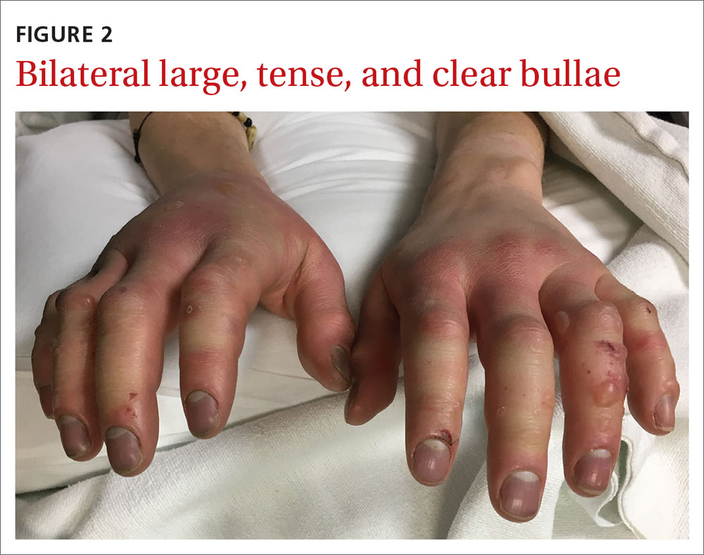
A 27-year-old man presented to the urgent care clinic with acute bilateral hand swelling, blisters, numbness, and pain. History taking revealed that these symptoms developed after he was locked outside of his apartment for 45 minutes in –22°C (–8°F) weather following a night of heavy drinking.
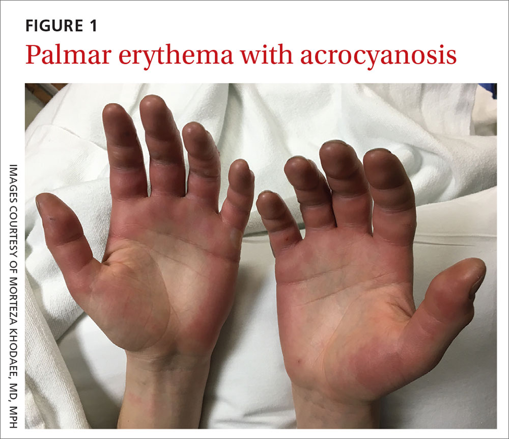
On physical examination, the patient had a temperature of 36.2°C (97.2°F) and a heart rate of 116 beats/min. He had
WHAT IS YOUR DIAGNOSIS?
HOW WOULD YOU TREAT THIS PATIENT?
Diagnosis: Second-degree frostbite
Frostbite is the result of tissue freezing, which generally occurs after prolonged exposure to freezing temperatures (typically –4°C or below).1,2 The majority (~90%) of frostbite injuries occur in the hands and feet; however, frostbite has also been observed in the face, perineum, buttocks, and male genitalia.3
Frostbite is a clinical diagnosis based on a history of sustained exposure to freezing temperatures, paresthesia of affected areas, and typical skin changes. Evidence is lacking regarding the epidemiology of frostbite within the general population.2
Pathophysiology. Intra- and extracellular ice crystal formation causes fluid and electrolyte disturbances, cell dehydration, lipid denaturation, and subsequent cell death.1 After thawing, progressive tissue ischemia can occur as a result of endothelial damage and dysfunction, intravascular sludging, increased inflammatory markers, an influx of free radicals, and microvascular thrombosis.1
Classification. Traditionally, frostbite has been classified according to a 4-tiered system based on tissue appearance after rewarming.2 First-degree frostbite is characterized by white plaques with surrounding erythema; second degree by edema and clear or cloudy vesicles; third degree by hemorrhagic bullae; and fourth degree by cold and hard tissue that eventually progresses to gangrene.2
A simpler scheme designates frostbite as either superficial (corresponding to first- or second-degree frostbite) or deep (corresponding to third- or fourth-degree frostbite) with presumed muscle and bone involvement.2
Continue to: Risk factors
Risk factors. Frostbite is often associated with risk factors such as alcohol or drug intoxication, vehicular failure or trauma, immobilizing trauma, psychiatric illness, homelessness, Raynaud phenomenon, peripheral vascular disease, diabetes, inadequate clothing, previous cold-weather injury, outdoor winter recreation, and the use of certain medications (eg, beta-blockers).1-3 Apart from environmental exposure, frostbite can also occur by direct contact with freezing materials, such as ice packs or industrial refrigerants.3
Differential includes nonfreezing injuries
Frostnip, pernio, and trench foot are other cold-weather injuries distinguished by the absence of tissue freezing.4 Raynaud phenomenon is a condition that is triggered by either cold temperatures or emotional stress.5
Frostnip is characterized by pallor and paresthesia of exposed areas. It may precede frostbite, but it quickly resolves after rewarming.2
Pernio occurs when skin is exposed to damp, cold, nonfreezing environments.6 It results in edematous and inflammatory skin lesions that may be painful, pruritic, violaceous, or erythematous.6 These lesions are typically found over the fingers, toes, nose, ears, buttocks, or thighs.4,6 Pernio may be classified as either primary or secondary disease.5 Primary pernio is considered idiopathic.6 Secondary pernio is thought to be either drug induced or due to underlying autoimmune diseases, such as hepatitis or cryopathy.6
Trench foot develops under similar conditions to pernio but requires exposure to a wet environment for at least 10 to 14 hours.7 It is characterized by foot pain, paresthesia, pruritus, edema, erythema, cyanosis, blisters, and even gangrene if left untreated.7
Continue to: Raynaud phenomenon
Raynaud phenomenon results from transient, acral vasocontraction and manifests as well-demarcated pallor, cyanosis, and then erythema as the affected body part reperfuses.5 Similar to pernio, it can be categorized as either primary or secondary.5 Primary phenomenon is idiopathic. Secondary phenomenon is thought to be a result of autoimmune disease, use of certain medications, occupational vibratory exposure, obstructive vascular disease, or infection
In the absence of a history of exposure to subfreezing temperatures, frostbite can be excluded from the differential diagnosis.
Treatment entails rewarming
The aim of frostbite treatment is to save injured cells and minimize tissue loss.1 This is accomplished through rapid rewarming and—in severe cases—reperfusion techniques.
Tissue should be rewarmed in a 37°C to 39°C water bath with povidone iodine or chlorhexidine added for antiseptic effect.1 All efforts should be made to avoid refreezing or trauma, as this could worsen the initial injury.2 Oral or intravenous hydration may be offered to optimize fluid status.1 Supplemental oxygen may be administered to maintain saturations above 90%.1 Nonsteroidal anti-inflammatory drugs are helpful for analgesia and anti-inflammatory effect, and opioids can be used for breakthrough pain.1 It is recommended that blisters be drained in a sterile fashion and that all affected tissue be covered with topical aloe vera and a loose dressing.1,2,4
Treatment of severe frostbite. Angiography should be performed on all patients with third- or fourth-degree frostbite.3 If imaging shows evidence of vascular occlusion, tissue plasminogen activator (tPA) and heparin can be initiated within 24 hours to reduce the risk for amputation.8-10
Continue to: Iloprost is another...
Iloprost is another proposed treatment for severe frostbite. It is a prostacyclin analog that may lower the amputation rate in patients with at least third-degree frostbite.11 Unlike tPA, iloprost may be given to trauma patients, and it can be used more than 24 hours after injury.2
In cases of fourth-degree frostbite that is not successfully reperfused, amputation is delayed until dry gangrene develops. This often takes weeks to months.12
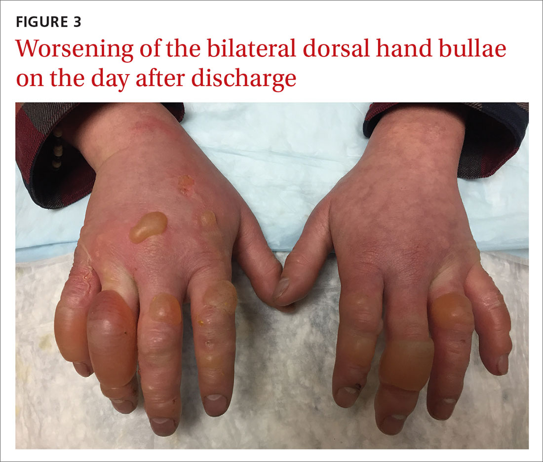
Our patient underwent rewarming and was orally rehydrated. He was discharged home with ibuprofen, oxycodone-acetaminophen, topical aloe vera, and loose dressings. His bullae enlarged the next day (FIGURE 3). One week later, his blisters were debrided and dressed with silver sulfadiazine at his plastic surgery follow-up. He experienced sensory deficits for a few months, but eventually made a full recovery after 6 months with no remaining sequelae.
ACKNOWLEDGEMENT
The authors thank Lisa Kim, MD, for her clinical care of this patient.
CORRESPONDENCE
Morteza Khodaee, MD, MPH, University of Colorado School of Medicine, Department of Family Medicine, AFW Clinic, 3055 Roslyn Street, Denver, CO 80238; morteza.khodaee@ cuanschutz.edu
1. Handford C, Thomas O, Imray CHE. Frostbite. Emerg Med Clin North Am. 2017;35:281-299.
2. Heil K, Thomas R, Robertson G, et al. Freezing and non-freezing cold weather injuries: a systematic review. Br Med Bull. 2016;117:79-93.
3. Millet JD, Brown RK, Levi B, et al. Frostbite: spectrum of imaging findings and guidelines for management. Radiographics. 2016;36:2154-2169.
4. Long WB 3rd, Edlich RF, Winters KL, et al. Cold injuries. J Long Term Eff Med Implants. 2005;15:67-78.
5. Baker JS, Miranpuri S. Perniosis: a case report with literature review. J Am Podiatr Med Assoc. 2016;106:138-140.
6. Bush JS, Watson S. Trench foot. Updated February 3, 2020. In: StatPearls [Internet]. Treasure Island, FL: StatPearls Publishing; 2020. www.ncbi.nlm.nih.gov/books/NBK482364/. Accessed April 22, 2020.
7. Musa R, Qurie A. Raynaud disease (Raynaud phenomenon, Raynaud syndrome). Updated February 14, 2019. In: StatPearls [Internet]. Treasure Island, FL: StatPearls Publishing; 2020. www.ncbi.nlm.nih.gov/books/NBK499833/. Accessed April 22, 2020.
8. Bruen KJ, Ballard JR, Morris SE, et al. Reduction of the incidence of amputation in frostbite injury with thrombolytic therapy. Arch Surg. 2007;142:546-551; discussion 551-553.
9. Gonzaga T, Jenabzadeh K, Anderson CP, et al. Use of intra-arterial thrombolytic therapy for acute treatment of frostbite in 62 patients with review of thrombolytic therapy in frostbite. J Burn Care Res. 2016;37:e323-e334.
10. Twomey JA, Peltier GL, Zera RT. An open-label study to evaluate the safety and efficacy of tissue plasminogen activator in treatment of severe frostbite. J Trauma. 2005;59:1350-1354; discussion 1354-1355.
11. Cauchy E, Cheguillaume B, Chetaille E. A controlled trial of a prostacyclin and rt-PA in the treatment of severe frostbite. N Engl J Med. 2011;364:189-190.
12. McIntosh SE, Opacic M, Freer L, et al; Wilderness Medical Society. Wilderness Medical Society practice guidelines for the prevention and treatment of frostbite: 2014 update. Wilderness Environ Med. 2014;25(4 suppl):S43-S54.
A 27-year-old man presented to the urgent care clinic with acute bilateral hand swelling, blisters, numbness, and pain. History taking revealed that these symptoms developed after he was locked outside of his apartment for 45 minutes in –22°C (–8°F) weather following a night of heavy drinking.
On physical examination, the patient had a temperature of 36.2°C (97.2°F) and a heart rate of 116 beats/min. He had
WHAT IS YOUR DIAGNOSIS?
HOW WOULD YOU TREAT THIS PATIENT?
Diagnosis: Second-degree frostbite
Frostbite is the result of tissue freezing, which generally occurs after prolonged exposure to freezing temperatures (typically –4°C or below).1,2 The majority (~90%) of frostbite injuries occur in the hands and feet; however, frostbite has also been observed in the face, perineum, buttocks, and male genitalia.3
Frostbite is a clinical diagnosis based on a history of sustained exposure to freezing temperatures, paresthesia of affected areas, and typical skin changes. Evidence is lacking regarding the epidemiology of frostbite within the general population.2
Pathophysiology. Intra- and extracellular ice crystal formation causes fluid and electrolyte disturbances, cell dehydration, lipid denaturation, and subsequent cell death.1 After thawing, progressive tissue ischemia can occur as a result of endothelial damage and dysfunction, intravascular sludging, increased inflammatory markers, an influx of free radicals, and microvascular thrombosis.1
Classification. Traditionally, frostbite has been classified according to a 4-tiered system based on tissue appearance after rewarming.2 First-degree frostbite is characterized by white plaques with surrounding erythema; second degree by edema and clear or cloudy vesicles; third degree by hemorrhagic bullae; and fourth degree by cold and hard tissue that eventually progresses to gangrene.2
A simpler scheme designates frostbite as either superficial (corresponding to first- or second-degree frostbite) or deep (corresponding to third- or fourth-degree frostbite) with presumed muscle and bone involvement.2
Continue to: Risk factors
Risk factors. Frostbite is often associated with risk factors such as alcohol or drug intoxication, vehicular failure or trauma, immobilizing trauma, psychiatric illness, homelessness, Raynaud phenomenon, peripheral vascular disease, diabetes, inadequate clothing, previous cold-weather injury, outdoor winter recreation, and the use of certain medications (eg, beta-blockers).1-3 Apart from environmental exposure, frostbite can also occur by direct contact with freezing materials, such as ice packs or industrial refrigerants.3
Differential includes nonfreezing injuries
Frostnip, pernio, and trench foot are other cold-weather injuries distinguished by the absence of tissue freezing.4 Raynaud phenomenon is a condition that is triggered by either cold temperatures or emotional stress.5
Frostnip is characterized by pallor and paresthesia of exposed areas. It may precede frostbite, but it quickly resolves after rewarming.2
Pernio occurs when skin is exposed to damp, cold, nonfreezing environments.6 It results in edematous and inflammatory skin lesions that may be painful, pruritic, violaceous, or erythematous.6 These lesions are typically found over the fingers, toes, nose, ears, buttocks, or thighs.4,6 Pernio may be classified as either primary or secondary disease.5 Primary pernio is considered idiopathic.6 Secondary pernio is thought to be either drug induced or due to underlying autoimmune diseases, such as hepatitis or cryopathy.6
Trench foot develops under similar conditions to pernio but requires exposure to a wet environment for at least 10 to 14 hours.7 It is characterized by foot pain, paresthesia, pruritus, edema, erythema, cyanosis, blisters, and even gangrene if left untreated.7
Continue to: Raynaud phenomenon
Raynaud phenomenon results from transient, acral vasocontraction and manifests as well-demarcated pallor, cyanosis, and then erythema as the affected body part reperfuses.5 Similar to pernio, it can be categorized as either primary or secondary.5 Primary phenomenon is idiopathic. Secondary phenomenon is thought to be a result of autoimmune disease, use of certain medications, occupational vibratory exposure, obstructive vascular disease, or infection
In the absence of a history of exposure to subfreezing temperatures, frostbite can be excluded from the differential diagnosis.
Treatment entails rewarming
The aim of frostbite treatment is to save injured cells and minimize tissue loss.1 This is accomplished through rapid rewarming and—in severe cases—reperfusion techniques.
Tissue should be rewarmed in a 37°C to 39°C water bath with povidone iodine or chlorhexidine added for antiseptic effect.1 All efforts should be made to avoid refreezing or trauma, as this could worsen the initial injury.2 Oral or intravenous hydration may be offered to optimize fluid status.1 Supplemental oxygen may be administered to maintain saturations above 90%.1 Nonsteroidal anti-inflammatory drugs are helpful for analgesia and anti-inflammatory effect, and opioids can be used for breakthrough pain.1 It is recommended that blisters be drained in a sterile fashion and that all affected tissue be covered with topical aloe vera and a loose dressing.1,2,4
Treatment of severe frostbite. Angiography should be performed on all patients with third- or fourth-degree frostbite.3 If imaging shows evidence of vascular occlusion, tissue plasminogen activator (tPA) and heparin can be initiated within 24 hours to reduce the risk for amputation.8-10
Continue to: Iloprost is another...
Iloprost is another proposed treatment for severe frostbite. It is a prostacyclin analog that may lower the amputation rate in patients with at least third-degree frostbite.11 Unlike tPA, iloprost may be given to trauma patients, and it can be used more than 24 hours after injury.2
In cases of fourth-degree frostbite that is not successfully reperfused, amputation is delayed until dry gangrene develops. This often takes weeks to months.12
Our patient underwent rewarming and was orally rehydrated. He was discharged home with ibuprofen, oxycodone-acetaminophen, topical aloe vera, and loose dressings. His bullae enlarged the next day (FIGURE 3). One week later, his blisters were debrided and dressed with silver sulfadiazine at his plastic surgery follow-up. He experienced sensory deficits for a few months, but eventually made a full recovery after 6 months with no remaining sequelae.
ACKNOWLEDGEMENT
The authors thank Lisa Kim, MD, for her clinical care of this patient.
CORRESPONDENCE
Morteza Khodaee, MD, MPH, University of Colorado School of Medicine, Department of Family Medicine, AFW Clinic, 3055 Roslyn Street, Denver, CO 80238; morteza.khodaee@ cuanschutz.edu
A 27-year-old man presented to the urgent care clinic with acute bilateral hand swelling, blisters, numbness, and pain. History taking revealed that these symptoms developed after he was locked outside of his apartment for 45 minutes in –22°C (–8°F) weather following a night of heavy drinking.
On physical examination, the patient had a temperature of 36.2°C (97.2°F) and a heart rate of 116 beats/min. He had
WHAT IS YOUR DIAGNOSIS?
HOW WOULD YOU TREAT THIS PATIENT?
Diagnosis: Second-degree frostbite
Frostbite is the result of tissue freezing, which generally occurs after prolonged exposure to freezing temperatures (typically –4°C or below).1,2 The majority (~90%) of frostbite injuries occur in the hands and feet; however, frostbite has also been observed in the face, perineum, buttocks, and male genitalia.3
Frostbite is a clinical diagnosis based on a history of sustained exposure to freezing temperatures, paresthesia of affected areas, and typical skin changes. Evidence is lacking regarding the epidemiology of frostbite within the general population.2
Pathophysiology. Intra- and extracellular ice crystal formation causes fluid and electrolyte disturbances, cell dehydration, lipid denaturation, and subsequent cell death.1 After thawing, progressive tissue ischemia can occur as a result of endothelial damage and dysfunction, intravascular sludging, increased inflammatory markers, an influx of free radicals, and microvascular thrombosis.1
Classification. Traditionally, frostbite has been classified according to a 4-tiered system based on tissue appearance after rewarming.2 First-degree frostbite is characterized by white plaques with surrounding erythema; second degree by edema and clear or cloudy vesicles; third degree by hemorrhagic bullae; and fourth degree by cold and hard tissue that eventually progresses to gangrene.2
A simpler scheme designates frostbite as either superficial (corresponding to first- or second-degree frostbite) or deep (corresponding to third- or fourth-degree frostbite) with presumed muscle and bone involvement.2
Continue to: Risk factors
Risk factors. Frostbite is often associated with risk factors such as alcohol or drug intoxication, vehicular failure or trauma, immobilizing trauma, psychiatric illness, homelessness, Raynaud phenomenon, peripheral vascular disease, diabetes, inadequate clothing, previous cold-weather injury, outdoor winter recreation, and the use of certain medications (eg, beta-blockers).1-3 Apart from environmental exposure, frostbite can also occur by direct contact with freezing materials, such as ice packs or industrial refrigerants.3
Differential includes nonfreezing injuries
Frostnip, pernio, and trench foot are other cold-weather injuries distinguished by the absence of tissue freezing.4 Raynaud phenomenon is a condition that is triggered by either cold temperatures or emotional stress.5
Frostnip is characterized by pallor and paresthesia of exposed areas. It may precede frostbite, but it quickly resolves after rewarming.2
Pernio occurs when skin is exposed to damp, cold, nonfreezing environments.6 It results in edematous and inflammatory skin lesions that may be painful, pruritic, violaceous, or erythematous.6 These lesions are typically found over the fingers, toes, nose, ears, buttocks, or thighs.4,6 Pernio may be classified as either primary or secondary disease.5 Primary pernio is considered idiopathic.6 Secondary pernio is thought to be either drug induced or due to underlying autoimmune diseases, such as hepatitis or cryopathy.6
Trench foot develops under similar conditions to pernio but requires exposure to a wet environment for at least 10 to 14 hours.7 It is characterized by foot pain, paresthesia, pruritus, edema, erythema, cyanosis, blisters, and even gangrene if left untreated.7
Continue to: Raynaud phenomenon
Raynaud phenomenon results from transient, acral vasocontraction and manifests as well-demarcated pallor, cyanosis, and then erythema as the affected body part reperfuses.5 Similar to pernio, it can be categorized as either primary or secondary.5 Primary phenomenon is idiopathic. Secondary phenomenon is thought to be a result of autoimmune disease, use of certain medications, occupational vibratory exposure, obstructive vascular disease, or infection
In the absence of a history of exposure to subfreezing temperatures, frostbite can be excluded from the differential diagnosis.
Treatment entails rewarming
The aim of frostbite treatment is to save injured cells and minimize tissue loss.1 This is accomplished through rapid rewarming and—in severe cases—reperfusion techniques.
Tissue should be rewarmed in a 37°C to 39°C water bath with povidone iodine or chlorhexidine added for antiseptic effect.1 All efforts should be made to avoid refreezing or trauma, as this could worsen the initial injury.2 Oral or intravenous hydration may be offered to optimize fluid status.1 Supplemental oxygen may be administered to maintain saturations above 90%.1 Nonsteroidal anti-inflammatory drugs are helpful for analgesia and anti-inflammatory effect, and opioids can be used for breakthrough pain.1 It is recommended that blisters be drained in a sterile fashion and that all affected tissue be covered with topical aloe vera and a loose dressing.1,2,4
Treatment of severe frostbite. Angiography should be performed on all patients with third- or fourth-degree frostbite.3 If imaging shows evidence of vascular occlusion, tissue plasminogen activator (tPA) and heparin can be initiated within 24 hours to reduce the risk for amputation.8-10
Continue to: Iloprost is another...
Iloprost is another proposed treatment for severe frostbite. It is a prostacyclin analog that may lower the amputation rate in patients with at least third-degree frostbite.11 Unlike tPA, iloprost may be given to trauma patients, and it can be used more than 24 hours after injury.2
In cases of fourth-degree frostbite that is not successfully reperfused, amputation is delayed until dry gangrene develops. This often takes weeks to months.12
Our patient underwent rewarming and was orally rehydrated. He was discharged home with ibuprofen, oxycodone-acetaminophen, topical aloe vera, and loose dressings. His bullae enlarged the next day (FIGURE 3). One week later, his blisters were debrided and dressed with silver sulfadiazine at his plastic surgery follow-up. He experienced sensory deficits for a few months, but eventually made a full recovery after 6 months with no remaining sequelae.
ACKNOWLEDGEMENT
The authors thank Lisa Kim, MD, for her clinical care of this patient.
CORRESPONDENCE
Morteza Khodaee, MD, MPH, University of Colorado School of Medicine, Department of Family Medicine, AFW Clinic, 3055 Roslyn Street, Denver, CO 80238; morteza.khodaee@ cuanschutz.edu
1. Handford C, Thomas O, Imray CHE. Frostbite. Emerg Med Clin North Am. 2017;35:281-299.
2. Heil K, Thomas R, Robertson G, et al. Freezing and non-freezing cold weather injuries: a systematic review. Br Med Bull. 2016;117:79-93.
3. Millet JD, Brown RK, Levi B, et al. Frostbite: spectrum of imaging findings and guidelines for management. Radiographics. 2016;36:2154-2169.
4. Long WB 3rd, Edlich RF, Winters KL, et al. Cold injuries. J Long Term Eff Med Implants. 2005;15:67-78.
5. Baker JS, Miranpuri S. Perniosis: a case report with literature review. J Am Podiatr Med Assoc. 2016;106:138-140.
6. Bush JS, Watson S. Trench foot. Updated February 3, 2020. In: StatPearls [Internet]. Treasure Island, FL: StatPearls Publishing; 2020. www.ncbi.nlm.nih.gov/books/NBK482364/. Accessed April 22, 2020.
7. Musa R, Qurie A. Raynaud disease (Raynaud phenomenon, Raynaud syndrome). Updated February 14, 2019. In: StatPearls [Internet]. Treasure Island, FL: StatPearls Publishing; 2020. www.ncbi.nlm.nih.gov/books/NBK499833/. Accessed April 22, 2020.
8. Bruen KJ, Ballard JR, Morris SE, et al. Reduction of the incidence of amputation in frostbite injury with thrombolytic therapy. Arch Surg. 2007;142:546-551; discussion 551-553.
9. Gonzaga T, Jenabzadeh K, Anderson CP, et al. Use of intra-arterial thrombolytic therapy for acute treatment of frostbite in 62 patients with review of thrombolytic therapy in frostbite. J Burn Care Res. 2016;37:e323-e334.
10. Twomey JA, Peltier GL, Zera RT. An open-label study to evaluate the safety and efficacy of tissue plasminogen activator in treatment of severe frostbite. J Trauma. 2005;59:1350-1354; discussion 1354-1355.
11. Cauchy E, Cheguillaume B, Chetaille E. A controlled trial of a prostacyclin and rt-PA in the treatment of severe frostbite. N Engl J Med. 2011;364:189-190.
12. McIntosh SE, Opacic M, Freer L, et al; Wilderness Medical Society. Wilderness Medical Society practice guidelines for the prevention and treatment of frostbite: 2014 update. Wilderness Environ Med. 2014;25(4 suppl):S43-S54.
1. Handford C, Thomas O, Imray CHE. Frostbite. Emerg Med Clin North Am. 2017;35:281-299.
2. Heil K, Thomas R, Robertson G, et al. Freezing and non-freezing cold weather injuries: a systematic review. Br Med Bull. 2016;117:79-93.
3. Millet JD, Brown RK, Levi B, et al. Frostbite: spectrum of imaging findings and guidelines for management. Radiographics. 2016;36:2154-2169.
4. Long WB 3rd, Edlich RF, Winters KL, et al. Cold injuries. J Long Term Eff Med Implants. 2005;15:67-78.
5. Baker JS, Miranpuri S. Perniosis: a case report with literature review. J Am Podiatr Med Assoc. 2016;106:138-140.
6. Bush JS, Watson S. Trench foot. Updated February 3, 2020. In: StatPearls [Internet]. Treasure Island, FL: StatPearls Publishing; 2020. www.ncbi.nlm.nih.gov/books/NBK482364/. Accessed April 22, 2020.
7. Musa R, Qurie A. Raynaud disease (Raynaud phenomenon, Raynaud syndrome). Updated February 14, 2019. In: StatPearls [Internet]. Treasure Island, FL: StatPearls Publishing; 2020. www.ncbi.nlm.nih.gov/books/NBK499833/. Accessed April 22, 2020.
8. Bruen KJ, Ballard JR, Morris SE, et al. Reduction of the incidence of amputation in frostbite injury with thrombolytic therapy. Arch Surg. 2007;142:546-551; discussion 551-553.
9. Gonzaga T, Jenabzadeh K, Anderson CP, et al. Use of intra-arterial thrombolytic therapy for acute treatment of frostbite in 62 patients with review of thrombolytic therapy in frostbite. J Burn Care Res. 2016;37:e323-e334.
10. Twomey JA, Peltier GL, Zera RT. An open-label study to evaluate the safety and efficacy of tissue plasminogen activator in treatment of severe frostbite. J Trauma. 2005;59:1350-1354; discussion 1354-1355.
11. Cauchy E, Cheguillaume B, Chetaille E. A controlled trial of a prostacyclin and rt-PA in the treatment of severe frostbite. N Engl J Med. 2011;364:189-190.
12. McIntosh SE, Opacic M, Freer L, et al; Wilderness Medical Society. Wilderness Medical Society practice guidelines for the prevention and treatment of frostbite: 2014 update. Wilderness Environ Med. 2014;25(4 suppl):S43-S54.
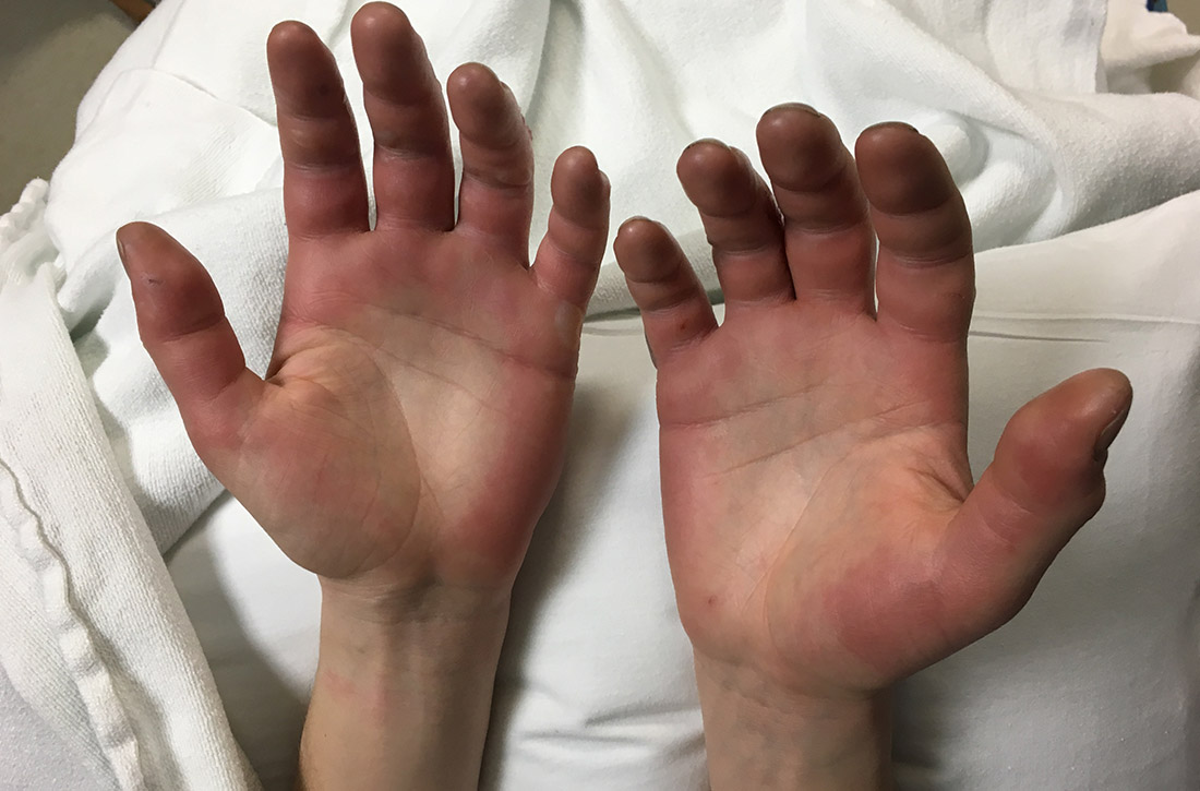
Immediate or delayed pushing in the second stage of labor?
ILLUSTRATIVE CASE
A 27-year-old G1P000 at term with an uncomplicated pregnancy has been laboring for 6 hours with an epidural in place and a reassuring fetal heart tracing. She is at –2 station with complete cervical dilation and effacement. Should she push now or delay pushing to allow for more descent?
More than 10,000 women give birth each day in the United States, yet few of our approaches to labor management are evidence based.2 For example, there are no clear guidelines on whether immediate pushing or delayed pushing (waiting 1-2 hours) in the second stage of labor (the time from complete cervical dilation to delivery of the fetus) leads to better outcomes.
A recent Cochrane review, which included very low- to moderate-quality trials of nulliparous and multiparous women using epidural analgesia showed that delayed pushing resulted in more vaginal deliveries, longer duration of second stage of labor, and shorter duration of pushing.3 But many of the trials included in this Cochrane review were noted to have study design limitations and significant heterogeneity.
A recent retrospective study found that delayed pushing resulted in longer duration of pushing and increased risks for cesarean section, operative vaginal delivery, and postpartum hemorrhage in nulliparous patients with and without epidurals.4 The World Health Organization recommends delayed pushing in women with epidural analgesia if time and fetal monitoring resources are available.5
STUDY SUMMARY
Does the timing of second stage pushing efforts affect outcomes?
This multicenter randomized controlled trial (RCT) evaluated the effect on spontaneous vaginal delivery of delayed pushing vs immediate pushing in 2404 term nulliparous women using epidural analgesia.1 Patients were ≥ 37 weeks’ gestation. Once patients achieved 10 cm of cervical dilation, they were randomized in a 1:1 ratio to either immediate pushing or to delayed (for 60 minutes) pushing (unless there was an irresistible urge to push or they were otherwise instructed by their provider).
Outcome and results. The primary outcome was spontaneous vaginal delivery without the use of any operative support. The mean time to pushing after complete cervical dilation was 19 minutes in the immediate pushing group and 60 minutes in the delayed group. There was no difference in the rate of spontaneous vaginal delivery between the immediate and delayed pushing groups (86% vs 87%, respectively; P = .67). The immediate pushing group had a shorter duration of second stage of labor (102 minutes vs 134 minutes; mean difference [MD] = –32 minutes; 95% confidence interval [CI], –37 to –27; P < .001) and a slightly longer duration of active pushing (84 minutes vs 75 minutes; MD = 9.2 minutes; 95% CI, 6-13; P < .001).
There was no significant difference in operative vaginal or cesarean deliveries. Postpartum hemorrhage was lower in the immediate pushing group (2.3% vs 4%; risk ratio [RR] = 0.6; 95% CI, 0.3-0.9; P = .03; number needed to treat [NNT] = 58), as was chorioamnionitis (6.7% vs 9.1%; RR = 0.7; 95% CI, 0.66-0.90; P = .005; NNT = 40). There was no significant difference in neonatal morbidity between groups. And in subgroup analysis, there was no significant difference in rates of vaginal delivery based on fetal position (occiput anterior, posterior, or transverse) or station (defined as high [< 2 cm] or low [≥ 2 cm]) between groups. Recruitment was stopped early at 75% because there was no difference in the primary outcome and there was concern regarding an increased risk of hemorrhage in the delayed pushing group.
Continue to: WHAT'S NEW
WHAT’S NEW
There’s no good reason to delay pushing
Delaying pushing once the cervix is completely dilated is not indicated, even for nulliparous women receiving epidural analgesia, as it does not decrease the rate of spontaneous vaginal delivery. It does, however, increase the length of second stage labor and the risk of postpartum hemorrhage and chorioamnionitis.
CAVEATS
Study was stopped early, and groups were unblinded
This study was stopped early, so it is not known if it was underpowered for some of the secondary outcomes. Also, it was not possible to blind the groups, so it is not clear if any bias in patient management or diagnosis resulted.
CHALLENGES TO IMPLEMENTATION
Will current practice and culture pose obstacles?
Although the overt challenges to enacting a policy of immediate pushing are minimal, the inertia of current practice and culture could affect the implementation of this strategy.
ACKNOWLEDGEMENT
The PURLs Surveillance System was supported in part by Grant Number UL1RR024999 from the National Center For Research Resources, a Clinical Translational Science Award to the University of Chicago. The content is solely the responsibility of the authors and does not necessarily represent the official views of the National Center For Research Resources or the National Institutes of Health.
1. Cahill AG, Srinivas SK, Tita ATN, et al. Effect of immediate vs delayed pushing on rates of spontaneous vaginal delivery among nulliparous women receiving neuraxial analgesia: a randomized clinical trial. JAMA. 2018;320:1444-1454.
2. Hamilton BE, Martin JA, Osterman MJK, et al. Births: provisional data for 2018. Vital Statistics Rapid Release. May 2019; Report No. 007. www.cdc.gov/nchs/data/vsrr/vsrr-007-508.pdf. Accessed April 22, 2020.
3. Lemos A, Amorim MM, Domales de Andrade A, et al. Pushing/bearing down methods for the second stage of labour. Cochrane Database Syst Rev. 2017;3:CD009124.
4. Yee LM, Sandoval G, Bailit J, et al. Maternal and neonatal outcomes with early compared with delayed pushing among nulliparous women. Obstet Gynecol. 2016;128:1039-1047.
5. WHO recommendations: intrapartum care for a positive childbirth experience. Geneva: World Health Organization; 2018. www.who.int/reproductivehealth/publications/intrapartum-care-guidelines/en/. Accessed April 22, 2020.
ILLUSTRATIVE CASE
A 27-year-old G1P000 at term with an uncomplicated pregnancy has been laboring for 6 hours with an epidural in place and a reassuring fetal heart tracing. She is at –2 station with complete cervical dilation and effacement. Should she push now or delay pushing to allow for more descent?
More than 10,000 women give birth each day in the United States, yet few of our approaches to labor management are evidence based.2 For example, there are no clear guidelines on whether immediate pushing or delayed pushing (waiting 1-2 hours) in the second stage of labor (the time from complete cervical dilation to delivery of the fetus) leads to better outcomes.
A recent Cochrane review, which included very low- to moderate-quality trials of nulliparous and multiparous women using epidural analgesia showed that delayed pushing resulted in more vaginal deliveries, longer duration of second stage of labor, and shorter duration of pushing.3 But many of the trials included in this Cochrane review were noted to have study design limitations and significant heterogeneity.
A recent retrospective study found that delayed pushing resulted in longer duration of pushing and increased risks for cesarean section, operative vaginal delivery, and postpartum hemorrhage in nulliparous patients with and without epidurals.4 The World Health Organization recommends delayed pushing in women with epidural analgesia if time and fetal monitoring resources are available.5
STUDY SUMMARY
Does the timing of second stage pushing efforts affect outcomes?
This multicenter randomized controlled trial (RCT) evaluated the effect on spontaneous vaginal delivery of delayed pushing vs immediate pushing in 2404 term nulliparous women using epidural analgesia.1 Patients were ≥ 37 weeks’ gestation. Once patients achieved 10 cm of cervical dilation, they were randomized in a 1:1 ratio to either immediate pushing or to delayed (for 60 minutes) pushing (unless there was an irresistible urge to push or they were otherwise instructed by their provider).
Outcome and results. The primary outcome was spontaneous vaginal delivery without the use of any operative support. The mean time to pushing after complete cervical dilation was 19 minutes in the immediate pushing group and 60 minutes in the delayed group. There was no difference in the rate of spontaneous vaginal delivery between the immediate and delayed pushing groups (86% vs 87%, respectively; P = .67). The immediate pushing group had a shorter duration of second stage of labor (102 minutes vs 134 minutes; mean difference [MD] = –32 minutes; 95% confidence interval [CI], –37 to –27; P < .001) and a slightly longer duration of active pushing (84 minutes vs 75 minutes; MD = 9.2 minutes; 95% CI, 6-13; P < .001).
There was no significant difference in operative vaginal or cesarean deliveries. Postpartum hemorrhage was lower in the immediate pushing group (2.3% vs 4%; risk ratio [RR] = 0.6; 95% CI, 0.3-0.9; P = .03; number needed to treat [NNT] = 58), as was chorioamnionitis (6.7% vs 9.1%; RR = 0.7; 95% CI, 0.66-0.90; P = .005; NNT = 40). There was no significant difference in neonatal morbidity between groups. And in subgroup analysis, there was no significant difference in rates of vaginal delivery based on fetal position (occiput anterior, posterior, or transverse) or station (defined as high [< 2 cm] or low [≥ 2 cm]) between groups. Recruitment was stopped early at 75% because there was no difference in the primary outcome and there was concern regarding an increased risk of hemorrhage in the delayed pushing group.
Continue to: WHAT'S NEW
WHAT’S NEW
There’s no good reason to delay pushing
Delaying pushing once the cervix is completely dilated is not indicated, even for nulliparous women receiving epidural analgesia, as it does not decrease the rate of spontaneous vaginal delivery. It does, however, increase the length of second stage labor and the risk of postpartum hemorrhage and chorioamnionitis.
CAVEATS
Study was stopped early, and groups were unblinded
This study was stopped early, so it is not known if it was underpowered for some of the secondary outcomes. Also, it was not possible to blind the groups, so it is not clear if any bias in patient management or diagnosis resulted.
CHALLENGES TO IMPLEMENTATION
Will current practice and culture pose obstacles?
Although the overt challenges to enacting a policy of immediate pushing are minimal, the inertia of current practice and culture could affect the implementation of this strategy.
ACKNOWLEDGEMENT
The PURLs Surveillance System was supported in part by Grant Number UL1RR024999 from the National Center For Research Resources, a Clinical Translational Science Award to the University of Chicago. The content is solely the responsibility of the authors and does not necessarily represent the official views of the National Center For Research Resources or the National Institutes of Health.
ILLUSTRATIVE CASE
A 27-year-old G1P000 at term with an uncomplicated pregnancy has been laboring for 6 hours with an epidural in place and a reassuring fetal heart tracing. She is at –2 station with complete cervical dilation and effacement. Should she push now or delay pushing to allow for more descent?
More than 10,000 women give birth each day in the United States, yet few of our approaches to labor management are evidence based.2 For example, there are no clear guidelines on whether immediate pushing or delayed pushing (waiting 1-2 hours) in the second stage of labor (the time from complete cervical dilation to delivery of the fetus) leads to better outcomes.
A recent Cochrane review, which included very low- to moderate-quality trials of nulliparous and multiparous women using epidural analgesia showed that delayed pushing resulted in more vaginal deliveries, longer duration of second stage of labor, and shorter duration of pushing.3 But many of the trials included in this Cochrane review were noted to have study design limitations and significant heterogeneity.
A recent retrospective study found that delayed pushing resulted in longer duration of pushing and increased risks for cesarean section, operative vaginal delivery, and postpartum hemorrhage in nulliparous patients with and without epidurals.4 The World Health Organization recommends delayed pushing in women with epidural analgesia if time and fetal monitoring resources are available.5
STUDY SUMMARY
Does the timing of second stage pushing efforts affect outcomes?
This multicenter randomized controlled trial (RCT) evaluated the effect on spontaneous vaginal delivery of delayed pushing vs immediate pushing in 2404 term nulliparous women using epidural analgesia.1 Patients were ≥ 37 weeks’ gestation. Once patients achieved 10 cm of cervical dilation, they were randomized in a 1:1 ratio to either immediate pushing or to delayed (for 60 minutes) pushing (unless there was an irresistible urge to push or they were otherwise instructed by their provider).
Outcome and results. The primary outcome was spontaneous vaginal delivery without the use of any operative support. The mean time to pushing after complete cervical dilation was 19 minutes in the immediate pushing group and 60 minutes in the delayed group. There was no difference in the rate of spontaneous vaginal delivery between the immediate and delayed pushing groups (86% vs 87%, respectively; P = .67). The immediate pushing group had a shorter duration of second stage of labor (102 minutes vs 134 minutes; mean difference [MD] = –32 minutes; 95% confidence interval [CI], –37 to –27; P < .001) and a slightly longer duration of active pushing (84 minutes vs 75 minutes; MD = 9.2 minutes; 95% CI, 6-13; P < .001).
There was no significant difference in operative vaginal or cesarean deliveries. Postpartum hemorrhage was lower in the immediate pushing group (2.3% vs 4%; risk ratio [RR] = 0.6; 95% CI, 0.3-0.9; P = .03; number needed to treat [NNT] = 58), as was chorioamnionitis (6.7% vs 9.1%; RR = 0.7; 95% CI, 0.66-0.90; P = .005; NNT = 40). There was no significant difference in neonatal morbidity between groups. And in subgroup analysis, there was no significant difference in rates of vaginal delivery based on fetal position (occiput anterior, posterior, or transverse) or station (defined as high [< 2 cm] or low [≥ 2 cm]) between groups. Recruitment was stopped early at 75% because there was no difference in the primary outcome and there was concern regarding an increased risk of hemorrhage in the delayed pushing group.
Continue to: WHAT'S NEW
WHAT’S NEW
There’s no good reason to delay pushing
Delaying pushing once the cervix is completely dilated is not indicated, even for nulliparous women receiving epidural analgesia, as it does not decrease the rate of spontaneous vaginal delivery. It does, however, increase the length of second stage labor and the risk of postpartum hemorrhage and chorioamnionitis.
CAVEATS
Study was stopped early, and groups were unblinded
This study was stopped early, so it is not known if it was underpowered for some of the secondary outcomes. Also, it was not possible to blind the groups, so it is not clear if any bias in patient management or diagnosis resulted.
CHALLENGES TO IMPLEMENTATION
Will current practice and culture pose obstacles?
Although the overt challenges to enacting a policy of immediate pushing are minimal, the inertia of current practice and culture could affect the implementation of this strategy.
ACKNOWLEDGEMENT
The PURLs Surveillance System was supported in part by Grant Number UL1RR024999 from the National Center For Research Resources, a Clinical Translational Science Award to the University of Chicago. The content is solely the responsibility of the authors and does not necessarily represent the official views of the National Center For Research Resources or the National Institutes of Health.
1. Cahill AG, Srinivas SK, Tita ATN, et al. Effect of immediate vs delayed pushing on rates of spontaneous vaginal delivery among nulliparous women receiving neuraxial analgesia: a randomized clinical trial. JAMA. 2018;320:1444-1454.
2. Hamilton BE, Martin JA, Osterman MJK, et al. Births: provisional data for 2018. Vital Statistics Rapid Release. May 2019; Report No. 007. www.cdc.gov/nchs/data/vsrr/vsrr-007-508.pdf. Accessed April 22, 2020.
3. Lemos A, Amorim MM, Domales de Andrade A, et al. Pushing/bearing down methods for the second stage of labour. Cochrane Database Syst Rev. 2017;3:CD009124.
4. Yee LM, Sandoval G, Bailit J, et al. Maternal and neonatal outcomes with early compared with delayed pushing among nulliparous women. Obstet Gynecol. 2016;128:1039-1047.
5. WHO recommendations: intrapartum care for a positive childbirth experience. Geneva: World Health Organization; 2018. www.who.int/reproductivehealth/publications/intrapartum-care-guidelines/en/. Accessed April 22, 2020.
1. Cahill AG, Srinivas SK, Tita ATN, et al. Effect of immediate vs delayed pushing on rates of spontaneous vaginal delivery among nulliparous women receiving neuraxial analgesia: a randomized clinical trial. JAMA. 2018;320:1444-1454.
2. Hamilton BE, Martin JA, Osterman MJK, et al. Births: provisional data for 2018. Vital Statistics Rapid Release. May 2019; Report No. 007. www.cdc.gov/nchs/data/vsrr/vsrr-007-508.pdf. Accessed April 22, 2020.
3. Lemos A, Amorim MM, Domales de Andrade A, et al. Pushing/bearing down methods for the second stage of labour. Cochrane Database Syst Rev. 2017;3:CD009124.
4. Yee LM, Sandoval G, Bailit J, et al. Maternal and neonatal outcomes with early compared with delayed pushing among nulliparous women. Obstet Gynecol. 2016;128:1039-1047.
5. WHO recommendations: intrapartum care for a positive childbirth experience. Geneva: World Health Organization; 2018. www.who.int/reproductivehealth/publications/intrapartum-care-guidelines/en/. Accessed April 22, 2020.
PRACTICE CHANGER
Recommend immediate, rather than delayed, pushing in the second stage of labor for nulliparous women receiving epidural analgesia. The rate of spontaneous vaginal delivery is the same, and there is a lower risk of postpartum hemorrhage and chorioamnionitis.
STRENGTH OF RECOMMENDATION
B: Based on an individual randomized controlled trial. 1
Cahill AG, Srinivas SK, Tita ATN, et al. Effect of immediate vs delayed pushing on rates of spontaneous vaginal delivery among nulliparous women receiving neuraxial analgesia: a randomized clinical trial. JAMA. 2018;320:1444-1454.

Whom should you screen for abdominal aortic aneurysm?
Too few patients are being screened for abdominal aortic aneurysm (AAA), resulting in severe morbidity and mortality. Many patients with AAA aren’t identified until they present with rupture, leading to mortality as high as 90%.1 Early detection is critical.
Medicare offers one-time free screening to eligible individuals > 65 years of age, and several professional organizations promote screening with published guidelines, which we discuss later in this article.
So who is at risk, who should be screened, and what is the best way to screen your patients?
Risk factors and sex differences
AAA has a prevalence of between 1% and 5% in men > 65 years old,2,3 and it is 4 to 6 times more common in men than women.4 Major risk factors include smoking, older age, family history, and genetic factors, while hypertension, history of coronary artery disease, hyperlipidemia, and peripheral arterial disease have weaker associations.3,4 Exercise and diabetes seem to have protective effects.5
The incidence and mortality of AAA increased between the 1950s and the mid-1990s; however, both indicators have decreased in numerous countries in the 21st century.6 Although the prevalence is much lower in women, they have a higher risk of rupture than men at equivalent lesion diameters.3 The prevalence of AAA in women who smoke and are > 70 years of age is > 1%.3
Silent but deadly
Most patients with AAA are asymptomatic. Their lesions are often detected incidentally on magnetic resonance imaging of the spine obtained for back pain, on an abdominal ultrasound (US) for gallstones, or on a routine computed tomography (CT) scan for the evaluation of abdominal pain. Some patients will experience vague abdominal discomfort from rapid expansion of an aneurysm prior to rupture, necessitating urgent repair. Also, some large aneurysms can erode into the spine and cause chronic back pain prior to rupture. An infrarenal abdominal aortic diameter > 30 mm defines an aneurysm,7 and once the diameter reaches 55 mm, the threat of rupture often justifies operative repair. (See “The preferred approach to repair.”)
SIDEBAR
The preferred approach to repair
Since the introduction of endovascular aneurysm repair (EVAR) in the latter part of the 20th century, it has become the standard of care for the surgical management of aneurysmal disease. Currently, > 80% of patients with an abdominal aortic aneurysm (AAA) who undergo repair are treated with EVAR.23
Typically, the AAA diameter is assessed via ultrasound. If repair is indicated, a computed tomography arteriogram is obtained to define the anatomy and help determine if the AAA is amenable to endografting. The most common contraindications to EVAR are either a short proximal neck (not enough distance below the renal arteries to safely anchor the stent graft) or an iliac artery diameter that is too small to allow delivery of the device. The operation can be performed under local, regional, or general anesthesia, and patients are usually discharged on the first postoperative day. These patients require lifelong surveillance due to the risk of delayed endoleak and reperfusion of the aneurysm sac.24
Ruptured aneurysms will classically manifest with severe abdominal and/or back pain. Often a ruptured aneurysm will be contained in the retroperitoneum, allowing the patient to remain hemodynamically stable for a period of time and thus providing a window of opportunity for emergent repair.
Continue to: What is the evidence that screening is effective?
What is the evidence that screening is effective?
In 1988, researchers in Chichester, England, randomized > 6000 men ages 65 to 80 years to either a control group or a group that was offered a one-time US screen for AAA. After 15 years of follow-up, no significant difference in AAA mortality was seen between the groups, although 26% of those invited for screening declined to participate and accounted for more than half of the AAA-related deaths in the group receiving an invitation for screening.8
The MASS Trial,9 another British study, began in 1997 and screened men ages 65 to 74. More than 67,000 men were randomized, with 1 group invited for AAA screening and the other serving as a control. The final report on this trial was published in 2012. After 13 years of follow-up, there was a 42% reduction in AAA-related deaths in the group invited for screening, a small reduction in all-cause mortality, and a significant reduction in risk of AAA rupture (hazard ratio = 0.57). The researchers noted that 216 patients would have to be invited for screening to prevent 1 death over 13 years. They also reported that 21% of the invited patients that had an AAA-related death had an initial scan that was negative for AAA (aortic diameter < 3 cm). However, despite this finding, screening still appeared to be beneficial.
Lindholdt et al10 randomized > 12,000 Danish men ages 64 to 73 to serve as controls or to be invited for US screening for AAA. After 13 years of follow-up, those invited for screening had a 66% relative risk reduction in AAA-related mortality, with screening considered cost effective. There were no differences between the groups in all-cause mortality. Conversely, the Western Australia Trial studied an older group of men, ages 64 to 83, but was unable to show a benefit of screening in lowering AAA-related mortality.11
Tikagi et al6 performed a meta-analysis on the data from the 4 trials above and reported up to 15 years of follow-up. Patients who attended screening sessions had a reduction in all-cause mortality with an odds ratio (OR) of 0.6, and a marked reduction in AAA-related mortality with an OR of 0.4. The favorable data on screening have prompted the United Kingdom and Sweden to offer screening to all men ≥ 65 years, based on the current estimate of a 1% prevalence of AAA, although screening is felt to remain cost effective down to a prevalence of 0.35%.12
Massachusetts General Hospital also reported13 that the detection rate of AAA increased, with the diagnosis made at smaller aneurysm dimensions, following publication of the US Preventive Services Task Force recommendations (reviewed below).
Continue to: When to screen
When to screen
Several professional organizations, as well as Medicare, have published AAA screening recommendations. While there are notable differences among them, their shared message is crucial: screen. Consider adding applicable reminders to your practice’s electronic medical record system to increase screening rates for eligible patients.
US Preventive Services Task Force 14
- Recommend one-time AAA screening with ultrasonography for men ages 65 to 75 years who have ever smoked (B recommendation).
- Selectively offer AAA screening to men ages 65 to 75 years who have never smoked, rather than routinely screening all men in this group (C recommendation). Individual attributes that could favor screening include a family history of AAA, the presence of other arterial aneurysms, and the number of risk factors for cardiovascular disease.
- Do not routinely screen women for AAA if they have never smoked and have no family history of AAA (D recommendation). For women ages 65 to 75 years who have ever smoked or have a family history of AAA, current evidence is insufficient to assess the balance of benefits and harms of screening for AAA (I statement). (See the related Practice Alert.)
Canadian Task Force on Preventive Health Care4
- Recommend one-time screening for AAA with US for men 65 to 80 years of age (weak level of recommendation; moderate-quality evidence).
- Do not recommend screening for men older than 80 years of age (weak recommendation; low quality of evidence).
- Do not recommend screening for women (strong recommendation; very low quality of evidence).
Society for Vascular Surgery15
- Recommend one-time US screening for AAA for men or women 65 to 75 years of age with a history of tobacco use (strong recommendation with high-quality evidence).
- Recommend one-time screening if there is a history of smoking for men or women > 75 years of age who are in good health and have not previously been screened. (Weak recommendation with low-quality evidence).
- Recommend one-time screening of men or women 65 to 75 years of age who are first-degree relatives of someone with AAA, or in those older than age 75 and in good health (weak recommendation with low-quality evidence).
How to screen
Physical exam. The abdominal aorta is often palpable in the epigastric region, and a thorough abdominal exam should include an attempt to detect it. It is critical that the patient be supine during palpation, to allow compression of the aorta against the lumbar spine. Even with a well-performed exam, however, its sensitivity is just 76% in the detection of AAA ≥ 5 cm.16
Continue to: Imaging
Imaging. US is the preferred imaging procedure when screening for AAA, given its high sensitivity and specificity.17 If US yields poor image quality, noncontrast CT is suggested, with magnetic resonance angiography being another alternative.18 Handheld US has the potential to supplement the physical exam, and has been shown to be a viable method to detect AAA in an outpatient primary care setting at a reasonable cost.19 Typically, a formal aortic US requires a patient to go without food or liquids for 8 hours before the procedure to obtain the best image; however, a good estimate of aortic diameter can be obtained without this restriction.
Despite Medicare coverage and recs, few people are screened
In 2007, Medicare started the SAAVE Program (Screening Aortic Aneurysm Very Effectively), offering a one-time US screening for AAA for eligible patients, as part of the Welcome to Medicare Program. Eligible individuals are men between 65 and 75 years of age with a history of smoking at least 100 cigarettes in their lifetime, and men or women in the same age group with a family history of AAA.2
Despite these recommendations, few patients receive screening. Centers for Medicare & Medicaid Services data show that < 10% of eligible men were screened between 2004 and 2008,20 and Olchanski et al21 report that < 1% of eligible patients were screened from 2005-2009. A simulation model estimates that 131 additional life years could be gained per 1000 patients screened if the utilization rate could be increased to 80%, a seemingly achievable goal.21 Moreover, expanding the screening program to include female smokers could increase 10-year life expectancy by 13%.21 Reasons for underutilization of this Medicare screening benefit may include lack of awareness by physicians and patients, costs of co-pays, and underutilization of the basic Welcome to Medicare exam.21
An additional consequence of low utilization of AAA screening is a high percentage of patients who are identified only late in the course of the disease. Mell et al22 report that 39% of patients undergoing AAA repair were identified < 6 months prior to surgery, a higher percentage than would be expected in a well-screened population. They also determined that slightly more than one-third of patients undergoing surgery for ruptured AAA had diagnostic imaging performed > 6 months prior to surgery, suggesting the possibility that these patients may not have been properly surveilled for aneurysm expansion, although the authors note that other potential explanations include delays in treatment due to comorbidities, and patient-related factors such as refusal of surgery or noncompliance with follow-up.
CORRESPONDENCE
Jeffrey S. Todd, MD, 127 McClanahan Street, Suite 300, Roanoke, VA 24014; jstodd@carilionclinic.org.
1. Assar AN, Zarins CK. Ruptured abdominal aortic aneurysm: a surgical emergency with many clinical presentations. Postgrad Med J. 2009;85:268-273.
2. SBU—Swedish agency for Health Technology Assessment and Assessment of Social Services. Screening for abdominal aortic aneurysm. 2018. www.sbu.se/en/publications/sbu-assesses/screening-for-abdominal-aortic-aneurysm/. Accessed April 24, 2020.
3. Spanos K, Labropoulos N, Giannoukas A. Abdominal aortic aneurysm screening: do we need to shift toward a targeted strategy? Angiology. 2018;69:192-194.
4. Canadian Task Force on Preventive Health Care. Recommendations on screening for abdominal aortic aneurysm in primary care. CMAJ. 2017;189:E1137-1145.
5. Stackelberg O, Wolk, A, Eliasson K, et al. Lifestyle and risk of screening-detected abdominal aortic aneurysm in men. J Am Heart Assoc. 2017;6:e004725.
6. Tikagi H, Ando T, Umemoto T; ALICE (All-Literature Investigation of Cardiovascular Evidence) group. Abdominal aortic aneurysm screening reduces all-cause mortality: make screening great again. Angiology. 2017;69:205-211.
7. Fleming C, Whitlock EP, Beil TL, et al. Screening for abdominal aortic aneurysm: a best-evidence systematic review for the US Preventive Services Task Force. Ann Intern Med. 2005;142:203-211.
8. Ashton HA, Gao L, Kim LG, et al. Fifteen-year follow-up of a randomized clinical trial of ultrasonographic screening for abdominal aortic aneurysms. Br J Surg. 2007;94:696-701.
9. Thompson SG, Ashton HA, Gao L, et al. Final follow-up of the Multicentre Aneurysm Screening Study (MASS) randomized trial of abdominal aortic aneurysm screening. Br J Surg. 2012;99:1649-1656.
10. Lindholdt JS, Sørenson J, Søgaard R et al. Long-term benefit and cost-effectiveness analysis of screening for abdominal aortic aneurysms from a randomized controlled trial. Br J Surg. 2010;97:826-834.
11. McCaul KA, Lawrence-Brown M, Dickinson JA, et al. Long-term outcomes of the Western Australian trial of screening for abdominal aortic aneurysms: secondary analysis of a randomized clinical trial. JAMA Intern Med. 2016;176:1761-1766.
12. Earnshaw JJ, Lees T. Update on screening for abdominal aortic aneurysm. Eur J Vasc Endovasc Surg. 2017;54:1-2.
13. Zucker EJ, Misono AS, Prabhakar AM. Abdominal aortic aneurysm screening practices: impact of the 2014 US Preventive Services Task Force recommendations. J Am Coll Radiol. 2017;14:868-874.
14. USPSTF. Screening for abdominal aortic aneurysm: US Preventive Services Task Force recommendation statement.
15. Chaikof EL, Dalman RL, Eskandari MK, et al. The Society for Vascular Surgery practice guidelines on the care of patients with an abdominal aortic aneurysm. J Vasc Surg. 2018;67:2-77.
16. Venkatasubramaniam AK, Mehta T, Chetter IC, et al. The value of abdominal examination in the diagnosis of abdominal aortic aneurysm. Eur J Vasc Endovasc Surg. 2004;24:56-60.
17. Lindholdt JS, Vammen S, Juul S, et al. The validity of ultrasonographic scanning as screening method for abdominal aortic aneurysm. Eur J Vasc Endovasc Surg. 1999;17:472-475.
18. Desjardins B, Dill KE, Flamm SD, et al. ACR Appropriateness Criteria®, pulsatile abdominal mass, suspected abdominal aortic aneurysm. Int J Cardiovasc Imaging. 2013;29:177-183.
19. Sisó-Almirall A, Kostov B, Navarro-González M, et al. Abdominal aortic aneurysm screening program using hand-held ultrasound in primary healthcare. PLoS One. 2017;12:e0176877.
20. Shreibati JB, Baker LC, Hlatky MA, et al. Impact of the Screening Abdominal Aortic Aneurysms Very Efficiently (SAAAVE) Act on abdominal ultrasonography use among Medicare beneficiaries. Arch Intern Med. 2012;172:1456-1462.
21. Olchanski N, Winn A, Cohen JT, et al. Abdominal aortic aneurysm screening: how many life years lost from underuse of the Medicare screening benefit? J Gen Intern Med. 2014;29:1155-1161.
22. Mell MW, Hlatky MA, Shreibati JB, et al. Late diagnosis of abdominal aortic aneurysms substantiates underutilization of abdominal aortic aneurysm screening for Medicare beneficiaries. J Vasc Surg. 2013;57:1519-1523.
23. England A, McWilliams R. Endovascular aortic aneurysm repair (EVAR). Ulster Med J. 2013;82:3-10.
24. Dillavou ED, Muluk SC, Makaroun MS. Improving aneurysm-related outcomes: nationwide benefits of endovascular repair. J Vasc Surg. 2006;43:446-451.
Too few patients are being screened for abdominal aortic aneurysm (AAA), resulting in severe morbidity and mortality. Many patients with AAA aren’t identified until they present with rupture, leading to mortality as high as 90%.1 Early detection is critical.
Medicare offers one-time free screening to eligible individuals > 65 years of age, and several professional organizations promote screening with published guidelines, which we discuss later in this article.
So who is at risk, who should be screened, and what is the best way to screen your patients?
Risk factors and sex differences
AAA has a prevalence of between 1% and 5% in men > 65 years old,2,3 and it is 4 to 6 times more common in men than women.4 Major risk factors include smoking, older age, family history, and genetic factors, while hypertension, history of coronary artery disease, hyperlipidemia, and peripheral arterial disease have weaker associations.3,4 Exercise and diabetes seem to have protective effects.5
The incidence and mortality of AAA increased between the 1950s and the mid-1990s; however, both indicators have decreased in numerous countries in the 21st century.6 Although the prevalence is much lower in women, they have a higher risk of rupture than men at equivalent lesion diameters.3 The prevalence of AAA in women who smoke and are > 70 years of age is > 1%.3
Silent but deadly
Most patients with AAA are asymptomatic. Their lesions are often detected incidentally on magnetic resonance imaging of the spine obtained for back pain, on an abdominal ultrasound (US) for gallstones, or on a routine computed tomography (CT) scan for the evaluation of abdominal pain. Some patients will experience vague abdominal discomfort from rapid expansion of an aneurysm prior to rupture, necessitating urgent repair. Also, some large aneurysms can erode into the spine and cause chronic back pain prior to rupture. An infrarenal abdominal aortic diameter > 30 mm defines an aneurysm,7 and once the diameter reaches 55 mm, the threat of rupture often justifies operative repair. (See “The preferred approach to repair.”)
SIDEBAR
The preferred approach to repair
Since the introduction of endovascular aneurysm repair (EVAR) in the latter part of the 20th century, it has become the standard of care for the surgical management of aneurysmal disease. Currently, > 80% of patients with an abdominal aortic aneurysm (AAA) who undergo repair are treated with EVAR.23
Typically, the AAA diameter is assessed via ultrasound. If repair is indicated, a computed tomography arteriogram is obtained to define the anatomy and help determine if the AAA is amenable to endografting. The most common contraindications to EVAR are either a short proximal neck (not enough distance below the renal arteries to safely anchor the stent graft) or an iliac artery diameter that is too small to allow delivery of the device. The operation can be performed under local, regional, or general anesthesia, and patients are usually discharged on the first postoperative day. These patients require lifelong surveillance due to the risk of delayed endoleak and reperfusion of the aneurysm sac.24
Ruptured aneurysms will classically manifest with severe abdominal and/or back pain. Often a ruptured aneurysm will be contained in the retroperitoneum, allowing the patient to remain hemodynamically stable for a period of time and thus providing a window of opportunity for emergent repair.
Continue to: What is the evidence that screening is effective?
What is the evidence that screening is effective?
In 1988, researchers in Chichester, England, randomized > 6000 men ages 65 to 80 years to either a control group or a group that was offered a one-time US screen for AAA. After 15 years of follow-up, no significant difference in AAA mortality was seen between the groups, although 26% of those invited for screening declined to participate and accounted for more than half of the AAA-related deaths in the group receiving an invitation for screening.8
The MASS Trial,9 another British study, began in 1997 and screened men ages 65 to 74. More than 67,000 men were randomized, with 1 group invited for AAA screening and the other serving as a control. The final report on this trial was published in 2012. After 13 years of follow-up, there was a 42% reduction in AAA-related deaths in the group invited for screening, a small reduction in all-cause mortality, and a significant reduction in risk of AAA rupture (hazard ratio = 0.57). The researchers noted that 216 patients would have to be invited for screening to prevent 1 death over 13 years. They also reported that 21% of the invited patients that had an AAA-related death had an initial scan that was negative for AAA (aortic diameter < 3 cm). However, despite this finding, screening still appeared to be beneficial.
Lindholdt et al10 randomized > 12,000 Danish men ages 64 to 73 to serve as controls or to be invited for US screening for AAA. After 13 years of follow-up, those invited for screening had a 66% relative risk reduction in AAA-related mortality, with screening considered cost effective. There were no differences between the groups in all-cause mortality. Conversely, the Western Australia Trial studied an older group of men, ages 64 to 83, but was unable to show a benefit of screening in lowering AAA-related mortality.11
Tikagi et al6 performed a meta-analysis on the data from the 4 trials above and reported up to 15 years of follow-up. Patients who attended screening sessions had a reduction in all-cause mortality with an odds ratio (OR) of 0.6, and a marked reduction in AAA-related mortality with an OR of 0.4. The favorable data on screening have prompted the United Kingdom and Sweden to offer screening to all men ≥ 65 years, based on the current estimate of a 1% prevalence of AAA, although screening is felt to remain cost effective down to a prevalence of 0.35%.12
Massachusetts General Hospital also reported13 that the detection rate of AAA increased, with the diagnosis made at smaller aneurysm dimensions, following publication of the US Preventive Services Task Force recommendations (reviewed below).
Continue to: When to screen
When to screen
Several professional organizations, as well as Medicare, have published AAA screening recommendations. While there are notable differences among them, their shared message is crucial: screen. Consider adding applicable reminders to your practice’s electronic medical record system to increase screening rates for eligible patients.
US Preventive Services Task Force 14
- Recommend one-time AAA screening with ultrasonography for men ages 65 to 75 years who have ever smoked (B recommendation).
- Selectively offer AAA screening to men ages 65 to 75 years who have never smoked, rather than routinely screening all men in this group (C recommendation). Individual attributes that could favor screening include a family history of AAA, the presence of other arterial aneurysms, and the number of risk factors for cardiovascular disease.
- Do not routinely screen women for AAA if they have never smoked and have no family history of AAA (D recommendation). For women ages 65 to 75 years who have ever smoked or have a family history of AAA, current evidence is insufficient to assess the balance of benefits and harms of screening for AAA (I statement). (See the related Practice Alert.)
Canadian Task Force on Preventive Health Care4
- Recommend one-time screening for AAA with US for men 65 to 80 years of age (weak level of recommendation; moderate-quality evidence).
- Do not recommend screening for men older than 80 years of age (weak recommendation; low quality of evidence).
- Do not recommend screening for women (strong recommendation; very low quality of evidence).
Society for Vascular Surgery15
- Recommend one-time US screening for AAA for men or women 65 to 75 years of age with a history of tobacco use (strong recommendation with high-quality evidence).
- Recommend one-time screening if there is a history of smoking for men or women > 75 years of age who are in good health and have not previously been screened. (Weak recommendation with low-quality evidence).
- Recommend one-time screening of men or women 65 to 75 years of age who are first-degree relatives of someone with AAA, or in those older than age 75 and in good health (weak recommendation with low-quality evidence).
How to screen
Physical exam. The abdominal aorta is often palpable in the epigastric region, and a thorough abdominal exam should include an attempt to detect it. It is critical that the patient be supine during palpation, to allow compression of the aorta against the lumbar spine. Even with a well-performed exam, however, its sensitivity is just 76% in the detection of AAA ≥ 5 cm.16
Continue to: Imaging
Imaging. US is the preferred imaging procedure when screening for AAA, given its high sensitivity and specificity.17 If US yields poor image quality, noncontrast CT is suggested, with magnetic resonance angiography being another alternative.18 Handheld US has the potential to supplement the physical exam, and has been shown to be a viable method to detect AAA in an outpatient primary care setting at a reasonable cost.19 Typically, a formal aortic US requires a patient to go without food or liquids for 8 hours before the procedure to obtain the best image; however, a good estimate of aortic diameter can be obtained without this restriction.
Despite Medicare coverage and recs, few people are screened
In 2007, Medicare started the SAAVE Program (Screening Aortic Aneurysm Very Effectively), offering a one-time US screening for AAA for eligible patients, as part of the Welcome to Medicare Program. Eligible individuals are men between 65 and 75 years of age with a history of smoking at least 100 cigarettes in their lifetime, and men or women in the same age group with a family history of AAA.2
Despite these recommendations, few patients receive screening. Centers for Medicare & Medicaid Services data show that < 10% of eligible men were screened between 2004 and 2008,20 and Olchanski et al21 report that < 1% of eligible patients were screened from 2005-2009. A simulation model estimates that 131 additional life years could be gained per 1000 patients screened if the utilization rate could be increased to 80%, a seemingly achievable goal.21 Moreover, expanding the screening program to include female smokers could increase 10-year life expectancy by 13%.21 Reasons for underutilization of this Medicare screening benefit may include lack of awareness by physicians and patients, costs of co-pays, and underutilization of the basic Welcome to Medicare exam.21
An additional consequence of low utilization of AAA screening is a high percentage of patients who are identified only late in the course of the disease. Mell et al22 report that 39% of patients undergoing AAA repair were identified < 6 months prior to surgery, a higher percentage than would be expected in a well-screened population. They also determined that slightly more than one-third of patients undergoing surgery for ruptured AAA had diagnostic imaging performed > 6 months prior to surgery, suggesting the possibility that these patients may not have been properly surveilled for aneurysm expansion, although the authors note that other potential explanations include delays in treatment due to comorbidities, and patient-related factors such as refusal of surgery or noncompliance with follow-up.
CORRESPONDENCE
Jeffrey S. Todd, MD, 127 McClanahan Street, Suite 300, Roanoke, VA 24014; jstodd@carilionclinic.org.
Too few patients are being screened for abdominal aortic aneurysm (AAA), resulting in severe morbidity and mortality. Many patients with AAA aren’t identified until they present with rupture, leading to mortality as high as 90%.1 Early detection is critical.
Medicare offers one-time free screening to eligible individuals > 65 years of age, and several professional organizations promote screening with published guidelines, which we discuss later in this article.
So who is at risk, who should be screened, and what is the best way to screen your patients?
Risk factors and sex differences
AAA has a prevalence of between 1% and 5% in men > 65 years old,2,3 and it is 4 to 6 times more common in men than women.4 Major risk factors include smoking, older age, family history, and genetic factors, while hypertension, history of coronary artery disease, hyperlipidemia, and peripheral arterial disease have weaker associations.3,4 Exercise and diabetes seem to have protective effects.5
The incidence and mortality of AAA increased between the 1950s and the mid-1990s; however, both indicators have decreased in numerous countries in the 21st century.6 Although the prevalence is much lower in women, they have a higher risk of rupture than men at equivalent lesion diameters.3 The prevalence of AAA in women who smoke and are > 70 years of age is > 1%.3
Silent but deadly
Most patients with AAA are asymptomatic. Their lesions are often detected incidentally on magnetic resonance imaging of the spine obtained for back pain, on an abdominal ultrasound (US) for gallstones, or on a routine computed tomography (CT) scan for the evaluation of abdominal pain. Some patients will experience vague abdominal discomfort from rapid expansion of an aneurysm prior to rupture, necessitating urgent repair. Also, some large aneurysms can erode into the spine and cause chronic back pain prior to rupture. An infrarenal abdominal aortic diameter > 30 mm defines an aneurysm,7 and once the diameter reaches 55 mm, the threat of rupture often justifies operative repair. (See “The preferred approach to repair.”)
SIDEBAR
The preferred approach to repair
Since the introduction of endovascular aneurysm repair (EVAR) in the latter part of the 20th century, it has become the standard of care for the surgical management of aneurysmal disease. Currently, > 80% of patients with an abdominal aortic aneurysm (AAA) who undergo repair are treated with EVAR.23
Typically, the AAA diameter is assessed via ultrasound. If repair is indicated, a computed tomography arteriogram is obtained to define the anatomy and help determine if the AAA is amenable to endografting. The most common contraindications to EVAR are either a short proximal neck (not enough distance below the renal arteries to safely anchor the stent graft) or an iliac artery diameter that is too small to allow delivery of the device. The operation can be performed under local, regional, or general anesthesia, and patients are usually discharged on the first postoperative day. These patients require lifelong surveillance due to the risk of delayed endoleak and reperfusion of the aneurysm sac.24
Ruptured aneurysms will classically manifest with severe abdominal and/or back pain. Often a ruptured aneurysm will be contained in the retroperitoneum, allowing the patient to remain hemodynamically stable for a period of time and thus providing a window of opportunity for emergent repair.
Continue to: What is the evidence that screening is effective?
What is the evidence that screening is effective?
In 1988, researchers in Chichester, England, randomized > 6000 men ages 65 to 80 years to either a control group or a group that was offered a one-time US screen for AAA. After 15 years of follow-up, no significant difference in AAA mortality was seen between the groups, although 26% of those invited for screening declined to participate and accounted for more than half of the AAA-related deaths in the group receiving an invitation for screening.8
The MASS Trial,9 another British study, began in 1997 and screened men ages 65 to 74. More than 67,000 men were randomized, with 1 group invited for AAA screening and the other serving as a control. The final report on this trial was published in 2012. After 13 years of follow-up, there was a 42% reduction in AAA-related deaths in the group invited for screening, a small reduction in all-cause mortality, and a significant reduction in risk of AAA rupture (hazard ratio = 0.57). The researchers noted that 216 patients would have to be invited for screening to prevent 1 death over 13 years. They also reported that 21% of the invited patients that had an AAA-related death had an initial scan that was negative for AAA (aortic diameter < 3 cm). However, despite this finding, screening still appeared to be beneficial.
Lindholdt et al10 randomized > 12,000 Danish men ages 64 to 73 to serve as controls or to be invited for US screening for AAA. After 13 years of follow-up, those invited for screening had a 66% relative risk reduction in AAA-related mortality, with screening considered cost effective. There were no differences between the groups in all-cause mortality. Conversely, the Western Australia Trial studied an older group of men, ages 64 to 83, but was unable to show a benefit of screening in lowering AAA-related mortality.11
Tikagi et al6 performed a meta-analysis on the data from the 4 trials above and reported up to 15 years of follow-up. Patients who attended screening sessions had a reduction in all-cause mortality with an odds ratio (OR) of 0.6, and a marked reduction in AAA-related mortality with an OR of 0.4. The favorable data on screening have prompted the United Kingdom and Sweden to offer screening to all men ≥ 65 years, based on the current estimate of a 1% prevalence of AAA, although screening is felt to remain cost effective down to a prevalence of 0.35%.12
Massachusetts General Hospital also reported13 that the detection rate of AAA increased, with the diagnosis made at smaller aneurysm dimensions, following publication of the US Preventive Services Task Force recommendations (reviewed below).
Continue to: When to screen
When to screen
Several professional organizations, as well as Medicare, have published AAA screening recommendations. While there are notable differences among them, their shared message is crucial: screen. Consider adding applicable reminders to your practice’s electronic medical record system to increase screening rates for eligible patients.
US Preventive Services Task Force 14
- Recommend one-time AAA screening with ultrasonography for men ages 65 to 75 years who have ever smoked (B recommendation).
- Selectively offer AAA screening to men ages 65 to 75 years who have never smoked, rather than routinely screening all men in this group (C recommendation). Individual attributes that could favor screening include a family history of AAA, the presence of other arterial aneurysms, and the number of risk factors for cardiovascular disease.
- Do not routinely screen women for AAA if they have never smoked and have no family history of AAA (D recommendation). For women ages 65 to 75 years who have ever smoked or have a family history of AAA, current evidence is insufficient to assess the balance of benefits and harms of screening for AAA (I statement). (See the related Practice Alert.)
Canadian Task Force on Preventive Health Care4
- Recommend one-time screening for AAA with US for men 65 to 80 years of age (weak level of recommendation; moderate-quality evidence).
- Do not recommend screening for men older than 80 years of age (weak recommendation; low quality of evidence).
- Do not recommend screening for women (strong recommendation; very low quality of evidence).
Society for Vascular Surgery15
- Recommend one-time US screening for AAA for men or women 65 to 75 years of age with a history of tobacco use (strong recommendation with high-quality evidence).
- Recommend one-time screening if there is a history of smoking for men or women > 75 years of age who are in good health and have not previously been screened. (Weak recommendation with low-quality evidence).
- Recommend one-time screening of men or women 65 to 75 years of age who are first-degree relatives of someone with AAA, or in those older than age 75 and in good health (weak recommendation with low-quality evidence).
How to screen
Physical exam. The abdominal aorta is often palpable in the epigastric region, and a thorough abdominal exam should include an attempt to detect it. It is critical that the patient be supine during palpation, to allow compression of the aorta against the lumbar spine. Even with a well-performed exam, however, its sensitivity is just 76% in the detection of AAA ≥ 5 cm.16
Continue to: Imaging
Imaging. US is the preferred imaging procedure when screening for AAA, given its high sensitivity and specificity.17 If US yields poor image quality, noncontrast CT is suggested, with magnetic resonance angiography being another alternative.18 Handheld US has the potential to supplement the physical exam, and has been shown to be a viable method to detect AAA in an outpatient primary care setting at a reasonable cost.19 Typically, a formal aortic US requires a patient to go without food or liquids for 8 hours before the procedure to obtain the best image; however, a good estimate of aortic diameter can be obtained without this restriction.
Despite Medicare coverage and recs, few people are screened
In 2007, Medicare started the SAAVE Program (Screening Aortic Aneurysm Very Effectively), offering a one-time US screening for AAA for eligible patients, as part of the Welcome to Medicare Program. Eligible individuals are men between 65 and 75 years of age with a history of smoking at least 100 cigarettes in their lifetime, and men or women in the same age group with a family history of AAA.2
Despite these recommendations, few patients receive screening. Centers for Medicare & Medicaid Services data show that < 10% of eligible men were screened between 2004 and 2008,20 and Olchanski et al21 report that < 1% of eligible patients were screened from 2005-2009. A simulation model estimates that 131 additional life years could be gained per 1000 patients screened if the utilization rate could be increased to 80%, a seemingly achievable goal.21 Moreover, expanding the screening program to include female smokers could increase 10-year life expectancy by 13%.21 Reasons for underutilization of this Medicare screening benefit may include lack of awareness by physicians and patients, costs of co-pays, and underutilization of the basic Welcome to Medicare exam.21
An additional consequence of low utilization of AAA screening is a high percentage of patients who are identified only late in the course of the disease. Mell et al22 report that 39% of patients undergoing AAA repair were identified < 6 months prior to surgery, a higher percentage than would be expected in a well-screened population. They also determined that slightly more than one-third of patients undergoing surgery for ruptured AAA had diagnostic imaging performed > 6 months prior to surgery, suggesting the possibility that these patients may not have been properly surveilled for aneurysm expansion, although the authors note that other potential explanations include delays in treatment due to comorbidities, and patient-related factors such as refusal of surgery or noncompliance with follow-up.
CORRESPONDENCE
Jeffrey S. Todd, MD, 127 McClanahan Street, Suite 300, Roanoke, VA 24014; jstodd@carilionclinic.org.
1. Assar AN, Zarins CK. Ruptured abdominal aortic aneurysm: a surgical emergency with many clinical presentations. Postgrad Med J. 2009;85:268-273.
2. SBU—Swedish agency for Health Technology Assessment and Assessment of Social Services. Screening for abdominal aortic aneurysm. 2018. www.sbu.se/en/publications/sbu-assesses/screening-for-abdominal-aortic-aneurysm/. Accessed April 24, 2020.
3. Spanos K, Labropoulos N, Giannoukas A. Abdominal aortic aneurysm screening: do we need to shift toward a targeted strategy? Angiology. 2018;69:192-194.
4. Canadian Task Force on Preventive Health Care. Recommendations on screening for abdominal aortic aneurysm in primary care. CMAJ. 2017;189:E1137-1145.
5. Stackelberg O, Wolk, A, Eliasson K, et al. Lifestyle and risk of screening-detected abdominal aortic aneurysm in men. J Am Heart Assoc. 2017;6:e004725.
6. Tikagi H, Ando T, Umemoto T; ALICE (All-Literature Investigation of Cardiovascular Evidence) group. Abdominal aortic aneurysm screening reduces all-cause mortality: make screening great again. Angiology. 2017;69:205-211.
7. Fleming C, Whitlock EP, Beil TL, et al. Screening for abdominal aortic aneurysm: a best-evidence systematic review for the US Preventive Services Task Force. Ann Intern Med. 2005;142:203-211.
8. Ashton HA, Gao L, Kim LG, et al. Fifteen-year follow-up of a randomized clinical trial of ultrasonographic screening for abdominal aortic aneurysms. Br J Surg. 2007;94:696-701.
9. Thompson SG, Ashton HA, Gao L, et al. Final follow-up of the Multicentre Aneurysm Screening Study (MASS) randomized trial of abdominal aortic aneurysm screening. Br J Surg. 2012;99:1649-1656.
10. Lindholdt JS, Sørenson J, Søgaard R et al. Long-term benefit and cost-effectiveness analysis of screening for abdominal aortic aneurysms from a randomized controlled trial. Br J Surg. 2010;97:826-834.
11. McCaul KA, Lawrence-Brown M, Dickinson JA, et al. Long-term outcomes of the Western Australian trial of screening for abdominal aortic aneurysms: secondary analysis of a randomized clinical trial. JAMA Intern Med. 2016;176:1761-1766.
12. Earnshaw JJ, Lees T. Update on screening for abdominal aortic aneurysm. Eur J Vasc Endovasc Surg. 2017;54:1-2.
13. Zucker EJ, Misono AS, Prabhakar AM. Abdominal aortic aneurysm screening practices: impact of the 2014 US Preventive Services Task Force recommendations. J Am Coll Radiol. 2017;14:868-874.
14. USPSTF. Screening for abdominal aortic aneurysm: US Preventive Services Task Force recommendation statement.
15. Chaikof EL, Dalman RL, Eskandari MK, et al. The Society for Vascular Surgery practice guidelines on the care of patients with an abdominal aortic aneurysm. J Vasc Surg. 2018;67:2-77.
16. Venkatasubramaniam AK, Mehta T, Chetter IC, et al. The value of abdominal examination in the diagnosis of abdominal aortic aneurysm. Eur J Vasc Endovasc Surg. 2004;24:56-60.
17. Lindholdt JS, Vammen S, Juul S, et al. The validity of ultrasonographic scanning as screening method for abdominal aortic aneurysm. Eur J Vasc Endovasc Surg. 1999;17:472-475.
18. Desjardins B, Dill KE, Flamm SD, et al. ACR Appropriateness Criteria®, pulsatile abdominal mass, suspected abdominal aortic aneurysm. Int J Cardiovasc Imaging. 2013;29:177-183.
19. Sisó-Almirall A, Kostov B, Navarro-González M, et al. Abdominal aortic aneurysm screening program using hand-held ultrasound in primary healthcare. PLoS One. 2017;12:e0176877.
20. Shreibati JB, Baker LC, Hlatky MA, et al. Impact of the Screening Abdominal Aortic Aneurysms Very Efficiently (SAAAVE) Act on abdominal ultrasonography use among Medicare beneficiaries. Arch Intern Med. 2012;172:1456-1462.
21. Olchanski N, Winn A, Cohen JT, et al. Abdominal aortic aneurysm screening: how many life years lost from underuse of the Medicare screening benefit? J Gen Intern Med. 2014;29:1155-1161.
22. Mell MW, Hlatky MA, Shreibati JB, et al. Late diagnosis of abdominal aortic aneurysms substantiates underutilization of abdominal aortic aneurysm screening for Medicare beneficiaries. J Vasc Surg. 2013;57:1519-1523.
23. England A, McWilliams R. Endovascular aortic aneurysm repair (EVAR). Ulster Med J. 2013;82:3-10.
24. Dillavou ED, Muluk SC, Makaroun MS. Improving aneurysm-related outcomes: nationwide benefits of endovascular repair. J Vasc Surg. 2006;43:446-451.
1. Assar AN, Zarins CK. Ruptured abdominal aortic aneurysm: a surgical emergency with many clinical presentations. Postgrad Med J. 2009;85:268-273.
2. SBU—Swedish agency for Health Technology Assessment and Assessment of Social Services. Screening for abdominal aortic aneurysm. 2018. www.sbu.se/en/publications/sbu-assesses/screening-for-abdominal-aortic-aneurysm/. Accessed April 24, 2020.
3. Spanos K, Labropoulos N, Giannoukas A. Abdominal aortic aneurysm screening: do we need to shift toward a targeted strategy? Angiology. 2018;69:192-194.
4. Canadian Task Force on Preventive Health Care. Recommendations on screening for abdominal aortic aneurysm in primary care. CMAJ. 2017;189:E1137-1145.
5. Stackelberg O, Wolk, A, Eliasson K, et al. Lifestyle and risk of screening-detected abdominal aortic aneurysm in men. J Am Heart Assoc. 2017;6:e004725.
6. Tikagi H, Ando T, Umemoto T; ALICE (All-Literature Investigation of Cardiovascular Evidence) group. Abdominal aortic aneurysm screening reduces all-cause mortality: make screening great again. Angiology. 2017;69:205-211.
7. Fleming C, Whitlock EP, Beil TL, et al. Screening for abdominal aortic aneurysm: a best-evidence systematic review for the US Preventive Services Task Force. Ann Intern Med. 2005;142:203-211.
8. Ashton HA, Gao L, Kim LG, et al. Fifteen-year follow-up of a randomized clinical trial of ultrasonographic screening for abdominal aortic aneurysms. Br J Surg. 2007;94:696-701.
9. Thompson SG, Ashton HA, Gao L, et al. Final follow-up of the Multicentre Aneurysm Screening Study (MASS) randomized trial of abdominal aortic aneurysm screening. Br J Surg. 2012;99:1649-1656.
10. Lindholdt JS, Sørenson J, Søgaard R et al. Long-term benefit and cost-effectiveness analysis of screening for abdominal aortic aneurysms from a randomized controlled trial. Br J Surg. 2010;97:826-834.
11. McCaul KA, Lawrence-Brown M, Dickinson JA, et al. Long-term outcomes of the Western Australian trial of screening for abdominal aortic aneurysms: secondary analysis of a randomized clinical trial. JAMA Intern Med. 2016;176:1761-1766.
12. Earnshaw JJ, Lees T. Update on screening for abdominal aortic aneurysm. Eur J Vasc Endovasc Surg. 2017;54:1-2.
13. Zucker EJ, Misono AS, Prabhakar AM. Abdominal aortic aneurysm screening practices: impact of the 2014 US Preventive Services Task Force recommendations. J Am Coll Radiol. 2017;14:868-874.
14. USPSTF. Screening for abdominal aortic aneurysm: US Preventive Services Task Force recommendation statement.
15. Chaikof EL, Dalman RL, Eskandari MK, et al. The Society for Vascular Surgery practice guidelines on the care of patients with an abdominal aortic aneurysm. J Vasc Surg. 2018;67:2-77.
16. Venkatasubramaniam AK, Mehta T, Chetter IC, et al. The value of abdominal examination in the diagnosis of abdominal aortic aneurysm. Eur J Vasc Endovasc Surg. 2004;24:56-60.
17. Lindholdt JS, Vammen S, Juul S, et al. The validity of ultrasonographic scanning as screening method for abdominal aortic aneurysm. Eur J Vasc Endovasc Surg. 1999;17:472-475.
18. Desjardins B, Dill KE, Flamm SD, et al. ACR Appropriateness Criteria®, pulsatile abdominal mass, suspected abdominal aortic aneurysm. Int J Cardiovasc Imaging. 2013;29:177-183.
19. Sisó-Almirall A, Kostov B, Navarro-González M, et al. Abdominal aortic aneurysm screening program using hand-held ultrasound in primary healthcare. PLoS One. 2017;12:e0176877.
20. Shreibati JB, Baker LC, Hlatky MA, et al. Impact of the Screening Abdominal Aortic Aneurysms Very Efficiently (SAAAVE) Act on abdominal ultrasonography use among Medicare beneficiaries. Arch Intern Med. 2012;172:1456-1462.
21. Olchanski N, Winn A, Cohen JT, et al. Abdominal aortic aneurysm screening: how many life years lost from underuse of the Medicare screening benefit? J Gen Intern Med. 2014;29:1155-1161.
22. Mell MW, Hlatky MA, Shreibati JB, et al. Late diagnosis of abdominal aortic aneurysms substantiates underutilization of abdominal aortic aneurysm screening for Medicare beneficiaries. J Vasc Surg. 2013;57:1519-1523.
23. England A, McWilliams R. Endovascular aortic aneurysm repair (EVAR). Ulster Med J. 2013;82:3-10.
24. Dillavou ED, Muluk SC, Makaroun MS. Improving aneurysm-related outcomes: nationwide benefits of endovascular repair. J Vasc Surg. 2006;43:446-451.
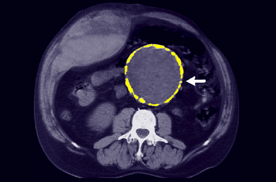
Does vitamin D supplementation reduce asthma exacerbations?
EVIDENCE SUMMARY
A Cochrane systematic review of vitamin D for managing asthma performed meta-analyses on RCTs that evaluated several outcomes.1 The review found improvement in the primary outcome of asthma exacerbations requiring systemic steroids, mainly in adult patients, and in the secondary outcomes of emergency department visits or hospitalization, in a mix of adults and children (TABLE1-6).
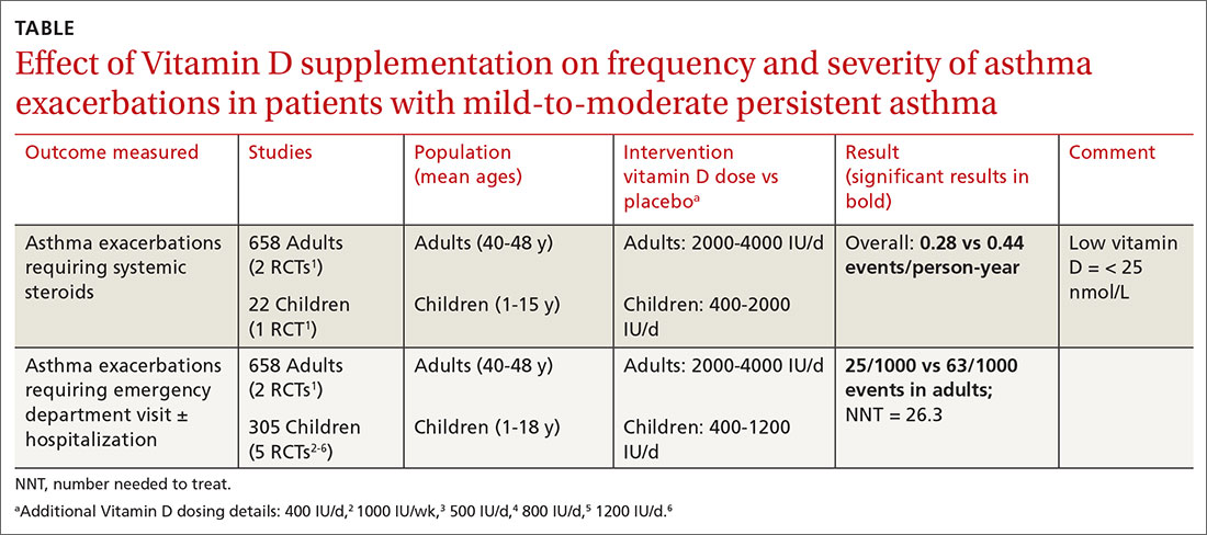
Most participants had mild-to-moderate asthma; trials lasted 4 to 12 months. Vitamin D dosage regimens varied, with a median daily dose of 900 IU/d (range, 400-4000 IU/d). Six RCTs were rated high-quality, and 1 had unclear risk of bias.
Supplementation reduced exacerbations in patients with low vitamin D levels
A subsequent (2017) systematic review and meta-analysis evaluating the primary outcome of exacerbations requiring steroids7 included another study8 (in addition to the 6 RCTs in the Cochrane review).
When researchers reanalyzed individual participant data from the trials in the Cochrane review, plus the additional RCT, to include baseline vitamin D levels, they found that vitamin D supplementation reduced exacerbations overall (NNT = 7.7) and in patients with low baseline vitamin D levels (25[OH] vitamin D < 25 nmol/L; 92 participants in 3 RCTs; NNT = 4.3) but not in patients with higher baseline levels (764 participants in 6 RCTs). Vitamin D supplementation reduced the asthma exacerbation rate in patients with low baseline vitamin D levels (0.19 vs 0.42 events per participant-year; P = .046).
Smaller benefit found on ED visits and hospitalizations
The Cochrane review, with 2 RCTs with adults (n = 658)1 and 5 RCTs with children (n = 305),2-6 evaluated whether Vitamin D reduced the need for emergency department visits and hospitalization with asthma exacerbations; they found a smaller benefit (NNT = 26.3).
Effects on FEV1, daily asthma symptoms, and serious adverse effects
Several RCTs included in the 2017 meta-analysis found no effect of vitamin D supplementation on FEV1, daily asthma symptoms (evaluated with the standardized Asthma Control Test Score), or reported serious adverse events.2-6,9,10 No deaths occurred in any trial.
Additional findings in children from lower-quality studies
A 2015 systematic review and meta-analysis of RCTs evaluating vitamin D supplementation for children with asthma found11:
- moderate-quality evidence for decreased emergency department visits (1 RCT from India, 100 children ages 3 to 14 years, decrease not specified; P = .015);
- low-quality evidence for reduced exacerbations (6 RCTs [3 RCTs also in Cochrane review], 507 children ages 3 to 17 years; risk ratio = 0.41; 95% confidence interval, 0.27-0.63); and
- low-quality evidence for reduced standardized asthma symptom scores (6 RCTs [2 RCTs also in Cochrane review], 231 children ages 3 to 17 years; amount of reduction not listed; P = .01).
Continue to: RECOMMENDATIONS
RECOMMENDATIONS
No published guidelines discuss using vitamin D in managing asthma. An American Academy of Family Physicians (AAFP) summary of the Cochrane systematic review recommends that family physicians await further studies and updated guidelines before recommending vitamin D for patients with asthma.12 The AAFP also points out that the Endocrine Society has recommended vitamin D supplementation for adults (1500-2000 IU/d) and children (at least 1000 IU/d) at risk for deficiency.
Editor's takeaway
In the meta-analyses highlighted here, researchers evaluated asthma patients with a wide range of ages, baseline vitamin D levels, and vitamin D supplementation protocols. Although vitamin D reduced asthma exacerbations requiring steroids overall, the effect was driven by 3 studies of patients with low baseline vitamin D levels. As a result, disentangling who might benefit the most remains a challenge. The conservative course for now is to manage asthma according to current guidelines and supplement vitamin D in patients at risk for, or with known, deficiency.
, , , . Vitamin D for the management of asthma. Cochrane Database Syst Rev. 2016;9:CD011511.
2. Jensen M, Mailhot G, Alos N, et al. Vitamin D intervention in preschoolers with viral-induced asthma (DIVA): a pilot randomised controlled trial. Trials. 2016;26:17:353.
, , , et al. Correlation of vitamin D with Foxp3 induction and steroid-sparing effect of immunotherapy in asthmatic children. Ann Allergy Asthma Immunol. 2012;109:329-335.
, , , et al. Vitamin D supplementation in children may prevent asthma exacerbation triggered by acute respiratory infection. J Allergy Clin Immunol. 2011;127:1294-1296.
, , , et al. Improved control of childhood asthma with low-dose, short-term vitamin D supplementation: a randomized, double-blind, placebo-controlled trial. Allergy. 2016;71:1001-1009.
, , , et al. Randomized trial of vitamin D supplementation to prevent seasonal influenza A in school children. Am J Clin Nutr. 2010;91:1255-1260.
7. Joliffe DA, Greenberg L, Hooper RL, et al. Vitamin D supplementation to prevent asthma exacerbations: a systematic review and meta-analysis of individual participant data. Lancet 2017;5:881-890.
8. Kerley CP, Hutchinson K, Cormical L, et al. Vitamin D3 for uncontrolled childhood asthma: a pilot study. Pediatr Allergy Immunol. 2016;27:404-412.
, , , et al. Effect of vitamin D3 on asthma treatment failures in adults with symptomatic asthma and lower vitamin D levels: the VIDA randomized clinical trial. JAMA. 2014;311:2083-2091.
, , , et al. Double-blind multi-centre randomised controlled trial of vitamin D3 supplementation in adults with inhaled corticosteroid-treated asthma (ViDiAs). Thorax. 2015:70:451-457.
11. Riverin B, Maguire J, Li P. Vitamin D supplementation for childhood asthma: a systematic review and meta-analysis. PLOS One. 2015;10:e0136841.
EVIDENCE SUMMARY
A Cochrane systematic review of vitamin D for managing asthma performed meta-analyses on RCTs that evaluated several outcomes.1 The review found improvement in the primary outcome of asthma exacerbations requiring systemic steroids, mainly in adult patients, and in the secondary outcomes of emergency department visits or hospitalization, in a mix of adults and children (TABLE1-6).
Most participants had mild-to-moderate asthma; trials lasted 4 to 12 months. Vitamin D dosage regimens varied, with a median daily dose of 900 IU/d (range, 400-4000 IU/d). Six RCTs were rated high-quality, and 1 had unclear risk of bias.
Supplementation reduced exacerbations in patients with low vitamin D levels
A subsequent (2017) systematic review and meta-analysis evaluating the primary outcome of exacerbations requiring steroids7 included another study8 (in addition to the 6 RCTs in the Cochrane review).
When researchers reanalyzed individual participant data from the trials in the Cochrane review, plus the additional RCT, to include baseline vitamin D levels, they found that vitamin D supplementation reduced exacerbations overall (NNT = 7.7) and in patients with low baseline vitamin D levels (25[OH] vitamin D < 25 nmol/L; 92 participants in 3 RCTs; NNT = 4.3) but not in patients with higher baseline levels (764 participants in 6 RCTs). Vitamin D supplementation reduced the asthma exacerbation rate in patients with low baseline vitamin D levels (0.19 vs 0.42 events per participant-year; P = .046).
Smaller benefit found on ED visits and hospitalizations
The Cochrane review, with 2 RCTs with adults (n = 658)1 and 5 RCTs with children (n = 305),2-6 evaluated whether Vitamin D reduced the need for emergency department visits and hospitalization with asthma exacerbations; they found a smaller benefit (NNT = 26.3).
Effects on FEV1, daily asthma symptoms, and serious adverse effects
Several RCTs included in the 2017 meta-analysis found no effect of vitamin D supplementation on FEV1, daily asthma symptoms (evaluated with the standardized Asthma Control Test Score), or reported serious adverse events.2-6,9,10 No deaths occurred in any trial.
Additional findings in children from lower-quality studies
A 2015 systematic review and meta-analysis of RCTs evaluating vitamin D supplementation for children with asthma found11:
- moderate-quality evidence for decreased emergency department visits (1 RCT from India, 100 children ages 3 to 14 years, decrease not specified; P = .015);
- low-quality evidence for reduced exacerbations (6 RCTs [3 RCTs also in Cochrane review], 507 children ages 3 to 17 years; risk ratio = 0.41; 95% confidence interval, 0.27-0.63); and
- low-quality evidence for reduced standardized asthma symptom scores (6 RCTs [2 RCTs also in Cochrane review], 231 children ages 3 to 17 years; amount of reduction not listed; P = .01).
Continue to: RECOMMENDATIONS
RECOMMENDATIONS
No published guidelines discuss using vitamin D in managing asthma. An American Academy of Family Physicians (AAFP) summary of the Cochrane systematic review recommends that family physicians await further studies and updated guidelines before recommending vitamin D for patients with asthma.12 The AAFP also points out that the Endocrine Society has recommended vitamin D supplementation for adults (1500-2000 IU/d) and children (at least 1000 IU/d) at risk for deficiency.
Editor's takeaway
In the meta-analyses highlighted here, researchers evaluated asthma patients with a wide range of ages, baseline vitamin D levels, and vitamin D supplementation protocols. Although vitamin D reduced asthma exacerbations requiring steroids overall, the effect was driven by 3 studies of patients with low baseline vitamin D levels. As a result, disentangling who might benefit the most remains a challenge. The conservative course for now is to manage asthma according to current guidelines and supplement vitamin D in patients at risk for, or with known, deficiency.
EVIDENCE SUMMARY
A Cochrane systematic review of vitamin D for managing asthma performed meta-analyses on RCTs that evaluated several outcomes.1 The review found improvement in the primary outcome of asthma exacerbations requiring systemic steroids, mainly in adult patients, and in the secondary outcomes of emergency department visits or hospitalization, in a mix of adults and children (TABLE1-6).
Most participants had mild-to-moderate asthma; trials lasted 4 to 12 months. Vitamin D dosage regimens varied, with a median daily dose of 900 IU/d (range, 400-4000 IU/d). Six RCTs were rated high-quality, and 1 had unclear risk of bias.
Supplementation reduced exacerbations in patients with low vitamin D levels
A subsequent (2017) systematic review and meta-analysis evaluating the primary outcome of exacerbations requiring steroids7 included another study8 (in addition to the 6 RCTs in the Cochrane review).
When researchers reanalyzed individual participant data from the trials in the Cochrane review, plus the additional RCT, to include baseline vitamin D levels, they found that vitamin D supplementation reduced exacerbations overall (NNT = 7.7) and in patients with low baseline vitamin D levels (25[OH] vitamin D < 25 nmol/L; 92 participants in 3 RCTs; NNT = 4.3) but not in patients with higher baseline levels (764 participants in 6 RCTs). Vitamin D supplementation reduced the asthma exacerbation rate in patients with low baseline vitamin D levels (0.19 vs 0.42 events per participant-year; P = .046).
Smaller benefit found on ED visits and hospitalizations
The Cochrane review, with 2 RCTs with adults (n = 658)1 and 5 RCTs with children (n = 305),2-6 evaluated whether Vitamin D reduced the need for emergency department visits and hospitalization with asthma exacerbations; they found a smaller benefit (NNT = 26.3).
Effects on FEV1, daily asthma symptoms, and serious adverse effects
Several RCTs included in the 2017 meta-analysis found no effect of vitamin D supplementation on FEV1, daily asthma symptoms (evaluated with the standardized Asthma Control Test Score), or reported serious adverse events.2-6,9,10 No deaths occurred in any trial.
Additional findings in children from lower-quality studies
A 2015 systematic review and meta-analysis of RCTs evaluating vitamin D supplementation for children with asthma found11:
- moderate-quality evidence for decreased emergency department visits (1 RCT from India, 100 children ages 3 to 14 years, decrease not specified; P = .015);
- low-quality evidence for reduced exacerbations (6 RCTs [3 RCTs also in Cochrane review], 507 children ages 3 to 17 years; risk ratio = 0.41; 95% confidence interval, 0.27-0.63); and
- low-quality evidence for reduced standardized asthma symptom scores (6 RCTs [2 RCTs also in Cochrane review], 231 children ages 3 to 17 years; amount of reduction not listed; P = .01).
Continue to: RECOMMENDATIONS
RECOMMENDATIONS
No published guidelines discuss using vitamin D in managing asthma. An American Academy of Family Physicians (AAFP) summary of the Cochrane systematic review recommends that family physicians await further studies and updated guidelines before recommending vitamin D for patients with asthma.12 The AAFP also points out that the Endocrine Society has recommended vitamin D supplementation for adults (1500-2000 IU/d) and children (at least 1000 IU/d) at risk for deficiency.
Editor's takeaway
In the meta-analyses highlighted here, researchers evaluated asthma patients with a wide range of ages, baseline vitamin D levels, and vitamin D supplementation protocols. Although vitamin D reduced asthma exacerbations requiring steroids overall, the effect was driven by 3 studies of patients with low baseline vitamin D levels. As a result, disentangling who might benefit the most remains a challenge. The conservative course for now is to manage asthma according to current guidelines and supplement vitamin D in patients at risk for, or with known, deficiency.
, , , . Vitamin D for the management of asthma. Cochrane Database Syst Rev. 2016;9:CD011511.
2. Jensen M, Mailhot G, Alos N, et al. Vitamin D intervention in preschoolers with viral-induced asthma (DIVA): a pilot randomised controlled trial. Trials. 2016;26:17:353.
, , , et al. Correlation of vitamin D with Foxp3 induction and steroid-sparing effect of immunotherapy in asthmatic children. Ann Allergy Asthma Immunol. 2012;109:329-335.
, , , et al. Vitamin D supplementation in children may prevent asthma exacerbation triggered by acute respiratory infection. J Allergy Clin Immunol. 2011;127:1294-1296.
, , , et al. Improved control of childhood asthma with low-dose, short-term vitamin D supplementation: a randomized, double-blind, placebo-controlled trial. Allergy. 2016;71:1001-1009.
, , , et al. Randomized trial of vitamin D supplementation to prevent seasonal influenza A in school children. Am J Clin Nutr. 2010;91:1255-1260.
7. Joliffe DA, Greenberg L, Hooper RL, et al. Vitamin D supplementation to prevent asthma exacerbations: a systematic review and meta-analysis of individual participant data. Lancet 2017;5:881-890.
8. Kerley CP, Hutchinson K, Cormical L, et al. Vitamin D3 for uncontrolled childhood asthma: a pilot study. Pediatr Allergy Immunol. 2016;27:404-412.
, , , et al. Effect of vitamin D3 on asthma treatment failures in adults with symptomatic asthma and lower vitamin D levels: the VIDA randomized clinical trial. JAMA. 2014;311:2083-2091.
, , , et al. Double-blind multi-centre randomised controlled trial of vitamin D3 supplementation in adults with inhaled corticosteroid-treated asthma (ViDiAs). Thorax. 2015:70:451-457.
11. Riverin B, Maguire J, Li P. Vitamin D supplementation for childhood asthma: a systematic review and meta-analysis. PLOS One. 2015;10:e0136841.
, , , . Vitamin D for the management of asthma. Cochrane Database Syst Rev. 2016;9:CD011511.
2. Jensen M, Mailhot G, Alos N, et al. Vitamin D intervention in preschoolers with viral-induced asthma (DIVA): a pilot randomised controlled trial. Trials. 2016;26:17:353.
, , , et al. Correlation of vitamin D with Foxp3 induction and steroid-sparing effect of immunotherapy in asthmatic children. Ann Allergy Asthma Immunol. 2012;109:329-335.
, , , et al. Vitamin D supplementation in children may prevent asthma exacerbation triggered by acute respiratory infection. J Allergy Clin Immunol. 2011;127:1294-1296.
, , , et al. Improved control of childhood asthma with low-dose, short-term vitamin D supplementation: a randomized, double-blind, placebo-controlled trial. Allergy. 2016;71:1001-1009.
, , , et al. Randomized trial of vitamin D supplementation to prevent seasonal influenza A in school children. Am J Clin Nutr. 2010;91:1255-1260.
7. Joliffe DA, Greenberg L, Hooper RL, et al. Vitamin D supplementation to prevent asthma exacerbations: a systematic review and meta-analysis of individual participant data. Lancet 2017;5:881-890.
8. Kerley CP, Hutchinson K, Cormical L, et al. Vitamin D3 for uncontrolled childhood asthma: a pilot study. Pediatr Allergy Immunol. 2016;27:404-412.
, , , et al. Effect of vitamin D3 on asthma treatment failures in adults with symptomatic asthma and lower vitamin D levels: the VIDA randomized clinical trial. JAMA. 2014;311:2083-2091.
, , , et al. Double-blind multi-centre randomised controlled trial of vitamin D3 supplementation in adults with inhaled corticosteroid-treated asthma (ViDiAs). Thorax. 2015:70:451-457.
11. Riverin B, Maguire J, Li P. Vitamin D supplementation for childhood asthma: a systematic review and meta-analysis. PLOS One. 2015;10:e0136841.
EVIDENCE-BASED ANSWER:
Yes, to some extent it does, and primarily in patients with low vitamin D levels. Supplementation reduces asthma exacerbations requiring systemic steroids by 30% overall in adults and children with mild-to-moderate asthma (number needed to treat [NNT] = 7.7). The outcome is driven by the effect in patients with vitamin D levels < 25 nmol/L (NNT = 4.3), however; supplementation doesn’t decrease exacerbations in patients with higher levels. Supplementation also reduces, by a smaller amount (NNT = 26.3), the odds of exacerbations requiring emergency department care or hospitalization (strength of recommendation [SOR]: A, meta-analysis of randomized controlled trials [RCTs]).
In children, vitamin D supplementation may also reduce exacerbations and improve symptom scores (SOR: C, low-quality RCTs).
Vitamin D doesn’t improve forced expiratory volume in 1 second (FEV1) or standardized asthma control test scores. Also, it isn’t associated with serious adverse effects (SOR: A, meta-analysis of RCTs).
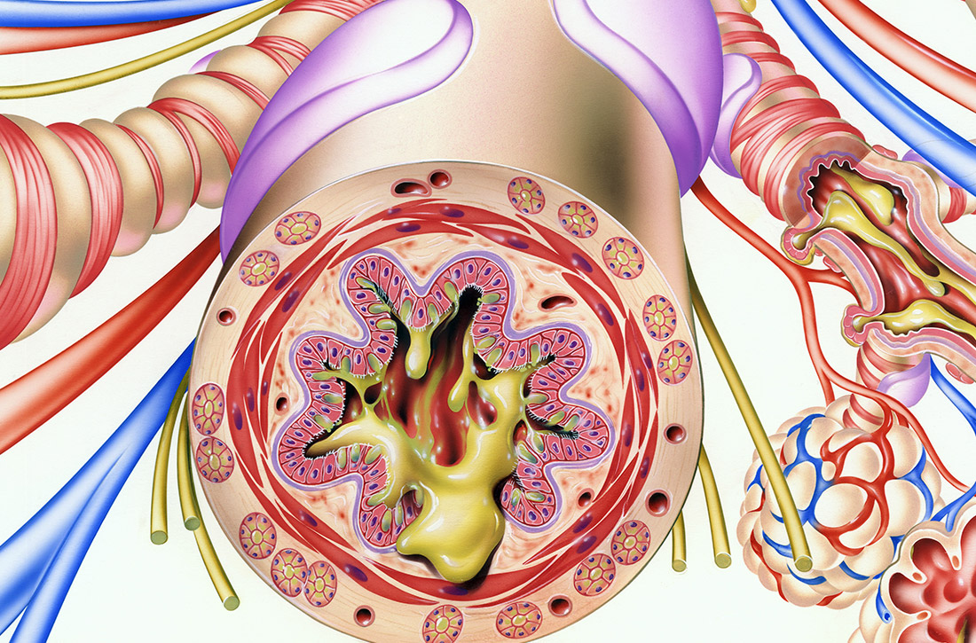
FDA approves olaparib/bevacizumab maintenance
The Food and Drug Administration has announced a new approved indication for olaparib (Lynparza) in adults with advanced epithelial ovarian, fallopian tube, or primary peritoneal cancer.

Olaparib is now FDA-approved for use in combination with bevacizumab as maintenance therapy in patients who responded to first-line platinum-based chemotherapy and whose cancer is homologous recombination deficiency positive, as defined by a deleterious or suspected deleterious BRCA mutation and/or genomic instability.
The FDA also approved the Myriad myChoice CDx test as a companion diagnostic for olaparib.
Trial results
The efficacy of olaparib and the myChoice CDx test were assessed in patients in the phase 3 PAOLA-1 trial (NCT02477644). The study enrolled patients with advanced high-grade epithelial ovarian, fallopian tube, or primary peritoneal cancer who had received first-line platinum-based chemotherapy and bevacizumab.
Patients were stratified by first-line treatment outcome and BRCA mutation status, as determined by prospective local testing. All available clinical samples were retrospectively tested with the Myriad myChoice CDx test.
The patients were randomized to receive olaparib at 300 mg orally twice daily in combination with bevacizumab at 15 mg/kg every 3 weeks (n = 537) or placebo plus bevacizumab (n = 269). Patients continued bevacizumab in the maintenance setting and started olaparib 3-9 weeks after their last chemotherapy dose. Olaparib could be continued for up to 2 years or until disease progression or unacceptable toxicity.
The median progression-free survival among the 387 patients with homologous recombination deficiency-positive tumors was 37.2 months in the olaparib arm and 17.7 months in the placebo arm (hazard ratio, 0.33), according to the prescribing information for olaparib.
Serious adverse events occurred in 31% of patients in the olaparib arm. The most common were hypertension (19%) and anemia (17%).
Dose interruptions from adverse events occurred in 54% of patients in the olaparib arm, and dose reductions from adverse events occurred in 41%.
The Food and Drug Administration has announced a new approved indication for olaparib (Lynparza) in adults with advanced epithelial ovarian, fallopian tube, or primary peritoneal cancer.
Olaparib is now FDA-approved for use in combination with bevacizumab as maintenance therapy in patients who responded to first-line platinum-based chemotherapy and whose cancer is homologous recombination deficiency positive, as defined by a deleterious or suspected deleterious BRCA mutation and/or genomic instability.
The FDA also approved the Myriad myChoice CDx test as a companion diagnostic for olaparib.
Trial results
The efficacy of olaparib and the myChoice CDx test were assessed in patients in the phase 3 PAOLA-1 trial (NCT02477644). The study enrolled patients with advanced high-grade epithelial ovarian, fallopian tube, or primary peritoneal cancer who had received first-line platinum-based chemotherapy and bevacizumab.
Patients were stratified by first-line treatment outcome and BRCA mutation status, as determined by prospective local testing. All available clinical samples were retrospectively tested with the Myriad myChoice CDx test.
The patients were randomized to receive olaparib at 300 mg orally twice daily in combination with bevacizumab at 15 mg/kg every 3 weeks (n = 537) or placebo plus bevacizumab (n = 269). Patients continued bevacizumab in the maintenance setting and started olaparib 3-9 weeks after their last chemotherapy dose. Olaparib could be continued for up to 2 years or until disease progression or unacceptable toxicity.
The median progression-free survival among the 387 patients with homologous recombination deficiency-positive tumors was 37.2 months in the olaparib arm and 17.7 months in the placebo arm (hazard ratio, 0.33), according to the prescribing information for olaparib.
Serious adverse events occurred in 31% of patients in the olaparib arm. The most common were hypertension (19%) and anemia (17%).
Dose interruptions from adverse events occurred in 54% of patients in the olaparib arm, and dose reductions from adverse events occurred in 41%.
The Food and Drug Administration has announced a new approved indication for olaparib (Lynparza) in adults with advanced epithelial ovarian, fallopian tube, or primary peritoneal cancer.
Olaparib is now FDA-approved for use in combination with bevacizumab as maintenance therapy in patients who responded to first-line platinum-based chemotherapy and whose cancer is homologous recombination deficiency positive, as defined by a deleterious or suspected deleterious BRCA mutation and/or genomic instability.
The FDA also approved the Myriad myChoice CDx test as a companion diagnostic for olaparib.
Trial results
The efficacy of olaparib and the myChoice CDx test were assessed in patients in the phase 3 PAOLA-1 trial (NCT02477644). The study enrolled patients with advanced high-grade epithelial ovarian, fallopian tube, or primary peritoneal cancer who had received first-line platinum-based chemotherapy and bevacizumab.
Patients were stratified by first-line treatment outcome and BRCA mutation status, as determined by prospective local testing. All available clinical samples were retrospectively tested with the Myriad myChoice CDx test.
The patients were randomized to receive olaparib at 300 mg orally twice daily in combination with bevacizumab at 15 mg/kg every 3 weeks (n = 537) or placebo plus bevacizumab (n = 269). Patients continued bevacizumab in the maintenance setting and started olaparib 3-9 weeks after their last chemotherapy dose. Olaparib could be continued for up to 2 years or until disease progression or unacceptable toxicity.
The median progression-free survival among the 387 patients with homologous recombination deficiency-positive tumors was 37.2 months in the olaparib arm and 17.7 months in the placebo arm (hazard ratio, 0.33), according to the prescribing information for olaparib.
Serious adverse events occurred in 31% of patients in the olaparib arm. The most common were hypertension (19%) and anemia (17%).
Dose interruptions from adverse events occurred in 54% of patients in the olaparib arm, and dose reductions from adverse events occurred in 41%.
Justices appear split over birth control mandate case
U.S. Supreme Court justices appear divided over whether the Trump administration acted properly when it expanded exemptions under the Affordable Care Act’s contraception mandate.

During oral arguments on May 6, the court expressed differing perspectives about the administration’s authority to allow for more exemptions under the health law’s birth control mandate and whether the expansions were reasonable. Justices heard the consolidated cases – Little Sisters of the Poor v. Pennsylvania and Trump v. Pennsylvania – by teleconference because of the COVID-19 pandemic. They are expected to make a decision by the summer.
Associate justice Ruth Bader Ginsburg, who participated in the telephone conference call from a hospital where she was recovering from a gallbladder condition, said the exemptions ignored the intent of Congress to provide women with comprehensive coverage through the ACA.
“The glaring feature of what the government has done in expanding this exemption is to toss to the winds entirely Congress’s instruction that women need and shall have seamless, no-cost, comprehensive coverage,” she said during oral arguments. “This leaves the women to hunt for other government programs that might cover them, and for those who are not covered by Medicaid or one of the other government programs, they can get contraceptive coverage only from paying out of their own pocket, which is exactly what Congress didn’t want to happen.”
Associate Justice Samuel Alito Jr., meanwhile, indicated that a lower court opinion that had blocked the exemptions from going forward conflicts with the Supreme Court’s ruling in a related case, Burwell v. Hobby Lobby.
“Explain to me why the Third Circuit’s analysis of the question of substantial burden is not squarely inconsistent with our reasoning in Hobby Lobby,” Associate Justice Alito said during oral arguments. “Hobby Lobby held that, if a person sincerely believes that it is immoral to perform an act that has the effect of enabling another person to commit an immoral act, a federal court does not have the right to say that this person is wrong on the question of moral complicity. That’s precisely the situation here. Reading the Third Circuit’s discussion of the substantial burden question, I wondered whether they had read that part of the Hobby Lobby decision.”
The dispute surrounding the ACA’s birth control mandate and the extent of exemptions afforded has gone on for a decade and has led to numerous legal challenges. The ACA initially required all employers to cover birth control for employees with no copayments, but exempted group health plans of religious employers. Those religious employers were primarily churches and other houses of worship. After a number of complaints and lawsuits, the Obama administration created a workaround for nonprofit religious employers not included in that exemption to opt out of the mandate. However, critics argued the process itself was a violation of their religious freedom.
The issue led to the case of Zubik v. Burwell, a legal challenge over the mandate exemption that went before the U.S. Supreme Court in March 2016. The issue was never resolved however, and in May 2016, the Supreme Court vacated the lower court rulings related to Zubik v. Burwell and remanded the case back to the four appeals courts that had originally ruled on the issue.
In 2018, the Trump administration announced new rules aimed at broadening exemptions to the ACA’s contraceptive mandate to entities that object to services covered by the mandate on the basis of “sincerely held religious beliefs.” A second rule allowed nonprofit organizations and small businesses that had nonreligious moral convictions against the mandate to opt out.
Thirteen states and the District of Columbia then sued the Trump administration over the rules, as well as Pennsylvania and New Jersey in a separate case. Little Sisters of the Poor, a religious nonprofit operating a home in Pittsburgh, intervened in the case as an aggrieved party. An appeal court temporarily barred the regulations from moving forward.
During oral arguments, Solicitor General for the Department of Justice Noel J. Francisco said the exemptions are lawful because they are authorized under a provision of the ACA as well as the Religious Freedom Restoration Act (RFRA).
“RFRA at the very least authorizes the religious exemption,” Mr. Francisco said during oral arguments.
Chief Deputy Attorney General for Pennsylvania Michael J. Fischer argued that the Trump administration’s moral and religious exemption rules rest on overly broad assertions of agency authority.
“First, the agencies twist a narrow delegation that allows the Health Resources and Services Administration to decide which preventive services insurers must cover under the Women’s Health Amendment into a grant of authority so broad it allows them to permit virtually any employer or college to opt out of providing contraceptive coverage entirely, including for reasons as amorphous as vaguely defined moral beliefs,” he said during oral arguments. “Second, the agencies claim that RFRA, a statute that limits government action, affirmatively authorizes them to permit employers to deny women their rights to contraceptive coverage even in the absence of a RFRA violation in the first place.”
U.S. Supreme Court justices appear divided over whether the Trump administration acted properly when it expanded exemptions under the Affordable Care Act’s contraception mandate.
During oral arguments on May 6, the court expressed differing perspectives about the administration’s authority to allow for more exemptions under the health law’s birth control mandate and whether the expansions were reasonable. Justices heard the consolidated cases – Little Sisters of the Poor v. Pennsylvania and Trump v. Pennsylvania – by teleconference because of the COVID-19 pandemic. They are expected to make a decision by the summer.
Associate justice Ruth Bader Ginsburg, who participated in the telephone conference call from a hospital where she was recovering from a gallbladder condition, said the exemptions ignored the intent of Congress to provide women with comprehensive coverage through the ACA.
“The glaring feature of what the government has done in expanding this exemption is to toss to the winds entirely Congress’s instruction that women need and shall have seamless, no-cost, comprehensive coverage,” she said during oral arguments. “This leaves the women to hunt for other government programs that might cover them, and for those who are not covered by Medicaid or one of the other government programs, they can get contraceptive coverage only from paying out of their own pocket, which is exactly what Congress didn’t want to happen.”
Associate Justice Samuel Alito Jr., meanwhile, indicated that a lower court opinion that had blocked the exemptions from going forward conflicts with the Supreme Court’s ruling in a related case, Burwell v. Hobby Lobby.
“Explain to me why the Third Circuit’s analysis of the question of substantial burden is not squarely inconsistent with our reasoning in Hobby Lobby,” Associate Justice Alito said during oral arguments. “Hobby Lobby held that, if a person sincerely believes that it is immoral to perform an act that has the effect of enabling another person to commit an immoral act, a federal court does not have the right to say that this person is wrong on the question of moral complicity. That’s precisely the situation here. Reading the Third Circuit’s discussion of the substantial burden question, I wondered whether they had read that part of the Hobby Lobby decision.”
The dispute surrounding the ACA’s birth control mandate and the extent of exemptions afforded has gone on for a decade and has led to numerous legal challenges. The ACA initially required all employers to cover birth control for employees with no copayments, but exempted group health plans of religious employers. Those religious employers were primarily churches and other houses of worship. After a number of complaints and lawsuits, the Obama administration created a workaround for nonprofit religious employers not included in that exemption to opt out of the mandate. However, critics argued the process itself was a violation of their religious freedom.
The issue led to the case of Zubik v. Burwell, a legal challenge over the mandate exemption that went before the U.S. Supreme Court in March 2016. The issue was never resolved however, and in May 2016, the Supreme Court vacated the lower court rulings related to Zubik v. Burwell and remanded the case back to the four appeals courts that had originally ruled on the issue.
In 2018, the Trump administration announced new rules aimed at broadening exemptions to the ACA’s contraceptive mandate to entities that object to services covered by the mandate on the basis of “sincerely held religious beliefs.” A second rule allowed nonprofit organizations and small businesses that had nonreligious moral convictions against the mandate to opt out.
Thirteen states and the District of Columbia then sued the Trump administration over the rules, as well as Pennsylvania and New Jersey in a separate case. Little Sisters of the Poor, a religious nonprofit operating a home in Pittsburgh, intervened in the case as an aggrieved party. An appeal court temporarily barred the regulations from moving forward.
During oral arguments, Solicitor General for the Department of Justice Noel J. Francisco said the exemptions are lawful because they are authorized under a provision of the ACA as well as the Religious Freedom Restoration Act (RFRA).
“RFRA at the very least authorizes the religious exemption,” Mr. Francisco said during oral arguments.
Chief Deputy Attorney General for Pennsylvania Michael J. Fischer argued that the Trump administration’s moral and religious exemption rules rest on overly broad assertions of agency authority.
“First, the agencies twist a narrow delegation that allows the Health Resources and Services Administration to decide which preventive services insurers must cover under the Women’s Health Amendment into a grant of authority so broad it allows them to permit virtually any employer or college to opt out of providing contraceptive coverage entirely, including for reasons as amorphous as vaguely defined moral beliefs,” he said during oral arguments. “Second, the agencies claim that RFRA, a statute that limits government action, affirmatively authorizes them to permit employers to deny women their rights to contraceptive coverage even in the absence of a RFRA violation in the first place.”
U.S. Supreme Court justices appear divided over whether the Trump administration acted properly when it expanded exemptions under the Affordable Care Act’s contraception mandate.
During oral arguments on May 6, the court expressed differing perspectives about the administration’s authority to allow for more exemptions under the health law’s birth control mandate and whether the expansions were reasonable. Justices heard the consolidated cases – Little Sisters of the Poor v. Pennsylvania and Trump v. Pennsylvania – by teleconference because of the COVID-19 pandemic. They are expected to make a decision by the summer.
Associate justice Ruth Bader Ginsburg, who participated in the telephone conference call from a hospital where she was recovering from a gallbladder condition, said the exemptions ignored the intent of Congress to provide women with comprehensive coverage through the ACA.
“The glaring feature of what the government has done in expanding this exemption is to toss to the winds entirely Congress’s instruction that women need and shall have seamless, no-cost, comprehensive coverage,” she said during oral arguments. “This leaves the women to hunt for other government programs that might cover them, and for those who are not covered by Medicaid or one of the other government programs, they can get contraceptive coverage only from paying out of their own pocket, which is exactly what Congress didn’t want to happen.”
Associate Justice Samuel Alito Jr., meanwhile, indicated that a lower court opinion that had blocked the exemptions from going forward conflicts with the Supreme Court’s ruling in a related case, Burwell v. Hobby Lobby.
“Explain to me why the Third Circuit’s analysis of the question of substantial burden is not squarely inconsistent with our reasoning in Hobby Lobby,” Associate Justice Alito said during oral arguments. “Hobby Lobby held that, if a person sincerely believes that it is immoral to perform an act that has the effect of enabling another person to commit an immoral act, a federal court does not have the right to say that this person is wrong on the question of moral complicity. That’s precisely the situation here. Reading the Third Circuit’s discussion of the substantial burden question, I wondered whether they had read that part of the Hobby Lobby decision.”
The dispute surrounding the ACA’s birth control mandate and the extent of exemptions afforded has gone on for a decade and has led to numerous legal challenges. The ACA initially required all employers to cover birth control for employees with no copayments, but exempted group health plans of religious employers. Those religious employers were primarily churches and other houses of worship. After a number of complaints and lawsuits, the Obama administration created a workaround for nonprofit religious employers not included in that exemption to opt out of the mandate. However, critics argued the process itself was a violation of their religious freedom.
The issue led to the case of Zubik v. Burwell, a legal challenge over the mandate exemption that went before the U.S. Supreme Court in March 2016. The issue was never resolved however, and in May 2016, the Supreme Court vacated the lower court rulings related to Zubik v. Burwell and remanded the case back to the four appeals courts that had originally ruled on the issue.
In 2018, the Trump administration announced new rules aimed at broadening exemptions to the ACA’s contraceptive mandate to entities that object to services covered by the mandate on the basis of “sincerely held religious beliefs.” A second rule allowed nonprofit organizations and small businesses that had nonreligious moral convictions against the mandate to opt out.
Thirteen states and the District of Columbia then sued the Trump administration over the rules, as well as Pennsylvania and New Jersey in a separate case. Little Sisters of the Poor, a religious nonprofit operating a home in Pittsburgh, intervened in the case as an aggrieved party. An appeal court temporarily barred the regulations from moving forward.
During oral arguments, Solicitor General for the Department of Justice Noel J. Francisco said the exemptions are lawful because they are authorized under a provision of the ACA as well as the Religious Freedom Restoration Act (RFRA).
“RFRA at the very least authorizes the religious exemption,” Mr. Francisco said during oral arguments.
Chief Deputy Attorney General for Pennsylvania Michael J. Fischer argued that the Trump administration’s moral and religious exemption rules rest on overly broad assertions of agency authority.
“First, the agencies twist a narrow delegation that allows the Health Resources and Services Administration to decide which preventive services insurers must cover under the Women’s Health Amendment into a grant of authority so broad it allows them to permit virtually any employer or college to opt out of providing contraceptive coverage entirely, including for reasons as amorphous as vaguely defined moral beliefs,” he said during oral arguments. “Second, the agencies claim that RFRA, a statute that limits government action, affirmatively authorizes them to permit employers to deny women their rights to contraceptive coverage even in the absence of a RFRA violation in the first place.”

Triple-antiviral combo speeds COVID-19 recovery
A triple-antiviral therapy regimen of interferon-beta1, lopinavir/ritonavir, and ribavirin shortened median time to COVID-19 viral negativity by 5 days in a small trial from Hong Kong.

In an open-label, randomized phase 2 trial in patients with mild or moderate COVID-19 infections, the median time to viral negativity by nasopharyngeal swab was 7 days for 86 patients assigned to receive a 14-day course of lopinavir 400 mg and ritonavir 100 mg every 12 hours, ribavirin 400 mg every 12 hours, and three doses of 8 million international units of interferon beta-1b on alternate days, compared with a median time to negativity of 12 days for patients treated with lopinavir/ritonavir alone (P = .0010), wrote Ivan Fan-Ngai Hung, MD, from Gleaneagles Hospital in Hong Kong, and colleagues.
“Triple-antiviral therapy with interferon beta-1b, lopinavir/ritonavir, and ribavirin were safe and superior to lopinavir/ritonavir alone in shortening virus shedding, alleviating symptoms, and facilitating discharge of patients with mild to moderate COVID-19,” they wrote in a study published online in The Lancet.
Patients who received the combination also had significantly shorter time to complete alleviation of symptoms as assessed by a National Early Warning Score 2 (NEWS2, a system for detecting clinical deterioration in patients with acute illnesses) score of 0 (4 vs. 8 days, respectively; hazard ratio 3.92, P < .0001), and to a Sequential Organ Failure Assessment (SOFA) score of 0 (3 vs. 8 days, HR 1.89, P = .041).
The median hospital stay was 9 days for patients treated with the combination, compared with 14.5 days for controls (HR 2.72, P = .016).
In most patients treated with the combination, SARS-CoV-2 viral load was effectively suppressed in all clinical specimens, including nasopharyngeal swabs, throat and posterior oropharyngeal saliva, and stool.
In addition, serum levels of interleukin 6 (IL-6) – an inflammatory cytokine implicated in the cytokine storm frequently seen in patients with severe COVID-19 infections – were significantly lower on treatment days 2, 6, and 8 in patients treated with the combination, compared with those treated with lopinavir/ritonavir alone.
“Our trial demonstrates that early treatment of mild to moderate COVID-19 with a triple combination of antiviral drugs may rapidly suppress the amount of virus in a patient’s body, relieve symptoms, and reduce the risk to health care workers by reducing the duration and quantity of viral shedding (when the virus is detectable and potentially transmissible). Furthermore, the treatment combination appeared safe and well tolerated by patients,” said lead investigator Professor Kwok-Yung Yuen from the University of Hong Kong, in a statement.
“Despite these encouraging findings,” he continued, “we must confirm in larger phase 3 trials that interferon beta-1b alone or in combination with other drugs is effective in patients with more severe illness (in whom the virus has had more time to replicate).”
Plausible rationale
Benjamin Medoff, MD, chief of the division of pulmonary and critical care medicine at Massachusetts General Hospital in Boston, who was not involved in the study, said in an interview that the biologic rationale for the combination is plausible.
“I think this is a promising study that suggests that a regimen of interferon beta-1b, lopinavir/ritonavir, and ribavirin can shorten the duration of infection and improve symptoms in COVID-19 patients especially if started early in disease, in less than 7 days of symptom onset,” he said in reply to a request for expert analysis.
“The open-label nature and small size of the study limits the broad use of the regimen as noted by the authors, and it’s important to emphasize that the subjects enrolled did not have very severe disease (not in the ICU). However, the study does suggest that a larger truly randomized study is warranted,” he said.
AIDS drugs repurposed
Lopinavir/ritonavir is commonly used to treat HIV/AIDS throughout the world, and the investigators had previously reported that the antiviral agents combined with ribavirin reduced deaths and the need for intensive ventilator support among patients with SARS-CoV, the betacoronavirus that causes severe acute respiratory syndrome (SARS), and antivirals have shown in vitro activity against both SARS-CoV and MERS-CoV, the closely related pathogen that causes Middle East respiratory syndrome.
“ However the viral load of SARS and MERS peaks at around day 7-10 after symptom onset, whereas the viral load of COVID-19 peaks at the time of presentation, similar to influenza. Experience from the treatment of patients with influenza who are admitted to hospital suggested that a combination of multiple antiviral drugs is more effective than single-drug treatments in this setting of patients with a high viral load at presentation,” the investigators wrote.
To test this, they enrolled adults patients admitted to one of six Hong Kong Hospitals for virologically confirmed COVID-19 infections from Feb. 10 through March 20, 2020.
A total of 86 patients were randomly assigned to the combination and 41 to lopinavir/ritonavir alone as controls, at doses described above.
Patients who entered the trial within less than 7 days of symptom onset received the triple combination, with interferon dosing adjusted according to the day that treatment started. Patients recruited 1 or 2 days after symptom onset received three doses of interferon, patients started on day 3 or 4 received two doses, and those started on days 5 or 6 received one interferon dose. Patients recruited 7 days or later from symptom onset did not receive interferon beta-1b because of its proinflammatory effects.
In post-hoc analysis by day of treatment initiation, clinical and virological outcomes (except stool samples) were superior in patients admitted less than 7 days after symptom onset for the 52 patients who received a least one interferon dose plus lopinavir/ritonavir and ribavirin, compared with 24 patients randomized to the control arm (lopinavir/ritonavir only). In contrast, among patients admitted and started on treatment at day 7 or later after symptom onset, there were no differences between those who received lopinavir/ritonavir alone or combined with ribavirin.
Adverse events were reported in 41 of 86 patients in the combination group and 20 of 41 patients in the control arm. The most common adverse events were diarrhea, occurring in 52 of all 127 patients, fever in 48, nausea in 43, and elevated alanine transaminase level in 18. The side effects generally resolved within 3 days of the start of treatments.
There were no serious adverse events reported in the combination group. One patient in the control group had impaired hepatic enzymes requiring discontinuation of treatment. No patients died during the study.
The study was funded by the Shaw Foundation, Richard and Carol Yu, May Tam Mak Mei Yin, and Sanming Project of Medicine. The authors and Dr. Medoff declared no competing interests.
SOURCE: Hung IFN et al. Lancet. 2020 May 8. doi: 10.1016/S0140-6736(20)31101-6.
A triple-antiviral therapy regimen of interferon-beta1, lopinavir/ritonavir, and ribavirin shortened median time to COVID-19 viral negativity by 5 days in a small trial from Hong Kong.
In an open-label, randomized phase 2 trial in patients with mild or moderate COVID-19 infections, the median time to viral negativity by nasopharyngeal swab was 7 days for 86 patients assigned to receive a 14-day course of lopinavir 400 mg and ritonavir 100 mg every 12 hours, ribavirin 400 mg every 12 hours, and three doses of 8 million international units of interferon beta-1b on alternate days, compared with a median time to negativity of 12 days for patients treated with lopinavir/ritonavir alone (P = .0010), wrote Ivan Fan-Ngai Hung, MD, from Gleaneagles Hospital in Hong Kong, and colleagues.
“Triple-antiviral therapy with interferon beta-1b, lopinavir/ritonavir, and ribavirin were safe and superior to lopinavir/ritonavir alone in shortening virus shedding, alleviating symptoms, and facilitating discharge of patients with mild to moderate COVID-19,” they wrote in a study published online in The Lancet.
Patients who received the combination also had significantly shorter time to complete alleviation of symptoms as assessed by a National Early Warning Score 2 (NEWS2, a system for detecting clinical deterioration in patients with acute illnesses) score of 0 (4 vs. 8 days, respectively; hazard ratio 3.92, P < .0001), and to a Sequential Organ Failure Assessment (SOFA) score of 0 (3 vs. 8 days, HR 1.89, P = .041).
The median hospital stay was 9 days for patients treated with the combination, compared with 14.5 days for controls (HR 2.72, P = .016).
In most patients treated with the combination, SARS-CoV-2 viral load was effectively suppressed in all clinical specimens, including nasopharyngeal swabs, throat and posterior oropharyngeal saliva, and stool.
In addition, serum levels of interleukin 6 (IL-6) – an inflammatory cytokine implicated in the cytokine storm frequently seen in patients with severe COVID-19 infections – were significantly lower on treatment days 2, 6, and 8 in patients treated with the combination, compared with those treated with lopinavir/ritonavir alone.
“Our trial demonstrates that early treatment of mild to moderate COVID-19 with a triple combination of antiviral drugs may rapidly suppress the amount of virus in a patient’s body, relieve symptoms, and reduce the risk to health care workers by reducing the duration and quantity of viral shedding (when the virus is detectable and potentially transmissible). Furthermore, the treatment combination appeared safe and well tolerated by patients,” said lead investigator Professor Kwok-Yung Yuen from the University of Hong Kong, in a statement.
“Despite these encouraging findings,” he continued, “we must confirm in larger phase 3 trials that interferon beta-1b alone or in combination with other drugs is effective in patients with more severe illness (in whom the virus has had more time to replicate).”
Plausible rationale
Benjamin Medoff, MD, chief of the division of pulmonary and critical care medicine at Massachusetts General Hospital in Boston, who was not involved in the study, said in an interview that the biologic rationale for the combination is plausible.
“I think this is a promising study that suggests that a regimen of interferon beta-1b, lopinavir/ritonavir, and ribavirin can shorten the duration of infection and improve symptoms in COVID-19 patients especially if started early in disease, in less than 7 days of symptom onset,” he said in reply to a request for expert analysis.
“The open-label nature and small size of the study limits the broad use of the regimen as noted by the authors, and it’s important to emphasize that the subjects enrolled did not have very severe disease (not in the ICU). However, the study does suggest that a larger truly randomized study is warranted,” he said.
AIDS drugs repurposed
Lopinavir/ritonavir is commonly used to treat HIV/AIDS throughout the world, and the investigators had previously reported that the antiviral agents combined with ribavirin reduced deaths and the need for intensive ventilator support among patients with SARS-CoV, the betacoronavirus that causes severe acute respiratory syndrome (SARS), and antivirals have shown in vitro activity against both SARS-CoV and MERS-CoV, the closely related pathogen that causes Middle East respiratory syndrome.
“ However the viral load of SARS and MERS peaks at around day 7-10 after symptom onset, whereas the viral load of COVID-19 peaks at the time of presentation, similar to influenza. Experience from the treatment of patients with influenza who are admitted to hospital suggested that a combination of multiple antiviral drugs is more effective than single-drug treatments in this setting of patients with a high viral load at presentation,” the investigators wrote.
To test this, they enrolled adults patients admitted to one of six Hong Kong Hospitals for virologically confirmed COVID-19 infections from Feb. 10 through March 20, 2020.
A total of 86 patients were randomly assigned to the combination and 41 to lopinavir/ritonavir alone as controls, at doses described above.
Patients who entered the trial within less than 7 days of symptom onset received the triple combination, with interferon dosing adjusted according to the day that treatment started. Patients recruited 1 or 2 days after symptom onset received three doses of interferon, patients started on day 3 or 4 received two doses, and those started on days 5 or 6 received one interferon dose. Patients recruited 7 days or later from symptom onset did not receive interferon beta-1b because of its proinflammatory effects.
In post-hoc analysis by day of treatment initiation, clinical and virological outcomes (except stool samples) were superior in patients admitted less than 7 days after symptom onset for the 52 patients who received a least one interferon dose plus lopinavir/ritonavir and ribavirin, compared with 24 patients randomized to the control arm (lopinavir/ritonavir only). In contrast, among patients admitted and started on treatment at day 7 or later after symptom onset, there were no differences between those who received lopinavir/ritonavir alone or combined with ribavirin.
Adverse events were reported in 41 of 86 patients in the combination group and 20 of 41 patients in the control arm. The most common adverse events were diarrhea, occurring in 52 of all 127 patients, fever in 48, nausea in 43, and elevated alanine transaminase level in 18. The side effects generally resolved within 3 days of the start of treatments.
There were no serious adverse events reported in the combination group. One patient in the control group had impaired hepatic enzymes requiring discontinuation of treatment. No patients died during the study.
The study was funded by the Shaw Foundation, Richard and Carol Yu, May Tam Mak Mei Yin, and Sanming Project of Medicine. The authors and Dr. Medoff declared no competing interests.
SOURCE: Hung IFN et al. Lancet. 2020 May 8. doi: 10.1016/S0140-6736(20)31101-6.
A triple-antiviral therapy regimen of interferon-beta1, lopinavir/ritonavir, and ribavirin shortened median time to COVID-19 viral negativity by 5 days in a small trial from Hong Kong.
In an open-label, randomized phase 2 trial in patients with mild or moderate COVID-19 infections, the median time to viral negativity by nasopharyngeal swab was 7 days for 86 patients assigned to receive a 14-day course of lopinavir 400 mg and ritonavir 100 mg every 12 hours, ribavirin 400 mg every 12 hours, and three doses of 8 million international units of interferon beta-1b on alternate days, compared with a median time to negativity of 12 days for patients treated with lopinavir/ritonavir alone (P = .0010), wrote Ivan Fan-Ngai Hung, MD, from Gleaneagles Hospital in Hong Kong, and colleagues.
“Triple-antiviral therapy with interferon beta-1b, lopinavir/ritonavir, and ribavirin were safe and superior to lopinavir/ritonavir alone in shortening virus shedding, alleviating symptoms, and facilitating discharge of patients with mild to moderate COVID-19,” they wrote in a study published online in The Lancet.
Patients who received the combination also had significantly shorter time to complete alleviation of symptoms as assessed by a National Early Warning Score 2 (NEWS2, a system for detecting clinical deterioration in patients with acute illnesses) score of 0 (4 vs. 8 days, respectively; hazard ratio 3.92, P < .0001), and to a Sequential Organ Failure Assessment (SOFA) score of 0 (3 vs. 8 days, HR 1.89, P = .041).
The median hospital stay was 9 days for patients treated with the combination, compared with 14.5 days for controls (HR 2.72, P = .016).
In most patients treated with the combination, SARS-CoV-2 viral load was effectively suppressed in all clinical specimens, including nasopharyngeal swabs, throat and posterior oropharyngeal saliva, and stool.
In addition, serum levels of interleukin 6 (IL-6) – an inflammatory cytokine implicated in the cytokine storm frequently seen in patients with severe COVID-19 infections – were significantly lower on treatment days 2, 6, and 8 in patients treated with the combination, compared with those treated with lopinavir/ritonavir alone.
“Our trial demonstrates that early treatment of mild to moderate COVID-19 with a triple combination of antiviral drugs may rapidly suppress the amount of virus in a patient’s body, relieve symptoms, and reduce the risk to health care workers by reducing the duration and quantity of viral shedding (when the virus is detectable and potentially transmissible). Furthermore, the treatment combination appeared safe and well tolerated by patients,” said lead investigator Professor Kwok-Yung Yuen from the University of Hong Kong, in a statement.
“Despite these encouraging findings,” he continued, “we must confirm in larger phase 3 trials that interferon beta-1b alone or in combination with other drugs is effective in patients with more severe illness (in whom the virus has had more time to replicate).”
Plausible rationale
Benjamin Medoff, MD, chief of the division of pulmonary and critical care medicine at Massachusetts General Hospital in Boston, who was not involved in the study, said in an interview that the biologic rationale for the combination is plausible.
“I think this is a promising study that suggests that a regimen of interferon beta-1b, lopinavir/ritonavir, and ribavirin can shorten the duration of infection and improve symptoms in COVID-19 patients especially if started early in disease, in less than 7 days of symptom onset,” he said in reply to a request for expert analysis.
“The open-label nature and small size of the study limits the broad use of the regimen as noted by the authors, and it’s important to emphasize that the subjects enrolled did not have very severe disease (not in the ICU). However, the study does suggest that a larger truly randomized study is warranted,” he said.
AIDS drugs repurposed
Lopinavir/ritonavir is commonly used to treat HIV/AIDS throughout the world, and the investigators had previously reported that the antiviral agents combined with ribavirin reduced deaths and the need for intensive ventilator support among patients with SARS-CoV, the betacoronavirus that causes severe acute respiratory syndrome (SARS), and antivirals have shown in vitro activity against both SARS-CoV and MERS-CoV, the closely related pathogen that causes Middle East respiratory syndrome.
“ However the viral load of SARS and MERS peaks at around day 7-10 after symptom onset, whereas the viral load of COVID-19 peaks at the time of presentation, similar to influenza. Experience from the treatment of patients with influenza who are admitted to hospital suggested that a combination of multiple antiviral drugs is more effective than single-drug treatments in this setting of patients with a high viral load at presentation,” the investigators wrote.
To test this, they enrolled adults patients admitted to one of six Hong Kong Hospitals for virologically confirmed COVID-19 infections from Feb. 10 through March 20, 2020.
A total of 86 patients were randomly assigned to the combination and 41 to lopinavir/ritonavir alone as controls, at doses described above.
Patients who entered the trial within less than 7 days of symptom onset received the triple combination, with interferon dosing adjusted according to the day that treatment started. Patients recruited 1 or 2 days after symptom onset received three doses of interferon, patients started on day 3 or 4 received two doses, and those started on days 5 or 6 received one interferon dose. Patients recruited 7 days or later from symptom onset did not receive interferon beta-1b because of its proinflammatory effects.
In post-hoc analysis by day of treatment initiation, clinical and virological outcomes (except stool samples) were superior in patients admitted less than 7 days after symptom onset for the 52 patients who received a least one interferon dose plus lopinavir/ritonavir and ribavirin, compared with 24 patients randomized to the control arm (lopinavir/ritonavir only). In contrast, among patients admitted and started on treatment at day 7 or later after symptom onset, there were no differences between those who received lopinavir/ritonavir alone or combined with ribavirin.
Adverse events were reported in 41 of 86 patients in the combination group and 20 of 41 patients in the control arm. The most common adverse events were diarrhea, occurring in 52 of all 127 patients, fever in 48, nausea in 43, and elevated alanine transaminase level in 18. The side effects generally resolved within 3 days of the start of treatments.
There were no serious adverse events reported in the combination group. One patient in the control group had impaired hepatic enzymes requiring discontinuation of treatment. No patients died during the study.
The study was funded by the Shaw Foundation, Richard and Carol Yu, May Tam Mak Mei Yin, and Sanming Project of Medicine. The authors and Dr. Medoff declared no competing interests.
SOURCE: Hung IFN et al. Lancet. 2020 May 8. doi: 10.1016/S0140-6736(20)31101-6.
FROM THE LANCET