User login
Overhead management during a crisis
– that is, the cost structure supporting the generation of revenue within your practice. Once you know your overhead, you can consider options for managing both costs and revenue during this critical period and beyond.

Based on the flood of questions I’ve received, it appears that many private practitioners do not know how to do those things. Those who do are prone to comparing their overhead figures with those of other offices or with some arbitrary national average. For example, an internist who has calculated his or her practice’s overhead at 65% is dismayed when a surgical colleague reports an overhead of only 35%. Or perhaps the internist reads that the “average” overhead for a practice of that size should never be more than, say, 50%.
First, it is essential to compare apples to apples. Medical practices have entirely different cost structures than do surgical practices. Within those categories, overheads can still vary widely. For example, a neurologist who spends most of the day doing consults in inpatient settings will have substantially different costs than does a dermatologist whose practice is almost entirely office based. Even within similar practices, what one office incorporates in its cost structure may be quite different than another. One may include performance bonuses, while another may factor in automobile allowances – or not. It is important to understand what you are comparing.
Once you have a firm understanding of your overhead, you must decide how to measure it. Typically, that is done as either a percentage (expenses divided by revenue) or as a straight dollar figure.
While everyone’s situation will be different, most accountants and practice consultants recommend looking at percentages. As I have written many times in the past, lower overhead cost, in dollars, doesn’t necessarily mean lower expenses. If your practice can generate more revenue by increasing your expenses, the higher revenue per dollar will result in a lower percentage.
For example, hypothetical Practice A generates $1,000,000 per year on costs of $500,000; Practice B generates $3,000,000 on costs of $1,000,000. Practice B has double the overhead costs of A; yet it brings in triple the revenue, generating more revenue per dollar spent, and making its overhead percentage lower (33% vs. 50%).
Of course, to manage your overhead percentage, you must look at both costs and revenue. Once again, everyone’s situation is different; but here are some general tips for managing costs:
- If you don’t have a budget, create one now, and measure your actual costs against it. Many private practices still operate without budgets, but you can’t manage what you don’t measure.
- Understand your costs. What drives them? What causes them to increase? Which ones are fixed, and which are variable?
- Get competitive bids on a regular basis for supplies, equipment, and outsourced services. Review your invoices monthly to ensure there is no “cost creep” – extra charges, or continued charges for discontinued items. One practice I worked with discovered that it was still making monthly lease payments on equipment that it had disposed of years before!
Equally important is managing revenue. To do this efficiently:
- Maximize documentation and coding. Other columnists and I have written extensively on this subject over the years.
- Ensure that your bookkeeping team challenges all claim denials, and follows up in a timely manner.
- Train your staff in effective patient collection techniques, and make sure they keep up on rule changes. If you haven’t started asking each patient for a credit card number, so that you can bill patient-owed portions after insurance payments come in, now would be a good time to start.
- Evaluate new ways of generating revenue; think outside the box.
Managing overhead requires conscious, consistent, and continuous oversight.

As I wrote in the May column, I sincerely hope that all of our practices will return to some semblance of normal in the coming months; but we cannot assume a best possible scenario. And even ideal scenarios suggest that overhead management will be more important than ever in future years.
As always, consult with your own attorney, accountant, and other business advisers before making any life-altering decisions.
Dr. Eastern practices dermatology and dermatologic surgery in Belleville, N.J. He is the author of numerous articles and textbook chapters, and is a longtime monthly columnist for Dermatology News. He has no disclosures. Write to him at dermnews@mdedge.com.
– that is, the cost structure supporting the generation of revenue within your practice. Once you know your overhead, you can consider options for managing both costs and revenue during this critical period and beyond.
Based on the flood of questions I’ve received, it appears that many private practitioners do not know how to do those things. Those who do are prone to comparing their overhead figures with those of other offices or with some arbitrary national average. For example, an internist who has calculated his or her practice’s overhead at 65% is dismayed when a surgical colleague reports an overhead of only 35%. Or perhaps the internist reads that the “average” overhead for a practice of that size should never be more than, say, 50%.
First, it is essential to compare apples to apples. Medical practices have entirely different cost structures than do surgical practices. Within those categories, overheads can still vary widely. For example, a neurologist who spends most of the day doing consults in inpatient settings will have substantially different costs than does a dermatologist whose practice is almost entirely office based. Even within similar practices, what one office incorporates in its cost structure may be quite different than another. One may include performance bonuses, while another may factor in automobile allowances – or not. It is important to understand what you are comparing.
Once you have a firm understanding of your overhead, you must decide how to measure it. Typically, that is done as either a percentage (expenses divided by revenue) or as a straight dollar figure.
While everyone’s situation will be different, most accountants and practice consultants recommend looking at percentages. As I have written many times in the past, lower overhead cost, in dollars, doesn’t necessarily mean lower expenses. If your practice can generate more revenue by increasing your expenses, the higher revenue per dollar will result in a lower percentage.
For example, hypothetical Practice A generates $1,000,000 per year on costs of $500,000; Practice B generates $3,000,000 on costs of $1,000,000. Practice B has double the overhead costs of A; yet it brings in triple the revenue, generating more revenue per dollar spent, and making its overhead percentage lower (33% vs. 50%).
Of course, to manage your overhead percentage, you must look at both costs and revenue. Once again, everyone’s situation is different; but here are some general tips for managing costs:
- If you don’t have a budget, create one now, and measure your actual costs against it. Many private practices still operate without budgets, but you can’t manage what you don’t measure.
- Understand your costs. What drives them? What causes them to increase? Which ones are fixed, and which are variable?
- Get competitive bids on a regular basis for supplies, equipment, and outsourced services. Review your invoices monthly to ensure there is no “cost creep” – extra charges, or continued charges for discontinued items. One practice I worked with discovered that it was still making monthly lease payments on equipment that it had disposed of years before!
Equally important is managing revenue. To do this efficiently:
- Maximize documentation and coding. Other columnists and I have written extensively on this subject over the years.
- Ensure that your bookkeeping team challenges all claim denials, and follows up in a timely manner.
- Train your staff in effective patient collection techniques, and make sure they keep up on rule changes. If you haven’t started asking each patient for a credit card number, so that you can bill patient-owed portions after insurance payments come in, now would be a good time to start.
- Evaluate new ways of generating revenue; think outside the box.
Managing overhead requires conscious, consistent, and continuous oversight.
As I wrote in the May column, I sincerely hope that all of our practices will return to some semblance of normal in the coming months; but we cannot assume a best possible scenario. And even ideal scenarios suggest that overhead management will be more important than ever in future years.
As always, consult with your own attorney, accountant, and other business advisers before making any life-altering decisions.
Dr. Eastern practices dermatology and dermatologic surgery in Belleville, N.J. He is the author of numerous articles and textbook chapters, and is a longtime monthly columnist for Dermatology News. He has no disclosures. Write to him at dermnews@mdedge.com.
– that is, the cost structure supporting the generation of revenue within your practice. Once you know your overhead, you can consider options for managing both costs and revenue during this critical period and beyond.
Based on the flood of questions I’ve received, it appears that many private practitioners do not know how to do those things. Those who do are prone to comparing their overhead figures with those of other offices or with some arbitrary national average. For example, an internist who has calculated his or her practice’s overhead at 65% is dismayed when a surgical colleague reports an overhead of only 35%. Or perhaps the internist reads that the “average” overhead for a practice of that size should never be more than, say, 50%.
First, it is essential to compare apples to apples. Medical practices have entirely different cost structures than do surgical practices. Within those categories, overheads can still vary widely. For example, a neurologist who spends most of the day doing consults in inpatient settings will have substantially different costs than does a dermatologist whose practice is almost entirely office based. Even within similar practices, what one office incorporates in its cost structure may be quite different than another. One may include performance bonuses, while another may factor in automobile allowances – or not. It is important to understand what you are comparing.
Once you have a firm understanding of your overhead, you must decide how to measure it. Typically, that is done as either a percentage (expenses divided by revenue) or as a straight dollar figure.
While everyone’s situation will be different, most accountants and practice consultants recommend looking at percentages. As I have written many times in the past, lower overhead cost, in dollars, doesn’t necessarily mean lower expenses. If your practice can generate more revenue by increasing your expenses, the higher revenue per dollar will result in a lower percentage.
For example, hypothetical Practice A generates $1,000,000 per year on costs of $500,000; Practice B generates $3,000,000 on costs of $1,000,000. Practice B has double the overhead costs of A; yet it brings in triple the revenue, generating more revenue per dollar spent, and making its overhead percentage lower (33% vs. 50%).
Of course, to manage your overhead percentage, you must look at both costs and revenue. Once again, everyone’s situation is different; but here are some general tips for managing costs:
- If you don’t have a budget, create one now, and measure your actual costs against it. Many private practices still operate without budgets, but you can’t manage what you don’t measure.
- Understand your costs. What drives them? What causes them to increase? Which ones are fixed, and which are variable?
- Get competitive bids on a regular basis for supplies, equipment, and outsourced services. Review your invoices monthly to ensure there is no “cost creep” – extra charges, or continued charges for discontinued items. One practice I worked with discovered that it was still making monthly lease payments on equipment that it had disposed of years before!
Equally important is managing revenue. To do this efficiently:
- Maximize documentation and coding. Other columnists and I have written extensively on this subject over the years.
- Ensure that your bookkeeping team challenges all claim denials, and follows up in a timely manner.
- Train your staff in effective patient collection techniques, and make sure they keep up on rule changes. If you haven’t started asking each patient for a credit card number, so that you can bill patient-owed portions after insurance payments come in, now would be a good time to start.
- Evaluate new ways of generating revenue; think outside the box.
Managing overhead requires conscious, consistent, and continuous oversight.
As I wrote in the May column, I sincerely hope that all of our practices will return to some semblance of normal in the coming months; but we cannot assume a best possible scenario. And even ideal scenarios suggest that overhead management will be more important than ever in future years.
As always, consult with your own attorney, accountant, and other business advisers before making any life-altering decisions.
Dr. Eastern practices dermatology and dermatologic surgery in Belleville, N.J. He is the author of numerous articles and textbook chapters, and is a longtime monthly columnist for Dermatology News. He has no disclosures. Write to him at dermnews@mdedge.com.
Health care costs nearly doubled for patients with NAFLD
The health care costs of patients with nonalcoholic fatty liver disease (NAFLD) were nearly twice that of matched population controls, according to the results of a longitudinal cohort study.
Patients with biopsy-confirmed nonalcoholic steatohepatitis (NASH) were hospitalized an average of 0.27 times per year versus 0.16 times for controls (P < .001), for an annual incremental cost of $635, reported Hannes Hagström, MD, PhD, of Karolinska University Hospital in Stockholm. Patients with NAFLD also made significantly more outpatient care visits than controls (P < .001), he said. “Patients with advanced fibrosis [had] the highest costs, suggesting that reducing fibrosis progression is important to reduce future health care costs” among patients with NASH, Dr. Hagström and his associates wrote in Clinical Gastroenterology and Hepatology.
The retrospective longitudinal cohort study included all 646 patients diagnosed with biopsy-confirmed NAFLD at two hospitals in Sweden between 1971 and 2019. Patients with other liver diseases were excluded, as were heavy drinkers: men who drank more than 30 g of alcohol (just under four units) daily and women who drank more than 20 g daily. Each patient with NAFLD was matched with 10 population controls matched by age, sex, and county of residence.
Over a mean of 19.9 years of follow-up (range, 0-40 years), patients with NASH were hospitalized a total of 3,478 times, an average of 5.4 hospitalizations per patient. Controls were hospitalized an average of 3.2 times during the same time period (P < .001 vs. NASH patients). “This corresponded to a higher incremental cost in NAFLD patients of $635 per year (95% confidence interval, $407-$864; P < .001),” the researchers reported.
Between 2001 and 2009, patients with NAFLD averaged 5.4 more outpatient visits than controls (P < .001), with annual averages of 1.46 versus 0.86 visits (P < .001). Consequently, patient with NASH incurred $255 more per year in annual outpatient care costs. Liver disease accounted for 6% of outpatient care costs among NASH patients versus 0.2% of costs among controls.
“Cumulative costs in the [fibrosis stage 3 and 4] subgroup were relatively matched with the control population until around year 4 after biopsy, when costs diverged,” the researchers said. “This could possibly be an effect of the larger F3 population developing cirrhosis and increasing costs due to decompensation events.”
They noted that the rising prevalence of NAFLD will further burden health care budgets. “Costs [among patients with NASH] were higher in conjunction with liver biopsy, which is why using noninvasive diagnostic methods (e.g., transient elastography) is likely to reduce total costs,” they added. Of note, although patients with NAFLD also incurred somewhat more per year in prescription costs, the difference was not statistically significant.
The study was supported by Stockholm City Council, the Bengt Ihre Foundation, the County Council of Östergötland, and Gilead. The researchers reported having no conflicts of interest.
SOURCE: Kim H et al. Clin Gastroenterol Hepatol. 2019 Sep 12. doi: 10.1016/j.cgh.2019.10.023.
The possibility of FDA approval of NASH-modifying drugs later this year brings the hope of improving outcomes for patients with NAFLD. Inevitably, the cost effectiveness of those drugs also will be scrutinized as we evaluate their impact in the coming years. To that end, Hagstrom et al. provide useful insight regarding the real-world costs of medical care among patients with histologically staged NAFLD in Sweden.

Their main finding is that medical costs for a patient with NAFLD over 20 years is double that for a random control patient from the general population.
It is worth taking a deeper dive into the factors that drove the cost differential. First, higher inpatient and outpatient specialty care costs accounted for the incremental cost of NAFLD care; drug costs were materially similar in the two groups, albeit examined over a very short time period in the study due to limited national registry data. Second, the cost differential was largest in the first year of diagnosis and attributed to the cost of liver biopsy and related expenses. Last, as one would expect, the cost differential was largest between patients who had stage 3-4 fibrosis, possibly explained by the costs of NASH-related complications.
While we hope that NASH-modifying drugs will reduce the risk of liver-specific complications, the cumulative financial impact of such therapies remains to be seen. On the one hand, short-term costs may increase because of the direct expense of the NASH-modifying drugs plus additional expenses related to management of side effects. In addition, it is likely patients treated with NASH-modifying drugs will need more frequent assessments of liver disease severity to evaluate whether the medication is working, which even if done noninvasively, is likely the add to medical costs. In the long term however, NASH-modifying treatments may reduce the risk of NAFLD complications over time, mitigating the cumulative cost of NAFLD care. The true net effect remains to be seen. In the meantime, we need further studies that quantify costs of NAFLD care - ideally by disease severity and that provide greater insight into the cost of caring for the complications of NASH progression, including liver disease clinical decompensations and transplant.
Maya Balakrishnan, MD, MPH, is an assistant professor, department of medicine, section of gastroenterology & hepatology, Baylor College of Medicine, Houston, and director of hepatology at Ben Taub General Hospital, Houston. She has no conflicts of interest.
The possibility of FDA approval of NASH-modifying drugs later this year brings the hope of improving outcomes for patients with NAFLD. Inevitably, the cost effectiveness of those drugs also will be scrutinized as we evaluate their impact in the coming years. To that end, Hagstrom et al. provide useful insight regarding the real-world costs of medical care among patients with histologically staged NAFLD in Sweden.
Their main finding is that medical costs for a patient with NAFLD over 20 years is double that for a random control patient from the general population.
It is worth taking a deeper dive into the factors that drove the cost differential. First, higher inpatient and outpatient specialty care costs accounted for the incremental cost of NAFLD care; drug costs were materially similar in the two groups, albeit examined over a very short time period in the study due to limited national registry data. Second, the cost differential was largest in the first year of diagnosis and attributed to the cost of liver biopsy and related expenses. Last, as one would expect, the cost differential was largest between patients who had stage 3-4 fibrosis, possibly explained by the costs of NASH-related complications.
While we hope that NASH-modifying drugs will reduce the risk of liver-specific complications, the cumulative financial impact of such therapies remains to be seen. On the one hand, short-term costs may increase because of the direct expense of the NASH-modifying drugs plus additional expenses related to management of side effects. In addition, it is likely patients treated with NASH-modifying drugs will need more frequent assessments of liver disease severity to evaluate whether the medication is working, which even if done noninvasively, is likely the add to medical costs. In the long term however, NASH-modifying treatments may reduce the risk of NAFLD complications over time, mitigating the cumulative cost of NAFLD care. The true net effect remains to be seen. In the meantime, we need further studies that quantify costs of NAFLD care - ideally by disease severity and that provide greater insight into the cost of caring for the complications of NASH progression, including liver disease clinical decompensations and transplant.
Maya Balakrishnan, MD, MPH, is an assistant professor, department of medicine, section of gastroenterology & hepatology, Baylor College of Medicine, Houston, and director of hepatology at Ben Taub General Hospital, Houston. She has no conflicts of interest.
The possibility of FDA approval of NASH-modifying drugs later this year brings the hope of improving outcomes for patients with NAFLD. Inevitably, the cost effectiveness of those drugs also will be scrutinized as we evaluate their impact in the coming years. To that end, Hagstrom et al. provide useful insight regarding the real-world costs of medical care among patients with histologically staged NAFLD in Sweden.
Their main finding is that medical costs for a patient with NAFLD over 20 years is double that for a random control patient from the general population.
It is worth taking a deeper dive into the factors that drove the cost differential. First, higher inpatient and outpatient specialty care costs accounted for the incremental cost of NAFLD care; drug costs were materially similar in the two groups, albeit examined over a very short time period in the study due to limited national registry data. Second, the cost differential was largest in the first year of diagnosis and attributed to the cost of liver biopsy and related expenses. Last, as one would expect, the cost differential was largest between patients who had stage 3-4 fibrosis, possibly explained by the costs of NASH-related complications.
While we hope that NASH-modifying drugs will reduce the risk of liver-specific complications, the cumulative financial impact of such therapies remains to be seen. On the one hand, short-term costs may increase because of the direct expense of the NASH-modifying drugs plus additional expenses related to management of side effects. In addition, it is likely patients treated with NASH-modifying drugs will need more frequent assessments of liver disease severity to evaluate whether the medication is working, which even if done noninvasively, is likely the add to medical costs. In the long term however, NASH-modifying treatments may reduce the risk of NAFLD complications over time, mitigating the cumulative cost of NAFLD care. The true net effect remains to be seen. In the meantime, we need further studies that quantify costs of NAFLD care - ideally by disease severity and that provide greater insight into the cost of caring for the complications of NASH progression, including liver disease clinical decompensations and transplant.
Maya Balakrishnan, MD, MPH, is an assistant professor, department of medicine, section of gastroenterology & hepatology, Baylor College of Medicine, Houston, and director of hepatology at Ben Taub General Hospital, Houston. She has no conflicts of interest.
The health care costs of patients with nonalcoholic fatty liver disease (NAFLD) were nearly twice that of matched population controls, according to the results of a longitudinal cohort study.
Patients with biopsy-confirmed nonalcoholic steatohepatitis (NASH) were hospitalized an average of 0.27 times per year versus 0.16 times for controls (P < .001), for an annual incremental cost of $635, reported Hannes Hagström, MD, PhD, of Karolinska University Hospital in Stockholm. Patients with NAFLD also made significantly more outpatient care visits than controls (P < .001), he said. “Patients with advanced fibrosis [had] the highest costs, suggesting that reducing fibrosis progression is important to reduce future health care costs” among patients with NASH, Dr. Hagström and his associates wrote in Clinical Gastroenterology and Hepatology.
The retrospective longitudinal cohort study included all 646 patients diagnosed with biopsy-confirmed NAFLD at two hospitals in Sweden between 1971 and 2019. Patients with other liver diseases were excluded, as were heavy drinkers: men who drank more than 30 g of alcohol (just under four units) daily and women who drank more than 20 g daily. Each patient with NAFLD was matched with 10 population controls matched by age, sex, and county of residence.
Over a mean of 19.9 years of follow-up (range, 0-40 years), patients with NASH were hospitalized a total of 3,478 times, an average of 5.4 hospitalizations per patient. Controls were hospitalized an average of 3.2 times during the same time period (P < .001 vs. NASH patients). “This corresponded to a higher incremental cost in NAFLD patients of $635 per year (95% confidence interval, $407-$864; P < .001),” the researchers reported.
Between 2001 and 2009, patients with NAFLD averaged 5.4 more outpatient visits than controls (P < .001), with annual averages of 1.46 versus 0.86 visits (P < .001). Consequently, patient with NASH incurred $255 more per year in annual outpatient care costs. Liver disease accounted for 6% of outpatient care costs among NASH patients versus 0.2% of costs among controls.
“Cumulative costs in the [fibrosis stage 3 and 4] subgroup were relatively matched with the control population until around year 4 after biopsy, when costs diverged,” the researchers said. “This could possibly be an effect of the larger F3 population developing cirrhosis and increasing costs due to decompensation events.”
They noted that the rising prevalence of NAFLD will further burden health care budgets. “Costs [among patients with NASH] were higher in conjunction with liver biopsy, which is why using noninvasive diagnostic methods (e.g., transient elastography) is likely to reduce total costs,” they added. Of note, although patients with NAFLD also incurred somewhat more per year in prescription costs, the difference was not statistically significant.
The study was supported by Stockholm City Council, the Bengt Ihre Foundation, the County Council of Östergötland, and Gilead. The researchers reported having no conflicts of interest.
SOURCE: Kim H et al. Clin Gastroenterol Hepatol. 2019 Sep 12. doi: 10.1016/j.cgh.2019.10.023.
The health care costs of patients with nonalcoholic fatty liver disease (NAFLD) were nearly twice that of matched population controls, according to the results of a longitudinal cohort study.
Patients with biopsy-confirmed nonalcoholic steatohepatitis (NASH) were hospitalized an average of 0.27 times per year versus 0.16 times for controls (P < .001), for an annual incremental cost of $635, reported Hannes Hagström, MD, PhD, of Karolinska University Hospital in Stockholm. Patients with NAFLD also made significantly more outpatient care visits than controls (P < .001), he said. “Patients with advanced fibrosis [had] the highest costs, suggesting that reducing fibrosis progression is important to reduce future health care costs” among patients with NASH, Dr. Hagström and his associates wrote in Clinical Gastroenterology and Hepatology.
The retrospective longitudinal cohort study included all 646 patients diagnosed with biopsy-confirmed NAFLD at two hospitals in Sweden between 1971 and 2019. Patients with other liver diseases were excluded, as were heavy drinkers: men who drank more than 30 g of alcohol (just under four units) daily and women who drank more than 20 g daily. Each patient with NAFLD was matched with 10 population controls matched by age, sex, and county of residence.
Over a mean of 19.9 years of follow-up (range, 0-40 years), patients with NASH were hospitalized a total of 3,478 times, an average of 5.4 hospitalizations per patient. Controls were hospitalized an average of 3.2 times during the same time period (P < .001 vs. NASH patients). “This corresponded to a higher incremental cost in NAFLD patients of $635 per year (95% confidence interval, $407-$864; P < .001),” the researchers reported.
Between 2001 and 2009, patients with NAFLD averaged 5.4 more outpatient visits than controls (P < .001), with annual averages of 1.46 versus 0.86 visits (P < .001). Consequently, patient with NASH incurred $255 more per year in annual outpatient care costs. Liver disease accounted for 6% of outpatient care costs among NASH patients versus 0.2% of costs among controls.
“Cumulative costs in the [fibrosis stage 3 and 4] subgroup were relatively matched with the control population until around year 4 after biopsy, when costs diverged,” the researchers said. “This could possibly be an effect of the larger F3 population developing cirrhosis and increasing costs due to decompensation events.”
They noted that the rising prevalence of NAFLD will further burden health care budgets. “Costs [among patients with NASH] were higher in conjunction with liver biopsy, which is why using noninvasive diagnostic methods (e.g., transient elastography) is likely to reduce total costs,” they added. Of note, although patients with NAFLD also incurred somewhat more per year in prescription costs, the difference was not statistically significant.
The study was supported by Stockholm City Council, the Bengt Ihre Foundation, the County Council of Östergötland, and Gilead. The researchers reported having no conflicts of interest.
SOURCE: Kim H et al. Clin Gastroenterol Hepatol. 2019 Sep 12. doi: 10.1016/j.cgh.2019.10.023.
FROM CLINICAL GASTROENTEROLOGY AND HEPATOLOGY
A surge in PTSD may be the ‘new normal’
The prolonged and unique stresses imparted by the COVID-19 pandemic has many predicting a significant rise in mental health issues in the weeks, months, and years ahead.
To understand how health care workers can best get ahead of this emerging crisis within a crisis, Medscape Psychiatry editorial director Bret Stetka, MD, spoke with Sheila Rauch, PhD, who’s with the Department of Psychiatry and Behavioral Sciences at the Emory University, Atlanta. The director of Mental Health Research and Program Evaluation at the Atlanta VA Medical Center, Dr. Rauch has studied the effects of and best treatments for posttraumatic stress disorder (PTSD) and anxiety disorders over the past 20 years.
Are we going to see a PTSD or anxiety epidemic as a result of the pandemic?
First, I think it’s really important that we prepare for the worst but hope for the best. But I would expect that, given the high levels of stress, the impact on resources, and other factors, we are going to see a pretty significant mental health impact over time. This could be the new normal for a while. Some of that will be PTSD, but there will also be other things. I would suspect that the resulting increase in rates of depression, traumatic grief, and loss is probably going to be a significant issue for years to come.
What will the anxiety we see as a result of COVID-19 look like compared with that seen in past disasters, like 9/11?
Most disasters in recent history, like 9/11, are single incidents. Something horrible happened, it impacted people at different levels, and we were able to start putting the pieces back together right away. The prolonged nature of this pandemic makes it even more variable given that the impact is going to be extended over time.
We’re also going to see a lot more people with compound impact – people who’ve lost their jobs, loved ones, maybe even their homes. All of those financial and resource losses put people in a higher risk category for negative mental health outcomes.
Is this analogous to the prolonged trauma that can occur with military service during war?
There is some similarity there. Combat is kind of an overarching context in which people experience trauma and, much like this pandemic, may or may not have traumatic exposures during it.
We’re asking health care workers to actually be in a role similar to what we ask of our military: going into danger, sometimes even without proper protective equipment, in order to save the lives of others. That’s also something we need to be factoring in as we plan to support those people and their families.
This is an ongoing incident, but is there a time window we need to be particularly worried about for seeing spikes in anxiety and PTSD?
I think we’re going to see variability on that. PTSD is a disorder that’s related to a specific incident or a couple of incidents that are similar. It’s a memory that’s haunting you.
For instance, typically if you have a combat veteran who has PTSD, they’ve been exposed to the overarching context of combat but then they have specific memories that are stuck. If they don’t have PTSD about 3-6 months after those incidents happen, then we would expect that they will not develop it, or it’s much less common that they would.
Depression has a very different course. It’s more prolonged and tends to grow with time.
Are you already seeing increased symptoms in your patients?
This is pretty similar to what we see in combat veterans. They’ll often be unhappy with the leadership decisions that were made as they were being deployed.
We’re also seeing lots more anger, sadness, and isolation now. Especially over the past couple of weeks, we’ve seen a rise in things like people reaching out for help in our intakes because we’re still open and doing phone assessments and telehealth with veterans and the veterans program.
In terms of interventions for this, what should psychiatrists, psychologists, and other clinicians be thinking about?
Right now, the best thing that we can do as mental health providers for people affected by the trauma is provide crisis intervention for those saying they are a danger to themselves and others. That means providing coping strategies and support. It also means making sure people are taking breaks and taking care of themselves, taking that little bit of time off so that they can go back, fully recharged, to their jobs and really stay there.
As we move forward, it will be clearer whether people are going to naturally recover, which most people will. For those who are going to have ongoing problems with time, we need to be getting ready as a system and as a country for those long-term mental health issues that are going to be coming up. And when I say long-term, it means the next 1-3 months. We want to be providing preventive interventions, versions of prolonged exposure, and other things that have shown some help in preventing PTSD. Psychological first aid is helpful.
There’s also an app called COVID Coach that the National Center for PTSD has created. That features a lot of positive coping resources together in one source.
Then when we get to the middle of that point and beyond it, we need to be ready to provide those evidence-based interventions for PTSD, depression, panic disorder, and other issues that are going to come out of this current situation.
But we were already short-staffed as far as mental health resources in general across the country, and especially in rural areas. So that means finding ways to efficiently use what we have through potentially briefer versions of interventions, through primary care, mental health, and other staff.
In what ways can primary care providers help?
There are versions of prolonged exposure therapy for primary care. That’s one of my big areas of research – increasing access. That would be something that we need to be building, by training and embedding mental health providers in primary care settings so that they can help to accommodate the increased need for access that’s going to be showing up for the next, I would suspect, several years with the pandemic.
Is there evidence that a prior episode of PTSD or traumatic experience like combat influences a subsequent reaction to a trauma like this?
It depends on how they manage. Research suggests that veterans or other people who have experienced trauma and naturally recovered, or who have gotten good treatment and remitted from that issue, are probably at no higher risk. But people who have subsyndromal PTSD or depression, or who are still experiencing symptoms from a history of trauma exposure, are maybe at a higher risk of having problems over time.
Do you have any guidance for healthcare providers on how to approach the pandemic with their patients, and also on how they can look after their own mental health?
In talking to patients, make sure that they have what they need. Ask if they’ve thought through how they’re going to cope if things get harder for them.
For people who have preexisting mental health issues, I’m talking with them about whether things have gotten worse. If they’re at high risk for suicide, I’m checking in to make sure that they’ve got new plans and ways to connect with people to reduce isolation, keeping in mind the social distancing that we’re asked to engage in so that they can do that safely.
It’s important to check and see if they have had any losses, whether it’s a financial loss or a personal loss of people that they care about. Also have them think through ways to stay entertained, which tends to help manage their own anxiety.
Every coping strategy we outline for patients also applies to mental health professionals. However, you would add to it the real need to take time to recharge, to take breaks, time off. It can feel overwhelming and like you need to just keep going. But the more that you get stuck in that mode of overdoing it, the less effective you’re going to be in helping people and also the more likely that you’ll be at risk of perhaps being one of the people that needs help.
It’s also important to make sure you’re staying connected with family and friends virtually, in whatever ways you can safely do that with social distancing.
So take a break to watch some Netflix now and then?
Yes!
A version of this article originally appeared on Medscape.com.
The prolonged and unique stresses imparted by the COVID-19 pandemic has many predicting a significant rise in mental health issues in the weeks, months, and years ahead.
To understand how health care workers can best get ahead of this emerging crisis within a crisis, Medscape Psychiatry editorial director Bret Stetka, MD, spoke with Sheila Rauch, PhD, who’s with the Department of Psychiatry and Behavioral Sciences at the Emory University, Atlanta. The director of Mental Health Research and Program Evaluation at the Atlanta VA Medical Center, Dr. Rauch has studied the effects of and best treatments for posttraumatic stress disorder (PTSD) and anxiety disorders over the past 20 years.
Are we going to see a PTSD or anxiety epidemic as a result of the pandemic?
First, I think it’s really important that we prepare for the worst but hope for the best. But I would expect that, given the high levels of stress, the impact on resources, and other factors, we are going to see a pretty significant mental health impact over time. This could be the new normal for a while. Some of that will be PTSD, but there will also be other things. I would suspect that the resulting increase in rates of depression, traumatic grief, and loss is probably going to be a significant issue for years to come.
What will the anxiety we see as a result of COVID-19 look like compared with that seen in past disasters, like 9/11?
Most disasters in recent history, like 9/11, are single incidents. Something horrible happened, it impacted people at different levels, and we were able to start putting the pieces back together right away. The prolonged nature of this pandemic makes it even more variable given that the impact is going to be extended over time.
We’re also going to see a lot more people with compound impact – people who’ve lost their jobs, loved ones, maybe even their homes. All of those financial and resource losses put people in a higher risk category for negative mental health outcomes.
Is this analogous to the prolonged trauma that can occur with military service during war?
There is some similarity there. Combat is kind of an overarching context in which people experience trauma and, much like this pandemic, may or may not have traumatic exposures during it.
We’re asking health care workers to actually be in a role similar to what we ask of our military: going into danger, sometimes even without proper protective equipment, in order to save the lives of others. That’s also something we need to be factoring in as we plan to support those people and their families.
This is an ongoing incident, but is there a time window we need to be particularly worried about for seeing spikes in anxiety and PTSD?
I think we’re going to see variability on that. PTSD is a disorder that’s related to a specific incident or a couple of incidents that are similar. It’s a memory that’s haunting you.
For instance, typically if you have a combat veteran who has PTSD, they’ve been exposed to the overarching context of combat but then they have specific memories that are stuck. If they don’t have PTSD about 3-6 months after those incidents happen, then we would expect that they will not develop it, or it’s much less common that they would.
Depression has a very different course. It’s more prolonged and tends to grow with time.
Are you already seeing increased symptoms in your patients?
This is pretty similar to what we see in combat veterans. They’ll often be unhappy with the leadership decisions that were made as they were being deployed.
We’re also seeing lots more anger, sadness, and isolation now. Especially over the past couple of weeks, we’ve seen a rise in things like people reaching out for help in our intakes because we’re still open and doing phone assessments and telehealth with veterans and the veterans program.
In terms of interventions for this, what should psychiatrists, psychologists, and other clinicians be thinking about?
Right now, the best thing that we can do as mental health providers for people affected by the trauma is provide crisis intervention for those saying they are a danger to themselves and others. That means providing coping strategies and support. It also means making sure people are taking breaks and taking care of themselves, taking that little bit of time off so that they can go back, fully recharged, to their jobs and really stay there.
As we move forward, it will be clearer whether people are going to naturally recover, which most people will. For those who are going to have ongoing problems with time, we need to be getting ready as a system and as a country for those long-term mental health issues that are going to be coming up. And when I say long-term, it means the next 1-3 months. We want to be providing preventive interventions, versions of prolonged exposure, and other things that have shown some help in preventing PTSD. Psychological first aid is helpful.
There’s also an app called COVID Coach that the National Center for PTSD has created. That features a lot of positive coping resources together in one source.
Then when we get to the middle of that point and beyond it, we need to be ready to provide those evidence-based interventions for PTSD, depression, panic disorder, and other issues that are going to come out of this current situation.
But we were already short-staffed as far as mental health resources in general across the country, and especially in rural areas. So that means finding ways to efficiently use what we have through potentially briefer versions of interventions, through primary care, mental health, and other staff.
In what ways can primary care providers help?
There are versions of prolonged exposure therapy for primary care. That’s one of my big areas of research – increasing access. That would be something that we need to be building, by training and embedding mental health providers in primary care settings so that they can help to accommodate the increased need for access that’s going to be showing up for the next, I would suspect, several years with the pandemic.
Is there evidence that a prior episode of PTSD or traumatic experience like combat influences a subsequent reaction to a trauma like this?
It depends on how they manage. Research suggests that veterans or other people who have experienced trauma and naturally recovered, or who have gotten good treatment and remitted from that issue, are probably at no higher risk. But people who have subsyndromal PTSD or depression, or who are still experiencing symptoms from a history of trauma exposure, are maybe at a higher risk of having problems over time.
Do you have any guidance for healthcare providers on how to approach the pandemic with their patients, and also on how they can look after their own mental health?
In talking to patients, make sure that they have what they need. Ask if they’ve thought through how they’re going to cope if things get harder for them.
For people who have preexisting mental health issues, I’m talking with them about whether things have gotten worse. If they’re at high risk for suicide, I’m checking in to make sure that they’ve got new plans and ways to connect with people to reduce isolation, keeping in mind the social distancing that we’re asked to engage in so that they can do that safely.
It’s important to check and see if they have had any losses, whether it’s a financial loss or a personal loss of people that they care about. Also have them think through ways to stay entertained, which tends to help manage their own anxiety.
Every coping strategy we outline for patients also applies to mental health professionals. However, you would add to it the real need to take time to recharge, to take breaks, time off. It can feel overwhelming and like you need to just keep going. But the more that you get stuck in that mode of overdoing it, the less effective you’re going to be in helping people and also the more likely that you’ll be at risk of perhaps being one of the people that needs help.
It’s also important to make sure you’re staying connected with family and friends virtually, in whatever ways you can safely do that with social distancing.
So take a break to watch some Netflix now and then?
Yes!
A version of this article originally appeared on Medscape.com.
The prolonged and unique stresses imparted by the COVID-19 pandemic has many predicting a significant rise in mental health issues in the weeks, months, and years ahead.
To understand how health care workers can best get ahead of this emerging crisis within a crisis, Medscape Psychiatry editorial director Bret Stetka, MD, spoke with Sheila Rauch, PhD, who’s with the Department of Psychiatry and Behavioral Sciences at the Emory University, Atlanta. The director of Mental Health Research and Program Evaluation at the Atlanta VA Medical Center, Dr. Rauch has studied the effects of and best treatments for posttraumatic stress disorder (PTSD) and anxiety disorders over the past 20 years.
Are we going to see a PTSD or anxiety epidemic as a result of the pandemic?
First, I think it’s really important that we prepare for the worst but hope for the best. But I would expect that, given the high levels of stress, the impact on resources, and other factors, we are going to see a pretty significant mental health impact over time. This could be the new normal for a while. Some of that will be PTSD, but there will also be other things. I would suspect that the resulting increase in rates of depression, traumatic grief, and loss is probably going to be a significant issue for years to come.
What will the anxiety we see as a result of COVID-19 look like compared with that seen in past disasters, like 9/11?
Most disasters in recent history, like 9/11, are single incidents. Something horrible happened, it impacted people at different levels, and we were able to start putting the pieces back together right away. The prolonged nature of this pandemic makes it even more variable given that the impact is going to be extended over time.
We’re also going to see a lot more people with compound impact – people who’ve lost their jobs, loved ones, maybe even their homes. All of those financial and resource losses put people in a higher risk category for negative mental health outcomes.
Is this analogous to the prolonged trauma that can occur with military service during war?
There is some similarity there. Combat is kind of an overarching context in which people experience trauma and, much like this pandemic, may or may not have traumatic exposures during it.
We’re asking health care workers to actually be in a role similar to what we ask of our military: going into danger, sometimes even without proper protective equipment, in order to save the lives of others. That’s also something we need to be factoring in as we plan to support those people and their families.
This is an ongoing incident, but is there a time window we need to be particularly worried about for seeing spikes in anxiety and PTSD?
I think we’re going to see variability on that. PTSD is a disorder that’s related to a specific incident or a couple of incidents that are similar. It’s a memory that’s haunting you.
For instance, typically if you have a combat veteran who has PTSD, they’ve been exposed to the overarching context of combat but then they have specific memories that are stuck. If they don’t have PTSD about 3-6 months after those incidents happen, then we would expect that they will not develop it, or it’s much less common that they would.
Depression has a very different course. It’s more prolonged and tends to grow with time.
Are you already seeing increased symptoms in your patients?
This is pretty similar to what we see in combat veterans. They’ll often be unhappy with the leadership decisions that were made as they were being deployed.
We’re also seeing lots more anger, sadness, and isolation now. Especially over the past couple of weeks, we’ve seen a rise in things like people reaching out for help in our intakes because we’re still open and doing phone assessments and telehealth with veterans and the veterans program.
In terms of interventions for this, what should psychiatrists, psychologists, and other clinicians be thinking about?
Right now, the best thing that we can do as mental health providers for people affected by the trauma is provide crisis intervention for those saying they are a danger to themselves and others. That means providing coping strategies and support. It also means making sure people are taking breaks and taking care of themselves, taking that little bit of time off so that they can go back, fully recharged, to their jobs and really stay there.
As we move forward, it will be clearer whether people are going to naturally recover, which most people will. For those who are going to have ongoing problems with time, we need to be getting ready as a system and as a country for those long-term mental health issues that are going to be coming up. And when I say long-term, it means the next 1-3 months. We want to be providing preventive interventions, versions of prolonged exposure, and other things that have shown some help in preventing PTSD. Psychological first aid is helpful.
There’s also an app called COVID Coach that the National Center for PTSD has created. That features a lot of positive coping resources together in one source.
Then when we get to the middle of that point and beyond it, we need to be ready to provide those evidence-based interventions for PTSD, depression, panic disorder, and other issues that are going to come out of this current situation.
But we were already short-staffed as far as mental health resources in general across the country, and especially in rural areas. So that means finding ways to efficiently use what we have through potentially briefer versions of interventions, through primary care, mental health, and other staff.
In what ways can primary care providers help?
There are versions of prolonged exposure therapy for primary care. That’s one of my big areas of research – increasing access. That would be something that we need to be building, by training and embedding mental health providers in primary care settings so that they can help to accommodate the increased need for access that’s going to be showing up for the next, I would suspect, several years with the pandemic.
Is there evidence that a prior episode of PTSD or traumatic experience like combat influences a subsequent reaction to a trauma like this?
It depends on how they manage. Research suggests that veterans or other people who have experienced trauma and naturally recovered, or who have gotten good treatment and remitted from that issue, are probably at no higher risk. But people who have subsyndromal PTSD or depression, or who are still experiencing symptoms from a history of trauma exposure, are maybe at a higher risk of having problems over time.
Do you have any guidance for healthcare providers on how to approach the pandemic with their patients, and also on how they can look after their own mental health?
In talking to patients, make sure that they have what they need. Ask if they’ve thought through how they’re going to cope if things get harder for them.
For people who have preexisting mental health issues, I’m talking with them about whether things have gotten worse. If they’re at high risk for suicide, I’m checking in to make sure that they’ve got new plans and ways to connect with people to reduce isolation, keeping in mind the social distancing that we’re asked to engage in so that they can do that safely.
It’s important to check and see if they have had any losses, whether it’s a financial loss or a personal loss of people that they care about. Also have them think through ways to stay entertained, which tends to help manage their own anxiety.
Every coping strategy we outline for patients also applies to mental health professionals. However, you would add to it the real need to take time to recharge, to take breaks, time off. It can feel overwhelming and like you need to just keep going. But the more that you get stuck in that mode of overdoing it, the less effective you’re going to be in helping people and also the more likely that you’ll be at risk of perhaps being one of the people that needs help.
It’s also important to make sure you’re staying connected with family and friends virtually, in whatever ways you can safely do that with social distancing.
So take a break to watch some Netflix now and then?
Yes!
A version of this article originally appeared on Medscape.com.
Modify risk factors to manage ICU delirium in patients with COVID-19
, and a bedside risk management strategy based on modifiable risk factors can help prevent lingering effects on cognition, according to an article published in Critical Care.

Several factors can contribute to an increased risk of ICU delirium in COVID-19 patients, wrote Katarzyna Kotfis, MD, of Pomeranian Medical University, Szczecin, Poland, and colleagues.
“In patients with COVID-19, delirium may be a manifestation of direct central nervous system invasion, induction of CNS inflammatory mediators, a secondary effect of other organ system failure, an effect of sedative strategies, prolonged mechanical ventilation time, or environmental factors, including social isolation,” they said.
Delirium in the context of COVID-19 can mean an early sign of infection, so patients should be screened using dedicated psychometric tools, the researchers wrote. Also, COVID-19 has been shown to cause pneumonia in elderly patients, who are at high risk for severe pulmonary disease related to COVID-19 and for ICU delirium generally, they said.
In addition, don’t underestimate the impact of social isolation created by quarantines, the researchers said.
“What is needed now, is not only high-quality ICU care, concentrated on providing adequate respiratory support to critically ill patients, but an identification of the source and degree of mental and spiritual suffering of patients as well as their families to provide the most ethical and person-centered care during this humanitarian crisis,” they emphasized. However, they acknowledged that nonpharmacologic interventions such as mobility outside the ICU room and interactions with family members are limited by the COVID-19 situation.
The researchers noted several mechanisms by which the COVID-19 virus may cause brain damage, including through the dysfunction of the renin-angiotensin system.
“Inflammatory response of the CNS to viral infection seems to be another important reason for poor neurological outcome and occurrence of delirium,” in COVID-19 patients, they said.
As for risk-reduction strategies, the researchers noted that “delirium in mechanically ventilated patients can be reduced dramatically to 50% using a culture of lighter sedation and mobilization via the implementation of the safety bundle called the ABCDEFs promoted by the Society of Critical Care Medicine in their ICU Liberation Collaborative,” although COVID-19 isolation is a barrier, they said.
The ABCDEF bundle consists of Assessment of pain, Both spontaneous awakening trials and spontaneous breathing trials, Choice of sedation, Delirium (hyperactive or hypoactive), Early mobility, and Family presence; all of which are challenging in the COVID-19 environment, the researchers said.
They advised implementing easy screening methods for delirium to reduce the burden on medical staff, and emphasized the importance of regular patient orientation, despite social separation from family and caregivers.
“No drugs can be recommended for the prevention or treatment of ICU delirium other than avoidance of overuse of potent psychoactive agents like sedatives and neuromuscular blockers (NMB) unless patients absolutely require such management,” they added.

“Delirium is so common and so hard to manage in the COVID-19 population,” Mangala Narasimhan, DO, of Northwell Health in New Hyde Park, N.Y., said in an interview. Delirium is impacted by many sources including a viral encephalopathy, the amount and duration of sedation medications, and prolonged intubation and hypoxemia, she said. “Managing the delirium allows you to wake the patient up successfully and without a lot of discoordination. This will help with weaning,” she noted. Barriers to delirium management for COVID-19 patients include the length of time on a ventilator, as well as amount of sedatives and paralytics, and the added issues of renal insufficiency, she noted. “How they can be addressed is thoughtful plans on the addition of long-term sedation for withdrawal symptoms, and anxiolytics for the profound anxiety associated with arousal from this type of sedation on ventilators, she said. The take-home message for clinicians is the need to perform weaning trials to manage delirium in the ICU. “We have to combat this delirium in order to be successful in taking these patients off of ventilators,” she said. Dr. Narasimhan added that more research is needed on areas including drug-to-drug interactions, duration of efficacy of various drugs, and how the virus affects the brain.

“Adherence to the ABCDEF bundle can reduce the incidence of delirium, from approximately 75% of mechanically ventilated patients to 50% or less,” David L. Bowton, MD, of Wake Forest Baptist Health in Winston-Salem, N.C., said in an interview.
“Importantly, in most studies, bundle adherence reduces mortality and ICU length of stay and lowers the total cost of care. However, isolation of patients and protection of staff, visitor restrictions, and potentially stressed staffing will likely alter how most institutions approach bundle compliance,” he said. “Gathering input from infection control clinicians and bedside providers from multiple disciplines that consider these factors to critically examine current bundle procedures and workflow will be essential to the creation and/or revision of bundle processes of care that maintain the integrity of the ABCDEF bundle yet preserve staff, patient, and family safety,” he said.
“We did not have strong evidence to suggest an optimal approach to treating delirium before the advent of the COVID-19 pandemic, so I do not believe we know what the best approach is in the current environment,” Dr. Bowton added. “Further, vigilance will be necessary to ensure that altered consciousness or cognition is ICU delirium and not attributable to another cause such as drug withdrawal, drug adverse effect, or primary central nervous system infection or immune response that mandates specific therapy,” he emphasized.
For clinicians, “this study reminds us of the importance of the ABCDEF bundle to improve outcomes of critical illness,” said Dr. Bowton. “It highlights the difficulties of providing frequent reassessment of pain, comfort, reassurance, and reorientation to critically ill patients. To me, it underscores the importance of each institution critically examining staffing needs and staffing roles to mitigate these difficulties and to explore novel methods of maintaining staff-patient and family-patient interactions to enhance compliance with all elements of the ABCDEF bundle while maintaining the safety of staff and families.”
Dr. Bowton added, “When necessary, explicit modifications to existing ABCDEF bundles should be developed and disseminated to provide realistic, readily understood guidance to achieve the best possible compliance with each bundle element. One potentially underrecognized issue will be the large, hopefully temporary, number of people requiring post–critical illness rehabilitation and mental health services,” he said. “In many regions these services are already underfunded and ill-equipped to handle an increased demand for these services,” he noted.
Additional research is needed in many areas, said Dr. Bowton. “While compliance with the ABCDEF bundle decreases the incidence and duration of delirium, decreases ICU length of stay, decreases duration of mechanical ventilation, and improves mortality, many questions remain. Individual elements of the bundle have been inconsistently associated with improved outcomes,” he said. “What is the relative importance of specific elements and what are the mechanisms by which they improve outcomes?” he asked. “We still do not know how to best achieve physical/functional recovery following critical illness, which, in light of these authors’ studies relating persisting physical debility to depression (Lancet Respir Med. 2014; 2[5]:369-79), may be a key component to improving long-term outcomes,” he said.
The study received no specific funding, although several coauthors disclosed grants from agencies including the National Center for Advancing Translational Sciences, National Institute of General Medical Sciences, National Heart, Lung, and Blood Institute, and National Institute on Aging. Dr. Narasimhan and Dr. Bowton had no financial conflicts to disclose.
SOURCE: Kotfis K et al. Critical Care. 2020 Apr 28. doi: 10.1186/s13054-020-02882-x.
, and a bedside risk management strategy based on modifiable risk factors can help prevent lingering effects on cognition, according to an article published in Critical Care.
Several factors can contribute to an increased risk of ICU delirium in COVID-19 patients, wrote Katarzyna Kotfis, MD, of Pomeranian Medical University, Szczecin, Poland, and colleagues.
“In patients with COVID-19, delirium may be a manifestation of direct central nervous system invasion, induction of CNS inflammatory mediators, a secondary effect of other organ system failure, an effect of sedative strategies, prolonged mechanical ventilation time, or environmental factors, including social isolation,” they said.
Delirium in the context of COVID-19 can mean an early sign of infection, so patients should be screened using dedicated psychometric tools, the researchers wrote. Also, COVID-19 has been shown to cause pneumonia in elderly patients, who are at high risk for severe pulmonary disease related to COVID-19 and for ICU delirium generally, they said.
In addition, don’t underestimate the impact of social isolation created by quarantines, the researchers said.
“What is needed now, is not only high-quality ICU care, concentrated on providing adequate respiratory support to critically ill patients, but an identification of the source and degree of mental and spiritual suffering of patients as well as their families to provide the most ethical and person-centered care during this humanitarian crisis,” they emphasized. However, they acknowledged that nonpharmacologic interventions such as mobility outside the ICU room and interactions with family members are limited by the COVID-19 situation.
The researchers noted several mechanisms by which the COVID-19 virus may cause brain damage, including through the dysfunction of the renin-angiotensin system.
“Inflammatory response of the CNS to viral infection seems to be another important reason for poor neurological outcome and occurrence of delirium,” in COVID-19 patients, they said.
As for risk-reduction strategies, the researchers noted that “delirium in mechanically ventilated patients can be reduced dramatically to 50% using a culture of lighter sedation and mobilization via the implementation of the safety bundle called the ABCDEFs promoted by the Society of Critical Care Medicine in their ICU Liberation Collaborative,” although COVID-19 isolation is a barrier, they said.
The ABCDEF bundle consists of Assessment of pain, Both spontaneous awakening trials and spontaneous breathing trials, Choice of sedation, Delirium (hyperactive or hypoactive), Early mobility, and Family presence; all of which are challenging in the COVID-19 environment, the researchers said.
They advised implementing easy screening methods for delirium to reduce the burden on medical staff, and emphasized the importance of regular patient orientation, despite social separation from family and caregivers.
“No drugs can be recommended for the prevention or treatment of ICU delirium other than avoidance of overuse of potent psychoactive agents like sedatives and neuromuscular blockers (NMB) unless patients absolutely require such management,” they added.
“Delirium is so common and so hard to manage in the COVID-19 population,” Mangala Narasimhan, DO, of Northwell Health in New Hyde Park, N.Y., said in an interview. Delirium is impacted by many sources including a viral encephalopathy, the amount and duration of sedation medications, and prolonged intubation and hypoxemia, she said. “Managing the delirium allows you to wake the patient up successfully and without a lot of discoordination. This will help with weaning,” she noted. Barriers to delirium management for COVID-19 patients include the length of time on a ventilator, as well as amount of sedatives and paralytics, and the added issues of renal insufficiency, she noted. “How they can be addressed is thoughtful plans on the addition of long-term sedation for withdrawal symptoms, and anxiolytics for the profound anxiety associated with arousal from this type of sedation on ventilators, she said. The take-home message for clinicians is the need to perform weaning trials to manage delirium in the ICU. “We have to combat this delirium in order to be successful in taking these patients off of ventilators,” she said. Dr. Narasimhan added that more research is needed on areas including drug-to-drug interactions, duration of efficacy of various drugs, and how the virus affects the brain.
“Adherence to the ABCDEF bundle can reduce the incidence of delirium, from approximately 75% of mechanically ventilated patients to 50% or less,” David L. Bowton, MD, of Wake Forest Baptist Health in Winston-Salem, N.C., said in an interview.
“Importantly, in most studies, bundle adherence reduces mortality and ICU length of stay and lowers the total cost of care. However, isolation of patients and protection of staff, visitor restrictions, and potentially stressed staffing will likely alter how most institutions approach bundle compliance,” he said. “Gathering input from infection control clinicians and bedside providers from multiple disciplines that consider these factors to critically examine current bundle procedures and workflow will be essential to the creation and/or revision of bundle processes of care that maintain the integrity of the ABCDEF bundle yet preserve staff, patient, and family safety,” he said.
“We did not have strong evidence to suggest an optimal approach to treating delirium before the advent of the COVID-19 pandemic, so I do not believe we know what the best approach is in the current environment,” Dr. Bowton added. “Further, vigilance will be necessary to ensure that altered consciousness or cognition is ICU delirium and not attributable to another cause such as drug withdrawal, drug adverse effect, or primary central nervous system infection or immune response that mandates specific therapy,” he emphasized.
For clinicians, “this study reminds us of the importance of the ABCDEF bundle to improve outcomes of critical illness,” said Dr. Bowton. “It highlights the difficulties of providing frequent reassessment of pain, comfort, reassurance, and reorientation to critically ill patients. To me, it underscores the importance of each institution critically examining staffing needs and staffing roles to mitigate these difficulties and to explore novel methods of maintaining staff-patient and family-patient interactions to enhance compliance with all elements of the ABCDEF bundle while maintaining the safety of staff and families.”
Dr. Bowton added, “When necessary, explicit modifications to existing ABCDEF bundles should be developed and disseminated to provide realistic, readily understood guidance to achieve the best possible compliance with each bundle element. One potentially underrecognized issue will be the large, hopefully temporary, number of people requiring post–critical illness rehabilitation and mental health services,” he said. “In many regions these services are already underfunded and ill-equipped to handle an increased demand for these services,” he noted.
Additional research is needed in many areas, said Dr. Bowton. “While compliance with the ABCDEF bundle decreases the incidence and duration of delirium, decreases ICU length of stay, decreases duration of mechanical ventilation, and improves mortality, many questions remain. Individual elements of the bundle have been inconsistently associated with improved outcomes,” he said. “What is the relative importance of specific elements and what are the mechanisms by which they improve outcomes?” he asked. “We still do not know how to best achieve physical/functional recovery following critical illness, which, in light of these authors’ studies relating persisting physical debility to depression (Lancet Respir Med. 2014; 2[5]:369-79), may be a key component to improving long-term outcomes,” he said.
The study received no specific funding, although several coauthors disclosed grants from agencies including the National Center for Advancing Translational Sciences, National Institute of General Medical Sciences, National Heart, Lung, and Blood Institute, and National Institute on Aging. Dr. Narasimhan and Dr. Bowton had no financial conflicts to disclose.
SOURCE: Kotfis K et al. Critical Care. 2020 Apr 28. doi: 10.1186/s13054-020-02882-x.
, and a bedside risk management strategy based on modifiable risk factors can help prevent lingering effects on cognition, according to an article published in Critical Care.
Several factors can contribute to an increased risk of ICU delirium in COVID-19 patients, wrote Katarzyna Kotfis, MD, of Pomeranian Medical University, Szczecin, Poland, and colleagues.
“In patients with COVID-19, delirium may be a manifestation of direct central nervous system invasion, induction of CNS inflammatory mediators, a secondary effect of other organ system failure, an effect of sedative strategies, prolonged mechanical ventilation time, or environmental factors, including social isolation,” they said.
Delirium in the context of COVID-19 can mean an early sign of infection, so patients should be screened using dedicated psychometric tools, the researchers wrote. Also, COVID-19 has been shown to cause pneumonia in elderly patients, who are at high risk for severe pulmonary disease related to COVID-19 and for ICU delirium generally, they said.
In addition, don’t underestimate the impact of social isolation created by quarantines, the researchers said.
“What is needed now, is not only high-quality ICU care, concentrated on providing adequate respiratory support to critically ill patients, but an identification of the source and degree of mental and spiritual suffering of patients as well as their families to provide the most ethical and person-centered care during this humanitarian crisis,” they emphasized. However, they acknowledged that nonpharmacologic interventions such as mobility outside the ICU room and interactions with family members are limited by the COVID-19 situation.
The researchers noted several mechanisms by which the COVID-19 virus may cause brain damage, including through the dysfunction of the renin-angiotensin system.
“Inflammatory response of the CNS to viral infection seems to be another important reason for poor neurological outcome and occurrence of delirium,” in COVID-19 patients, they said.
As for risk-reduction strategies, the researchers noted that “delirium in mechanically ventilated patients can be reduced dramatically to 50% using a culture of lighter sedation and mobilization via the implementation of the safety bundle called the ABCDEFs promoted by the Society of Critical Care Medicine in their ICU Liberation Collaborative,” although COVID-19 isolation is a barrier, they said.
The ABCDEF bundle consists of Assessment of pain, Both spontaneous awakening trials and spontaneous breathing trials, Choice of sedation, Delirium (hyperactive or hypoactive), Early mobility, and Family presence; all of which are challenging in the COVID-19 environment, the researchers said.
They advised implementing easy screening methods for delirium to reduce the burden on medical staff, and emphasized the importance of regular patient orientation, despite social separation from family and caregivers.
“No drugs can be recommended for the prevention or treatment of ICU delirium other than avoidance of overuse of potent psychoactive agents like sedatives and neuromuscular blockers (NMB) unless patients absolutely require such management,” they added.
“Delirium is so common and so hard to manage in the COVID-19 population,” Mangala Narasimhan, DO, of Northwell Health in New Hyde Park, N.Y., said in an interview. Delirium is impacted by many sources including a viral encephalopathy, the amount and duration of sedation medications, and prolonged intubation and hypoxemia, she said. “Managing the delirium allows you to wake the patient up successfully and without a lot of discoordination. This will help with weaning,” she noted. Barriers to delirium management for COVID-19 patients include the length of time on a ventilator, as well as amount of sedatives and paralytics, and the added issues of renal insufficiency, she noted. “How they can be addressed is thoughtful plans on the addition of long-term sedation for withdrawal symptoms, and anxiolytics for the profound anxiety associated with arousal from this type of sedation on ventilators, she said. The take-home message for clinicians is the need to perform weaning trials to manage delirium in the ICU. “We have to combat this delirium in order to be successful in taking these patients off of ventilators,” she said. Dr. Narasimhan added that more research is needed on areas including drug-to-drug interactions, duration of efficacy of various drugs, and how the virus affects the brain.
“Adherence to the ABCDEF bundle can reduce the incidence of delirium, from approximately 75% of mechanically ventilated patients to 50% or less,” David L. Bowton, MD, of Wake Forest Baptist Health in Winston-Salem, N.C., said in an interview.
“Importantly, in most studies, bundle adherence reduces mortality and ICU length of stay and lowers the total cost of care. However, isolation of patients and protection of staff, visitor restrictions, and potentially stressed staffing will likely alter how most institutions approach bundle compliance,” he said. “Gathering input from infection control clinicians and bedside providers from multiple disciplines that consider these factors to critically examine current bundle procedures and workflow will be essential to the creation and/or revision of bundle processes of care that maintain the integrity of the ABCDEF bundle yet preserve staff, patient, and family safety,” he said.
“We did not have strong evidence to suggest an optimal approach to treating delirium before the advent of the COVID-19 pandemic, so I do not believe we know what the best approach is in the current environment,” Dr. Bowton added. “Further, vigilance will be necessary to ensure that altered consciousness or cognition is ICU delirium and not attributable to another cause such as drug withdrawal, drug adverse effect, or primary central nervous system infection or immune response that mandates specific therapy,” he emphasized.
For clinicians, “this study reminds us of the importance of the ABCDEF bundle to improve outcomes of critical illness,” said Dr. Bowton. “It highlights the difficulties of providing frequent reassessment of pain, comfort, reassurance, and reorientation to critically ill patients. To me, it underscores the importance of each institution critically examining staffing needs and staffing roles to mitigate these difficulties and to explore novel methods of maintaining staff-patient and family-patient interactions to enhance compliance with all elements of the ABCDEF bundle while maintaining the safety of staff and families.”
Dr. Bowton added, “When necessary, explicit modifications to existing ABCDEF bundles should be developed and disseminated to provide realistic, readily understood guidance to achieve the best possible compliance with each bundle element. One potentially underrecognized issue will be the large, hopefully temporary, number of people requiring post–critical illness rehabilitation and mental health services,” he said. “In many regions these services are already underfunded and ill-equipped to handle an increased demand for these services,” he noted.
Additional research is needed in many areas, said Dr. Bowton. “While compliance with the ABCDEF bundle decreases the incidence and duration of delirium, decreases ICU length of stay, decreases duration of mechanical ventilation, and improves mortality, many questions remain. Individual elements of the bundle have been inconsistently associated with improved outcomes,” he said. “What is the relative importance of specific elements and what are the mechanisms by which they improve outcomes?” he asked. “We still do not know how to best achieve physical/functional recovery following critical illness, which, in light of these authors’ studies relating persisting physical debility to depression (Lancet Respir Med. 2014; 2[5]:369-79), may be a key component to improving long-term outcomes,” he said.
The study received no specific funding, although several coauthors disclosed grants from agencies including the National Center for Advancing Translational Sciences, National Institute of General Medical Sciences, National Heart, Lung, and Blood Institute, and National Institute on Aging. Dr. Narasimhan and Dr. Bowton had no financial conflicts to disclose.
SOURCE: Kotfis K et al. Critical Care. 2020 Apr 28. doi: 10.1186/s13054-020-02882-x.
FROM CRITICAL CARE
U.S. is poised to produce a COVID-19 vaccine, but don’t expect it soon
Manufacturers will begin producing COVID-19 vaccine doses in anticipation of approval so that if a product gets the okay for usage, distribution can begin quickly, according to Anthony S. Fauci, MD, director of the National Institute of Allergy and Infectious Diseases.

“We will be producing vaccine at risk, which means we’ll be [investing] considerable resources in developing doses even before we know any given candidate or candidates work,” he testified during a May 12, 2020, hearing of the Senate Health, Education, Labor, and Pensions Committee.
During the hearing, Dr. Fauci did not elaborate on how the production at risk would be undertaken, what criteria would be in place for selecting which candidates would be in the pipeline, or how much would be spent on the advanced production of these vaccines.
And while Dr. Fauci, a member of the White House coronavirus task force, remained optimistic that one or more vaccine candidates would ultimately be viable, he cautioned that there remain many unknowns that could slow the development of a vaccine for COVID-19.
“I must warn that there’s also the possibility of negative consequences that certain vaccines can actually enhance the negative effect of the infection,” he said. “The big unknown is efficacy. Will it be present or absent and how durable will it be?”
It’s unlikely that either a vaccine or an effective treatment will be available in the next 3 months, Dr. Fauci told the committee.
Sen. Lamar Alexander (R-Tenn.), the committee chairman, asked Dr. Fauci what he would say to college, primary, and secondary school administrators about how the availability of treatments and vaccines could influence the ability to reopen campuses to students. Dr. Fauci replied that the idea of having treatments or a vaccine available to facilitate the reentry of students in the fall term would be “a bit of a bridge too far.”
The emphasis in the coming months should be on testing, contact tracing, and isolation of those infected with the virus, Dr. Fauci said.
Manufacturers will begin producing COVID-19 vaccine doses in anticipation of approval so that if a product gets the okay for usage, distribution can begin quickly, according to Anthony S. Fauci, MD, director of the National Institute of Allergy and Infectious Diseases.
“We will be producing vaccine at risk, which means we’ll be [investing] considerable resources in developing doses even before we know any given candidate or candidates work,” he testified during a May 12, 2020, hearing of the Senate Health, Education, Labor, and Pensions Committee.
During the hearing, Dr. Fauci did not elaborate on how the production at risk would be undertaken, what criteria would be in place for selecting which candidates would be in the pipeline, or how much would be spent on the advanced production of these vaccines.
And while Dr. Fauci, a member of the White House coronavirus task force, remained optimistic that one or more vaccine candidates would ultimately be viable, he cautioned that there remain many unknowns that could slow the development of a vaccine for COVID-19.
“I must warn that there’s also the possibility of negative consequences that certain vaccines can actually enhance the negative effect of the infection,” he said. “The big unknown is efficacy. Will it be present or absent and how durable will it be?”
It’s unlikely that either a vaccine or an effective treatment will be available in the next 3 months, Dr. Fauci told the committee.
Sen. Lamar Alexander (R-Tenn.), the committee chairman, asked Dr. Fauci what he would say to college, primary, and secondary school administrators about how the availability of treatments and vaccines could influence the ability to reopen campuses to students. Dr. Fauci replied that the idea of having treatments or a vaccine available to facilitate the reentry of students in the fall term would be “a bit of a bridge too far.”
The emphasis in the coming months should be on testing, contact tracing, and isolation of those infected with the virus, Dr. Fauci said.
Manufacturers will begin producing COVID-19 vaccine doses in anticipation of approval so that if a product gets the okay for usage, distribution can begin quickly, according to Anthony S. Fauci, MD, director of the National Institute of Allergy and Infectious Diseases.
“We will be producing vaccine at risk, which means we’ll be [investing] considerable resources in developing doses even before we know any given candidate or candidates work,” he testified during a May 12, 2020, hearing of the Senate Health, Education, Labor, and Pensions Committee.
During the hearing, Dr. Fauci did not elaborate on how the production at risk would be undertaken, what criteria would be in place for selecting which candidates would be in the pipeline, or how much would be spent on the advanced production of these vaccines.
And while Dr. Fauci, a member of the White House coronavirus task force, remained optimistic that one or more vaccine candidates would ultimately be viable, he cautioned that there remain many unknowns that could slow the development of a vaccine for COVID-19.
“I must warn that there’s also the possibility of negative consequences that certain vaccines can actually enhance the negative effect of the infection,” he said. “The big unknown is efficacy. Will it be present or absent and how durable will it be?”
It’s unlikely that either a vaccine or an effective treatment will be available in the next 3 months, Dr. Fauci told the committee.
Sen. Lamar Alexander (R-Tenn.), the committee chairman, asked Dr. Fauci what he would say to college, primary, and secondary school administrators about how the availability of treatments and vaccines could influence the ability to reopen campuses to students. Dr. Fauci replied that the idea of having treatments or a vaccine available to facilitate the reentry of students in the fall term would be “a bit of a bridge too far.”
The emphasis in the coming months should be on testing, contact tracing, and isolation of those infected with the virus, Dr. Fauci said.
States vary in vulnerability to COVID-19 impact
West Virginia’s large elderly population and high rates of chronic kidney disease, cardiovascular disease, diabetes, and COPD make it the most vulnerable state to the coronavirus, according to a new analysis.

Vulnerability to the virus “isn’t just health related, though, as many people are harmed by the economic effects of the pandemic,” personal finance website WalletHub said May 12.
“It’s important for the U.S. to dedicate a large portion of its resources to providing medical support during the coronavirus pandemic, but we should also support people who don’t have adequate housing or enough money to survive the pandemic,” said WalletHub analyst Jill Gonzalez.
WalletHub graded each state on 28 measures – including share of obese adults, share of homes lacking access to basic hygienic facilities, and biggest increases in unemployment because of COVID-19 – grouped into three dimensions of vulnerability: medical (60% of the total score), housing (15%), and financial (25%).
Using those measures, Louisiana is the most vulnerable state after West Virginia, followed by Mississippi, Arkansas, and Alabama. All 5 states finished in the top 6 for medical vulnerability, and 4 were in the top 10 for financial vulnerability, but only 1 (Arkansas) was in the top 10 for housing vulnerability, WalletHub said.
Among the three vulnerability dimensions, West Virginia was first in medical, Hawaii (33rd overall) was first in housing, and Louisiana was first in financial. Utah is the least vulnerable state, overall, and the least vulnerable states in each dimension are, respectively, Colorado (50th overall), the District of Columbia (29th overall), and Iowa (45th overall), the report showed.
A look at the individual metrics WalletHub used shows some serious disparities:
- New Jersey’s unemployment recipiency rate of 57.2%, the highest in the country, is 6.1 times higher than North Carolina’s 9.3%.
- The highest uninsured rate, 17.4% in Texas, is 6.2 times higher than in Massachusetts, which is the lowest at 2.8%.
- In California, the share of the homeless population that is unsheltered (71.7%) is more than 33 times higher than in North Dakota (2.2%).
“The financial damage caused by COVID-19 is leaving many Americans without the means to pay their bills and purchase necessities. … The U.S. must continue to support its financially vulnerable populations even after the virus has subsided,” Ms. Gonzalez said.
West Virginia’s large elderly population and high rates of chronic kidney disease, cardiovascular disease, diabetes, and COPD make it the most vulnerable state to the coronavirus, according to a new analysis.
Vulnerability to the virus “isn’t just health related, though, as many people are harmed by the economic effects of the pandemic,” personal finance website WalletHub said May 12.
“It’s important for the U.S. to dedicate a large portion of its resources to providing medical support during the coronavirus pandemic, but we should also support people who don’t have adequate housing or enough money to survive the pandemic,” said WalletHub analyst Jill Gonzalez.
WalletHub graded each state on 28 measures – including share of obese adults, share of homes lacking access to basic hygienic facilities, and biggest increases in unemployment because of COVID-19 – grouped into three dimensions of vulnerability: medical (60% of the total score), housing (15%), and financial (25%).
Using those measures, Louisiana is the most vulnerable state after West Virginia, followed by Mississippi, Arkansas, and Alabama. All 5 states finished in the top 6 for medical vulnerability, and 4 were in the top 10 for financial vulnerability, but only 1 (Arkansas) was in the top 10 for housing vulnerability, WalletHub said.
Among the three vulnerability dimensions, West Virginia was first in medical, Hawaii (33rd overall) was first in housing, and Louisiana was first in financial. Utah is the least vulnerable state, overall, and the least vulnerable states in each dimension are, respectively, Colorado (50th overall), the District of Columbia (29th overall), and Iowa (45th overall), the report showed.
A look at the individual metrics WalletHub used shows some serious disparities:
- New Jersey’s unemployment recipiency rate of 57.2%, the highest in the country, is 6.1 times higher than North Carolina’s 9.3%.
- The highest uninsured rate, 17.4% in Texas, is 6.2 times higher than in Massachusetts, which is the lowest at 2.8%.
- In California, the share of the homeless population that is unsheltered (71.7%) is more than 33 times higher than in North Dakota (2.2%).
“The financial damage caused by COVID-19 is leaving many Americans without the means to pay their bills and purchase necessities. … The U.S. must continue to support its financially vulnerable populations even after the virus has subsided,” Ms. Gonzalez said.
West Virginia’s large elderly population and high rates of chronic kidney disease, cardiovascular disease, diabetes, and COPD make it the most vulnerable state to the coronavirus, according to a new analysis.
Vulnerability to the virus “isn’t just health related, though, as many people are harmed by the economic effects of the pandemic,” personal finance website WalletHub said May 12.
“It’s important for the U.S. to dedicate a large portion of its resources to providing medical support during the coronavirus pandemic, but we should also support people who don’t have adequate housing or enough money to survive the pandemic,” said WalletHub analyst Jill Gonzalez.
WalletHub graded each state on 28 measures – including share of obese adults, share of homes lacking access to basic hygienic facilities, and biggest increases in unemployment because of COVID-19 – grouped into three dimensions of vulnerability: medical (60% of the total score), housing (15%), and financial (25%).
Using those measures, Louisiana is the most vulnerable state after West Virginia, followed by Mississippi, Arkansas, and Alabama. All 5 states finished in the top 6 for medical vulnerability, and 4 were in the top 10 for financial vulnerability, but only 1 (Arkansas) was in the top 10 for housing vulnerability, WalletHub said.
Among the three vulnerability dimensions, West Virginia was first in medical, Hawaii (33rd overall) was first in housing, and Louisiana was first in financial. Utah is the least vulnerable state, overall, and the least vulnerable states in each dimension are, respectively, Colorado (50th overall), the District of Columbia (29th overall), and Iowa (45th overall), the report showed.
A look at the individual metrics WalletHub used shows some serious disparities:
- New Jersey’s unemployment recipiency rate of 57.2%, the highest in the country, is 6.1 times higher than North Carolina’s 9.3%.
- The highest uninsured rate, 17.4% in Texas, is 6.2 times higher than in Massachusetts, which is the lowest at 2.8%.
- In California, the share of the homeless population that is unsheltered (71.7%) is more than 33 times higher than in North Dakota (2.2%).
“The financial damage caused by COVID-19 is leaving many Americans without the means to pay their bills and purchase necessities. … The U.S. must continue to support its financially vulnerable populations even after the virus has subsided,” Ms. Gonzalez said.
ACR gives guidance on rheumatic disease management during pandemic
When COVID-19 is suspected or confirmed in a patient with a rheumatic disease, treatment with hydroxychloroquine may be continued, but other treatments may need to be stopped or held temporarily, according to new guidance issued by the American College of Rheumatology.

That includes disease-modifying treatment with antirheumatic drugs such as sulfasalazine, methotrexate, leflunomide, and the Janus kinase (JAK) inhibitors, as well as immunosuppressants and non-interleukin (IL)-6 biologics, and this is regardless of how severe the COVID-19 illness is. NSAIDs should also be stopped if there are respiratory symptoms.
The advice is slightly less drastic if someone with stable rheumatic disease has probably been exposed to the severe acute respiratory syndrome coronavirus 2 (SARS-CoV-2) or are asymptomatic. In those patients, DMARDs may be continued, although there is uncertainty over whether there is a need to temporarily stop methotrexate or leflunomide. Interruption of immunosuppressive, non–IL-6, and JAK inhibitor treatment is advised pending a negative SARS-CoV-2 test result, assuming the patient’s rheumatic disease is stable.
Impetus for ACR COVID-19 guidance
“One of the earliest challenges for rheumatologists during the COVID-19 pandemic was determining how to advise our patients who were taking immunosuppressive medications and were concerned as to whether or not to discontinue their therapy,” ACR President Ellen Gravallese, MD, said in an interview about the ACR Clinical Guidance Document, which is published online in Arthritis & Rheumatology.
“A second challenge was keeping our patients safe from exposure to the virus, while still seeing those patients in person who required office visits,” added Dr. Gravallese, who is chief of the division of rheumatology, inflammation, and immunity at Brigham and Women’s Hospital in Boston.
She continued: “The ACR Clinical Guidance Document was prepared in order to assist rheumatologists with decisions as to how to handle current medications during different phases of a patient’s exposure to the SARS-CoV-2 virus.”
But with very little evidence available on how to manage COVID-19 patients generally, let alone specifically in those with rheumatic diseases, “it became evident that any recommendations made would need to be done in a thoughtful and organized manner, evaluating the evidence that was available and obtaining the advice of experts in infectious disease, epidemiology, and in the use of biologic and nonbiologic agents for rheumatic disease,” she said.
As such, the ACR convened a task force of 10 rheumatologists and 4 infectious disease specialists from North America to look at how best to manage patients with rheumatic disease during the COVID-19 pandemic.
“Our charge was to develop a guidance document for the care of adult rheumatic disease patients in the context of COVID-19 and not per se to provide guidance for the treatment of COVID-19,” explained task force member and the corresponding author for the guidance, Ted R. Mikuls, MD, MSPH, of the University of Nebraska Medical Center, Omaha.
Dr. Mikuls, who was speaking at a virtual town hall meeting hosted by the ACR on May 6, noted that the guidance was obviously based on the best consensus of the available data and as such represented a “living document” that “would change and be added to” as necessary.
General recommendations for adult rheumatic disease management
In terms of general recommendations for the management of adult rheumatic disease patients, Dr. Mikuls said that six statements had been made “specific to risk assessment, prevention of infection, and best practices related to glucocorticoid use and the use of ACE [angiotensin-converting enzyme] inhibitors and ARBs [angiotensin II receptor blockers] during the pandemic.”
For example, general advice is to counsel patients to keep up general preventive measures such as social distancing and regular hand washing, reducing the number of in-person health care visits, and undertaking other means to try to prevent potential SARS-CoV-2 exposure. As for general treatment advice, glucocorticoids should be used at their lowest doses possible and should not be abruptly stopped, and antihypertensive treatment should be used as indicated.
Additional guidance statements include those that address the treatment of patients with stable rheumatic disease in the absence of infection or known exposure to SARS-CoV-2, with guidance specific to the treatment of systemic lupus erythematosus (SLE), and those with newly diagnosed or active rheumatic disease.
SLE and inflammatory arthritis recommendations
“There are several sections within the guidance document that address the treatment of patients with systemic lupus erythematosus during this pandemic,” Dr. Gravallese pointed out. “In general, it is recommended that lupus patients who are currently taking hydroxychloroquine can remain on the therapy prior to and during infection and that newly diagnosed patients with lupus can be placed on this medication at full dose. It is recommended that pregnant patients with lupus remain on therapy with this drug.”
She also observed that, for the treatment of active inflammatory arthritis, “the recommendations were written to address specific medications that could be used in this setting. In general, the task force recommendations were guided by the importance of controlling inflammation prior to exposure to the virus, even during this pandemic.
Guidance raises questions
During the ACR’s town hall meeting, the task force answered several questions raised by the guidance, such as the reasoning behind recommending that the use of traditional DMARDs be discontinued in patients with confirmed SARS-CoV-2 infection.

Dr. Mikuls observed: “Maybe if you just read the guidance statements it isn’t terribly intuitive.” There was a lot of discussion about whether or not conventional DMARDs were immunosuppressive, and even though they may not have such effects, it was decided to err on the side of caution.
“I think the task force felt that, with a COVID-19–positive patient, there is a concern of potentially confusing adverse effects related to medicines or conflate those with problems from the infection,” he said. Although rare, examples of those issues could be drug-induced hypersensitivity, hypersensitivity pneumonitis, or gastrointestinal side effects of hepatitis, all of which have been described in COVID-19. “Not only could it cause confusion, but it could maybe worsen those sequalae of COVID-19,” he said.
“I think the other part of this answer was that the panel really felt that the risk in terms of the flaring of the underlying rheumatic disease was likely to be pretty low given the finite time frame you’d be taking about – usually a time frame of 2-3 weeks you’d be holding the agent – so I think that is really why the task force ended up with that recommendation.”
Similarly, for the JAK inhibitors, the decision was to err on the side of caution when COVID-19 was suspected or confirmed. “Not so much because of the risk of thromboembolic disease, but concerns over immunosuppression that these drugs carry with them and also the fact the JAK inhibitors are probably inhibitors of type 1 interferons, which play a significant role in viral immunity and could potentially have a negative impact,” said Stanley Cohen, MD, who practices rheumatology in the Dallas area.
“On the flipside, there is interest in some of the JAK inhibitors as a potential treatment for COVID-19,” Dr. Cohen said, referring to anecdotal evidence for baricitinib (Olumiant).
Michael Weinblatt, MD, of Brigham and Women’s Hospital, addressed the recent concern over the use of NSAIDs by the public.
“There’s been a lot in the lay press that NSAIDs – because of the effects on receptors in the lung – could lead to deleterious outcomes in patients with COVID and there’s very little data to support this.
“We did recommend that NSAIDs be held in the hospitalized patient and that wasn’t because of the COVID-19 issue, it really was just medical practice, and we didn’t want to confound the care of these really sick patients with potential toxicities from NSAIDs. But as far as routine rheumatological care in your outpatients, we did not recommend that nonsteroidals be stopped if they were tolerated.”
One part of the guidance that might already need revision is the recommendation on the continued use of hydroxychloroquine in patients who develop COVID-19.
“Our guidance document says it’s OK; we were all in very strong agreement to continue hydroxychloroquine in our patients with COVID-19 because at that point, just a couple of weeks ago, we thought it was part of the potential treatment,” Karen Costenbader, MD, MPH, of Brigham and Women’s Hospital, said during the town hall meeting.
“Now the pendulum has swung the other way, and we’re worried about maybe we shouldn’t be continuing it because COVID-19 patients will be getting many other medications,” Dr. Costenbader said, and these may affect the QT-interval. “They will not be getting azithromycin because the pendulum swung the other way on that one too, but definitely on many other medications when they are sick.”
Potentially, she added, “if the rheumatic disease is under good control the inpatient physicians could decide whether they should continue [hydroxychloroquine] or not. If the COVID-19 is a mild disease, I would say we probably could continue in accordance with what we put in the document, but we will have to revisit this as well.”
Guidance is a ‘living document’
“We will be providing updates to the Clinical Guidance Document as the need arises,” Dr. Gravallese emphasized. While the general recommendations are unlikely to change very much, “the task force will be interested in seeing the results of all new data, but the results of randomized, clinical trials will be particularly important as they become available,” she said. In particular, randomized, controlled trials of glucocorticoids and IL-6 receptor blockade for use in COVID-19 will be of great importance.
“In this initial document, we could not take on all of the medical scenarios our members will face. For example, we could not take on recommendations for the pediatric population as this group of patients has a very different response than adults to the SARS-CoV-2 virus,” Dr. Gravallese acknowledged. The plan is to provide guidance for that group of patients soon.
In addition, the ACR Executive Committee has appointed a Practice and Advocacy Task Force that will “address issues rheumatologists face on the practice side, including advice regarding how to effectively use telemedicine, address the frequency and safety of infusions, determine urgent versus nonurgent issues that would or would not require face-to-face visits, and help with financial challenges.”
The American College of Rheumatology supported the guidance-development process. Dr. Mikuls, Dr. Weinblatt, Dr. Cohen, and Dr. Costenbader each disclosed research support or consultancies with multiple pharmaceutical companies. Dr. Gravallese had no disclosures.
SOURCE: Mikuls TR et al. Arthritis Rheumatol. 2020 Apr 29. doi: 10.1002/art.41301.
When COVID-19 is suspected or confirmed in a patient with a rheumatic disease, treatment with hydroxychloroquine may be continued, but other treatments may need to be stopped or held temporarily, according to new guidance issued by the American College of Rheumatology.
That includes disease-modifying treatment with antirheumatic drugs such as sulfasalazine, methotrexate, leflunomide, and the Janus kinase (JAK) inhibitors, as well as immunosuppressants and non-interleukin (IL)-6 biologics, and this is regardless of how severe the COVID-19 illness is. NSAIDs should also be stopped if there are respiratory symptoms.
The advice is slightly less drastic if someone with stable rheumatic disease has probably been exposed to the severe acute respiratory syndrome coronavirus 2 (SARS-CoV-2) or are asymptomatic. In those patients, DMARDs may be continued, although there is uncertainty over whether there is a need to temporarily stop methotrexate or leflunomide. Interruption of immunosuppressive, non–IL-6, and JAK inhibitor treatment is advised pending a negative SARS-CoV-2 test result, assuming the patient’s rheumatic disease is stable.
Impetus for ACR COVID-19 guidance
“One of the earliest challenges for rheumatologists during the COVID-19 pandemic was determining how to advise our patients who were taking immunosuppressive medications and were concerned as to whether or not to discontinue their therapy,” ACR President Ellen Gravallese, MD, said in an interview about the ACR Clinical Guidance Document, which is published online in Arthritis & Rheumatology.
“A second challenge was keeping our patients safe from exposure to the virus, while still seeing those patients in person who required office visits,” added Dr. Gravallese, who is chief of the division of rheumatology, inflammation, and immunity at Brigham and Women’s Hospital in Boston.
She continued: “The ACR Clinical Guidance Document was prepared in order to assist rheumatologists with decisions as to how to handle current medications during different phases of a patient’s exposure to the SARS-CoV-2 virus.”
But with very little evidence available on how to manage COVID-19 patients generally, let alone specifically in those with rheumatic diseases, “it became evident that any recommendations made would need to be done in a thoughtful and organized manner, evaluating the evidence that was available and obtaining the advice of experts in infectious disease, epidemiology, and in the use of biologic and nonbiologic agents for rheumatic disease,” she said.
As such, the ACR convened a task force of 10 rheumatologists and 4 infectious disease specialists from North America to look at how best to manage patients with rheumatic disease during the COVID-19 pandemic.
“Our charge was to develop a guidance document for the care of adult rheumatic disease patients in the context of COVID-19 and not per se to provide guidance for the treatment of COVID-19,” explained task force member and the corresponding author for the guidance, Ted R. Mikuls, MD, MSPH, of the University of Nebraska Medical Center, Omaha.
Dr. Mikuls, who was speaking at a virtual town hall meeting hosted by the ACR on May 6, noted that the guidance was obviously based on the best consensus of the available data and as such represented a “living document” that “would change and be added to” as necessary.
General recommendations for adult rheumatic disease management
In terms of general recommendations for the management of adult rheumatic disease patients, Dr. Mikuls said that six statements had been made “specific to risk assessment, prevention of infection, and best practices related to glucocorticoid use and the use of ACE [angiotensin-converting enzyme] inhibitors and ARBs [angiotensin II receptor blockers] during the pandemic.”
For example, general advice is to counsel patients to keep up general preventive measures such as social distancing and regular hand washing, reducing the number of in-person health care visits, and undertaking other means to try to prevent potential SARS-CoV-2 exposure. As for general treatment advice, glucocorticoids should be used at their lowest doses possible and should not be abruptly stopped, and antihypertensive treatment should be used as indicated.
Additional guidance statements include those that address the treatment of patients with stable rheumatic disease in the absence of infection or known exposure to SARS-CoV-2, with guidance specific to the treatment of systemic lupus erythematosus (SLE), and those with newly diagnosed or active rheumatic disease.
SLE and inflammatory arthritis recommendations
“There are several sections within the guidance document that address the treatment of patients with systemic lupus erythematosus during this pandemic,” Dr. Gravallese pointed out. “In general, it is recommended that lupus patients who are currently taking hydroxychloroquine can remain on the therapy prior to and during infection and that newly diagnosed patients with lupus can be placed on this medication at full dose. It is recommended that pregnant patients with lupus remain on therapy with this drug.”
She also observed that, for the treatment of active inflammatory arthritis, “the recommendations were written to address specific medications that could be used in this setting. In general, the task force recommendations were guided by the importance of controlling inflammation prior to exposure to the virus, even during this pandemic.
Guidance raises questions
During the ACR’s town hall meeting, the task force answered several questions raised by the guidance, such as the reasoning behind recommending that the use of traditional DMARDs be discontinued in patients with confirmed SARS-CoV-2 infection.
Dr. Mikuls observed: “Maybe if you just read the guidance statements it isn’t terribly intuitive.” There was a lot of discussion about whether or not conventional DMARDs were immunosuppressive, and even though they may not have such effects, it was decided to err on the side of caution.
“I think the task force felt that, with a COVID-19–positive patient, there is a concern of potentially confusing adverse effects related to medicines or conflate those with problems from the infection,” he said. Although rare, examples of those issues could be drug-induced hypersensitivity, hypersensitivity pneumonitis, or gastrointestinal side effects of hepatitis, all of which have been described in COVID-19. “Not only could it cause confusion, but it could maybe worsen those sequalae of COVID-19,” he said.
“I think the other part of this answer was that the panel really felt that the risk in terms of the flaring of the underlying rheumatic disease was likely to be pretty low given the finite time frame you’d be taking about – usually a time frame of 2-3 weeks you’d be holding the agent – so I think that is really why the task force ended up with that recommendation.”
Similarly, for the JAK inhibitors, the decision was to err on the side of caution when COVID-19 was suspected or confirmed. “Not so much because of the risk of thromboembolic disease, but concerns over immunosuppression that these drugs carry with them and also the fact the JAK inhibitors are probably inhibitors of type 1 interferons, which play a significant role in viral immunity and could potentially have a negative impact,” said Stanley Cohen, MD, who practices rheumatology in the Dallas area.
“On the flipside, there is interest in some of the JAK inhibitors as a potential treatment for COVID-19,” Dr. Cohen said, referring to anecdotal evidence for baricitinib (Olumiant).
Michael Weinblatt, MD, of Brigham and Women’s Hospital, addressed the recent concern over the use of NSAIDs by the public.
“There’s been a lot in the lay press that NSAIDs – because of the effects on receptors in the lung – could lead to deleterious outcomes in patients with COVID and there’s very little data to support this.
“We did recommend that NSAIDs be held in the hospitalized patient and that wasn’t because of the COVID-19 issue, it really was just medical practice, and we didn’t want to confound the care of these really sick patients with potential toxicities from NSAIDs. But as far as routine rheumatological care in your outpatients, we did not recommend that nonsteroidals be stopped if they were tolerated.”
One part of the guidance that might already need revision is the recommendation on the continued use of hydroxychloroquine in patients who develop COVID-19.
“Our guidance document says it’s OK; we were all in very strong agreement to continue hydroxychloroquine in our patients with COVID-19 because at that point, just a couple of weeks ago, we thought it was part of the potential treatment,” Karen Costenbader, MD, MPH, of Brigham and Women’s Hospital, said during the town hall meeting.
“Now the pendulum has swung the other way, and we’re worried about maybe we shouldn’t be continuing it because COVID-19 patients will be getting many other medications,” Dr. Costenbader said, and these may affect the QT-interval. “They will not be getting azithromycin because the pendulum swung the other way on that one too, but definitely on many other medications when they are sick.”
Potentially, she added, “if the rheumatic disease is under good control the inpatient physicians could decide whether they should continue [hydroxychloroquine] or not. If the COVID-19 is a mild disease, I would say we probably could continue in accordance with what we put in the document, but we will have to revisit this as well.”
Guidance is a ‘living document’
“We will be providing updates to the Clinical Guidance Document as the need arises,” Dr. Gravallese emphasized. While the general recommendations are unlikely to change very much, “the task force will be interested in seeing the results of all new data, but the results of randomized, clinical trials will be particularly important as they become available,” she said. In particular, randomized, controlled trials of glucocorticoids and IL-6 receptor blockade for use in COVID-19 will be of great importance.
“In this initial document, we could not take on all of the medical scenarios our members will face. For example, we could not take on recommendations for the pediatric population as this group of patients has a very different response than adults to the SARS-CoV-2 virus,” Dr. Gravallese acknowledged. The plan is to provide guidance for that group of patients soon.
In addition, the ACR Executive Committee has appointed a Practice and Advocacy Task Force that will “address issues rheumatologists face on the practice side, including advice regarding how to effectively use telemedicine, address the frequency and safety of infusions, determine urgent versus nonurgent issues that would or would not require face-to-face visits, and help with financial challenges.”
The American College of Rheumatology supported the guidance-development process. Dr. Mikuls, Dr. Weinblatt, Dr. Cohen, and Dr. Costenbader each disclosed research support or consultancies with multiple pharmaceutical companies. Dr. Gravallese had no disclosures.
SOURCE: Mikuls TR et al. Arthritis Rheumatol. 2020 Apr 29. doi: 10.1002/art.41301.
When COVID-19 is suspected or confirmed in a patient with a rheumatic disease, treatment with hydroxychloroquine may be continued, but other treatments may need to be stopped or held temporarily, according to new guidance issued by the American College of Rheumatology.
That includes disease-modifying treatment with antirheumatic drugs such as sulfasalazine, methotrexate, leflunomide, and the Janus kinase (JAK) inhibitors, as well as immunosuppressants and non-interleukin (IL)-6 biologics, and this is regardless of how severe the COVID-19 illness is. NSAIDs should also be stopped if there are respiratory symptoms.
The advice is slightly less drastic if someone with stable rheumatic disease has probably been exposed to the severe acute respiratory syndrome coronavirus 2 (SARS-CoV-2) or are asymptomatic. In those patients, DMARDs may be continued, although there is uncertainty over whether there is a need to temporarily stop methotrexate or leflunomide. Interruption of immunosuppressive, non–IL-6, and JAK inhibitor treatment is advised pending a negative SARS-CoV-2 test result, assuming the patient’s rheumatic disease is stable.
Impetus for ACR COVID-19 guidance
“One of the earliest challenges for rheumatologists during the COVID-19 pandemic was determining how to advise our patients who were taking immunosuppressive medications and were concerned as to whether or not to discontinue their therapy,” ACR President Ellen Gravallese, MD, said in an interview about the ACR Clinical Guidance Document, which is published online in Arthritis & Rheumatology.
“A second challenge was keeping our patients safe from exposure to the virus, while still seeing those patients in person who required office visits,” added Dr. Gravallese, who is chief of the division of rheumatology, inflammation, and immunity at Brigham and Women’s Hospital in Boston.
She continued: “The ACR Clinical Guidance Document was prepared in order to assist rheumatologists with decisions as to how to handle current medications during different phases of a patient’s exposure to the SARS-CoV-2 virus.”
But with very little evidence available on how to manage COVID-19 patients generally, let alone specifically in those with rheumatic diseases, “it became evident that any recommendations made would need to be done in a thoughtful and organized manner, evaluating the evidence that was available and obtaining the advice of experts in infectious disease, epidemiology, and in the use of biologic and nonbiologic agents for rheumatic disease,” she said.
As such, the ACR convened a task force of 10 rheumatologists and 4 infectious disease specialists from North America to look at how best to manage patients with rheumatic disease during the COVID-19 pandemic.
“Our charge was to develop a guidance document for the care of adult rheumatic disease patients in the context of COVID-19 and not per se to provide guidance for the treatment of COVID-19,” explained task force member and the corresponding author for the guidance, Ted R. Mikuls, MD, MSPH, of the University of Nebraska Medical Center, Omaha.
Dr. Mikuls, who was speaking at a virtual town hall meeting hosted by the ACR on May 6, noted that the guidance was obviously based on the best consensus of the available data and as such represented a “living document” that “would change and be added to” as necessary.
General recommendations for adult rheumatic disease management
In terms of general recommendations for the management of adult rheumatic disease patients, Dr. Mikuls said that six statements had been made “specific to risk assessment, prevention of infection, and best practices related to glucocorticoid use and the use of ACE [angiotensin-converting enzyme] inhibitors and ARBs [angiotensin II receptor blockers] during the pandemic.”
For example, general advice is to counsel patients to keep up general preventive measures such as social distancing and regular hand washing, reducing the number of in-person health care visits, and undertaking other means to try to prevent potential SARS-CoV-2 exposure. As for general treatment advice, glucocorticoids should be used at their lowest doses possible and should not be abruptly stopped, and antihypertensive treatment should be used as indicated.
Additional guidance statements include those that address the treatment of patients with stable rheumatic disease in the absence of infection or known exposure to SARS-CoV-2, with guidance specific to the treatment of systemic lupus erythematosus (SLE), and those with newly diagnosed or active rheumatic disease.
SLE and inflammatory arthritis recommendations
“There are several sections within the guidance document that address the treatment of patients with systemic lupus erythematosus during this pandemic,” Dr. Gravallese pointed out. “In general, it is recommended that lupus patients who are currently taking hydroxychloroquine can remain on the therapy prior to and during infection and that newly diagnosed patients with lupus can be placed on this medication at full dose. It is recommended that pregnant patients with lupus remain on therapy with this drug.”
She also observed that, for the treatment of active inflammatory arthritis, “the recommendations were written to address specific medications that could be used in this setting. In general, the task force recommendations were guided by the importance of controlling inflammation prior to exposure to the virus, even during this pandemic.
Guidance raises questions
During the ACR’s town hall meeting, the task force answered several questions raised by the guidance, such as the reasoning behind recommending that the use of traditional DMARDs be discontinued in patients with confirmed SARS-CoV-2 infection.
Dr. Mikuls observed: “Maybe if you just read the guidance statements it isn’t terribly intuitive.” There was a lot of discussion about whether or not conventional DMARDs were immunosuppressive, and even though they may not have such effects, it was decided to err on the side of caution.
“I think the task force felt that, with a COVID-19–positive patient, there is a concern of potentially confusing adverse effects related to medicines or conflate those with problems from the infection,” he said. Although rare, examples of those issues could be drug-induced hypersensitivity, hypersensitivity pneumonitis, or gastrointestinal side effects of hepatitis, all of which have been described in COVID-19. “Not only could it cause confusion, but it could maybe worsen those sequalae of COVID-19,” he said.
“I think the other part of this answer was that the panel really felt that the risk in terms of the flaring of the underlying rheumatic disease was likely to be pretty low given the finite time frame you’d be taking about – usually a time frame of 2-3 weeks you’d be holding the agent – so I think that is really why the task force ended up with that recommendation.”
Similarly, for the JAK inhibitors, the decision was to err on the side of caution when COVID-19 was suspected or confirmed. “Not so much because of the risk of thromboembolic disease, but concerns over immunosuppression that these drugs carry with them and also the fact the JAK inhibitors are probably inhibitors of type 1 interferons, which play a significant role in viral immunity and could potentially have a negative impact,” said Stanley Cohen, MD, who practices rheumatology in the Dallas area.
“On the flipside, there is interest in some of the JAK inhibitors as a potential treatment for COVID-19,” Dr. Cohen said, referring to anecdotal evidence for baricitinib (Olumiant).
Michael Weinblatt, MD, of Brigham and Women’s Hospital, addressed the recent concern over the use of NSAIDs by the public.
“There’s been a lot in the lay press that NSAIDs – because of the effects on receptors in the lung – could lead to deleterious outcomes in patients with COVID and there’s very little data to support this.
“We did recommend that NSAIDs be held in the hospitalized patient and that wasn’t because of the COVID-19 issue, it really was just medical practice, and we didn’t want to confound the care of these really sick patients with potential toxicities from NSAIDs. But as far as routine rheumatological care in your outpatients, we did not recommend that nonsteroidals be stopped if they were tolerated.”
One part of the guidance that might already need revision is the recommendation on the continued use of hydroxychloroquine in patients who develop COVID-19.
“Our guidance document says it’s OK; we were all in very strong agreement to continue hydroxychloroquine in our patients with COVID-19 because at that point, just a couple of weeks ago, we thought it was part of the potential treatment,” Karen Costenbader, MD, MPH, of Brigham and Women’s Hospital, said during the town hall meeting.
“Now the pendulum has swung the other way, and we’re worried about maybe we shouldn’t be continuing it because COVID-19 patients will be getting many other medications,” Dr. Costenbader said, and these may affect the QT-interval. “They will not be getting azithromycin because the pendulum swung the other way on that one too, but definitely on many other medications when they are sick.”
Potentially, she added, “if the rheumatic disease is under good control the inpatient physicians could decide whether they should continue [hydroxychloroquine] or not. If the COVID-19 is a mild disease, I would say we probably could continue in accordance with what we put in the document, but we will have to revisit this as well.”
Guidance is a ‘living document’
“We will be providing updates to the Clinical Guidance Document as the need arises,” Dr. Gravallese emphasized. While the general recommendations are unlikely to change very much, “the task force will be interested in seeing the results of all new data, but the results of randomized, clinical trials will be particularly important as they become available,” she said. In particular, randomized, controlled trials of glucocorticoids and IL-6 receptor blockade for use in COVID-19 will be of great importance.
“In this initial document, we could not take on all of the medical scenarios our members will face. For example, we could not take on recommendations for the pediatric population as this group of patients has a very different response than adults to the SARS-CoV-2 virus,” Dr. Gravallese acknowledged. The plan is to provide guidance for that group of patients soon.
In addition, the ACR Executive Committee has appointed a Practice and Advocacy Task Force that will “address issues rheumatologists face on the practice side, including advice regarding how to effectively use telemedicine, address the frequency and safety of infusions, determine urgent versus nonurgent issues that would or would not require face-to-face visits, and help with financial challenges.”
The American College of Rheumatology supported the guidance-development process. Dr. Mikuls, Dr. Weinblatt, Dr. Cohen, and Dr. Costenbader each disclosed research support or consultancies with multiple pharmaceutical companies. Dr. Gravallese had no disclosures.
SOURCE: Mikuls TR et al. Arthritis Rheumatol. 2020 Apr 29. doi: 10.1002/art.41301.
FROM ARTHRITIS & RHEUMATOLOGY
Ranitidine did not show higher cancer risk than famotidine in study
People who had taken either of these drugs had a higher risk for cancer, but they also were more likely to have risk factors for this disease such as obesity or a history of smoking, an investigator said.

The findings from Nabeeha Mohyuddin, MD, an internal medicine resident at Allegheny Health Network in Pittsburgh, appear to be at odds with warnings from the U.S. Food and Drug Administration. The FDA in April called on manufacturers to pull all versions of ranitidine because of a problem with probable contamination by human carcinogen n-nitrosodimethylamine (NDMA). The FDA and the European Medicines Agency last year announced investigations of this NDMA contamination.
Dr. Mohyuddin and colleagues used IBM’s Explorys database, which includes data collected from EMRs from more than 40 U.S. health systems, to see if ranitidine use appeared connected to cancer diagnoses. Dr. Mohyuddin presented the findings in an abstract released as part of the annual Digestive Disease Week®, which was canceled because of COVID-19. The researchers identified records for 1.62 million users of ranitidine, 3.37 million users of famotidine, and 59.63 million people who did not use either H2 blocker.
The incidence of cancer was respectively 14.69%, 21.24%, and 5.38% for the ranitidine group, the famotidine group, and the group representing the general population without use of either H2 blocker. Among subjects without risk factors including smoking, obesity, alcohol use, family history, cirrhosis, and gastroesophageal reflux disease, ranitidine users did not have an increased risk of cancer, compared with famotidine users (odds ratio, 0.77; 95% confidence interval, 0.76-0.77; P < .001), Dr. Mohyuddin said in an email interview after her presentation.
“The results need to be interpreted with caution given that this is a retrospective study and it’s the first of its kind,” she said. “Further studies will be definitely needed on this to definitively answer the question, ‘does ranitidine have an increased risk of cancer?’ ”
People in the study who used H2 blockers tended to be older and were more likely to have other risk factors for cancers, according to the abstract:
- Of the ranitidine group, about 33.6% were older than 65, 74.4% were smokers, and 8.9% had a body mass index above 30 kg/m2.
- Of the famotidine group, about 38.3% were older than 65, 76.9% were smokers and 10.8% had a BMI above 30.
- Of the general population, about 23.9% were older than 65, 27.5% were smokers, and 1.69% were obese.
The Explorys database accounts for only 15%-19% of the entire U.S. population and that could be an explanation for why the percentage for obesity in the population seems spuriously low, Dr. Mohyuddin said in an email exchange.
Additionally, it pulls in patients through diagnosis codes, and if a different code for obesity was entered, those patients may not have been accounted for, she said.
Dhyanesh A. Patel, MD, assistant professor of medicine at Vanderbilt University, Nashville, Tenn., said it was a surprise to see the rate of cancer reported for famotidine users in the abstract presented at DDW.
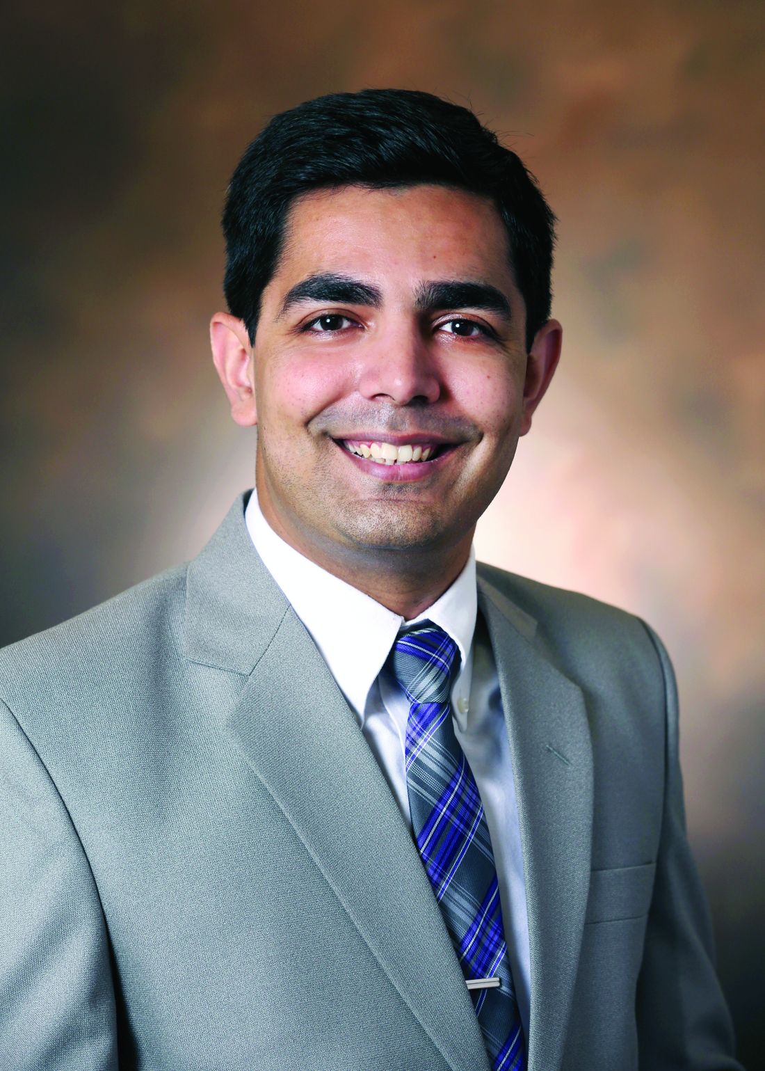
“But these databases are so prone to multiple biases that it’s really hard to tease out. You’d really have to do prospective studies where you follow these patients and then control for variables to know the true risk,” Dr. Patel said in an interview.
In many cases, for example, patients may start on H2 blockers after reporting symptoms that sound like indigestion, Patel said.
“And then the next week, they actually get an upper endoscopy and they get diagnosed with gastric cancer,” Patel said. “In the database, it may seem to be that ranitidine was started and the patient was diagnosed with gastric cancer. So now the gastric cancer gets associated with ranitidine.”
Physicians have had many options to use instead of ranitidine since the FDA last year announced its concerns about the drug, Dr. Patel said.
“Patients can easily switch over to a similar histamine blocker or you can use one of the proton pump inhibitors that haven’t been found to have this impurity in them,” he said. “Both of those are good options for patients.”
Further research
Manufacturers have been withdrawing their ranitidine products from the market since the reports of contamination surfaced last year. Several drugmakers, including Pfizer and Perrigo, have reported facing lawsuits connected with claims of cancer or increased cancer risk from ranitidine. In February 2020, a federal judicial panel opted to use a procedure, known as multidistrict litigation (MDL), to streamline the handling of these many cases. They were put before the U.S. District Court for the Southern District of Florida, labeled as In Re: Zantac (Ranitidine) NDMA Litigation.
A spokesman for FDA declined MDedge’s request for comment on Dr. Mohyuddin’s DDW presentation. He said the agency tends not to offer its views on work done by scientists outside of the FDA.
Valisure, an online pharmacy that runs quality checks on the medicines it dispenses, in September 2019 petitioned the FDA for a withdrawal of ranitidine due to concerns about NDMA. In its petition, Valisure reported finding notable levels of NDMA in ranitidine tablets, but not detecting it in testing of famotidine and several similar drugs. In an emailed statement, David Light, founder and CEO of Valisure, said the structure of Dr. Mohyuddin’s research limited its ability to detect cancer correlations because of the large and generalized study population.
“When comparing millions of people on medications which are both over-the-counter and prescription, any epidemiological impact will very likely be eclipsed by the sheer variations of exposure and a wide variety of confounding factors,” Mr. Light wrote.
There are a “vast number of variables that aren’t controlled for in such a massive and broadly defined cohort,” he added.
More focused and controlled studies will be needed to best evaluate NDMA and ranitidine, according to Mr. Light. “We are also investigating this issue with researchers at Memorial Sloan Kettering Cancer Center and plan to publish results soon,” he said.
Dr. Mohyuddin and Dr. Patel did not disclose financial conflicts in connection with ranitidine.
SOURCE: Mohyuddin N et al. DDW 2020. Abstract Tu1360.
People who had taken either of these drugs had a higher risk for cancer, but they also were more likely to have risk factors for this disease such as obesity or a history of smoking, an investigator said.
The findings from Nabeeha Mohyuddin, MD, an internal medicine resident at Allegheny Health Network in Pittsburgh, appear to be at odds with warnings from the U.S. Food and Drug Administration. The FDA in April called on manufacturers to pull all versions of ranitidine because of a problem with probable contamination by human carcinogen n-nitrosodimethylamine (NDMA). The FDA and the European Medicines Agency last year announced investigations of this NDMA contamination.
Dr. Mohyuddin and colleagues used IBM’s Explorys database, which includes data collected from EMRs from more than 40 U.S. health systems, to see if ranitidine use appeared connected to cancer diagnoses. Dr. Mohyuddin presented the findings in an abstract released as part of the annual Digestive Disease Week®, which was canceled because of COVID-19. The researchers identified records for 1.62 million users of ranitidine, 3.37 million users of famotidine, and 59.63 million people who did not use either H2 blocker.
The incidence of cancer was respectively 14.69%, 21.24%, and 5.38% for the ranitidine group, the famotidine group, and the group representing the general population without use of either H2 blocker. Among subjects without risk factors including smoking, obesity, alcohol use, family history, cirrhosis, and gastroesophageal reflux disease, ranitidine users did not have an increased risk of cancer, compared with famotidine users (odds ratio, 0.77; 95% confidence interval, 0.76-0.77; P < .001), Dr. Mohyuddin said in an email interview after her presentation.
“The results need to be interpreted with caution given that this is a retrospective study and it’s the first of its kind,” she said. “Further studies will be definitely needed on this to definitively answer the question, ‘does ranitidine have an increased risk of cancer?’ ”
People in the study who used H2 blockers tended to be older and were more likely to have other risk factors for cancers, according to the abstract:
- Of the ranitidine group, about 33.6% were older than 65, 74.4% were smokers, and 8.9% had a body mass index above 30 kg/m2.
- Of the famotidine group, about 38.3% were older than 65, 76.9% were smokers and 10.8% had a BMI above 30.
- Of the general population, about 23.9% were older than 65, 27.5% were smokers, and 1.69% were obese.
The Explorys database accounts for only 15%-19% of the entire U.S. population and that could be an explanation for why the percentage for obesity in the population seems spuriously low, Dr. Mohyuddin said in an email exchange.
Additionally, it pulls in patients through diagnosis codes, and if a different code for obesity was entered, those patients may not have been accounted for, she said.
Dhyanesh A. Patel, MD, assistant professor of medicine at Vanderbilt University, Nashville, Tenn., said it was a surprise to see the rate of cancer reported for famotidine users in the abstract presented at DDW.
“But these databases are so prone to multiple biases that it’s really hard to tease out. You’d really have to do prospective studies where you follow these patients and then control for variables to know the true risk,” Dr. Patel said in an interview.
In many cases, for example, patients may start on H2 blockers after reporting symptoms that sound like indigestion, Patel said.
“And then the next week, they actually get an upper endoscopy and they get diagnosed with gastric cancer,” Patel said. “In the database, it may seem to be that ranitidine was started and the patient was diagnosed with gastric cancer. So now the gastric cancer gets associated with ranitidine.”
Physicians have had many options to use instead of ranitidine since the FDA last year announced its concerns about the drug, Dr. Patel said.
“Patients can easily switch over to a similar histamine blocker or you can use one of the proton pump inhibitors that haven’t been found to have this impurity in them,” he said. “Both of those are good options for patients.”
Further research
Manufacturers have been withdrawing their ranitidine products from the market since the reports of contamination surfaced last year. Several drugmakers, including Pfizer and Perrigo, have reported facing lawsuits connected with claims of cancer or increased cancer risk from ranitidine. In February 2020, a federal judicial panel opted to use a procedure, known as multidistrict litigation (MDL), to streamline the handling of these many cases. They were put before the U.S. District Court for the Southern District of Florida, labeled as In Re: Zantac (Ranitidine) NDMA Litigation.
A spokesman for FDA declined MDedge’s request for comment on Dr. Mohyuddin’s DDW presentation. He said the agency tends not to offer its views on work done by scientists outside of the FDA.
Valisure, an online pharmacy that runs quality checks on the medicines it dispenses, in September 2019 petitioned the FDA for a withdrawal of ranitidine due to concerns about NDMA. In its petition, Valisure reported finding notable levels of NDMA in ranitidine tablets, but not detecting it in testing of famotidine and several similar drugs. In an emailed statement, David Light, founder and CEO of Valisure, said the structure of Dr. Mohyuddin’s research limited its ability to detect cancer correlations because of the large and generalized study population.
“When comparing millions of people on medications which are both over-the-counter and prescription, any epidemiological impact will very likely be eclipsed by the sheer variations of exposure and a wide variety of confounding factors,” Mr. Light wrote.
There are a “vast number of variables that aren’t controlled for in such a massive and broadly defined cohort,” he added.
More focused and controlled studies will be needed to best evaluate NDMA and ranitidine, according to Mr. Light. “We are also investigating this issue with researchers at Memorial Sloan Kettering Cancer Center and plan to publish results soon,” he said.
Dr. Mohyuddin and Dr. Patel did not disclose financial conflicts in connection with ranitidine.
SOURCE: Mohyuddin N et al. DDW 2020. Abstract Tu1360.
People who had taken either of these drugs had a higher risk for cancer, but they also were more likely to have risk factors for this disease such as obesity or a history of smoking, an investigator said.
The findings from Nabeeha Mohyuddin, MD, an internal medicine resident at Allegheny Health Network in Pittsburgh, appear to be at odds with warnings from the U.S. Food and Drug Administration. The FDA in April called on manufacturers to pull all versions of ranitidine because of a problem with probable contamination by human carcinogen n-nitrosodimethylamine (NDMA). The FDA and the European Medicines Agency last year announced investigations of this NDMA contamination.
Dr. Mohyuddin and colleagues used IBM’s Explorys database, which includes data collected from EMRs from more than 40 U.S. health systems, to see if ranitidine use appeared connected to cancer diagnoses. Dr. Mohyuddin presented the findings in an abstract released as part of the annual Digestive Disease Week®, which was canceled because of COVID-19. The researchers identified records for 1.62 million users of ranitidine, 3.37 million users of famotidine, and 59.63 million people who did not use either H2 blocker.
The incidence of cancer was respectively 14.69%, 21.24%, and 5.38% for the ranitidine group, the famotidine group, and the group representing the general population without use of either H2 blocker. Among subjects without risk factors including smoking, obesity, alcohol use, family history, cirrhosis, and gastroesophageal reflux disease, ranitidine users did not have an increased risk of cancer, compared with famotidine users (odds ratio, 0.77; 95% confidence interval, 0.76-0.77; P < .001), Dr. Mohyuddin said in an email interview after her presentation.
“The results need to be interpreted with caution given that this is a retrospective study and it’s the first of its kind,” she said. “Further studies will be definitely needed on this to definitively answer the question, ‘does ranitidine have an increased risk of cancer?’ ”
People in the study who used H2 blockers tended to be older and were more likely to have other risk factors for cancers, according to the abstract:
- Of the ranitidine group, about 33.6% were older than 65, 74.4% were smokers, and 8.9% had a body mass index above 30 kg/m2.
- Of the famotidine group, about 38.3% were older than 65, 76.9% were smokers and 10.8% had a BMI above 30.
- Of the general population, about 23.9% were older than 65, 27.5% were smokers, and 1.69% were obese.
The Explorys database accounts for only 15%-19% of the entire U.S. population and that could be an explanation for why the percentage for obesity in the population seems spuriously low, Dr. Mohyuddin said in an email exchange.
Additionally, it pulls in patients through diagnosis codes, and if a different code for obesity was entered, those patients may not have been accounted for, she said.
Dhyanesh A. Patel, MD, assistant professor of medicine at Vanderbilt University, Nashville, Tenn., said it was a surprise to see the rate of cancer reported for famotidine users in the abstract presented at DDW.
“But these databases are so prone to multiple biases that it’s really hard to tease out. You’d really have to do prospective studies where you follow these patients and then control for variables to know the true risk,” Dr. Patel said in an interview.
In many cases, for example, patients may start on H2 blockers after reporting symptoms that sound like indigestion, Patel said.
“And then the next week, they actually get an upper endoscopy and they get diagnosed with gastric cancer,” Patel said. “In the database, it may seem to be that ranitidine was started and the patient was diagnosed with gastric cancer. So now the gastric cancer gets associated with ranitidine.”
Physicians have had many options to use instead of ranitidine since the FDA last year announced its concerns about the drug, Dr. Patel said.
“Patients can easily switch over to a similar histamine blocker or you can use one of the proton pump inhibitors that haven’t been found to have this impurity in them,” he said. “Both of those are good options for patients.”
Further research
Manufacturers have been withdrawing their ranitidine products from the market since the reports of contamination surfaced last year. Several drugmakers, including Pfizer and Perrigo, have reported facing lawsuits connected with claims of cancer or increased cancer risk from ranitidine. In February 2020, a federal judicial panel opted to use a procedure, known as multidistrict litigation (MDL), to streamline the handling of these many cases. They were put before the U.S. District Court for the Southern District of Florida, labeled as In Re: Zantac (Ranitidine) NDMA Litigation.
A spokesman for FDA declined MDedge’s request for comment on Dr. Mohyuddin’s DDW presentation. He said the agency tends not to offer its views on work done by scientists outside of the FDA.
Valisure, an online pharmacy that runs quality checks on the medicines it dispenses, in September 2019 petitioned the FDA for a withdrawal of ranitidine due to concerns about NDMA. In its petition, Valisure reported finding notable levels of NDMA in ranitidine tablets, but not detecting it in testing of famotidine and several similar drugs. In an emailed statement, David Light, founder and CEO of Valisure, said the structure of Dr. Mohyuddin’s research limited its ability to detect cancer correlations because of the large and generalized study population.
“When comparing millions of people on medications which are both over-the-counter and prescription, any epidemiological impact will very likely be eclipsed by the sheer variations of exposure and a wide variety of confounding factors,” Mr. Light wrote.
There are a “vast number of variables that aren’t controlled for in such a massive and broadly defined cohort,” he added.
More focused and controlled studies will be needed to best evaluate NDMA and ranitidine, according to Mr. Light. “We are also investigating this issue with researchers at Memorial Sloan Kettering Cancer Center and plan to publish results soon,” he said.
Dr. Mohyuddin and Dr. Patel did not disclose financial conflicts in connection with ranitidine.
SOURCE: Mohyuddin N et al. DDW 2020. Abstract Tu1360.
FROM DDW 2020
Sleeve gastrectomy, antiobesity drugs underutilized
Despite an increasing rate of obesity in the United States, sleeve gastrectomy and postoperative antiobesity pharmacotherapy remain significantly underutilized, according to investigators.
A retrospective study involving almost 3 million adults with obesity found that only 0.94% had undergone sleeve gastrectomy, with 5.6% of those receiving weight-loss drugs after discharge, reported lead author Raj Shah, MD, of University Hospitals Cleveland Medical Center, and colleagues.
“While obesity has increased exponentially in the past decade, the trends of bariatric procedures and postoperative pharmacotherapy in this timeline is not well established,” the investigators wrote in an abstract released as part of the annual Digestive Disease Week, which was canceled because of COVID-19.

According to coauthor Abbinaya Elangovan, MD, of MetroHealth Medical Center, Cleveland, existing data suggest a practice gap.
“We know from published studies that antiobesity measures – both surgical and pharmacotherapeutic – do not match the rates of obesity,” Dr. Elangovan said. “We wanted to see how many of the morbidly obese [patients] who get bariatric surgery get started on antiobesity pharmacotherapy. We selected sleeve gastrectomy, as that is the most common bariatric procedure performed in the United States in recent times.”
The investigators began by retrospectively screening 2,717,000 individuals with a body mass index (in kg/m2) of at least 40 who entered the IBM Explorys database from 2010 to 2019. Out of this group, 25,540 individuals (0.94%) had undergone sleeve gastrectomy. Annual rates of the procedure increased from 0.06% in 2010 to 0.4% in 2019 (P < .0001).
Of the 25,540 patients who underwent sleeve gastrectomy, 1,440 (5.6%) were prescribed antiobesity medication after surgery, with about half (47%) of these prescriptions written within a year. The most common medication was phentermine (66%), followed by bupropion/naltrexone (16%) and phentermine/topiramate (14.4%).
Dr. Elangovan said that the rates of surgery and antiobesity pharmacotherapy found in the study were “sparse” compared with rates of obesity.
“[Future studies need] to find the barriers to antiobesity pharmacotherapy,” Dr. Elangovan said. “We know from some of the published studies that there are differences in provider perceptions, as well as patient populations who get the therapy.”
The present analysis showed that women, African Americans, and patients with commercial insurance were significantly more likely to receive postoperative weight-loss medications than other patient subgroups.
“I think insurance could be a potential concern,” Dr. Elangovan said. “This has been shown previously in the literature.” She also suggested that women may be accessing obesity-related health care more often than men.
Discussing steps to improve interventions for patients with obesity, Dr. Elangovan emphasized the amount of data supporting antiobesity pharmacotherapy.
“We know from studies published so far that combining pharmacotherapy with behavioral modifications has a greater percentage of success, compared to behavioral modifications by themselves,” Dr. Elangovan said.
According to Dr. Elangovan, primary care providers play a key role in connecting obese patients with the treatments they need, requiring familiarity with existing guidelines.
“It helps if practicing clinicians, especially primary care providers, are familiar with bariatric surgery criteria and institution policies,” Dr. Elangovan said. “It has been shown in some studies that limited experience in prescribing and concern for adverse reactions could affect the prescription of antiobesity pharmacotherapy. Targeted interventions such as educational programs may increase the appropriate usage of medications.”
Dr. Smith disclosed a relationship with US Endoscopy.
SOURCE: Shah R et al. DDW 2020, Abstract 791.
Despite an increasing rate of obesity in the United States, sleeve gastrectomy and postoperative antiobesity pharmacotherapy remain significantly underutilized, according to investigators.
A retrospective study involving almost 3 million adults with obesity found that only 0.94% had undergone sleeve gastrectomy, with 5.6% of those receiving weight-loss drugs after discharge, reported lead author Raj Shah, MD, of University Hospitals Cleveland Medical Center, and colleagues.
“While obesity has increased exponentially in the past decade, the trends of bariatric procedures and postoperative pharmacotherapy in this timeline is not well established,” the investigators wrote in an abstract released as part of the annual Digestive Disease Week, which was canceled because of COVID-19.
According to coauthor Abbinaya Elangovan, MD, of MetroHealth Medical Center, Cleveland, existing data suggest a practice gap.
“We know from published studies that antiobesity measures – both surgical and pharmacotherapeutic – do not match the rates of obesity,” Dr. Elangovan said. “We wanted to see how many of the morbidly obese [patients] who get bariatric surgery get started on antiobesity pharmacotherapy. We selected sleeve gastrectomy, as that is the most common bariatric procedure performed in the United States in recent times.”
The investigators began by retrospectively screening 2,717,000 individuals with a body mass index (in kg/m2) of at least 40 who entered the IBM Explorys database from 2010 to 2019. Out of this group, 25,540 individuals (0.94%) had undergone sleeve gastrectomy. Annual rates of the procedure increased from 0.06% in 2010 to 0.4% in 2019 (P < .0001).
Of the 25,540 patients who underwent sleeve gastrectomy, 1,440 (5.6%) were prescribed antiobesity medication after surgery, with about half (47%) of these prescriptions written within a year. The most common medication was phentermine (66%), followed by bupropion/naltrexone (16%) and phentermine/topiramate (14.4%).
Dr. Elangovan said that the rates of surgery and antiobesity pharmacotherapy found in the study were “sparse” compared with rates of obesity.
“[Future studies need] to find the barriers to antiobesity pharmacotherapy,” Dr. Elangovan said. “We know from some of the published studies that there are differences in provider perceptions, as well as patient populations who get the therapy.”
The present analysis showed that women, African Americans, and patients with commercial insurance were significantly more likely to receive postoperative weight-loss medications than other patient subgroups.
“I think insurance could be a potential concern,” Dr. Elangovan said. “This has been shown previously in the literature.” She also suggested that women may be accessing obesity-related health care more often than men.
Discussing steps to improve interventions for patients with obesity, Dr. Elangovan emphasized the amount of data supporting antiobesity pharmacotherapy.
“We know from studies published so far that combining pharmacotherapy with behavioral modifications has a greater percentage of success, compared to behavioral modifications by themselves,” Dr. Elangovan said.
According to Dr. Elangovan, primary care providers play a key role in connecting obese patients with the treatments they need, requiring familiarity with existing guidelines.
“It helps if practicing clinicians, especially primary care providers, are familiar with bariatric surgery criteria and institution policies,” Dr. Elangovan said. “It has been shown in some studies that limited experience in prescribing and concern for adverse reactions could affect the prescription of antiobesity pharmacotherapy. Targeted interventions such as educational programs may increase the appropriate usage of medications.”
Dr. Smith disclosed a relationship with US Endoscopy.
SOURCE: Shah R et al. DDW 2020, Abstract 791.
Despite an increasing rate of obesity in the United States, sleeve gastrectomy and postoperative antiobesity pharmacotherapy remain significantly underutilized, according to investigators.
A retrospective study involving almost 3 million adults with obesity found that only 0.94% had undergone sleeve gastrectomy, with 5.6% of those receiving weight-loss drugs after discharge, reported lead author Raj Shah, MD, of University Hospitals Cleveland Medical Center, and colleagues.
“While obesity has increased exponentially in the past decade, the trends of bariatric procedures and postoperative pharmacotherapy in this timeline is not well established,” the investigators wrote in an abstract released as part of the annual Digestive Disease Week, which was canceled because of COVID-19.
According to coauthor Abbinaya Elangovan, MD, of MetroHealth Medical Center, Cleveland, existing data suggest a practice gap.
“We know from published studies that antiobesity measures – both surgical and pharmacotherapeutic – do not match the rates of obesity,” Dr. Elangovan said. “We wanted to see how many of the morbidly obese [patients] who get bariatric surgery get started on antiobesity pharmacotherapy. We selected sleeve gastrectomy, as that is the most common bariatric procedure performed in the United States in recent times.”
The investigators began by retrospectively screening 2,717,000 individuals with a body mass index (in kg/m2) of at least 40 who entered the IBM Explorys database from 2010 to 2019. Out of this group, 25,540 individuals (0.94%) had undergone sleeve gastrectomy. Annual rates of the procedure increased from 0.06% in 2010 to 0.4% in 2019 (P < .0001).
Of the 25,540 patients who underwent sleeve gastrectomy, 1,440 (5.6%) were prescribed antiobesity medication after surgery, with about half (47%) of these prescriptions written within a year. The most common medication was phentermine (66%), followed by bupropion/naltrexone (16%) and phentermine/topiramate (14.4%).
Dr. Elangovan said that the rates of surgery and antiobesity pharmacotherapy found in the study were “sparse” compared with rates of obesity.
“[Future studies need] to find the barriers to antiobesity pharmacotherapy,” Dr. Elangovan said. “We know from some of the published studies that there are differences in provider perceptions, as well as patient populations who get the therapy.”
The present analysis showed that women, African Americans, and patients with commercial insurance were significantly more likely to receive postoperative weight-loss medications than other patient subgroups.
“I think insurance could be a potential concern,” Dr. Elangovan said. “This has been shown previously in the literature.” She also suggested that women may be accessing obesity-related health care more often than men.
Discussing steps to improve interventions for patients with obesity, Dr. Elangovan emphasized the amount of data supporting antiobesity pharmacotherapy.
“We know from studies published so far that combining pharmacotherapy with behavioral modifications has a greater percentage of success, compared to behavioral modifications by themselves,” Dr. Elangovan said.
According to Dr. Elangovan, primary care providers play a key role in connecting obese patients with the treatments they need, requiring familiarity with existing guidelines.
“It helps if practicing clinicians, especially primary care providers, are familiar with bariatric surgery criteria and institution policies,” Dr. Elangovan said. “It has been shown in some studies that limited experience in prescribing and concern for adverse reactions could affect the prescription of antiobesity pharmacotherapy. Targeted interventions such as educational programs may increase the appropriate usage of medications.”
Dr. Smith disclosed a relationship with US Endoscopy.
SOURCE: Shah R et al. DDW 2020, Abstract 791.
FROM DDW 2020
Bronchoscopy guideline for COVID-19 pandemic: Use sparingly
With little evidence available on the role of bronchoscopy during the COVID-19 pandemic, an expert panel has published a guideline recommending its spare use in COVID-19 patients and those with suspected COVID-19 infection.
The panel stated that in the context of the COVID-19 crisis, bronchoscopy and other aerosol-generating procedures put health care workers (HCWs) at particularly high risk of exposure and infection. They recommended deferring bronchoscopy in nonurgent cases, and advised practitioners to wear personal protective equipment when performing bronchoscopy, even on asymptomatic patients.
The guideline and expert panel report have been published online in the journal Chest. CHEST and the American Association for Bronchology and Interventional Pulmonology participated in selecting the 14 panelists. “The recommendation and suggestions outlined in this document were specifically created to address what were felt to be clinically common and urgent questions that frontline clinicians are likely to face,” wrote lead author and panel cochair Momen M. Wahidi, MD, MBA, of Duke University, Durham, N.C., and colleagues.
Only one of the six recommendations is based on graded evidence; the remainder are ungraded consensus-based statements. The guideline consists of the following recommendations for performing or using bronchoscopy:
- HCWs in the procedure or recovery rooms should wear either an N-95 respirator or powered air-purifying respirator (PAPR) when performing bronchoscopy on patients suspected or confirmed to have COVID-19. They should wear personal protective equipment (PPE) that includes a face shield, gown, and gloves, and they should discard N-95 respirators after performing bronchoscopy.
- A nasopharyngeal specimen in COVID-19 suspects should be obtained before performing bronchoscopy. If the patient has severe or progressive disease that requires intubation but an additional specimen is needed to confirm COVID-19 or another diagnosis that could change the treatment course, an option would be lower-respiratory specimen from the endotracheal aspirate or bronchoscopy with bronchoalveolar lavage.
- HCWs should wear an N-95 or PAPR when doing bronchoscopy on asymptomatic patients in an area with community spread of COVID-19 – again, with the PPE designated in the first recommendation.
- Test for COVID-19 before doing bronchoscopy on asymptomatic patients. Defer nonurgent bronchoscopy if the test is positive. If it’s negative, follow the recommendations regarding respirators and PPE when doing bronchoscopy.
- Perform timely bronchoscopy when indicated even in an area with known community spread of COVID-19. This is the only graded recommendation among the six (Grade 2C) and may be the most nuanced. Local teams should develop strategies for using bronchoscopy in their setting, taking into account local resources and availability of PPE, and they should send noninfected cancer patients from resource-depleted hospitals to other centers.
- Base the timing of bronchoscopy in patients recovering after COVID-19 on the indication for the procedure, disease severity, and time duration since symptoms resolved. The recommendation noted that the exact timing is still unknown, but that a wait of at least 30 days after symptoms recede is “reasonable.”
The expert panel added a noteworthy caveat to the recommendations. “We would like to stress that these protective strategies can be rendered completely ineffective if proper training on donning and doffing is not provided to HCW,” Dr. Wahidi and colleagues wrote. “Proper personnel instruction and practice for wearing PPE should receive as much attention by health facilities as the chosen strategy for protection.”
Dr. Wahidi and colleagues have no financial relationships to disclose.
SOURCE: Wahidi MM et al. CHEST. 2020 Apr 30. doi: 10.1016/j.chest.2020.04.036.
With little evidence available on the role of bronchoscopy during the COVID-19 pandemic, an expert panel has published a guideline recommending its spare use in COVID-19 patients and those with suspected COVID-19 infection.
The panel stated that in the context of the COVID-19 crisis, bronchoscopy and other aerosol-generating procedures put health care workers (HCWs) at particularly high risk of exposure and infection. They recommended deferring bronchoscopy in nonurgent cases, and advised practitioners to wear personal protective equipment when performing bronchoscopy, even on asymptomatic patients.
The guideline and expert panel report have been published online in the journal Chest. CHEST and the American Association for Bronchology and Interventional Pulmonology participated in selecting the 14 panelists. “The recommendation and suggestions outlined in this document were specifically created to address what were felt to be clinically common and urgent questions that frontline clinicians are likely to face,” wrote lead author and panel cochair Momen M. Wahidi, MD, MBA, of Duke University, Durham, N.C., and colleagues.
Only one of the six recommendations is based on graded evidence; the remainder are ungraded consensus-based statements. The guideline consists of the following recommendations for performing or using bronchoscopy:
- HCWs in the procedure or recovery rooms should wear either an N-95 respirator or powered air-purifying respirator (PAPR) when performing bronchoscopy on patients suspected or confirmed to have COVID-19. They should wear personal protective equipment (PPE) that includes a face shield, gown, and gloves, and they should discard N-95 respirators after performing bronchoscopy.
- A nasopharyngeal specimen in COVID-19 suspects should be obtained before performing bronchoscopy. If the patient has severe or progressive disease that requires intubation but an additional specimen is needed to confirm COVID-19 or another diagnosis that could change the treatment course, an option would be lower-respiratory specimen from the endotracheal aspirate or bronchoscopy with bronchoalveolar lavage.
- HCWs should wear an N-95 or PAPR when doing bronchoscopy on asymptomatic patients in an area with community spread of COVID-19 – again, with the PPE designated in the first recommendation.
- Test for COVID-19 before doing bronchoscopy on asymptomatic patients. Defer nonurgent bronchoscopy if the test is positive. If it’s negative, follow the recommendations regarding respirators and PPE when doing bronchoscopy.
- Perform timely bronchoscopy when indicated even in an area with known community spread of COVID-19. This is the only graded recommendation among the six (Grade 2C) and may be the most nuanced. Local teams should develop strategies for using bronchoscopy in their setting, taking into account local resources and availability of PPE, and they should send noninfected cancer patients from resource-depleted hospitals to other centers.
- Base the timing of bronchoscopy in patients recovering after COVID-19 on the indication for the procedure, disease severity, and time duration since symptoms resolved. The recommendation noted that the exact timing is still unknown, but that a wait of at least 30 days after symptoms recede is “reasonable.”
The expert panel added a noteworthy caveat to the recommendations. “We would like to stress that these protective strategies can be rendered completely ineffective if proper training on donning and doffing is not provided to HCW,” Dr. Wahidi and colleagues wrote. “Proper personnel instruction and practice for wearing PPE should receive as much attention by health facilities as the chosen strategy for protection.”
Dr. Wahidi and colleagues have no financial relationships to disclose.
SOURCE: Wahidi MM et al. CHEST. 2020 Apr 30. doi: 10.1016/j.chest.2020.04.036.
With little evidence available on the role of bronchoscopy during the COVID-19 pandemic, an expert panel has published a guideline recommending its spare use in COVID-19 patients and those with suspected COVID-19 infection.
The panel stated that in the context of the COVID-19 crisis, bronchoscopy and other aerosol-generating procedures put health care workers (HCWs) at particularly high risk of exposure and infection. They recommended deferring bronchoscopy in nonurgent cases, and advised practitioners to wear personal protective equipment when performing bronchoscopy, even on asymptomatic patients.
The guideline and expert panel report have been published online in the journal Chest. CHEST and the American Association for Bronchology and Interventional Pulmonology participated in selecting the 14 panelists. “The recommendation and suggestions outlined in this document were specifically created to address what were felt to be clinically common and urgent questions that frontline clinicians are likely to face,” wrote lead author and panel cochair Momen M. Wahidi, MD, MBA, of Duke University, Durham, N.C., and colleagues.
Only one of the six recommendations is based on graded evidence; the remainder are ungraded consensus-based statements. The guideline consists of the following recommendations for performing or using bronchoscopy:
- HCWs in the procedure or recovery rooms should wear either an N-95 respirator or powered air-purifying respirator (PAPR) when performing bronchoscopy on patients suspected or confirmed to have COVID-19. They should wear personal protective equipment (PPE) that includes a face shield, gown, and gloves, and they should discard N-95 respirators after performing bronchoscopy.
- A nasopharyngeal specimen in COVID-19 suspects should be obtained before performing bronchoscopy. If the patient has severe or progressive disease that requires intubation but an additional specimen is needed to confirm COVID-19 or another diagnosis that could change the treatment course, an option would be lower-respiratory specimen from the endotracheal aspirate or bronchoscopy with bronchoalveolar lavage.
- HCWs should wear an N-95 or PAPR when doing bronchoscopy on asymptomatic patients in an area with community spread of COVID-19 – again, with the PPE designated in the first recommendation.
- Test for COVID-19 before doing bronchoscopy on asymptomatic patients. Defer nonurgent bronchoscopy if the test is positive. If it’s negative, follow the recommendations regarding respirators and PPE when doing bronchoscopy.
- Perform timely bronchoscopy when indicated even in an area with known community spread of COVID-19. This is the only graded recommendation among the six (Grade 2C) and may be the most nuanced. Local teams should develop strategies for using bronchoscopy in their setting, taking into account local resources and availability of PPE, and they should send noninfected cancer patients from resource-depleted hospitals to other centers.
- Base the timing of bronchoscopy in patients recovering after COVID-19 on the indication for the procedure, disease severity, and time duration since symptoms resolved. The recommendation noted that the exact timing is still unknown, but that a wait of at least 30 days after symptoms recede is “reasonable.”
The expert panel added a noteworthy caveat to the recommendations. “We would like to stress that these protective strategies can be rendered completely ineffective if proper training on donning and doffing is not provided to HCW,” Dr. Wahidi and colleagues wrote. “Proper personnel instruction and practice for wearing PPE should receive as much attention by health facilities as the chosen strategy for protection.”
Dr. Wahidi and colleagues have no financial relationships to disclose.
SOURCE: Wahidi MM et al. CHEST. 2020 Apr 30. doi: 10.1016/j.chest.2020.04.036.
FROM CHEST