User login
Doug Brunk is a San Diego-based award-winning reporter who began covering health care in 1991. Before joining the company, he wrote for the health sciences division of Columbia University and was an associate editor at Contemporary Long Term Care magazine when it won a Jesse H. Neal Award. His work has been syndicated by the Los Angeles Times and he is the author of two books related to the University of Kentucky Wildcats men's basketball program. Doug has a master’s degree in magazine journalism from the S.I. Newhouse School of Public Communications at Syracuse University. Follow him on Twitter @dougbrunk.
Integrative care is the future of psychiatric care
KAUAI, HAWAII – The future of psychiatry in the era of health care reform will involve more team-based integrative care than ever before, according to Dr. James H. Scully Jr.
That means a shift away from the fee-for-service, volume-based model of care to which psychiatrists are accustomed. "We’re going to have to change the way we do business in order to survive," said Dr. Scully, CEO and medical director of the American Psychiatric Association. "We have to change our availability. We can’t say ‘I’ll see the patient in a couple of months’ like we do now sometimes. We’ll have to say ‘I’ll be there this afternoon’ and structure our clinical work around that, not only to do good liaison work but to be able to see the patient."

Volume-based fee-for-service health care "is a great risk," he said at the annual meeting of the American College of Psychiatrists. "That’s not sustainable [under] the Affordable Care Act. The insurance companies don’t want to do it anymore and certainly the companies who buy health insurance for their employees don’t want to spend money in the way they’ve been spending it."
The Center for Medicare and Medicaid Innovation (CMMI) – part of the Centers for Medicare and Medicaid Services – is funding numerous pilot programs aimed at fostering integrated care in primary and specialty care.
Dr. Scully offered examples of projects that are well developed, in his opinion: North Carolina Center of Excellence in Integrated Care; the Depression Improvement Across Minnesota, Offering a New Direction (DIAMOND) project; Integrated Behavioral Health Project in California; Improving Mood–Promoting Access to Collaborative Treatment (IMPACT) in Washington State; Mental Health Integration Program, also in Washington State; and TEAMcare, a multidisciplinary collaboration between the University of Washington and the Group Health Research Institute.
"These are all grant funded, so the question is, are they sustainable in the current way we pay for health care?" Dr. Scully asked. "Probably not. We have to change the way we pay for the services. Service delivery models and service payment models are two aspects of how this will get dealt with."
At the same time, mounting evidence from published studies is demonstrating that integrated health care can improve outcomes and lower cost. For example, in one randomized, controlled trial, diabetes patients who participated in the IMPACT program experienced fewer days of depression over a 2 year period compared with patients who received usual care (Diabetes Care 2006;29:265-70).
New payment models also are being developed under the ACA, Dr. Scully said. Many models include bundled payment for episodes of care "where you get a fee for caring for an episode or for a period of time."
Global capitation and partial capitation are being studied as well. "There are lots of different variations to change away from fee-for-service to a different kind of model to get paid for what we do," Dr. Scully said. "It’s an exciting time for us. We can’t walk away from [health care reform efforts]. We could, but I think it would be at our peril in the long-term. I think we have to participate in this and show some leadership. I’m optimistic."
Dr. Scully said that he had no relevant financial conflicts to disclose.
KAUAI, HAWAII – The future of psychiatry in the era of health care reform will involve more team-based integrative care than ever before, according to Dr. James H. Scully Jr.
That means a shift away from the fee-for-service, volume-based model of care to which psychiatrists are accustomed. "We’re going to have to change the way we do business in order to survive," said Dr. Scully, CEO and medical director of the American Psychiatric Association. "We have to change our availability. We can’t say ‘I’ll see the patient in a couple of months’ like we do now sometimes. We’ll have to say ‘I’ll be there this afternoon’ and structure our clinical work around that, not only to do good liaison work but to be able to see the patient."
Volume-based fee-for-service health care "is a great risk," he said at the annual meeting of the American College of Psychiatrists. "That’s not sustainable [under] the Affordable Care Act. The insurance companies don’t want to do it anymore and certainly the companies who buy health insurance for their employees don’t want to spend money in the way they’ve been spending it."
The Center for Medicare and Medicaid Innovation (CMMI) – part of the Centers for Medicare and Medicaid Services – is funding numerous pilot programs aimed at fostering integrated care in primary and specialty care.
Dr. Scully offered examples of projects that are well developed, in his opinion: North Carolina Center of Excellence in Integrated Care; the Depression Improvement Across Minnesota, Offering a New Direction (DIAMOND) project; Integrated Behavioral Health Project in California; Improving Mood–Promoting Access to Collaborative Treatment (IMPACT) in Washington State; Mental Health Integration Program, also in Washington State; and TEAMcare, a multidisciplinary collaboration between the University of Washington and the Group Health Research Institute.
"These are all grant funded, so the question is, are they sustainable in the current way we pay for health care?" Dr. Scully asked. "Probably not. We have to change the way we pay for the services. Service delivery models and service payment models are two aspects of how this will get dealt with."
At the same time, mounting evidence from published studies is demonstrating that integrated health care can improve outcomes and lower cost. For example, in one randomized, controlled trial, diabetes patients who participated in the IMPACT program experienced fewer days of depression over a 2 year period compared with patients who received usual care (Diabetes Care 2006;29:265-70).
New payment models also are being developed under the ACA, Dr. Scully said. Many models include bundled payment for episodes of care "where you get a fee for caring for an episode or for a period of time."
Global capitation and partial capitation are being studied as well. "There are lots of different variations to change away from fee-for-service to a different kind of model to get paid for what we do," Dr. Scully said. "It’s an exciting time for us. We can’t walk away from [health care reform efforts]. We could, but I think it would be at our peril in the long-term. I think we have to participate in this and show some leadership. I’m optimistic."
Dr. Scully said that he had no relevant financial conflicts to disclose.
AT THE AMERICAN COLLEGE OF PSYCHIATRISTS ANNUAL MEETING

DSM-5 expected to be more 'user-friendly'
When the DSM-5 is unveiled at the annual meeting of the American Psychiatric Association in May, Dr. David J. Kupfer hopes that clinicians will find a more user-friendly document compared with the DSM-IV.
At the annual meeting of the American College of Psychiatrists, Dr. Kupfer, chair of the DSM-5 task force and professor of psychiatry at the University of Pittsburgh, said the DSM-IV’s organizational structure "failed to reflect shared features or symptoms of related disorders and diagnostic groups, like psychotic disorders with bipolar disorders or internalizing and externalizing disorders. This led us to restructure DSM-5 in a way that better reflects these interrelationships, within and across diagnostic chapters."

The DSM-IV also was limited, he said, because it promoted a strict categorical approach to making diagnoses, the notion that "either you have it or you don’t," Dr. Kupfer said at the annual meeting of the American College of Psychiatrists. "This tends not to capture the variations of disorders that we see in real life. As a consequence, more ‘not otherwise specified’ designations were used than probably were necessary." Nor did the DSM-IV adequately address lifespan perspective, he said, including variations of symptom presentation across the developmental trajectory, or cultural perspectives. "That’s a deficiency, I think."
The 20-chapter DSM-5 – an 8-year effort that involved input from about 400 clinicians at 13 international conferences – "represents an opportunity to integrate cross-cutting symptomatic descriptions which better reflect the true presentation of disorders and may reduce reliance on ‘not otherwise specified’ diagnoses," Dr. Kupfer said. Its chapter structure, criteria revisions, and text outline "actively address age and development as part of diagnosis and classification," he said. "Culture is similarly discussed more explicitly to bring greater attention to cultural variations in symptom presentations."
Dr. Kupfer described the DSM-5 as a "living document," meaning that it will be more amenable to updates in psychiatry and neuroscience, and less susceptible to becoming outdated, compared with its predecessors. "We do not want to wait for 20 or 25 years for the next change to be made in the DSM," he emphasized. "We want to take advantage of advances that are likely to be made in certain areas of diagnostic nomenclature that can be put into the DSM and give us more objective criteria than we currently have for most of our disorders."
At the meeting, Dr. Kupfer and Dr. Darrel A. Regier, vice chair of the DSM-5 task force, highlighted select changes from DSM-IV that clinicians can expect to find in DSM-5. For example, autism spectrum disorder is now a single diagnosis. "The concern within the clinical and research field was that it was not possible to consistently break out autism, Asperger’s disorder, and pervasive developmental disorder not otherwise specified," said Dr. Regier, director of the American Psychiatric Institute for Research and Education and director of the division of research at the American Psychiatric Association. "There was a universal agreement that this needed to be seen as a spectrum of disorders that would be assessed on the basis of two domains: One was social communication and the other was restricted repetitive behaviors and interests.
"If one would rank people on the basis of their impairment in those two areas, that would be much more informative for guiding treatment and educational programs. That was a critical concern," he said.
The DSM-5 includes the addition of a specifier for all neurodevelopmental disorders associated with known medical or genetic conditions, or environmental factors. Specifiers are also included for specific learning disorders in reading, writing, and math.
In schizophrenia, special treatment of bizarre delusions and special hallucinations in criterion A (characteristic symptoms) has been eliminated. "With DSM-IV, you could get a diagnosis of schizophrenia if you just had bizarre delusions, but research shows that there is very poor reliability in separating bizarre and nonbizarre delusions," Dr. Regier said. Now, at least one psychotic symptom is required for a diagnosis of schizophrenia. "You have to have delusions, hallucinations, or disorganized speech in order to meet criteria," he said.
In a related development, catatonia exists as a specifier for neurodevelopmental, psychotic, mood, and other mental disorders, as well as for catatonia due to another medical condition.
In bipolar disorder, increased energy/activity as a criterion A symptom of hypomania/mania has been included. "Although those aspects have been included as symptoms previously, what is now recommended is that they become part of the criterion A, along with changes in mood," Dr. Kupfer said. The DSM-5 also includes a "with mixed features" specifier for manic, hypomanic, and major depressive episodes, which "better reflects what clinicians see and what they need to diagnose."
Depressive disorders are now organized in a dedicated chapter separate from bipolar and related disorders. In major depressive episode, the bereavement exclusion has been eliminated. "The basic message in the bereavement exclusion from DSM-IV was that we as clinicians could not diagnose major depression during the first 2 months following a bereavement," Dr. Kupfer said. "This would be independent of how the person might be suffering during that 2-month period. The other thing that seemed to be implied, which was very unfortunate, was that a number of people concluded that bereavement may only last 2 months, when in fact all of us know that bereavement often lasts a lot longer than 2 months." The DSM-5 includes a criteria note "that allows one to think about the presence of major depression while someone is also experiencing a significant loss."
Anxiety disorders are now organized in a dedicated chapter separate from other anxiety-related disorders. "With panic attacks" is a specifier for any mental disorder, and panic disorder and agoraphobia have become unlinked.
Another set of changes that were made in the DSM-5 related to either new disorders or in named disorders. For example, disruptive mood dysregulation disorder (DMDD) is a newcomer that addresses presentations of severe, nonepisodic irritability that has contributed to an upsurge of pediatric bipolar disorders. In DMDD, "symptoms overlap with oppositional defiance disorder but are considered more severe," Dr. Kupfer said. Meanwhile, premenstrual dysphoric disorder has been elevated from the appendix to the depressive disorders section of DSM-5, while binge eating disorder has been elevated from the appendix to the feeding and eating disorders section of the document.
Hoarding disorder is another newcomer to the DSM-5. "This is one of major public health significance because every department of public health in every county in the country has to deal with a hoarding issue, whether it’s animal-related or other forms of excessive acquisition," Dr. Regier said.
One change to posttraumatic stress disorder diagnoses includes removal of the A2 criteria, "which was that an individual not only has to be exposed an overwhelming stress but they have to react with horror or disgust," Dr. Regier said. "What was happening is that soldiers who are trained to immediately deal with horrendous experiences would say that their training ‘kicked in.’ They didn’t have the reaction – the A2 criteria – yet they subsequently would have clear criteria for PTSD. There was a need to eliminate that criteria to focus on four symptom clusters that filled out the syndrome." Now, the avoidance/numbing cluster has been divided into two distinct clusters: avoidance and persistent negative alterations in cognition and mood.
Dr. Regier predicted that the DSM-5 will make a significant contribution to assisting clinicians with diagnosing neurocognitive disorders. "An enormous amount of information has emerged in the area of neurocognitive disorders [in terms of] early differentiation of a probable Alzheimer’s disease versus a frontal temporal dementia diagnosis and differentiating dementia with Lewy bodies versus vascular dementia," he said. Going forward, he continued: "I think the biggest challenge is going to be making the distinction between mild dementia and normality. We’re working to develop a computer-assisted neurocognitive test that clinicians can use in their office to do some screening in this area. It would also be helpful for looking at cognitive impairment in schizophrenia. That’s in the future, but it’s an area we know we need to move forward with."
Neither Dr. Kupfer nor Dr. Regier had relevant financial conflicts to disclose.
When the DSM-5 is unveiled at the annual meeting of the American Psychiatric Association in May, Dr. David J. Kupfer hopes that clinicians will find a more user-friendly document compared with the DSM-IV.
At the annual meeting of the American College of Psychiatrists, Dr. Kupfer, chair of the DSM-5 task force and professor of psychiatry at the University of Pittsburgh, said the DSM-IV’s organizational structure "failed to reflect shared features or symptoms of related disorders and diagnostic groups, like psychotic disorders with bipolar disorders or internalizing and externalizing disorders. This led us to restructure DSM-5 in a way that better reflects these interrelationships, within and across diagnostic chapters."
The DSM-IV also was limited, he said, because it promoted a strict categorical approach to making diagnoses, the notion that "either you have it or you don’t," Dr. Kupfer said at the annual meeting of the American College of Psychiatrists. "This tends not to capture the variations of disorders that we see in real life. As a consequence, more ‘not otherwise specified’ designations were used than probably were necessary." Nor did the DSM-IV adequately address lifespan perspective, he said, including variations of symptom presentation across the developmental trajectory, or cultural perspectives. "That’s a deficiency, I think."
The 20-chapter DSM-5 – an 8-year effort that involved input from about 400 clinicians at 13 international conferences – "represents an opportunity to integrate cross-cutting symptomatic descriptions which better reflect the true presentation of disorders and may reduce reliance on ‘not otherwise specified’ diagnoses," Dr. Kupfer said. Its chapter structure, criteria revisions, and text outline "actively address age and development as part of diagnosis and classification," he said. "Culture is similarly discussed more explicitly to bring greater attention to cultural variations in symptom presentations."
Dr. Kupfer described the DSM-5 as a "living document," meaning that it will be more amenable to updates in psychiatry and neuroscience, and less susceptible to becoming outdated, compared with its predecessors. "We do not want to wait for 20 or 25 years for the next change to be made in the DSM," he emphasized. "We want to take advantage of advances that are likely to be made in certain areas of diagnostic nomenclature that can be put into the DSM and give us more objective criteria than we currently have for most of our disorders."
At the meeting, Dr. Kupfer and Dr. Darrel A. Regier, vice chair of the DSM-5 task force, highlighted select changes from DSM-IV that clinicians can expect to find in DSM-5. For example, autism spectrum disorder is now a single diagnosis. "The concern within the clinical and research field was that it was not possible to consistently break out autism, Asperger’s disorder, and pervasive developmental disorder not otherwise specified," said Dr. Regier, director of the American Psychiatric Institute for Research and Education and director of the division of research at the American Psychiatric Association. "There was a universal agreement that this needed to be seen as a spectrum of disorders that would be assessed on the basis of two domains: One was social communication and the other was restricted repetitive behaviors and interests.
"If one would rank people on the basis of their impairment in those two areas, that would be much more informative for guiding treatment and educational programs. That was a critical concern," he said.
The DSM-5 includes the addition of a specifier for all neurodevelopmental disorders associated with known medical or genetic conditions, or environmental factors. Specifiers are also included for specific learning disorders in reading, writing, and math.
In schizophrenia, special treatment of bizarre delusions and special hallucinations in criterion A (characteristic symptoms) has been eliminated. "With DSM-IV, you could get a diagnosis of schizophrenia if you just had bizarre delusions, but research shows that there is very poor reliability in separating bizarre and nonbizarre delusions," Dr. Regier said. Now, at least one psychotic symptom is required for a diagnosis of schizophrenia. "You have to have delusions, hallucinations, or disorganized speech in order to meet criteria," he said.
In a related development, catatonia exists as a specifier for neurodevelopmental, psychotic, mood, and other mental disorders, as well as for catatonia due to another medical condition.
In bipolar disorder, increased energy/activity as a criterion A symptom of hypomania/mania has been included. "Although those aspects have been included as symptoms previously, what is now recommended is that they become part of the criterion A, along with changes in mood," Dr. Kupfer said. The DSM-5 also includes a "with mixed features" specifier for manic, hypomanic, and major depressive episodes, which "better reflects what clinicians see and what they need to diagnose."
Depressive disorders are now organized in a dedicated chapter separate from bipolar and related disorders. In major depressive episode, the bereavement exclusion has been eliminated. "The basic message in the bereavement exclusion from DSM-IV was that we as clinicians could not diagnose major depression during the first 2 months following a bereavement," Dr. Kupfer said. "This would be independent of how the person might be suffering during that 2-month period. The other thing that seemed to be implied, which was very unfortunate, was that a number of people concluded that bereavement may only last 2 months, when in fact all of us know that bereavement often lasts a lot longer than 2 months." The DSM-5 includes a criteria note "that allows one to think about the presence of major depression while someone is also experiencing a significant loss."
Anxiety disorders are now organized in a dedicated chapter separate from other anxiety-related disorders. "With panic attacks" is a specifier for any mental disorder, and panic disorder and agoraphobia have become unlinked.
Another set of changes that were made in the DSM-5 related to either new disorders or in named disorders. For example, disruptive mood dysregulation disorder (DMDD) is a newcomer that addresses presentations of severe, nonepisodic irritability that has contributed to an upsurge of pediatric bipolar disorders. In DMDD, "symptoms overlap with oppositional defiance disorder but are considered more severe," Dr. Kupfer said. Meanwhile, premenstrual dysphoric disorder has been elevated from the appendix to the depressive disorders section of DSM-5, while binge eating disorder has been elevated from the appendix to the feeding and eating disorders section of the document.
Hoarding disorder is another newcomer to the DSM-5. "This is one of major public health significance because every department of public health in every county in the country has to deal with a hoarding issue, whether it’s animal-related or other forms of excessive acquisition," Dr. Regier said.
One change to posttraumatic stress disorder diagnoses includes removal of the A2 criteria, "which was that an individual not only has to be exposed an overwhelming stress but they have to react with horror or disgust," Dr. Regier said. "What was happening is that soldiers who are trained to immediately deal with horrendous experiences would say that their training ‘kicked in.’ They didn’t have the reaction – the A2 criteria – yet they subsequently would have clear criteria for PTSD. There was a need to eliminate that criteria to focus on four symptom clusters that filled out the syndrome." Now, the avoidance/numbing cluster has been divided into two distinct clusters: avoidance and persistent negative alterations in cognition and mood.
Dr. Regier predicted that the DSM-5 will make a significant contribution to assisting clinicians with diagnosing neurocognitive disorders. "An enormous amount of information has emerged in the area of neurocognitive disorders [in terms of] early differentiation of a probable Alzheimer’s disease versus a frontal temporal dementia diagnosis and differentiating dementia with Lewy bodies versus vascular dementia," he said. Going forward, he continued: "I think the biggest challenge is going to be making the distinction between mild dementia and normality. We’re working to develop a computer-assisted neurocognitive test that clinicians can use in their office to do some screening in this area. It would also be helpful for looking at cognitive impairment in schizophrenia. That’s in the future, but it’s an area we know we need to move forward with."
Neither Dr. Kupfer nor Dr. Regier had relevant financial conflicts to disclose.
EXPERT ANALYSIS FROM THE AMERICAN COLLEGE OF PSYCHIATRISTS ANNUAL MEETING

Physician reputation management is a tricky business
LAS VEGAS – Enhancing your online reputation is a tricky business, an industry that Robert Baxter said attracts a lot of sharks.
"A lot of people in this field are not very good at what they do," Mr. Baxter said at the annual meeting of the American Academy of Cosmetic Surgery. "They’re going to take your money and promise things they can’t deliver. Be careful."

It can also be expensive, especially when it comes to repairing your reputation. That part of the business "is so expensive that I personally stay out of it," said Mr. Baxter, a Miami Beach–based consultant who is widely considered a leading expert in physician reputation management.
"If you have a big problem, like a situation with a state medical board or negative appearance on a news station, it could cost $5,000-$10,000 per month for a minimum of 6 months to push the negative items down," he said. "There are no tried and true methods for what you do each time, either. It’s a case by case situation. For a posting on a review site, it helps if you can figure out who that patient is and get an idea of the psychology of how much trouble that person could actually cause."
To have your reputation look good online, Mr. Baxter recommended standardizing, claiming, and optimizing pages about you on the Web, including your Google Places page, your pages on review sites, your own practice website, your YouTube page, and your LinkedIn account. That way, the online information about you appears cohesive, "and you have a better opportunity to rank and control more of your name space," he explained.
What’s more, you should go after the increasing number of mobile visitors because "targeting mobile users is one of the biggest goals for both Apple and Google," Mr. Baxter said.
Another way to enhance your reputation is to enlist the aid of patients who are willing to write a review of your practice on sites such as Yelp.com or RateMDs.com. It helps to have patients post positive reviews and comments on the sites they hold accounts with. "If someone is an active Yelp user, they should post on Yelp, while someone who has a Google Plus account should post on Google," he noted.
According to Mr. Baxter, Google is moving toward the concept of the "semantic web," whereby the search engine works more like the human brain so that it "knows" what you might be looking for. "It’s no longer just about links as it relates to reviews and rankings," commented Mr. Baxter. "It’s about what’s being said about you and what’s written online in general."
To get a sense of your current online reputation, he recommended typing your name and "reviews" into the subject line of the search engine. "That’s going to give you a good representation of what your name space looks like as it relates to your reputation," he said.
Mr. Baxter also advises clients to use marquee phrases. "So if, for instance, you’re a dermatologist in San Diego, you should search for San Diego dermatologist to see both how you are ranking and how your reputation looks alongside your listing," he said. "You should also pay close attention to how your competitors look, because if someone is doing really well with reviews, you want to emulate what they’re doing."
Mr. Baxter said he had no relevant financial conflicts to disclose.
LAS VEGAS – Enhancing your online reputation is a tricky business, an industry that Robert Baxter said attracts a lot of sharks.
"A lot of people in this field are not very good at what they do," Mr. Baxter said at the annual meeting of the American Academy of Cosmetic Surgery. "They’re going to take your money and promise things they can’t deliver. Be careful."
It can also be expensive, especially when it comes to repairing your reputation. That part of the business "is so expensive that I personally stay out of it," said Mr. Baxter, a Miami Beach–based consultant who is widely considered a leading expert in physician reputation management.
"If you have a big problem, like a situation with a state medical board or negative appearance on a news station, it could cost $5,000-$10,000 per month for a minimum of 6 months to push the negative items down," he said. "There are no tried and true methods for what you do each time, either. It’s a case by case situation. For a posting on a review site, it helps if you can figure out who that patient is and get an idea of the psychology of how much trouble that person could actually cause."
To have your reputation look good online, Mr. Baxter recommended standardizing, claiming, and optimizing pages about you on the Web, including your Google Places page, your pages on review sites, your own practice website, your YouTube page, and your LinkedIn account. That way, the online information about you appears cohesive, "and you have a better opportunity to rank and control more of your name space," he explained.
What’s more, you should go after the increasing number of mobile visitors because "targeting mobile users is one of the biggest goals for both Apple and Google," Mr. Baxter said.
Another way to enhance your reputation is to enlist the aid of patients who are willing to write a review of your practice on sites such as Yelp.com or RateMDs.com. It helps to have patients post positive reviews and comments on the sites they hold accounts with. "If someone is an active Yelp user, they should post on Yelp, while someone who has a Google Plus account should post on Google," he noted.
According to Mr. Baxter, Google is moving toward the concept of the "semantic web," whereby the search engine works more like the human brain so that it "knows" what you might be looking for. "It’s no longer just about links as it relates to reviews and rankings," commented Mr. Baxter. "It’s about what’s being said about you and what’s written online in general."
To get a sense of your current online reputation, he recommended typing your name and "reviews" into the subject line of the search engine. "That’s going to give you a good representation of what your name space looks like as it relates to your reputation," he said.
Mr. Baxter also advises clients to use marquee phrases. "So if, for instance, you’re a dermatologist in San Diego, you should search for San Diego dermatologist to see both how you are ranking and how your reputation looks alongside your listing," he said. "You should also pay close attention to how your competitors look, because if someone is doing really well with reviews, you want to emulate what they’re doing."
Mr. Baxter said he had no relevant financial conflicts to disclose.
AT THE ANNUAL MEETING OF THE AMERICAN ACADEMY OF COSMETIC SURGERY

Lasers expand options for vascular lesion treatment
LAS VEGAS – The 595-nm pulsed dye laser, which allows for the application of 8 micropulses instead of a single pulse is one go-to device for treating vascular lesions, according to Dr. Melanie Palm.
"This allows me to use higher fluences without some of that eggplant purple discoloration or purpura that I would get if I used higher fluences in earlier generations of this laser," Dr. Palm said at the annual meeting of the American Academy of Cosmetic Surgery.

For example, when treating nasal telangiectasias, Dr. Palm said she sets the parameters to a fluence of 13-15 J/cm2, a pulse width of 40 milliseconds, and a spot size of 7 mm. "Using this new platform, I don’t get any of the purpura that you would expect with the more traditional 585-nm pulsed dye laser," said Dr. Palm, a dermatologist in Solana Beach, Calif.
Dr. Palm said she also has used the 595-nm pulsed dye laser (PDL) to treat rosacea, cherry angiomas, venous lakes, vascular malformations, postinflammatory erythema, striae distensae, scars, and purpura. "I will often combine treatments," she continued. For scars, she may combine 5-fluorouracil and intralesional Kenalog (triamcinolone), and immediately treat with the 595-nm PDL set to a fluence of 8 J/cm2, a pulse width of 10 milliseconds, and a spot size of 7 mm. For recalcitrant warts, she will often try intralesional bleomycin combined with the 595-nm PDL set to a fluence of 1-15 J/cm2, a pulse width of 1.5 milliseconds, and a spot size of 7 mm. "If the 595-nm PDL is the only laser in your office, you can use it to treat solar lentigines and other pigmentary disorders with some success," Dr. Palm said. "I also use it a lot for posttreatment bruising."
Intense pulsed light (IPL) is another technology Dr. Palm said she uses to treat vascular lesions. When discussing this technology with her patients, "I set the expectation that this is going to involve multiple treatments," Dr. Palm said. "I’ll often show them right after treatment that the vessels have gone into vasospasm. They have disappeared, but they will come back, and it will be several weeks before they see improvement."
Dr. Palm said she typically uses lidocaine cream as a numbing agent to improve patient comfort prior to IPL procedures. "But if patients want a stronger numbing agent, I mix lidocaine with tetracaine, which has a tendency to cause flushing," she said. "You can also use a hair dryer to aggravate erythema on the face prior to treatment."
Dr. Palm said she often uses the 515-nm filter with IPL energy applied in triple pulses to treat facial erythema. For facial telangiectasias, she typically uses the 560-nm filter with IPL energy applied in double pulses. "For stubborn spots, I switch to a smaller treatment hand piece, which creates higher fluence," she said.
Dr. Palm said she advises clinicians to be aggressive in treating postoperative scars. "If I see some redness, I’ll often treat as early as 1 month after treatment, using either a PDL or an IPL," she said. If she uses a PDL, she sets it to a fluence of 7-10 J/cm2, a pulse width of 10 milliseconds, and a spot size of 7 mm. If she uses an IPL, she employs a 560-nm filter, and sets the device to a fluence of 16-18 J/cm2 and a pulse width of 4 milliseconds.
To treat postprocedural bruising, Dr. Palm said she may use a PDL set to a fluence of 6 J/cm2, a pulse width of 6 milliseconds, and a spot size of 10 mm. If she opts to treat the bruising with an IPL, she employs a 560-nm filter and sets the device to a fluence of 13-15 J/cm2 and a pulse width of 4 milliseconds, and applies it in a double-pulse fashion. "You want to titrate the fluence inversely to the degree of bruising," Dr. Palm advised. "If you have an intense bruise, you want to decrease the fluence. If it’s a light bruise, you want to use higher fluences," she said. "I typically use a single pulse. You want to avoid pulse stacking because you can make the bruising worse. I don’t just treat where the bruise is. I treat within a centimeter around the bruised area as well."
Dr. Palm also discussed her experience using the Q-switched Nd:YAG double-frequency 532-nm laser as "a peel" to treat facial redness. "It’s usually a single-pass treatment that uses a double-frequency 1,064 Nd:YAG platform," she said. "I typically use an 8-mm hand piece set to a fluence of 3.5-5 J/cm2. Results are usually apparent within one to two treatments," she noted.
Dr. Palm disclosed that she is a speaker for Valeant, Medicis, and Lumenis. She is also a consultant for Lutronic.
LAS VEGAS – The 595-nm pulsed dye laser, which allows for the application of 8 micropulses instead of a single pulse is one go-to device for treating vascular lesions, according to Dr. Melanie Palm.
"This allows me to use higher fluences without some of that eggplant purple discoloration or purpura that I would get if I used higher fluences in earlier generations of this laser," Dr. Palm said at the annual meeting of the American Academy of Cosmetic Surgery.
For example, when treating nasal telangiectasias, Dr. Palm said she sets the parameters to a fluence of 13-15 J/cm2, a pulse width of 40 milliseconds, and a spot size of 7 mm. "Using this new platform, I don’t get any of the purpura that you would expect with the more traditional 585-nm pulsed dye laser," said Dr. Palm, a dermatologist in Solana Beach, Calif.
Dr. Palm said she also has used the 595-nm pulsed dye laser (PDL) to treat rosacea, cherry angiomas, venous lakes, vascular malformations, postinflammatory erythema, striae distensae, scars, and purpura. "I will often combine treatments," she continued. For scars, she may combine 5-fluorouracil and intralesional Kenalog (triamcinolone), and immediately treat with the 595-nm PDL set to a fluence of 8 J/cm2, a pulse width of 10 milliseconds, and a spot size of 7 mm. For recalcitrant warts, she will often try intralesional bleomycin combined with the 595-nm PDL set to a fluence of 1-15 J/cm2, a pulse width of 1.5 milliseconds, and a spot size of 7 mm. "If the 595-nm PDL is the only laser in your office, you can use it to treat solar lentigines and other pigmentary disorders with some success," Dr. Palm said. "I also use it a lot for posttreatment bruising."
Intense pulsed light (IPL) is another technology Dr. Palm said she uses to treat vascular lesions. When discussing this technology with her patients, "I set the expectation that this is going to involve multiple treatments," Dr. Palm said. "I’ll often show them right after treatment that the vessels have gone into vasospasm. They have disappeared, but they will come back, and it will be several weeks before they see improvement."
Dr. Palm said she typically uses lidocaine cream as a numbing agent to improve patient comfort prior to IPL procedures. "But if patients want a stronger numbing agent, I mix lidocaine with tetracaine, which has a tendency to cause flushing," she said. "You can also use a hair dryer to aggravate erythema on the face prior to treatment."
Dr. Palm said she often uses the 515-nm filter with IPL energy applied in triple pulses to treat facial erythema. For facial telangiectasias, she typically uses the 560-nm filter with IPL energy applied in double pulses. "For stubborn spots, I switch to a smaller treatment hand piece, which creates higher fluence," she said.
Dr. Palm said she advises clinicians to be aggressive in treating postoperative scars. "If I see some redness, I’ll often treat as early as 1 month after treatment, using either a PDL or an IPL," she said. If she uses a PDL, she sets it to a fluence of 7-10 J/cm2, a pulse width of 10 milliseconds, and a spot size of 7 mm. If she uses an IPL, she employs a 560-nm filter, and sets the device to a fluence of 16-18 J/cm2 and a pulse width of 4 milliseconds.
To treat postprocedural bruising, Dr. Palm said she may use a PDL set to a fluence of 6 J/cm2, a pulse width of 6 milliseconds, and a spot size of 10 mm. If she opts to treat the bruising with an IPL, she employs a 560-nm filter and sets the device to a fluence of 13-15 J/cm2 and a pulse width of 4 milliseconds, and applies it in a double-pulse fashion. "You want to titrate the fluence inversely to the degree of bruising," Dr. Palm advised. "If you have an intense bruise, you want to decrease the fluence. If it’s a light bruise, you want to use higher fluences," she said. "I typically use a single pulse. You want to avoid pulse stacking because you can make the bruising worse. I don’t just treat where the bruise is. I treat within a centimeter around the bruised area as well."
Dr. Palm also discussed her experience using the Q-switched Nd:YAG double-frequency 532-nm laser as "a peel" to treat facial redness. "It’s usually a single-pass treatment that uses a double-frequency 1,064 Nd:YAG platform," she said. "I typically use an 8-mm hand piece set to a fluence of 3.5-5 J/cm2. Results are usually apparent within one to two treatments," she noted.
Dr. Palm disclosed that she is a speaker for Valeant, Medicis, and Lumenis. She is also a consultant for Lutronic.
EXPERT ANALYSIS FROM THE ANNUAL MEETING OF THE AMERICAN ACADEMY OF COSMETIC SURGERY

Don't make these mistakes when marketing your practice
LAS VEGAS – Do your receptionists, patient coordinators, and other staff members represent you and your practice well? If not, it might be time for you to remind them that their role comes down to supporting you.
"Not knowing who’s on your team is a common marketing mistake," Catherine Maley said at the annual meeting of the American Academy of Cosmetic Surgery. "Nothing is more important in a cosmetic dermatology practice than having the right team in place. Your team is going to make or break your practice, because they are going to spend more time with your patients than you are."

Your staff must represent and promote you as the best choice, Ms. Maley said. "They have to embrace aesthetics," she emphasized. "I’ve been in offices where I’ve heard the receptionist say, ‘I would never get Botox.’ I have also heard a patient care coordinator say, ‘Just so you know, that’s not his best procedure. I would probably go somewhere else for that.’ "
Ms. Maley, a marketing strategist with Sausalito, Calif.–based Cosmetic Image Marketing, said that clinicians can find out which of their staff are true team players by staging a "refer a friend" contest in January or September, which are traditionally slow months for cosmetic dermatology practices. For the contest, employees have 30 days to distribute referral cards to family, friends, and other people in their social network. "At the end of 30 days, have a party and the employee who brings in the most referrals wins a prize – maybe an iPad or cash," Ms. Maley said. "Those who gave you the most referrals you know are on your team. Those who never participated aren’t."
Ms. Maley noted several other common mistakes clinicians make in marketing their practices, including:
• Ignoring your patients. Indifference "costs you, and it allows the gate to be open for your competitors," said Ms. Maley, who is also author of the book "Your Aesthetic Practice: What Your Patients Are Saying" (Sausalito: Cosmetic Imaging Marketing, 2011). "You have a captive market of aging baby boomers, so you want to keep them," she said. "For example, let’s say a patient shows up for a simple peel procedure or to buy a product. If they like what they got, they’re likely to start working their way up to injectables, laser procedures, or skin-tightening procedures. Keep them coming with messages by direct mail, e-mail, and social media," she said.
Although the Internet is fast and easy, don’t put all your eggs in that basket, Ms. Maley added. "You are missing out on half the patients who aren’t reading their e-mail and who can’t get past a computer firewall at work." Direct mail, she continued, offers an opportunity for one-on-one communication with the patient, "which is golden." Face time also builds trust. "The more face time you have with patients, the more they feel like they know you," she said.
• Assuming your patients will refer. Ms. Maley estimated that almost everyone knows about 150 people in their general geographic area, including service providers, family, friends, colleagues, and neighbors. "What if each patient referred one person to you?" she asked. "That could double your patient database. It’s worth asking patients who know you, like you, and trust you to refer at least one person to you."
While asking for referrals may seem awkward for some, she recommended displaying a sign in your waiting room that reads: "We love you as a patient. We would love to have more patients just like you." Another positive gesture is to hand patients before and after photos on a card following their treatment sessions, along with a handwritten note from you that says, "Thank you for your trust."
Patient surveys also can help you gauge how you’re doing, but keep them short, such as, "What’s one thing we could have done to improve your experience today?"
• Taking a "one-size-fits-all" approach. Gone are the days when an advertisement in your local newspaper with a menu of services is considered sufficient. Instead, "create one message to a specific market using the one media channel they are most likely in," Ms. Maley said. "For example, a woman in her 60s who is considering a facelift is more likely to respond to a one-on-one phone call or direct mail. Her daughter who is considering blepharoplasty is likely to research the procedure on the Internet."
• Having no marketing plan. Ms. Maley recommended carving out dedicated time with staff and perhaps a marketing consultant to devise a strategy for attracting high-value patients. "First, you have to attract them," she said. "Then, you have to qualify them to make sure they have the financial and emotional wherewithal to want what you offer. Then, you have to convert them to procedures, retain them for a lifetime, obtain testimonials and reviews, and encourage referrals. If you can set up a system that works just like protocols for surgery, it becomes an automatic system," she noted.
Ms. Maley said she had no relevant financial disclosures.
LAS VEGAS – Do your receptionists, patient coordinators, and other staff members represent you and your practice well? If not, it might be time for you to remind them that their role comes down to supporting you.
"Not knowing who’s on your team is a common marketing mistake," Catherine Maley said at the annual meeting of the American Academy of Cosmetic Surgery. "Nothing is more important in a cosmetic dermatology practice than having the right team in place. Your team is going to make or break your practice, because they are going to spend more time with your patients than you are."
Your staff must represent and promote you as the best choice, Ms. Maley said. "They have to embrace aesthetics," she emphasized. "I’ve been in offices where I’ve heard the receptionist say, ‘I would never get Botox.’ I have also heard a patient care coordinator say, ‘Just so you know, that’s not his best procedure. I would probably go somewhere else for that.’ "
Ms. Maley, a marketing strategist with Sausalito, Calif.–based Cosmetic Image Marketing, said that clinicians can find out which of their staff are true team players by staging a "refer a friend" contest in January or September, which are traditionally slow months for cosmetic dermatology practices. For the contest, employees have 30 days to distribute referral cards to family, friends, and other people in their social network. "At the end of 30 days, have a party and the employee who brings in the most referrals wins a prize – maybe an iPad or cash," Ms. Maley said. "Those who gave you the most referrals you know are on your team. Those who never participated aren’t."
Ms. Maley noted several other common mistakes clinicians make in marketing their practices, including:
• Ignoring your patients. Indifference "costs you, and it allows the gate to be open for your competitors," said Ms. Maley, who is also author of the book "Your Aesthetic Practice: What Your Patients Are Saying" (Sausalito: Cosmetic Imaging Marketing, 2011). "You have a captive market of aging baby boomers, so you want to keep them," she said. "For example, let’s say a patient shows up for a simple peel procedure or to buy a product. If they like what they got, they’re likely to start working their way up to injectables, laser procedures, or skin-tightening procedures. Keep them coming with messages by direct mail, e-mail, and social media," she said.
Although the Internet is fast and easy, don’t put all your eggs in that basket, Ms. Maley added. "You are missing out on half the patients who aren’t reading their e-mail and who can’t get past a computer firewall at work." Direct mail, she continued, offers an opportunity for one-on-one communication with the patient, "which is golden." Face time also builds trust. "The more face time you have with patients, the more they feel like they know you," she said.
• Assuming your patients will refer. Ms. Maley estimated that almost everyone knows about 150 people in their general geographic area, including service providers, family, friends, colleagues, and neighbors. "What if each patient referred one person to you?" she asked. "That could double your patient database. It’s worth asking patients who know you, like you, and trust you to refer at least one person to you."
While asking for referrals may seem awkward for some, she recommended displaying a sign in your waiting room that reads: "We love you as a patient. We would love to have more patients just like you." Another positive gesture is to hand patients before and after photos on a card following their treatment sessions, along with a handwritten note from you that says, "Thank you for your trust."
Patient surveys also can help you gauge how you’re doing, but keep them short, such as, "What’s one thing we could have done to improve your experience today?"
• Taking a "one-size-fits-all" approach. Gone are the days when an advertisement in your local newspaper with a menu of services is considered sufficient. Instead, "create one message to a specific market using the one media channel they are most likely in," Ms. Maley said. "For example, a woman in her 60s who is considering a facelift is more likely to respond to a one-on-one phone call or direct mail. Her daughter who is considering blepharoplasty is likely to research the procedure on the Internet."
• Having no marketing plan. Ms. Maley recommended carving out dedicated time with staff and perhaps a marketing consultant to devise a strategy for attracting high-value patients. "First, you have to attract them," she said. "Then, you have to qualify them to make sure they have the financial and emotional wherewithal to want what you offer. Then, you have to convert them to procedures, retain them for a lifetime, obtain testimonials and reviews, and encourage referrals. If you can set up a system that works just like protocols for surgery, it becomes an automatic system," she noted.
Ms. Maley said she had no relevant financial disclosures.
EXPERT ANALYSIS FROM THE ANNUAL MEETING OF THE AMERICAN ACADEMY OF COSMETIC SURGERY

No forehead paralysis seen after microdroplet technique
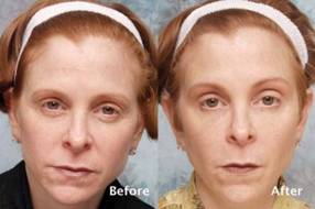
LAS VEGAS – A technique that involves injecting tiny, closely placed amounts of botulinum toxin A to balance the actions of the muscles around the eyebrows yielded natural-looking outcomes without forehead paralysis, based on data from a 5-year study.
"Doctors have mistakenly adopted maximal forehead paralysis as a desirable treatment endpoint," Dr. Kenneth D. Steinsapir said at the annual meeting of the American Academy of Cosmetic Surgery. "Forehead paralysis as a result of cosmetic botulinum toxin is feared by the public and lampooned by the media."
Over the years, onabotulinumtoxinA treatments have evolved to create maximal frontalis brow lifts while minimizing the risk of eyelid ptosis. As a result, "the forehead is smooth, but the central forehead is also ptotic," said Dr. Steinsapir of the department of ophthalmology at the University of California, Los Angeles. "There can be recruitment lines on the side of the forehead, which can make for an undesirable treatment effect."
In 2006, Dr. Steinsapir first described the microdroplet botulinum toxin forehead lift, a technique he developed for treating eyebrow depressors that leaves the brow elevator untreated. "I hypothesized that very small quantities of botulinum toxic can be injected and effectively trapped between the skin and the underlying orbicularis oculi muscle in the brow, and in the crow’s feet area as well," said Dr. Steinsapir, who also maintains a private cosmetic surgery practice in Beverly Hills, Calif. "This weakens the eyebrow depressors, allowing the frontalis muscle to lift unopposed, lifting the brows. Forehead movement is preserved and unwanted diffusion responsible for eyelid ptosis is prevented."
Between August 2006 and July 2011, Dr. Steinsapir performed 574 consecutive microdroplet botulinum toxin forehead lift treatments on 175 women and 53 men with a mean age of 45 years. A typical treatment involves 10 mcL of injectable saline containing 0.33 U of botulinum toxin A using the product formulation of Botox or Xeomin. About 100 microinjections are needed to complete the pattern, and all patients in the study received 33 units of Botox exclusively.
Dr. Steinsapir reported that there were no cases of treatment-induced upper eyelid or eyebrow ptosis or cases of diplopia, "which established that this treatment can be safely performed." Of the 574 patients, 49 returned for follow-up appointments between 10 and 45 days after treatment. Before and after images were used to assess the effect of treatment on the upper eyelid margin reflex distance, the tarsal platform show, and the brow position central to the cornea. Dr. Steinsapir used National Institutes of Health imaging software to perform quantitative image analysis and validated facial scales to assess the brow and forehead before and after treatment.
There was no significant change in the margin reflex distance after treatment in the 49 patients who returned for follow-up, Dr. Steinsapir said. "There was a slight trend to minimal brow elevation, and the tarsal platform show was essentially unchanged after treatment," he said. "It’s my clinical impression that the principal effect of the treatment is the softening of the brow pinch that commonly purses the brow and brings an unintentional negative affect to the face."
Dr. Steinsapir acknowledged that the procedure requires a learning curve "and a need to educate patients regarding the effect of treatment. This is more labor intensive than standard treatment methods." Dr. Steinsapir has developed a detailed training video that is available online on his website, and he said that he is working on a treatment atlas.
The microdroplet botulinum toxin forehead lift "presents the first alternative to standard periocular treatments that cause unwarranted forehead paralysis, brow flare, or muscle activation," Dr. Steinsapir said. "By controlling the depth, volume, and dose of agent, very controlled brow shaping and lifting can be performed to create aesthetic improvement with natural results, including preservation of forehead movement," he noted.
Dr. Steinsapir received a United States patent on the microdroplet method. He said that he hopes to license the technique to a drug company for the development of a Food and Drug Administration–approved indication, so the treatment can be directly marketed to consumers. He had no other relevant financial conflicts to disclose.
Dr. Kenneth D. Steinsapir, American Academy of Cosmetic Surgery, cosmetic botulinum toxin, onabotulinumtoxinA, maximal frontalis brow lifts, eyelid ptosis, ptotic, microdroplet botulinum toxin forehead lift, eyebrow depressors,
LAS VEGAS – A technique that involves injecting tiny, closely placed amounts of botulinum toxin A to balance the actions of the muscles around the eyebrows yielded natural-looking outcomes without forehead paralysis, based on data from a 5-year study.
"Doctors have mistakenly adopted maximal forehead paralysis as a desirable treatment endpoint," Dr. Kenneth D. Steinsapir said at the annual meeting of the American Academy of Cosmetic Surgery. "Forehead paralysis as a result of cosmetic botulinum toxin is feared by the public and lampooned by the media."
Over the years, onabotulinumtoxinA treatments have evolved to create maximal frontalis brow lifts while minimizing the risk of eyelid ptosis. As a result, "the forehead is smooth, but the central forehead is also ptotic," said Dr. Steinsapir of the department of ophthalmology at the University of California, Los Angeles. "There can be recruitment lines on the side of the forehead, which can make for an undesirable treatment effect."
In 2006, Dr. Steinsapir first described the microdroplet botulinum toxin forehead lift, a technique he developed for treating eyebrow depressors that leaves the brow elevator untreated. "I hypothesized that very small quantities of botulinum toxic can be injected and effectively trapped between the skin and the underlying orbicularis oculi muscle in the brow, and in the crow’s feet area as well," said Dr. Steinsapir, who also maintains a private cosmetic surgery practice in Beverly Hills, Calif. "This weakens the eyebrow depressors, allowing the frontalis muscle to lift unopposed, lifting the brows. Forehead movement is preserved and unwanted diffusion responsible for eyelid ptosis is prevented."
Between August 2006 and July 2011, Dr. Steinsapir performed 574 consecutive microdroplet botulinum toxin forehead lift treatments on 175 women and 53 men with a mean age of 45 years. A typical treatment involves 10 mcL of injectable saline containing 0.33 U of botulinum toxin A using the product formulation of Botox or Xeomin. About 100 microinjections are needed to complete the pattern, and all patients in the study received 33 units of Botox exclusively.
Dr. Steinsapir reported that there were no cases of treatment-induced upper eyelid or eyebrow ptosis or cases of diplopia, "which established that this treatment can be safely performed." Of the 574 patients, 49 returned for follow-up appointments between 10 and 45 days after treatment. Before and after images were used to assess the effect of treatment on the upper eyelid margin reflex distance, the tarsal platform show, and the brow position central to the cornea. Dr. Steinsapir used National Institutes of Health imaging software to perform quantitative image analysis and validated facial scales to assess the brow and forehead before and after treatment.
There was no significant change in the margin reflex distance after treatment in the 49 patients who returned for follow-up, Dr. Steinsapir said. "There was a slight trend to minimal brow elevation, and the tarsal platform show was essentially unchanged after treatment," he said. "It’s my clinical impression that the principal effect of the treatment is the softening of the brow pinch that commonly purses the brow and brings an unintentional negative affect to the face."
Dr. Steinsapir acknowledged that the procedure requires a learning curve "and a need to educate patients regarding the effect of treatment. This is more labor intensive than standard treatment methods." Dr. Steinsapir has developed a detailed training video that is available online on his website, and he said that he is working on a treatment atlas.
The microdroplet botulinum toxin forehead lift "presents the first alternative to standard periocular treatments that cause unwarranted forehead paralysis, brow flare, or muscle activation," Dr. Steinsapir said. "By controlling the depth, volume, and dose of agent, very controlled brow shaping and lifting can be performed to create aesthetic improvement with natural results, including preservation of forehead movement," he noted.
Dr. Steinsapir received a United States patent on the microdroplet method. He said that he hopes to license the technique to a drug company for the development of a Food and Drug Administration–approved indication, so the treatment can be directly marketed to consumers. He had no other relevant financial conflicts to disclose.
Dr. Kenneth D. Steinsapir, American Academy of Cosmetic Surgery, cosmetic botulinum toxin, onabotulinumtoxinA, maximal frontalis brow lifts, eyelid ptosis, ptotic, microdroplet botulinum toxin forehead lift, eyebrow depressors,
Dr. Kenneth D. Steinsapir, American Academy of Cosmetic Surgery, cosmetic botulinum toxin, onabotulinumtoxinA, maximal frontalis brow lifts, eyelid ptosis, ptotic, microdroplet botulinum toxin forehead lift, eyebrow depressors,
AT THE ANNUAL MEETING OF THE AMERICAN ACADEMY OF COSMETIC SURGERY
Major Finding: Patients who underwent the microdroplet botulinum toxin forehead lift experienced no cases of treatment-induced upper eyelid or eyebrow ptosis or cases of diplopia.
Data Source: A 5-year study of the technique performed on 574 consecutive patients with a mean age of 45 years.
Disclosures: Dr. Steinsapir received a United States patent on the microdroplet method. He said that he hopes to license the technique to a drug company for the development of an FDA-approved indication so the treatment can be directly marketed to consumers. He had no other relevant financial conflicts to disclose.

Surgeon, respect the levator muscle
LAS VEGAS – Knowing and respecting the anatomy of the levator muscle can help clinicians steer clear of complications from blepharoplasty and manage ptosis, according to Dr. Marc S. Cohen.
"It’s very helpful if you have a good understanding of how to find the levator muscle during eyelid surgery," said Dr. Cohen, an ophthalmic plastic surgeon at the Wills Eye Institute, Philadelphia. "In order to do this, you need to understand the relationship between the levator and the other eyelid structures."

The levator muscle elevates the eyelid and helps form the eyelid crease. It also creates the margin contour. As the levator muscle approaches the eyelid, it changes direction from vertically oriented to horizontally oriented. The muscle then advances inferiorly toward the eyelid margin, "and for the final centimeter or so, it becomes a fibrous aponeurosis, which attaches to the tarsus posteriorly," said Dr. Cohen, who also has a private cosmetic surgery practice. Behind the levator muscle are Müller’s muscle and the conjunctiva.
Whether a surgeon performs blepharoplasty with a CO2 laser, a blade, cautery, or radiofrequency, the first structure encountered posteriorly is the orbicularis oculi muscle, which closes the eyelid. "It’s highly vascular, and is the site where most of the bleeding occurs during blepharoplasty," Dr. Cohen said at the annual meeting of the American Academy of Cosmetic Surgery.
The next layer contains the orbital septum. "It’s important to understand that the septum does not travel all the way to the eyelid margin," he added. "The septum starts at the orbital rim and attaches to the levator muscle. This layer really has two structures: the septum and the levator. Behind the septum are the eyelid fat pads."
In a dissection above and behind in the eyelid, the septum and the fat precede the levator muscle. However, in the inferior eyelid, the levator is just deep to the orbicularis muscle. Beneath the fat, the levator muscle moves posteriorly into the orbit; this causes it to narrow.
"Lateral to the muscle at this point is the lacrimal gland, but medially is just orbital fat," Dr. Cohen said. Upon reaching the orbicularis muscle, the goal is to protect the levator muscle. "The levator muscle is protected by septum fat superiorly, whereas more inferiorly the levator fuses with the orbicularis, so this is a danger zone," Dr. Cohen said. "Laterally is the lacrimal gland and supramedially is the safest point, because there you have the fat, and nothing else to really worry about superficially. So what you do is press on the globe through the eyelid, have the fat prolapse forward, and dissect there."
Reattaching the levator muscle can be tricky in the context of levator resection ptosis surgery, said Dr. Cohen. "Where you make the attachment is going to affect the contour postoperatively," he said. "Grasp the tarsus and pull it upward to see if you have obtained a natural curve. If you grasp it at the wrong point, you’ll have a curve that’s not aesthetically pleasing," he cautioned.
"When you get the right point, that is where you are going to put the sutures to reattach the levator. A double-armed 6-0 suture is passed in a horizontal mattress fashion, partial thickness, through the tarsus. The suture is then passed in a posterior to anterior direction, which shortens the levator muscle."
Placement of the suture determines how much the muscle shortens. "The suture is then temporarily tied, and the patient is asked to open their eyes to assess the height and the contour," Dr. Cohen said. "If you need to adjust height vertically, you can move the suture vertically on the levator muscle. If there’s a problem with the contour, you change the fixation point to the tarsus. Then the suture is permanently tied and the skin is closed."
Dr. Cohen warned about the risk of complications from blepharoplasty in patients with active Graves’ disease, a common autoimmune condition that can cause hyperthyroidism and fibrosis of the extraocular tissues. The severe form of Graves’ disease can cause eyelid retraction, difficulty closing the eyes, double vision, and anterior displacement of the globes. "Many patients present with much more subtle findings," he noted. "For example, fibrosis of the levator with lid retraction is a common presentation in women aged 40-60 – the same demographic that tends to have blepharoplasty. It’s often subtle and underdiagnosed."
Patients with undiagnosed Graves’ disease prior to a blepharoplasty "can develop signs and symptoms which are indistinguishable from the complications of blepharoplasty," Dr. Cohen said. "You need to make the diagnosis before surgery and make sure the disease has stabilized before you do any surgery. That happens on average in about 18 months but is variable."
Dr. Cohen disclosed that he is a member of the advisory board for Allergan and that he is a speaker for Allergan and Medicis.
LAS VEGAS – Knowing and respecting the anatomy of the levator muscle can help clinicians steer clear of complications from blepharoplasty and manage ptosis, according to Dr. Marc S. Cohen.
"It’s very helpful if you have a good understanding of how to find the levator muscle during eyelid surgery," said Dr. Cohen, an ophthalmic plastic surgeon at the Wills Eye Institute, Philadelphia. "In order to do this, you need to understand the relationship between the levator and the other eyelid structures."
The levator muscle elevates the eyelid and helps form the eyelid crease. It also creates the margin contour. As the levator muscle approaches the eyelid, it changes direction from vertically oriented to horizontally oriented. The muscle then advances inferiorly toward the eyelid margin, "and for the final centimeter or so, it becomes a fibrous aponeurosis, which attaches to the tarsus posteriorly," said Dr. Cohen, who also has a private cosmetic surgery practice. Behind the levator muscle are Müller’s muscle and the conjunctiva.
Whether a surgeon performs blepharoplasty with a CO2 laser, a blade, cautery, or radiofrequency, the first structure encountered posteriorly is the orbicularis oculi muscle, which closes the eyelid. "It’s highly vascular, and is the site where most of the bleeding occurs during blepharoplasty," Dr. Cohen said at the annual meeting of the American Academy of Cosmetic Surgery.
The next layer contains the orbital septum. "It’s important to understand that the septum does not travel all the way to the eyelid margin," he added. "The septum starts at the orbital rim and attaches to the levator muscle. This layer really has two structures: the septum and the levator. Behind the septum are the eyelid fat pads."
In a dissection above and behind in the eyelid, the septum and the fat precede the levator muscle. However, in the inferior eyelid, the levator is just deep to the orbicularis muscle. Beneath the fat, the levator muscle moves posteriorly into the orbit; this causes it to narrow.
"Lateral to the muscle at this point is the lacrimal gland, but medially is just orbital fat," Dr. Cohen said. Upon reaching the orbicularis muscle, the goal is to protect the levator muscle. "The levator muscle is protected by septum fat superiorly, whereas more inferiorly the levator fuses with the orbicularis, so this is a danger zone," Dr. Cohen said. "Laterally is the lacrimal gland and supramedially is the safest point, because there you have the fat, and nothing else to really worry about superficially. So what you do is press on the globe through the eyelid, have the fat prolapse forward, and dissect there."
Reattaching the levator muscle can be tricky in the context of levator resection ptosis surgery, said Dr. Cohen. "Where you make the attachment is going to affect the contour postoperatively," he said. "Grasp the tarsus and pull it upward to see if you have obtained a natural curve. If you grasp it at the wrong point, you’ll have a curve that’s not aesthetically pleasing," he cautioned.
"When you get the right point, that is where you are going to put the sutures to reattach the levator. A double-armed 6-0 suture is passed in a horizontal mattress fashion, partial thickness, through the tarsus. The suture is then passed in a posterior to anterior direction, which shortens the levator muscle."
Placement of the suture determines how much the muscle shortens. "The suture is then temporarily tied, and the patient is asked to open their eyes to assess the height and the contour," Dr. Cohen said. "If you need to adjust height vertically, you can move the suture vertically on the levator muscle. If there’s a problem with the contour, you change the fixation point to the tarsus. Then the suture is permanently tied and the skin is closed."
Dr. Cohen warned about the risk of complications from blepharoplasty in patients with active Graves’ disease, a common autoimmune condition that can cause hyperthyroidism and fibrosis of the extraocular tissues. The severe form of Graves’ disease can cause eyelid retraction, difficulty closing the eyes, double vision, and anterior displacement of the globes. "Many patients present with much more subtle findings," he noted. "For example, fibrosis of the levator with lid retraction is a common presentation in women aged 40-60 – the same demographic that tends to have blepharoplasty. It’s often subtle and underdiagnosed."
Patients with undiagnosed Graves’ disease prior to a blepharoplasty "can develop signs and symptoms which are indistinguishable from the complications of blepharoplasty," Dr. Cohen said. "You need to make the diagnosis before surgery and make sure the disease has stabilized before you do any surgery. That happens on average in about 18 months but is variable."
Dr. Cohen disclosed that he is a member of the advisory board for Allergan and that he is a speaker for Allergan and Medicis.
EXPERT ANALYSIS FROM THE ANNUAL MEETING OF THE AMERICAN ACADEMY OF COSMETIC SURGERY

Robotic surgery called 'in the destiny of humanity'
The way Dr. Pier Cristoforo Giulianotti sees it, robots will play an increasingly prominent role in the future of surgery. He should know. Dr. Giulianotti, the Lloyd Nyhus Professor of Surgery and chief, division of minimally invasive, general, and robotic surgery at the University of Illinois at Chicago, pioneered robotic lung resection in 2002 and was the first surgeon to perform a robotic Whipple procedure in 2001.
"In my opinion there is no way back from robotic surgery," he said in an interview. "It’s in the destiny of humanity, for the simple reason: to perform more precisely and to overcome our limitations – not only manual limitations, the ability to control movements at a microscopic level, for example – but also diagnostic limitations. The computer and the artificial intelligence of the future will integrate our senses and our mechanical abilities, so we will be able to perform more precisely on selected targets."

Currently, most abdominal procedures performed at the University of Illinois Medical Center are done robotically, including the Whipple procedure, splenectomy, total gastrectomy, lung lobectomy, colorectal surgery, thyroidectomy, adrenalectomy, esophagectomy, major hepatectomies, and common bile duct procedures. The robot "has enormous benefits for the patients," Dr. Giulianotti said. "Open procedures involve a longer postoperative stay and more complications, and it takes more time to enter adjuvant oncological treatment after surgery, and there is less blood loss."
In Dr. Giulianotti’s published experience of 134 robotic-assisted laparoscopic pancreatic surgery procedures, morbidity was 26%, mortality was 2.2%, the conversion rate was 10.4%, and the fistula rate was 20.9%. The majority were grade A fistulas not requiring any treatment (Surg. Endosc. 2010; 24:1646-57). He said that robotic surgery "enables difficult technical maneuvers to be performed that facilitate the success of pancreatic minimally invasive surgery. The results in this series demonstrate feasibility and safety with clinical outcomes."
In a separate study, investigators who compared 32 open vs. 28 laparoscopic vs. 17 robotic distal pancreatectomies found that all three procedures resulted in a similar cost, while the robotic group had a shorter hospital stay, a higher rate of spleen-preserving cases, and an increased operative time, compared with patients in the other two groups (Surgery 2010;148:814-23).
Pelvic indications that are becoming the gold standard for robotic surgery include robotic prostatectomy and resection for rectal cancer. "When you have a very deep and complex surgical field like the pelvis, and you need to do a radical resection for cancer while at the same time sparing nerves that can be important for urinary function, the robot combines three-dimensional vision plus improved functionality of the instruments, allowing for a more precise dissection," Dr. Giulianotti said.

Dr. Leela M. Prasad, chief of the division of colon and rectal surgery at the University of Illinois at Chicago, described robotic-assisted surgery for rectal cancer as "far superior to anything we know right now," especially in overweight males. According to combined results from three published studies, circumferential margin positivity ranged from 0 to 7%, which, Dr. Prasad said, is significantly lower than the average number of positive margins reported following open and laparoscopic rectal surgery procedures (14% and 16%, respectively) (Ann. Surg. Oncol. 2007;14:3168-73; Surg. Oncol. 2009;16:1480-7; Dis. Colon Rectum 2009;52:1824-30).
At the present time, however, in his clinical experience there are no significant differences in outcomes between robotic-assisted and laparoscopically assisted open surgery for colon cancer. "We looked at our data, and it did not make any difference robotically or laparoscopically in terms of length of stay, lymph node harvest, or survival," he said. A recent study from Korean investigators supports those findings (Surg. Endosc. 2012 Dec. 13).
Dr. Giulianotti estimated that there are more than 1,600 robotic surgery systems in the United States. Some of them are devoted to urologic or gynecologic applications, "but in the majority of hospitals there is a growing tendency to use the robot as part of a multidisciplinary program," he said. "That means at the same institution different surgical teams on different days are using the robot for different indications. Of the 1,600 systems out there, I would estimate that about half are used for general surgery."
While hospital administrators have warmed up to the idea of initiating robotic surgery programs at their institutions in recent years, Dr. Giulianotti and Dr. Prasad both called for a culture change in terms of how surgeons and medical educators think about the robot’s place in clinical medicine. "There is a cultural resistance in accepting big changes in surgery," Dr. Giulianotti said. "Academic medical institutions can play a key role here. They need to start teaching robotics in a mandatory way by offering training opportunities in a lab. We also believe that simulation has a big future in this kind of training."
The economic investment required to launch a robotic surgery program is another challenge. "Some hospitals are only concerned about the cost," he said. "When you are doing the same procedure laparoscopically, the cost is probably about 30% less. In the future I hope we can better impact the overall economic aspect of surgical procedures. That means speeding up the postoperative and outpatient treatment of some pathologies. We are already doing Nissen fundoplication and gallbladder removal with robotic surgery, and patients are being discharged a few hours after the surgery."
Dr. Prasad said the field will be poised to further advance when makers of robotic surgery devices and instruments improve on existing technology. "I am using the same robot and instruments as I was using 3 years ago," he said. "I think we need new technologies – including a smaller robot to cut down the costs and speed up the operations. This will make it easier for patients."
To launch a successful robotic surgery program in this day and age, "you need a good team of nurses and surgeons who are dedicated to doing robotic surgery," Dr. Prasad concluded. "It is dangerous in the hands of a surgeon who is not qualified."
Dr. Giulianotti said that he had no relevant financial conflicts to disclose. Dr. Prasad disclosed that he has received honoraria from Intuitive Surgical, Ethicon Endo-Surgery, and Covidien.
I would like to congratulate Dr. Pier Cristoforo Giulianotti for his work to date demonstrating the effectiveness of robotic pancreatic surgery. Clearly the “robot,” as it is affectionately termed, is here to stay.
It has clear applications in all surgical subspecialties and is being embraced by surgeons, administrators, and patients as an important tool. It is a platform that will facilitate future developments that are certain to change the way we practice our craft.
Dr. John Sweeney |
Given the focus being placed on the value of health care deliveredin the United States, it is incumbent upon surgeons to continuously evaluate the quality and costs of each intervention that we offer to our patients. To many, this type of evaluative process might seem to come up short when it comes to robotic applications for many minimally invasive procedures. Quality in most studies has been equivalent, while the costs associated with this expensive technology can be very significant. Because this new piece of technology is the first step in a new direction, we must continue to place the time, effort and cost into refining, enhancing and improving it’s applications as the technology stands today. However unless we are able to accomplish this goal, I fear we fall into the trap of “using a Cadillac for a golf cart” which is something that the U.S. health care system cannot afford nor sustain.
Dr. John Sweeney, is an ACS Fellow and the W. Dean Warren Distinguished Chair in Surgery at Emory University, Atlanta.
The way Dr. Pier Cristoforo Giulianotti sees it, robots will play an increasingly prominent role in the future of surgery. He should know. Dr. Giulianotti, the Lloyd Nyhus Professor of Surgery and chief, division of minimally invasive, general, and robotic surgery at the University of Illinois at Chicago, pioneered robotic lung resection in 2002 and was the first surgeon to perform a robotic Whipple procedure in 2001.
"In my opinion there is no way back from robotic surgery," he said in an interview. "It’s in the destiny of humanity, for the simple reason: to perform more precisely and to overcome our limitations – not only manual limitations, the ability to control movements at a microscopic level, for example – but also diagnostic limitations. The computer and the artificial intelligence of the future will integrate our senses and our mechanical abilities, so we will be able to perform more precisely on selected targets."
Currently, most abdominal procedures performed at the University of Illinois Medical Center are done robotically, including the Whipple procedure, splenectomy, total gastrectomy, lung lobectomy, colorectal surgery, thyroidectomy, adrenalectomy, esophagectomy, major hepatectomies, and common bile duct procedures. The robot "has enormous benefits for the patients," Dr. Giulianotti said. "Open procedures involve a longer postoperative stay and more complications, and it takes more time to enter adjuvant oncological treatment after surgery, and there is less blood loss."
In Dr. Giulianotti’s published experience of 134 robotic-assisted laparoscopic pancreatic surgery procedures, morbidity was 26%, mortality was 2.2%, the conversion rate was 10.4%, and the fistula rate was 20.9%. The majority were grade A fistulas not requiring any treatment (Surg. Endosc. 2010; 24:1646-57). He said that robotic surgery "enables difficult technical maneuvers to be performed that facilitate the success of pancreatic minimally invasive surgery. The results in this series demonstrate feasibility and safety with clinical outcomes."
In a separate study, investigators who compared 32 open vs. 28 laparoscopic vs. 17 robotic distal pancreatectomies found that all three procedures resulted in a similar cost, while the robotic group had a shorter hospital stay, a higher rate of spleen-preserving cases, and an increased operative time, compared with patients in the other two groups (Surgery 2010;148:814-23).
Pelvic indications that are becoming the gold standard for robotic surgery include robotic prostatectomy and resection for rectal cancer. "When you have a very deep and complex surgical field like the pelvis, and you need to do a radical resection for cancer while at the same time sparing nerves that can be important for urinary function, the robot combines three-dimensional vision plus improved functionality of the instruments, allowing for a more precise dissection," Dr. Giulianotti said.
Dr. Leela M. Prasad, chief of the division of colon and rectal surgery at the University of Illinois at Chicago, described robotic-assisted surgery for rectal cancer as "far superior to anything we know right now," especially in overweight males. According to combined results from three published studies, circumferential margin positivity ranged from 0 to 7%, which, Dr. Prasad said, is significantly lower than the average number of positive margins reported following open and laparoscopic rectal surgery procedures (14% and 16%, respectively) (Ann. Surg. Oncol. 2007;14:3168-73; Surg. Oncol. 2009;16:1480-7; Dis. Colon Rectum 2009;52:1824-30).
At the present time, however, in his clinical experience there are no significant differences in outcomes between robotic-assisted and laparoscopically assisted open surgery for colon cancer. "We looked at our data, and it did not make any difference robotically or laparoscopically in terms of length of stay, lymph node harvest, or survival," he said. A recent study from Korean investigators supports those findings (Surg. Endosc. 2012 Dec. 13).
Dr. Giulianotti estimated that there are more than 1,600 robotic surgery systems in the United States. Some of them are devoted to urologic or gynecologic applications, "but in the majority of hospitals there is a growing tendency to use the robot as part of a multidisciplinary program," he said. "That means at the same institution different surgical teams on different days are using the robot for different indications. Of the 1,600 systems out there, I would estimate that about half are used for general surgery."
While hospital administrators have warmed up to the idea of initiating robotic surgery programs at their institutions in recent years, Dr. Giulianotti and Dr. Prasad both called for a culture change in terms of how surgeons and medical educators think about the robot’s place in clinical medicine. "There is a cultural resistance in accepting big changes in surgery," Dr. Giulianotti said. "Academic medical institutions can play a key role here. They need to start teaching robotics in a mandatory way by offering training opportunities in a lab. We also believe that simulation has a big future in this kind of training."
The economic investment required to launch a robotic surgery program is another challenge. "Some hospitals are only concerned about the cost," he said. "When you are doing the same procedure laparoscopically, the cost is probably about 30% less. In the future I hope we can better impact the overall economic aspect of surgical procedures. That means speeding up the postoperative and outpatient treatment of some pathologies. We are already doing Nissen fundoplication and gallbladder removal with robotic surgery, and patients are being discharged a few hours after the surgery."
Dr. Prasad said the field will be poised to further advance when makers of robotic surgery devices and instruments improve on existing technology. "I am using the same robot and instruments as I was using 3 years ago," he said. "I think we need new technologies – including a smaller robot to cut down the costs and speed up the operations. This will make it easier for patients."
To launch a successful robotic surgery program in this day and age, "you need a good team of nurses and surgeons who are dedicated to doing robotic surgery," Dr. Prasad concluded. "It is dangerous in the hands of a surgeon who is not qualified."
Dr. Giulianotti said that he had no relevant financial conflicts to disclose. Dr. Prasad disclosed that he has received honoraria from Intuitive Surgical, Ethicon Endo-Surgery, and Covidien.
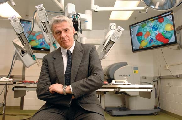
Your waiting room 'is an extension of you'
LAS VEGAS – You might not equate your waiting room with patient satisfaction, but marketing expert Risa Goldman Luksa says there may no greater reflection of your practice.
"Your waiting room is really an extension of you," Mrs. Luksa said at the annual meeting of the American Academy of Cosmetic Surgery. "Patients spend a lot of time in your waiting room, so make sure it conveys the impression you want to give. You want to keep it clean and professional. It’s a good idea for someone on your staff to walk through the waiting room every 30 minutes to make sure it is tidy. Pick up cups if someone left them, as well as any trash, newspapers, or magazines left behind."
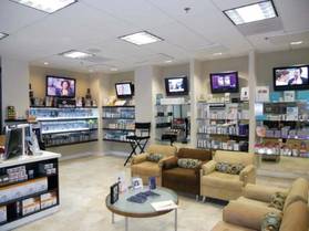
If you are running behind schedule, that person can remind patients that you haven’t forgotten about them. "It’s also a good idea to provide coffee, water, and other light refreshments," said Mrs. Luksa. "If you do that, make sure your front office staff asks patients if they can get them something to drink or eat. These are nice touches."
Mrs. Luksa, founder and president of Los Angeles-based Goldman Marketing Group, offered the following strategies to enhance the waiting room experience for patients:
• Ditch the magazines. "You’re not a nail salon," she said. "You’re selling high-end procedures, so I would get rid of all the magazines and display your own marketing materials instead. Your waiting room is your golden marketing opportunity, so don’t let it advertise for other people."
• Ditch the clocks. Just like the casino operators in Las Vegas prefer that you not keep track of time, "You don’t want people to be watching the clock and keeping track how late you might be running," Mrs. Luksa said.

• Create a peaceful atmosphere. Having a television show or radio station blaring, or allowing patients to talk on their cell phones while they wait can be distracting. "Move your practice telephones away from the front desk, especially if they’re loud and the phones ring off the hook," Mrs. Luksa advised. "When you do this, it allows people at the front desk to give their full attention to people in the waiting room. This is important. You want them to have that full attention. Equally, it allows whoever is answering the phones to give their full attention to people calling in."
• Offer amenities that showcase your practice. Branded water bottles are a nice touch, as are flash drives, containers of hand sanitizer, and tubes of Chap Stick. "These are all cost-effective solutions," Mrs. Luksa said. "Most of them you can order for $1 or $2 apiece." She also recommends investing in a custom entrance rug that sports your practice logo.
• Offer free Wi-Fi. Since most practices are likely to have a wireless Internet network, "why not let your patients know about that and give them a password? While they’re waiting, it’s really nice if they can log on and surf the Web on their iPhones."
• Use clean-looking intake and consent forms. "You would be surprised at how many offices I have walked into and they are using blotchy photocopies [of these forms]," Mrs. Luksa said. "Over the years, they get messy looking and they’re hard to read. Make your intake forms and your consent forms are clean looking and professional. Make sure they contain your branding and make the forms as short as possible."
• Ask patients to fill out a brief cosmetic questionnaire. This contains a checklist of services offered by your practice, along with a statement at the top of the page inviting the patient to please check any procedure below you are interested in learning about. "This is a great opportunity to cross market your services without a strong sales approach," said Mrs. Luksa. "Make sure to use these forms in your patient consultation and to record the results in your electronic health records for future targeted marketing efforts."
• Printed newsletters have a place. While some may view this means of communication as old-fashioned, a printed newsletter "is another great source of unique and relevant reading material for patients," Mrs. Luksa said. "You can repurpose the blogs on your website so you’re not re-creating something from scratch. It’s also a good way to advertise your promotions. Plus, you can easily spin it into an e-newsletter to e-mail to your patient database, which is great for patient retention."
• Publish hardback picture books that contain before and after pictures of the services you offer. Mrs. Luksa described this as "a classy way of educating patients about what you do and what the procedure entails" before their consultation with you. The same result can be achieved with an iPad book and/or a DVD reel, which eliminates printing costs.
• Display retail products. People won’t know what cosmetic products you sell unless they can see them. "Walk through any department store," Mrs. Luksa said. "Women love touching and sampling the products. They’ll want to do the same in your office. Create and display packages of products to up-sell."
• Ask for referrals. Word of mouth is crucial, especially in today’s economic environment. "You don’t want to come off as being needy, but don’t be shy to ask for referrals when the situation feels appropriate," Mrs. Luksa noted. "Maybe display a sign that says, ‘the highest compliment we can receive is the referral of your family and friends.’ The same goes for asking for reviews on Yelp and other review sites, which almost every doctor could use more of."
Mrs. Luksa said that she had no relevant financial conflicts to disclose.
LAS VEGAS – You might not equate your waiting room with patient satisfaction, but marketing expert Risa Goldman Luksa says there may no greater reflection of your practice.
"Your waiting room is really an extension of you," Mrs. Luksa said at the annual meeting of the American Academy of Cosmetic Surgery. "Patients spend a lot of time in your waiting room, so make sure it conveys the impression you want to give. You want to keep it clean and professional. It’s a good idea for someone on your staff to walk through the waiting room every 30 minutes to make sure it is tidy. Pick up cups if someone left them, as well as any trash, newspapers, or magazines left behind."
If you are running behind schedule, that person can remind patients that you haven’t forgotten about them. "It’s also a good idea to provide coffee, water, and other light refreshments," said Mrs. Luksa. "If you do that, make sure your front office staff asks patients if they can get them something to drink or eat. These are nice touches."
Mrs. Luksa, founder and president of Los Angeles-based Goldman Marketing Group, offered the following strategies to enhance the waiting room experience for patients:
• Ditch the magazines. "You’re not a nail salon," she said. "You’re selling high-end procedures, so I would get rid of all the magazines and display your own marketing materials instead. Your waiting room is your golden marketing opportunity, so don’t let it advertise for other people."
• Ditch the clocks. Just like the casino operators in Las Vegas prefer that you not keep track of time, "You don’t want people to be watching the clock and keeping track how late you might be running," Mrs. Luksa said.
• Create a peaceful atmosphere. Having a television show or radio station blaring, or allowing patients to talk on their cell phones while they wait can be distracting. "Move your practice telephones away from the front desk, especially if they’re loud and the phones ring off the hook," Mrs. Luksa advised. "When you do this, it allows people at the front desk to give their full attention to people in the waiting room. This is important. You want them to have that full attention. Equally, it allows whoever is answering the phones to give their full attention to people calling in."
• Offer amenities that showcase your practice. Branded water bottles are a nice touch, as are flash drives, containers of hand sanitizer, and tubes of Chap Stick. "These are all cost-effective solutions," Mrs. Luksa said. "Most of them you can order for $1 or $2 apiece." She also recommends investing in a custom entrance rug that sports your practice logo.
• Offer free Wi-Fi. Since most practices are likely to have a wireless Internet network, "why not let your patients know about that and give them a password? While they’re waiting, it’s really nice if they can log on and surf the Web on their iPhones."
• Use clean-looking intake and consent forms. "You would be surprised at how many offices I have walked into and they are using blotchy photocopies [of these forms]," Mrs. Luksa said. "Over the years, they get messy looking and they’re hard to read. Make your intake forms and your consent forms are clean looking and professional. Make sure they contain your branding and make the forms as short as possible."
• Ask patients to fill out a brief cosmetic questionnaire. This contains a checklist of services offered by your practice, along with a statement at the top of the page inviting the patient to please check any procedure below you are interested in learning about. "This is a great opportunity to cross market your services without a strong sales approach," said Mrs. Luksa. "Make sure to use these forms in your patient consultation and to record the results in your electronic health records for future targeted marketing efforts."
• Printed newsletters have a place. While some may view this means of communication as old-fashioned, a printed newsletter "is another great source of unique and relevant reading material for patients," Mrs. Luksa said. "You can repurpose the blogs on your website so you’re not re-creating something from scratch. It’s also a good way to advertise your promotions. Plus, you can easily spin it into an e-newsletter to e-mail to your patient database, which is great for patient retention."
• Publish hardback picture books that contain before and after pictures of the services you offer. Mrs. Luksa described this as "a classy way of educating patients about what you do and what the procedure entails" before their consultation with you. The same result can be achieved with an iPad book and/or a DVD reel, which eliminates printing costs.
• Display retail products. People won’t know what cosmetic products you sell unless they can see them. "Walk through any department store," Mrs. Luksa said. "Women love touching and sampling the products. They’ll want to do the same in your office. Create and display packages of products to up-sell."
• Ask for referrals. Word of mouth is crucial, especially in today’s economic environment. "You don’t want to come off as being needy, but don’t be shy to ask for referrals when the situation feels appropriate," Mrs. Luksa noted. "Maybe display a sign that says, ‘the highest compliment we can receive is the referral of your family and friends.’ The same goes for asking for reviews on Yelp and other review sites, which almost every doctor could use more of."
Mrs. Luksa said that she had no relevant financial conflicts to disclose.
EXPERT ANALYSIS FROM THE ANNUAL MEETING OF THE AMERICAN ACADEMY OF COSMETIC SURGERY

Tofacitinib found beneficial for subset of RA patients
Among adults with treatment-refractory rheumatoid arthritis, the investigative oral Janus kinase inhibitor tofacitinib taken at doses of 5 and 10 mg twice a day in combination with methotrexate produced rapid and clinically meaningful improvements in signs and symptoms of the disease as well as in physical function at 6 months, results from a large multicenter phase III trial showed.
In the study, funded by Pfizer, makers of tofacitinib, and published online Jan. 5, 2013, in The Lancet, investigators at 82 centers in 13 countries evaluated 399 adult patients with moderate to severe arthritis who were unresponsive to tumor necrosis factor inhibitors. The patients were randomly assigned in a 2:2:1:1 ratio to tofacitinib 5 mg twice a day (133 patients); tofacitinib 10 mg twice a day (134 patients); placebo (132) for 3 months then advanced to 5 mg tofacitinib twice a day; or placebo for 3 months then advanced to 10 mg tofacitinib twice a day, all with stable doses of methotrexate (Lancet 2013 Jan. 5 [doi:10.1016/S0140-6736(12)61424-X]). The three primary endpoints were the American College of Rheumatology–20 (ACR20) response rate, the mean change from baseline in the Health Assessment Questionnaire–Disability Index (HAQ-DI), and Disease Activity Score of less than 2.6 in 28 joints (DAS28 less than 2.6).
After 3 months, ACR20 response rates were 41.7% in the 5-mg tofacitinib twice-a-day group and 48.1% in the 10-mg tofacitinib twice-a-day group compared with 24.4% in the placebo group. Improvements from baseline in the HAQ-DI were -0.43 in the 5-mg tofacitinib twice-a-day group and -0.46 in the 10-mg tofacitinib twice-a-day group compared with -0.18 in the placebo group. At the same time, DA28 less than 2.6 rates were 6.7% in the 5-mg tofacitinib twice-a-day group and 8.8% in the 10-mg tofacitinib twice-a-day group compared with 1.7% in the placebo group.
The researchers, who were led by Dr. Gerd R. Burmester, a rheumatologist and professor of medicine at Charité Hospital in Berlin, also observed changes in laboratory parameters for both dosing levels compared with placebo, including decreases in mean neutrophil counts and increases in mean HDL and LDL concentration. "Whether changes in lipid levels associated with immune modulatory therapy are necessarily associated with increased cardiovascular risk is unclear," they wrote. Further studies are warranted "to achieve better understanding of the mechanism underlying the lipid changes seen with tofacitinib in patients with rheumatoid arthritis," they added.
The most common safety events observed across all tofacitinib groups during the first 3 months were diarrhea (4.9%), nasopharyngitis (4.1%), headache (4.1%), and urinary tract infection (3%). Nausea was the most common adverse event seen in the placebo group (6.8%).
Dr. Burmester and his associates acknowledged certain limitations of the study, including the fact that most of the patients were white (83%) and from North America or Europe (88%). "Because these patients had severe treatment-refractory rheumatoid arthritis, placebo treatment duration was limited to 3 months; therefore, definitive conclusions about the long-term efficacy and safety of tofacitinib can only be made after additional data are available for longer treatment durations," they wrote.
Pfizer funded the study. Dr. Gerd R. Burmester of Charité in Berlin has a financial relationship with Pfizer as do many of the coauthors.
Although these clinical results are of considerable interest, safety data will be needed for a final assessment of the risk/benefit ratio. Infectious adverse events represent the major risk, explained by the effect of tofacitinib on innate and acquired immunity. Because of findings from mouse studies that indicated an increased risk of reactivation of tuberculosis with TNF inhibition, patients at risk of tuberculosis are now excluded from rheumatoid arthritis trials. Inhibition of granulopoiesis and production of chemokines for neutrophils could explain neutropenic events. Lipid changes raise the question of a possible effect on increased cardiovascular risk. With regard to liver function, significant increases in transaminase concentrations were also noted. This will have to be carefully monitored because of the risk of acute liver failure with small molecules.
Rheumatoid arthritis is a heterogeneous disease, as shown in this study by Burmester and his colleagues by a response rate close to 50% in this refractory population. Use of biomarkers could improve the response, however. Although samples for biomarkers were obtained during the trial, unfortunately no results were provided. In development of small molecules, industry should focus more on the early detection of the few individuals with a genetic predisposition for developing severe adverse reactions.
Short-term trials have shown efficacy on inflammatory signs and symptoms. The next step is the demonstration of an effect on bone and cartilage destruction, and even on joint repair. Long-term follow-up studies are needed to assess further risks such as the possible onset of cancer, and also possible protection from cardiovascular events.
Dr. Pierre Miossec is with the department of clinical immunology and rheumatology and immunogenomics and inflammation research unit at the University of Lyon, France. He stated that he had no relevant financial conflicts to disclose. This text was extracted from a guest editorial published online Jan. 5, 2013, in The Lancet (Lancet 2013 Jan 4. pii: S0140-6736(12)61722-X. doi: 10.1016/S0140-6736(12)61722-X. [Epub ahead of print])
Among adults with treatment-refractory rheumatoid arthritis, the investigative oral Janus kinase inhibitor tofacitinib taken at doses of 5 and 10 mg twice a day in combination with methotrexate produced rapid and clinically meaningful improvements in signs and symptoms of the disease as well as in physical function at 6 months, results from a large multicenter phase III trial showed.
In the study, funded by Pfizer, makers of tofacitinib, and published online Jan. 5, 2013, in The Lancet, investigators at 82 centers in 13 countries evaluated 399 adult patients with moderate to severe arthritis who were unresponsive to tumor necrosis factor inhibitors. The patients were randomly assigned in a 2:2:1:1 ratio to tofacitinib 5 mg twice a day (133 patients); tofacitinib 10 mg twice a day (134 patients); placebo (132) for 3 months then advanced to 5 mg tofacitinib twice a day; or placebo for 3 months then advanced to 10 mg tofacitinib twice a day, all with stable doses of methotrexate (Lancet 2013 Jan. 5 [doi:10.1016/S0140-6736(12)61424-X]). The three primary endpoints were the American College of Rheumatology–20 (ACR20) response rate, the mean change from baseline in the Health Assessment Questionnaire–Disability Index (HAQ-DI), and Disease Activity Score of less than 2.6 in 28 joints (DAS28 less than 2.6).
After 3 months, ACR20 response rates were 41.7% in the 5-mg tofacitinib twice-a-day group and 48.1% in the 10-mg tofacitinib twice-a-day group compared with 24.4% in the placebo group. Improvements from baseline in the HAQ-DI were -0.43 in the 5-mg tofacitinib twice-a-day group and -0.46 in the 10-mg tofacitinib twice-a-day group compared with -0.18 in the placebo group. At the same time, DA28 less than 2.6 rates were 6.7% in the 5-mg tofacitinib twice-a-day group and 8.8% in the 10-mg tofacitinib twice-a-day group compared with 1.7% in the placebo group.
The researchers, who were led by Dr. Gerd R. Burmester, a rheumatologist and professor of medicine at Charité Hospital in Berlin, also observed changes in laboratory parameters for both dosing levels compared with placebo, including decreases in mean neutrophil counts and increases in mean HDL and LDL concentration. "Whether changes in lipid levels associated with immune modulatory therapy are necessarily associated with increased cardiovascular risk is unclear," they wrote. Further studies are warranted "to achieve better understanding of the mechanism underlying the lipid changes seen with tofacitinib in patients with rheumatoid arthritis," they added.
The most common safety events observed across all tofacitinib groups during the first 3 months were diarrhea (4.9%), nasopharyngitis (4.1%), headache (4.1%), and urinary tract infection (3%). Nausea was the most common adverse event seen in the placebo group (6.8%).
Dr. Burmester and his associates acknowledged certain limitations of the study, including the fact that most of the patients were white (83%) and from North America or Europe (88%). "Because these patients had severe treatment-refractory rheumatoid arthritis, placebo treatment duration was limited to 3 months; therefore, definitive conclusions about the long-term efficacy and safety of tofacitinib can only be made after additional data are available for longer treatment durations," they wrote.
Pfizer funded the study. Dr. Gerd R. Burmester of Charité in Berlin has a financial relationship with Pfizer as do many of the coauthors.
FROM THE LANCET
Major Finding: After 3 months of treatment, ACR20 response rates were 41.7% for patients taking tofacitinib 5 mg twice a day, 48.1% for those taking 10 mg twice a day, and 24.4% for those taking placebo.
Data Source: A study of 399 adults with moderate to severe RA who participated in a double-blind, phase III study at 82 centers in 13 countries.
Disclosures: Pfizer funded the study. Dr. Gerd R. Burmester of Charité in Berlin has a financial relationship with Pfizer as do many of the coauthors.