User login
Team tracks cell therapy using MRI
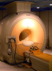
Scientists say they have devised a way to track the movements, destination, and persistence of a broad range of cellular therapies, without resorting to invasive procedures.
In Magnetic Resonance in Medicine, the researchers described the first human tests using a perfluorocarbon (PFC) tracer in combination with non-invasive magnetic resonance imaging (MRI) to track therapeutic immune cells injected into patients with colorectal cancer.
“Initially, we see this technique used for clinical trials that involve tests of new cell therapies,” said study author Eric T. Ahrens, PhD, of the University of California, San Diego.
“Clinical development of cell therapies can be accelerated by providing feedback regarding cell motility, optimal delivery routes, individual therapeutic doses, and engraftment success.”
Currently, there is no accepted way to image cells in the human body that covers a broad range of cell types and diseases. Earlier techniques have used metal ion-based vascular MRI contrast agents and radioisotopes.
The former have proven difficult to differentiate in vivo. The latter raise concerns about radiation toxicity and do not provide the anatomical detail available with MRIs.
“This is the first human PFC cell tracking agent, which is a new way to do MRI cell tracking,” Dr Ahrens said. “It’s the first example of a clinical MRI agent designed specifically for cell tracking.”
He and his colleagues used a PFC tracer agent and an MRI technique that directly detects fluorine atoms in labeled cells. Fluorine atoms naturally occur in extremely low concentrations in the body, making it easier to observe cells labeled with fluorine using MRI.
In this case, the modified and labeled dendritic cells were first prepared from white blood cells extracted from the patient. The cells were then injected into patients with stage 4 metastatic colorectal cancer to stimulate an anticancer T-cell immune response.
The researchers did not assess the efficacy of the cell therapy, only their ability to detect the labeled cells and monitor what happened to them. Dr Ahrens said the technique worked as expected, with the surprising finding that only half of the delivered cell vaccine remained at the inoculation site after 24 hours.
“The imaging agent technology has been shown to be able to tag any cell type that is of interest,” Dr Ahrens said. “It is a platform imaging technology for a wide range of diseases and applications.”
“Non-invasive cell tracking may help lower regulatory barriers,” he added. “For example, new stem cell therapies can be slow to obtain regulatory approvals, in part, because it is difficult, if not impossible, with current approaches to verify survival and location of transplanted cells.”
“And cell therapy trials generally have a high cost per patient. Tools that allow the investigator to gain a ‘richer’ data set from individual patients mean it may be possible to reduce patient numbers enrolled in a trial, thus reducing total trial cost.” 
Scientists say they have devised a way to track the movements, destination, and persistence of a broad range of cellular therapies, without resorting to invasive procedures.
In Magnetic Resonance in Medicine, the researchers described the first human tests using a perfluorocarbon (PFC) tracer in combination with non-invasive magnetic resonance imaging (MRI) to track therapeutic immune cells injected into patients with colorectal cancer.
“Initially, we see this technique used for clinical trials that involve tests of new cell therapies,” said study author Eric T. Ahrens, PhD, of the University of California, San Diego.
“Clinical development of cell therapies can be accelerated by providing feedback regarding cell motility, optimal delivery routes, individual therapeutic doses, and engraftment success.”
Currently, there is no accepted way to image cells in the human body that covers a broad range of cell types and diseases. Earlier techniques have used metal ion-based vascular MRI contrast agents and radioisotopes.
The former have proven difficult to differentiate in vivo. The latter raise concerns about radiation toxicity and do not provide the anatomical detail available with MRIs.
“This is the first human PFC cell tracking agent, which is a new way to do MRI cell tracking,” Dr Ahrens said. “It’s the first example of a clinical MRI agent designed specifically for cell tracking.”
He and his colleagues used a PFC tracer agent and an MRI technique that directly detects fluorine atoms in labeled cells. Fluorine atoms naturally occur in extremely low concentrations in the body, making it easier to observe cells labeled with fluorine using MRI.
In this case, the modified and labeled dendritic cells were first prepared from white blood cells extracted from the patient. The cells were then injected into patients with stage 4 metastatic colorectal cancer to stimulate an anticancer T-cell immune response.
The researchers did not assess the efficacy of the cell therapy, only their ability to detect the labeled cells and monitor what happened to them. Dr Ahrens said the technique worked as expected, with the surprising finding that only half of the delivered cell vaccine remained at the inoculation site after 24 hours.
“The imaging agent technology has been shown to be able to tag any cell type that is of interest,” Dr Ahrens said. “It is a platform imaging technology for a wide range of diseases and applications.”
“Non-invasive cell tracking may help lower regulatory barriers,” he added. “For example, new stem cell therapies can be slow to obtain regulatory approvals, in part, because it is difficult, if not impossible, with current approaches to verify survival and location of transplanted cells.”
“And cell therapy trials generally have a high cost per patient. Tools that allow the investigator to gain a ‘richer’ data set from individual patients mean it may be possible to reduce patient numbers enrolled in a trial, thus reducing total trial cost.”
Scientists say they have devised a way to track the movements, destination, and persistence of a broad range of cellular therapies, without resorting to invasive procedures.
In Magnetic Resonance in Medicine, the researchers described the first human tests using a perfluorocarbon (PFC) tracer in combination with non-invasive magnetic resonance imaging (MRI) to track therapeutic immune cells injected into patients with colorectal cancer.
“Initially, we see this technique used for clinical trials that involve tests of new cell therapies,” said study author Eric T. Ahrens, PhD, of the University of California, San Diego.
“Clinical development of cell therapies can be accelerated by providing feedback regarding cell motility, optimal delivery routes, individual therapeutic doses, and engraftment success.”
Currently, there is no accepted way to image cells in the human body that covers a broad range of cell types and diseases. Earlier techniques have used metal ion-based vascular MRI contrast agents and radioisotopes.
The former have proven difficult to differentiate in vivo. The latter raise concerns about radiation toxicity and do not provide the anatomical detail available with MRIs.
“This is the first human PFC cell tracking agent, which is a new way to do MRI cell tracking,” Dr Ahrens said. “It’s the first example of a clinical MRI agent designed specifically for cell tracking.”
He and his colleagues used a PFC tracer agent and an MRI technique that directly detects fluorine atoms in labeled cells. Fluorine atoms naturally occur in extremely low concentrations in the body, making it easier to observe cells labeled with fluorine using MRI.
In this case, the modified and labeled dendritic cells were first prepared from white blood cells extracted from the patient. The cells were then injected into patients with stage 4 metastatic colorectal cancer to stimulate an anticancer T-cell immune response.
The researchers did not assess the efficacy of the cell therapy, only their ability to detect the labeled cells and monitor what happened to them. Dr Ahrens said the technique worked as expected, with the surprising finding that only half of the delivered cell vaccine remained at the inoculation site after 24 hours.
“The imaging agent technology has been shown to be able to tag any cell type that is of interest,” Dr Ahrens said. “It is a platform imaging technology for a wide range of diseases and applications.”
“Non-invasive cell tracking may help lower regulatory barriers,” he added. “For example, new stem cell therapies can be slow to obtain regulatory approvals, in part, because it is difficult, if not impossible, with current approaches to verify survival and location of transplanted cells.”
“And cell therapy trials generally have a high cost per patient. Tools that allow the investigator to gain a ‘richer’ data set from individual patients mean it may be possible to reduce patient numbers enrolled in a trial, thus reducing total trial cost.”
Drugs demonstrate inconsistent synergy in CLL, MCL

PHILADELPHIA—The Bcl-2 inhibitor ABT-199 and the Bruton tyrosine kinase inhibitor ibrutinib can have a synergistic effect against mantle cell lymphoma (MCL) and chronic lymphocytic leukemia (CLL), preclinical data suggest.
In one set of experiments with MCL and CLL samples, the drugs induced apoptosis at a much higher rate when used together than when used alone.
However, in other experiments with CLL samples, ABT-199 and ibrutinib did not consistently display synergistic cytoxicity.
The researchers said this indicates substantial patient heterogeneity in response to the combination that may be due to variations in the genetic landscape.
Michael J. Weber, PhD, of the University of Virginia School of Medicine in Charlottesville, and his colleagues presented this research at the AACR conference Hematologic Malignancies: Translating Discoveries to Novel Therapies.
“We took an empirical but systematic approach to identify combinations of drugs that might improve the ability of ibrutinib to kill cancer cells,” Dr Weber said. “The combination of ibrutinib and ABT-199 was, by far, the most effective in our assays, and we are in the very earliest stages of planning a clinical trial to test this combination in the clinic.”
In previous studies, Dr Weber and his colleagues found that ibrutinib synergized with both ABT-199 and the proteasome inhibitor carfilzomib to kill MCL cell lines. In this study, the team assessed the effects of exposure to these 2 combinations on samples from patients with MCL or CLL.
The researchers found that apoptosis occurred in 23% of cells exposed to ABT-199 and ibrutinib in combination, compared to 3.8% of cells exposed to ibrutinib and 3% exposed to ABT-199.
The combination of ibrutinib and carfilzomib also increased apoptosis in MCL and CLL cells compared to either agent alone, though to a lesser degree than the ABT-199 combination. Apoptosis occurred in 5.5% of cells exposed to ibrutinib and carfilzomib, 3.8% exposed to ibrutinib, and 1.7% exposed to carfilzomib.
The researchers observed minimal apoptosis in normal T cells, both with the single agents and the combinations.
Further analysis showed that ibrutinib and ABT-199 worked synergistically to cause apoptosis in leukemic cells from 5 of 9 patients with CLL.
According to Dr Weber, the variable response to this combination points to the importance of understanding how these combinations work, so we can match the treatments with the most appropriate patients.
“Ibrutinib and ABT-199 target different pathways involved in promoting cancer cell survival and growth,” Dr Weber said.
“This is very intriguing because, in most instances where cancer cells are resistant to a particular molecularly targeted drug, we find that cancer cells adapt and find new ways to reactivate the pathway being targeted by the drug and that combinations of drugs targeting this pathway in different ways can improve outcomes. Here, we found that targeting a pathway outside the primary pathway was effective.”
This study was funded by the University of Virginia Cancer Center. Dr Weber declared no conflicts of interest.
PHILADELPHIA—The Bcl-2 inhibitor ABT-199 and the Bruton tyrosine kinase inhibitor ibrutinib can have a synergistic effect against mantle cell lymphoma (MCL) and chronic lymphocytic leukemia (CLL), preclinical data suggest.
In one set of experiments with MCL and CLL samples, the drugs induced apoptosis at a much higher rate when used together than when used alone.
However, in other experiments with CLL samples, ABT-199 and ibrutinib did not consistently display synergistic cytoxicity.
The researchers said this indicates substantial patient heterogeneity in response to the combination that may be due to variations in the genetic landscape.
Michael J. Weber, PhD, of the University of Virginia School of Medicine in Charlottesville, and his colleagues presented this research at the AACR conference Hematologic Malignancies: Translating Discoveries to Novel Therapies.
“We took an empirical but systematic approach to identify combinations of drugs that might improve the ability of ibrutinib to kill cancer cells,” Dr Weber said. “The combination of ibrutinib and ABT-199 was, by far, the most effective in our assays, and we are in the very earliest stages of planning a clinical trial to test this combination in the clinic.”
In previous studies, Dr Weber and his colleagues found that ibrutinib synergized with both ABT-199 and the proteasome inhibitor carfilzomib to kill MCL cell lines. In this study, the team assessed the effects of exposure to these 2 combinations on samples from patients with MCL or CLL.
The researchers found that apoptosis occurred in 23% of cells exposed to ABT-199 and ibrutinib in combination, compared to 3.8% of cells exposed to ibrutinib and 3% exposed to ABT-199.
The combination of ibrutinib and carfilzomib also increased apoptosis in MCL and CLL cells compared to either agent alone, though to a lesser degree than the ABT-199 combination. Apoptosis occurred in 5.5% of cells exposed to ibrutinib and carfilzomib, 3.8% exposed to ibrutinib, and 1.7% exposed to carfilzomib.
The researchers observed minimal apoptosis in normal T cells, both with the single agents and the combinations.
Further analysis showed that ibrutinib and ABT-199 worked synergistically to cause apoptosis in leukemic cells from 5 of 9 patients with CLL.
According to Dr Weber, the variable response to this combination points to the importance of understanding how these combinations work, so we can match the treatments with the most appropriate patients.
“Ibrutinib and ABT-199 target different pathways involved in promoting cancer cell survival and growth,” Dr Weber said.
“This is very intriguing because, in most instances where cancer cells are resistant to a particular molecularly targeted drug, we find that cancer cells adapt and find new ways to reactivate the pathway being targeted by the drug and that combinations of drugs targeting this pathway in different ways can improve outcomes. Here, we found that targeting a pathway outside the primary pathway was effective.”
This study was funded by the University of Virginia Cancer Center. Dr Weber declared no conflicts of interest.
PHILADELPHIA—The Bcl-2 inhibitor ABT-199 and the Bruton tyrosine kinase inhibitor ibrutinib can have a synergistic effect against mantle cell lymphoma (MCL) and chronic lymphocytic leukemia (CLL), preclinical data suggest.
In one set of experiments with MCL and CLL samples, the drugs induced apoptosis at a much higher rate when used together than when used alone.
However, in other experiments with CLL samples, ABT-199 and ibrutinib did not consistently display synergistic cytoxicity.
The researchers said this indicates substantial patient heterogeneity in response to the combination that may be due to variations in the genetic landscape.
Michael J. Weber, PhD, of the University of Virginia School of Medicine in Charlottesville, and his colleagues presented this research at the AACR conference Hematologic Malignancies: Translating Discoveries to Novel Therapies.
“We took an empirical but systematic approach to identify combinations of drugs that might improve the ability of ibrutinib to kill cancer cells,” Dr Weber said. “The combination of ibrutinib and ABT-199 was, by far, the most effective in our assays, and we are in the very earliest stages of planning a clinical trial to test this combination in the clinic.”
In previous studies, Dr Weber and his colleagues found that ibrutinib synergized with both ABT-199 and the proteasome inhibitor carfilzomib to kill MCL cell lines. In this study, the team assessed the effects of exposure to these 2 combinations on samples from patients with MCL or CLL.
The researchers found that apoptosis occurred in 23% of cells exposed to ABT-199 and ibrutinib in combination, compared to 3.8% of cells exposed to ibrutinib and 3% exposed to ABT-199.
The combination of ibrutinib and carfilzomib also increased apoptosis in MCL and CLL cells compared to either agent alone, though to a lesser degree than the ABT-199 combination. Apoptosis occurred in 5.5% of cells exposed to ibrutinib and carfilzomib, 3.8% exposed to ibrutinib, and 1.7% exposed to carfilzomib.
The researchers observed minimal apoptosis in normal T cells, both with the single agents and the combinations.
Further analysis showed that ibrutinib and ABT-199 worked synergistically to cause apoptosis in leukemic cells from 5 of 9 patients with CLL.
According to Dr Weber, the variable response to this combination points to the importance of understanding how these combinations work, so we can match the treatments with the most appropriate patients.
“Ibrutinib and ABT-199 target different pathways involved in promoting cancer cell survival and growth,” Dr Weber said.
“This is very intriguing because, in most instances where cancer cells are resistant to a particular molecularly targeted drug, we find that cancer cells adapt and find new ways to reactivate the pathway being targeted by the drug and that combinations of drugs targeting this pathway in different ways can improve outcomes. Here, we found that targeting a pathway outside the primary pathway was effective.”
This study was funded by the University of Virginia Cancer Center. Dr Weber declared no conflicts of interest.
Proper transfusion practice prevents CMV

Credit: Vera Kratochvil
New research confirms that transfusing leukoreduced, cytomegalovirus (CMV)-seronegative blood products prevents CMV transmission in very-low-birth-weight (VLBW) infants.
The study showed that, with this approach, maternal breast milk becomes the primary source of postnatal CMV infection.
“Previously, the risk of CMV infection from blood transfusion of seronegative or leukoreduced transfusions was estimated to be 1% to 3%,” said Cassandra Josephson, MD, of Emory University School of Medicine in Atlanta, Georgia.
“We showed that, using blood components that are both CMV-seronegative and leukoreduced, we can effectively prevent the transfusion-transmission of CMV. Therefore, we believe that this is the safest approach to reduce the risk of CMV infection when giving transfusions to VLBW infants.”
Dr Josephson and her colleagues described this research in JAMA Pediatrics.
The researchers evaluated 462 mothers and 539 VLBW infants who were admitted to 3 neonatal intensive care units between January 2010 and June 2013.
A majority of mothers had a history of CMV infection prior to delivery (CMV sero-prevalence of 76.2%). The infants were enrolled in the study within 5 days of birth and had not received a blood transfusion at that time.
The infants were tested for congenital infection at birth and again at 5 additional intervals between birth and 90 days, discharge, or death.
Twenty-nine of the infants had CMV infection (cumulative incidence of 6.9% at 12 weeks). Five infants with CMV infection developed severe disease or died.
Although 2061 transfusions were administered to 310 of the infants (57.5%), the blood products were CMV-seronegative and leukoreduced, and none of the CMV infections was linked to transfusion.
Twenty-seven of 28 infections acquired after birth occurred among infants fed CMV-positive breast milk.
Dr Josephson and her colleagues estimate that between 1 in 5 and 1 in 10 VLBW infants who are fed CMV-positive breast milk from mothers with a history of CMV infection will develop postnatal CMV infection.
The American Academy of Pediatrics currently states that the value of routinely feeding breast milk from CMV-seropositive mothers to preterm infants outweighs the risks of clinical disease from CMV.
But the researchers noted that new strategies to prevent breast milk transmission of CMV are needed because freezing and thawing breast milk did not completely prevent transmission in this study.
The team said alternative approaches to prevent breast milk transmission of CMV could include routine CMV-serologic testing of pregnant mothers to enable counseling regarding the risk of infection, closer surveillance of infants with CMV-positive mothers, and pasteurization of breast milk until a corrected gestational age of 34 weeks (as recommended by the Austrian Society of Pediatrics).
In addition, routine screening for postnatal CMV infection may help identify infants who are likely to develop symptomatic disease.
The researchers also said the frequency of CMV infection in this study raises significant concern about the potential consequences of CMV infection among VLBW infants and points to the need for large, long-term follow-up studies of neurological outcomes in infants with postnatal CMV infection.
Credit: Vera Kratochvil
New research confirms that transfusing leukoreduced, cytomegalovirus (CMV)-seronegative blood products prevents CMV transmission in very-low-birth-weight (VLBW) infants.
The study showed that, with this approach, maternal breast milk becomes the primary source of postnatal CMV infection.
“Previously, the risk of CMV infection from blood transfusion of seronegative or leukoreduced transfusions was estimated to be 1% to 3%,” said Cassandra Josephson, MD, of Emory University School of Medicine in Atlanta, Georgia.
“We showed that, using blood components that are both CMV-seronegative and leukoreduced, we can effectively prevent the transfusion-transmission of CMV. Therefore, we believe that this is the safest approach to reduce the risk of CMV infection when giving transfusions to VLBW infants.”
Dr Josephson and her colleagues described this research in JAMA Pediatrics.
The researchers evaluated 462 mothers and 539 VLBW infants who were admitted to 3 neonatal intensive care units between January 2010 and June 2013.
A majority of mothers had a history of CMV infection prior to delivery (CMV sero-prevalence of 76.2%). The infants were enrolled in the study within 5 days of birth and had not received a blood transfusion at that time.
The infants were tested for congenital infection at birth and again at 5 additional intervals between birth and 90 days, discharge, or death.
Twenty-nine of the infants had CMV infection (cumulative incidence of 6.9% at 12 weeks). Five infants with CMV infection developed severe disease or died.
Although 2061 transfusions were administered to 310 of the infants (57.5%), the blood products were CMV-seronegative and leukoreduced, and none of the CMV infections was linked to transfusion.
Twenty-seven of 28 infections acquired after birth occurred among infants fed CMV-positive breast milk.
Dr Josephson and her colleagues estimate that between 1 in 5 and 1 in 10 VLBW infants who are fed CMV-positive breast milk from mothers with a history of CMV infection will develop postnatal CMV infection.
The American Academy of Pediatrics currently states that the value of routinely feeding breast milk from CMV-seropositive mothers to preterm infants outweighs the risks of clinical disease from CMV.
But the researchers noted that new strategies to prevent breast milk transmission of CMV are needed because freezing and thawing breast milk did not completely prevent transmission in this study.
The team said alternative approaches to prevent breast milk transmission of CMV could include routine CMV-serologic testing of pregnant mothers to enable counseling regarding the risk of infection, closer surveillance of infants with CMV-positive mothers, and pasteurization of breast milk until a corrected gestational age of 34 weeks (as recommended by the Austrian Society of Pediatrics).
In addition, routine screening for postnatal CMV infection may help identify infants who are likely to develop symptomatic disease.
The researchers also said the frequency of CMV infection in this study raises significant concern about the potential consequences of CMV infection among VLBW infants and points to the need for large, long-term follow-up studies of neurological outcomes in infants with postnatal CMV infection.
Credit: Vera Kratochvil
New research confirms that transfusing leukoreduced, cytomegalovirus (CMV)-seronegative blood products prevents CMV transmission in very-low-birth-weight (VLBW) infants.
The study showed that, with this approach, maternal breast milk becomes the primary source of postnatal CMV infection.
“Previously, the risk of CMV infection from blood transfusion of seronegative or leukoreduced transfusions was estimated to be 1% to 3%,” said Cassandra Josephson, MD, of Emory University School of Medicine in Atlanta, Georgia.
“We showed that, using blood components that are both CMV-seronegative and leukoreduced, we can effectively prevent the transfusion-transmission of CMV. Therefore, we believe that this is the safest approach to reduce the risk of CMV infection when giving transfusions to VLBW infants.”
Dr Josephson and her colleagues described this research in JAMA Pediatrics.
The researchers evaluated 462 mothers and 539 VLBW infants who were admitted to 3 neonatal intensive care units between January 2010 and June 2013.
A majority of mothers had a history of CMV infection prior to delivery (CMV sero-prevalence of 76.2%). The infants were enrolled in the study within 5 days of birth and had not received a blood transfusion at that time.
The infants were tested for congenital infection at birth and again at 5 additional intervals between birth and 90 days, discharge, or death.
Twenty-nine of the infants had CMV infection (cumulative incidence of 6.9% at 12 weeks). Five infants with CMV infection developed severe disease or died.
Although 2061 transfusions were administered to 310 of the infants (57.5%), the blood products were CMV-seronegative and leukoreduced, and none of the CMV infections was linked to transfusion.
Twenty-seven of 28 infections acquired after birth occurred among infants fed CMV-positive breast milk.
Dr Josephson and her colleagues estimate that between 1 in 5 and 1 in 10 VLBW infants who are fed CMV-positive breast milk from mothers with a history of CMV infection will develop postnatal CMV infection.
The American Academy of Pediatrics currently states that the value of routinely feeding breast milk from CMV-seropositive mothers to preterm infants outweighs the risks of clinical disease from CMV.
But the researchers noted that new strategies to prevent breast milk transmission of CMV are needed because freezing and thawing breast milk did not completely prevent transmission in this study.
The team said alternative approaches to prevent breast milk transmission of CMV could include routine CMV-serologic testing of pregnant mothers to enable counseling regarding the risk of infection, closer surveillance of infants with CMV-positive mothers, and pasteurization of breast milk until a corrected gestational age of 34 weeks (as recommended by the Austrian Society of Pediatrics).
In addition, routine screening for postnatal CMV infection may help identify infants who are likely to develop symptomatic disease.
The researchers also said the frequency of CMV infection in this study raises significant concern about the potential consequences of CMV infection among VLBW infants and points to the need for large, long-term follow-up studies of neurological outcomes in infants with postnatal CMV infection.
Blood testing system wins marketing authorization in US
Credit: Juan D. Alfonso
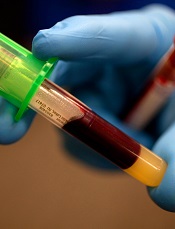
The US Food and Drug Administration (FDA) has granted marketing authorization for the T2Candida® Panel and the T2Dx® Instrument, a system that provides direct detection of 5 yeast pathogens in whole blood samples.
The system can detect Candida albicans, Candida tropicalis, Candida parapsilosis, Candida glabrata, and Candida krusei in patients with symptoms of, or medical conditions predisposing them to, invasive fungal infections.
It can take up to 6 days to detect yeast pathogens using blood culture, and even more time to identify the specific type of yeast present.
The T2Candida system can identify specific Candida pathogens from a single blood sample within 3 to 5 hours.
However, false positive results are possible with this system, so physicians should perform blood cultures to confirm T2Candida results, according to the FDA. Still, a negative test result may provide timely data that allows physicians to avoid or suspend unnecessary antifungal treatment.
“By testing 1 blood sample for 5 yeast pathogens—and getting results within a few hours—physicians can initiate appropriate antifungal treatment earlier and potentially reduce patient illness and decrease the risk of dying from these infections,” said Alberto Gutierrez, director of the Office of In Vitro Diagnostics and Radiological Health at the FDA’s Center for Devices and Radiological Health.
How the system works
The T2Candida panel and T2Dx instrument are powered by T2MR, a miniaturized, magnetic-resonance-based diagnostic approach that measures how water molecules react in the presence of magnetic fields.
The system uses blood-compatible polymerase chain reaction to amplify Candida DNA, which then binds to superparamagnetic nanoparticles coated with a complementary DNA strand. The binding event causes the nanoparticles to cluster, which changes the sample’s T2 magnetic resonance signal.
If the system detects yeast DNA, it can then determine the species category to which the DNA belongs, which helps point healthcare providers to the appropriate treatment.
T2Dx is a fully automated, bench-top instrument. To perform a test, the patient sample is snapped onto a disposable test cartridge, which is preloaded with the necessary reagents. The cartridge is then inserted into T2Dx, which processes the sample and delivers a diagnostic test result.
Studies and FDA review
The FDA reviewed the T2Candida panel and the T2Dx instrument through the agency’s de novo classification process, a regulatory pathway for certain novel, low- to moderate-risk medical devices.
The FDA based its review on a clinical study of 1500 patients, in which the T2Candida system correctly categorized nearly 100% of the negative specimens as negative for the presence of yeast.
In a separate study of 300 blood samples with specific concentrations of yeast, the system correctly identified the organism in 84% to 96% of the positive specimens.
The T2Candida panel and T2Dx instrument are manufactured by T2 Biosystems, Inc.
Credit: Juan D. Alfonso
The US Food and Drug Administration (FDA) has granted marketing authorization for the T2Candida® Panel and the T2Dx® Instrument, a system that provides direct detection of 5 yeast pathogens in whole blood samples.
The system can detect Candida albicans, Candida tropicalis, Candida parapsilosis, Candida glabrata, and Candida krusei in patients with symptoms of, or medical conditions predisposing them to, invasive fungal infections.
It can take up to 6 days to detect yeast pathogens using blood culture, and even more time to identify the specific type of yeast present.
The T2Candida system can identify specific Candida pathogens from a single blood sample within 3 to 5 hours.
However, false positive results are possible with this system, so physicians should perform blood cultures to confirm T2Candida results, according to the FDA. Still, a negative test result may provide timely data that allows physicians to avoid or suspend unnecessary antifungal treatment.
“By testing 1 blood sample for 5 yeast pathogens—and getting results within a few hours—physicians can initiate appropriate antifungal treatment earlier and potentially reduce patient illness and decrease the risk of dying from these infections,” said Alberto Gutierrez, director of the Office of In Vitro Diagnostics and Radiological Health at the FDA’s Center for Devices and Radiological Health.
How the system works
The T2Candida panel and T2Dx instrument are powered by T2MR, a miniaturized, magnetic-resonance-based diagnostic approach that measures how water molecules react in the presence of magnetic fields.
The system uses blood-compatible polymerase chain reaction to amplify Candida DNA, which then binds to superparamagnetic nanoparticles coated with a complementary DNA strand. The binding event causes the nanoparticles to cluster, which changes the sample’s T2 magnetic resonance signal.
If the system detects yeast DNA, it can then determine the species category to which the DNA belongs, which helps point healthcare providers to the appropriate treatment.
T2Dx is a fully automated, bench-top instrument. To perform a test, the patient sample is snapped onto a disposable test cartridge, which is preloaded with the necessary reagents. The cartridge is then inserted into T2Dx, which processes the sample and delivers a diagnostic test result.
Studies and FDA review
The FDA reviewed the T2Candida panel and the T2Dx instrument through the agency’s de novo classification process, a regulatory pathway for certain novel, low- to moderate-risk medical devices.
The FDA based its review on a clinical study of 1500 patients, in which the T2Candida system correctly categorized nearly 100% of the negative specimens as negative for the presence of yeast.
In a separate study of 300 blood samples with specific concentrations of yeast, the system correctly identified the organism in 84% to 96% of the positive specimens.
The T2Candida panel and T2Dx instrument are manufactured by T2 Biosystems, Inc.
Credit: Juan D. Alfonso
The US Food and Drug Administration (FDA) has granted marketing authorization for the T2Candida® Panel and the T2Dx® Instrument, a system that provides direct detection of 5 yeast pathogens in whole blood samples.
The system can detect Candida albicans, Candida tropicalis, Candida parapsilosis, Candida glabrata, and Candida krusei in patients with symptoms of, or medical conditions predisposing them to, invasive fungal infections.
It can take up to 6 days to detect yeast pathogens using blood culture, and even more time to identify the specific type of yeast present.
The T2Candida system can identify specific Candida pathogens from a single blood sample within 3 to 5 hours.
However, false positive results are possible with this system, so physicians should perform blood cultures to confirm T2Candida results, according to the FDA. Still, a negative test result may provide timely data that allows physicians to avoid or suspend unnecessary antifungal treatment.
“By testing 1 blood sample for 5 yeast pathogens—and getting results within a few hours—physicians can initiate appropriate antifungal treatment earlier and potentially reduce patient illness and decrease the risk of dying from these infections,” said Alberto Gutierrez, director of the Office of In Vitro Diagnostics and Radiological Health at the FDA’s Center for Devices and Radiological Health.
How the system works
The T2Candida panel and T2Dx instrument are powered by T2MR, a miniaturized, magnetic-resonance-based diagnostic approach that measures how water molecules react in the presence of magnetic fields.
The system uses blood-compatible polymerase chain reaction to amplify Candida DNA, which then binds to superparamagnetic nanoparticles coated with a complementary DNA strand. The binding event causes the nanoparticles to cluster, which changes the sample’s T2 magnetic resonance signal.
If the system detects yeast DNA, it can then determine the species category to which the DNA belongs, which helps point healthcare providers to the appropriate treatment.
T2Dx is a fully automated, bench-top instrument. To perform a test, the patient sample is snapped onto a disposable test cartridge, which is preloaded with the necessary reagents. The cartridge is then inserted into T2Dx, which processes the sample and delivers a diagnostic test result.
Studies and FDA review
The FDA reviewed the T2Candida panel and the T2Dx instrument through the agency’s de novo classification process, a regulatory pathway for certain novel, low- to moderate-risk medical devices.
The FDA based its review on a clinical study of 1500 patients, in which the T2Candida system correctly categorized nearly 100% of the negative specimens as negative for the presence of yeast.
In a separate study of 300 blood samples with specific concentrations of yeast, the system correctly identified the organism in 84% to 96% of the positive specimens.
The T2Candida panel and T2Dx instrument are manufactured by T2 Biosystems, Inc.
Exercise boosts anticancer effects of doxorubicin

Credit: Aaron Logan
Exercising during doxorubicin treatment can amplify the drug’s cancer-fighting ability, according to preclinical research.
Previous studies showed that adopting an exercise regimen before receiving doxorubicin could protect against the cardiac side effects associated with the drug.
Now, researchers have reported that exercising during doxorubicin treatment does not protect the heart, but it does help shrink tumors in mice.
Joseph Libonati, PhD, of the University of Pennsylvania in Philadelphia, and his colleagues reported these findings in the American Journal of Physiology—Regulatory, Integrative and Comparative Physiology.
The researchers conducted experiments with 4 groups of mice, all of which received an injection of melanoma cells.
During the next 2 weeks, 2 of the groups received doxorubicin in 2 doses, while the other 2 groups received placebo injections.
A treated group and a placebo group were put on exercise regimens, walking for 45 minutes 5 days a week on mouse-sized treadmills, while the rest of the mice remained sedentary.
After the 2-week trial, the researchers examined the animals’ hearts using echocardiogram and tissue analysis.
As expected, doxorubicin reduced the heart’s function and size and increased fibrosis. Mice that exercised were not protected from this damage.
“We looked, and the exercise didn’t do anything to the heart; it didn’t worsen it, it didn’t help it,” Dr Libonati said. “But the tumor data—I find them actually amazing.”
The mice that received doxorubicin and exercised had significantly smaller tumors after 2 weeks than mice that only received doxorubicin (P<0.05).
Further studies will investigate exactly how exercise enhances the effect of doxorubicin, but the researchers believe it could be, in part, because exercise increases blood flow to the tumor, bringing with it more of the drug in the bloodstream.
“If exercise helps in this way, you could potentially use a smaller dose of the drug and get fewer side effects,” Dr Libonati said.
Gaining a clearer understanding of the many ways that exercise affects various systems of the body could also pave the way for developing drugs that mimic the effects of exercise.
“People don’t take a drug and then sit down all day,” Dr Libonati said. “Something as simple as moving affects how drugs are metabolized. We’re only just beginning to understand the complexities.”
Credit: Aaron Logan
Exercising during doxorubicin treatment can amplify the drug’s cancer-fighting ability, according to preclinical research.
Previous studies showed that adopting an exercise regimen before receiving doxorubicin could protect against the cardiac side effects associated with the drug.
Now, researchers have reported that exercising during doxorubicin treatment does not protect the heart, but it does help shrink tumors in mice.
Joseph Libonati, PhD, of the University of Pennsylvania in Philadelphia, and his colleagues reported these findings in the American Journal of Physiology—Regulatory, Integrative and Comparative Physiology.
The researchers conducted experiments with 4 groups of mice, all of which received an injection of melanoma cells.
During the next 2 weeks, 2 of the groups received doxorubicin in 2 doses, while the other 2 groups received placebo injections.
A treated group and a placebo group were put on exercise regimens, walking for 45 minutes 5 days a week on mouse-sized treadmills, while the rest of the mice remained sedentary.
After the 2-week trial, the researchers examined the animals’ hearts using echocardiogram and tissue analysis.
As expected, doxorubicin reduced the heart’s function and size and increased fibrosis. Mice that exercised were not protected from this damage.
“We looked, and the exercise didn’t do anything to the heart; it didn’t worsen it, it didn’t help it,” Dr Libonati said. “But the tumor data—I find them actually amazing.”
The mice that received doxorubicin and exercised had significantly smaller tumors after 2 weeks than mice that only received doxorubicin (P<0.05).
Further studies will investigate exactly how exercise enhances the effect of doxorubicin, but the researchers believe it could be, in part, because exercise increases blood flow to the tumor, bringing with it more of the drug in the bloodstream.
“If exercise helps in this way, you could potentially use a smaller dose of the drug and get fewer side effects,” Dr Libonati said.
Gaining a clearer understanding of the many ways that exercise affects various systems of the body could also pave the way for developing drugs that mimic the effects of exercise.
“People don’t take a drug and then sit down all day,” Dr Libonati said. “Something as simple as moving affects how drugs are metabolized. We’re only just beginning to understand the complexities.”
Credit: Aaron Logan
Exercising during doxorubicin treatment can amplify the drug’s cancer-fighting ability, according to preclinical research.
Previous studies showed that adopting an exercise regimen before receiving doxorubicin could protect against the cardiac side effects associated with the drug.
Now, researchers have reported that exercising during doxorubicin treatment does not protect the heart, but it does help shrink tumors in mice.
Joseph Libonati, PhD, of the University of Pennsylvania in Philadelphia, and his colleagues reported these findings in the American Journal of Physiology—Regulatory, Integrative and Comparative Physiology.
The researchers conducted experiments with 4 groups of mice, all of which received an injection of melanoma cells.
During the next 2 weeks, 2 of the groups received doxorubicin in 2 doses, while the other 2 groups received placebo injections.
A treated group and a placebo group were put on exercise regimens, walking for 45 minutes 5 days a week on mouse-sized treadmills, while the rest of the mice remained sedentary.
After the 2-week trial, the researchers examined the animals’ hearts using echocardiogram and tissue analysis.
As expected, doxorubicin reduced the heart’s function and size and increased fibrosis. Mice that exercised were not protected from this damage.
“We looked, and the exercise didn’t do anything to the heart; it didn’t worsen it, it didn’t help it,” Dr Libonati said. “But the tumor data—I find them actually amazing.”
The mice that received doxorubicin and exercised had significantly smaller tumors after 2 weeks than mice that only received doxorubicin (P<0.05).
Further studies will investigate exactly how exercise enhances the effect of doxorubicin, but the researchers believe it could be, in part, because exercise increases blood flow to the tumor, bringing with it more of the drug in the bloodstream.
“If exercise helps in this way, you could potentially use a smaller dose of the drug and get fewer side effects,” Dr Libonati said.
Gaining a clearer understanding of the many ways that exercise affects various systems of the body could also pave the way for developing drugs that mimic the effects of exercise.
“People don’t take a drug and then sit down all day,” Dr Libonati said. “Something as simple as moving affects how drugs are metabolized. We’re only just beginning to understand the complexities.”
FDA approves 30-day pack of anticoagulant

Credit: NHS
The US Food and Drug Administration (FDA) has approved the Xarelto Starter Pack™ for the treatment of deep vein thrombosis (DVT) and/or pulmonary embolism (PE).
The pack provides a 30-day supply of Xarelto (rivaroxaban), which gives patients time to follow up with their primary care physician after leaving the hospital, without missing treatment.
Patients have the greatest risk of recurrence in the first 30 days after a DVT or PE.
“The starter pack will make a difference for patients impacted by blood clots by offering a 30-day supply of the drug in a new, convenient package, which may help support patients as they transition from the hospital to outpatient care,” said Paul Burton, MD, PhD, Vice President of Medical Affairs at Janssen, the company developing Xarelto.
The Xarelto Starter Pack™ will be available at pharmacies in October.
Xarelto is a factor Xa inhibitor that is FDA-approved to treat and prevent the recurrence of DVT/PE, as thromboprophylaxis in patients who have undergone knee or hip replacement surgery, and to reduce the risk of stroke in patients with non-valvular atrial fibrillation.
For DVT/PE patients, Xarelto is given twice daily at 15 mg with food for the first 21 days. On day 22, patients transition to a once-daily dose of 20 mg with food for the remainder of treatment.
Xarelto has a boxed warning stating that premature discontinuation of the drug increases the risk of thrombotic events, and epidural or spinal hematomas have occurred in Xarelto-treated patients who are receiving neuraxial anesthesia or undergoing spinal puncture.
For more details on Xarelto, see the full prescribing information.
Credit: NHS
The US Food and Drug Administration (FDA) has approved the Xarelto Starter Pack™ for the treatment of deep vein thrombosis (DVT) and/or pulmonary embolism (PE).
The pack provides a 30-day supply of Xarelto (rivaroxaban), which gives patients time to follow up with their primary care physician after leaving the hospital, without missing treatment.
Patients have the greatest risk of recurrence in the first 30 days after a DVT or PE.
“The starter pack will make a difference for patients impacted by blood clots by offering a 30-day supply of the drug in a new, convenient package, which may help support patients as they transition from the hospital to outpatient care,” said Paul Burton, MD, PhD, Vice President of Medical Affairs at Janssen, the company developing Xarelto.
The Xarelto Starter Pack™ will be available at pharmacies in October.
Xarelto is a factor Xa inhibitor that is FDA-approved to treat and prevent the recurrence of DVT/PE, as thromboprophylaxis in patients who have undergone knee or hip replacement surgery, and to reduce the risk of stroke in patients with non-valvular atrial fibrillation.
For DVT/PE patients, Xarelto is given twice daily at 15 mg with food for the first 21 days. On day 22, patients transition to a once-daily dose of 20 mg with food for the remainder of treatment.
Xarelto has a boxed warning stating that premature discontinuation of the drug increases the risk of thrombotic events, and epidural or spinal hematomas have occurred in Xarelto-treated patients who are receiving neuraxial anesthesia or undergoing spinal puncture.
For more details on Xarelto, see the full prescribing information.
Credit: NHS
The US Food and Drug Administration (FDA) has approved the Xarelto Starter Pack™ for the treatment of deep vein thrombosis (DVT) and/or pulmonary embolism (PE).
The pack provides a 30-day supply of Xarelto (rivaroxaban), which gives patients time to follow up with their primary care physician after leaving the hospital, without missing treatment.
Patients have the greatest risk of recurrence in the first 30 days after a DVT or PE.
“The starter pack will make a difference for patients impacted by blood clots by offering a 30-day supply of the drug in a new, convenient package, which may help support patients as they transition from the hospital to outpatient care,” said Paul Burton, MD, PhD, Vice President of Medical Affairs at Janssen, the company developing Xarelto.
The Xarelto Starter Pack™ will be available at pharmacies in October.
Xarelto is a factor Xa inhibitor that is FDA-approved to treat and prevent the recurrence of DVT/PE, as thromboprophylaxis in patients who have undergone knee or hip replacement surgery, and to reduce the risk of stroke in patients with non-valvular atrial fibrillation.
For DVT/PE patients, Xarelto is given twice daily at 15 mg with food for the first 21 days. On day 22, patients transition to a once-daily dose of 20 mg with food for the remainder of treatment.
Xarelto has a boxed warning stating that premature discontinuation of the drug increases the risk of thrombotic events, and epidural or spinal hematomas have occurred in Xarelto-treated patients who are receiving neuraxial anesthesia or undergoing spinal puncture.
For more details on Xarelto, see the full prescribing information.
Combo improves PFS in multiple myeloma

Credit: NIH
Adding panobinostat to treatment with bortezomib and dexamethasone can improve progression-free survival (PFS) in previously treated patients with multiple myeloma, results of the PANORAMA-1 trial suggest.
The combination conferred a 4-month improvement in median PFS when compared to bortezomib and dexamethasone plus placebo.
There was no significant difference in overall survival between the treatment groups, but researchers said these data are not mature.
“The PANORAMA-1 study is the first phase 3 trial to show the superiority of [panobinostat] plus bortezomib and dexamethasone over one of the standard 2-drug regimens for patients with relapsing and/or refractory multiple myeloma,” said lead study investigator Jesus San-Miguel, MD, of Clínica Universidad de Navarra in Pamplona, Spain.
Dr San-Miguel and his colleagues reported the results of PANORAMA-1 in The Lancet Oncology. The trial was sponsored by Novartis Pharmaceuticals, the company developing panobinostat.
The trial included 768 patients with relapsed or relapsed and refractory multiple myeloma who had failed at least 1 prior treatment.
Three-hundred and eighty-seven patients were randomized to treatment with panobinostat, bortezomib, and dexamethasone. And 381 patients were randomized to receive placebo, bortezomib, and dexamethasone.
The median follow up was 6.47 months in the panobinostat arm 5.59 months in the placebo arm.
The study’s primary endpoint was PFS. And the median PFS was significantly longer in the panobinostat arm than the placebo arm—11.99 months and 8.08 months, respectively (P<0.0001).
The median overall survival, on the other hand, was similar between the treatment arms. It was 33.64 months in the panobinostat arm and 30.39 months in the placebo arm (P=0.26).
Likewise, the overall response rate was similar between the treatment arms—60.7% with panobinostat and 54.6% with placebo (P=0.09). But the rate of complete or near-complete response was higher with panobinostat—27.6% and 15.7%, respectively (P=0.00006).
The rate of serious adverse events was 60% in the panobinostat arm and 42% in the placebo arm.
The most common grade 3/4 adverse events were thrombocytopenia (67% and 31%, respectively), lymphopenia (53% and 40%, respectively), neutropenia (35% and 11%, respectively), diarrhea (26% and 8%, respectively), and neuropathy (18% and 15%, respectively).
Based on these data, panobinostat was granted priority review by the US Food and Drug Administration in May. Priority review is given to therapies that may offer major advances in treatment. 
Credit: NIH
Adding panobinostat to treatment with bortezomib and dexamethasone can improve progression-free survival (PFS) in previously treated patients with multiple myeloma, results of the PANORAMA-1 trial suggest.
The combination conferred a 4-month improvement in median PFS when compared to bortezomib and dexamethasone plus placebo.
There was no significant difference in overall survival between the treatment groups, but researchers said these data are not mature.
“The PANORAMA-1 study is the first phase 3 trial to show the superiority of [panobinostat] plus bortezomib and dexamethasone over one of the standard 2-drug regimens for patients with relapsing and/or refractory multiple myeloma,” said lead study investigator Jesus San-Miguel, MD, of Clínica Universidad de Navarra in Pamplona, Spain.
Dr San-Miguel and his colleagues reported the results of PANORAMA-1 in The Lancet Oncology. The trial was sponsored by Novartis Pharmaceuticals, the company developing panobinostat.
The trial included 768 patients with relapsed or relapsed and refractory multiple myeloma who had failed at least 1 prior treatment.
Three-hundred and eighty-seven patients were randomized to treatment with panobinostat, bortezomib, and dexamethasone. And 381 patients were randomized to receive placebo, bortezomib, and dexamethasone.
The median follow up was 6.47 months in the panobinostat arm 5.59 months in the placebo arm.
The study’s primary endpoint was PFS. And the median PFS was significantly longer in the panobinostat arm than the placebo arm—11.99 months and 8.08 months, respectively (P<0.0001).
The median overall survival, on the other hand, was similar between the treatment arms. It was 33.64 months in the panobinostat arm and 30.39 months in the placebo arm (P=0.26).
Likewise, the overall response rate was similar between the treatment arms—60.7% with panobinostat and 54.6% with placebo (P=0.09). But the rate of complete or near-complete response was higher with panobinostat—27.6% and 15.7%, respectively (P=0.00006).
The rate of serious adverse events was 60% in the panobinostat arm and 42% in the placebo arm.
The most common grade 3/4 adverse events were thrombocytopenia (67% and 31%, respectively), lymphopenia (53% and 40%, respectively), neutropenia (35% and 11%, respectively), diarrhea (26% and 8%, respectively), and neuropathy (18% and 15%, respectively).
Based on these data, panobinostat was granted priority review by the US Food and Drug Administration in May. Priority review is given to therapies that may offer major advances in treatment.
Credit: NIH
Adding panobinostat to treatment with bortezomib and dexamethasone can improve progression-free survival (PFS) in previously treated patients with multiple myeloma, results of the PANORAMA-1 trial suggest.
The combination conferred a 4-month improvement in median PFS when compared to bortezomib and dexamethasone plus placebo.
There was no significant difference in overall survival between the treatment groups, but researchers said these data are not mature.
“The PANORAMA-1 study is the first phase 3 trial to show the superiority of [panobinostat] plus bortezomib and dexamethasone over one of the standard 2-drug regimens for patients with relapsing and/or refractory multiple myeloma,” said lead study investigator Jesus San-Miguel, MD, of Clínica Universidad de Navarra in Pamplona, Spain.
Dr San-Miguel and his colleagues reported the results of PANORAMA-1 in The Lancet Oncology. The trial was sponsored by Novartis Pharmaceuticals, the company developing panobinostat.
The trial included 768 patients with relapsed or relapsed and refractory multiple myeloma who had failed at least 1 prior treatment.
Three-hundred and eighty-seven patients were randomized to treatment with panobinostat, bortezomib, and dexamethasone. And 381 patients were randomized to receive placebo, bortezomib, and dexamethasone.
The median follow up was 6.47 months in the panobinostat arm 5.59 months in the placebo arm.
The study’s primary endpoint was PFS. And the median PFS was significantly longer in the panobinostat arm than the placebo arm—11.99 months and 8.08 months, respectively (P<0.0001).
The median overall survival, on the other hand, was similar between the treatment arms. It was 33.64 months in the panobinostat arm and 30.39 months in the placebo arm (P=0.26).
Likewise, the overall response rate was similar between the treatment arms—60.7% with panobinostat and 54.6% with placebo (P=0.09). But the rate of complete or near-complete response was higher with panobinostat—27.6% and 15.7%, respectively (P=0.00006).
The rate of serious adverse events was 60% in the panobinostat arm and 42% in the placebo arm.
The most common grade 3/4 adverse events were thrombocytopenia (67% and 31%, respectively), lymphopenia (53% and 40%, respectively), neutropenia (35% and 11%, respectively), diarrhea (26% and 8%, respectively), and neuropathy (18% and 15%, respectively).
Based on these data, panobinostat was granted priority review by the US Food and Drug Administration in May. Priority review is given to therapies that may offer major advances in treatment.
Studies explain gene’s role in thrombosis, hemostasis
Credit: Kevin MacKenzie
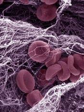
Two preclinical studies provide new insight into the activity of STXBP5, a gene that has been linked to changes in von Willebrand factor (VWF).
One research group found evidence suggesting that STXBP5 regulates endothelial exocytosis and thrombosis.
Another group’s work indicated that STXBP5 is required for normal arterial hemostasis, as it contributes to platelet packaging and secretion.
Both studies appear in The Journal of Clinical Investigation.
Charles Lowenstein, MD, of the University of Rochester in New York, and his colleagues began their research with the theory that STXBP5 inhibits endothelial cell exocytosis.
The group found that STXBP5 is expressed in human endothelial cells, and reducing STXBP5 increases exocytosis of VWF and P-selectin.
STXBP5-knockout mice had higher levels of VWF in their plasma, increased P-selectin translocation, and more platelet-endothelial interactions. This suggests that defective STXBP5 is a risk factor for thrombosis.
However, STXBP5-knockout mice also exhibited prolonged bleeding and impaired thrombosis. They had defects in platelet secretion and activation as well.
Sidney (Wally) Whiteheart, PhD, of the University of Kentucky in Lexington, and his colleagues helped to explain these findings with their research.
The group showed that platelets lacking SXTBP5 failed to function correctly, and SXTBP5 was required for platelets to assist in normal clot formation. This suggests STXBP5 plays different roles in endothelial cells and platelets.
Specifically, the researchers found that STXBP5 interacts with core secretion machinery complexes and the platelet cytoskeleton.
And platelets from STXBP5-knockout mice exhibited defects in granule secretion. These platelets had altered granule cargo levels, despite having normal morphology and granule numbers.
Like Dr Lowenstein’s group, Dr Whiteheart and his colleagues observed dramatic bleeding and defective hemostasis in STXBP5-knockout mice. Transplant experiments suggested these defects were due to a loss of STXBP5 in bone marrow-derived cells.
Credit: Kevin MacKenzie
Two preclinical studies provide new insight into the activity of STXBP5, a gene that has been linked to changes in von Willebrand factor (VWF).
One research group found evidence suggesting that STXBP5 regulates endothelial exocytosis and thrombosis.
Another group’s work indicated that STXBP5 is required for normal arterial hemostasis, as it contributes to platelet packaging and secretion.
Both studies appear in The Journal of Clinical Investigation.
Charles Lowenstein, MD, of the University of Rochester in New York, and his colleagues began their research with the theory that STXBP5 inhibits endothelial cell exocytosis.
The group found that STXBP5 is expressed in human endothelial cells, and reducing STXBP5 increases exocytosis of VWF and P-selectin.
STXBP5-knockout mice had higher levels of VWF in their plasma, increased P-selectin translocation, and more platelet-endothelial interactions. This suggests that defective STXBP5 is a risk factor for thrombosis.
However, STXBP5-knockout mice also exhibited prolonged bleeding and impaired thrombosis. They had defects in platelet secretion and activation as well.
Sidney (Wally) Whiteheart, PhD, of the University of Kentucky in Lexington, and his colleagues helped to explain these findings with their research.
The group showed that platelets lacking SXTBP5 failed to function correctly, and SXTBP5 was required for platelets to assist in normal clot formation. This suggests STXBP5 plays different roles in endothelial cells and platelets.
Specifically, the researchers found that STXBP5 interacts with core secretion machinery complexes and the platelet cytoskeleton.
And platelets from STXBP5-knockout mice exhibited defects in granule secretion. These platelets had altered granule cargo levels, despite having normal morphology and granule numbers.
Like Dr Lowenstein’s group, Dr Whiteheart and his colleagues observed dramatic bleeding and defective hemostasis in STXBP5-knockout mice. Transplant experiments suggested these defects were due to a loss of STXBP5 in bone marrow-derived cells.
Credit: Kevin MacKenzie
Two preclinical studies provide new insight into the activity of STXBP5, a gene that has been linked to changes in von Willebrand factor (VWF).
One research group found evidence suggesting that STXBP5 regulates endothelial exocytosis and thrombosis.
Another group’s work indicated that STXBP5 is required for normal arterial hemostasis, as it contributes to platelet packaging and secretion.
Both studies appear in The Journal of Clinical Investigation.
Charles Lowenstein, MD, of the University of Rochester in New York, and his colleagues began their research with the theory that STXBP5 inhibits endothelial cell exocytosis.
The group found that STXBP5 is expressed in human endothelial cells, and reducing STXBP5 increases exocytosis of VWF and P-selectin.
STXBP5-knockout mice had higher levels of VWF in their plasma, increased P-selectin translocation, and more platelet-endothelial interactions. This suggests that defective STXBP5 is a risk factor for thrombosis.
However, STXBP5-knockout mice also exhibited prolonged bleeding and impaired thrombosis. They had defects in platelet secretion and activation as well.
Sidney (Wally) Whiteheart, PhD, of the University of Kentucky in Lexington, and his colleagues helped to explain these findings with their research.
The group showed that platelets lacking SXTBP5 failed to function correctly, and SXTBP5 was required for platelets to assist in normal clot formation. This suggests STXBP5 plays different roles in endothelial cells and platelets.
Specifically, the researchers found that STXBP5 interacts with core secretion machinery complexes and the platelet cytoskeleton.
And platelets from STXBP5-knockout mice exhibited defects in granule secretion. These platelets had altered granule cargo levels, despite having normal morphology and granule numbers.
Like Dr Lowenstein’s group, Dr Whiteheart and his colleagues observed dramatic bleeding and defective hemostasis in STXBP5-knockout mice. Transplant experiments suggested these defects were due to a loss of STXBP5 in bone marrow-derived cells.
Library can help cut cost of syringe pumps

Credit: Darren Baker
Researchers have published an open-source library of designs that could allow scientists to cut the cost of syringe pumps.
These syringe-pump designs can be made on a RepRap 3D printer for the cost of the plastic filament, and the designs are customizable.
“Not only have we designed a single syringe pump, we’ve designed all future syringe pumps,” said Joshua Pearce, PhD, of Michigan Technological University in Houghton.
“Scientists can customize the design of a pump for exactly what they are doing, just by changing a couple of numbers in the software.”
Dr Pearce and his colleagues described their work creating the library of designs in PLOS ONE. The hardware plans, designs, and source code for the pumps are available on Appropedia.
The library includes recipes for most parts of a syringe pump. Scientists will have to buy the small electric stepper motor that drives the liquid, some simple hardware, and the syringe itself.
The researchers also incorporated a low-cost, credit card-sized Raspberry Pi computer as a wireless controller.
“That way, you can link the syringe pump to the network, sit on a beach in Hawaii, and control your lab,” Dr Pearce said. “Plenty of people can have access, and you can run multiple experiments at the same time. Our entire single-pump system costs only $50 and can replace pumps that run between $250 and $2500.”
It costs more to make a double-pump system, about $120, but it replaces a commercial system that costs $5000.
And Dr Pearce believes someone will find a way to make the pumps even better.
“I’m sure someone will improve our designs and share their results with us and the rest of the community,” he said. “That’s the beauty and power of open source.”
Credit: Darren Baker
Researchers have published an open-source library of designs that could allow scientists to cut the cost of syringe pumps.
These syringe-pump designs can be made on a RepRap 3D printer for the cost of the plastic filament, and the designs are customizable.
“Not only have we designed a single syringe pump, we’ve designed all future syringe pumps,” said Joshua Pearce, PhD, of Michigan Technological University in Houghton.
“Scientists can customize the design of a pump for exactly what they are doing, just by changing a couple of numbers in the software.”
Dr Pearce and his colleagues described their work creating the library of designs in PLOS ONE. The hardware plans, designs, and source code for the pumps are available on Appropedia.
The library includes recipes for most parts of a syringe pump. Scientists will have to buy the small electric stepper motor that drives the liquid, some simple hardware, and the syringe itself.
The researchers also incorporated a low-cost, credit card-sized Raspberry Pi computer as a wireless controller.
“That way, you can link the syringe pump to the network, sit on a beach in Hawaii, and control your lab,” Dr Pearce said. “Plenty of people can have access, and you can run multiple experiments at the same time. Our entire single-pump system costs only $50 and can replace pumps that run between $250 and $2500.”
It costs more to make a double-pump system, about $120, but it replaces a commercial system that costs $5000.
And Dr Pearce believes someone will find a way to make the pumps even better.
“I’m sure someone will improve our designs and share their results with us and the rest of the community,” he said. “That’s the beauty and power of open source.”
Credit: Darren Baker
Researchers have published an open-source library of designs that could allow scientists to cut the cost of syringe pumps.
These syringe-pump designs can be made on a RepRap 3D printer for the cost of the plastic filament, and the designs are customizable.
“Not only have we designed a single syringe pump, we’ve designed all future syringe pumps,” said Joshua Pearce, PhD, of Michigan Technological University in Houghton.
“Scientists can customize the design of a pump for exactly what they are doing, just by changing a couple of numbers in the software.”
Dr Pearce and his colleagues described their work creating the library of designs in PLOS ONE. The hardware plans, designs, and source code for the pumps are available on Appropedia.
The library includes recipes for most parts of a syringe pump. Scientists will have to buy the small electric stepper motor that drives the liquid, some simple hardware, and the syringe itself.
The researchers also incorporated a low-cost, credit card-sized Raspberry Pi computer as a wireless controller.
“That way, you can link the syringe pump to the network, sit on a beach in Hawaii, and control your lab,” Dr Pearce said. “Plenty of people can have access, and you can run multiple experiments at the same time. Our entire single-pump system costs only $50 and can replace pumps that run between $250 and $2500.”
It costs more to make a double-pump system, about $120, but it replaces a commercial system that costs $5000.
And Dr Pearce believes someone will find a way to make the pumps even better.
“I’m sure someone will improve our designs and share their results with us and the rest of the community,” he said. “That’s the beauty and power of open source.”
Idelalisib approved to treat CLL, FL in EU
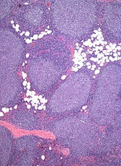
The European Commission has granted marketing authorization for the PI3K delta inhibitor idelalisib (Zydelig) to treat chronic lymphocytic leukemia (CLL) and follicular lymphoma (FL) in the European Union.
The drug is now approved for use in combination with rituximab for CLL patients who have received at least 1 prior therapy or as first-line treatment in CLL patients who have 17p deletion or TP53 mutation and are not eligible for chemo-immunotherapy.
Idelalisib is also approved as monotherapy for FL patients who were refractory to 2 prior lines of treatment.
These approvals are based on data from 2 clinical trials—Study 116 and Study 101-09.
Study 116: Idelalisib in CLL
This phase 3 trial was stopped early because idelalisib had a significant impact on progression-free survival.
The study included 220 CLL patients who could not receive chemotherapy. Half were randomized to receive idelalisib plus rituximab, and the other half were randomized to rituximab plus placebo.
Patients in the rituximab-idelalisib arm had a much higher overall response rate than patients in the rituximab-placebo arm—81% and 13%, respectively (P<0.001). There were no complete responses.
At 24 weeks, the rate of progression-free survival was 93% in the rituximab-idelalisib arm and 46% in the rituximab-placebo arm (P<0.001). The median progression-free survival was 5.5 months in the rituximab-placebo arm and not reached in the rituximab-idelalisib arm (P<0.001).
At 12 months, the overall survival rate was 92% in the rituximab-idelalisib arm and 80% in the rituximab-placebo arm (P=0.02).
Most adverse events, in either treatment arm, were grade 2 or lower. The most common events in the rituximab-idelalisib arm were pyrexia, fatigue, nausea, chills, and diarrhea. In the rituximab-placebo arm, the most common events were infusion-related reactions, fatigue, cough, nausea, and dyspnea.
There were more serious adverse events in the rituximab-idelalisib arm than in the rituximab-placebo arm—40% and 35%, respectively. The most frequent serious events were pneumonia, pyrexia, and febrile neutropenia (in both treatment arms).
Study 101-09: Idelalisib in FL
This phase 2 trial enrolled 125 patients with indolent non-Hodgkin lymphoma who were refractory to rituximab and chemotherapy containing an alkylating agent. Patients received idelalisib monotherapy.
Of the 72 subjects with FL, 54% achieved a response, and 8% had a complete response. The median duration of response was not reached (range, 0-14.8 months).
Improvements in survival or disease-related symptoms have not been established.
In all patients, the most common grade 3 or higher adverse events were neutropenia (27%), elevations in aminotransferase levels (13%), diarrhea (13%), and pneumonia (7%).
Idelalisib is under development by Gilead Sciences. The drug is already approved in the US for the aforementioned indications, as well as to treat small lymphocytic lymphoma.
The European Commission has granted marketing authorization for the PI3K delta inhibitor idelalisib (Zydelig) to treat chronic lymphocytic leukemia (CLL) and follicular lymphoma (FL) in the European Union.
The drug is now approved for use in combination with rituximab for CLL patients who have received at least 1 prior therapy or as first-line treatment in CLL patients who have 17p deletion or TP53 mutation and are not eligible for chemo-immunotherapy.
Idelalisib is also approved as monotherapy for FL patients who were refractory to 2 prior lines of treatment.
These approvals are based on data from 2 clinical trials—Study 116 and Study 101-09.
Study 116: Idelalisib in CLL
This phase 3 trial was stopped early because idelalisib had a significant impact on progression-free survival.
The study included 220 CLL patients who could not receive chemotherapy. Half were randomized to receive idelalisib plus rituximab, and the other half were randomized to rituximab plus placebo.
Patients in the rituximab-idelalisib arm had a much higher overall response rate than patients in the rituximab-placebo arm—81% and 13%, respectively (P<0.001). There were no complete responses.
At 24 weeks, the rate of progression-free survival was 93% in the rituximab-idelalisib arm and 46% in the rituximab-placebo arm (P<0.001). The median progression-free survival was 5.5 months in the rituximab-placebo arm and not reached in the rituximab-idelalisib arm (P<0.001).
At 12 months, the overall survival rate was 92% in the rituximab-idelalisib arm and 80% in the rituximab-placebo arm (P=0.02).
Most adverse events, in either treatment arm, were grade 2 or lower. The most common events in the rituximab-idelalisib arm were pyrexia, fatigue, nausea, chills, and diarrhea. In the rituximab-placebo arm, the most common events were infusion-related reactions, fatigue, cough, nausea, and dyspnea.
There were more serious adverse events in the rituximab-idelalisib arm than in the rituximab-placebo arm—40% and 35%, respectively. The most frequent serious events were pneumonia, pyrexia, and febrile neutropenia (in both treatment arms).
Study 101-09: Idelalisib in FL
This phase 2 trial enrolled 125 patients with indolent non-Hodgkin lymphoma who were refractory to rituximab and chemotherapy containing an alkylating agent. Patients received idelalisib monotherapy.
Of the 72 subjects with FL, 54% achieved a response, and 8% had a complete response. The median duration of response was not reached (range, 0-14.8 months).
Improvements in survival or disease-related symptoms have not been established.
In all patients, the most common grade 3 or higher adverse events were neutropenia (27%), elevations in aminotransferase levels (13%), diarrhea (13%), and pneumonia (7%).
Idelalisib is under development by Gilead Sciences. The drug is already approved in the US for the aforementioned indications, as well as to treat small lymphocytic lymphoma.
The European Commission has granted marketing authorization for the PI3K delta inhibitor idelalisib (Zydelig) to treat chronic lymphocytic leukemia (CLL) and follicular lymphoma (FL) in the European Union.
The drug is now approved for use in combination with rituximab for CLL patients who have received at least 1 prior therapy or as first-line treatment in CLL patients who have 17p deletion or TP53 mutation and are not eligible for chemo-immunotherapy.
Idelalisib is also approved as monotherapy for FL patients who were refractory to 2 prior lines of treatment.
These approvals are based on data from 2 clinical trials—Study 116 and Study 101-09.
Study 116: Idelalisib in CLL
This phase 3 trial was stopped early because idelalisib had a significant impact on progression-free survival.
The study included 220 CLL patients who could not receive chemotherapy. Half were randomized to receive idelalisib plus rituximab, and the other half were randomized to rituximab plus placebo.
Patients in the rituximab-idelalisib arm had a much higher overall response rate than patients in the rituximab-placebo arm—81% and 13%, respectively (P<0.001). There were no complete responses.
At 24 weeks, the rate of progression-free survival was 93% in the rituximab-idelalisib arm and 46% in the rituximab-placebo arm (P<0.001). The median progression-free survival was 5.5 months in the rituximab-placebo arm and not reached in the rituximab-idelalisib arm (P<0.001).
At 12 months, the overall survival rate was 92% in the rituximab-idelalisib arm and 80% in the rituximab-placebo arm (P=0.02).
Most adverse events, in either treatment arm, were grade 2 or lower. The most common events in the rituximab-idelalisib arm were pyrexia, fatigue, nausea, chills, and diarrhea. In the rituximab-placebo arm, the most common events were infusion-related reactions, fatigue, cough, nausea, and dyspnea.
There were more serious adverse events in the rituximab-idelalisib arm than in the rituximab-placebo arm—40% and 35%, respectively. The most frequent serious events were pneumonia, pyrexia, and febrile neutropenia (in both treatment arms).
Study 101-09: Idelalisib in FL
This phase 2 trial enrolled 125 patients with indolent non-Hodgkin lymphoma who were refractory to rituximab and chemotherapy containing an alkylating agent. Patients received idelalisib monotherapy.
Of the 72 subjects with FL, 54% achieved a response, and 8% had a complete response. The median duration of response was not reached (range, 0-14.8 months).
Improvements in survival or disease-related symptoms have not been established.
In all patients, the most common grade 3 or higher adverse events were neutropenia (27%), elevations in aminotransferase levels (13%), diarrhea (13%), and pneumonia (7%).
Idelalisib is under development by Gilead Sciences. The drug is already approved in the US for the aforementioned indications, as well as to treat small lymphocytic lymphoma.