User login
Preventing cancer-related infection

Credit: CDC/Kimberly Smith
and Christine Ford
NEW YORK—Hand washing is still the single most effective method to prevent the transmission of infection, but additional measures can reduce the risk of cancer-related infections, according to a speaker at the NCCN 9th Annual Congress: Hematologic Malignancies.
In her presentation, Laura Zitella, RN, of the Stanford Cancer Institute in California, discussed current recommendations for pharmacologic and non-pharmacologic infection prophylaxis.
She noted that NCCN Guidelines on the Prevention and Treatment of Cancer-Related Infections state that the highest risk of infection is in patients undergoing allogeneic hematopoietic stem cell transplant (HSCT), acute leukemia patients undergoing induction or consolidation therapy, patients receiving alemtuzumab therapy, patients with graft-vs-host disease (GVHD) treated with high-dose steroids, and patients with neutropenia anticipated to last greater than 10 days.
Antibiotic prophylaxis
Prior to 2005, Zitella said, no survival benefit was observed for antibiotic prophylaxis. All of this changed with the results of a meta-analysis. The analysis included 95 randomized, controlled trials and 9283 patients, the majority having acute leukemia or undergoing HSCT.
For the first time, antibiotic prophylaxis was shown to confer a survival benefit. In neutropenic patients, prophylaxis reduced overall mortality by 33% and infection-related mortality by 42%, compared with placebo or no treatment.
Prophylaxis is not recommended for low-risk neutropenic patients, Zitella said, because it is not proven to decrease morality.
And the drugs of choice are levofloxacin (500-750 mg PO daily) or ciprofloxacin (500-750 mg PO twice daily).
Colony-stimulating factors
Consensus guidelines for the use of colony-stimulating factors (CSFs) are a compilation of ASCO, EORTC, ESMO, and NCCN guidelines.
CSFs may be used prophylactically to prevent chemotherapy-induced neutropenia, febrile neutropenia, and infection. They reduce the duration of hospitalization, the duration of parenteral antibiotics, and have shown a survival benefit.
CSFs are recommended if the risk of febrile neutropenia is 20% or greater. CSFs are not routinely recommended for patients undergoing radiation treatment, acute myeloid leukemia induction, or patients with Hodgkin lymphoma.
Antifungal prophylaxis
Zitella noted that fluconazole is the best-studied antifungal prophylaxis and is recommended as the primary prophylaxis for HSCT patients.
In double-blind, placebo-controlled trials, fluconazole reduced mucosal candidiasis and invasive Candida infections in patients undergoing HSCT. And it improved survival at day 110 after transplant.
Posaconazole prophylaxis has proven effective in patients with acute myeloid leukemia or myelodysplastic syndromes undergoing intensive chemotherapy. The drug reduced invasive fungal infections, including aspergillosis, and improved survival.
For patients with GVHD on immunosuppressive therapy, posaconazole and fluconazole prophylaxis were equivalent in preventing invasive fungal infections. However, posaconazole reduced the incidence of invasive aspergillosis and fungal-related mortality.
The NCCN guidelines, Zitella said, spell out which antifungal agents should be used for each disease or therapeutic intervention.
Antiviral prophylaxis
HSV and VZV
Patients requiring antiviral prophylaxis for herpes simplex virus (HSV) and varicella zoster virus (VZV) should be seropositive and have acute leukemia, GVHD treated with steroids, prior HSV reactivation under treatment, or have undergone HSCT.
Zitella pointed out that patients treated with proteasome inhibitors, such as bortezomib, alemtuzumab, or purine analaogs, such as fludarabine, are more at risk and should also receive antiviral prophylaxis.
Recommended drugs include valacyclovir, acyclovir, or famciclovir.
CMV
Cytomegalovirus-positive (CMV+) patients at high risk include those who have received an allogeneic HSCT or treatment with alemtuzumab.
Zitella explained that for these patients, prophylaxis is uncommon, and a pre-emptive strategy should be used, including testing 3 to 6 months after transplant or in the setting of GVHD and 2 months after alemtuzumab therapy.
CMV viremia should be treated with valganciclovir, ganciclovir, foscarnet, or cidofovir.
HBV
Zitella noted that 30% of the world population has been infected with hepatitis B virus (HBV), and reactivation during cancer treatment can lead to fulminant hepatitis and death.
NCCN recommends that patients undergoing immunosuppressive therapy, allogeneic HSCT candidates, patients receiving anti-CD20 monoclonal antibodies, those treated with alemtuzumab, and patients receiving systemic therapy who have an obvious risk factor for HBV infection should be tested.
Entecavir, tenofovir, adefovir, telbivudine, or lamivudine may be used to prevent HBV reactivation.
Pneumocystis pneumonia prophylaxis
Patients undergoing allogeneic HSCT, patients with acute lymphoblastic leukemia, those treated with alemtuzumab, and those with a CD4 count below 200 cells/mcL should receive pneumocystis pneumonia prophylaxis.
Trimethorpim/sulfamethoxazole is the drug of choice. Atovaquone, dapsone, and inhaled or IV pentamidine are alternatives.
Vaccines
Zitella pointed out that recommended vaccines include influenza, pneumococcal, and tetanus, diphtheria, and acellular pertussis.
She cautioned that live attenuated vaccines should not be given to cancer patients. Other vaccines to avoid include smallpox; measles, mumps, and rubella; varicella zoster; rotavirus; yellow fever; oral typhoid; BCG; and oral polio vaccine.
Neutropenic precautions
Low microbial diets are a hot topic among patients undergoing cancer treatment, Zitella said. Fresh fruits and vegetables used to be restricted, but no studies show that dietary restrictions decrease the risk of infection.
Zitella stressed, however, that standard food safety recommendations of the USDA/FDA should be followed.
She also noted that HEPA filtration is protective against molds in high-risk patients, antiseptic bathing has contradictory evidence, the benefit of laminar airflow is unclear, and protective isolation has not been proven to reduce the risk of infection. 
Credit: CDC/Kimberly Smith
and Christine Ford
NEW YORK—Hand washing is still the single most effective method to prevent the transmission of infection, but additional measures can reduce the risk of cancer-related infections, according to a speaker at the NCCN 9th Annual Congress: Hematologic Malignancies.
In her presentation, Laura Zitella, RN, of the Stanford Cancer Institute in California, discussed current recommendations for pharmacologic and non-pharmacologic infection prophylaxis.
She noted that NCCN Guidelines on the Prevention and Treatment of Cancer-Related Infections state that the highest risk of infection is in patients undergoing allogeneic hematopoietic stem cell transplant (HSCT), acute leukemia patients undergoing induction or consolidation therapy, patients receiving alemtuzumab therapy, patients with graft-vs-host disease (GVHD) treated with high-dose steroids, and patients with neutropenia anticipated to last greater than 10 days.
Antibiotic prophylaxis
Prior to 2005, Zitella said, no survival benefit was observed for antibiotic prophylaxis. All of this changed with the results of a meta-analysis. The analysis included 95 randomized, controlled trials and 9283 patients, the majority having acute leukemia or undergoing HSCT.
For the first time, antibiotic prophylaxis was shown to confer a survival benefit. In neutropenic patients, prophylaxis reduced overall mortality by 33% and infection-related mortality by 42%, compared with placebo or no treatment.
Prophylaxis is not recommended for low-risk neutropenic patients, Zitella said, because it is not proven to decrease morality.
And the drugs of choice are levofloxacin (500-750 mg PO daily) or ciprofloxacin (500-750 mg PO twice daily).
Colony-stimulating factors
Consensus guidelines for the use of colony-stimulating factors (CSFs) are a compilation of ASCO, EORTC, ESMO, and NCCN guidelines.
CSFs may be used prophylactically to prevent chemotherapy-induced neutropenia, febrile neutropenia, and infection. They reduce the duration of hospitalization, the duration of parenteral antibiotics, and have shown a survival benefit.
CSFs are recommended if the risk of febrile neutropenia is 20% or greater. CSFs are not routinely recommended for patients undergoing radiation treatment, acute myeloid leukemia induction, or patients with Hodgkin lymphoma.
Antifungal prophylaxis
Zitella noted that fluconazole is the best-studied antifungal prophylaxis and is recommended as the primary prophylaxis for HSCT patients.
In double-blind, placebo-controlled trials, fluconazole reduced mucosal candidiasis and invasive Candida infections in patients undergoing HSCT. And it improved survival at day 110 after transplant.
Posaconazole prophylaxis has proven effective in patients with acute myeloid leukemia or myelodysplastic syndromes undergoing intensive chemotherapy. The drug reduced invasive fungal infections, including aspergillosis, and improved survival.
For patients with GVHD on immunosuppressive therapy, posaconazole and fluconazole prophylaxis were equivalent in preventing invasive fungal infections. However, posaconazole reduced the incidence of invasive aspergillosis and fungal-related mortality.
The NCCN guidelines, Zitella said, spell out which antifungal agents should be used for each disease or therapeutic intervention.
Antiviral prophylaxis
HSV and VZV
Patients requiring antiviral prophylaxis for herpes simplex virus (HSV) and varicella zoster virus (VZV) should be seropositive and have acute leukemia, GVHD treated with steroids, prior HSV reactivation under treatment, or have undergone HSCT.
Zitella pointed out that patients treated with proteasome inhibitors, such as bortezomib, alemtuzumab, or purine analaogs, such as fludarabine, are more at risk and should also receive antiviral prophylaxis.
Recommended drugs include valacyclovir, acyclovir, or famciclovir.
CMV
Cytomegalovirus-positive (CMV+) patients at high risk include those who have received an allogeneic HSCT or treatment with alemtuzumab.
Zitella explained that for these patients, prophylaxis is uncommon, and a pre-emptive strategy should be used, including testing 3 to 6 months after transplant or in the setting of GVHD and 2 months after alemtuzumab therapy.
CMV viremia should be treated with valganciclovir, ganciclovir, foscarnet, or cidofovir.
HBV
Zitella noted that 30% of the world population has been infected with hepatitis B virus (HBV), and reactivation during cancer treatment can lead to fulminant hepatitis and death.
NCCN recommends that patients undergoing immunosuppressive therapy, allogeneic HSCT candidates, patients receiving anti-CD20 monoclonal antibodies, those treated with alemtuzumab, and patients receiving systemic therapy who have an obvious risk factor for HBV infection should be tested.
Entecavir, tenofovir, adefovir, telbivudine, or lamivudine may be used to prevent HBV reactivation.
Pneumocystis pneumonia prophylaxis
Patients undergoing allogeneic HSCT, patients with acute lymphoblastic leukemia, those treated with alemtuzumab, and those with a CD4 count below 200 cells/mcL should receive pneumocystis pneumonia prophylaxis.
Trimethorpim/sulfamethoxazole is the drug of choice. Atovaquone, dapsone, and inhaled or IV pentamidine are alternatives.
Vaccines
Zitella pointed out that recommended vaccines include influenza, pneumococcal, and tetanus, diphtheria, and acellular pertussis.
She cautioned that live attenuated vaccines should not be given to cancer patients. Other vaccines to avoid include smallpox; measles, mumps, and rubella; varicella zoster; rotavirus; yellow fever; oral typhoid; BCG; and oral polio vaccine.
Neutropenic precautions
Low microbial diets are a hot topic among patients undergoing cancer treatment, Zitella said. Fresh fruits and vegetables used to be restricted, but no studies show that dietary restrictions decrease the risk of infection.
Zitella stressed, however, that standard food safety recommendations of the USDA/FDA should be followed.
She also noted that HEPA filtration is protective against molds in high-risk patients, antiseptic bathing has contradictory evidence, the benefit of laminar airflow is unclear, and protective isolation has not been proven to reduce the risk of infection.
Credit: CDC/Kimberly Smith
and Christine Ford
NEW YORK—Hand washing is still the single most effective method to prevent the transmission of infection, but additional measures can reduce the risk of cancer-related infections, according to a speaker at the NCCN 9th Annual Congress: Hematologic Malignancies.
In her presentation, Laura Zitella, RN, of the Stanford Cancer Institute in California, discussed current recommendations for pharmacologic and non-pharmacologic infection prophylaxis.
She noted that NCCN Guidelines on the Prevention and Treatment of Cancer-Related Infections state that the highest risk of infection is in patients undergoing allogeneic hematopoietic stem cell transplant (HSCT), acute leukemia patients undergoing induction or consolidation therapy, patients receiving alemtuzumab therapy, patients with graft-vs-host disease (GVHD) treated with high-dose steroids, and patients with neutropenia anticipated to last greater than 10 days.
Antibiotic prophylaxis
Prior to 2005, Zitella said, no survival benefit was observed for antibiotic prophylaxis. All of this changed with the results of a meta-analysis. The analysis included 95 randomized, controlled trials and 9283 patients, the majority having acute leukemia or undergoing HSCT.
For the first time, antibiotic prophylaxis was shown to confer a survival benefit. In neutropenic patients, prophylaxis reduced overall mortality by 33% and infection-related mortality by 42%, compared with placebo or no treatment.
Prophylaxis is not recommended for low-risk neutropenic patients, Zitella said, because it is not proven to decrease morality.
And the drugs of choice are levofloxacin (500-750 mg PO daily) or ciprofloxacin (500-750 mg PO twice daily).
Colony-stimulating factors
Consensus guidelines for the use of colony-stimulating factors (CSFs) are a compilation of ASCO, EORTC, ESMO, and NCCN guidelines.
CSFs may be used prophylactically to prevent chemotherapy-induced neutropenia, febrile neutropenia, and infection. They reduce the duration of hospitalization, the duration of parenteral antibiotics, and have shown a survival benefit.
CSFs are recommended if the risk of febrile neutropenia is 20% or greater. CSFs are not routinely recommended for patients undergoing radiation treatment, acute myeloid leukemia induction, or patients with Hodgkin lymphoma.
Antifungal prophylaxis
Zitella noted that fluconazole is the best-studied antifungal prophylaxis and is recommended as the primary prophylaxis for HSCT patients.
In double-blind, placebo-controlled trials, fluconazole reduced mucosal candidiasis and invasive Candida infections in patients undergoing HSCT. And it improved survival at day 110 after transplant.
Posaconazole prophylaxis has proven effective in patients with acute myeloid leukemia or myelodysplastic syndromes undergoing intensive chemotherapy. The drug reduced invasive fungal infections, including aspergillosis, and improved survival.
For patients with GVHD on immunosuppressive therapy, posaconazole and fluconazole prophylaxis were equivalent in preventing invasive fungal infections. However, posaconazole reduced the incidence of invasive aspergillosis and fungal-related mortality.
The NCCN guidelines, Zitella said, spell out which antifungal agents should be used for each disease or therapeutic intervention.
Antiviral prophylaxis
HSV and VZV
Patients requiring antiviral prophylaxis for herpes simplex virus (HSV) and varicella zoster virus (VZV) should be seropositive and have acute leukemia, GVHD treated with steroids, prior HSV reactivation under treatment, or have undergone HSCT.
Zitella pointed out that patients treated with proteasome inhibitors, such as bortezomib, alemtuzumab, or purine analaogs, such as fludarabine, are more at risk and should also receive antiviral prophylaxis.
Recommended drugs include valacyclovir, acyclovir, or famciclovir.
CMV
Cytomegalovirus-positive (CMV+) patients at high risk include those who have received an allogeneic HSCT or treatment with alemtuzumab.
Zitella explained that for these patients, prophylaxis is uncommon, and a pre-emptive strategy should be used, including testing 3 to 6 months after transplant or in the setting of GVHD and 2 months after alemtuzumab therapy.
CMV viremia should be treated with valganciclovir, ganciclovir, foscarnet, or cidofovir.
HBV
Zitella noted that 30% of the world population has been infected with hepatitis B virus (HBV), and reactivation during cancer treatment can lead to fulminant hepatitis and death.
NCCN recommends that patients undergoing immunosuppressive therapy, allogeneic HSCT candidates, patients receiving anti-CD20 monoclonal antibodies, those treated with alemtuzumab, and patients receiving systemic therapy who have an obvious risk factor for HBV infection should be tested.
Entecavir, tenofovir, adefovir, telbivudine, or lamivudine may be used to prevent HBV reactivation.
Pneumocystis pneumonia prophylaxis
Patients undergoing allogeneic HSCT, patients with acute lymphoblastic leukemia, those treated with alemtuzumab, and those with a CD4 count below 200 cells/mcL should receive pneumocystis pneumonia prophylaxis.
Trimethorpim/sulfamethoxazole is the drug of choice. Atovaquone, dapsone, and inhaled or IV pentamidine are alternatives.
Vaccines
Zitella pointed out that recommended vaccines include influenza, pneumococcal, and tetanus, diphtheria, and acellular pertussis.
She cautioned that live attenuated vaccines should not be given to cancer patients. Other vaccines to avoid include smallpox; measles, mumps, and rubella; varicella zoster; rotavirus; yellow fever; oral typhoid; BCG; and oral polio vaccine.
Neutropenic precautions
Low microbial diets are a hot topic among patients undergoing cancer treatment, Zitella said. Fresh fruits and vegetables used to be restricted, but no studies show that dietary restrictions decrease the risk of infection.
Zitella stressed, however, that standard food safety recommendations of the USDA/FDA should be followed.
She also noted that HEPA filtration is protective against molds in high-risk patients, antiseptic bathing has contradictory evidence, the benefit of laminar airflow is unclear, and protective isolation has not been proven to reduce the risk of infection.
Speaker offers alternatives for sustainable TKI use in CML

NEW YORK—If any oncology drugs warrant a high price, BCR-ABL tyrosine kinase inhibitors (TKIs) could make a strong case, according to a speaker at the NCCN 9th Annual Congress: Hematologic Malignancies.
TKIs have revolutionized the therapeutic landscape in chronic myeloid leukemia (CML), said Neil P. Shah, MD, PhD, of the UCSF
Helen Diller Family Comprehensive Cancer Center in San Francisco.
Five-year overall survival is 93% in CML patients treated with imatinib. And 80,000 people are estimated to be living with CML in 2014, a number that is expected to double by 2035.
Yet the increase in life expectancy places a burden on the healthcare system, Dr Shah said, particularly since the price of BCR-ABL TKIs has risen dramatically.
A year of imatinib therapy circa 2001 cost approximately $30,000, and by 2013, approximately $92,000. Second-generation (2G) TKIs cost even more, at about $115,000 annually, and the third-generation TKI ponatinib costs about $138,000 annually.
Confounding the financial picture will be the advent of generic imatinib, which will be available in the US in early 2016.
Generic imatinib will, presumably, be less expensive than the brand-name drug. However, it is not clear whether it will be as effective or what impact it will have on the 2G and 3G TKIs.
With all that in mind, Dr Shah provided some options for maximizing therapeutic outcomes in a fiscally responsible manner.
He based his potential solutions on evidence-based premises. First, imatinib is a highly active therapy for many chronic-phase CML patients. The 8-year update of the IRIS study shows that 53% of patients sustain complete cytogenetic remission (CCyR) with imatinib.
Second, patients who are likely to do well on TKI therapy can be identified early based on their BCR-ABL PCR levels after 3 months of therapy.
This important 3-month milestone has been incorporated into the NCCN guidelines. Patients who achieve BCR-ABL/ABL levels of 10% or less have a significantly better overall survival than those who have levels greater than 10%.
Third, the ENESTnd and DASISION studies demonstrated that nilotinib and dasatinib elicit achievement of important treatment milestones (PCR<10% at 3 months and CCyR after 12 months) in a significantly higher proportion of patients than imatinib. But to date, neither agent has produced a significant improvement in overall survival.
And fourth, while nilotinib and dasatinib have an undisputable clinical benefit over imatinib in terms of transformation to accelerated phase/blast crisis (AP/BC), these agents cost between $23,000 and $31,000 more than imatinib for an annual course of treatment.
Dr Shah calculated that to achieve approximately 10 fewer AP/BC transformations over 5 years, approximately 300 patients would need to be treated at an additional cost of $37.5 million. This equates to $3.75 million in additional costs for 1 transformation event spared over 5 years.
“We do have to keep in mind transformation events are not cheap,” he said. “So when these happen, patients need to be hospitalized for chemotherapy, typically, or they have to go to allogeneic stem cell transplantation.”
So Dr Shah proffered 3 potential approaches for fiscally sustainable TKI use.
The first approach would be to initiate imatinib in most, if not all, chronic-phase CML patients. If they achieve BCR-ABL levels <10% at 3 months or partial CyR after 12 months, then continue imatinib. If not, switch to a 2G TKI.
Dr Shah noted that 40% to 60% of patients will have a deep, durable response to imatinib.
The second approach would be to initiate 2G TKIs in newly diagnosed patients. And when they achieve CCyR or major molecular response (MMR), enroll them in a clinical trial.
These trials could investigate a switch to imatinib, use of a lower dose of the 2G TKI, a switch to interferon-alfa, the addition of a CYP3A4 inhibitor (such as grapefruit) and a lower dose, or another investigational option.
And the third approach would be to initiate imatinib or an approved 2G TKI in newly diagnosed patients. When they achieve targeted molecular remission, enroll them in a treatment-discontinuation trial.
Dr Shah pointed out that in the STIM study, 61% of patients remained in complete molecular remission 60 months after discontinuing imatinib, and 40% experienced treatment-free survival.
Taking into account the cost of imatinib and the number of months without treatment, STIM investigators estimated the savings to be €4,587,500, or approximately $6 to $7 million.
Patients who relapsed 6 to 9 months after discontinuing imatinib tended to respond well to a rechallenge with imatinib. But Dr Shah cautioned that, at present, discontinuation should only be performed in the context of a clinical trial.
NEW YORK—If any oncology drugs warrant a high price, BCR-ABL tyrosine kinase inhibitors (TKIs) could make a strong case, according to a speaker at the NCCN 9th Annual Congress: Hematologic Malignancies.
TKIs have revolutionized the therapeutic landscape in chronic myeloid leukemia (CML), said Neil P. Shah, MD, PhD, of the UCSF
Helen Diller Family Comprehensive Cancer Center in San Francisco.
Five-year overall survival is 93% in CML patients treated with imatinib. And 80,000 people are estimated to be living with CML in 2014, a number that is expected to double by 2035.
Yet the increase in life expectancy places a burden on the healthcare system, Dr Shah said, particularly since the price of BCR-ABL TKIs has risen dramatically.
A year of imatinib therapy circa 2001 cost approximately $30,000, and by 2013, approximately $92,000. Second-generation (2G) TKIs cost even more, at about $115,000 annually, and the third-generation TKI ponatinib costs about $138,000 annually.
Confounding the financial picture will be the advent of generic imatinib, which will be available in the US in early 2016.
Generic imatinib will, presumably, be less expensive than the brand-name drug. However, it is not clear whether it will be as effective or what impact it will have on the 2G and 3G TKIs.
With all that in mind, Dr Shah provided some options for maximizing therapeutic outcomes in a fiscally responsible manner.
He based his potential solutions on evidence-based premises. First, imatinib is a highly active therapy for many chronic-phase CML patients. The 8-year update of the IRIS study shows that 53% of patients sustain complete cytogenetic remission (CCyR) with imatinib.
Second, patients who are likely to do well on TKI therapy can be identified early based on their BCR-ABL PCR levels after 3 months of therapy.
This important 3-month milestone has been incorporated into the NCCN guidelines. Patients who achieve BCR-ABL/ABL levels of 10% or less have a significantly better overall survival than those who have levels greater than 10%.
Third, the ENESTnd and DASISION studies demonstrated that nilotinib and dasatinib elicit achievement of important treatment milestones (PCR<10% at 3 months and CCyR after 12 months) in a significantly higher proportion of patients than imatinib. But to date, neither agent has produced a significant improvement in overall survival.
And fourth, while nilotinib and dasatinib have an undisputable clinical benefit over imatinib in terms of transformation to accelerated phase/blast crisis (AP/BC), these agents cost between $23,000 and $31,000 more than imatinib for an annual course of treatment.
Dr Shah calculated that to achieve approximately 10 fewer AP/BC transformations over 5 years, approximately 300 patients would need to be treated at an additional cost of $37.5 million. This equates to $3.75 million in additional costs for 1 transformation event spared over 5 years.
“We do have to keep in mind transformation events are not cheap,” he said. “So when these happen, patients need to be hospitalized for chemotherapy, typically, or they have to go to allogeneic stem cell transplantation.”
So Dr Shah proffered 3 potential approaches for fiscally sustainable TKI use.
The first approach would be to initiate imatinib in most, if not all, chronic-phase CML patients. If they achieve BCR-ABL levels <10% at 3 months or partial CyR after 12 months, then continue imatinib. If not, switch to a 2G TKI.
Dr Shah noted that 40% to 60% of patients will have a deep, durable response to imatinib.
The second approach would be to initiate 2G TKIs in newly diagnosed patients. And when they achieve CCyR or major molecular response (MMR), enroll them in a clinical trial.
These trials could investigate a switch to imatinib, use of a lower dose of the 2G TKI, a switch to interferon-alfa, the addition of a CYP3A4 inhibitor (such as grapefruit) and a lower dose, or another investigational option.
And the third approach would be to initiate imatinib or an approved 2G TKI in newly diagnosed patients. When they achieve targeted molecular remission, enroll them in a treatment-discontinuation trial.
Dr Shah pointed out that in the STIM study, 61% of patients remained in complete molecular remission 60 months after discontinuing imatinib, and 40% experienced treatment-free survival.
Taking into account the cost of imatinib and the number of months without treatment, STIM investigators estimated the savings to be €4,587,500, or approximately $6 to $7 million.
Patients who relapsed 6 to 9 months after discontinuing imatinib tended to respond well to a rechallenge with imatinib. But Dr Shah cautioned that, at present, discontinuation should only be performed in the context of a clinical trial.
NEW YORK—If any oncology drugs warrant a high price, BCR-ABL tyrosine kinase inhibitors (TKIs) could make a strong case, according to a speaker at the NCCN 9th Annual Congress: Hematologic Malignancies.
TKIs have revolutionized the therapeutic landscape in chronic myeloid leukemia (CML), said Neil P. Shah, MD, PhD, of the UCSF
Helen Diller Family Comprehensive Cancer Center in San Francisco.
Five-year overall survival is 93% in CML patients treated with imatinib. And 80,000 people are estimated to be living with CML in 2014, a number that is expected to double by 2035.
Yet the increase in life expectancy places a burden on the healthcare system, Dr Shah said, particularly since the price of BCR-ABL TKIs has risen dramatically.
A year of imatinib therapy circa 2001 cost approximately $30,000, and by 2013, approximately $92,000. Second-generation (2G) TKIs cost even more, at about $115,000 annually, and the third-generation TKI ponatinib costs about $138,000 annually.
Confounding the financial picture will be the advent of generic imatinib, which will be available in the US in early 2016.
Generic imatinib will, presumably, be less expensive than the brand-name drug. However, it is not clear whether it will be as effective or what impact it will have on the 2G and 3G TKIs.
With all that in mind, Dr Shah provided some options for maximizing therapeutic outcomes in a fiscally responsible manner.
He based his potential solutions on evidence-based premises. First, imatinib is a highly active therapy for many chronic-phase CML patients. The 8-year update of the IRIS study shows that 53% of patients sustain complete cytogenetic remission (CCyR) with imatinib.
Second, patients who are likely to do well on TKI therapy can be identified early based on their BCR-ABL PCR levels after 3 months of therapy.
This important 3-month milestone has been incorporated into the NCCN guidelines. Patients who achieve BCR-ABL/ABL levels of 10% or less have a significantly better overall survival than those who have levels greater than 10%.
Third, the ENESTnd and DASISION studies demonstrated that nilotinib and dasatinib elicit achievement of important treatment milestones (PCR<10% at 3 months and CCyR after 12 months) in a significantly higher proportion of patients than imatinib. But to date, neither agent has produced a significant improvement in overall survival.
And fourth, while nilotinib and dasatinib have an undisputable clinical benefit over imatinib in terms of transformation to accelerated phase/blast crisis (AP/BC), these agents cost between $23,000 and $31,000 more than imatinib for an annual course of treatment.
Dr Shah calculated that to achieve approximately 10 fewer AP/BC transformations over 5 years, approximately 300 patients would need to be treated at an additional cost of $37.5 million. This equates to $3.75 million in additional costs for 1 transformation event spared over 5 years.
“We do have to keep in mind transformation events are not cheap,” he said. “So when these happen, patients need to be hospitalized for chemotherapy, typically, or they have to go to allogeneic stem cell transplantation.”
So Dr Shah proffered 3 potential approaches for fiscally sustainable TKI use.
The first approach would be to initiate imatinib in most, if not all, chronic-phase CML patients. If they achieve BCR-ABL levels <10% at 3 months or partial CyR after 12 months, then continue imatinib. If not, switch to a 2G TKI.
Dr Shah noted that 40% to 60% of patients will have a deep, durable response to imatinib.
The second approach would be to initiate 2G TKIs in newly diagnosed patients. And when they achieve CCyR or major molecular response (MMR), enroll them in a clinical trial.
These trials could investigate a switch to imatinib, use of a lower dose of the 2G TKI, a switch to interferon-alfa, the addition of a CYP3A4 inhibitor (such as grapefruit) and a lower dose, or another investigational option.
And the third approach would be to initiate imatinib or an approved 2G TKI in newly diagnosed patients. When they achieve targeted molecular remission, enroll them in a treatment-discontinuation trial.
Dr Shah pointed out that in the STIM study, 61% of patients remained in complete molecular remission 60 months after discontinuing imatinib, and 40% experienced treatment-free survival.
Taking into account the cost of imatinib and the number of months without treatment, STIM investigators estimated the savings to be €4,587,500, or approximately $6 to $7 million.
Patients who relapsed 6 to 9 months after discontinuing imatinib tended to respond well to a rechallenge with imatinib. But Dr Shah cautioned that, at present, discontinuation should only be performed in the context of a clinical trial.
Split decision on role of upfront transplant in MM

Credit: Chad McNeeley
NEW YORK—A debate on the pros and cons of upfront transplant in symptomatic multiple myeloma (MM) yielded a split decision from the audience during the NCCN 9th Annual Congress: Hematologic Malignancies.
Sergio Giralt, MD, of Memorial Sloan Kettering Cancer Center in New York, argued for upfront transplant, pointing out that long-term MM survivors have transplant as upfront therapy.
Kenneth Anderson, MD, of Dana Farber/Brigham and Women’s Cancer Center in Boston, took the stance that, in the past 10 years, there has been a
revolution in novel therapies that has significantly improved survival in MM.
For upfront transplant
Dr Giralt cited the 36-month follow-up of the E4A03 landmark analysis of patients who went off therapy after 4 cycles of lenalidomide/dexamethasone to pursue early stem cell transplant and those who continued treatment until disease progression.
Regardless of whether the patients were younger than 65 years or between 65 and 70, the patients who had an early transplant had superior progression-free survival (PFS) and overall survival (OS) compared to those who did not.
Dr Giralt added that bortezomib should be a component of induction therapy prior to autologous stem cell transplant (ASCT). Even though there is no survival benefit with bortezomib-based regimens, he said, there is significant improvement in PFS, as shown in a meta-analysis of phase 3 European studies.
The E4A03 landmark study also determined that the more intense the treatment, the better the outcome. So patients with double ASCT had a significantly longer PFS than patients who only had a single transplant.
This held true for OS as well, and included patients with 17p deletion and/or t(4;14) who failed to achieve complete remission after bortezomib-based induction regimens.
An analysis of 27,987 MM patients with a median age of 68 years (range, 19 to 90) revealed that of the patients who survived 10 years or more, 16.5% had ASCT as part of their initial therapy.
Dr Giralt concluded that the preponderance of evidence supports high-dose melphalan and ASCT as upfront consolidation therapy for MM. And until results of randomized trials investigating combination therapies are reported, melphalan consolidation should be considered the standard of care for all eligible patients with MM.
Against upfront transplant
Dr Anderson countered with data showing limited or no improvement in survival with ASCT, including evidence from studies by Attal, Fermand, Blade, Child, and Barlogie.
ASCT confers only modest PFS advantage, he said, showing results of the Barlogie study in which patients undergoing ASCT had a 25-month PFS, compared with a 21-month PFS with VBMCP (vincristine, carmustine, melphalan, cyclophosphamide, and prednisone).
“In the last 10 to 15 years, there has been a revolution in myeloma,” Dr Anderson said. “We have a lot of novel agents here today, and we have even more coming. It’s a hugely exciting time.”
Dr Anderson pointed out that since the introduction of novel agents, survival has improved between 2006 and 2010, compared to the period between 2001 and 2005, and particularly in patients older than 65 years (P=0.001).
Transplant has also changed, he said. Novel therapies have been integrated into the transplant paradigm, either before, as induction and consolidation therapy, or after, as maintenance. He indicated that this begs the question as to whether we really need the transplant component.
There has also been unprecedented use of triplets in combination therapy, Dr Anderson said, resulting in overall response rates upwards of 90%. For example, carfilzomib in combination with lenalidomide and dexamethasone prompted an overall response rate of 94%, with a stringent complete response (CR), CR, and near CR of 53%.
“It’s a new day in myeloma,” he said. “It’s taken us a long time, but we’re worrying about minimal residual disease (MRD) now. We’re worried about getting to the endpoint of 1 myeloma cell in 1 million normal cells.”
The point is, he added, that with novel therapies, such as carfilzomib, lenalidomide, and dexamethasone, patients who achieve a complete response can become MRD negative, suggesting an unprecedented extent of response without transplant.
MRD negativity may also be accomplished with oral agents, such as ixazomib. The depth of response with ixazomib increases over the course of treatment, with 27% achieving stringent CR or CR with a median duration of response of 13.8 months, and 82% of patients attaining MRD-negative status.
“In the absence of transplant,” Dr Anderson said, “this is an unprecedented response.”
Dr Anderson also pointed out that in the era of novel agents, there is no difference in outcome between early or delayed transplant. The 4-year OS in transplant-eligible patients who received initial therapy with lenalidomide was 80%, regardless of the timing of ASCT.
And in one trial, patients who received a delayed transplant fared better in OS than those transplanted early.
Dr Anderson said there is a parallel, international phase 3 study underway (IFM/DFCI2009) that will provide an answer to the debate on upfront transplant in the not-too-distant future.
Credit: Chad McNeeley
NEW YORK—A debate on the pros and cons of upfront transplant in symptomatic multiple myeloma (MM) yielded a split decision from the audience during the NCCN 9th Annual Congress: Hematologic Malignancies.
Sergio Giralt, MD, of Memorial Sloan Kettering Cancer Center in New York, argued for upfront transplant, pointing out that long-term MM survivors have transplant as upfront therapy.
Kenneth Anderson, MD, of Dana Farber/Brigham and Women’s Cancer Center in Boston, took the stance that, in the past 10 years, there has been a
revolution in novel therapies that has significantly improved survival in MM.
For upfront transplant
Dr Giralt cited the 36-month follow-up of the E4A03 landmark analysis of patients who went off therapy after 4 cycles of lenalidomide/dexamethasone to pursue early stem cell transplant and those who continued treatment until disease progression.
Regardless of whether the patients were younger than 65 years or between 65 and 70, the patients who had an early transplant had superior progression-free survival (PFS) and overall survival (OS) compared to those who did not.
Dr Giralt added that bortezomib should be a component of induction therapy prior to autologous stem cell transplant (ASCT). Even though there is no survival benefit with bortezomib-based regimens, he said, there is significant improvement in PFS, as shown in a meta-analysis of phase 3 European studies.
The E4A03 landmark study also determined that the more intense the treatment, the better the outcome. So patients with double ASCT had a significantly longer PFS than patients who only had a single transplant.
This held true for OS as well, and included patients with 17p deletion and/or t(4;14) who failed to achieve complete remission after bortezomib-based induction regimens.
An analysis of 27,987 MM patients with a median age of 68 years (range, 19 to 90) revealed that of the patients who survived 10 years or more, 16.5% had ASCT as part of their initial therapy.
Dr Giralt concluded that the preponderance of evidence supports high-dose melphalan and ASCT as upfront consolidation therapy for MM. And until results of randomized trials investigating combination therapies are reported, melphalan consolidation should be considered the standard of care for all eligible patients with MM.
Against upfront transplant
Dr Anderson countered with data showing limited or no improvement in survival with ASCT, including evidence from studies by Attal, Fermand, Blade, Child, and Barlogie.
ASCT confers only modest PFS advantage, he said, showing results of the Barlogie study in which patients undergoing ASCT had a 25-month PFS, compared with a 21-month PFS with VBMCP (vincristine, carmustine, melphalan, cyclophosphamide, and prednisone).
“In the last 10 to 15 years, there has been a revolution in myeloma,” Dr Anderson said. “We have a lot of novel agents here today, and we have even more coming. It’s a hugely exciting time.”
Dr Anderson pointed out that since the introduction of novel agents, survival has improved between 2006 and 2010, compared to the period between 2001 and 2005, and particularly in patients older than 65 years (P=0.001).
Transplant has also changed, he said. Novel therapies have been integrated into the transplant paradigm, either before, as induction and consolidation therapy, or after, as maintenance. He indicated that this begs the question as to whether we really need the transplant component.
There has also been unprecedented use of triplets in combination therapy, Dr Anderson said, resulting in overall response rates upwards of 90%. For example, carfilzomib in combination with lenalidomide and dexamethasone prompted an overall response rate of 94%, with a stringent complete response (CR), CR, and near CR of 53%.
“It’s a new day in myeloma,” he said. “It’s taken us a long time, but we’re worrying about minimal residual disease (MRD) now. We’re worried about getting to the endpoint of 1 myeloma cell in 1 million normal cells.”
The point is, he added, that with novel therapies, such as carfilzomib, lenalidomide, and dexamethasone, patients who achieve a complete response can become MRD negative, suggesting an unprecedented extent of response without transplant.
MRD negativity may also be accomplished with oral agents, such as ixazomib. The depth of response with ixazomib increases over the course of treatment, with 27% achieving stringent CR or CR with a median duration of response of 13.8 months, and 82% of patients attaining MRD-negative status.
“In the absence of transplant,” Dr Anderson said, “this is an unprecedented response.”
Dr Anderson also pointed out that in the era of novel agents, there is no difference in outcome between early or delayed transplant. The 4-year OS in transplant-eligible patients who received initial therapy with lenalidomide was 80%, regardless of the timing of ASCT.
And in one trial, patients who received a delayed transplant fared better in OS than those transplanted early.
Dr Anderson said there is a parallel, international phase 3 study underway (IFM/DFCI2009) that will provide an answer to the debate on upfront transplant in the not-too-distant future.
Credit: Chad McNeeley
NEW YORK—A debate on the pros and cons of upfront transplant in symptomatic multiple myeloma (MM) yielded a split decision from the audience during the NCCN 9th Annual Congress: Hematologic Malignancies.
Sergio Giralt, MD, of Memorial Sloan Kettering Cancer Center in New York, argued for upfront transplant, pointing out that long-term MM survivors have transplant as upfront therapy.
Kenneth Anderson, MD, of Dana Farber/Brigham and Women’s Cancer Center in Boston, took the stance that, in the past 10 years, there has been a
revolution in novel therapies that has significantly improved survival in MM.
For upfront transplant
Dr Giralt cited the 36-month follow-up of the E4A03 landmark analysis of patients who went off therapy after 4 cycles of lenalidomide/dexamethasone to pursue early stem cell transplant and those who continued treatment until disease progression.
Regardless of whether the patients were younger than 65 years or between 65 and 70, the patients who had an early transplant had superior progression-free survival (PFS) and overall survival (OS) compared to those who did not.
Dr Giralt added that bortezomib should be a component of induction therapy prior to autologous stem cell transplant (ASCT). Even though there is no survival benefit with bortezomib-based regimens, he said, there is significant improvement in PFS, as shown in a meta-analysis of phase 3 European studies.
The E4A03 landmark study also determined that the more intense the treatment, the better the outcome. So patients with double ASCT had a significantly longer PFS than patients who only had a single transplant.
This held true for OS as well, and included patients with 17p deletion and/or t(4;14) who failed to achieve complete remission after bortezomib-based induction regimens.
An analysis of 27,987 MM patients with a median age of 68 years (range, 19 to 90) revealed that of the patients who survived 10 years or more, 16.5% had ASCT as part of their initial therapy.
Dr Giralt concluded that the preponderance of evidence supports high-dose melphalan and ASCT as upfront consolidation therapy for MM. And until results of randomized trials investigating combination therapies are reported, melphalan consolidation should be considered the standard of care for all eligible patients with MM.
Against upfront transplant
Dr Anderson countered with data showing limited or no improvement in survival with ASCT, including evidence from studies by Attal, Fermand, Blade, Child, and Barlogie.
ASCT confers only modest PFS advantage, he said, showing results of the Barlogie study in which patients undergoing ASCT had a 25-month PFS, compared with a 21-month PFS with VBMCP (vincristine, carmustine, melphalan, cyclophosphamide, and prednisone).
“In the last 10 to 15 years, there has been a revolution in myeloma,” Dr Anderson said. “We have a lot of novel agents here today, and we have even more coming. It’s a hugely exciting time.”
Dr Anderson pointed out that since the introduction of novel agents, survival has improved between 2006 and 2010, compared to the period between 2001 and 2005, and particularly in patients older than 65 years (P=0.001).
Transplant has also changed, he said. Novel therapies have been integrated into the transplant paradigm, either before, as induction and consolidation therapy, or after, as maintenance. He indicated that this begs the question as to whether we really need the transplant component.
There has also been unprecedented use of triplets in combination therapy, Dr Anderson said, resulting in overall response rates upwards of 90%. For example, carfilzomib in combination with lenalidomide and dexamethasone prompted an overall response rate of 94%, with a stringent complete response (CR), CR, and near CR of 53%.
“It’s a new day in myeloma,” he said. “It’s taken us a long time, but we’re worrying about minimal residual disease (MRD) now. We’re worried about getting to the endpoint of 1 myeloma cell in 1 million normal cells.”
The point is, he added, that with novel therapies, such as carfilzomib, lenalidomide, and dexamethasone, patients who achieve a complete response can become MRD negative, suggesting an unprecedented extent of response without transplant.
MRD negativity may also be accomplished with oral agents, such as ixazomib. The depth of response with ixazomib increases over the course of treatment, with 27% achieving stringent CR or CR with a median duration of response of 13.8 months, and 82% of patients attaining MRD-negative status.
“In the absence of transplant,” Dr Anderson said, “this is an unprecedented response.”
Dr Anderson also pointed out that in the era of novel agents, there is no difference in outcome between early or delayed transplant. The 4-year OS in transplant-eligible patients who received initial therapy with lenalidomide was 80%, regardless of the timing of ASCT.
And in one trial, patients who received a delayed transplant fared better in OS than those transplanted early.
Dr Anderson said there is a parallel, international phase 3 study underway (IFM/DFCI2009) that will provide an answer to the debate on upfront transplant in the not-too-distant future.
Drug could treat a range of blood cancers
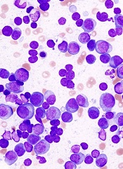
PHILADELPHIA—A drug that targets the ribosome may be active in a broad range of hematologic malignancies, researchers say.
The drug, CX-5461, inhibits the protein RNA polymerase I (Pol I), which is consistently upregulated in hematologic and other cancers.
CX-5461 significantly prolonged survival in mouse models of refractory acute myeloid leukemia (AML) and multiple myeloma (MM). It also synergized with everolimus to extend survival in mice with B-cell lymphoma.
Furthermore, the drug did not elicit severe adverse effects.
“We were excited to find that therapeutic doses of CX-5461 had little effect on normal cells in our experiments,” said Ross D. Hannan, PhD, of the Peter MacCallum Cancer Centre in Melbourne, Australia,
“Prior to these studies, few people would have guessed that such a therapeutic window could be obtained by targeting a so-called house-keeping protein that is essential to all cells for survival.”
Dr Hannan and his colleagues presented these findings in a poster at the AACR conference Hematologic Malignancies: Translating Discoveries to Novel Therapies.
The researchers previously showed that cancer cells are much more dependent on ribosome biogenesis than normal cells. And blocking the accelerated reading of ribosomal genes in mice—using CX5461—can cause lymphoma and leukemia cells to die, while sparing normal cells.
With their latest research, the group expanded upon these findings by testing CX-5461 in MLL-driven AML, V*κ-Myc-driven MM, and Eμ-Myc lymphoma.
They found that CX-5461 improved overall survival in MLL/ENL Nras leukemic mice, compared to placebo and standard therapy. The median survival was 17 days for vehicle-treated mice, 21 days for mice treated with cytarabine and doxorubicin, and 36 days for mice that received CX-5461 (P<0.0001 for vehicle vs CX-5461).
CX-5461 treated MLL-driven AML by inducing apoptosis, delaying cell-cycle progression, and promoting differentiation.
The researchers also found the therapeutic benefit of CX-5461 is not p53-dependent, which contradicts their previous findings. Human AML cell lines and primary patient samples were sensitive to CX-5461 independent of p53 status.
And MLL/ENL Nras p53-/- leukemic mice had significantly prolonged survival when treated with CX-5461, compared to vehicle-treated controls. The median survival was 11 days and 24 days, respectively (P<0.0001).
Likewise, CX-5461 significantly prolonged survival in mice bearing V*κ-Myc MM. The median survival was 103.5 days for controls and 175 days for mice that received CX-5461 (P<0.0001).
Finally, the researchers showed that CX-5461 synergizes with everolimus to treat Eμ-Myc lymphoma. The median survival was 15 days in control mice, 18 days in mice that received everolimus, 32 days in mice treated with CX-5461, and 54 days in mice that received both drugs (P<0.0001 for CX-5461 vs the combination).
“These results provide further rationale for the first-in-human phase 1 clinical trial that we initiated in July 2013 testing CX-5461 for patients with advanced hematological malignancies, including AML and multiple myeloma,” Dr Hannan said.
His group’s preclinical research was funded by the National Health and Medical Research Council, Australia; the Leukaemia Foundation of Australia; and Cancer Council Victoria, Melbourne, Australia. Senhwa Biosciences, the makers of CX-5461, provided the drug.
PHILADELPHIA—A drug that targets the ribosome may be active in a broad range of hematologic malignancies, researchers say.
The drug, CX-5461, inhibits the protein RNA polymerase I (Pol I), which is consistently upregulated in hematologic and other cancers.
CX-5461 significantly prolonged survival in mouse models of refractory acute myeloid leukemia (AML) and multiple myeloma (MM). It also synergized with everolimus to extend survival in mice with B-cell lymphoma.
Furthermore, the drug did not elicit severe adverse effects.
“We were excited to find that therapeutic doses of CX-5461 had little effect on normal cells in our experiments,” said Ross D. Hannan, PhD, of the Peter MacCallum Cancer Centre in Melbourne, Australia,
“Prior to these studies, few people would have guessed that such a therapeutic window could be obtained by targeting a so-called house-keeping protein that is essential to all cells for survival.”
Dr Hannan and his colleagues presented these findings in a poster at the AACR conference Hematologic Malignancies: Translating Discoveries to Novel Therapies.
The researchers previously showed that cancer cells are much more dependent on ribosome biogenesis than normal cells. And blocking the accelerated reading of ribosomal genes in mice—using CX5461—can cause lymphoma and leukemia cells to die, while sparing normal cells.
With their latest research, the group expanded upon these findings by testing CX-5461 in MLL-driven AML, V*κ-Myc-driven MM, and Eμ-Myc lymphoma.
They found that CX-5461 improved overall survival in MLL/ENL Nras leukemic mice, compared to placebo and standard therapy. The median survival was 17 days for vehicle-treated mice, 21 days for mice treated with cytarabine and doxorubicin, and 36 days for mice that received CX-5461 (P<0.0001 for vehicle vs CX-5461).
CX-5461 treated MLL-driven AML by inducing apoptosis, delaying cell-cycle progression, and promoting differentiation.
The researchers also found the therapeutic benefit of CX-5461 is not p53-dependent, which contradicts their previous findings. Human AML cell lines and primary patient samples were sensitive to CX-5461 independent of p53 status.
And MLL/ENL Nras p53-/- leukemic mice had significantly prolonged survival when treated with CX-5461, compared to vehicle-treated controls. The median survival was 11 days and 24 days, respectively (P<0.0001).
Likewise, CX-5461 significantly prolonged survival in mice bearing V*κ-Myc MM. The median survival was 103.5 days for controls and 175 days for mice that received CX-5461 (P<0.0001).
Finally, the researchers showed that CX-5461 synergizes with everolimus to treat Eμ-Myc lymphoma. The median survival was 15 days in control mice, 18 days in mice that received everolimus, 32 days in mice treated with CX-5461, and 54 days in mice that received both drugs (P<0.0001 for CX-5461 vs the combination).
“These results provide further rationale for the first-in-human phase 1 clinical trial that we initiated in July 2013 testing CX-5461 for patients with advanced hematological malignancies, including AML and multiple myeloma,” Dr Hannan said.
His group’s preclinical research was funded by the National Health and Medical Research Council, Australia; the Leukaemia Foundation of Australia; and Cancer Council Victoria, Melbourne, Australia. Senhwa Biosciences, the makers of CX-5461, provided the drug.
PHILADELPHIA—A drug that targets the ribosome may be active in a broad range of hematologic malignancies, researchers say.
The drug, CX-5461, inhibits the protein RNA polymerase I (Pol I), which is consistently upregulated in hematologic and other cancers.
CX-5461 significantly prolonged survival in mouse models of refractory acute myeloid leukemia (AML) and multiple myeloma (MM). It also synergized with everolimus to extend survival in mice with B-cell lymphoma.
Furthermore, the drug did not elicit severe adverse effects.
“We were excited to find that therapeutic doses of CX-5461 had little effect on normal cells in our experiments,” said Ross D. Hannan, PhD, of the Peter MacCallum Cancer Centre in Melbourne, Australia,
“Prior to these studies, few people would have guessed that such a therapeutic window could be obtained by targeting a so-called house-keeping protein that is essential to all cells for survival.”
Dr Hannan and his colleagues presented these findings in a poster at the AACR conference Hematologic Malignancies: Translating Discoveries to Novel Therapies.
The researchers previously showed that cancer cells are much more dependent on ribosome biogenesis than normal cells. And blocking the accelerated reading of ribosomal genes in mice—using CX5461—can cause lymphoma and leukemia cells to die, while sparing normal cells.
With their latest research, the group expanded upon these findings by testing CX-5461 in MLL-driven AML, V*κ-Myc-driven MM, and Eμ-Myc lymphoma.
They found that CX-5461 improved overall survival in MLL/ENL Nras leukemic mice, compared to placebo and standard therapy. The median survival was 17 days for vehicle-treated mice, 21 days for mice treated with cytarabine and doxorubicin, and 36 days for mice that received CX-5461 (P<0.0001 for vehicle vs CX-5461).
CX-5461 treated MLL-driven AML by inducing apoptosis, delaying cell-cycle progression, and promoting differentiation.
The researchers also found the therapeutic benefit of CX-5461 is not p53-dependent, which contradicts their previous findings. Human AML cell lines and primary patient samples were sensitive to CX-5461 independent of p53 status.
And MLL/ENL Nras p53-/- leukemic mice had significantly prolonged survival when treated with CX-5461, compared to vehicle-treated controls. The median survival was 11 days and 24 days, respectively (P<0.0001).
Likewise, CX-5461 significantly prolonged survival in mice bearing V*κ-Myc MM. The median survival was 103.5 days for controls and 175 days for mice that received CX-5461 (P<0.0001).
Finally, the researchers showed that CX-5461 synergizes with everolimus to treat Eμ-Myc lymphoma. The median survival was 15 days in control mice, 18 days in mice that received everolimus, 32 days in mice treated with CX-5461, and 54 days in mice that received both drugs (P<0.0001 for CX-5461 vs the combination).
“These results provide further rationale for the first-in-human phase 1 clinical trial that we initiated in July 2013 testing CX-5461 for patients with advanced hematological malignancies, including AML and multiple myeloma,” Dr Hannan said.
His group’s preclinical research was funded by the National Health and Medical Research Council, Australia; the Leukaemia Foundation of Australia; and Cancer Council Victoria, Melbourne, Australia. Senhwa Biosciences, the makers of CX-5461, provided the drug.
Study reveals mutation that causes aplastic anemia

of women in a family
By studying 3 generations of a family plagued by blood disorders, researchers discovered a genetic mutation that causes aplastic anemia.
The team performed whole-exome sequencing on DNA from the families and identified an inherited mutation on the ACD gene, which codes for the telomere-binding protein TPP1.
The mutation disrupts the interactions between telomeres and telomerase, which causes blood cells to die and results in aplastic anemia.
“Identifying this causal defect may help suggest future molecular-based treatments that bypass the gene defect and restore blood cell production,” said Hakon Hakonarson, MD, PhD, of The Children’s Hospital of Philadelphia in Pennsylvania.
Dr Hakonarson and his colleagues described this research in Blood.
The team studied an Australian family with aplastic anemia and other hematopoietic disorders, including leukemia. Whole-exome sequencing of the family’s DNA revealed an inherited mutation on the ACD gene.
The mutation is an amino acid deletion in the TEL patch of TPP1 (ΔK170). All of the family members with this mutation had short telomeres, and those with wild-type TPP1 did not.
The researchers introduced TPP1 with the ΔK170 mutation into 293T cells and found the protein could localize to telomeres but failed to recruit telomerase. The team said this indicates a causal relationship between the mutation and bone marrow disorders.
Without access to telomerase to help maintain telomeres, blood cells lose their structural integrity and die, resulting in bone marrow failure and aplastic anemia.
Nine other genes were previously found to play a role in bone marrow failure disorders. The current study adds ACD to the list and is the first time the gene has been shown to have a disease-causing role.
“This improved understanding of the underlying molecular mechanisms may suggest new approaches to treating disorders such as aplastic anemia,” Dr Hakonarson said. “For instance, investigators may identify other avenues for recruiting telomerase to telomeres to restore its protective function.”
of women in a family
By studying 3 generations of a family plagued by blood disorders, researchers discovered a genetic mutation that causes aplastic anemia.
The team performed whole-exome sequencing on DNA from the families and identified an inherited mutation on the ACD gene, which codes for the telomere-binding protein TPP1.
The mutation disrupts the interactions between telomeres and telomerase, which causes blood cells to die and results in aplastic anemia.
“Identifying this causal defect may help suggest future molecular-based treatments that bypass the gene defect and restore blood cell production,” said Hakon Hakonarson, MD, PhD, of The Children’s Hospital of Philadelphia in Pennsylvania.
Dr Hakonarson and his colleagues described this research in Blood.
The team studied an Australian family with aplastic anemia and other hematopoietic disorders, including leukemia. Whole-exome sequencing of the family’s DNA revealed an inherited mutation on the ACD gene.
The mutation is an amino acid deletion in the TEL patch of TPP1 (ΔK170). All of the family members with this mutation had short telomeres, and those with wild-type TPP1 did not.
The researchers introduced TPP1 with the ΔK170 mutation into 293T cells and found the protein could localize to telomeres but failed to recruit telomerase. The team said this indicates a causal relationship between the mutation and bone marrow disorders.
Without access to telomerase to help maintain telomeres, blood cells lose their structural integrity and die, resulting in bone marrow failure and aplastic anemia.
Nine other genes were previously found to play a role in bone marrow failure disorders. The current study adds ACD to the list and is the first time the gene has been shown to have a disease-causing role.
“This improved understanding of the underlying molecular mechanisms may suggest new approaches to treating disorders such as aplastic anemia,” Dr Hakonarson said. “For instance, investigators may identify other avenues for recruiting telomerase to telomeres to restore its protective function.”
of women in a family
By studying 3 generations of a family plagued by blood disorders, researchers discovered a genetic mutation that causes aplastic anemia.
The team performed whole-exome sequencing on DNA from the families and identified an inherited mutation on the ACD gene, which codes for the telomere-binding protein TPP1.
The mutation disrupts the interactions between telomeres and telomerase, which causes blood cells to die and results in aplastic anemia.
“Identifying this causal defect may help suggest future molecular-based treatments that bypass the gene defect and restore blood cell production,” said Hakon Hakonarson, MD, PhD, of The Children’s Hospital of Philadelphia in Pennsylvania.
Dr Hakonarson and his colleagues described this research in Blood.
The team studied an Australian family with aplastic anemia and other hematopoietic disorders, including leukemia. Whole-exome sequencing of the family’s DNA revealed an inherited mutation on the ACD gene.
The mutation is an amino acid deletion in the TEL patch of TPP1 (ΔK170). All of the family members with this mutation had short telomeres, and those with wild-type TPP1 did not.
The researchers introduced TPP1 with the ΔK170 mutation into 293T cells and found the protein could localize to telomeres but failed to recruit telomerase. The team said this indicates a causal relationship between the mutation and bone marrow disorders.
Without access to telomerase to help maintain telomeres, blood cells lose their structural integrity and die, resulting in bone marrow failure and aplastic anemia.
Nine other genes were previously found to play a role in bone marrow failure disorders. The current study adds ACD to the list and is the first time the gene has been shown to have a disease-causing role.
“This improved understanding of the underlying molecular mechanisms may suggest new approaches to treating disorders such as aplastic anemia,” Dr Hakonarson said. “For instance, investigators may identify other avenues for recruiting telomerase to telomeres to restore its protective function.”
Method can detect drivers of AML
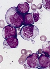
PHILADELPHIA—Super-enhancer profiling can unearth biomarkers and therapeutic targets for acute myeloid leukemia (AML), according to research presented at the AACR conference Hematologic Malignancies: Translating Discoveries to Novel Therapies.
Researchers used high-throughput ChIP sequencing to identify super-enhancer domains in a cohort of AML patients.
And this revealed both known and previously unknown genes that are important for AML disease biology.
Eric Olson, PhD, and his colleagues from Syros Pharmaceuticals in Watertown, Massachusetts, presented this research during one of the meeting’s poster sessions.
The investigators explained that super-enhancers are a class of densely clustered cis-regulatory elements that are key to initiating and maintaining cell-type-specific gene expression in cancer and other settings. Tumor cells acquire super-enhancers at key oncogenes and at genes that participate in the acquisition of hallmark capabilities in cancer.
So the researchers set out to identify and characterize super-enhancer domains in a cohort of AML patients.
The team collected primary AML samples and performed chromatin fragmentation, chromatin immunoprecipitation, and DNA purification and sequencing.
They then mapped enhancer regions and characterized enhancer profiles. This revealed AML-specific super-enhancers and associated genes.
For example, in one patient, the investigators identified 392 AML-specific super-enhancers, which were associated with 11 genes important for AML disease biology: HOXA7, LMO2, HLX, MYADM, ETV6, AFF1, RUNX1, GFI1, SPI1, MEIS1, and MYB.
In another patient, the team identified 279 AML-specific super-enhancers that were associated with 9 genes: MLLT10, AKT3, FLT3, ETV6, KLF13, RELA, FOSB, BMI1, and RUNX1.
The researchers said these findings suggest that super-enhancer profiling provides a new option for identifying biomarkers and therapeutic targets in AML and other malignancies.
“Syros’s gene control platform can systematically and efficiently identify known and previously unrecognized tumor biomarkers and cancer dependencies directly from patient tissue,” Dr Olson said. “Our data demonstrate unique gene control elements in AML patient subsets that hold promise in the classification and treatment of AML.”
PHILADELPHIA—Super-enhancer profiling can unearth biomarkers and therapeutic targets for acute myeloid leukemia (AML), according to research presented at the AACR conference Hematologic Malignancies: Translating Discoveries to Novel Therapies.
Researchers used high-throughput ChIP sequencing to identify super-enhancer domains in a cohort of AML patients.
And this revealed both known and previously unknown genes that are important for AML disease biology.
Eric Olson, PhD, and his colleagues from Syros Pharmaceuticals in Watertown, Massachusetts, presented this research during one of the meeting’s poster sessions.
The investigators explained that super-enhancers are a class of densely clustered cis-regulatory elements that are key to initiating and maintaining cell-type-specific gene expression in cancer and other settings. Tumor cells acquire super-enhancers at key oncogenes and at genes that participate in the acquisition of hallmark capabilities in cancer.
So the researchers set out to identify and characterize super-enhancer domains in a cohort of AML patients.
The team collected primary AML samples and performed chromatin fragmentation, chromatin immunoprecipitation, and DNA purification and sequencing.
They then mapped enhancer regions and characterized enhancer profiles. This revealed AML-specific super-enhancers and associated genes.
For example, in one patient, the investigators identified 392 AML-specific super-enhancers, which were associated with 11 genes important for AML disease biology: HOXA7, LMO2, HLX, MYADM, ETV6, AFF1, RUNX1, GFI1, SPI1, MEIS1, and MYB.
In another patient, the team identified 279 AML-specific super-enhancers that were associated with 9 genes: MLLT10, AKT3, FLT3, ETV6, KLF13, RELA, FOSB, BMI1, and RUNX1.
The researchers said these findings suggest that super-enhancer profiling provides a new option for identifying biomarkers and therapeutic targets in AML and other malignancies.
“Syros’s gene control platform can systematically and efficiently identify known and previously unrecognized tumor biomarkers and cancer dependencies directly from patient tissue,” Dr Olson said. “Our data demonstrate unique gene control elements in AML patient subsets that hold promise in the classification and treatment of AML.”
PHILADELPHIA—Super-enhancer profiling can unearth biomarkers and therapeutic targets for acute myeloid leukemia (AML), according to research presented at the AACR conference Hematologic Malignancies: Translating Discoveries to Novel Therapies.
Researchers used high-throughput ChIP sequencing to identify super-enhancer domains in a cohort of AML patients.
And this revealed both known and previously unknown genes that are important for AML disease biology.
Eric Olson, PhD, and his colleagues from Syros Pharmaceuticals in Watertown, Massachusetts, presented this research during one of the meeting’s poster sessions.
The investigators explained that super-enhancers are a class of densely clustered cis-regulatory elements that are key to initiating and maintaining cell-type-specific gene expression in cancer and other settings. Tumor cells acquire super-enhancers at key oncogenes and at genes that participate in the acquisition of hallmark capabilities in cancer.
So the researchers set out to identify and characterize super-enhancer domains in a cohort of AML patients.
The team collected primary AML samples and performed chromatin fragmentation, chromatin immunoprecipitation, and DNA purification and sequencing.
They then mapped enhancer regions and characterized enhancer profiles. This revealed AML-specific super-enhancers and associated genes.
For example, in one patient, the investigators identified 392 AML-specific super-enhancers, which were associated with 11 genes important for AML disease biology: HOXA7, LMO2, HLX, MYADM, ETV6, AFF1, RUNX1, GFI1, SPI1, MEIS1, and MYB.
In another patient, the team identified 279 AML-specific super-enhancers that were associated with 9 genes: MLLT10, AKT3, FLT3, ETV6, KLF13, RELA, FOSB, BMI1, and RUNX1.
The researchers said these findings suggest that super-enhancer profiling provides a new option for identifying biomarkers and therapeutic targets in AML and other malignancies.
“Syros’s gene control platform can systematically and efficiently identify known and previously unrecognized tumor biomarkers and cancer dependencies directly from patient tissue,” Dr Olson said. “Our data demonstrate unique gene control elements in AML patient subsets that hold promise in the classification and treatment of AML.”
Self-monitoring coagulometers get thumbs-up from NICE

The UK’s National Institute for Health and Care Excellence (NICE) has published a guidance recommending 2 technologies that enable patients on long-term anticoagulant therapy to self-monitor their clotting time.
The guidance supports use of the Coaguchek XS system (Roche Diagnostics) and the InRatio2 PT/INR Monitor (Alere) as options for some adults with atrial
fibrillation or heart valve disease who are on long-term anticoagulant therapy.
“The evidence shows that greater use of self-monitoring offers clinical and patient benefit and, over time, is likely to result in reductions in heart attacks and strokes caused by blood clots,” said Carole Longson, NICE Health Technology Evaluation Centre Director.
“Because self-monitoring provides almost instant results, self-monitoring can reduce anxiety, provide a sense of control for the patient, and remove the need to frequently attend clinics or hospitals.”
About the Coaguchek XS system
The Coaguchek XS system (Roche Diagnostics) consists of a meter and specifically designed test strips that can analyze a blood sample (fresh capillary blood or fresh untreated whole venous blood) and calculate the prothrombin time (PT) and the international normalized ratio (INR).
A code chip, which contains calibration data and the expiration date of the test strips, is inserted into the meter before it is switched on. Once the device is switched on, a test strip is inserted, and the blood sample is applied.
The test result is displayed approximately 1 minute after application of the sample, and the device automatically stores the result in its memory. The user is guided through the process by on-screen graphical instructions.
About the InRatio2 PT/INR Monitor
The INRatio2 PT/INR monitor (Alere) does a modified version of the 1-stage PT test using a recombinant human thromboplastin reagent. The clot formed in the reaction is detected by the change in the electrical impedance of the sample during the coagulation process.
The system consists of a monitor and disposable test strips. The monitor provides a user interface, heats the test strip to the appropriate reaction temperature, measures the impedance of blood samples, and calculates and reports PT and INR results.
Instructions and test results are displayed on an LCD. The monitor can store test results so that past results can be reviewed.
The UK’s National Institute for Health and Care Excellence (NICE) has published a guidance recommending 2 technologies that enable patients on long-term anticoagulant therapy to self-monitor their clotting time.
The guidance supports use of the Coaguchek XS system (Roche Diagnostics) and the InRatio2 PT/INR Monitor (Alere) as options for some adults with atrial
fibrillation or heart valve disease who are on long-term anticoagulant therapy.
“The evidence shows that greater use of self-monitoring offers clinical and patient benefit and, over time, is likely to result in reductions in heart attacks and strokes caused by blood clots,” said Carole Longson, NICE Health Technology Evaluation Centre Director.
“Because self-monitoring provides almost instant results, self-monitoring can reduce anxiety, provide a sense of control for the patient, and remove the need to frequently attend clinics or hospitals.”
About the Coaguchek XS system
The Coaguchek XS system (Roche Diagnostics) consists of a meter and specifically designed test strips that can analyze a blood sample (fresh capillary blood or fresh untreated whole venous blood) and calculate the prothrombin time (PT) and the international normalized ratio (INR).
A code chip, which contains calibration data and the expiration date of the test strips, is inserted into the meter before it is switched on. Once the device is switched on, a test strip is inserted, and the blood sample is applied.
The test result is displayed approximately 1 minute after application of the sample, and the device automatically stores the result in its memory. The user is guided through the process by on-screen graphical instructions.
About the InRatio2 PT/INR Monitor
The INRatio2 PT/INR monitor (Alere) does a modified version of the 1-stage PT test using a recombinant human thromboplastin reagent. The clot formed in the reaction is detected by the change in the electrical impedance of the sample during the coagulation process.
The system consists of a monitor and disposable test strips. The monitor provides a user interface, heats the test strip to the appropriate reaction temperature, measures the impedance of blood samples, and calculates and reports PT and INR results.
Instructions and test results are displayed on an LCD. The monitor can store test results so that past results can be reviewed.
The UK’s National Institute for Health and Care Excellence (NICE) has published a guidance recommending 2 technologies that enable patients on long-term anticoagulant therapy to self-monitor their clotting time.
The guidance supports use of the Coaguchek XS system (Roche Diagnostics) and the InRatio2 PT/INR Monitor (Alere) as options for some adults with atrial
fibrillation or heart valve disease who are on long-term anticoagulant therapy.
“The evidence shows that greater use of self-monitoring offers clinical and patient benefit and, over time, is likely to result in reductions in heart attacks and strokes caused by blood clots,” said Carole Longson, NICE Health Technology Evaluation Centre Director.
“Because self-monitoring provides almost instant results, self-monitoring can reduce anxiety, provide a sense of control for the patient, and remove the need to frequently attend clinics or hospitals.”
About the Coaguchek XS system
The Coaguchek XS system (Roche Diagnostics) consists of a meter and specifically designed test strips that can analyze a blood sample (fresh capillary blood or fresh untreated whole venous blood) and calculate the prothrombin time (PT) and the international normalized ratio (INR).
A code chip, which contains calibration data and the expiration date of the test strips, is inserted into the meter before it is switched on. Once the device is switched on, a test strip is inserted, and the blood sample is applied.
The test result is displayed approximately 1 minute after application of the sample, and the device automatically stores the result in its memory. The user is guided through the process by on-screen graphical instructions.
About the InRatio2 PT/INR Monitor
The INRatio2 PT/INR monitor (Alere) does a modified version of the 1-stage PT test using a recombinant human thromboplastin reagent. The clot formed in the reaction is detected by the change in the electrical impedance of the sample during the coagulation process.
The system consists of a monitor and disposable test strips. The monitor provides a user interface, heats the test strip to the appropriate reaction temperature, measures the impedance of blood samples, and calculates and reports PT and INR results.
Instructions and test results are displayed on an LCD. The monitor can store test results so that past results can be reviewed.
Murine studies support use of TKIs in ALL subtype

PHILADELPHIA—Experiments in mice reinforce the idea that tyrosine kinase inhibitors (TKIs) can treat patients with Ph-like acute lymphoblastic leukemia (ALL).
Investigators recently identified genomic alterations in Ph-like ALL that suggest these patients might respond to TKIs, and tests in a small number of patients supported this theory.
Now, preclinical results show that kinase fusions in Ph-like ALL activate signaling pathways differently, and this affects sensitivity to TKIs.
Kathryn Roberts, PhD, of St Jude Children’s Research Hospital in Memphis, Tennessee, and her colleagues presented these results at the AACR conference Hematologic Malignancies: Translating Discoveries to Novel Therapies.
“We recently described a subtype of B-cell acute lymphoblastic leukemia with very poor outcome that is characterized by genetic alterations involving tyrosine kinases, termed Ph-like ALL,” Dr Roberts said. “We wanted to examine whether these alterations contribute to the development of Ph-like ALL and determine if they could be targeted with tyrosine kinase inhibitors.”
“We showed, for the first time, that the kinase alterations we tested contribute to the development of Ph-like ALL, and that Ph-like ALL can be treated effectively with tyrosine kinase inhibitors in animal models. These findings provide a strong rationale for treating Ph-like ALL patients with targeted therapies to improve their survival.”
Dr Roberts and her colleagues first introduced kinase alterations—RCSD1-ABL2, SSBP2-CSF1R, or PAX5-JAK2—in IL-7-dependent, Arf-/- mouse pre-B cells expressing IK6.
They found that each fusion conferred cytokine-independent growth in vitro. And mice that received transplants of pre-B cells expressing RCSD1-ABL2 or SSBP2-CSF1R developed ALL with a pre-B immunophenotype.
The investigators then assessed the activation of kinase signaling pathways and TKI sensitivity in Arf-/- pre-B cells and human leukemic cells harvested from xenografted mice expressing ETV6-ABL1, RANBP2-ABL1, PAG1-ABL2, RCSD1-ABL2, SSBP2-CSF1R, IGH-EPOR, ATF7IP-JAK2, and PAX5-JAK2.
In both cell types, signaling pathway activation and TKI sensitivity differed according to the kinase fusion.
Cells expressing ABL1-class kinase fusions (ABL1, ABL2, CSF1R, and PDGFRB) exhibited pSTAT5 activation that was inhibited by imatinib or dasatinib. But in cells expressing ATF7IP-JAK2, PAX5-JAK2, or IGH-EPOR, pSTAT5 activation was only inhibited by ruxolitinib.
Finally, the investigators tested dasatinib in xenograft models of ETV6-ABL1, RCSD1-ABL2, PAG1-ABL2, or SSBP2-CSF1R ALL.
They found that treated mice had significantly lower leukemic burdens and splenic weights than control mice. And STAT5 phosphorylation was attenuated in cells from treated mice.
“Our studies show that different FDA-approved TKIs such as imatinib, dasatinib, ruxolitinib, or crizotinib could potentially be used to treat Ph-like ALL patients, depending on the type of kinase alterations their tumors bear,” Dr Roberts said.
“We were able to gain a better understanding of the genetics underlying Ph-like ALL, and our studies could help identify patients who will not respond optimally to current therapy. By knowing the exact genetic alteration upfront, we may be able to implement different therapeutic strategies to improve the survival rate of future patients with ALL.”
PHILADELPHIA—Experiments in mice reinforce the idea that tyrosine kinase inhibitors (TKIs) can treat patients with Ph-like acute lymphoblastic leukemia (ALL).
Investigators recently identified genomic alterations in Ph-like ALL that suggest these patients might respond to TKIs, and tests in a small number of patients supported this theory.
Now, preclinical results show that kinase fusions in Ph-like ALL activate signaling pathways differently, and this affects sensitivity to TKIs.
Kathryn Roberts, PhD, of St Jude Children’s Research Hospital in Memphis, Tennessee, and her colleagues presented these results at the AACR conference Hematologic Malignancies: Translating Discoveries to Novel Therapies.
“We recently described a subtype of B-cell acute lymphoblastic leukemia with very poor outcome that is characterized by genetic alterations involving tyrosine kinases, termed Ph-like ALL,” Dr Roberts said. “We wanted to examine whether these alterations contribute to the development of Ph-like ALL and determine if they could be targeted with tyrosine kinase inhibitors.”
“We showed, for the first time, that the kinase alterations we tested contribute to the development of Ph-like ALL, and that Ph-like ALL can be treated effectively with tyrosine kinase inhibitors in animal models. These findings provide a strong rationale for treating Ph-like ALL patients with targeted therapies to improve their survival.”
Dr Roberts and her colleagues first introduced kinase alterations—RCSD1-ABL2, SSBP2-CSF1R, or PAX5-JAK2—in IL-7-dependent, Arf-/- mouse pre-B cells expressing IK6.
They found that each fusion conferred cytokine-independent growth in vitro. And mice that received transplants of pre-B cells expressing RCSD1-ABL2 or SSBP2-CSF1R developed ALL with a pre-B immunophenotype.
The investigators then assessed the activation of kinase signaling pathways and TKI sensitivity in Arf-/- pre-B cells and human leukemic cells harvested from xenografted mice expressing ETV6-ABL1, RANBP2-ABL1, PAG1-ABL2, RCSD1-ABL2, SSBP2-CSF1R, IGH-EPOR, ATF7IP-JAK2, and PAX5-JAK2.
In both cell types, signaling pathway activation and TKI sensitivity differed according to the kinase fusion.
Cells expressing ABL1-class kinase fusions (ABL1, ABL2, CSF1R, and PDGFRB) exhibited pSTAT5 activation that was inhibited by imatinib or dasatinib. But in cells expressing ATF7IP-JAK2, PAX5-JAK2, or IGH-EPOR, pSTAT5 activation was only inhibited by ruxolitinib.
Finally, the investigators tested dasatinib in xenograft models of ETV6-ABL1, RCSD1-ABL2, PAG1-ABL2, or SSBP2-CSF1R ALL.
They found that treated mice had significantly lower leukemic burdens and splenic weights than control mice. And STAT5 phosphorylation was attenuated in cells from treated mice.
“Our studies show that different FDA-approved TKIs such as imatinib, dasatinib, ruxolitinib, or crizotinib could potentially be used to treat Ph-like ALL patients, depending on the type of kinase alterations their tumors bear,” Dr Roberts said.
“We were able to gain a better understanding of the genetics underlying Ph-like ALL, and our studies could help identify patients who will not respond optimally to current therapy. By knowing the exact genetic alteration upfront, we may be able to implement different therapeutic strategies to improve the survival rate of future patients with ALL.”
PHILADELPHIA—Experiments in mice reinforce the idea that tyrosine kinase inhibitors (TKIs) can treat patients with Ph-like acute lymphoblastic leukemia (ALL).
Investigators recently identified genomic alterations in Ph-like ALL that suggest these patients might respond to TKIs, and tests in a small number of patients supported this theory.
Now, preclinical results show that kinase fusions in Ph-like ALL activate signaling pathways differently, and this affects sensitivity to TKIs.
Kathryn Roberts, PhD, of St Jude Children’s Research Hospital in Memphis, Tennessee, and her colleagues presented these results at the AACR conference Hematologic Malignancies: Translating Discoveries to Novel Therapies.
“We recently described a subtype of B-cell acute lymphoblastic leukemia with very poor outcome that is characterized by genetic alterations involving tyrosine kinases, termed Ph-like ALL,” Dr Roberts said. “We wanted to examine whether these alterations contribute to the development of Ph-like ALL and determine if they could be targeted with tyrosine kinase inhibitors.”
“We showed, for the first time, that the kinase alterations we tested contribute to the development of Ph-like ALL, and that Ph-like ALL can be treated effectively with tyrosine kinase inhibitors in animal models. These findings provide a strong rationale for treating Ph-like ALL patients with targeted therapies to improve their survival.”
Dr Roberts and her colleagues first introduced kinase alterations—RCSD1-ABL2, SSBP2-CSF1R, or PAX5-JAK2—in IL-7-dependent, Arf-/- mouse pre-B cells expressing IK6.
They found that each fusion conferred cytokine-independent growth in vitro. And mice that received transplants of pre-B cells expressing RCSD1-ABL2 or SSBP2-CSF1R developed ALL with a pre-B immunophenotype.
The investigators then assessed the activation of kinase signaling pathways and TKI sensitivity in Arf-/- pre-B cells and human leukemic cells harvested from xenografted mice expressing ETV6-ABL1, RANBP2-ABL1, PAG1-ABL2, RCSD1-ABL2, SSBP2-CSF1R, IGH-EPOR, ATF7IP-JAK2, and PAX5-JAK2.
In both cell types, signaling pathway activation and TKI sensitivity differed according to the kinase fusion.
Cells expressing ABL1-class kinase fusions (ABL1, ABL2, CSF1R, and PDGFRB) exhibited pSTAT5 activation that was inhibited by imatinib or dasatinib. But in cells expressing ATF7IP-JAK2, PAX5-JAK2, or IGH-EPOR, pSTAT5 activation was only inhibited by ruxolitinib.
Finally, the investigators tested dasatinib in xenograft models of ETV6-ABL1, RCSD1-ABL2, PAG1-ABL2, or SSBP2-CSF1R ALL.
They found that treated mice had significantly lower leukemic burdens and splenic weights than control mice. And STAT5 phosphorylation was attenuated in cells from treated mice.
“Our studies show that different FDA-approved TKIs such as imatinib, dasatinib, ruxolitinib, or crizotinib could potentially be used to treat Ph-like ALL patients, depending on the type of kinase alterations their tumors bear,” Dr Roberts said.
“We were able to gain a better understanding of the genetics underlying Ph-like ALL, and our studies could help identify patients who will not respond optimally to current therapy. By knowing the exact genetic alteration upfront, we may be able to implement different therapeutic strategies to improve the survival rate of future patients with ALL.”
ETBs prove effective against lymphoma and myeloma

Credit: Rhoda Baer
PHILADELPHIA—A pair of engineered toxin bodies (ETBs) can successfully treat Burkitt lymphoma and multiple myeloma, according to preclinical research presented at the AACR conference Hematologic Malignancies: Translating Discoveries to Novel Therapies.
The ETBs, known as MT-4007 and MT-4007-D, work by targeting CD38.
They greatly reduced tumor burden and improved survival in mouse models. And they were well-tolerated, even at the highest doses administered.
“In this study, we found that the growth of human cancer cells in mice was substantially decreased, or the cells were even eliminated, following treatment with our investigational CD38-targeted therapy,” said Erin K. Willert, PhD, of Molecular Templates Inc., in Georgetown, Texas.
Dr Willert and her colleagues explained that ETBs are derived from the ribosome-inactivating alpha subunit of Shiga-like toxin 1 (SLT-1A). They have been engineered to contain a target binding domain fused to a modified SLT-1A protein, which allows for delivery to a cell surface target—in this case, CD38.
Upon binding to a CD38-expressing cell, the ETB enters the cell, routes to the cytosol, halts protein synthesis, and kills the cell.
The researchers first tested MT-4007 and MT-4007-D in a range of human cell lines. The agents exhibited cytotoxicity in CD38+ Burkitt lymphoma and multiple myeloma cell lines (H929, Daudi, ST486, and Raji). But neither agent proved cytotoxic in CD38- cell lines (U266, SKBR3, and HCC1954).
The team then moved on to test MT-4007 in a mouse model of Burkitt lymphoma. Following injection with Daudi-Luc cells, mice received no treatment or MT-4007 at 0.05 mg/kg, 0.5 mg/kg, or 2 mg/kg on days 3, 5, 8, 10, and 12.
Treated mice exhibited significantly reduced tumor burden compared to controls. The mean tumor burden for mice that received MT-4007 at 0.05 mg/kg was 29% of the control tumor burden (P<0.0001). It was 0.4% for mice that received 0.50 mg/kg (P<0.0001) and 0.02% for mice that received 2 mg/kg (P<0.0001).
In a model of multiple myeloma, MT-4007-D provided a dose-dependent delay in tumor growth. After receiving injections of H929 cells, mice received no treatment or MT-4007-D at 0.5 mg/kg, 2 mg/kg, or 3 mg/kg on days 1, 3, 5, 8, 10, and 12.
The researchers assessed efficacy by measuring the time to endpoint, which was a tumor volume of 2000 mm3. The median time to endpoint was 22.3 days in controls, 21.2 days in the 0.5 mg/kg arm (not significant), 24.5 days in the 2 mg/kg arm (P=0.004), and 26.2 days in the 3 mg/kg arm (P=0.04).
The team assessed safety using body weight. They found that all treated groups of mice maintained a stable weight, suggesting MT-4007-D is well-tolerated.
The researchers also noted that, in a previous dose-finding study, the maximum-tolerated dose of MT-4007 was not reached at the highest dose administered to mice (2 mg/kg), which suggests MT-4007 is well tolerated as well.
Finally, Dr Willert and her colleagues found that MT-4007 extends survival in models of Burkitt lymphoma. The team euthanized mice if they had a greater than 20% loss in body weight or symptoms such as hind limb paralysis.
In the control group, all 10 mice died, and the median survival was 34 days. In the 0.5 mg/kg treatment group, 5 mice died, and the median survival was 59.5 days (P=0.0002).
One mouse died in the 0.5 mg/kg group (P<0.0001), and none of the mice died in the 2 mg/kg group (P<0.0001). The median survival was undefined for both groups.
Dr Willert said these results suggest the ETBs should be moved forward to clinical trials in CD38+ B-cell malignancies such as multiple myeloma. And because the ETBs work differently from other treatments, they might prove effective in relapsed or refractory patients.
However, more preclinical research is needed before the ETBs can be tested in patients. MT-4007-D is under investigation in preclinical studies now.
This research was funded by Molecular Templates Inc., makers of MT-4007 and MT-4007-D.
Credit: Rhoda Baer
PHILADELPHIA—A pair of engineered toxin bodies (ETBs) can successfully treat Burkitt lymphoma and multiple myeloma, according to preclinical research presented at the AACR conference Hematologic Malignancies: Translating Discoveries to Novel Therapies.
The ETBs, known as MT-4007 and MT-4007-D, work by targeting CD38.
They greatly reduced tumor burden and improved survival in mouse models. And they were well-tolerated, even at the highest doses administered.
“In this study, we found that the growth of human cancer cells in mice was substantially decreased, or the cells were even eliminated, following treatment with our investigational CD38-targeted therapy,” said Erin K. Willert, PhD, of Molecular Templates Inc., in Georgetown, Texas.
Dr Willert and her colleagues explained that ETBs are derived from the ribosome-inactivating alpha subunit of Shiga-like toxin 1 (SLT-1A). They have been engineered to contain a target binding domain fused to a modified SLT-1A protein, which allows for delivery to a cell surface target—in this case, CD38.
Upon binding to a CD38-expressing cell, the ETB enters the cell, routes to the cytosol, halts protein synthesis, and kills the cell.
The researchers first tested MT-4007 and MT-4007-D in a range of human cell lines. The agents exhibited cytotoxicity in CD38+ Burkitt lymphoma and multiple myeloma cell lines (H929, Daudi, ST486, and Raji). But neither agent proved cytotoxic in CD38- cell lines (U266, SKBR3, and HCC1954).
The team then moved on to test MT-4007 in a mouse model of Burkitt lymphoma. Following injection with Daudi-Luc cells, mice received no treatment or MT-4007 at 0.05 mg/kg, 0.5 mg/kg, or 2 mg/kg on days 3, 5, 8, 10, and 12.
Treated mice exhibited significantly reduced tumor burden compared to controls. The mean tumor burden for mice that received MT-4007 at 0.05 mg/kg was 29% of the control tumor burden (P<0.0001). It was 0.4% for mice that received 0.50 mg/kg (P<0.0001) and 0.02% for mice that received 2 mg/kg (P<0.0001).
In a model of multiple myeloma, MT-4007-D provided a dose-dependent delay in tumor growth. After receiving injections of H929 cells, mice received no treatment or MT-4007-D at 0.5 mg/kg, 2 mg/kg, or 3 mg/kg on days 1, 3, 5, 8, 10, and 12.
The researchers assessed efficacy by measuring the time to endpoint, which was a tumor volume of 2000 mm3. The median time to endpoint was 22.3 days in controls, 21.2 days in the 0.5 mg/kg arm (not significant), 24.5 days in the 2 mg/kg arm (P=0.004), and 26.2 days in the 3 mg/kg arm (P=0.04).
The team assessed safety using body weight. They found that all treated groups of mice maintained a stable weight, suggesting MT-4007-D is well-tolerated.
The researchers also noted that, in a previous dose-finding study, the maximum-tolerated dose of MT-4007 was not reached at the highest dose administered to mice (2 mg/kg), which suggests MT-4007 is well tolerated as well.
Finally, Dr Willert and her colleagues found that MT-4007 extends survival in models of Burkitt lymphoma. The team euthanized mice if they had a greater than 20% loss in body weight or symptoms such as hind limb paralysis.
In the control group, all 10 mice died, and the median survival was 34 days. In the 0.5 mg/kg treatment group, 5 mice died, and the median survival was 59.5 days (P=0.0002).
One mouse died in the 0.5 mg/kg group (P<0.0001), and none of the mice died in the 2 mg/kg group (P<0.0001). The median survival was undefined for both groups.
Dr Willert said these results suggest the ETBs should be moved forward to clinical trials in CD38+ B-cell malignancies such as multiple myeloma. And because the ETBs work differently from other treatments, they might prove effective in relapsed or refractory patients.
However, more preclinical research is needed before the ETBs can be tested in patients. MT-4007-D is under investigation in preclinical studies now.
This research was funded by Molecular Templates Inc., makers of MT-4007 and MT-4007-D.
Credit: Rhoda Baer
PHILADELPHIA—A pair of engineered toxin bodies (ETBs) can successfully treat Burkitt lymphoma and multiple myeloma, according to preclinical research presented at the AACR conference Hematologic Malignancies: Translating Discoveries to Novel Therapies.
The ETBs, known as MT-4007 and MT-4007-D, work by targeting CD38.
They greatly reduced tumor burden and improved survival in mouse models. And they were well-tolerated, even at the highest doses administered.
“In this study, we found that the growth of human cancer cells in mice was substantially decreased, or the cells were even eliminated, following treatment with our investigational CD38-targeted therapy,” said Erin K. Willert, PhD, of Molecular Templates Inc., in Georgetown, Texas.
Dr Willert and her colleagues explained that ETBs are derived from the ribosome-inactivating alpha subunit of Shiga-like toxin 1 (SLT-1A). They have been engineered to contain a target binding domain fused to a modified SLT-1A protein, which allows for delivery to a cell surface target—in this case, CD38.
Upon binding to a CD38-expressing cell, the ETB enters the cell, routes to the cytosol, halts protein synthesis, and kills the cell.
The researchers first tested MT-4007 and MT-4007-D in a range of human cell lines. The agents exhibited cytotoxicity in CD38+ Burkitt lymphoma and multiple myeloma cell lines (H929, Daudi, ST486, and Raji). But neither agent proved cytotoxic in CD38- cell lines (U266, SKBR3, and HCC1954).
The team then moved on to test MT-4007 in a mouse model of Burkitt lymphoma. Following injection with Daudi-Luc cells, mice received no treatment or MT-4007 at 0.05 mg/kg, 0.5 mg/kg, or 2 mg/kg on days 3, 5, 8, 10, and 12.
Treated mice exhibited significantly reduced tumor burden compared to controls. The mean tumor burden for mice that received MT-4007 at 0.05 mg/kg was 29% of the control tumor burden (P<0.0001). It was 0.4% for mice that received 0.50 mg/kg (P<0.0001) and 0.02% for mice that received 2 mg/kg (P<0.0001).
In a model of multiple myeloma, MT-4007-D provided a dose-dependent delay in tumor growth. After receiving injections of H929 cells, mice received no treatment or MT-4007-D at 0.5 mg/kg, 2 mg/kg, or 3 mg/kg on days 1, 3, 5, 8, 10, and 12.
The researchers assessed efficacy by measuring the time to endpoint, which was a tumor volume of 2000 mm3. The median time to endpoint was 22.3 days in controls, 21.2 days in the 0.5 mg/kg arm (not significant), 24.5 days in the 2 mg/kg arm (P=0.004), and 26.2 days in the 3 mg/kg arm (P=0.04).
The team assessed safety using body weight. They found that all treated groups of mice maintained a stable weight, suggesting MT-4007-D is well-tolerated.
The researchers also noted that, in a previous dose-finding study, the maximum-tolerated dose of MT-4007 was not reached at the highest dose administered to mice (2 mg/kg), which suggests MT-4007 is well tolerated as well.
Finally, Dr Willert and her colleagues found that MT-4007 extends survival in models of Burkitt lymphoma. The team euthanized mice if they had a greater than 20% loss in body weight or symptoms such as hind limb paralysis.
In the control group, all 10 mice died, and the median survival was 34 days. In the 0.5 mg/kg treatment group, 5 mice died, and the median survival was 59.5 days (P=0.0002).
One mouse died in the 0.5 mg/kg group (P<0.0001), and none of the mice died in the 2 mg/kg group (P<0.0001). The median survival was undefined for both groups.
Dr Willert said these results suggest the ETBs should be moved forward to clinical trials in CD38+ B-cell malignancies such as multiple myeloma. And because the ETBs work differently from other treatments, they might prove effective in relapsed or refractory patients.
However, more preclinical research is needed before the ETBs can be tested in patients. MT-4007-D is under investigation in preclinical studies now.
This research was funded by Molecular Templates Inc., makers of MT-4007 and MT-4007-D.
Platelets respond to their surroundings, study shows
Credit: Andre E.X. Brown
Platelets can sense and respond to their surroundings, according to research published in PNAS.
Researchers reported that platelets can detect mechanical aspects of their environment and transduce those cues into biological signals.
Experiments showed that platelets could sense the stiffness of a fibrin/fibrinogen substrate, and increasing stiffness was associated with increased platelet adhesion, spreading, and activation.
“Platelets are smarter than we give them credit for, in that they are able to sense the physical characteristics of their environment and respond in a graduated way,” said study author Wilbur Lam, MD, PhD, of the Emory University School of Medicine in Atlanta, Georgia.
He and his colleagues were able to separate physical and biochemical effects on platelet behavior by forming polymer gels with different degrees of stiffness and then overlaying each with the same coating of fibrinogen.
With stiffer gels, the researchers observed an increase in platelet adhesion, spreading, and activation. This behavior was most pronounced when the concentration of fibrinogen was relatively low.
“This variability helps to explain platelet behavior in the 3D context of a clot in the body, which can be quite heterogenous in makeup,” Dr Lam said.
The researchers were also able to dissect platelet biochemistry by allowing the platelets to adhere and then spread on the various gels under the influence of drugs that interfere with different biochemical steps.
The team found that integrins, which engage the fibrinogen, and the protein Rac1 are involved in the initial mechanical sensing during adhesion. Myosin and actin, components of the cytoskeleton, are responsible for platelet spreading.
“We found that the initial adhesion and later spreading are separable, because different biochemical pathways are involved in each step,” Dr Lam said. “Our data show that mechanosensing can occur and plays important roles even when the cellular structural building blocks are fairly basic, even when the nucleus is absent.”
The researchers believe these findings could influence the design of medical devices, as modifying the stiffness of materials used in these devices might reduce the formation of blood clots. The results could also guide the refinement of anticoagulant therapy.
Credit: Andre E.X. Brown
Platelets can sense and respond to their surroundings, according to research published in PNAS.
Researchers reported that platelets can detect mechanical aspects of their environment and transduce those cues into biological signals.
Experiments showed that platelets could sense the stiffness of a fibrin/fibrinogen substrate, and increasing stiffness was associated with increased platelet adhesion, spreading, and activation.
“Platelets are smarter than we give them credit for, in that they are able to sense the physical characteristics of their environment and respond in a graduated way,” said study author Wilbur Lam, MD, PhD, of the Emory University School of Medicine in Atlanta, Georgia.
He and his colleagues were able to separate physical and biochemical effects on platelet behavior by forming polymer gels with different degrees of stiffness and then overlaying each with the same coating of fibrinogen.
With stiffer gels, the researchers observed an increase in platelet adhesion, spreading, and activation. This behavior was most pronounced when the concentration of fibrinogen was relatively low.
“This variability helps to explain platelet behavior in the 3D context of a clot in the body, which can be quite heterogenous in makeup,” Dr Lam said.
The researchers were also able to dissect platelet biochemistry by allowing the platelets to adhere and then spread on the various gels under the influence of drugs that interfere with different biochemical steps.
The team found that integrins, which engage the fibrinogen, and the protein Rac1 are involved in the initial mechanical sensing during adhesion. Myosin and actin, components of the cytoskeleton, are responsible for platelet spreading.
“We found that the initial adhesion and later spreading are separable, because different biochemical pathways are involved in each step,” Dr Lam said. “Our data show that mechanosensing can occur and plays important roles even when the cellular structural building blocks are fairly basic, even when the nucleus is absent.”
The researchers believe these findings could influence the design of medical devices, as modifying the stiffness of materials used in these devices might reduce the formation of blood clots. The results could also guide the refinement of anticoagulant therapy.
Credit: Andre E.X. Brown
Platelets can sense and respond to their surroundings, according to research published in PNAS.
Researchers reported that platelets can detect mechanical aspects of their environment and transduce those cues into biological signals.
Experiments showed that platelets could sense the stiffness of a fibrin/fibrinogen substrate, and increasing stiffness was associated with increased platelet adhesion, spreading, and activation.
“Platelets are smarter than we give them credit for, in that they are able to sense the physical characteristics of their environment and respond in a graduated way,” said study author Wilbur Lam, MD, PhD, of the Emory University School of Medicine in Atlanta, Georgia.
He and his colleagues were able to separate physical and biochemical effects on platelet behavior by forming polymer gels with different degrees of stiffness and then overlaying each with the same coating of fibrinogen.
With stiffer gels, the researchers observed an increase in platelet adhesion, spreading, and activation. This behavior was most pronounced when the concentration of fibrinogen was relatively low.
“This variability helps to explain platelet behavior in the 3D context of a clot in the body, which can be quite heterogenous in makeup,” Dr Lam said.
The researchers were also able to dissect platelet biochemistry by allowing the platelets to adhere and then spread on the various gels under the influence of drugs that interfere with different biochemical steps.
The team found that integrins, which engage the fibrinogen, and the protein Rac1 are involved in the initial mechanical sensing during adhesion. Myosin and actin, components of the cytoskeleton, are responsible for platelet spreading.
“We found that the initial adhesion and later spreading are separable, because different biochemical pathways are involved in each step,” Dr Lam said. “Our data show that mechanosensing can occur and plays important roles even when the cellular structural building blocks are fairly basic, even when the nucleus is absent.”
The researchers believe these findings could influence the design of medical devices, as modifying the stiffness of materials used in these devices might reduce the formation of blood clots. The results could also guide the refinement of anticoagulant therapy.