User login
Targeting drug resistance earlier
Credit: PNAS
PHILADELPHIA—A combination treatment strategy that takes tumor evolution into account could help us avoid drug resistance in hematologic malignancies, researchers say.
Preclinical experiments suggest we can prevent resistance by starting secondary treatment prior to relapse.
For example, a patient receiving dasatinib for acute lymphoblastic leukemia (ALL) could benefit from receiving crizotinib or foretinib during the early stages of clonal evolution.
The researchers described this strategy in a poster at the AACR conference Hematologic Malignancies: Translating Discoveries to Novel Therapies.
“Our goal is to identify vulnerabilities in cancer across stages of tumor evolution while it is developing resistance to initial treatment, to help guide the design of drug combination strategies,” said Douglas Lauffenburger, PhD, of the Koch Institute for Integrative Cancer Research at MIT in Boston.
“There may be many stages in a tumor evolution while under treatment that may make them vulnerable to already existing therapies. Rather than waiting for the tumor to become resistant to the first treatment and then thinking about a second-line drug to use, we can capitalize on opportunities that exploit vulnerabilities at different early stages, as the tumor is evolving to become resistant to the first drug.”
Dr Lauffenburger and his colleagues used a combination of computational and experimental approaches to identify drugs likely to be effective against a murine ALL cell line as the cells evolve.
To develop drug combinations based on the characteristics of evolving tumors, the researchers used escalating doses of imatinib, dasatinib, nilotinib, foretinib, and crizotinib on ALL cells.
As some cells exhibited resistance to a particular drug, the team treated the resistant cells with other drugs to check for cross-resistance. They found that resistant cells surviving at low multiples of the original drug dose actually demonstrated sensitization to certain other drugs, with the sensitization abrogated at higher doses.
Specifically, cell populations that were resistant to dasatinib at 1x and 2x IC90 became even more sensitive to crizotinib and foretinib. However, the sensitivity was lost at IC90 4x and above.
“Instead of only looking for the most resistant population of ALL cells at the end of this selection process, we monitored for drug sensitivity of the cells at each stage of the dose escalation,” Dr Lauffenburger explained.
“This led us to discover the vulnerabilities of a tumor at different stages of clonal evolution, a phenomenon we would have missed if we only analyzed for drug sensitivity at the last stage of this process, which is equivalent to when a patient has relapsed.” 
Credit: PNAS
PHILADELPHIA—A combination treatment strategy that takes tumor evolution into account could help us avoid drug resistance in hematologic malignancies, researchers say.
Preclinical experiments suggest we can prevent resistance by starting secondary treatment prior to relapse.
For example, a patient receiving dasatinib for acute lymphoblastic leukemia (ALL) could benefit from receiving crizotinib or foretinib during the early stages of clonal evolution.
The researchers described this strategy in a poster at the AACR conference Hematologic Malignancies: Translating Discoveries to Novel Therapies.
“Our goal is to identify vulnerabilities in cancer across stages of tumor evolution while it is developing resistance to initial treatment, to help guide the design of drug combination strategies,” said Douglas Lauffenburger, PhD, of the Koch Institute for Integrative Cancer Research at MIT in Boston.
“There may be many stages in a tumor evolution while under treatment that may make them vulnerable to already existing therapies. Rather than waiting for the tumor to become resistant to the first treatment and then thinking about a second-line drug to use, we can capitalize on opportunities that exploit vulnerabilities at different early stages, as the tumor is evolving to become resistant to the first drug.”
Dr Lauffenburger and his colleagues used a combination of computational and experimental approaches to identify drugs likely to be effective against a murine ALL cell line as the cells evolve.
To develop drug combinations based on the characteristics of evolving tumors, the researchers used escalating doses of imatinib, dasatinib, nilotinib, foretinib, and crizotinib on ALL cells.
As some cells exhibited resistance to a particular drug, the team treated the resistant cells with other drugs to check for cross-resistance. They found that resistant cells surviving at low multiples of the original drug dose actually demonstrated sensitization to certain other drugs, with the sensitization abrogated at higher doses.
Specifically, cell populations that were resistant to dasatinib at 1x and 2x IC90 became even more sensitive to crizotinib and foretinib. However, the sensitivity was lost at IC90 4x and above.
“Instead of only looking for the most resistant population of ALL cells at the end of this selection process, we monitored for drug sensitivity of the cells at each stage of the dose escalation,” Dr Lauffenburger explained.
“This led us to discover the vulnerabilities of a tumor at different stages of clonal evolution, a phenomenon we would have missed if we only analyzed for drug sensitivity at the last stage of this process, which is equivalent to when a patient has relapsed.”
Credit: PNAS
PHILADELPHIA—A combination treatment strategy that takes tumor evolution into account could help us avoid drug resistance in hematologic malignancies, researchers say.
Preclinical experiments suggest we can prevent resistance by starting secondary treatment prior to relapse.
For example, a patient receiving dasatinib for acute lymphoblastic leukemia (ALL) could benefit from receiving crizotinib or foretinib during the early stages of clonal evolution.
The researchers described this strategy in a poster at the AACR conference Hematologic Malignancies: Translating Discoveries to Novel Therapies.
“Our goal is to identify vulnerabilities in cancer across stages of tumor evolution while it is developing resistance to initial treatment, to help guide the design of drug combination strategies,” said Douglas Lauffenburger, PhD, of the Koch Institute for Integrative Cancer Research at MIT in Boston.
“There may be many stages in a tumor evolution while under treatment that may make them vulnerable to already existing therapies. Rather than waiting for the tumor to become resistant to the first treatment and then thinking about a second-line drug to use, we can capitalize on opportunities that exploit vulnerabilities at different early stages, as the tumor is evolving to become resistant to the first drug.”
Dr Lauffenburger and his colleagues used a combination of computational and experimental approaches to identify drugs likely to be effective against a murine ALL cell line as the cells evolve.
To develop drug combinations based on the characteristics of evolving tumors, the researchers used escalating doses of imatinib, dasatinib, nilotinib, foretinib, and crizotinib on ALL cells.
As some cells exhibited resistance to a particular drug, the team treated the resistant cells with other drugs to check for cross-resistance. They found that resistant cells surviving at low multiples of the original drug dose actually demonstrated sensitization to certain other drugs, with the sensitization abrogated at higher doses.
Specifically, cell populations that were resistant to dasatinib at 1x and 2x IC90 became even more sensitive to crizotinib and foretinib. However, the sensitivity was lost at IC90 4x and above.
“Instead of only looking for the most resistant population of ALL cells at the end of this selection process, we monitored for drug sensitivity of the cells at each stage of the dose escalation,” Dr Lauffenburger explained.
“This led us to discover the vulnerabilities of a tumor at different stages of clonal evolution, a phenomenon we would have missed if we only analyzed for drug sensitivity at the last stage of this process, which is equivalent to when a patient has relapsed.”
Five genes linked to risk of severe malaria
Credit: Peter H. Seeberger
Investigators have identified 5 genes that appear to affect a person’s susceptibility to severe malaria.
The group’s analyses suggest that single-nucleotide polymorphisms (SNPs) in HBB, ABO, ATP2B4, G6PD, and CD40LG are associated with an increased or decreased risk of severe malaria, cerebral malaria, and severe malarial anemia.
This knowledge could lead to new therapeutics or vaccines to target severe malaria.
“[This study] has allowed for investigation of genes that influence susceptibility to malaria on a scale that has previously not been achieved,” said Sarah Dunstan, PhD, of the Oxford University Clinical Research Unit at the Hospital for Tropical Diseases in Ho Chi Minh City, Vietnam.
“It involved a large number of severe malaria patients from multiple countries, which allows us to identify genes that truly have an effect on whether or not you develop severe malaria.”
Dr Dunstan and her colleagues detailed this research in Nature Genetics.
The team collected data on 11,890 cases of severe malaria due to Plasmodium falciparum and 17,441 control subjects across 12 locations in countries of Africa, Asia, and Oceania.
The researchers tested 55 SNPs in 27 gene regions that were associated with severe malaria in previous research: ABO, ADORA2B, ATP2B4, C6, CD36, CD40LG, CR1, ACKR1 (DARC), G6PD, GNAS, HBB, ICAM1, IL1A, IL1B, IL4, IL10, IL13, IL22, IRF1, LTA, NOS2, SPTB, TLR1, TLR4, TLR6, TLR9, and TNF.
The team found evidence of associations with the HBB, ABO, ATP2B4, G6PD, and CD40LG loci, but no association for the other 22 loci.
HBB
The HBB gene encodes β-globin, which has 3 variants associated with resistance to malaria: hemoglobin S (HbS), hemoglobin C (HbC), and hemoglobin E (HbE). The SNP responsible for HbE, rs33950507, was rare or absent in most groups, so the sample was too small to estimate association with severe malaria.
However, for HbS (SNP rs334), heterozygotes had a reduced risk of severe malaria (odds ratio [OR]=0.14; P=1.6×10−225), cerebral malaria (OR=0.11; P=4.7×10−88), and severe malarial anemia (OR=0.11; P=9.3×10−65).
For HbC (SNP rs33930165), each copy of the derived allele reduced the risk of severe malaria by 29% (OR=0.71; P=6.9×10⁻⁹), cerebral malaria by 28% (OR=0.72; P=0.01), and severe malarial anemia by 26% (OR=0.74; P=2.1×10⁻³).
ABO
The ABO gene encodes the glycosyltransferase enzyme that determines ABO blood group. Blood group O was associated with a lower risk of severe malaria (OR=0.74; P=5.0×10⁻³²), cerebral malaria (OR=0.73; P=8.9×10⁻¹⁶), and severe malarial anemia (OR=0.68; P=7.9×10⁻¹⁴).
The researchers also found that rs8176746, an SNP in ABO that determines the production of B antigens, was associated with an increased risk of severe malaria (OR=1.25; P=2.0×10⁻¹⁷).
G6PD
G6PD is an X-linked gene encoding glucose-6-phosphate dehydrogenase. The investigators found that deficiency in G6PD (rs1050828 [G6PD c.202C>T]) can reduce the risk of cerebral malaria but increase the risk of severe malarial anemia.
There was an increased risk of severe malarial anemia in male hemizygotes (OR=1.49; P=3.6×10⁻⁵) and in female homozygotes (OR=1.94; P=1.9×10⁻³), as well as a decreased risk of cerebral malaria in female heterozygotes (OR=0.87; P=0.06) and male hemizygotes (OR=0.81; P=0.01).
ATP2B4
ATP2B4 encodes a calcium transporter found in the plasma membrane of erythrocytes. The derived alleles of rs10900585 and rs55868763 were associated with an increased risk of severe malaria, and the derived alleles of rs4951074 and rs1541255 were associated with a decreased risk.
Subjects with at least one copy of the derived allele at rs10900585 had an OR of 1.32 for severe malaria (P=1.7×10⁻⁹), and those homozygous for the derived allele at rs4951074 had an OR of 0.77 (P=7.6×10⁻⁷). Findings were similar for cerebral malaria and severe malarial anemia.
CD40LG
CD40LG is a gene on the X chromosome that has previously been associated with severe malaria. Homozygotes for the derived allele of rs3092945 had a decreased risk of severe malaria (OR=0.85; P=1.1×10⁻⁶) when the researchers analyzed data from the study sites together.
However, the results differed when they analyzed sites individually. For instance, homozygotes had a reduced risk of severe malaria in The Gambia (OR=0.54; P=2.3×10⁻²²) and an increased risk in Kenya (OR=1.42; P=7.8×10⁻⁶).
These findings suggest the role of common human genetic disorders in severe malaria is more complex than we thought, Dr Dunstan said. But the results should enable a better understanding of the mechanisms and processes at work during progression to severe disease.
Credit: Peter H. Seeberger
Investigators have identified 5 genes that appear to affect a person’s susceptibility to severe malaria.
The group’s analyses suggest that single-nucleotide polymorphisms (SNPs) in HBB, ABO, ATP2B4, G6PD, and CD40LG are associated with an increased or decreased risk of severe malaria, cerebral malaria, and severe malarial anemia.
This knowledge could lead to new therapeutics or vaccines to target severe malaria.
“[This study] has allowed for investigation of genes that influence susceptibility to malaria on a scale that has previously not been achieved,” said Sarah Dunstan, PhD, of the Oxford University Clinical Research Unit at the Hospital for Tropical Diseases in Ho Chi Minh City, Vietnam.
“It involved a large number of severe malaria patients from multiple countries, which allows us to identify genes that truly have an effect on whether or not you develop severe malaria.”
Dr Dunstan and her colleagues detailed this research in Nature Genetics.
The team collected data on 11,890 cases of severe malaria due to Plasmodium falciparum and 17,441 control subjects across 12 locations in countries of Africa, Asia, and Oceania.
The researchers tested 55 SNPs in 27 gene regions that were associated with severe malaria in previous research: ABO, ADORA2B, ATP2B4, C6, CD36, CD40LG, CR1, ACKR1 (DARC), G6PD, GNAS, HBB, ICAM1, IL1A, IL1B, IL4, IL10, IL13, IL22, IRF1, LTA, NOS2, SPTB, TLR1, TLR4, TLR6, TLR9, and TNF.
The team found evidence of associations with the HBB, ABO, ATP2B4, G6PD, and CD40LG loci, but no association for the other 22 loci.
HBB
The HBB gene encodes β-globin, which has 3 variants associated with resistance to malaria: hemoglobin S (HbS), hemoglobin C (HbC), and hemoglobin E (HbE). The SNP responsible for HbE, rs33950507, was rare or absent in most groups, so the sample was too small to estimate association with severe malaria.
However, for HbS (SNP rs334), heterozygotes had a reduced risk of severe malaria (odds ratio [OR]=0.14; P=1.6×10−225), cerebral malaria (OR=0.11; P=4.7×10−88), and severe malarial anemia (OR=0.11; P=9.3×10−65).
For HbC (SNP rs33930165), each copy of the derived allele reduced the risk of severe malaria by 29% (OR=0.71; P=6.9×10⁻⁹), cerebral malaria by 28% (OR=0.72; P=0.01), and severe malarial anemia by 26% (OR=0.74; P=2.1×10⁻³).
ABO
The ABO gene encodes the glycosyltransferase enzyme that determines ABO blood group. Blood group O was associated with a lower risk of severe malaria (OR=0.74; P=5.0×10⁻³²), cerebral malaria (OR=0.73; P=8.9×10⁻¹⁶), and severe malarial anemia (OR=0.68; P=7.9×10⁻¹⁴).
The researchers also found that rs8176746, an SNP in ABO that determines the production of B antigens, was associated with an increased risk of severe malaria (OR=1.25; P=2.0×10⁻¹⁷).
G6PD
G6PD is an X-linked gene encoding glucose-6-phosphate dehydrogenase. The investigators found that deficiency in G6PD (rs1050828 [G6PD c.202C>T]) can reduce the risk of cerebral malaria but increase the risk of severe malarial anemia.
There was an increased risk of severe malarial anemia in male hemizygotes (OR=1.49; P=3.6×10⁻⁵) and in female homozygotes (OR=1.94; P=1.9×10⁻³), as well as a decreased risk of cerebral malaria in female heterozygotes (OR=0.87; P=0.06) and male hemizygotes (OR=0.81; P=0.01).
ATP2B4
ATP2B4 encodes a calcium transporter found in the plasma membrane of erythrocytes. The derived alleles of rs10900585 and rs55868763 were associated with an increased risk of severe malaria, and the derived alleles of rs4951074 and rs1541255 were associated with a decreased risk.
Subjects with at least one copy of the derived allele at rs10900585 had an OR of 1.32 for severe malaria (P=1.7×10⁻⁹), and those homozygous for the derived allele at rs4951074 had an OR of 0.77 (P=7.6×10⁻⁷). Findings were similar for cerebral malaria and severe malarial anemia.
CD40LG
CD40LG is a gene on the X chromosome that has previously been associated with severe malaria. Homozygotes for the derived allele of rs3092945 had a decreased risk of severe malaria (OR=0.85; P=1.1×10⁻⁶) when the researchers analyzed data from the study sites together.
However, the results differed when they analyzed sites individually. For instance, homozygotes had a reduced risk of severe malaria in The Gambia (OR=0.54; P=2.3×10⁻²²) and an increased risk in Kenya (OR=1.42; P=7.8×10⁻⁶).
These findings suggest the role of common human genetic disorders in severe malaria is more complex than we thought, Dr Dunstan said. But the results should enable a better understanding of the mechanisms and processes at work during progression to severe disease.
Credit: Peter H. Seeberger
Investigators have identified 5 genes that appear to affect a person’s susceptibility to severe malaria.
The group’s analyses suggest that single-nucleotide polymorphisms (SNPs) in HBB, ABO, ATP2B4, G6PD, and CD40LG are associated with an increased or decreased risk of severe malaria, cerebral malaria, and severe malarial anemia.
This knowledge could lead to new therapeutics or vaccines to target severe malaria.
“[This study] has allowed for investigation of genes that influence susceptibility to malaria on a scale that has previously not been achieved,” said Sarah Dunstan, PhD, of the Oxford University Clinical Research Unit at the Hospital for Tropical Diseases in Ho Chi Minh City, Vietnam.
“It involved a large number of severe malaria patients from multiple countries, which allows us to identify genes that truly have an effect on whether or not you develop severe malaria.”
Dr Dunstan and her colleagues detailed this research in Nature Genetics.
The team collected data on 11,890 cases of severe malaria due to Plasmodium falciparum and 17,441 control subjects across 12 locations in countries of Africa, Asia, and Oceania.
The researchers tested 55 SNPs in 27 gene regions that were associated with severe malaria in previous research: ABO, ADORA2B, ATP2B4, C6, CD36, CD40LG, CR1, ACKR1 (DARC), G6PD, GNAS, HBB, ICAM1, IL1A, IL1B, IL4, IL10, IL13, IL22, IRF1, LTA, NOS2, SPTB, TLR1, TLR4, TLR6, TLR9, and TNF.
The team found evidence of associations with the HBB, ABO, ATP2B4, G6PD, and CD40LG loci, but no association for the other 22 loci.
HBB
The HBB gene encodes β-globin, which has 3 variants associated with resistance to malaria: hemoglobin S (HbS), hemoglobin C (HbC), and hemoglobin E (HbE). The SNP responsible for HbE, rs33950507, was rare or absent in most groups, so the sample was too small to estimate association with severe malaria.
However, for HbS (SNP rs334), heterozygotes had a reduced risk of severe malaria (odds ratio [OR]=0.14; P=1.6×10−225), cerebral malaria (OR=0.11; P=4.7×10−88), and severe malarial anemia (OR=0.11; P=9.3×10−65).
For HbC (SNP rs33930165), each copy of the derived allele reduced the risk of severe malaria by 29% (OR=0.71; P=6.9×10⁻⁹), cerebral malaria by 28% (OR=0.72; P=0.01), and severe malarial anemia by 26% (OR=0.74; P=2.1×10⁻³).
ABO
The ABO gene encodes the glycosyltransferase enzyme that determines ABO blood group. Blood group O was associated with a lower risk of severe malaria (OR=0.74; P=5.0×10⁻³²), cerebral malaria (OR=0.73; P=8.9×10⁻¹⁶), and severe malarial anemia (OR=0.68; P=7.9×10⁻¹⁴).
The researchers also found that rs8176746, an SNP in ABO that determines the production of B antigens, was associated with an increased risk of severe malaria (OR=1.25; P=2.0×10⁻¹⁷).
G6PD
G6PD is an X-linked gene encoding glucose-6-phosphate dehydrogenase. The investigators found that deficiency in G6PD (rs1050828 [G6PD c.202C>T]) can reduce the risk of cerebral malaria but increase the risk of severe malarial anemia.
There was an increased risk of severe malarial anemia in male hemizygotes (OR=1.49; P=3.6×10⁻⁵) and in female homozygotes (OR=1.94; P=1.9×10⁻³), as well as a decreased risk of cerebral malaria in female heterozygotes (OR=0.87; P=0.06) and male hemizygotes (OR=0.81; P=0.01).
ATP2B4
ATP2B4 encodes a calcium transporter found in the plasma membrane of erythrocytes. The derived alleles of rs10900585 and rs55868763 were associated with an increased risk of severe malaria, and the derived alleles of rs4951074 and rs1541255 were associated with a decreased risk.
Subjects with at least one copy of the derived allele at rs10900585 had an OR of 1.32 for severe malaria (P=1.7×10⁻⁹), and those homozygous for the derived allele at rs4951074 had an OR of 0.77 (P=7.6×10⁻⁷). Findings were similar for cerebral malaria and severe malarial anemia.
CD40LG
CD40LG is a gene on the X chromosome that has previously been associated with severe malaria. Homozygotes for the derived allele of rs3092945 had a decreased risk of severe malaria (OR=0.85; P=1.1×10⁻⁶) when the researchers analyzed data from the study sites together.
However, the results differed when they analyzed sites individually. For instance, homozygotes had a reduced risk of severe malaria in The Gambia (OR=0.54; P=2.3×10⁻²²) and an increased risk in Kenya (OR=1.42; P=7.8×10⁻⁶).
These findings suggest the role of common human genetic disorders in severe malaria is more complex than we thought, Dr Dunstan said. But the results should enable a better understanding of the mechanisms and processes at work during progression to severe disease.
Study points to potential anemia treatment

Credit: Georges Seguin
Acetate, the major component of vinegar, can stimulate erythropoiesis in anemic mice, according to research published in Nature Medicine.
The study suggests acetate supplements could eventually be a suitable addition or even an alternative to erythropoietin (EPO) therapy.
“[W]e may be able to treat acutely or chronically anemic patients with acetate supplements and thereby reduce the need for blood transfusions or erythropoietin therapy,” said Joseph Garcia, MD, PhD, of the UT Southwestern Medical Center in Dallas, Texas.
He and his colleagues began this study by identifying a pathway that controls erythropoiesis in conditions of stress, such as hypoxia.
Studying genetically modified mice, the researchers discovered that hypoxia stimulates the production of acetate. Acetate, in turn, activates a molecular pathway that ultimately results in erythropoiesis by triggering EPO production.
EPO is regulated by the stress-responsive transcription factor hypoxia-inducible factor-2 (HIF-2). And the researchers had previously shown that the lysine acetyltransferase CREB-binding protein (CBP) is required for HIF-2a acetylation and efficient HIF-2-dependent EPO induction during hypoxia.
With this study, the team found that these processes require acetate-dependent acetyl CoA synthetase 2 (ACSS2) as well.
Experiments showed that ACSS2 is required for HIF-2a acetylation, CBP-HIF-2a complex formation, CBP-HIF-2a recruitment to the EPO enhancer, and efficient induction of EPO gene expression.
The researchers administered acetate to acutely anemic mice and found the treatment augments stress erythropoiesis in an ACSS2-dependent manner.
When they administered acetate to mice with acquired and inherited chronic anemia, the team observed increases in EPO expression and resting hematocrit.
“Our study shows that acetate functions as a biochemical ‘flare,’” Dr Garcia said, “linking changes in cell metabolism that occur during hypoxia with the activation of a selective stress signaling pathway.”
Credit: Georges Seguin
Acetate, the major component of vinegar, can stimulate erythropoiesis in anemic mice, according to research published in Nature Medicine.
The study suggests acetate supplements could eventually be a suitable addition or even an alternative to erythropoietin (EPO) therapy.
“[W]e may be able to treat acutely or chronically anemic patients with acetate supplements and thereby reduce the need for blood transfusions or erythropoietin therapy,” said Joseph Garcia, MD, PhD, of the UT Southwestern Medical Center in Dallas, Texas.
He and his colleagues began this study by identifying a pathway that controls erythropoiesis in conditions of stress, such as hypoxia.
Studying genetically modified mice, the researchers discovered that hypoxia stimulates the production of acetate. Acetate, in turn, activates a molecular pathway that ultimately results in erythropoiesis by triggering EPO production.
EPO is regulated by the stress-responsive transcription factor hypoxia-inducible factor-2 (HIF-2). And the researchers had previously shown that the lysine acetyltransferase CREB-binding protein (CBP) is required for HIF-2a acetylation and efficient HIF-2-dependent EPO induction during hypoxia.
With this study, the team found that these processes require acetate-dependent acetyl CoA synthetase 2 (ACSS2) as well.
Experiments showed that ACSS2 is required for HIF-2a acetylation, CBP-HIF-2a complex formation, CBP-HIF-2a recruitment to the EPO enhancer, and efficient induction of EPO gene expression.
The researchers administered acetate to acutely anemic mice and found the treatment augments stress erythropoiesis in an ACSS2-dependent manner.
When they administered acetate to mice with acquired and inherited chronic anemia, the team observed increases in EPO expression and resting hematocrit.
“Our study shows that acetate functions as a biochemical ‘flare,’” Dr Garcia said, “linking changes in cell metabolism that occur during hypoxia with the activation of a selective stress signaling pathway.”
Credit: Georges Seguin
Acetate, the major component of vinegar, can stimulate erythropoiesis in anemic mice, according to research published in Nature Medicine.
The study suggests acetate supplements could eventually be a suitable addition or even an alternative to erythropoietin (EPO) therapy.
“[W]e may be able to treat acutely or chronically anemic patients with acetate supplements and thereby reduce the need for blood transfusions or erythropoietin therapy,” said Joseph Garcia, MD, PhD, of the UT Southwestern Medical Center in Dallas, Texas.
He and his colleagues began this study by identifying a pathway that controls erythropoiesis in conditions of stress, such as hypoxia.
Studying genetically modified mice, the researchers discovered that hypoxia stimulates the production of acetate. Acetate, in turn, activates a molecular pathway that ultimately results in erythropoiesis by triggering EPO production.
EPO is regulated by the stress-responsive transcription factor hypoxia-inducible factor-2 (HIF-2). And the researchers had previously shown that the lysine acetyltransferase CREB-binding protein (CBP) is required for HIF-2a acetylation and efficient HIF-2-dependent EPO induction during hypoxia.
With this study, the team found that these processes require acetate-dependent acetyl CoA synthetase 2 (ACSS2) as well.
Experiments showed that ACSS2 is required for HIF-2a acetylation, CBP-HIF-2a complex formation, CBP-HIF-2a recruitment to the EPO enhancer, and efficient induction of EPO gene expression.
The researchers administered acetate to acutely anemic mice and found the treatment augments stress erythropoiesis in an ACSS2-dependent manner.
When they administered acetate to mice with acquired and inherited chronic anemia, the team observed increases in EPO expression and resting hematocrit.
“Our study shows that acetate functions as a biochemical ‘flare,’” Dr Garcia said, “linking changes in cell metabolism that occur during hypoxia with the activation of a selective stress signaling pathway.”
Combo shows potential as frontline therapy in PTCL

Credit: NIH
MADRID—Follow-up data from a phase 1 trial suggest brentuximab vedotin plus chemotherapy may be a feasible frontline option for patients with peripheral T-cell lymphoma (PTCL).
At the ESMO 2014 Congress, investigators presented a 2-year durability analysis from a trial of brentuximab vedotin plus cyclophosphamide, doxorubicin, and prednisone (BV+CHP) in patients newly diagnosed with PTCL.
The estimated 2-year overall survival rate was 80% in these patients. And the median progression-free survival was not reached.
Michelle Fanale, MD, of The University of Texas MD Anderson Cancer Center in Houston, and her colleagues reported these results as abstract 944O.
The research was sponsored by Seattle Genetics Inc. and Takeda Pharmaceuticals International, the companies co-developing brentuximab vedotin (Adcetris).
In this trial, patients received 1 of 2 treatment regimens. The first was sequential treatment (once every 3 weeks) with brentuximab vedotin at 1.8 mg/kg for 2 cycles, followed by cyclophosphamide, doxorubicin, vincristine, and prednisone (CHOP) for 6 cycles.
The second was combination BV+CHP every 3 weeks for 6 cycles. Patients who achieved at least a partial response after 6 cycles of treatment were eligible to receive continued single-agent brentuximab vedotin for up to 10 additional 3-week cycles.
Earlier results with both treatment regimens were published in the Journal of Clinical Oncology. At ESMO, Dr Fanale presented 2-year results among the 26 patients who received BV+CHP.
The median patient age was 56 years. Nineteen patients had systemic anaplastic large-cell lymphoma (sALCL), including 16 patients (62%) with ALK-negative disease.
Two patients had PTCL not otherwise specified, 2 had angioimmunoblastic T-cell lymphoma, 2 had adult T-cell leukemia/lymphoma, and 1 had enteropathy-associated T-cell lymphoma. The majority of patients had advanced-stage disease and/or were considered high risk.
All 26 patients had an objective response to BV+CHP, including 23 patients (88%) with a complete response. All 23 patients who achieved a complete remission demonstrated normalized glucose uptake by PET.
The median observation time was 27.1 months from the first dose of therapy. The estimated 2-year progression-free survival rate was 54%, with no patients receiving a consolidative stem cell transplant. And the estimated 2-year overall survival rate was 80%.
The most common treatment-emergent adverse events of any grade occurring in more than 40% of patients were peripheral sensory neuropathy, nausea, fatigue, hair loss, diarrhea, and shortness of breath.
Based on the results of this study, Seattle Genetics and Takeda initiated a global phase 3 study called ECHELON-2. This randomized, double-blind, placebo-controlled, multicenter trial was designed to investigate BV+CHP vs CHOP as frontline therapy in patients with CD30-positive PTCL.
The study is currently enrolling patients. It is expected to enroll 300 patients, who will be randomized to receive either treatment every 3 weeks for 6 to 8 cycles.
Credit: NIH
MADRID—Follow-up data from a phase 1 trial suggest brentuximab vedotin plus chemotherapy may be a feasible frontline option for patients with peripheral T-cell lymphoma (PTCL).
At the ESMO 2014 Congress, investigators presented a 2-year durability analysis from a trial of brentuximab vedotin plus cyclophosphamide, doxorubicin, and prednisone (BV+CHP) in patients newly diagnosed with PTCL.
The estimated 2-year overall survival rate was 80% in these patients. And the median progression-free survival was not reached.
Michelle Fanale, MD, of The University of Texas MD Anderson Cancer Center in Houston, and her colleagues reported these results as abstract 944O.
The research was sponsored by Seattle Genetics Inc. and Takeda Pharmaceuticals International, the companies co-developing brentuximab vedotin (Adcetris).
In this trial, patients received 1 of 2 treatment regimens. The first was sequential treatment (once every 3 weeks) with brentuximab vedotin at 1.8 mg/kg for 2 cycles, followed by cyclophosphamide, doxorubicin, vincristine, and prednisone (CHOP) for 6 cycles.
The second was combination BV+CHP every 3 weeks for 6 cycles. Patients who achieved at least a partial response after 6 cycles of treatment were eligible to receive continued single-agent brentuximab vedotin for up to 10 additional 3-week cycles.
Earlier results with both treatment regimens were published in the Journal of Clinical Oncology. At ESMO, Dr Fanale presented 2-year results among the 26 patients who received BV+CHP.
The median patient age was 56 years. Nineteen patients had systemic anaplastic large-cell lymphoma (sALCL), including 16 patients (62%) with ALK-negative disease.
Two patients had PTCL not otherwise specified, 2 had angioimmunoblastic T-cell lymphoma, 2 had adult T-cell leukemia/lymphoma, and 1 had enteropathy-associated T-cell lymphoma. The majority of patients had advanced-stage disease and/or were considered high risk.
All 26 patients had an objective response to BV+CHP, including 23 patients (88%) with a complete response. All 23 patients who achieved a complete remission demonstrated normalized glucose uptake by PET.
The median observation time was 27.1 months from the first dose of therapy. The estimated 2-year progression-free survival rate was 54%, with no patients receiving a consolidative stem cell transplant. And the estimated 2-year overall survival rate was 80%.
The most common treatment-emergent adverse events of any grade occurring in more than 40% of patients were peripheral sensory neuropathy, nausea, fatigue, hair loss, diarrhea, and shortness of breath.
Based on the results of this study, Seattle Genetics and Takeda initiated a global phase 3 study called ECHELON-2. This randomized, double-blind, placebo-controlled, multicenter trial was designed to investigate BV+CHP vs CHOP as frontline therapy in patients with CD30-positive PTCL.
The study is currently enrolling patients. It is expected to enroll 300 patients, who will be randomized to receive either treatment every 3 weeks for 6 to 8 cycles.
Credit: NIH
MADRID—Follow-up data from a phase 1 trial suggest brentuximab vedotin plus chemotherapy may be a feasible frontline option for patients with peripheral T-cell lymphoma (PTCL).
At the ESMO 2014 Congress, investigators presented a 2-year durability analysis from a trial of brentuximab vedotin plus cyclophosphamide, doxorubicin, and prednisone (BV+CHP) in patients newly diagnosed with PTCL.
The estimated 2-year overall survival rate was 80% in these patients. And the median progression-free survival was not reached.
Michelle Fanale, MD, of The University of Texas MD Anderson Cancer Center in Houston, and her colleagues reported these results as abstract 944O.
The research was sponsored by Seattle Genetics Inc. and Takeda Pharmaceuticals International, the companies co-developing brentuximab vedotin (Adcetris).
In this trial, patients received 1 of 2 treatment regimens. The first was sequential treatment (once every 3 weeks) with brentuximab vedotin at 1.8 mg/kg for 2 cycles, followed by cyclophosphamide, doxorubicin, vincristine, and prednisone (CHOP) for 6 cycles.
The second was combination BV+CHP every 3 weeks for 6 cycles. Patients who achieved at least a partial response after 6 cycles of treatment were eligible to receive continued single-agent brentuximab vedotin for up to 10 additional 3-week cycles.
Earlier results with both treatment regimens were published in the Journal of Clinical Oncology. At ESMO, Dr Fanale presented 2-year results among the 26 patients who received BV+CHP.
The median patient age was 56 years. Nineteen patients had systemic anaplastic large-cell lymphoma (sALCL), including 16 patients (62%) with ALK-negative disease.
Two patients had PTCL not otherwise specified, 2 had angioimmunoblastic T-cell lymphoma, 2 had adult T-cell leukemia/lymphoma, and 1 had enteropathy-associated T-cell lymphoma. The majority of patients had advanced-stage disease and/or were considered high risk.
All 26 patients had an objective response to BV+CHP, including 23 patients (88%) with a complete response. All 23 patients who achieved a complete remission demonstrated normalized glucose uptake by PET.
The median observation time was 27.1 months from the first dose of therapy. The estimated 2-year progression-free survival rate was 54%, with no patients receiving a consolidative stem cell transplant. And the estimated 2-year overall survival rate was 80%.
The most common treatment-emergent adverse events of any grade occurring in more than 40% of patients were peripheral sensory neuropathy, nausea, fatigue, hair loss, diarrhea, and shortness of breath.
Based on the results of this study, Seattle Genetics and Takeda initiated a global phase 3 study called ECHELON-2. This randomized, double-blind, placebo-controlled, multicenter trial was designed to investigate BV+CHP vs CHOP as frontline therapy in patients with CD30-positive PTCL.
The study is currently enrolling patients. It is expected to enroll 300 patients, who will be randomized to receive either treatment every 3 weeks for 6 to 8 cycles.
Drug can prevent chemo-induced nausea, vomiting

Credit: Rhoda Baer
MADRID—A small molecule called rolapitant can prevent nausea and vomiting in patients receiving cisplatin-based chemotherapy, results of a phase 3 trial suggest.
When given prior to chemotherapy, rolapitant induced a complete response in about 70% of patients.
These patients had no emesis after chemotherapy and did not require any rescue medication.
“This agent makes a significant difference in the way people tolerate their chemotherapy,” said Martin Chasen, MD, of Ottawa Hospital Cancer Centre in Canada.
“Patients experienced no loss in quality of life, and, in fact, many saw meaningful improvements. One of the patients in the rolapitant cohort reported that he had just finished 18 holes of golf one week after receiving chemotherapy. This is in sharp contrast to many patients on current standard anti-emetics that are too ill to get out of bed within a week after each cycle of cisplatin.”
Dr Chasen and his colleagues reported these results at the ESMO 2014 Congress (abstract LBA47_PR).
The team had set out to evaluate rolapitant, a novel antagonist of the NK-1 receptor, for the prevention of severe nausea and vomiting often experienced by patients receiving cisplatin-based chemotherapy, which may cause dose reductions and treatment discontinuation.
The trial included 532 patients who were randomized 1:1 to receive rolapitant plus granisetron/dexamethasone or placebo plus granisetron/dexamethasone prior to chemotherapy.
The primary endpoint was complete response (defined as the patient having no emesis and not requiring any rescue medication) in the delayed phase (>24-120 hours) post-chemotherapy. Key secondary endpoints included complete response during the acute phase (0-24 hours) and overall (0-120 hours).
The trial met its primary endpoint, with 72.7% of patients receiving rolapitant achieving a complete response in the delayed phase, compared to 58.4% of those receiving placebo (P<0.001).
Rolapitant also improved the complete response rate compared to placebo in the acute phase—83.7% and 73.7%, respectively (P=0.005).
Overall, the complete response rates were 70.1% and 56.5%, respectively (P=0.001).
Patients receiving rolapitant tended to report that chemotherapy had less of an impact on their daily quality of life, although the difference between the treatment arms was not significant—72.8% vs 67.8% (P=0.231).
“Rolapitant demonstrated a significant effect in both the acute and delayed phases,” Dr Chasen noted. “Our primary endpoint was achieved in the delayed phase—an incredible result.”
“We know that the NK-1 receptor in the brain must be blocked to control nausea and vomiting. Rolapitant is an exceptionally long-term receptor blocker that binds to the receptor and remains in place for up to 120 hours, therefore not allowing the chemotherapy to induce nausea and vomiting.”
Dr Chasen added that rolapitant may prove effective in patients receiving less emetogenic cancer treatments as well.
Credit: Rhoda Baer
MADRID—A small molecule called rolapitant can prevent nausea and vomiting in patients receiving cisplatin-based chemotherapy, results of a phase 3 trial suggest.
When given prior to chemotherapy, rolapitant induced a complete response in about 70% of patients.
These patients had no emesis after chemotherapy and did not require any rescue medication.
“This agent makes a significant difference in the way people tolerate their chemotherapy,” said Martin Chasen, MD, of Ottawa Hospital Cancer Centre in Canada.
“Patients experienced no loss in quality of life, and, in fact, many saw meaningful improvements. One of the patients in the rolapitant cohort reported that he had just finished 18 holes of golf one week after receiving chemotherapy. This is in sharp contrast to many patients on current standard anti-emetics that are too ill to get out of bed within a week after each cycle of cisplatin.”
Dr Chasen and his colleagues reported these results at the ESMO 2014 Congress (abstract LBA47_PR).
The team had set out to evaluate rolapitant, a novel antagonist of the NK-1 receptor, for the prevention of severe nausea and vomiting often experienced by patients receiving cisplatin-based chemotherapy, which may cause dose reductions and treatment discontinuation.
The trial included 532 patients who were randomized 1:1 to receive rolapitant plus granisetron/dexamethasone or placebo plus granisetron/dexamethasone prior to chemotherapy.
The primary endpoint was complete response (defined as the patient having no emesis and not requiring any rescue medication) in the delayed phase (>24-120 hours) post-chemotherapy. Key secondary endpoints included complete response during the acute phase (0-24 hours) and overall (0-120 hours).
The trial met its primary endpoint, with 72.7% of patients receiving rolapitant achieving a complete response in the delayed phase, compared to 58.4% of those receiving placebo (P<0.001).
Rolapitant also improved the complete response rate compared to placebo in the acute phase—83.7% and 73.7%, respectively (P=0.005).
Overall, the complete response rates were 70.1% and 56.5%, respectively (P=0.001).
Patients receiving rolapitant tended to report that chemotherapy had less of an impact on their daily quality of life, although the difference between the treatment arms was not significant—72.8% vs 67.8% (P=0.231).
“Rolapitant demonstrated a significant effect in both the acute and delayed phases,” Dr Chasen noted. “Our primary endpoint was achieved in the delayed phase—an incredible result.”
“We know that the NK-1 receptor in the brain must be blocked to control nausea and vomiting. Rolapitant is an exceptionally long-term receptor blocker that binds to the receptor and remains in place for up to 120 hours, therefore not allowing the chemotherapy to induce nausea and vomiting.”
Dr Chasen added that rolapitant may prove effective in patients receiving less emetogenic cancer treatments as well.
Credit: Rhoda Baer
MADRID—A small molecule called rolapitant can prevent nausea and vomiting in patients receiving cisplatin-based chemotherapy, results of a phase 3 trial suggest.
When given prior to chemotherapy, rolapitant induced a complete response in about 70% of patients.
These patients had no emesis after chemotherapy and did not require any rescue medication.
“This agent makes a significant difference in the way people tolerate their chemotherapy,” said Martin Chasen, MD, of Ottawa Hospital Cancer Centre in Canada.
“Patients experienced no loss in quality of life, and, in fact, many saw meaningful improvements. One of the patients in the rolapitant cohort reported that he had just finished 18 holes of golf one week after receiving chemotherapy. This is in sharp contrast to many patients on current standard anti-emetics that are too ill to get out of bed within a week after each cycle of cisplatin.”
Dr Chasen and his colleagues reported these results at the ESMO 2014 Congress (abstract LBA47_PR).
The team had set out to evaluate rolapitant, a novel antagonist of the NK-1 receptor, for the prevention of severe nausea and vomiting often experienced by patients receiving cisplatin-based chemotherapy, which may cause dose reductions and treatment discontinuation.
The trial included 532 patients who were randomized 1:1 to receive rolapitant plus granisetron/dexamethasone or placebo plus granisetron/dexamethasone prior to chemotherapy.
The primary endpoint was complete response (defined as the patient having no emesis and not requiring any rescue medication) in the delayed phase (>24-120 hours) post-chemotherapy. Key secondary endpoints included complete response during the acute phase (0-24 hours) and overall (0-120 hours).
The trial met its primary endpoint, with 72.7% of patients receiving rolapitant achieving a complete response in the delayed phase, compared to 58.4% of those receiving placebo (P<0.001).
Rolapitant also improved the complete response rate compared to placebo in the acute phase—83.7% and 73.7%, respectively (P=0.005).
Overall, the complete response rates were 70.1% and 56.5%, respectively (P=0.001).
Patients receiving rolapitant tended to report that chemotherapy had less of an impact on their daily quality of life, although the difference between the treatment arms was not significant—72.8% vs 67.8% (P=0.231).
“Rolapitant demonstrated a significant effect in both the acute and delayed phases,” Dr Chasen noted. “Our primary endpoint was achieved in the delayed phase—an incredible result.”
“We know that the NK-1 receptor in the brain must be blocked to control nausea and vomiting. Rolapitant is an exceptionally long-term receptor blocker that binds to the receptor and remains in place for up to 120 hours, therefore not allowing the chemotherapy to induce nausea and vomiting.”
Dr Chasen added that rolapitant may prove effective in patients receiving less emetogenic cancer treatments as well.
Research could aid platelet production
in the bone marrow
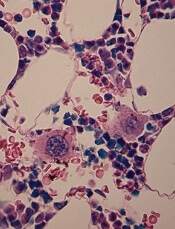
Scientists say they’ve shed new light on the mechanism of platelet formation, paving the way to accelerating and enhancing platelet production using stem cells.
The group uncovered their findings by studying the effects of shear stress on megakaryocyte maturation and the formation of preplatelets, platelet-like particles, and megakaryocyte microparticles.
“Until recently, these microparticles were viewed as inconsequential cell debris,” said Terry Papoutsakis, PhD, of the University of Delaware in Newark.
“We now know that they play a significant biological role in platelet formation. The enhanced generation of preplatelets and platelet-like particles under shear stress correlates with physiological observations—in healthy adults, both acute and prolonged exercise leads to elevated platelet counts.”
“Now, these findings can be used to develop better bioreactor technologies for producing platelets, preplatelets, platelet-like particles, and megakaryocyte microparticles for transfusion medicine, using stem cells as starting material.”
Dr Papoutsakis and his colleagues described these findings in Blood.
The researchers discovered that shear stress accelerated DNA synthesis of immature megakaryocytes, and this was dependent upon exposure time and the shear stress level.
Physiological shear stress increased the formation of preplatelets and platelet-like particles up to 10.8-fold. And it increased megakaryocyte microparticle production up to 47-fold. Platelet-like particles generated under shear flow showed improved function.
Experiments also revealed that phosphatidylserine exposure and caspase-3 activation were enhanced by shear stress. But inhibiting caspase-3 reduced the formation of preplatelets, platelet-like particles, and megakaryocyte microparticles.
Finally, the researchers found that coculturing megakaryocyte microparticles with hematopoietic stem and progenitor cells promoted differentiation to mature megakaryocytes that synthesized α- and dense-granules, and formed preplatelets without exogenous thrombopoietin.
The team noted that, unlike platelets themselves, these microparticles can be frozen, which will enable them to be stored and used for platelet production on an as-needed basis.
“Knowing that these microparticles have a biological function opens the door to other applications, including genetic therapies,” Dr Papoutsakis said. “We’re hopeful that our discovery can break the vicious cycle of [immune thrombocytopenia] as well as other conditions that cause reduced platelet count and cause life-threatening bleeding.”
in the bone marrow
Scientists say they’ve shed new light on the mechanism of platelet formation, paving the way to accelerating and enhancing platelet production using stem cells.
The group uncovered their findings by studying the effects of shear stress on megakaryocyte maturation and the formation of preplatelets, platelet-like particles, and megakaryocyte microparticles.
“Until recently, these microparticles were viewed as inconsequential cell debris,” said Terry Papoutsakis, PhD, of the University of Delaware in Newark.
“We now know that they play a significant biological role in platelet formation. The enhanced generation of preplatelets and platelet-like particles under shear stress correlates with physiological observations—in healthy adults, both acute and prolonged exercise leads to elevated platelet counts.”
“Now, these findings can be used to develop better bioreactor technologies for producing platelets, preplatelets, platelet-like particles, and megakaryocyte microparticles for transfusion medicine, using stem cells as starting material.”
Dr Papoutsakis and his colleagues described these findings in Blood.
The researchers discovered that shear stress accelerated DNA synthesis of immature megakaryocytes, and this was dependent upon exposure time and the shear stress level.
Physiological shear stress increased the formation of preplatelets and platelet-like particles up to 10.8-fold. And it increased megakaryocyte microparticle production up to 47-fold. Platelet-like particles generated under shear flow showed improved function.
Experiments also revealed that phosphatidylserine exposure and caspase-3 activation were enhanced by shear stress. But inhibiting caspase-3 reduced the formation of preplatelets, platelet-like particles, and megakaryocyte microparticles.
Finally, the researchers found that coculturing megakaryocyte microparticles with hematopoietic stem and progenitor cells promoted differentiation to mature megakaryocytes that synthesized α- and dense-granules, and formed preplatelets without exogenous thrombopoietin.
The team noted that, unlike platelets themselves, these microparticles can be frozen, which will enable them to be stored and used for platelet production on an as-needed basis.
“Knowing that these microparticles have a biological function opens the door to other applications, including genetic therapies,” Dr Papoutsakis said. “We’re hopeful that our discovery can break the vicious cycle of [immune thrombocytopenia] as well as other conditions that cause reduced platelet count and cause life-threatening bleeding.”
in the bone marrow
Scientists say they’ve shed new light on the mechanism of platelet formation, paving the way to accelerating and enhancing platelet production using stem cells.
The group uncovered their findings by studying the effects of shear stress on megakaryocyte maturation and the formation of preplatelets, platelet-like particles, and megakaryocyte microparticles.
“Until recently, these microparticles were viewed as inconsequential cell debris,” said Terry Papoutsakis, PhD, of the University of Delaware in Newark.
“We now know that they play a significant biological role in platelet formation. The enhanced generation of preplatelets and platelet-like particles under shear stress correlates with physiological observations—in healthy adults, both acute and prolonged exercise leads to elevated platelet counts.”
“Now, these findings can be used to develop better bioreactor technologies for producing platelets, preplatelets, platelet-like particles, and megakaryocyte microparticles for transfusion medicine, using stem cells as starting material.”
Dr Papoutsakis and his colleagues described these findings in Blood.
The researchers discovered that shear stress accelerated DNA synthesis of immature megakaryocytes, and this was dependent upon exposure time and the shear stress level.
Physiological shear stress increased the formation of preplatelets and platelet-like particles up to 10.8-fold. And it increased megakaryocyte microparticle production up to 47-fold. Platelet-like particles generated under shear flow showed improved function.
Experiments also revealed that phosphatidylserine exposure and caspase-3 activation were enhanced by shear stress. But inhibiting caspase-3 reduced the formation of preplatelets, platelet-like particles, and megakaryocyte microparticles.
Finally, the researchers found that coculturing megakaryocyte microparticles with hematopoietic stem and progenitor cells promoted differentiation to mature megakaryocytes that synthesized α- and dense-granules, and formed preplatelets without exogenous thrombopoietin.
The team noted that, unlike platelets themselves, these microparticles can be frozen, which will enable them to be stored and used for platelet production on an as-needed basis.
“Knowing that these microparticles have a biological function opens the door to other applications, including genetic therapies,” Dr Papoutsakis said. “We’re hopeful that our discovery can break the vicious cycle of [immune thrombocytopenia] as well as other conditions that cause reduced platelet count and cause life-threatening bleeding.”
Hypercalcemia may be indicator for hematologic cancers
Credit: Graham Colm
Hypercalcemia could be an early indication of cancer, according to a study published in the British Journal of Cancer.
The connection between hypercalcemia and cancer is well known, but this study shows the condition can predate cancer diagnosis in primary care.
The association between hypercalcemia and cancers was particularly strong in men. And myeloma and other hematologic malignancies were among the most common cancers associated with hypercalcemia.
“All previous studies on hypercalcemia and cancer had been carried out with patients who had already been diagnosed with cancer; hypercalcemia was seen as a late effect of the cancer,” said study author Fergus Hamilton, of the University of Bristol in the UK.
“We wanted to look at the issue from a different perspective and find out if high calcium levels in blood could be used as an early indicator of cancer and, therefore, in the diagnosis of cancer.”
So the researchers analyzed the electronic records of 54,267 patients with elevated calcium levels and found that hypercalcemia was strongly associated with cancer, especially in males.
The positive predictive values for cancer in men were 11.5% for calcium levels between 2.60 and 2.79 mmol l-1, 27.9% for 2.8-2.99 mmol l-1, and 50% for >3.0 mmol l-1. In women, the corresponding values were 4.1%, 8.7%, and 16.7%, respectively.
In men, the most common cancers associated with hypercalcemia were lung (34%), prostate (21%), colorectal (8%), myeloma (8%), and other hematologic cancers (8%). There were 12 other cancer types recorded as well (19%).
In women, the most common cancers were myeloma (24%), breast (18%), other hematologic cancers (10%), lung (8%), and metastatic cancer with unknown primary (8%). There were 16 other cancers recorded among women (32%).
The researchers found no difference in calcium levels among the different cancers.
“We were surprised by the gender difference,” Dr Hamilton said. “There are a number of possible explanations for this, but we think it might be because women are much more likely to have hyperparathyroidism, another cause of hypercalcemia. Men rarely get this condition, so their hypercalcemia is more likely to be due to cancer.”
Credit: Graham Colm
Hypercalcemia could be an early indication of cancer, according to a study published in the British Journal of Cancer.
The connection between hypercalcemia and cancer is well known, but this study shows the condition can predate cancer diagnosis in primary care.
The association between hypercalcemia and cancers was particularly strong in men. And myeloma and other hematologic malignancies were among the most common cancers associated with hypercalcemia.
“All previous studies on hypercalcemia and cancer had been carried out with patients who had already been diagnosed with cancer; hypercalcemia was seen as a late effect of the cancer,” said study author Fergus Hamilton, of the University of Bristol in the UK.
“We wanted to look at the issue from a different perspective and find out if high calcium levels in blood could be used as an early indicator of cancer and, therefore, in the diagnosis of cancer.”
So the researchers analyzed the electronic records of 54,267 patients with elevated calcium levels and found that hypercalcemia was strongly associated with cancer, especially in males.
The positive predictive values for cancer in men were 11.5% for calcium levels between 2.60 and 2.79 mmol l-1, 27.9% for 2.8-2.99 mmol l-1, and 50% for >3.0 mmol l-1. In women, the corresponding values were 4.1%, 8.7%, and 16.7%, respectively.
In men, the most common cancers associated with hypercalcemia were lung (34%), prostate (21%), colorectal (8%), myeloma (8%), and other hematologic cancers (8%). There were 12 other cancer types recorded as well (19%).
In women, the most common cancers were myeloma (24%), breast (18%), other hematologic cancers (10%), lung (8%), and metastatic cancer with unknown primary (8%). There were 16 other cancers recorded among women (32%).
The researchers found no difference in calcium levels among the different cancers.
“We were surprised by the gender difference,” Dr Hamilton said. “There are a number of possible explanations for this, but we think it might be because women are much more likely to have hyperparathyroidism, another cause of hypercalcemia. Men rarely get this condition, so their hypercalcemia is more likely to be due to cancer.”
Credit: Graham Colm
Hypercalcemia could be an early indication of cancer, according to a study published in the British Journal of Cancer.
The connection between hypercalcemia and cancer is well known, but this study shows the condition can predate cancer diagnosis in primary care.
The association between hypercalcemia and cancers was particularly strong in men. And myeloma and other hematologic malignancies were among the most common cancers associated with hypercalcemia.
“All previous studies on hypercalcemia and cancer had been carried out with patients who had already been diagnosed with cancer; hypercalcemia was seen as a late effect of the cancer,” said study author Fergus Hamilton, of the University of Bristol in the UK.
“We wanted to look at the issue from a different perspective and find out if high calcium levels in blood could be used as an early indicator of cancer and, therefore, in the diagnosis of cancer.”
So the researchers analyzed the electronic records of 54,267 patients with elevated calcium levels and found that hypercalcemia was strongly associated with cancer, especially in males.
The positive predictive values for cancer in men were 11.5% for calcium levels between 2.60 and 2.79 mmol l-1, 27.9% for 2.8-2.99 mmol l-1, and 50% for >3.0 mmol l-1. In women, the corresponding values were 4.1%, 8.7%, and 16.7%, respectively.
In men, the most common cancers associated with hypercalcemia were lung (34%), prostate (21%), colorectal (8%), myeloma (8%), and other hematologic cancers (8%). There were 12 other cancer types recorded as well (19%).
In women, the most common cancers were myeloma (24%), breast (18%), other hematologic cancers (10%), lung (8%), and metastatic cancer with unknown primary (8%). There were 16 other cancers recorded among women (32%).
The researchers found no difference in calcium levels among the different cancers.
“We were surprised by the gender difference,” Dr Hamilton said. “There are a number of possible explanations for this, but we think it might be because women are much more likely to have hyperparathyroidism, another cause of hypercalcemia. Men rarely get this condition, so their hypercalcemia is more likely to be due to cancer.”
Rivaroxaban rivals standard for VTE in cancer

Credit: CDC
MADRID—A pooled analysis of 2 studies suggests rivaroxaban prevents recurrent venous thromboembolism (VTE) in cancer patients as effectively as standard therapy, while conferring a lower risk of major bleeding.
The analysis included data from the EINSTEIN-DVT and EINSTEIN-PE trials, which were funded by the companies developing rivaroxaban (Xarelto), Bayer HealthCare Pharmaceuticals and Janssen Research & Development, LLC.
The results were published in Lancet Haematology and presented at the ESMO 2014 Congress (abstract LBA48).
The EINSTEIN-PE study included 3449 subjects with acute symptomatic pulmonary embolism (PE), with or without symptomatic deep vein thrombosis (DVT), who received anticoagulant therapy for 6 or 12 months.
The EINSTEIN-DVT study included 4833 patients who had acute symptomatic DVT, but no symptoms of PE, and received treatment for 3, 6, or 12 months.
In both studies, patients received either oral rivaroxaban (15 mg twice daily for 3 weeks, followed by 20 mg once daily) or standard therapy—enoxaparin (1.0 mg/kg twice daily) followed by a vitamin K antagonist (warfarin or acenocoumarol).
A total of 8281 patients were randomized in these studies—4150 to rivaroxaban and 4131 to standard therapy. Of the 655 patients (7.9%) with active cancer, 462 (5.6%) had cancer at baseline, and 193 (2.3%) were diagnosed during the study.
For this analysis, Martin H. Prins, MD, PhD, of Maastricht University Medical Center in The Netherlands, and his colleagues compared the 2 treatments in patients with active cancer.
VTE recurred in 4.5% (16/354) of the patients who were randomized to rivaroxaban and 6.6% (20/301) of patients randomized to standard therapy (hazard ratio [HR]=0.67).
The mortality rate was 16.4% (58/354) among patients randomized to rivaroxaban and 17.6% (53/301) among those randomized to standard therapy (HR=0.93).
Major bleeding occurred in 2.3% (8/353) of patients who received rivaroxaban and 5% (15/298) who received standard therapy (HR=0.42). And clinically relevant bleeding occurred in 13.6% (48/353) and 16.4% (49/298), respectively (HR=0.80).
Given these results, Dr Prins and his colleagues concluded that rivaroxaban can be considered an alternative to standard therapy in patients with cancer-associated VTE.
The team also said there is a need for a head-to-head comparison of rivaroxaban and long-term low-molecular-weight heparin in this patient population.
Credit: CDC
MADRID—A pooled analysis of 2 studies suggests rivaroxaban prevents recurrent venous thromboembolism (VTE) in cancer patients as effectively as standard therapy, while conferring a lower risk of major bleeding.
The analysis included data from the EINSTEIN-DVT and EINSTEIN-PE trials, which were funded by the companies developing rivaroxaban (Xarelto), Bayer HealthCare Pharmaceuticals and Janssen Research & Development, LLC.
The results were published in Lancet Haematology and presented at the ESMO 2014 Congress (abstract LBA48).
The EINSTEIN-PE study included 3449 subjects with acute symptomatic pulmonary embolism (PE), with or without symptomatic deep vein thrombosis (DVT), who received anticoagulant therapy for 6 or 12 months.
The EINSTEIN-DVT study included 4833 patients who had acute symptomatic DVT, but no symptoms of PE, and received treatment for 3, 6, or 12 months.
In both studies, patients received either oral rivaroxaban (15 mg twice daily for 3 weeks, followed by 20 mg once daily) or standard therapy—enoxaparin (1.0 mg/kg twice daily) followed by a vitamin K antagonist (warfarin or acenocoumarol).
A total of 8281 patients were randomized in these studies—4150 to rivaroxaban and 4131 to standard therapy. Of the 655 patients (7.9%) with active cancer, 462 (5.6%) had cancer at baseline, and 193 (2.3%) were diagnosed during the study.
For this analysis, Martin H. Prins, MD, PhD, of Maastricht University Medical Center in The Netherlands, and his colleagues compared the 2 treatments in patients with active cancer.
VTE recurred in 4.5% (16/354) of the patients who were randomized to rivaroxaban and 6.6% (20/301) of patients randomized to standard therapy (hazard ratio [HR]=0.67).
The mortality rate was 16.4% (58/354) among patients randomized to rivaroxaban and 17.6% (53/301) among those randomized to standard therapy (HR=0.93).
Major bleeding occurred in 2.3% (8/353) of patients who received rivaroxaban and 5% (15/298) who received standard therapy (HR=0.42). And clinically relevant bleeding occurred in 13.6% (48/353) and 16.4% (49/298), respectively (HR=0.80).
Given these results, Dr Prins and his colleagues concluded that rivaroxaban can be considered an alternative to standard therapy in patients with cancer-associated VTE.
The team also said there is a need for a head-to-head comparison of rivaroxaban and long-term low-molecular-weight heparin in this patient population.
Credit: CDC
MADRID—A pooled analysis of 2 studies suggests rivaroxaban prevents recurrent venous thromboembolism (VTE) in cancer patients as effectively as standard therapy, while conferring a lower risk of major bleeding.
The analysis included data from the EINSTEIN-DVT and EINSTEIN-PE trials, which were funded by the companies developing rivaroxaban (Xarelto), Bayer HealthCare Pharmaceuticals and Janssen Research & Development, LLC.
The results were published in Lancet Haematology and presented at the ESMO 2014 Congress (abstract LBA48).
The EINSTEIN-PE study included 3449 subjects with acute symptomatic pulmonary embolism (PE), with or without symptomatic deep vein thrombosis (DVT), who received anticoagulant therapy for 6 or 12 months.
The EINSTEIN-DVT study included 4833 patients who had acute symptomatic DVT, but no symptoms of PE, and received treatment for 3, 6, or 12 months.
In both studies, patients received either oral rivaroxaban (15 mg twice daily for 3 weeks, followed by 20 mg once daily) or standard therapy—enoxaparin (1.0 mg/kg twice daily) followed by a vitamin K antagonist (warfarin or acenocoumarol).
A total of 8281 patients were randomized in these studies—4150 to rivaroxaban and 4131 to standard therapy. Of the 655 patients (7.9%) with active cancer, 462 (5.6%) had cancer at baseline, and 193 (2.3%) were diagnosed during the study.
For this analysis, Martin H. Prins, MD, PhD, of Maastricht University Medical Center in The Netherlands, and his colleagues compared the 2 treatments in patients with active cancer.
VTE recurred in 4.5% (16/354) of the patients who were randomized to rivaroxaban and 6.6% (20/301) of patients randomized to standard therapy (hazard ratio [HR]=0.67).
The mortality rate was 16.4% (58/354) among patients randomized to rivaroxaban and 17.6% (53/301) among those randomized to standard therapy (HR=0.93).
Major bleeding occurred in 2.3% (8/353) of patients who received rivaroxaban and 5% (15/298) who received standard therapy (HR=0.42). And clinically relevant bleeding occurred in 13.6% (48/353) and 16.4% (49/298), respectively (HR=0.80).
Given these results, Dr Prins and his colleagues concluded that rivaroxaban can be considered an alternative to standard therapy in patients with cancer-associated VTE.
The team also said there is a need for a head-to-head comparison of rivaroxaban and long-term low-molecular-weight heparin in this patient population.
FDA approves cancer drugs faster, study shows

MADRID—The US Food and Drug Administration (FDA) tends to approve cancer drugs faster than Health Canada and the European Medicines Agency (EMA),
according to a study presented at the ESMO 2014 Congress.
On average, the FDA approved antineoplastic agents about 6 to 8 months faster than the EMA and Health Canada, researchers found.
One of the drugs studied had been FDA-approved for more than 4.5 years before the EMA and Health Canada authorized its use.
The researchers said these results suggest a need for a coordinated international approach to reduce the disparity in approval times.
“There needs to be a dialogue amongst industry, regulatory agencies, patient bodies, [the] research community, and oncology professionals on how best we can reduce the time to approval while ensuring safety for approved drugs,” said study investigator Sunil Verma, MD, of Sunnybrook Odette Cancer Center in Toronto, Canada.
A previous study, published in NEJM in 2012, showed that, between 2001 and 2010, the FDA tended to approve all types of drugs faster than the EMA and Health Canada.
Dr Verma and Nardin Samuel, an MD/PhD student at the University of Toronto, focused their study on cancer drugs and presented their findings at ESMO as abstract 1036O_PR.
The pair analyzed approval data for 41 antineoplastic agents and found the average time to FDA approval for these drugs was 6 months shorter than for the EMA and 7.6 months shorter than for Health Canada.
Azacitidine, which is approved to treat hematologic malignancies, had the greatest delay between FDA and Health Canada approval, at 66.1 months. The EMA approved azacitidine 10.3 months earlier than Health Canada but 55.8 months after the FDA.
The fastest approval time among the drugs studied was for cabazitaxel, which was approved for metastatic prostate cancer by the FDA just 17 days after the drug’s manufacturer filed for approval. In Canada and the European Union, the times to approval for cabazitaxel were 11.63 months and 11.03 months, respectively.
“It is not clear why there were these differences, but they are of some concern . . . ,” said David Cameron, MD, of the Edinburgh Cancer Research Centre in the UK, who was not involved in this research.
“[T]hey suggest that, in the absence of data to the contrary, there may be bureaucratic rather than medical/scientific reasons for differential geographical approval timelines, which, of course, will lead to differential geographical benefits from new agents.”
Dr Cameron added that more work is needed to understand the reasons for these differences, as well as assess any potential impact on patients.
MADRID—The US Food and Drug Administration (FDA) tends to approve cancer drugs faster than Health Canada and the European Medicines Agency (EMA),
according to a study presented at the ESMO 2014 Congress.
On average, the FDA approved antineoplastic agents about 6 to 8 months faster than the EMA and Health Canada, researchers found.
One of the drugs studied had been FDA-approved for more than 4.5 years before the EMA and Health Canada authorized its use.
The researchers said these results suggest a need for a coordinated international approach to reduce the disparity in approval times.
“There needs to be a dialogue amongst industry, regulatory agencies, patient bodies, [the] research community, and oncology professionals on how best we can reduce the time to approval while ensuring safety for approved drugs,” said study investigator Sunil Verma, MD, of Sunnybrook Odette Cancer Center in Toronto, Canada.
A previous study, published in NEJM in 2012, showed that, between 2001 and 2010, the FDA tended to approve all types of drugs faster than the EMA and Health Canada.
Dr Verma and Nardin Samuel, an MD/PhD student at the University of Toronto, focused their study on cancer drugs and presented their findings at ESMO as abstract 1036O_PR.
The pair analyzed approval data for 41 antineoplastic agents and found the average time to FDA approval for these drugs was 6 months shorter than for the EMA and 7.6 months shorter than for Health Canada.
Azacitidine, which is approved to treat hematologic malignancies, had the greatest delay between FDA and Health Canada approval, at 66.1 months. The EMA approved azacitidine 10.3 months earlier than Health Canada but 55.8 months after the FDA.
The fastest approval time among the drugs studied was for cabazitaxel, which was approved for metastatic prostate cancer by the FDA just 17 days after the drug’s manufacturer filed for approval. In Canada and the European Union, the times to approval for cabazitaxel were 11.63 months and 11.03 months, respectively.
“It is not clear why there were these differences, but they are of some concern . . . ,” said David Cameron, MD, of the Edinburgh Cancer Research Centre in the UK, who was not involved in this research.
“[T]hey suggest that, in the absence of data to the contrary, there may be bureaucratic rather than medical/scientific reasons for differential geographical approval timelines, which, of course, will lead to differential geographical benefits from new agents.”
Dr Cameron added that more work is needed to understand the reasons for these differences, as well as assess any potential impact on patients.
MADRID—The US Food and Drug Administration (FDA) tends to approve cancer drugs faster than Health Canada and the European Medicines Agency (EMA),
according to a study presented at the ESMO 2014 Congress.
On average, the FDA approved antineoplastic agents about 6 to 8 months faster than the EMA and Health Canada, researchers found.
One of the drugs studied had been FDA-approved for more than 4.5 years before the EMA and Health Canada authorized its use.
The researchers said these results suggest a need for a coordinated international approach to reduce the disparity in approval times.
“There needs to be a dialogue amongst industry, regulatory agencies, patient bodies, [the] research community, and oncology professionals on how best we can reduce the time to approval while ensuring safety for approved drugs,” said study investigator Sunil Verma, MD, of Sunnybrook Odette Cancer Center in Toronto, Canada.
A previous study, published in NEJM in 2012, showed that, between 2001 and 2010, the FDA tended to approve all types of drugs faster than the EMA and Health Canada.
Dr Verma and Nardin Samuel, an MD/PhD student at the University of Toronto, focused their study on cancer drugs and presented their findings at ESMO as abstract 1036O_PR.
The pair analyzed approval data for 41 antineoplastic agents and found the average time to FDA approval for these drugs was 6 months shorter than for the EMA and 7.6 months shorter than for Health Canada.
Azacitidine, which is approved to treat hematologic malignancies, had the greatest delay between FDA and Health Canada approval, at 66.1 months. The EMA approved azacitidine 10.3 months earlier than Health Canada but 55.8 months after the FDA.
The fastest approval time among the drugs studied was for cabazitaxel, which was approved for metastatic prostate cancer by the FDA just 17 days after the drug’s manufacturer filed for approval. In Canada and the European Union, the times to approval for cabazitaxel were 11.63 months and 11.03 months, respectively.
“It is not clear why there were these differences, but they are of some concern . . . ,” said David Cameron, MD, of the Edinburgh Cancer Research Centre in the UK, who was not involved in this research.
“[T]hey suggest that, in the absence of data to the contrary, there may be bureaucratic rather than medical/scientific reasons for differential geographical approval timelines, which, of course, will lead to differential geographical benefits from new agents.”
Dr Cameron added that more work is needed to understand the reasons for these differences, as well as assess any potential impact on patients.
Compound can inhibit metastasis in multiple myeloma

Credit: Daniel E. Sabath
A novel compound can prevent metastasis in mouse models of multiple myeloma (MM), according to research published in Cell Reports.
Investigators discovered that this compound, olaptesed pegol, can inhibit stromal cell-derived factor-1 (SDF-1), which attracts certain cells to new locations within the bone marrow.
By blocking the activity of SDF-1, olaptesed pegol renders the bone marrow uninviting to MM cells and prevents metastasis.
“Metastasis remains one of the most formidable complications we face as cancer researchers and physicians,” said study author Irene Ghobrial, MD, of the Dana-Farber Cancer Institute in Boston.
“Improvements in the treatment of metastatic cancers have, for the most part, not been nearly as dramatic as in primary disease.”
Dr Ghobrial and her colleagues studied MM because it is metastatic by nature. Myeloma cells originate in the bone marrow, depart for the bloodstream, and eventually return to the bones, where they form numerous colonies.
The team found that mice with advanced stages of MM had higher levels of SDF-1 at sites in the bones where metastasis had occurred.
“We reasoned that by neutralizing SDF-1, we could change the bone marrow environment to make it less receptive for multiple myeloma cells, reduce myeloma cells’ affinity for the marrow, and thereby inhibit the progression of the disease,” said Aldo Roccaro, MD, PhD, also of Dana-Farber.
Working with the German biotechnology company NOXXON Pharma, the investigators tested olaptesed pegol (a PEGylated mirror-image L-oligonucleotide), which binds to SDF-1.
The team found that olaptesed pegol modulates bone marrow niches and prevents MM cells from homing and engrafting to bone.
This slowed disease progression and prolonged survival in the animals, both compared to control mice and mice treated with AMD3100.
The investigators said it isn’t completely clear what becomes of the blood-borne MM cells that are prevented from metastasizing.
“We know that myeloma cells can’t survive for long if they’re circulating in the blood and can’t adhere to other tissue,” Dr Ghobrial said. “We saw no evidence that they had metastasized and begun to grow in other tissue either.”
“Our findings clearly document a therapeutic effect of olaptesed pegol in a mouse model of advanced myeloma. It is now being tested in a clinical trial of multiple myeloma patients, with more trials to come.”
Credit: Daniel E. Sabath
A novel compound can prevent metastasis in mouse models of multiple myeloma (MM), according to research published in Cell Reports.
Investigators discovered that this compound, olaptesed pegol, can inhibit stromal cell-derived factor-1 (SDF-1), which attracts certain cells to new locations within the bone marrow.
By blocking the activity of SDF-1, olaptesed pegol renders the bone marrow uninviting to MM cells and prevents metastasis.
“Metastasis remains one of the most formidable complications we face as cancer researchers and physicians,” said study author Irene Ghobrial, MD, of the Dana-Farber Cancer Institute in Boston.
“Improvements in the treatment of metastatic cancers have, for the most part, not been nearly as dramatic as in primary disease.”
Dr Ghobrial and her colleagues studied MM because it is metastatic by nature. Myeloma cells originate in the bone marrow, depart for the bloodstream, and eventually return to the bones, where they form numerous colonies.
The team found that mice with advanced stages of MM had higher levels of SDF-1 at sites in the bones where metastasis had occurred.
“We reasoned that by neutralizing SDF-1, we could change the bone marrow environment to make it less receptive for multiple myeloma cells, reduce myeloma cells’ affinity for the marrow, and thereby inhibit the progression of the disease,” said Aldo Roccaro, MD, PhD, also of Dana-Farber.
Working with the German biotechnology company NOXXON Pharma, the investigators tested olaptesed pegol (a PEGylated mirror-image L-oligonucleotide), which binds to SDF-1.
The team found that olaptesed pegol modulates bone marrow niches and prevents MM cells from homing and engrafting to bone.
This slowed disease progression and prolonged survival in the animals, both compared to control mice and mice treated with AMD3100.
The investigators said it isn’t completely clear what becomes of the blood-borne MM cells that are prevented from metastasizing.
“We know that myeloma cells can’t survive for long if they’re circulating in the blood and can’t adhere to other tissue,” Dr Ghobrial said. “We saw no evidence that they had metastasized and begun to grow in other tissue either.”
“Our findings clearly document a therapeutic effect of olaptesed pegol in a mouse model of advanced myeloma. It is now being tested in a clinical trial of multiple myeloma patients, with more trials to come.”
Credit: Daniel E. Sabath
A novel compound can prevent metastasis in mouse models of multiple myeloma (MM), according to research published in Cell Reports.
Investigators discovered that this compound, olaptesed pegol, can inhibit stromal cell-derived factor-1 (SDF-1), which attracts certain cells to new locations within the bone marrow.
By blocking the activity of SDF-1, olaptesed pegol renders the bone marrow uninviting to MM cells and prevents metastasis.
“Metastasis remains one of the most formidable complications we face as cancer researchers and physicians,” said study author Irene Ghobrial, MD, of the Dana-Farber Cancer Institute in Boston.
“Improvements in the treatment of metastatic cancers have, for the most part, not been nearly as dramatic as in primary disease.”
Dr Ghobrial and her colleagues studied MM because it is metastatic by nature. Myeloma cells originate in the bone marrow, depart for the bloodstream, and eventually return to the bones, where they form numerous colonies.
The team found that mice with advanced stages of MM had higher levels of SDF-1 at sites in the bones where metastasis had occurred.
“We reasoned that by neutralizing SDF-1, we could change the bone marrow environment to make it less receptive for multiple myeloma cells, reduce myeloma cells’ affinity for the marrow, and thereby inhibit the progression of the disease,” said Aldo Roccaro, MD, PhD, also of Dana-Farber.
Working with the German biotechnology company NOXXON Pharma, the investigators tested olaptesed pegol (a PEGylated mirror-image L-oligonucleotide), which binds to SDF-1.
The team found that olaptesed pegol modulates bone marrow niches and prevents MM cells from homing and engrafting to bone.
This slowed disease progression and prolonged survival in the animals, both compared to control mice and mice treated with AMD3100.
The investigators said it isn’t completely clear what becomes of the blood-borne MM cells that are prevented from metastasizing.
“We know that myeloma cells can’t survive for long if they’re circulating in the blood and can’t adhere to other tissue,” Dr Ghobrial said. “We saw no evidence that they had metastasized and begun to grow in other tissue either.”
“Our findings clearly document a therapeutic effect of olaptesed pegol in a mouse model of advanced myeloma. It is now being tested in a clinical trial of multiple myeloma patients, with more trials to come.”