User login
Novel capsule could replace injections

Credit: USDA
Scientists have created a novel drug capsule coated with tiny needles that can inject drugs directly into the lining of the stomach after the capsule is swallowed.
In experiments with pigs, the capsule delivered insulin more efficiently than an injection under the skin, and there were no harmful side effects as the capsule passed through the digestive system.
The researchers anticipate the capsule would be most useful for delivering biopharmaceuticals such as antibodies to treat cancers and other disorders.
“This could be a way that the patient can circumvent the need to have an infusion or subcutaneous administration of a drug,” said Giovanni Traverso, MB BChir, PhD, of Massachusetts General Hospital in Boston.
He and his colleagues described their capsule in the Journal of Pharmaceutical Sciences.
The team had set out to design a capsule that would serve as a platform for the delivery of a wide range of therapeutics, prevent degradation of the drugs, and inject the payload directly into the lining of the gastrointestinal tract.
Their prototype acrylic capsule, 2 cm long and 1 cm in diameter, includes a reservoir for the drug and is coated with hollow, stainless steel needles about 5 mm long.
Previous studies of accidental ingestion of sharp objects in human patients have suggested that it could be safe to swallow a capsule coated with short needles. Because there are no pain receptors in the gastrointestinal tract, patients would not feel any pain from the drug injection.
To test whether this type of capsule could allow safe and effective drug delivery, the researchers tested it in pigs, with insulin as the drug payload.
It took more than a week for the capsules to move through the entire digestive tract, and the researchers found no traces of tissue damage, supporting the potential safety of this novel approach.
They also found the microneedles successfully injected insulin into the lining of the stomach, small intestine, and colon, causing the animals’ blood glucose levels to drop. This reduction in blood glucose was faster and larger than the drop seen when the same amount of glucose was given by subcutaneous injection.
“The kinetics are much better, and much faster-onset, than those seen with traditional under-the-skin administration,” Dr Traverso said. “For molecules that are particularly difficult to absorb, this would be a way of actually administering them at much higher efficiency.”
This approach could also be used to administer vaccines that normally have to be injected, the researchers said.
The team now plans to modify the capsule so that peristalsis would slowly squeeze the drug out of the capsule as it travels through the digestive tract.
They are also working on capsules with needles made of degradable polymers and sugar that would break off and become embedded in the gut lining, where they would slowly disintegrate and release the drug. This would further minimize any safety concern. 
Credit: USDA
Scientists have created a novel drug capsule coated with tiny needles that can inject drugs directly into the lining of the stomach after the capsule is swallowed.
In experiments with pigs, the capsule delivered insulin more efficiently than an injection under the skin, and there were no harmful side effects as the capsule passed through the digestive system.
The researchers anticipate the capsule would be most useful for delivering biopharmaceuticals such as antibodies to treat cancers and other disorders.
“This could be a way that the patient can circumvent the need to have an infusion or subcutaneous administration of a drug,” said Giovanni Traverso, MB BChir, PhD, of Massachusetts General Hospital in Boston.
He and his colleagues described their capsule in the Journal of Pharmaceutical Sciences.
The team had set out to design a capsule that would serve as a platform for the delivery of a wide range of therapeutics, prevent degradation of the drugs, and inject the payload directly into the lining of the gastrointestinal tract.
Their prototype acrylic capsule, 2 cm long and 1 cm in diameter, includes a reservoir for the drug and is coated with hollow, stainless steel needles about 5 mm long.
Previous studies of accidental ingestion of sharp objects in human patients have suggested that it could be safe to swallow a capsule coated with short needles. Because there are no pain receptors in the gastrointestinal tract, patients would not feel any pain from the drug injection.
To test whether this type of capsule could allow safe and effective drug delivery, the researchers tested it in pigs, with insulin as the drug payload.
It took more than a week for the capsules to move through the entire digestive tract, and the researchers found no traces of tissue damage, supporting the potential safety of this novel approach.
They also found the microneedles successfully injected insulin into the lining of the stomach, small intestine, and colon, causing the animals’ blood glucose levels to drop. This reduction in blood glucose was faster and larger than the drop seen when the same amount of glucose was given by subcutaneous injection.
“The kinetics are much better, and much faster-onset, than those seen with traditional under-the-skin administration,” Dr Traverso said. “For molecules that are particularly difficult to absorb, this would be a way of actually administering them at much higher efficiency.”
This approach could also be used to administer vaccines that normally have to be injected, the researchers said.
The team now plans to modify the capsule so that peristalsis would slowly squeeze the drug out of the capsule as it travels through the digestive tract.
They are also working on capsules with needles made of degradable polymers and sugar that would break off and become embedded in the gut lining, where they would slowly disintegrate and release the drug. This would further minimize any safety concern.
Credit: USDA
Scientists have created a novel drug capsule coated with tiny needles that can inject drugs directly into the lining of the stomach after the capsule is swallowed.
In experiments with pigs, the capsule delivered insulin more efficiently than an injection under the skin, and there were no harmful side effects as the capsule passed through the digestive system.
The researchers anticipate the capsule would be most useful for delivering biopharmaceuticals such as antibodies to treat cancers and other disorders.
“This could be a way that the patient can circumvent the need to have an infusion or subcutaneous administration of a drug,” said Giovanni Traverso, MB BChir, PhD, of Massachusetts General Hospital in Boston.
He and his colleagues described their capsule in the Journal of Pharmaceutical Sciences.
The team had set out to design a capsule that would serve as a platform for the delivery of a wide range of therapeutics, prevent degradation of the drugs, and inject the payload directly into the lining of the gastrointestinal tract.
Their prototype acrylic capsule, 2 cm long and 1 cm in diameter, includes a reservoir for the drug and is coated with hollow, stainless steel needles about 5 mm long.
Previous studies of accidental ingestion of sharp objects in human patients have suggested that it could be safe to swallow a capsule coated with short needles. Because there are no pain receptors in the gastrointestinal tract, patients would not feel any pain from the drug injection.
To test whether this type of capsule could allow safe and effective drug delivery, the researchers tested it in pigs, with insulin as the drug payload.
It took more than a week for the capsules to move through the entire digestive tract, and the researchers found no traces of tissue damage, supporting the potential safety of this novel approach.
They also found the microneedles successfully injected insulin into the lining of the stomach, small intestine, and colon, causing the animals’ blood glucose levels to drop. This reduction in blood glucose was faster and larger than the drop seen when the same amount of glucose was given by subcutaneous injection.
“The kinetics are much better, and much faster-onset, than those seen with traditional under-the-skin administration,” Dr Traverso said. “For molecules that are particularly difficult to absorb, this would be a way of actually administering them at much higher efficiency.”
This approach could also be used to administer vaccines that normally have to be injected, the researchers said.
The team now plans to modify the capsule so that peristalsis would slowly squeeze the drug out of the capsule as it travels through the digestive tract.
They are also working on capsules with needles made of degradable polymers and sugar that would break off and become embedded in the gut lining, where they would slowly disintegrate and release the drug. This would further minimize any safety concern.
Ibrutinib fights chronic GVHD in mice

Preclinical research suggests the anticancer agent ibrutinib can ameliorate chronic graft-vs-host disease (GVHD).
Ibrutinib reduced the symptoms and progression of chronic GVHD in mouse models, and it decreased the activation of T and B cells isolated from patients with chronic GVHD.
These results indicate that T and B cells drive chronic GVHD and ibrutinib should be explored as a treatment option for human GVHD, according to investigators.
Bruce Blazar, MD, of the University of Minnesota in Minneapolis, and his colleagues described this work in The Journal of Clinical Investigation.
The team noted that CD4+ T cells and B cells mediate chronic GVHD, so it follows that targeting these populations might inhibit chronic GVHD pathogenesis.
As ibrutinib targets Th2 cells and B cells, the investigators decided to test whether the drug could reverse established chronic GVHD in 2 mouse models—a model of T cell–driven sclerodermatous chronic GVHD and an alloantibody-driven, multiorgan-system chronic GVHD model that induces bronchiolar obliterans.
Sclerodermatous chronic GVHD
The researchers first found that ibrutinib can reduce the clinical signs of sclerodermatous chronic GVHD. Fourteen days after treatment began, vehicle-treated mice and those that received cyclosporine exhibited sclerodermatous lesions, hair loss, hunched posture, and scabbing. But ibrutinib-treated mice did not.
Animals treated with ibrutinib had a significantly lower overall intensity of chronic GVHD—as measured by body weight, posture, mobility, hair loss, skin lesions, and vitality—than vehicle-treated mice (P=0.0184).
Ibrutinib also extended the median time to chronic GVHD progression by 14 days when compared to control. Thirty-three percent of ibrutinib-treated mice remained progression-free, compared to 12% of vehicle-treated mice (P<0.02).
Overall survival was 100% among ibrutinib-treated mice, 82% for cyclosporine-treated mice, and 88% for vehicle-treated mice.
The investigators also discovered that prolonged administration of ibrutinib is needed to reap the maximum therapeutic benefit in sclerodermatous chronic GVHD. Withdrawing treatment at day 60 enabled clinical breakthrough of chronic GVHD in a single mouse.
Alloantibody-driven chronic GVHD
In this model, ibrutinib inhibited the development of bronchiolar obliterans, as measured by pulmonary resistance (P=0.0090), elastance (P=0.0019), and compliance (P=0.0071).
In addition, there was less peribroncheolar collagen fibrosis among ibrutinib-treated animals and a significant reduction in pulmonary fibrosis (P<0.0001) compared to vehicle-treated controls.
However, continued therapy was necessary to see a long-term benefit with ibrutinib. Prophylactic ibrutinib given from day -2 to day 28 was not effective against chronic GVHD or bronchiolar obliterans.
Lastly, the investigators found that ibrutinib reduced the overall size, cellularity, and number of germinal center reactions (P<0.001), and the drug eliminated pulmonary immunoglobulin deposition (P<0.001).
Verifying the results
Additional experiments showed that mice lacking Bruton tyrosine kinase and IL-2 inducible T-cell kinase (both of which are inhibited by ibrutinib) did not develop chronic GVHD, which suggests these molecules are necessary for the condition to occur.
The investigators also discovered that ibrutinib reduced the activation of T and B cells from patients with active chronic GVHD.
CD4+ T cells pretreated with ibrutinib had lower surface expression of CD69 after ex vivo T-cell receptor stimulation using anti-CD3 (P=0.033). And purified B cells pretreated with ibrutinib showed lower levels of pBTK-Y223, pPLCγ2-Y1217, and pERK1/2.
Dr Blazar and his colleagues said these results indicate that B cells and T cells drive chronic GVHD and suggest that ibrutinib should be considered for testing in clinical trials of chronic GVHD.
Preclinical research suggests the anticancer agent ibrutinib can ameliorate chronic graft-vs-host disease (GVHD).
Ibrutinib reduced the symptoms and progression of chronic GVHD in mouse models, and it decreased the activation of T and B cells isolated from patients with chronic GVHD.
These results indicate that T and B cells drive chronic GVHD and ibrutinib should be explored as a treatment option for human GVHD, according to investigators.
Bruce Blazar, MD, of the University of Minnesota in Minneapolis, and his colleagues described this work in The Journal of Clinical Investigation.
The team noted that CD4+ T cells and B cells mediate chronic GVHD, so it follows that targeting these populations might inhibit chronic GVHD pathogenesis.
As ibrutinib targets Th2 cells and B cells, the investigators decided to test whether the drug could reverse established chronic GVHD in 2 mouse models—a model of T cell–driven sclerodermatous chronic GVHD and an alloantibody-driven, multiorgan-system chronic GVHD model that induces bronchiolar obliterans.
Sclerodermatous chronic GVHD
The researchers first found that ibrutinib can reduce the clinical signs of sclerodermatous chronic GVHD. Fourteen days after treatment began, vehicle-treated mice and those that received cyclosporine exhibited sclerodermatous lesions, hair loss, hunched posture, and scabbing. But ibrutinib-treated mice did not.
Animals treated with ibrutinib had a significantly lower overall intensity of chronic GVHD—as measured by body weight, posture, mobility, hair loss, skin lesions, and vitality—than vehicle-treated mice (P=0.0184).
Ibrutinib also extended the median time to chronic GVHD progression by 14 days when compared to control. Thirty-three percent of ibrutinib-treated mice remained progression-free, compared to 12% of vehicle-treated mice (P<0.02).
Overall survival was 100% among ibrutinib-treated mice, 82% for cyclosporine-treated mice, and 88% for vehicle-treated mice.
The investigators also discovered that prolonged administration of ibrutinib is needed to reap the maximum therapeutic benefit in sclerodermatous chronic GVHD. Withdrawing treatment at day 60 enabled clinical breakthrough of chronic GVHD in a single mouse.
Alloantibody-driven chronic GVHD
In this model, ibrutinib inhibited the development of bronchiolar obliterans, as measured by pulmonary resistance (P=0.0090), elastance (P=0.0019), and compliance (P=0.0071).
In addition, there was less peribroncheolar collagen fibrosis among ibrutinib-treated animals and a significant reduction in pulmonary fibrosis (P<0.0001) compared to vehicle-treated controls.
However, continued therapy was necessary to see a long-term benefit with ibrutinib. Prophylactic ibrutinib given from day -2 to day 28 was not effective against chronic GVHD or bronchiolar obliterans.
Lastly, the investigators found that ibrutinib reduced the overall size, cellularity, and number of germinal center reactions (P<0.001), and the drug eliminated pulmonary immunoglobulin deposition (P<0.001).
Verifying the results
Additional experiments showed that mice lacking Bruton tyrosine kinase and IL-2 inducible T-cell kinase (both of which are inhibited by ibrutinib) did not develop chronic GVHD, which suggests these molecules are necessary for the condition to occur.
The investigators also discovered that ibrutinib reduced the activation of T and B cells from patients with active chronic GVHD.
CD4+ T cells pretreated with ibrutinib had lower surface expression of CD69 after ex vivo T-cell receptor stimulation using anti-CD3 (P=0.033). And purified B cells pretreated with ibrutinib showed lower levels of pBTK-Y223, pPLCγ2-Y1217, and pERK1/2.
Dr Blazar and his colleagues said these results indicate that B cells and T cells drive chronic GVHD and suggest that ibrutinib should be considered for testing in clinical trials of chronic GVHD.
Preclinical research suggests the anticancer agent ibrutinib can ameliorate chronic graft-vs-host disease (GVHD).
Ibrutinib reduced the symptoms and progression of chronic GVHD in mouse models, and it decreased the activation of T and B cells isolated from patients with chronic GVHD.
These results indicate that T and B cells drive chronic GVHD and ibrutinib should be explored as a treatment option for human GVHD, according to investigators.
Bruce Blazar, MD, of the University of Minnesota in Minneapolis, and his colleagues described this work in The Journal of Clinical Investigation.
The team noted that CD4+ T cells and B cells mediate chronic GVHD, so it follows that targeting these populations might inhibit chronic GVHD pathogenesis.
As ibrutinib targets Th2 cells and B cells, the investigators decided to test whether the drug could reverse established chronic GVHD in 2 mouse models—a model of T cell–driven sclerodermatous chronic GVHD and an alloantibody-driven, multiorgan-system chronic GVHD model that induces bronchiolar obliterans.
Sclerodermatous chronic GVHD
The researchers first found that ibrutinib can reduce the clinical signs of sclerodermatous chronic GVHD. Fourteen days after treatment began, vehicle-treated mice and those that received cyclosporine exhibited sclerodermatous lesions, hair loss, hunched posture, and scabbing. But ibrutinib-treated mice did not.
Animals treated with ibrutinib had a significantly lower overall intensity of chronic GVHD—as measured by body weight, posture, mobility, hair loss, skin lesions, and vitality—than vehicle-treated mice (P=0.0184).
Ibrutinib also extended the median time to chronic GVHD progression by 14 days when compared to control. Thirty-three percent of ibrutinib-treated mice remained progression-free, compared to 12% of vehicle-treated mice (P<0.02).
Overall survival was 100% among ibrutinib-treated mice, 82% for cyclosporine-treated mice, and 88% for vehicle-treated mice.
The investigators also discovered that prolonged administration of ibrutinib is needed to reap the maximum therapeutic benefit in sclerodermatous chronic GVHD. Withdrawing treatment at day 60 enabled clinical breakthrough of chronic GVHD in a single mouse.
Alloantibody-driven chronic GVHD
In this model, ibrutinib inhibited the development of bronchiolar obliterans, as measured by pulmonary resistance (P=0.0090), elastance (P=0.0019), and compliance (P=0.0071).
In addition, there was less peribroncheolar collagen fibrosis among ibrutinib-treated animals and a significant reduction in pulmonary fibrosis (P<0.0001) compared to vehicle-treated controls.
However, continued therapy was necessary to see a long-term benefit with ibrutinib. Prophylactic ibrutinib given from day -2 to day 28 was not effective against chronic GVHD or bronchiolar obliterans.
Lastly, the investigators found that ibrutinib reduced the overall size, cellularity, and number of germinal center reactions (P<0.001), and the drug eliminated pulmonary immunoglobulin deposition (P<0.001).
Verifying the results
Additional experiments showed that mice lacking Bruton tyrosine kinase and IL-2 inducible T-cell kinase (both of which are inhibited by ibrutinib) did not develop chronic GVHD, which suggests these molecules are necessary for the condition to occur.
The investigators also discovered that ibrutinib reduced the activation of T and B cells from patients with active chronic GVHD.
CD4+ T cells pretreated with ibrutinib had lower surface expression of CD69 after ex vivo T-cell receptor stimulation using anti-CD3 (P=0.033). And purified B cells pretreated with ibrutinib showed lower levels of pBTK-Y223, pPLCγ2-Y1217, and pERK1/2.
Dr Blazar and his colleagues said these results indicate that B cells and T cells drive chronic GVHD and suggest that ibrutinib should be considered for testing in clinical trials of chronic GVHD.
Group detects new steps in hematopoiesis

in the bone marrow
Researchers say they have discovered previously undetected steps in hematopoiesis, establishing that a highly complex series of events determine the fate of closely related populations of blood progenitor cells.
Their study revealed thousands of differences in gene expression between blood cell types.
These differences result from many specific events that are crucial for normal blood development, and errors in this process can lead to blood disorders.
The researchers described their work in Science.
The team sequenced RNA from 8 primary human hematopoietic progenitor populations representing the classical myeloid commitment stages of hematopoiesis and the main lymphoid stage.
This revealed 6711 genes and 10,724 transcripts enriched in non-protein-coding elements at early stages of differentiation.
The researchers also discovered the extent to which RNA is cut and pasted together in different ways during hematopoiesis, leading to specific forms of proteins for each of these stages.
“We have identified thousands of novel places where the RNA is processed in an alternative way,” said study author Willem Ouwehand, MD, PhD, of the University of Cambridge in the UK.
Specifically, the team identified 7881 novel splice junctions and 2301 differentially used alternative splicing events enriched in genes involved in regulatory processes.
“Such events changed the amount, structure, and behavior of proteins derived from a single gene,” said study author Wendy Erber, MD, DPhil, of the University of Western Australia in Crawley.
“Alternative proteins could drive stem cells towards becoming different mature blood cells.”
Until this study, hematopoiesis was relatively well understood at the level of DNA. What was not known was how the genetic information in DNA was then transcribed to generate RNA, leading to protein formation.
The researchers illustrated the importance of alternative RNA splicing in blood cell development by studying the role that 2 different forms of the same transcription factor—NFIB—play in megakaryocyte formation.
The team said their findings could have significant applications for patients with blood disorders. The results could aid the design of diagnostics and new therapies, as well as prove valuable for studies in stem cell transplant and for discovering the genetic basis of rare, inherited hematologic disorders.
in the bone marrow
Researchers say they have discovered previously undetected steps in hematopoiesis, establishing that a highly complex series of events determine the fate of closely related populations of blood progenitor cells.
Their study revealed thousands of differences in gene expression between blood cell types.
These differences result from many specific events that are crucial for normal blood development, and errors in this process can lead to blood disorders.
The researchers described their work in Science.
The team sequenced RNA from 8 primary human hematopoietic progenitor populations representing the classical myeloid commitment stages of hematopoiesis and the main lymphoid stage.
This revealed 6711 genes and 10,724 transcripts enriched in non-protein-coding elements at early stages of differentiation.
The researchers also discovered the extent to which RNA is cut and pasted together in different ways during hematopoiesis, leading to specific forms of proteins for each of these stages.
“We have identified thousands of novel places where the RNA is processed in an alternative way,” said study author Willem Ouwehand, MD, PhD, of the University of Cambridge in the UK.
Specifically, the team identified 7881 novel splice junctions and 2301 differentially used alternative splicing events enriched in genes involved in regulatory processes.
“Such events changed the amount, structure, and behavior of proteins derived from a single gene,” said study author Wendy Erber, MD, DPhil, of the University of Western Australia in Crawley.
“Alternative proteins could drive stem cells towards becoming different mature blood cells.”
Until this study, hematopoiesis was relatively well understood at the level of DNA. What was not known was how the genetic information in DNA was then transcribed to generate RNA, leading to protein formation.
The researchers illustrated the importance of alternative RNA splicing in blood cell development by studying the role that 2 different forms of the same transcription factor—NFIB—play in megakaryocyte formation.
The team said their findings could have significant applications for patients with blood disorders. The results could aid the design of diagnostics and new therapies, as well as prove valuable for studies in stem cell transplant and for discovering the genetic basis of rare, inherited hematologic disorders.
in the bone marrow
Researchers say they have discovered previously undetected steps in hematopoiesis, establishing that a highly complex series of events determine the fate of closely related populations of blood progenitor cells.
Their study revealed thousands of differences in gene expression between blood cell types.
These differences result from many specific events that are crucial for normal blood development, and errors in this process can lead to blood disorders.
The researchers described their work in Science.
The team sequenced RNA from 8 primary human hematopoietic progenitor populations representing the classical myeloid commitment stages of hematopoiesis and the main lymphoid stage.
This revealed 6711 genes and 10,724 transcripts enriched in non-protein-coding elements at early stages of differentiation.
The researchers also discovered the extent to which RNA is cut and pasted together in different ways during hematopoiesis, leading to specific forms of proteins for each of these stages.
“We have identified thousands of novel places where the RNA is processed in an alternative way,” said study author Willem Ouwehand, MD, PhD, of the University of Cambridge in the UK.
Specifically, the team identified 7881 novel splice junctions and 2301 differentially used alternative splicing events enriched in genes involved in regulatory processes.
“Such events changed the amount, structure, and behavior of proteins derived from a single gene,” said study author Wendy Erber, MD, DPhil, of the University of Western Australia in Crawley.
“Alternative proteins could drive stem cells towards becoming different mature blood cells.”
Until this study, hematopoiesis was relatively well understood at the level of DNA. What was not known was how the genetic information in DNA was then transcribed to generate RNA, leading to protein formation.
The researchers illustrated the importance of alternative RNA splicing in blood cell development by studying the role that 2 different forms of the same transcription factor—NFIB—play in megakaryocyte formation.
The team said their findings could have significant applications for patients with blood disorders. The results could aid the design of diagnostics and new therapies, as well as prove valuable for studies in stem cell transplant and for discovering the genetic basis of rare, inherited hematologic disorders.
HHS identifies known and likely carcinogens

Credit: Trevor MacInnis
The US Department of Health and Human Services (HHS) has identified 1 chemical substance as a known human carcinogen and 3 additional substances as likely carcinogens.
Ortho-toluidine, which is used to make rubber chemicals, pesticides, and dyes, has been shown to cause urinary bladder cancer and is now listed as a known human carcinogen.
The 3 substances that are likely to be human carcinogens are 1-bromopropane, cumene, and pentachlorophenol.
1-bromopropane is used as a cleaning solvent and spray adhesive. Cumene is used to make phenol and acetone, and it is found in fuel products and tobacco smoke. Pentachlorophenol is a mixture used to preserve wood.
Exposure to pentachlorophenol is associated with an increased risk of non-Hodgkin lymphoma in humans and solid tumor malignancies in mice. Cumene and 1-bromopropane have been linked to solid tumor malignancies in mice as well.
All 4 substances are listed in the HHS’s 13th Report on Carcinogens, a science-based document prepared by the National Toxicology Program (NTP) that identifies chemical, biological, and physical agents considered to be cancer hazards for people living in the US.
The new report has a total of 243 listings, which includes known carcinogens and substances “reasonably anticipated” to be carcinogens.
“Identifying substances in our environment that can make people vulnerable to cancer will help in prevention efforts,” said Linda Birnbaum, PhD, director of the National Institute of Environmental Health Sciences and the NTP.
“This report provides a valuable resource for health regulatory and research agencies, and it empowers the public with information people can use to reduce exposure to cancer-causing substances.”
New known carcinogen
Since 1983, ortho-toluidine has been listed in the HHS’s Report on Carcinogens as reasonably anticipated to be a human carcinogen. However, new cancer studies led the NTP to reevaluate and reclassify ortho-toluidine. It is now classified as a known human carcinogen, based on clinical studies showing it causes urinary bladder cancer.
Ortho-toluidine is a synthetic chemical produced in other countries and imported into the US by several companies in high volumes. It is primarily used to make rubber chemicals, pesticides, and dyes. It is also used in some consumer and medical products.
People are mainly exposed through the workplace, by skin contact and/or inhalation when using ortho-toluidine. They can also be exposed outside the workplace through sources such as tobacco smoke.
Three new substances likely to be carcinogenic
Pentachlorophenol
Pentachlorophenol and byproducts of its synthesis are complex mixtures of chemicals used as wood preservatives. Because virtually everyone exposed to pentachlorophenol is also exposed to its synthesis byproducts, they were evaluated together.
In the US, pentachlorophenol has been regulated since the 1980s as a restricted-use pesticide. It is used industrially for treating utility poles, wood pilings, fence posts, and lumber or timber for construction.
Most exposure has occurred in settings where workers treat lumber or come in contact with treated lumber. People may also be exposed to this mixture from breathing contaminated air or dust, or from contact with contaminated soil.
Exposure to this mixture was associated with an increased risk of non-Hodgkin lymphoma in clinical studies. In mice, it has been shown to cause tumors in the liver and other organs.
1-bromopropane
1-bromopropane is a liquid used as a solvent in many commercial industries. It is used as a cleaner for optics, electronics, and metals, as well as a solvent for aerosol-applied adhesives such as those used in foam cushion manufacturing.
It is also used in dry cleaning and in solvent sprays for aircraft maintenance. Workers in certain occupations may be more exposed to 1-bromopropane than the general population.
The NTP did not identify any clinical studies that evaluated the relationship between human cancer and exposure to 1-bromopropane. However, inhalation exposure to 1-bromopropane in rodents caused tumors in several organs, including the skin, lungs, and large intestine.
Cumene
Cumene is a flammable and volatile liquid with a gasoline-like odor. It is a natural component of coal tar and petroleum, and is found in tobacco smoke. It is used primarily to make acetone and phenol.
People are mainly exposed to cumene through the environment and in workplaces that use or produce cumene. It can be found in emissions from petroleum products.
Inhalation exposure to cumene caused lung tumors in male and female mice, and liver tumors in female mice. The NTP did not identify any clinical studies evaluating the relationship between cancer and exposure to cumene.
Credit: Trevor MacInnis
The US Department of Health and Human Services (HHS) has identified 1 chemical substance as a known human carcinogen and 3 additional substances as likely carcinogens.
Ortho-toluidine, which is used to make rubber chemicals, pesticides, and dyes, has been shown to cause urinary bladder cancer and is now listed as a known human carcinogen.
The 3 substances that are likely to be human carcinogens are 1-bromopropane, cumene, and pentachlorophenol.
1-bromopropane is used as a cleaning solvent and spray adhesive. Cumene is used to make phenol and acetone, and it is found in fuel products and tobacco smoke. Pentachlorophenol is a mixture used to preserve wood.
Exposure to pentachlorophenol is associated with an increased risk of non-Hodgkin lymphoma in humans and solid tumor malignancies in mice. Cumene and 1-bromopropane have been linked to solid tumor malignancies in mice as well.
All 4 substances are listed in the HHS’s 13th Report on Carcinogens, a science-based document prepared by the National Toxicology Program (NTP) that identifies chemical, biological, and physical agents considered to be cancer hazards for people living in the US.
The new report has a total of 243 listings, which includes known carcinogens and substances “reasonably anticipated” to be carcinogens.
“Identifying substances in our environment that can make people vulnerable to cancer will help in prevention efforts,” said Linda Birnbaum, PhD, director of the National Institute of Environmental Health Sciences and the NTP.
“This report provides a valuable resource for health regulatory and research agencies, and it empowers the public with information people can use to reduce exposure to cancer-causing substances.”
New known carcinogen
Since 1983, ortho-toluidine has been listed in the HHS’s Report on Carcinogens as reasonably anticipated to be a human carcinogen. However, new cancer studies led the NTP to reevaluate and reclassify ortho-toluidine. It is now classified as a known human carcinogen, based on clinical studies showing it causes urinary bladder cancer.
Ortho-toluidine is a synthetic chemical produced in other countries and imported into the US by several companies in high volumes. It is primarily used to make rubber chemicals, pesticides, and dyes. It is also used in some consumer and medical products.
People are mainly exposed through the workplace, by skin contact and/or inhalation when using ortho-toluidine. They can also be exposed outside the workplace through sources such as tobacco smoke.
Three new substances likely to be carcinogenic
Pentachlorophenol
Pentachlorophenol and byproducts of its synthesis are complex mixtures of chemicals used as wood preservatives. Because virtually everyone exposed to pentachlorophenol is also exposed to its synthesis byproducts, they were evaluated together.
In the US, pentachlorophenol has been regulated since the 1980s as a restricted-use pesticide. It is used industrially for treating utility poles, wood pilings, fence posts, and lumber or timber for construction.
Most exposure has occurred in settings where workers treat lumber or come in contact with treated lumber. People may also be exposed to this mixture from breathing contaminated air or dust, or from contact with contaminated soil.
Exposure to this mixture was associated with an increased risk of non-Hodgkin lymphoma in clinical studies. In mice, it has been shown to cause tumors in the liver and other organs.
1-bromopropane
1-bromopropane is a liquid used as a solvent in many commercial industries. It is used as a cleaner for optics, electronics, and metals, as well as a solvent for aerosol-applied adhesives such as those used in foam cushion manufacturing.
It is also used in dry cleaning and in solvent sprays for aircraft maintenance. Workers in certain occupations may be more exposed to 1-bromopropane than the general population.
The NTP did not identify any clinical studies that evaluated the relationship between human cancer and exposure to 1-bromopropane. However, inhalation exposure to 1-bromopropane in rodents caused tumors in several organs, including the skin, lungs, and large intestine.
Cumene
Cumene is a flammable and volatile liquid with a gasoline-like odor. It is a natural component of coal tar and petroleum, and is found in tobacco smoke. It is used primarily to make acetone and phenol.
People are mainly exposed to cumene through the environment and in workplaces that use or produce cumene. It can be found in emissions from petroleum products.
Inhalation exposure to cumene caused lung tumors in male and female mice, and liver tumors in female mice. The NTP did not identify any clinical studies evaluating the relationship between cancer and exposure to cumene.
Credit: Trevor MacInnis
The US Department of Health and Human Services (HHS) has identified 1 chemical substance as a known human carcinogen and 3 additional substances as likely carcinogens.
Ortho-toluidine, which is used to make rubber chemicals, pesticides, and dyes, has been shown to cause urinary bladder cancer and is now listed as a known human carcinogen.
The 3 substances that are likely to be human carcinogens are 1-bromopropane, cumene, and pentachlorophenol.
1-bromopropane is used as a cleaning solvent and spray adhesive. Cumene is used to make phenol and acetone, and it is found in fuel products and tobacco smoke. Pentachlorophenol is a mixture used to preserve wood.
Exposure to pentachlorophenol is associated with an increased risk of non-Hodgkin lymphoma in humans and solid tumor malignancies in mice. Cumene and 1-bromopropane have been linked to solid tumor malignancies in mice as well.
All 4 substances are listed in the HHS’s 13th Report on Carcinogens, a science-based document prepared by the National Toxicology Program (NTP) that identifies chemical, biological, and physical agents considered to be cancer hazards for people living in the US.
The new report has a total of 243 listings, which includes known carcinogens and substances “reasonably anticipated” to be carcinogens.
“Identifying substances in our environment that can make people vulnerable to cancer will help in prevention efforts,” said Linda Birnbaum, PhD, director of the National Institute of Environmental Health Sciences and the NTP.
“This report provides a valuable resource for health regulatory and research agencies, and it empowers the public with information people can use to reduce exposure to cancer-causing substances.”
New known carcinogen
Since 1983, ortho-toluidine has been listed in the HHS’s Report on Carcinogens as reasonably anticipated to be a human carcinogen. However, new cancer studies led the NTP to reevaluate and reclassify ortho-toluidine. It is now classified as a known human carcinogen, based on clinical studies showing it causes urinary bladder cancer.
Ortho-toluidine is a synthetic chemical produced in other countries and imported into the US by several companies in high volumes. It is primarily used to make rubber chemicals, pesticides, and dyes. It is also used in some consumer and medical products.
People are mainly exposed through the workplace, by skin contact and/or inhalation when using ortho-toluidine. They can also be exposed outside the workplace through sources such as tobacco smoke.
Three new substances likely to be carcinogenic
Pentachlorophenol
Pentachlorophenol and byproducts of its synthesis are complex mixtures of chemicals used as wood preservatives. Because virtually everyone exposed to pentachlorophenol is also exposed to its synthesis byproducts, they were evaluated together.
In the US, pentachlorophenol has been regulated since the 1980s as a restricted-use pesticide. It is used industrially for treating utility poles, wood pilings, fence posts, and lumber or timber for construction.
Most exposure has occurred in settings where workers treat lumber or come in contact with treated lumber. People may also be exposed to this mixture from breathing contaminated air or dust, or from contact with contaminated soil.
Exposure to this mixture was associated with an increased risk of non-Hodgkin lymphoma in clinical studies. In mice, it has been shown to cause tumors in the liver and other organs.
1-bromopropane
1-bromopropane is a liquid used as a solvent in many commercial industries. It is used as a cleaner for optics, electronics, and metals, as well as a solvent for aerosol-applied adhesives such as those used in foam cushion manufacturing.
It is also used in dry cleaning and in solvent sprays for aircraft maintenance. Workers in certain occupations may be more exposed to 1-bromopropane than the general population.
The NTP did not identify any clinical studies that evaluated the relationship between human cancer and exposure to 1-bromopropane. However, inhalation exposure to 1-bromopropane in rodents caused tumors in several organs, including the skin, lungs, and large intestine.
Cumene
Cumene is a flammable and volatile liquid with a gasoline-like odor. It is a natural component of coal tar and petroleum, and is found in tobacco smoke. It is used primarily to make acetone and phenol.
People are mainly exposed to cumene through the environment and in workplaces that use or produce cumene. It can be found in emissions from petroleum products.
Inhalation exposure to cumene caused lung tumors in male and female mice, and liver tumors in female mice. The NTP did not identify any clinical studies evaluating the relationship between cancer and exposure to cumene.
Results of VTE prophylaxis vary in otolaryngology patients

The effectiveness of thromboprophylaxis in otolaryngology patients undergoing surgery differs based on patient risk and the procedure, a new study suggests.
Overall, the incidence of venous thromboembolism (VTE) was similar between patients who received prophylaxis and those who did not.
However, prophylaxis reduced the incidence of VTE in patients with a high Caprini risk score and those who underwent microvascular free tissue reconstruction.
On the other hand, there was an increased risk of bleeding complications associated with prophylaxis among patients who underwent microvascular free tissue reconstruction and in the cohort overall.
Vinita Bahl, DMD, of the University of Michigan Health System in Ann Arbor, and his colleagues conducted this research and recounted the results in JAMA Otolaryngology–Head & Neck Surgery.
The researchers analyzed 3498 patients treated by surgeons at an academic medical center between September 2003 and June 2010. The team assessed the incidence of VTE and bleeding complications in the 30 days after surgery.
In all, 1482 patients received VTE prophylaxis. Most (96.8%) received subcutaneous unfractionated heparin (5000 IU 2-3 times daily), 3.1% received enoxaparin (30-40 mg daily), and 0.1% received fondaparinux (2.5 mg daily).
The incidence of VTE among all patients was 1.3%—0.74% were deep vein thromboses and 0.66% were pulmonary emboli. The overall incidence of bleeding complications was 2.2%.
VTE occurred in 1.2% of patients who received prophylaxis and 1.3% of patients who did not (P=0.75).
Bleeding complications occurred in 3.5% and 1.2% of patients, respectively (P<0.001).
Patients with Caprini VTE risk scores greater than 7 were less likely to develop a VTE if they received perioperative prophylaxis—5.3% vs 10.4% (P=0.06).
Prophylaxis also decreased the incidence of VTE among patients who underwent free tissue transfer—2.1% vs 7.7% (P=0.002). But it increased bleeding complications—11.9% vs 4.5% (P=0.01).
Bleeding complications were associated with concomitant use of antiplatelet medications and VTE prophylaxis.
In all other patients, prophylaxis did not affect the incidence of VTE or bleeding. VTE occurred in 1% of treated and 0.6% of untreated patients (P=0.12). And bleeding occurred in 1.5% and 0.9% of patients, respectively (P=0.15).
These results suggest the Caprini risk assessment model is an effective tool to stratify otolaryngology patients according to VTE risk, the researchers said.
However, they believe additional research is needed before recommendations can be made for patients undergoing free tissue reconstruction, as prophylaxis reduced their risk of VTE but increased their risk of bleeding.
The effectiveness of thromboprophylaxis in otolaryngology patients undergoing surgery differs based on patient risk and the procedure, a new study suggests.
Overall, the incidence of venous thromboembolism (VTE) was similar between patients who received prophylaxis and those who did not.
However, prophylaxis reduced the incidence of VTE in patients with a high Caprini risk score and those who underwent microvascular free tissue reconstruction.
On the other hand, there was an increased risk of bleeding complications associated with prophylaxis among patients who underwent microvascular free tissue reconstruction and in the cohort overall.
Vinita Bahl, DMD, of the University of Michigan Health System in Ann Arbor, and his colleagues conducted this research and recounted the results in JAMA Otolaryngology–Head & Neck Surgery.
The researchers analyzed 3498 patients treated by surgeons at an academic medical center between September 2003 and June 2010. The team assessed the incidence of VTE and bleeding complications in the 30 days after surgery.
In all, 1482 patients received VTE prophylaxis. Most (96.8%) received subcutaneous unfractionated heparin (5000 IU 2-3 times daily), 3.1% received enoxaparin (30-40 mg daily), and 0.1% received fondaparinux (2.5 mg daily).
The incidence of VTE among all patients was 1.3%—0.74% were deep vein thromboses and 0.66% were pulmonary emboli. The overall incidence of bleeding complications was 2.2%.
VTE occurred in 1.2% of patients who received prophylaxis and 1.3% of patients who did not (P=0.75).
Bleeding complications occurred in 3.5% and 1.2% of patients, respectively (P<0.001).
Patients with Caprini VTE risk scores greater than 7 were less likely to develop a VTE if they received perioperative prophylaxis—5.3% vs 10.4% (P=0.06).
Prophylaxis also decreased the incidence of VTE among patients who underwent free tissue transfer—2.1% vs 7.7% (P=0.002). But it increased bleeding complications—11.9% vs 4.5% (P=0.01).
Bleeding complications were associated with concomitant use of antiplatelet medications and VTE prophylaxis.
In all other patients, prophylaxis did not affect the incidence of VTE or bleeding. VTE occurred in 1% of treated and 0.6% of untreated patients (P=0.12). And bleeding occurred in 1.5% and 0.9% of patients, respectively (P=0.15).
These results suggest the Caprini risk assessment model is an effective tool to stratify otolaryngology patients according to VTE risk, the researchers said.
However, they believe additional research is needed before recommendations can be made for patients undergoing free tissue reconstruction, as prophylaxis reduced their risk of VTE but increased their risk of bleeding.
The effectiveness of thromboprophylaxis in otolaryngology patients undergoing surgery differs based on patient risk and the procedure, a new study suggests.
Overall, the incidence of venous thromboembolism (VTE) was similar between patients who received prophylaxis and those who did not.
However, prophylaxis reduced the incidence of VTE in patients with a high Caprini risk score and those who underwent microvascular free tissue reconstruction.
On the other hand, there was an increased risk of bleeding complications associated with prophylaxis among patients who underwent microvascular free tissue reconstruction and in the cohort overall.
Vinita Bahl, DMD, of the University of Michigan Health System in Ann Arbor, and his colleagues conducted this research and recounted the results in JAMA Otolaryngology–Head & Neck Surgery.
The researchers analyzed 3498 patients treated by surgeons at an academic medical center between September 2003 and June 2010. The team assessed the incidence of VTE and bleeding complications in the 30 days after surgery.
In all, 1482 patients received VTE prophylaxis. Most (96.8%) received subcutaneous unfractionated heparin (5000 IU 2-3 times daily), 3.1% received enoxaparin (30-40 mg daily), and 0.1% received fondaparinux (2.5 mg daily).
The incidence of VTE among all patients was 1.3%—0.74% were deep vein thromboses and 0.66% were pulmonary emboli. The overall incidence of bleeding complications was 2.2%.
VTE occurred in 1.2% of patients who received prophylaxis and 1.3% of patients who did not (P=0.75).
Bleeding complications occurred in 3.5% and 1.2% of patients, respectively (P<0.001).
Patients with Caprini VTE risk scores greater than 7 were less likely to develop a VTE if they received perioperative prophylaxis—5.3% vs 10.4% (P=0.06).
Prophylaxis also decreased the incidence of VTE among patients who underwent free tissue transfer—2.1% vs 7.7% (P=0.002). But it increased bleeding complications—11.9% vs 4.5% (P=0.01).
Bleeding complications were associated with concomitant use of antiplatelet medications and VTE prophylaxis.
In all other patients, prophylaxis did not affect the incidence of VTE or bleeding. VTE occurred in 1% of treated and 0.6% of untreated patients (P=0.12). And bleeding occurred in 1.5% and 0.9% of patients, respectively (P=0.15).
These results suggest the Caprini risk assessment model is an effective tool to stratify otolaryngology patients according to VTE risk, the researchers said.
However, they believe additional research is needed before recommendations can be made for patients undergoing free tissue reconstruction, as prophylaxis reduced their risk of VTE but increased their risk of bleeding.
Dendritic cells promote Myc-driven lymphoma
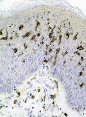
Studies have shown that dendritic cells (DCs) can contribute to tumor growth and help shield the tumor from the immune system in colon, stomach, breast, and prostate cancer.
Now, researchers have found evidence suggesting this phenomenon also occurs in Myc-driven lymphomas.
The team has also identified the molecular mechanism that induces the immune cells to promote tumor growth.
They reported these findings in Nature Communications.
Uta Höpken, PhD, of the Max Delbrück Center for Molecular Medicine in Berlin, Germany, and her colleagues investigated how DCs drive tumor in mouse models of Eµ-Myc lymphoma.
The team began by depleting DCs in these mice and found that tumor growth was delayed—the first clue that DCs are indeed associated with lymphoma growth.
Next, the researchers found that, after contact with lymphoma cells, the DCs increasingly secrete immunomodulatory cytokines and growth factors. The cytokine secretion takes place in the spleen and lymph nodes.
Dr Höpken and her colleagues previously demonstrated that various forms of lymphoma cells settle in the lymph nodes and in the spleen, where they create their own survival niche. This process is regulated by selective cytokines and growth factors the researchers identified a few years ago.
“In these niches, almost everything is already there that the lymphoma cells as malignant B cells need to survive, including blood vessels and connective tissue cells [stromal cells],” Dr Höpken said. “The survival substances secreted by the DCs optimize the niche so that the tumors can grow better.”
This also means the DCs prevent the T lymphocytes from exercising their defensive function. Normally, healthy B or T cells settle in the respective B- or T-cell niches of the spleen and the lymph nodes to be made fit for immune defense.
“What is paradoxical is that the mouse lymphoma cells we studied—malignant B cells—found their survival niche in the T-cell zones of the lymph nodes and the spleen and not in the B-cell zones,” Dr Höpken said.
After making contact with the lymphoma cells, the DCs increasingly upregulate C/EBPβ, a transcription factor that promotes the production of cytokines that mediate inflammation.
The researchers found that C/EBPβ regulates DCs. Without it, the cells could not secrete inflammatory cytokines. C/EBPβ also indirectly blocks apoptosis in the lymphoma cells, allowing the cancer cells to grow unchecked.
The team pointed out that, even if their model of Eµ-Myc lymphoma is not entirely comparable to B-cell lymphomas in humans, it shows that lymphoma cells and DCs interact—a previously unknown molecular mechanism.
Furthermore, these findings may have clinical applications. The researchers noted that the immunomodulatory agent lenalidomide induces downregulation of C/EBPβ, which is secreted by cancer cells.
So Dr Höpken and her colleagues believe it might be appropriate to approve the use of lenalidomide for patients with Myc B-cell lymphoma, as an addition to their existing treatment, to strengthen their immune defense.
Studies have shown that dendritic cells (DCs) can contribute to tumor growth and help shield the tumor from the immune system in colon, stomach, breast, and prostate cancer.
Now, researchers have found evidence suggesting this phenomenon also occurs in Myc-driven lymphomas.
The team has also identified the molecular mechanism that induces the immune cells to promote tumor growth.
They reported these findings in Nature Communications.
Uta Höpken, PhD, of the Max Delbrück Center for Molecular Medicine in Berlin, Germany, and her colleagues investigated how DCs drive tumor in mouse models of Eµ-Myc lymphoma.
The team began by depleting DCs in these mice and found that tumor growth was delayed—the first clue that DCs are indeed associated with lymphoma growth.
Next, the researchers found that, after contact with lymphoma cells, the DCs increasingly secrete immunomodulatory cytokines and growth factors. The cytokine secretion takes place in the spleen and lymph nodes.
Dr Höpken and her colleagues previously demonstrated that various forms of lymphoma cells settle in the lymph nodes and in the spleen, where they create their own survival niche. This process is regulated by selective cytokines and growth factors the researchers identified a few years ago.
“In these niches, almost everything is already there that the lymphoma cells as malignant B cells need to survive, including blood vessels and connective tissue cells [stromal cells],” Dr Höpken said. “The survival substances secreted by the DCs optimize the niche so that the tumors can grow better.”
This also means the DCs prevent the T lymphocytes from exercising their defensive function. Normally, healthy B or T cells settle in the respective B- or T-cell niches of the spleen and the lymph nodes to be made fit for immune defense.
“What is paradoxical is that the mouse lymphoma cells we studied—malignant B cells—found their survival niche in the T-cell zones of the lymph nodes and the spleen and not in the B-cell zones,” Dr Höpken said.
After making contact with the lymphoma cells, the DCs increasingly upregulate C/EBPβ, a transcription factor that promotes the production of cytokines that mediate inflammation.
The researchers found that C/EBPβ regulates DCs. Without it, the cells could not secrete inflammatory cytokines. C/EBPβ also indirectly blocks apoptosis in the lymphoma cells, allowing the cancer cells to grow unchecked.
The team pointed out that, even if their model of Eµ-Myc lymphoma is not entirely comparable to B-cell lymphomas in humans, it shows that lymphoma cells and DCs interact—a previously unknown molecular mechanism.
Furthermore, these findings may have clinical applications. The researchers noted that the immunomodulatory agent lenalidomide induces downregulation of C/EBPβ, which is secreted by cancer cells.
So Dr Höpken and her colleagues believe it might be appropriate to approve the use of lenalidomide for patients with Myc B-cell lymphoma, as an addition to their existing treatment, to strengthen their immune defense.
Studies have shown that dendritic cells (DCs) can contribute to tumor growth and help shield the tumor from the immune system in colon, stomach, breast, and prostate cancer.
Now, researchers have found evidence suggesting this phenomenon also occurs in Myc-driven lymphomas.
The team has also identified the molecular mechanism that induces the immune cells to promote tumor growth.
They reported these findings in Nature Communications.
Uta Höpken, PhD, of the Max Delbrück Center for Molecular Medicine in Berlin, Germany, and her colleagues investigated how DCs drive tumor in mouse models of Eµ-Myc lymphoma.
The team began by depleting DCs in these mice and found that tumor growth was delayed—the first clue that DCs are indeed associated with lymphoma growth.
Next, the researchers found that, after contact with lymphoma cells, the DCs increasingly secrete immunomodulatory cytokines and growth factors. The cytokine secretion takes place in the spleen and lymph nodes.
Dr Höpken and her colleagues previously demonstrated that various forms of lymphoma cells settle in the lymph nodes and in the spleen, where they create their own survival niche. This process is regulated by selective cytokines and growth factors the researchers identified a few years ago.
“In these niches, almost everything is already there that the lymphoma cells as malignant B cells need to survive, including blood vessels and connective tissue cells [stromal cells],” Dr Höpken said. “The survival substances secreted by the DCs optimize the niche so that the tumors can grow better.”
This also means the DCs prevent the T lymphocytes from exercising their defensive function. Normally, healthy B or T cells settle in the respective B- or T-cell niches of the spleen and the lymph nodes to be made fit for immune defense.
“What is paradoxical is that the mouse lymphoma cells we studied—malignant B cells—found their survival niche in the T-cell zones of the lymph nodes and the spleen and not in the B-cell zones,” Dr Höpken said.
After making contact with the lymphoma cells, the DCs increasingly upregulate C/EBPβ, a transcription factor that promotes the production of cytokines that mediate inflammation.
The researchers found that C/EBPβ regulates DCs. Without it, the cells could not secrete inflammatory cytokines. C/EBPβ also indirectly blocks apoptosis in the lymphoma cells, allowing the cancer cells to grow unchecked.
The team pointed out that, even if their model of Eµ-Myc lymphoma is not entirely comparable to B-cell lymphomas in humans, it shows that lymphoma cells and DCs interact—a previously unknown molecular mechanism.
Furthermore, these findings may have clinical applications. The researchers noted that the immunomodulatory agent lenalidomide induces downregulation of C/EBPβ, which is secreted by cancer cells.
So Dr Höpken and her colleagues believe it might be appropriate to approve the use of lenalidomide for patients with Myc B-cell lymphoma, as an addition to their existing treatment, to strengthen their immune defense.
Product appears safe, effective in rel/ref ALL

Credit: Bill Branson
Eryaspase, a product consisting of L-asparaginase encapsulated in red blood cells, may be safer and more effective than native E coli L-asparaginase, results of a phase 2/3 study suggest.
In patients with relapsed or refractory acute lymphoblastic leukemia (ALL), eryaspase given in combination with chemotherapy produced a higher complete response rate and fewer allergic reactions than native E coli L-asparaginase in combination with chemotherapy.
Eryaspase was also well-tolerated in patients who had previously experienced allergic reactions to L-asparaginase.
ERYTECH Pharma, the company developing eryaspase, recently announced these results from the GRASPIVOTALL trial.
The study included 80 children and adults with relapsed or refractory ALL, who were randomized to 1 of 3 treatment arms.
In the first 2 arms, researchers compared eryaspase to native E coli L-asparaginase, both in combination with standard chemotherapy (COOPRALL), in patients without prior allergies to L-asparaginase. In the third arm, the team evaluated eryaspase for patients who have experienced allergic reactions related to asparaginase in their first-line treatment.
The primary endpoint of the study consisted of 2 objectives: (a) superior safety, expressed as a significant reduction of the incidence of allergic reactions with eryaspase compared to native L-asparaginase, and (b) noninferior duration of asparaginase activity above the threshold of 100 IU/L during the induction phase in the nonallergic patients. Both endpoints needed to be met for the study to be considered positive.
The main secondary efficacy endpoints included complete response, minimal residual disease, event-free survival, and overall survival.
At 1 year of follow-up, both primary endpoints were met. There was a significant reduction of allergic reactions in the eryaspase arm compared to the native L-asparaginase arm—0% (0/26) and 42.9% (12/28), respectively (P<0.001).
And there was a significant increase in the duration of circulating asparaginase activity in the eryaspase arm compared to the native L-asparaginase arm. Asparaginase levels were maintained above 100 IU/L for an average of 20.5 days with up to 2 injections during the first month of treatment with eryaspase, compared to 9.2 days in the native L-asparaginase arm, with up to 8 injections of L-asparaginase (P<0.001).
In addition, the complete response rate was higher with eryaspase. At the end of the induction phase, 71.4% of patients (n=15) in the eryaspase arm had a complete response, compared to 42.3% of patients (n=11) in the native L-asparaginase arm.
Finally, the study showed that eryaspase is well-tolerated by patients with previous allergies to L-asparaginase. Two of the 26 patients with prior allergies to L-asparaginase experienced mild allergic reactions to eryaspase.
“The results of this study are an important step forward for the treatment of ALL patients that are at risk to receive L-asparaginase, which remains an important unmet medical need,” said Yves Betrand, MD, of the Institute for Pediatric Hematology and Oncology in Lyon, France.
“The virtual absence of allergic reactions, also in patients with prior allergies to L-asparaginase, is very encouraging.”
The analysis of additional secondary and exploratory endpoints for this study is ongoing. ERYTECH said results will be available later this year and are set to be presented at an upcoming scientific conference.
Based on the results of this and earlier studies of eryaspase, ERYTECH intends to submit its application for European Marketing Authorization in the first half of 2015.
The company also plans to accelerate the product’s development in ALL in the US and to launch phase 2 clinical trials in additional oncology indications with high unmet medical need. A phase 2b study of eryaspase in acute myeloid leukemia is already underway, with more than half of the patients enrolled. 
Credit: Bill Branson
Eryaspase, a product consisting of L-asparaginase encapsulated in red blood cells, may be safer and more effective than native E coli L-asparaginase, results of a phase 2/3 study suggest.
In patients with relapsed or refractory acute lymphoblastic leukemia (ALL), eryaspase given in combination with chemotherapy produced a higher complete response rate and fewer allergic reactions than native E coli L-asparaginase in combination with chemotherapy.
Eryaspase was also well-tolerated in patients who had previously experienced allergic reactions to L-asparaginase.
ERYTECH Pharma, the company developing eryaspase, recently announced these results from the GRASPIVOTALL trial.
The study included 80 children and adults with relapsed or refractory ALL, who were randomized to 1 of 3 treatment arms.
In the first 2 arms, researchers compared eryaspase to native E coli L-asparaginase, both in combination with standard chemotherapy (COOPRALL), in patients without prior allergies to L-asparaginase. In the third arm, the team evaluated eryaspase for patients who have experienced allergic reactions related to asparaginase in their first-line treatment.
The primary endpoint of the study consisted of 2 objectives: (a) superior safety, expressed as a significant reduction of the incidence of allergic reactions with eryaspase compared to native L-asparaginase, and (b) noninferior duration of asparaginase activity above the threshold of 100 IU/L during the induction phase in the nonallergic patients. Both endpoints needed to be met for the study to be considered positive.
The main secondary efficacy endpoints included complete response, minimal residual disease, event-free survival, and overall survival.
At 1 year of follow-up, both primary endpoints were met. There was a significant reduction of allergic reactions in the eryaspase arm compared to the native L-asparaginase arm—0% (0/26) and 42.9% (12/28), respectively (P<0.001).
And there was a significant increase in the duration of circulating asparaginase activity in the eryaspase arm compared to the native L-asparaginase arm. Asparaginase levels were maintained above 100 IU/L for an average of 20.5 days with up to 2 injections during the first month of treatment with eryaspase, compared to 9.2 days in the native L-asparaginase arm, with up to 8 injections of L-asparaginase (P<0.001).
In addition, the complete response rate was higher with eryaspase. At the end of the induction phase, 71.4% of patients (n=15) in the eryaspase arm had a complete response, compared to 42.3% of patients (n=11) in the native L-asparaginase arm.
Finally, the study showed that eryaspase is well-tolerated by patients with previous allergies to L-asparaginase. Two of the 26 patients with prior allergies to L-asparaginase experienced mild allergic reactions to eryaspase.
“The results of this study are an important step forward for the treatment of ALL patients that are at risk to receive L-asparaginase, which remains an important unmet medical need,” said Yves Betrand, MD, of the Institute for Pediatric Hematology and Oncology in Lyon, France.
“The virtual absence of allergic reactions, also in patients with prior allergies to L-asparaginase, is very encouraging.”
The analysis of additional secondary and exploratory endpoints for this study is ongoing. ERYTECH said results will be available later this year and are set to be presented at an upcoming scientific conference.
Based on the results of this and earlier studies of eryaspase, ERYTECH intends to submit its application for European Marketing Authorization in the first half of 2015.
The company also plans to accelerate the product’s development in ALL in the US and to launch phase 2 clinical trials in additional oncology indications with high unmet medical need. A phase 2b study of eryaspase in acute myeloid leukemia is already underway, with more than half of the patients enrolled.
Credit: Bill Branson
Eryaspase, a product consisting of L-asparaginase encapsulated in red blood cells, may be safer and more effective than native E coli L-asparaginase, results of a phase 2/3 study suggest.
In patients with relapsed or refractory acute lymphoblastic leukemia (ALL), eryaspase given in combination with chemotherapy produced a higher complete response rate and fewer allergic reactions than native E coli L-asparaginase in combination with chemotherapy.
Eryaspase was also well-tolerated in patients who had previously experienced allergic reactions to L-asparaginase.
ERYTECH Pharma, the company developing eryaspase, recently announced these results from the GRASPIVOTALL trial.
The study included 80 children and adults with relapsed or refractory ALL, who were randomized to 1 of 3 treatment arms.
In the first 2 arms, researchers compared eryaspase to native E coli L-asparaginase, both in combination with standard chemotherapy (COOPRALL), in patients without prior allergies to L-asparaginase. In the third arm, the team evaluated eryaspase for patients who have experienced allergic reactions related to asparaginase in their first-line treatment.
The primary endpoint of the study consisted of 2 objectives: (a) superior safety, expressed as a significant reduction of the incidence of allergic reactions with eryaspase compared to native L-asparaginase, and (b) noninferior duration of asparaginase activity above the threshold of 100 IU/L during the induction phase in the nonallergic patients. Both endpoints needed to be met for the study to be considered positive.
The main secondary efficacy endpoints included complete response, minimal residual disease, event-free survival, and overall survival.
At 1 year of follow-up, both primary endpoints were met. There was a significant reduction of allergic reactions in the eryaspase arm compared to the native L-asparaginase arm—0% (0/26) and 42.9% (12/28), respectively (P<0.001).
And there was a significant increase in the duration of circulating asparaginase activity in the eryaspase arm compared to the native L-asparaginase arm. Asparaginase levels were maintained above 100 IU/L for an average of 20.5 days with up to 2 injections during the first month of treatment with eryaspase, compared to 9.2 days in the native L-asparaginase arm, with up to 8 injections of L-asparaginase (P<0.001).
In addition, the complete response rate was higher with eryaspase. At the end of the induction phase, 71.4% of patients (n=15) in the eryaspase arm had a complete response, compared to 42.3% of patients (n=11) in the native L-asparaginase arm.
Finally, the study showed that eryaspase is well-tolerated by patients with previous allergies to L-asparaginase. Two of the 26 patients with prior allergies to L-asparaginase experienced mild allergic reactions to eryaspase.
“The results of this study are an important step forward for the treatment of ALL patients that are at risk to receive L-asparaginase, which remains an important unmet medical need,” said Yves Betrand, MD, of the Institute for Pediatric Hematology and Oncology in Lyon, France.
“The virtual absence of allergic reactions, also in patients with prior allergies to L-asparaginase, is very encouraging.”
The analysis of additional secondary and exploratory endpoints for this study is ongoing. ERYTECH said results will be available later this year and are set to be presented at an upcoming scientific conference.
Based on the results of this and earlier studies of eryaspase, ERYTECH intends to submit its application for European Marketing Authorization in the first half of 2015.
The company also plans to accelerate the product’s development in ALL in the US and to launch phase 2 clinical trials in additional oncology indications with high unmet medical need. A phase 2b study of eryaspase in acute myeloid leukemia is already underway, with more than half of the patients enrolled.
Cancer treatment during pregnancy can be safe
MADRID—Two new studies suggest that children exposed to chemotherapy or radiotherapy in the womb can be spared negative long-term effects.
At a median age of 6, most of the children exposed to radiation in utero had neuropsychological, behavioral, and general health outcomes that were within normal ranges.
And children who were exposed to chemotherapy in the womb had normal mental and cardiac health at a median age of 2.
“When chemotherapy is administered after the first trimester of pregnancy, we cannot discern any problems in the children,” said Frederic Amant, MD, PhD, of University Hospitals Leuven in Belgium.
“Fear about the risks of chemotherapy administration should not be a reason to terminate a pregnancy, delay cancer treatment for the mother, or to deliver a baby prematurely.”
Dr Amant and his colleagues presented these findings (abstract 267PD_PR) and their findings on radiation (abstract 49LBA_PR) at the ESMO 2014 Congress.
Outcomes with chemotherapy
In the first study, the researchers recruited 38 children from the International Network for Cancer, Infertility and Pregnancy registry who were prenatally exposed to chemotherapy.
Most of the mothers had breast (61%) or hematologic (22%) cancers. Most were treated with anthracyclines (61%) and had received an average of 4 cycles (range, 1-7) of treatment.
The researchers assessed mental development and cardiac health in the children of these subjects, comparing them to 38 control children who were not exposed to chemotherapy.
At a median age of almost 2 years, mental development was in the normal range for both groups of children, and development was not significantly different between the groups. The mean Mental Development Index score was 99.13 for exposed children and 101.47 for controls.
Cardiac dimensions and functions were within normal ranges for both groups as well. Mean fractional shortening was 36% (range, 32-42) for exposed children and 39% (range, 32-51) for controls. Although the difference was signficant (P=0.004), the researchers said it was not clinically relevant.
“This paper points to the very important issue of long-term safety of prenatal exposure to chemotherapy and reinforces the notion that chemotherapy during gestation does not endanger the fetus and her or his subsequent development,” said Fedro Alessandro Peccatori, MD, PhD, of the European Institute of Oncology in Milan, Italy, who was not involved in this study.
“To further ameliorate neonatal outcome, a special effort should be made to prolong pregnancy duration, and stringent long-term follow-up should be pursued to confirm these findings. Meanwhile, specific measures to support prematurely delivered babies and their families should be implemented.”
Dr Amant said future studies will explore the effects of specific chemotherapy types in detail and include longer-term follow-up.
Outcomes with radiation
In a second study, Dr Amant and his colleagues explored the impact of radiotherapy on the children of women with cancer.
The study included 16 children, with a median age of 6 years, who had been exposed to radiotherapy in utero. The median maternal irradiation was 48 Gy (range, 12-70), and the median estimated fetal irradiation was 91 mGy (range, 0-1690).
Neuropsychological, behavioral, and general health outcomes were within normal ranges for most of the children. And the researchers found no linear relationship between the fetal dose of radiation and cognitive outcome.
However, there was a negative linear relationship between the gestational age at radiotherapy exposure and verbal intelligence, based on results in 8 children. Two of these children were exposed to radiotherapy in the third trimester and had verbal intelligence scores outside the normal range.
One of the 2 children had low scores on all variables of cognitive development, but other pregnancy-related complications are confounding factors. The child’s mother suffered from an aggressive non-Hodgkin tumor of the brain that impacted her general state, and she had a preterm delivery.
Dr Amant noted that this is based on a very small number of children, so the results should be interpreted with caution.
“We cannot exclude that there might be an impact of prenatal radiotherapy exposure,” he said, “but larger series are needed to further investigate this relationship.”
MADRID—Two new studies suggest that children exposed to chemotherapy or radiotherapy in the womb can be spared negative long-term effects.
At a median age of 6, most of the children exposed to radiation in utero had neuropsychological, behavioral, and general health outcomes that were within normal ranges.
And children who were exposed to chemotherapy in the womb had normal mental and cardiac health at a median age of 2.
“When chemotherapy is administered after the first trimester of pregnancy, we cannot discern any problems in the children,” said Frederic Amant, MD, PhD, of University Hospitals Leuven in Belgium.
“Fear about the risks of chemotherapy administration should not be a reason to terminate a pregnancy, delay cancer treatment for the mother, or to deliver a baby prematurely.”
Dr Amant and his colleagues presented these findings (abstract 267PD_PR) and their findings on radiation (abstract 49LBA_PR) at the ESMO 2014 Congress.
Outcomes with chemotherapy
In the first study, the researchers recruited 38 children from the International Network for Cancer, Infertility and Pregnancy registry who were prenatally exposed to chemotherapy.
Most of the mothers had breast (61%) or hematologic (22%) cancers. Most were treated with anthracyclines (61%) and had received an average of 4 cycles (range, 1-7) of treatment.
The researchers assessed mental development and cardiac health in the children of these subjects, comparing them to 38 control children who were not exposed to chemotherapy.
At a median age of almost 2 years, mental development was in the normal range for both groups of children, and development was not significantly different between the groups. The mean Mental Development Index score was 99.13 for exposed children and 101.47 for controls.
Cardiac dimensions and functions were within normal ranges for both groups as well. Mean fractional shortening was 36% (range, 32-42) for exposed children and 39% (range, 32-51) for controls. Although the difference was signficant (P=0.004), the researchers said it was not clinically relevant.
“This paper points to the very important issue of long-term safety of prenatal exposure to chemotherapy and reinforces the notion that chemotherapy during gestation does not endanger the fetus and her or his subsequent development,” said Fedro Alessandro Peccatori, MD, PhD, of the European Institute of Oncology in Milan, Italy, who was not involved in this study.
“To further ameliorate neonatal outcome, a special effort should be made to prolong pregnancy duration, and stringent long-term follow-up should be pursued to confirm these findings. Meanwhile, specific measures to support prematurely delivered babies and their families should be implemented.”
Dr Amant said future studies will explore the effects of specific chemotherapy types in detail and include longer-term follow-up.
Outcomes with radiation
In a second study, Dr Amant and his colleagues explored the impact of radiotherapy on the children of women with cancer.
The study included 16 children, with a median age of 6 years, who had been exposed to radiotherapy in utero. The median maternal irradiation was 48 Gy (range, 12-70), and the median estimated fetal irradiation was 91 mGy (range, 0-1690).
Neuropsychological, behavioral, and general health outcomes were within normal ranges for most of the children. And the researchers found no linear relationship between the fetal dose of radiation and cognitive outcome.
However, there was a negative linear relationship between the gestational age at radiotherapy exposure and verbal intelligence, based on results in 8 children. Two of these children were exposed to radiotherapy in the third trimester and had verbal intelligence scores outside the normal range.
One of the 2 children had low scores on all variables of cognitive development, but other pregnancy-related complications are confounding factors. The child’s mother suffered from an aggressive non-Hodgkin tumor of the brain that impacted her general state, and she had a preterm delivery.
Dr Amant noted that this is based on a very small number of children, so the results should be interpreted with caution.
“We cannot exclude that there might be an impact of prenatal radiotherapy exposure,” he said, “but larger series are needed to further investigate this relationship.”
MADRID—Two new studies suggest that children exposed to chemotherapy or radiotherapy in the womb can be spared negative long-term effects.
At a median age of 6, most of the children exposed to radiation in utero had neuropsychological, behavioral, and general health outcomes that were within normal ranges.
And children who were exposed to chemotherapy in the womb had normal mental and cardiac health at a median age of 2.
“When chemotherapy is administered after the first trimester of pregnancy, we cannot discern any problems in the children,” said Frederic Amant, MD, PhD, of University Hospitals Leuven in Belgium.
“Fear about the risks of chemotherapy administration should not be a reason to terminate a pregnancy, delay cancer treatment for the mother, or to deliver a baby prematurely.”
Dr Amant and his colleagues presented these findings (abstract 267PD_PR) and their findings on radiation (abstract 49LBA_PR) at the ESMO 2014 Congress.
Outcomes with chemotherapy
In the first study, the researchers recruited 38 children from the International Network for Cancer, Infertility and Pregnancy registry who were prenatally exposed to chemotherapy.
Most of the mothers had breast (61%) or hematologic (22%) cancers. Most were treated with anthracyclines (61%) and had received an average of 4 cycles (range, 1-7) of treatment.
The researchers assessed mental development and cardiac health in the children of these subjects, comparing them to 38 control children who were not exposed to chemotherapy.
At a median age of almost 2 years, mental development was in the normal range for both groups of children, and development was not significantly different between the groups. The mean Mental Development Index score was 99.13 for exposed children and 101.47 for controls.
Cardiac dimensions and functions were within normal ranges for both groups as well. Mean fractional shortening was 36% (range, 32-42) for exposed children and 39% (range, 32-51) for controls. Although the difference was signficant (P=0.004), the researchers said it was not clinically relevant.
“This paper points to the very important issue of long-term safety of prenatal exposure to chemotherapy and reinforces the notion that chemotherapy during gestation does not endanger the fetus and her or his subsequent development,” said Fedro Alessandro Peccatori, MD, PhD, of the European Institute of Oncology in Milan, Italy, who was not involved in this study.
“To further ameliorate neonatal outcome, a special effort should be made to prolong pregnancy duration, and stringent long-term follow-up should be pursued to confirm these findings. Meanwhile, specific measures to support prematurely delivered babies and their families should be implemented.”
Dr Amant said future studies will explore the effects of specific chemotherapy types in detail and include longer-term follow-up.
Outcomes with radiation
In a second study, Dr Amant and his colleagues explored the impact of radiotherapy on the children of women with cancer.
The study included 16 children, with a median age of 6 years, who had been exposed to radiotherapy in utero. The median maternal irradiation was 48 Gy (range, 12-70), and the median estimated fetal irradiation was 91 mGy (range, 0-1690).
Neuropsychological, behavioral, and general health outcomes were within normal ranges for most of the children. And the researchers found no linear relationship between the fetal dose of radiation and cognitive outcome.
However, there was a negative linear relationship between the gestational age at radiotherapy exposure and verbal intelligence, based on results in 8 children. Two of these children were exposed to radiotherapy in the third trimester and had verbal intelligence scores outside the normal range.
One of the 2 children had low scores on all variables of cognitive development, but other pregnancy-related complications are confounding factors. The child’s mother suffered from an aggressive non-Hodgkin tumor of the brain that impacted her general state, and she had a preterm delivery.
Dr Amant noted that this is based on a very small number of children, so the results should be interpreted with caution.
“We cannot exclude that there might be an impact of prenatal radiotherapy exposure,” he said, “but larger series are needed to further investigate this relationship.”
Consolidation can improve PFS in HL
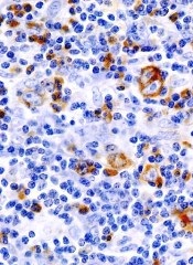
Consolidation therapy with brentuximab vedotin can improve progression-free survival (PFS) for Hodgkin lymphoma (HL) patients who have undergone a transplant, according to a phase 3 study.
The trial, known as AETHERA, is a comparison of single-agent brentuximab vedotin to placebo in patients with HL who were at risk of relapse following autologous stem cell transplant (ASCT).
Brentuximab vedotin conferred a 75% improvement in PFS over placebo.
However, there was no significant difference in overall survival between the 2 treatment arms.
These results were recently announced by Seattle Genetics Inc. and Takeda Pharmaceutical Company Limited, the companies developing brentuximab vedotin (Adcetris).
The companies said more complete results from AETHERA will be presented at the 2014 ASH Annual Meeting in December.
AETHERA is a randomized, double-blind, placebo-controlled study designed to evaluate the potential of brentuximab vedotin to extend PFS post-ASCT in patients with HL who have at least one risk factor for progression. In addition to the primary endpoint of PFS, secondary endpoints included overall survival, safety, and tolerability.
Patients were eligible if they had risk factors for residual HL, defined as a history of refractory HL, those who relapse or progress within a year of receiving frontline chemotherapy, and/or those who have disease outside of the lymph nodes at the time of pre-ASCT relapse.
The study included 329 patients who received brentuximab vedotin or placebo every 3 weeks for up to a year.
The researchers assessed PFS a minimum of 2 years after the initiation of treatment for all patients. There was a significant improvement in PFS with brentuximab vedotin compared to placebo (hazard ratio=0.57; P=0.001).
However, a prespecified interim analysis of overall survival showed no significant difference between the treatment arms.
Patients in both arms who experienced progression received a variety of subsequent therapies. Most patients on the placebo arm received brentuximab vedotin after progression.
A further analysis of overall survival is planned in 2016. The safety profile of brentuximab vedotin in the AETHERA trial was generally consistent with the existing prescribing information.
“We anticipate reporting more complete AETHERA data at the ASH Annual Meeting in December and intend to submit a supplemental Biologics License Application to the FDA in 2015 for approval in this setting,” said Clay B. Siegall, PhD, President and Chief Executive Officer of Seattle Genetics.
The FDA has already granted brentuximab vedotin accelerated approval to treat HL patients after ASCT failure or after the failure of at least 2 prior multiagent chemotherapy regimens in patients who are not ASCT candidates. The FDA also granted the drug accelerated approval to treat systemic anaplastic large cell lymphoma after the failure of at least 1 prior multiagent chemotherapy regimen.
The European Commission granted brentuximab vedotin conditional marketing authorization for the same indications. In both cases, the drug can gain full, traditional approval once studies have shown it confers a clinical benefit.
Brentuximab vedotin has a boxed warning detailing the risk of progressive multifocal leukoencephalopathy associated with use of the drug. The drug has also been shown to pose a risk of pulmonary toxicity when combined with bleomycin.
Consolidation therapy with brentuximab vedotin can improve progression-free survival (PFS) for Hodgkin lymphoma (HL) patients who have undergone a transplant, according to a phase 3 study.
The trial, known as AETHERA, is a comparison of single-agent brentuximab vedotin to placebo in patients with HL who were at risk of relapse following autologous stem cell transplant (ASCT).
Brentuximab vedotin conferred a 75% improvement in PFS over placebo.
However, there was no significant difference in overall survival between the 2 treatment arms.
These results were recently announced by Seattle Genetics Inc. and Takeda Pharmaceutical Company Limited, the companies developing brentuximab vedotin (Adcetris).
The companies said more complete results from AETHERA will be presented at the 2014 ASH Annual Meeting in December.
AETHERA is a randomized, double-blind, placebo-controlled study designed to evaluate the potential of brentuximab vedotin to extend PFS post-ASCT in patients with HL who have at least one risk factor for progression. In addition to the primary endpoint of PFS, secondary endpoints included overall survival, safety, and tolerability.
Patients were eligible if they had risk factors for residual HL, defined as a history of refractory HL, those who relapse or progress within a year of receiving frontline chemotherapy, and/or those who have disease outside of the lymph nodes at the time of pre-ASCT relapse.
The study included 329 patients who received brentuximab vedotin or placebo every 3 weeks for up to a year.
The researchers assessed PFS a minimum of 2 years after the initiation of treatment for all patients. There was a significant improvement in PFS with brentuximab vedotin compared to placebo (hazard ratio=0.57; P=0.001).
However, a prespecified interim analysis of overall survival showed no significant difference between the treatment arms.
Patients in both arms who experienced progression received a variety of subsequent therapies. Most patients on the placebo arm received brentuximab vedotin after progression.
A further analysis of overall survival is planned in 2016. The safety profile of brentuximab vedotin in the AETHERA trial was generally consistent with the existing prescribing information.
“We anticipate reporting more complete AETHERA data at the ASH Annual Meeting in December and intend to submit a supplemental Biologics License Application to the FDA in 2015 for approval in this setting,” said Clay B. Siegall, PhD, President and Chief Executive Officer of Seattle Genetics.
The FDA has already granted brentuximab vedotin accelerated approval to treat HL patients after ASCT failure or after the failure of at least 2 prior multiagent chemotherapy regimens in patients who are not ASCT candidates. The FDA also granted the drug accelerated approval to treat systemic anaplastic large cell lymphoma after the failure of at least 1 prior multiagent chemotherapy regimen.
The European Commission granted brentuximab vedotin conditional marketing authorization for the same indications. In both cases, the drug can gain full, traditional approval once studies have shown it confers a clinical benefit.
Brentuximab vedotin has a boxed warning detailing the risk of progressive multifocal leukoencephalopathy associated with use of the drug. The drug has also been shown to pose a risk of pulmonary toxicity when combined with bleomycin.
Consolidation therapy with brentuximab vedotin can improve progression-free survival (PFS) for Hodgkin lymphoma (HL) patients who have undergone a transplant, according to a phase 3 study.
The trial, known as AETHERA, is a comparison of single-agent brentuximab vedotin to placebo in patients with HL who were at risk of relapse following autologous stem cell transplant (ASCT).
Brentuximab vedotin conferred a 75% improvement in PFS over placebo.
However, there was no significant difference in overall survival between the 2 treatment arms.
These results were recently announced by Seattle Genetics Inc. and Takeda Pharmaceutical Company Limited, the companies developing brentuximab vedotin (Adcetris).
The companies said more complete results from AETHERA will be presented at the 2014 ASH Annual Meeting in December.
AETHERA is a randomized, double-blind, placebo-controlled study designed to evaluate the potential of brentuximab vedotin to extend PFS post-ASCT in patients with HL who have at least one risk factor for progression. In addition to the primary endpoint of PFS, secondary endpoints included overall survival, safety, and tolerability.
Patients were eligible if they had risk factors for residual HL, defined as a history of refractory HL, those who relapse or progress within a year of receiving frontline chemotherapy, and/or those who have disease outside of the lymph nodes at the time of pre-ASCT relapse.
The study included 329 patients who received brentuximab vedotin or placebo every 3 weeks for up to a year.
The researchers assessed PFS a minimum of 2 years after the initiation of treatment for all patients. There was a significant improvement in PFS with brentuximab vedotin compared to placebo (hazard ratio=0.57; P=0.001).
However, a prespecified interim analysis of overall survival showed no significant difference between the treatment arms.
Patients in both arms who experienced progression received a variety of subsequent therapies. Most patients on the placebo arm received brentuximab vedotin after progression.
A further analysis of overall survival is planned in 2016. The safety profile of brentuximab vedotin in the AETHERA trial was generally consistent with the existing prescribing information.
“We anticipate reporting more complete AETHERA data at the ASH Annual Meeting in December and intend to submit a supplemental Biologics License Application to the FDA in 2015 for approval in this setting,” said Clay B. Siegall, PhD, President and Chief Executive Officer of Seattle Genetics.
The FDA has already granted brentuximab vedotin accelerated approval to treat HL patients after ASCT failure or after the failure of at least 2 prior multiagent chemotherapy regimens in patients who are not ASCT candidates. The FDA also granted the drug accelerated approval to treat systemic anaplastic large cell lymphoma after the failure of at least 1 prior multiagent chemotherapy regimen.
The European Commission granted brentuximab vedotin conditional marketing authorization for the same indications. In both cases, the drug can gain full, traditional approval once studies have shown it confers a clinical benefit.
Brentuximab vedotin has a boxed warning detailing the risk of progressive multifocal leukoencephalopathy associated with use of the drug. The drug has also been shown to pose a risk of pulmonary toxicity when combined with bleomycin.
Maintenance may be unnecessary in FL

Credit: Bill Branson
New research suggests maintenance therapy may not be necessary for patients with follicular lymphoma (FL) who have a low tumor burden.
Investigators compared rituximab re-treatment with rituximab maintenance in nearly 300 FL patients, and results showed no significant difference between the treatment groups in the time to disease recurrence.
The researchers also noted that the re-treatment strategy was more cost-effective.
“For those 2 reasons, we recommend a retreatment strategy over a maintenance strategy in this patient population,” said Brad S. Kahl, MD, of the University of Wisconsin in Madison.
Dr Kahl and his colleagues described this research—the RESORT trial—in the Journal of Clinical Oncology. Early results from this trial were previously presented at the 2011 ASH Annual Meeting.
The team evaluated 289 patients with previously untreated, low-tumor-burden FL. All patients responded to initial treatment with rituximab (4 doses).
Patients were then randomized to receive maintenance therapy—a single dose of rituximab every 3 months until treatment failure—or rituximab re-treatment upon disease recurrence. Patients receiving re-treatment could receive rituximab every time they experienced progression, until treatment failure.
The median number of rituximab doses was 4 in the re-treatment arm and 18 in the maintenance arm. Three-year freedom from cytotoxic therapy was 84% in the re-treatment arm and 95% in the maintenance arm (P=0.03).
There was no significant difference between the arms in the time to disease recurrence. With a median follow-up of 4.5 years, the estimated median time to treatment failure was 3.9 years in the re-treatment arm and 4.3 years in the maintenance arm (P=0.54).
The researchers found no difference in health-related quality of life or anxiety between the treatment arms.
They also said grade 3 to 5 adverse events were infrequent in both arms. One patient developed progressive multifocal leukoencephalopathy after the 15th maintenance dose of rituximab and died.
Second malignancies were reported in 16 patients receiving re-treatment and 14 patients on maintenance therapy, but there were no obvious trends toward specific cancers.
“The study shows that a rituximab re-treatment strategy provides comparable disease control to a maintenance strategy in low-tumor-burden follicular lymphoma,” Dr Kahl said. “In addition, a re-treatment strategy is more cost-effective, as it requires about a quarter as much drug utilization.”
The study was accompanied by an editorial saying these results should change clinical practice.
Credit: Bill Branson
New research suggests maintenance therapy may not be necessary for patients with follicular lymphoma (FL) who have a low tumor burden.
Investigators compared rituximab re-treatment with rituximab maintenance in nearly 300 FL patients, and results showed no significant difference between the treatment groups in the time to disease recurrence.
The researchers also noted that the re-treatment strategy was more cost-effective.
“For those 2 reasons, we recommend a retreatment strategy over a maintenance strategy in this patient population,” said Brad S. Kahl, MD, of the University of Wisconsin in Madison.
Dr Kahl and his colleagues described this research—the RESORT trial—in the Journal of Clinical Oncology. Early results from this trial were previously presented at the 2011 ASH Annual Meeting.
The team evaluated 289 patients with previously untreated, low-tumor-burden FL. All patients responded to initial treatment with rituximab (4 doses).
Patients were then randomized to receive maintenance therapy—a single dose of rituximab every 3 months until treatment failure—or rituximab re-treatment upon disease recurrence. Patients receiving re-treatment could receive rituximab every time they experienced progression, until treatment failure.
The median number of rituximab doses was 4 in the re-treatment arm and 18 in the maintenance arm. Three-year freedom from cytotoxic therapy was 84% in the re-treatment arm and 95% in the maintenance arm (P=0.03).
There was no significant difference between the arms in the time to disease recurrence. With a median follow-up of 4.5 years, the estimated median time to treatment failure was 3.9 years in the re-treatment arm and 4.3 years in the maintenance arm (P=0.54).
The researchers found no difference in health-related quality of life or anxiety between the treatment arms.
They also said grade 3 to 5 adverse events were infrequent in both arms. One patient developed progressive multifocal leukoencephalopathy after the 15th maintenance dose of rituximab and died.
Second malignancies were reported in 16 patients receiving re-treatment and 14 patients on maintenance therapy, but there were no obvious trends toward specific cancers.
“The study shows that a rituximab re-treatment strategy provides comparable disease control to a maintenance strategy in low-tumor-burden follicular lymphoma,” Dr Kahl said. “In addition, a re-treatment strategy is more cost-effective, as it requires about a quarter as much drug utilization.”
The study was accompanied by an editorial saying these results should change clinical practice.
Credit: Bill Branson
New research suggests maintenance therapy may not be necessary for patients with follicular lymphoma (FL) who have a low tumor burden.
Investigators compared rituximab re-treatment with rituximab maintenance in nearly 300 FL patients, and results showed no significant difference between the treatment groups in the time to disease recurrence.
The researchers also noted that the re-treatment strategy was more cost-effective.
“For those 2 reasons, we recommend a retreatment strategy over a maintenance strategy in this patient population,” said Brad S. Kahl, MD, of the University of Wisconsin in Madison.
Dr Kahl and his colleagues described this research—the RESORT trial—in the Journal of Clinical Oncology. Early results from this trial were previously presented at the 2011 ASH Annual Meeting.
The team evaluated 289 patients with previously untreated, low-tumor-burden FL. All patients responded to initial treatment with rituximab (4 doses).
Patients were then randomized to receive maintenance therapy—a single dose of rituximab every 3 months until treatment failure—or rituximab re-treatment upon disease recurrence. Patients receiving re-treatment could receive rituximab every time they experienced progression, until treatment failure.
The median number of rituximab doses was 4 in the re-treatment arm and 18 in the maintenance arm. Three-year freedom from cytotoxic therapy was 84% in the re-treatment arm and 95% in the maintenance arm (P=0.03).
There was no significant difference between the arms in the time to disease recurrence. With a median follow-up of 4.5 years, the estimated median time to treatment failure was 3.9 years in the re-treatment arm and 4.3 years in the maintenance arm (P=0.54).
The researchers found no difference in health-related quality of life or anxiety between the treatment arms.
They also said grade 3 to 5 adverse events were infrequent in both arms. One patient developed progressive multifocal leukoencephalopathy after the 15th maintenance dose of rituximab and died.
Second malignancies were reported in 16 patients receiving re-treatment and 14 patients on maintenance therapy, but there were no obvious trends toward specific cancers.
“The study shows that a rituximab re-treatment strategy provides comparable disease control to a maintenance strategy in low-tumor-burden follicular lymphoma,” Dr Kahl said. “In addition, a re-treatment strategy is more cost-effective, as it requires about a quarter as much drug utilization.”
The study was accompanied by an editorial saying these results should change clinical practice.