User login
Molecule enables ‘robust’ expansion of cord blood cells
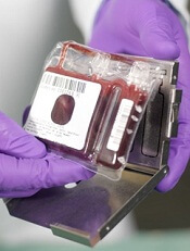
Credit: NHS
Investigators say they have identified a molecule that allows for robust ex vivo expansion of human cord blood (CB) cells.
CB cells expanded with this molecule, known as UM171, were capable of human hematopoietic reconstitution in NSG mice, an effect that lasted more than 6 months.
The researchers believe UM171 acts by enhancing the long-term-hematopoietic stem cell (LT-HSC) self-renewal machinery independently of AhR suppression.
Guy Sauvageau, MD, PhD, of the University of Montreal in Quebec, Canada, and his colleagues identified UM171 and described the discovery in Science.
The team first screened a library of 5280 low-molecular-weight compounds looking for those with the ability to expand human CD34+CD45RA- mobilized peripheral blood cells, which are enriched in LT-HSCs.
They got 7 hits, and only 2 of these—UM729 and UM118428—did not suppress the AhR pathway. The researchers selected UM729 for further characterization and optimization because it demonstrated superior activity in expanding CD34+CD45RA- cells.
The investigators analyzed more than 300 newly synthesized analogs of UM729 and identified one that was 10 to 20 times more potent than UM729. That compound was UM171.
UM171 could expand CD34+CD45RA- cells at concentrations of 17 nM to 19 nM. The highest expansion of multipotent progenitors and long-term culture-initiating cells occurred on day 12.
The effect of UM171 required its constant presence in the media, and the compound lacked direct mitogenic activity. UM171 did not affect the division rate of phenotypically primitive cell populations.
The researchers compared UM171 to SR1 (a compound known to promote self-renewal of HSCs) in fed-batch culture. They found that frequencies of CD34+ CB cells were similar in cultures containing SR1 and those containing UM171. But CD34+CD45RA- cells were more abundant with UM171 (P<0.005).
The team then evaluated LT-HSC populations. Twenty weeks after CD34+ CB cells were transplanted into mice, LT-HSC frequencies were similar in mice that received control and SR1-expanded cells. But LT-HSC frequencies were 13-fold higher in the mice that received UM171-expanded cells.
Next, the investigators assessed human hematopoietic reconstitution in NSG mice transplanted with fresh or expanded cells. They observed 2 patterns of reconstitution. One was from predominantly lymphomyeloid LT-HSCs that occurred with high cell doses in most conditions.
And the other was from LT-HSCs that display a lymphoid-deficient differentiation phenotype mostly observed with UM171 treatment, with or without SR1. However, UM171 did not negatively affect B lymphopoiesis or the frequency or number of lymphomyeloid LT-HSCs.
The impact of UM171 on LT-HSCs was preserved at 30 weeks post-transplant. And myeloid cell output was slightly augmented, a phenomenon that has been observed with normal, unexpanded cells.
The researchers also transplanted UM171-treated LT-HSC populations into secondary recipients. And they found the cells were still competent, but they had no advantage over unmanipulated CD34+ cells.
A clinical study of UM171 and a new type of bioreactor developed for stem cell culture is set to begin this December. The cells will be expanded at Maisonneuve-Rosemont Hospital, and grafts will be distributed to patients in Montreal, Quebec City, and Vancouver. The investigators expect tangible results will be available in December 2015. 
Credit: NHS
Investigators say they have identified a molecule that allows for robust ex vivo expansion of human cord blood (CB) cells.
CB cells expanded with this molecule, known as UM171, were capable of human hematopoietic reconstitution in NSG mice, an effect that lasted more than 6 months.
The researchers believe UM171 acts by enhancing the long-term-hematopoietic stem cell (LT-HSC) self-renewal machinery independently of AhR suppression.
Guy Sauvageau, MD, PhD, of the University of Montreal in Quebec, Canada, and his colleagues identified UM171 and described the discovery in Science.
The team first screened a library of 5280 low-molecular-weight compounds looking for those with the ability to expand human CD34+CD45RA- mobilized peripheral blood cells, which are enriched in LT-HSCs.
They got 7 hits, and only 2 of these—UM729 and UM118428—did not suppress the AhR pathway. The researchers selected UM729 for further characterization and optimization because it demonstrated superior activity in expanding CD34+CD45RA- cells.
The investigators analyzed more than 300 newly synthesized analogs of UM729 and identified one that was 10 to 20 times more potent than UM729. That compound was UM171.
UM171 could expand CD34+CD45RA- cells at concentrations of 17 nM to 19 nM. The highest expansion of multipotent progenitors and long-term culture-initiating cells occurred on day 12.
The effect of UM171 required its constant presence in the media, and the compound lacked direct mitogenic activity. UM171 did not affect the division rate of phenotypically primitive cell populations.
The researchers compared UM171 to SR1 (a compound known to promote self-renewal of HSCs) in fed-batch culture. They found that frequencies of CD34+ CB cells were similar in cultures containing SR1 and those containing UM171. But CD34+CD45RA- cells were more abundant with UM171 (P<0.005).
The team then evaluated LT-HSC populations. Twenty weeks after CD34+ CB cells were transplanted into mice, LT-HSC frequencies were similar in mice that received control and SR1-expanded cells. But LT-HSC frequencies were 13-fold higher in the mice that received UM171-expanded cells.
Next, the investigators assessed human hematopoietic reconstitution in NSG mice transplanted with fresh or expanded cells. They observed 2 patterns of reconstitution. One was from predominantly lymphomyeloid LT-HSCs that occurred with high cell doses in most conditions.
And the other was from LT-HSCs that display a lymphoid-deficient differentiation phenotype mostly observed with UM171 treatment, with or without SR1. However, UM171 did not negatively affect B lymphopoiesis or the frequency or number of lymphomyeloid LT-HSCs.
The impact of UM171 on LT-HSCs was preserved at 30 weeks post-transplant. And myeloid cell output was slightly augmented, a phenomenon that has been observed with normal, unexpanded cells.
The researchers also transplanted UM171-treated LT-HSC populations into secondary recipients. And they found the cells were still competent, but they had no advantage over unmanipulated CD34+ cells.
A clinical study of UM171 and a new type of bioreactor developed for stem cell culture is set to begin this December. The cells will be expanded at Maisonneuve-Rosemont Hospital, and grafts will be distributed to patients in Montreal, Quebec City, and Vancouver. The investigators expect tangible results will be available in December 2015.
Credit: NHS
Investigators say they have identified a molecule that allows for robust ex vivo expansion of human cord blood (CB) cells.
CB cells expanded with this molecule, known as UM171, were capable of human hematopoietic reconstitution in NSG mice, an effect that lasted more than 6 months.
The researchers believe UM171 acts by enhancing the long-term-hematopoietic stem cell (LT-HSC) self-renewal machinery independently of AhR suppression.
Guy Sauvageau, MD, PhD, of the University of Montreal in Quebec, Canada, and his colleagues identified UM171 and described the discovery in Science.
The team first screened a library of 5280 low-molecular-weight compounds looking for those with the ability to expand human CD34+CD45RA- mobilized peripheral blood cells, which are enriched in LT-HSCs.
They got 7 hits, and only 2 of these—UM729 and UM118428—did not suppress the AhR pathway. The researchers selected UM729 for further characterization and optimization because it demonstrated superior activity in expanding CD34+CD45RA- cells.
The investigators analyzed more than 300 newly synthesized analogs of UM729 and identified one that was 10 to 20 times more potent than UM729. That compound was UM171.
UM171 could expand CD34+CD45RA- cells at concentrations of 17 nM to 19 nM. The highest expansion of multipotent progenitors and long-term culture-initiating cells occurred on day 12.
The effect of UM171 required its constant presence in the media, and the compound lacked direct mitogenic activity. UM171 did not affect the division rate of phenotypically primitive cell populations.
The researchers compared UM171 to SR1 (a compound known to promote self-renewal of HSCs) in fed-batch culture. They found that frequencies of CD34+ CB cells were similar in cultures containing SR1 and those containing UM171. But CD34+CD45RA- cells were more abundant with UM171 (P<0.005).
The team then evaluated LT-HSC populations. Twenty weeks after CD34+ CB cells were transplanted into mice, LT-HSC frequencies were similar in mice that received control and SR1-expanded cells. But LT-HSC frequencies were 13-fold higher in the mice that received UM171-expanded cells.
Next, the investigators assessed human hematopoietic reconstitution in NSG mice transplanted with fresh or expanded cells. They observed 2 patterns of reconstitution. One was from predominantly lymphomyeloid LT-HSCs that occurred with high cell doses in most conditions.
And the other was from LT-HSCs that display a lymphoid-deficient differentiation phenotype mostly observed with UM171 treatment, with or without SR1. However, UM171 did not negatively affect B lymphopoiesis or the frequency or number of lymphomyeloid LT-HSCs.
The impact of UM171 on LT-HSCs was preserved at 30 weeks post-transplant. And myeloid cell output was slightly augmented, a phenomenon that has been observed with normal, unexpanded cells.
The researchers also transplanted UM171-treated LT-HSC populations into secondary recipients. And they found the cells were still competent, but they had no advantage over unmanipulated CD34+ cells.
A clinical study of UM171 and a new type of bioreactor developed for stem cell culture is set to begin this December. The cells will be expanded at Maisonneuve-Rosemont Hospital, and grafts will be distributed to patients in Montreal, Quebec City, and Vancouver. The investigators expect tangible results will be available in December 2015.
Predicting chemo drugs’ effects on male fertility

Credit: Rhoda Baer
A mathematical formula may help physicians predict which cancer survivors are likely to have normal sperm production following chemotherapy with alkylating agents.
Researchers developed the formula to calculate survivors’ cumulative treatment exposure to alkylating agents as a cyclophosphamide-equivalent dose (CED).
The team used this formula to evaluate a cohort of cancer survivors and found that CEDs of 4 g/m2 or less were associated with normal sperm concentrations.
An account of this research was published in The Lancet Oncology.
“Based on these results, we would recommend pretreatment fertility preservation be offered, whenever clinically possible, to any male whose projected treatment is expected to include a cyclophosphamide-equivalent dose greater than 4 g/m2,” said study author Daniel Green, MD, of St Jude Children’s Research Hospital in Memphis, Tennessee.
Dr Green and his colleagues had studied 214 male survivors of childhood cancer who had a median age of 29. Subjects had received cyclophosphamide and other alkylating agents, but their treatment did not include radiation.
The researchers performed semen analysis in the survivors a median of 21 years from their diagnosis. The analysis showed that 47.6% of survivors had normal sperm concentrations (at least 15 million per milliliter), 27.6% had low sperm counts, and 24.8% produced no sperm.
To determine the impact of alkylating agents on the survivors’ sperm production, the researchers calculated each survivor’s cumulative treatment exposure as a CED.
They did this using the following formula: CED (mg/m2)= 1.0 (cumulative cyclophosphamide dose [mg/m2]) + 0.244 (cumulative ifosfamide dose [mg/m2]) + 0.857 (cumulative procarbazine dose [mg/m2]) + 14.286 (cumulative chlorambucil dose [mg/m2]) + 15.0 (cumulative carmustine dose [mg/m2]) + 16.0 (cumulative lomustine dose [mg/m2]) + 40 (cumulative melphalan dose [mg/m2]) + 50 (cumulative thiotepa dose [mg/m2]) + 100 cumulative chlormethine dose [mg/m2]) + 8.823 (cumulative busulfan dose [mg/m2]).
The researchers could not identify a uniformly safe or toxic dose of these drugs, but they found that survivor sperm concentrations generally decreased as cumulative exposure to alkylating agents increased.
And CEDs of 4 g/m2 or less were associated with normal sperm concentrations. Almost 89% of survivors whose CED was 4 g/m2 or less had a normal sperm count. Their sperm were also more likely than sperm from other survivors to look and behave normally.
Dr Green did note that, although a lower cumulative dose of alkylating agents was associated with normal sperm production, outcomes for individual patients were unpredictable and varied.
Credit: Rhoda Baer
A mathematical formula may help physicians predict which cancer survivors are likely to have normal sperm production following chemotherapy with alkylating agents.
Researchers developed the formula to calculate survivors’ cumulative treatment exposure to alkylating agents as a cyclophosphamide-equivalent dose (CED).
The team used this formula to evaluate a cohort of cancer survivors and found that CEDs of 4 g/m2 or less were associated with normal sperm concentrations.
An account of this research was published in The Lancet Oncology.
“Based on these results, we would recommend pretreatment fertility preservation be offered, whenever clinically possible, to any male whose projected treatment is expected to include a cyclophosphamide-equivalent dose greater than 4 g/m2,” said study author Daniel Green, MD, of St Jude Children’s Research Hospital in Memphis, Tennessee.
Dr Green and his colleagues had studied 214 male survivors of childhood cancer who had a median age of 29. Subjects had received cyclophosphamide and other alkylating agents, but their treatment did not include radiation.
The researchers performed semen analysis in the survivors a median of 21 years from their diagnosis. The analysis showed that 47.6% of survivors had normal sperm concentrations (at least 15 million per milliliter), 27.6% had low sperm counts, and 24.8% produced no sperm.
To determine the impact of alkylating agents on the survivors’ sperm production, the researchers calculated each survivor’s cumulative treatment exposure as a CED.
They did this using the following formula: CED (mg/m2)= 1.0 (cumulative cyclophosphamide dose [mg/m2]) + 0.244 (cumulative ifosfamide dose [mg/m2]) + 0.857 (cumulative procarbazine dose [mg/m2]) + 14.286 (cumulative chlorambucil dose [mg/m2]) + 15.0 (cumulative carmustine dose [mg/m2]) + 16.0 (cumulative lomustine dose [mg/m2]) + 40 (cumulative melphalan dose [mg/m2]) + 50 (cumulative thiotepa dose [mg/m2]) + 100 cumulative chlormethine dose [mg/m2]) + 8.823 (cumulative busulfan dose [mg/m2]).
The researchers could not identify a uniformly safe or toxic dose of these drugs, but they found that survivor sperm concentrations generally decreased as cumulative exposure to alkylating agents increased.
And CEDs of 4 g/m2 or less were associated with normal sperm concentrations. Almost 89% of survivors whose CED was 4 g/m2 or less had a normal sperm count. Their sperm were also more likely than sperm from other survivors to look and behave normally.
Dr Green did note that, although a lower cumulative dose of alkylating agents was associated with normal sperm production, outcomes for individual patients were unpredictable and varied.
Credit: Rhoda Baer
A mathematical formula may help physicians predict which cancer survivors are likely to have normal sperm production following chemotherapy with alkylating agents.
Researchers developed the formula to calculate survivors’ cumulative treatment exposure to alkylating agents as a cyclophosphamide-equivalent dose (CED).
The team used this formula to evaluate a cohort of cancer survivors and found that CEDs of 4 g/m2 or less were associated with normal sperm concentrations.
An account of this research was published in The Lancet Oncology.
“Based on these results, we would recommend pretreatment fertility preservation be offered, whenever clinically possible, to any male whose projected treatment is expected to include a cyclophosphamide-equivalent dose greater than 4 g/m2,” said study author Daniel Green, MD, of St Jude Children’s Research Hospital in Memphis, Tennessee.
Dr Green and his colleagues had studied 214 male survivors of childhood cancer who had a median age of 29. Subjects had received cyclophosphamide and other alkylating agents, but their treatment did not include radiation.
The researchers performed semen analysis in the survivors a median of 21 years from their diagnosis. The analysis showed that 47.6% of survivors had normal sperm concentrations (at least 15 million per milliliter), 27.6% had low sperm counts, and 24.8% produced no sperm.
To determine the impact of alkylating agents on the survivors’ sperm production, the researchers calculated each survivor’s cumulative treatment exposure as a CED.
They did this using the following formula: CED (mg/m2)= 1.0 (cumulative cyclophosphamide dose [mg/m2]) + 0.244 (cumulative ifosfamide dose [mg/m2]) + 0.857 (cumulative procarbazine dose [mg/m2]) + 14.286 (cumulative chlorambucil dose [mg/m2]) + 15.0 (cumulative carmustine dose [mg/m2]) + 16.0 (cumulative lomustine dose [mg/m2]) + 40 (cumulative melphalan dose [mg/m2]) + 50 (cumulative thiotepa dose [mg/m2]) + 100 cumulative chlormethine dose [mg/m2]) + 8.823 (cumulative busulfan dose [mg/m2]).
The researchers could not identify a uniformly safe or toxic dose of these drugs, but they found that survivor sperm concentrations generally decreased as cumulative exposure to alkylating agents increased.
And CEDs of 4 g/m2 or less were associated with normal sperm concentrations. Almost 89% of survivors whose CED was 4 g/m2 or less had a normal sperm count. Their sperm were also more likely than sperm from other survivors to look and behave normally.
Dr Green did note that, although a lower cumulative dose of alkylating agents was associated with normal sperm production, outcomes for individual patients were unpredictable and varied.
Disrupting equilibrium to fight AML

Credit: Lance Liotta
Understanding the relationship between mutated and wild-type genes could lead to new therapies for acute myeloid leukemia (AML), according to researchers.
The group discovered that RUNX1/ETO, generated by the chromosomal translocation t(8;21), regulates a leukemia-propagating transcriptional network via a dynamic equilibrium with RUNX1.
But depleting RUNX1/ETO prompts the formation of a transcriptional network that promotes myeloid differentiation.
Constanze Bonifer, PhD, of the University of Birmingham in the UK, and her colleagues detailed this work in Cell Reports.
The researchers likened normal hematopoiesis to a production line, with genes acting as regulators to control each step of blood formation. When a mutation occurs in the relevant regulator genes, the finely balanced order of the production line is disrupted, with drastic consequences.
Using digital footprinting and chromatin immunoprecipitation sequencing, the team showed that RUNX1/ETO switches off hundreds of other genes, many of them regulators themselves. As a consequence of the drastically altered production line, normal blood formation cannot happen, and leukemic cells form.
“Understanding how these rogue regulators operate is essential,” Dr Bonifer said. “[T]hese leukemic cells have one mutated gene and one unchanged one that would make the normal regulator. What happens in the leukemic cell is fundamentally a battle for supremacy between the two regulators, and the mutated one wins much of the time.”
“This is compounded by the normal regulator, which tries to compensate for defeat, and, in doing so, changes the output of genes that would be otherwise unaffected by the abnormal regulator. Quite simply, the result is a real mess. The cells are confused and can’t develop into mature blood cells.”
More specifically, the researchers found that the transcriptional program underlying leukemic propagation is regulated by a dynamic equilibrium between RUNX1/ETO and RUNX1 complexes.
Disrupting this equilibrium in t(8;21) cells by depleting RUNX1/ETO led to a global redistribution of transcription factor complexes within pre-existing open chromatin. And this prompted the formation of a transcriptional network that drives myeloid differentiation.
“If targeting [RUNX1/ETO] can reverse the changes it is making to the cellular production line, then it would ultimately point towards new avenues for a more precise treatment of leukemia,” said study author Olaf Heidenreich, PhD, of Newcastle University in the UK.
“Knowing that the production line can be restored to normal function gives us real hope. Of course, that is much easier to do in the lab than it is in the human body. But now we know how this works, we can look to deliver inhibitors to those mutated regulators. Creating one that works is the next step we have to overcome.”
Credit: Lance Liotta
Understanding the relationship between mutated and wild-type genes could lead to new therapies for acute myeloid leukemia (AML), according to researchers.
The group discovered that RUNX1/ETO, generated by the chromosomal translocation t(8;21), regulates a leukemia-propagating transcriptional network via a dynamic equilibrium with RUNX1.
But depleting RUNX1/ETO prompts the formation of a transcriptional network that promotes myeloid differentiation.
Constanze Bonifer, PhD, of the University of Birmingham in the UK, and her colleagues detailed this work in Cell Reports.
The researchers likened normal hematopoiesis to a production line, with genes acting as regulators to control each step of blood formation. When a mutation occurs in the relevant regulator genes, the finely balanced order of the production line is disrupted, with drastic consequences.
Using digital footprinting and chromatin immunoprecipitation sequencing, the team showed that RUNX1/ETO switches off hundreds of other genes, many of them regulators themselves. As a consequence of the drastically altered production line, normal blood formation cannot happen, and leukemic cells form.
“Understanding how these rogue regulators operate is essential,” Dr Bonifer said. “[T]hese leukemic cells have one mutated gene and one unchanged one that would make the normal regulator. What happens in the leukemic cell is fundamentally a battle for supremacy between the two regulators, and the mutated one wins much of the time.”
“This is compounded by the normal regulator, which tries to compensate for defeat, and, in doing so, changes the output of genes that would be otherwise unaffected by the abnormal regulator. Quite simply, the result is a real mess. The cells are confused and can’t develop into mature blood cells.”
More specifically, the researchers found that the transcriptional program underlying leukemic propagation is regulated by a dynamic equilibrium between RUNX1/ETO and RUNX1 complexes.
Disrupting this equilibrium in t(8;21) cells by depleting RUNX1/ETO led to a global redistribution of transcription factor complexes within pre-existing open chromatin. And this prompted the formation of a transcriptional network that drives myeloid differentiation.
“If targeting [RUNX1/ETO] can reverse the changes it is making to the cellular production line, then it would ultimately point towards new avenues for a more precise treatment of leukemia,” said study author Olaf Heidenreich, PhD, of Newcastle University in the UK.
“Knowing that the production line can be restored to normal function gives us real hope. Of course, that is much easier to do in the lab than it is in the human body. But now we know how this works, we can look to deliver inhibitors to those mutated regulators. Creating one that works is the next step we have to overcome.”
Credit: Lance Liotta
Understanding the relationship between mutated and wild-type genes could lead to new therapies for acute myeloid leukemia (AML), according to researchers.
The group discovered that RUNX1/ETO, generated by the chromosomal translocation t(8;21), regulates a leukemia-propagating transcriptional network via a dynamic equilibrium with RUNX1.
But depleting RUNX1/ETO prompts the formation of a transcriptional network that promotes myeloid differentiation.
Constanze Bonifer, PhD, of the University of Birmingham in the UK, and her colleagues detailed this work in Cell Reports.
The researchers likened normal hematopoiesis to a production line, with genes acting as regulators to control each step of blood formation. When a mutation occurs in the relevant regulator genes, the finely balanced order of the production line is disrupted, with drastic consequences.
Using digital footprinting and chromatin immunoprecipitation sequencing, the team showed that RUNX1/ETO switches off hundreds of other genes, many of them regulators themselves. As a consequence of the drastically altered production line, normal blood formation cannot happen, and leukemic cells form.
“Understanding how these rogue regulators operate is essential,” Dr Bonifer said. “[T]hese leukemic cells have one mutated gene and one unchanged one that would make the normal regulator. What happens in the leukemic cell is fundamentally a battle for supremacy between the two regulators, and the mutated one wins much of the time.”
“This is compounded by the normal regulator, which tries to compensate for defeat, and, in doing so, changes the output of genes that would be otherwise unaffected by the abnormal regulator. Quite simply, the result is a real mess. The cells are confused and can’t develop into mature blood cells.”
More specifically, the researchers found that the transcriptional program underlying leukemic propagation is regulated by a dynamic equilibrium between RUNX1/ETO and RUNX1 complexes.
Disrupting this equilibrium in t(8;21) cells by depleting RUNX1/ETO led to a global redistribution of transcription factor complexes within pre-existing open chromatin. And this prompted the formation of a transcriptional network that drives myeloid differentiation.
“If targeting [RUNX1/ETO] can reverse the changes it is making to the cellular production line, then it would ultimately point towards new avenues for a more precise treatment of leukemia,” said study author Olaf Heidenreich, PhD, of Newcastle University in the UK.
“Knowing that the production line can be restored to normal function gives us real hope. Of course, that is much easier to do in the lab than it is in the human body. But now we know how this works, we can look to deliver inhibitors to those mutated regulators. Creating one that works is the next step we have to overcome.”
Longer duration of triple therapy confers no benefit, trial suggests

Credit: Sage Ross
WASHINGTON, DC—Six weeks of triple anticoagulant therapy may be sufficient in patients who have received a drug-eluting stent.
Results of the ISAR-TRIPLE trial revealed no significant differences in net clinical outcomes for patients who received 6 weeks of triple anticoagulant therapy and those who received 6 months of the therapy.
Nikolaus Sarafoff, MD, of Deutsches Herzzentrum Munich and Klinikum der Universität Munich in Germany, presented these results at TCT 2014.
“The shortening of triple therapy neither reduced the incidence of TIMI major bleeding nor increased the incidence of the composite of ischemic events,” Dr Sarafoff said. “These results suggest that physicians should weigh the trade-off between ischemic and bleeding risk when choosing the shorter or longer duration of triple therapy.”
For the ISAR-TRIPLE trial, Dr Sarafoff and his colleagues randomized 614 patients in a 1:1 fashion to either 6 weeks or 6 months of clopidogrel therapy, in addition to aspirin plus an oral anticoagulant (phenprocoumon or warfarin).
The primary endpoint was a composite of death, myocardial infarction, definite stent thrombosis, stroke, or TIMI major bleeding at 9 months.
As secondary endpoints, the researchers assessed TIMI major bleeding separately from a composite endpoint of cardiac death, myocardial infarction, stent thrombosis, or ischemic stroke.
The primary endpoint occurred in 9.8% of patients in the 6-week treatment group and 8.8% of patients in the 6-month group (P=0.63.)
The composite of cardiac death, myocardial infarction, stent thrombosis, or ischemic stroke occurred at a similar rate in both the 6-week and 6-month groups—4.0% and 4.3%, respectively (P=0.87).
And the same was true for TIMI major bleeding, which occurred in 5.3% of patients in the 6-week group and 4.0% in the 6-month group (P=0.44).
This trial was funded by Deutsches Herzzentrum München. Dr Sarafoff reported fees for lectures or traveling from Lilly/Daiichi Sankyo, Boehringer Ingelheim, Bayer Healthcare, Boston Scientific, Biotronik, and Medtronic.
Credit: Sage Ross
WASHINGTON, DC—Six weeks of triple anticoagulant therapy may be sufficient in patients who have received a drug-eluting stent.
Results of the ISAR-TRIPLE trial revealed no significant differences in net clinical outcomes for patients who received 6 weeks of triple anticoagulant therapy and those who received 6 months of the therapy.
Nikolaus Sarafoff, MD, of Deutsches Herzzentrum Munich and Klinikum der Universität Munich in Germany, presented these results at TCT 2014.
“The shortening of triple therapy neither reduced the incidence of TIMI major bleeding nor increased the incidence of the composite of ischemic events,” Dr Sarafoff said. “These results suggest that physicians should weigh the trade-off between ischemic and bleeding risk when choosing the shorter or longer duration of triple therapy.”
For the ISAR-TRIPLE trial, Dr Sarafoff and his colleagues randomized 614 patients in a 1:1 fashion to either 6 weeks or 6 months of clopidogrel therapy, in addition to aspirin plus an oral anticoagulant (phenprocoumon or warfarin).
The primary endpoint was a composite of death, myocardial infarction, definite stent thrombosis, stroke, or TIMI major bleeding at 9 months.
As secondary endpoints, the researchers assessed TIMI major bleeding separately from a composite endpoint of cardiac death, myocardial infarction, stent thrombosis, or ischemic stroke.
The primary endpoint occurred in 9.8% of patients in the 6-week treatment group and 8.8% of patients in the 6-month group (P=0.63.)
The composite of cardiac death, myocardial infarction, stent thrombosis, or ischemic stroke occurred at a similar rate in both the 6-week and 6-month groups—4.0% and 4.3%, respectively (P=0.87).
And the same was true for TIMI major bleeding, which occurred in 5.3% of patients in the 6-week group and 4.0% in the 6-month group (P=0.44).
This trial was funded by Deutsches Herzzentrum München. Dr Sarafoff reported fees for lectures or traveling from Lilly/Daiichi Sankyo, Boehringer Ingelheim, Bayer Healthcare, Boston Scientific, Biotronik, and Medtronic.
Credit: Sage Ross
WASHINGTON, DC—Six weeks of triple anticoagulant therapy may be sufficient in patients who have received a drug-eluting stent.
Results of the ISAR-TRIPLE trial revealed no significant differences in net clinical outcomes for patients who received 6 weeks of triple anticoagulant therapy and those who received 6 months of the therapy.
Nikolaus Sarafoff, MD, of Deutsches Herzzentrum Munich and Klinikum der Universität Munich in Germany, presented these results at TCT 2014.
“The shortening of triple therapy neither reduced the incidence of TIMI major bleeding nor increased the incidence of the composite of ischemic events,” Dr Sarafoff said. “These results suggest that physicians should weigh the trade-off between ischemic and bleeding risk when choosing the shorter or longer duration of triple therapy.”
For the ISAR-TRIPLE trial, Dr Sarafoff and his colleagues randomized 614 patients in a 1:1 fashion to either 6 weeks or 6 months of clopidogrel therapy, in addition to aspirin plus an oral anticoagulant (phenprocoumon or warfarin).
The primary endpoint was a composite of death, myocardial infarction, definite stent thrombosis, stroke, or TIMI major bleeding at 9 months.
As secondary endpoints, the researchers assessed TIMI major bleeding separately from a composite endpoint of cardiac death, myocardial infarction, stent thrombosis, or ischemic stroke.
The primary endpoint occurred in 9.8% of patients in the 6-week treatment group and 8.8% of patients in the 6-month group (P=0.63.)
The composite of cardiac death, myocardial infarction, stent thrombosis, or ischemic stroke occurred at a similar rate in both the 6-week and 6-month groups—4.0% and 4.3%, respectively (P=0.87).
And the same was true for TIMI major bleeding, which occurred in 5.3% of patients in the 6-week group and 4.0% in the 6-month group (P=0.44).
This trial was funded by Deutsches Herzzentrum München. Dr Sarafoff reported fees for lectures or traveling from Lilly/Daiichi Sankyo, Boehringer Ingelheim, Bayer Healthcare, Boston Scientific, Biotronik, and Medtronic.
Study supports new gold standard for FL
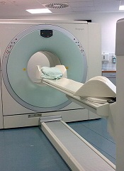
PET-CT should be the new standard for response assessment in patients with follicular lymphoma (FL), according to researchers.
The group found evidence suggesting that PET-CT is more accurate than conventional CT in measuring treatment response and predicting survival in patients with FL.
“Our findings have important implications for patients with follicular lymphoma,” said study author Judith Trotman, MBChB, of the University of Sydney in Australia.
“Compared to conventional CT scanning, PET-CT is more accurate in mapping out the lymphoma and better identifies the majority of patients who have a prolonged remission after treatment.”
Dr Trotman and her colleagues reported these findings in The Lancet Haematology. The results will also be discussed at the International Workshop on PET in Lymphoma in Menton, France, which is taking place September 19-20.
By assessing imaging performed in 3 clinical trials, the researchers examined the link between PET-CT status and survival following first-line immunochemotherapy for advanced FL.
Independent, masked reviewers evaluated scans of 246 patients who underwent both PET-CT and traditional CT imaging within 3 months of their last dose of therapy. Patients were followed for a median of 54.8 months.
Seventeen percent of patients had a positive post-induction PET scan, according to a cutoff of 4 or higher on the 5PS.
When the researchers compared patients with a positive PET scan to those with a negative scan, the hazard ratio (HR) for progression-free survival (PFS) was 3.9 (P<0.0001). For overall survival, the HR was 6.7 (P=0.0002).
The 4-year PFS was 23.2% in patients with a positive PET scan and 63.4% in those who had a negative PET scan (P<0.0001). The 4-year overall survival was 87.2% and 97.1%, respectively (P<0.0001).
The researchers also discovered that conventional CT-based response—complete response or unconfirmed complete response compared to partial response—was weakly predictive of PFS. The HR was 1.7 (P=0.017).
“Our study shows that PET-CT is much better in evaluating treatment response and is an early predictor of survival,” Dr Trotman said. “This greater accuracy will assist physicians to more effectively monitor their patients.”
“We expect this research will result in PET-CT imaging replacing CT, becoming the new gold standard to evaluate patients with follicular lymphoma after treatment. Importantly, it will be a platform for future studies of response-adapted therapies aimed to improve the poor outcomes for those patients who remain PET-positive.”
This study may also pave the way for several new research opportunities, according to Bruce Cheson, MD, of Georgetown University in Washington DC, who wrote a comment article related to this study.
“One such possibility would be to assess if an early reaction to the PET scan result improves patient outcome,” he wrote. “Thus, patients with a positive PET scan after induction therapy could be randomly assigned to either deferred treatment until disease progression or immediate intervention.”
“A preferable alternative would be to introduce a unique agent at that time, such as the newly developed small molecules (eg, idelalisib, ibrutinib, or ABT-199) in a novel combination.”
PET-CT should be the new standard for response assessment in patients with follicular lymphoma (FL), according to researchers.
The group found evidence suggesting that PET-CT is more accurate than conventional CT in measuring treatment response and predicting survival in patients with FL.
“Our findings have important implications for patients with follicular lymphoma,” said study author Judith Trotman, MBChB, of the University of Sydney in Australia.
“Compared to conventional CT scanning, PET-CT is more accurate in mapping out the lymphoma and better identifies the majority of patients who have a prolonged remission after treatment.”
Dr Trotman and her colleagues reported these findings in The Lancet Haematology. The results will also be discussed at the International Workshop on PET in Lymphoma in Menton, France, which is taking place September 19-20.
By assessing imaging performed in 3 clinical trials, the researchers examined the link between PET-CT status and survival following first-line immunochemotherapy for advanced FL.
Independent, masked reviewers evaluated scans of 246 patients who underwent both PET-CT and traditional CT imaging within 3 months of their last dose of therapy. Patients were followed for a median of 54.8 months.
Seventeen percent of patients had a positive post-induction PET scan, according to a cutoff of 4 or higher on the 5PS.
When the researchers compared patients with a positive PET scan to those with a negative scan, the hazard ratio (HR) for progression-free survival (PFS) was 3.9 (P<0.0001). For overall survival, the HR was 6.7 (P=0.0002).
The 4-year PFS was 23.2% in patients with a positive PET scan and 63.4% in those who had a negative PET scan (P<0.0001). The 4-year overall survival was 87.2% and 97.1%, respectively (P<0.0001).
The researchers also discovered that conventional CT-based response—complete response or unconfirmed complete response compared to partial response—was weakly predictive of PFS. The HR was 1.7 (P=0.017).
“Our study shows that PET-CT is much better in evaluating treatment response and is an early predictor of survival,” Dr Trotman said. “This greater accuracy will assist physicians to more effectively monitor their patients.”
“We expect this research will result in PET-CT imaging replacing CT, becoming the new gold standard to evaluate patients with follicular lymphoma after treatment. Importantly, it will be a platform for future studies of response-adapted therapies aimed to improve the poor outcomes for those patients who remain PET-positive.”
This study may also pave the way for several new research opportunities, according to Bruce Cheson, MD, of Georgetown University in Washington DC, who wrote a comment article related to this study.
“One such possibility would be to assess if an early reaction to the PET scan result improves patient outcome,” he wrote. “Thus, patients with a positive PET scan after induction therapy could be randomly assigned to either deferred treatment until disease progression or immediate intervention.”
“A preferable alternative would be to introduce a unique agent at that time, such as the newly developed small molecules (eg, idelalisib, ibrutinib, or ABT-199) in a novel combination.”
PET-CT should be the new standard for response assessment in patients with follicular lymphoma (FL), according to researchers.
The group found evidence suggesting that PET-CT is more accurate than conventional CT in measuring treatment response and predicting survival in patients with FL.
“Our findings have important implications for patients with follicular lymphoma,” said study author Judith Trotman, MBChB, of the University of Sydney in Australia.
“Compared to conventional CT scanning, PET-CT is more accurate in mapping out the lymphoma and better identifies the majority of patients who have a prolonged remission after treatment.”
Dr Trotman and her colleagues reported these findings in The Lancet Haematology. The results will also be discussed at the International Workshop on PET in Lymphoma in Menton, France, which is taking place September 19-20.
By assessing imaging performed in 3 clinical trials, the researchers examined the link between PET-CT status and survival following first-line immunochemotherapy for advanced FL.
Independent, masked reviewers evaluated scans of 246 patients who underwent both PET-CT and traditional CT imaging within 3 months of their last dose of therapy. Patients were followed for a median of 54.8 months.
Seventeen percent of patients had a positive post-induction PET scan, according to a cutoff of 4 or higher on the 5PS.
When the researchers compared patients with a positive PET scan to those with a negative scan, the hazard ratio (HR) for progression-free survival (PFS) was 3.9 (P<0.0001). For overall survival, the HR was 6.7 (P=0.0002).
The 4-year PFS was 23.2% in patients with a positive PET scan and 63.4% in those who had a negative PET scan (P<0.0001). The 4-year overall survival was 87.2% and 97.1%, respectively (P<0.0001).
The researchers also discovered that conventional CT-based response—complete response or unconfirmed complete response compared to partial response—was weakly predictive of PFS. The HR was 1.7 (P=0.017).
“Our study shows that PET-CT is much better in evaluating treatment response and is an early predictor of survival,” Dr Trotman said. “This greater accuracy will assist physicians to more effectively monitor their patients.”
“We expect this research will result in PET-CT imaging replacing CT, becoming the new gold standard to evaluate patients with follicular lymphoma after treatment. Importantly, it will be a platform for future studies of response-adapted therapies aimed to improve the poor outcomes for those patients who remain PET-positive.”
This study may also pave the way for several new research opportunities, according to Bruce Cheson, MD, of Georgetown University in Washington DC, who wrote a comment article related to this study.
“One such possibility would be to assess if an early reaction to the PET scan result improves patient outcome,” he wrote. “Thus, patients with a positive PET scan after induction therapy could be randomly assigned to either deferred treatment until disease progression or immediate intervention.”
“A preferable alternative would be to introduce a unique agent at that time, such as the newly developed small molecules (eg, idelalisib, ibrutinib, or ABT-199) in a novel combination.”
Group investigates link between implants and ALCL

Credit: FDA
A newly published review provides insight into anaplastic large-cell lymphoma (ALCL) associated with breast implants.
Previous reports have demonstrated a link between breast implants and ALCL, but the reasons why implants may contribute to this type of lymphoma have remained unclear.
So Suzanne Turner, PhD, of the University of Cambridge in the UK, and her colleagues evaluated the 71 recorded cases of implant-associated ALCL (iALCL).
The researchers recounted their findings in Mutation Research/Reviews in Mutations Research.
The team calculated that the absolute risk of developing iALCL is low, ranging from 1:500,000 to 1:3,000,000 patients with breast implants per year.
Among the 71 cases of iALCL, the average patient was 50 years of age. The average time from breast augmentation/reconstruction to iALCL diagnosis was 10 years.
Most of the cases presented in the capsule surrounding the implant, as part of the periprosthetic fluid or the capsule itself.
The majority of cases were ALK-negative but had a good prognosis. Of the 49 cases where information on the patients’ progress was available, there were 5 deaths.
Some patients received chemotherapy and radiotherapy, but many achieved remission once the implant and surrounding tissue were removed. This suggests it is the body’s abnormal immune response to the implant that is causing iALCL, the researchers said.
There is some evidence to suggest that having any type of prosthetic implant can increase the risk of lymphoma. And some researchers have suggested that lifestyle differences between patients with and without breast implants may be at the root of iALCL.
But Dr Turner and her colleagues said the biological features of lymphomas in the presence of implants, such as the lack of ALK and the proximity to the implant, suggest a real link.
“It’s becoming clear that implant-related ALCL is a distinct clinical entity in itself,” Dr Turner said. “There are still unanswered questions, and only by getting to the bottom of this very rare disease will we be able to find alternative ways to treat it.”
Credit: FDA
A newly published review provides insight into anaplastic large-cell lymphoma (ALCL) associated with breast implants.
Previous reports have demonstrated a link between breast implants and ALCL, but the reasons why implants may contribute to this type of lymphoma have remained unclear.
So Suzanne Turner, PhD, of the University of Cambridge in the UK, and her colleagues evaluated the 71 recorded cases of implant-associated ALCL (iALCL).
The researchers recounted their findings in Mutation Research/Reviews in Mutations Research.
The team calculated that the absolute risk of developing iALCL is low, ranging from 1:500,000 to 1:3,000,000 patients with breast implants per year.
Among the 71 cases of iALCL, the average patient was 50 years of age. The average time from breast augmentation/reconstruction to iALCL diagnosis was 10 years.
Most of the cases presented in the capsule surrounding the implant, as part of the periprosthetic fluid or the capsule itself.
The majority of cases were ALK-negative but had a good prognosis. Of the 49 cases where information on the patients’ progress was available, there were 5 deaths.
Some patients received chemotherapy and radiotherapy, but many achieved remission once the implant and surrounding tissue were removed. This suggests it is the body’s abnormal immune response to the implant that is causing iALCL, the researchers said.
There is some evidence to suggest that having any type of prosthetic implant can increase the risk of lymphoma. And some researchers have suggested that lifestyle differences between patients with and without breast implants may be at the root of iALCL.
But Dr Turner and her colleagues said the biological features of lymphomas in the presence of implants, such as the lack of ALK and the proximity to the implant, suggest a real link.
“It’s becoming clear that implant-related ALCL is a distinct clinical entity in itself,” Dr Turner said. “There are still unanswered questions, and only by getting to the bottom of this very rare disease will we be able to find alternative ways to treat it.”
Credit: FDA
A newly published review provides insight into anaplastic large-cell lymphoma (ALCL) associated with breast implants.
Previous reports have demonstrated a link between breast implants and ALCL, but the reasons why implants may contribute to this type of lymphoma have remained unclear.
So Suzanne Turner, PhD, of the University of Cambridge in the UK, and her colleagues evaluated the 71 recorded cases of implant-associated ALCL (iALCL).
The researchers recounted their findings in Mutation Research/Reviews in Mutations Research.
The team calculated that the absolute risk of developing iALCL is low, ranging from 1:500,000 to 1:3,000,000 patients with breast implants per year.
Among the 71 cases of iALCL, the average patient was 50 years of age. The average time from breast augmentation/reconstruction to iALCL diagnosis was 10 years.
Most of the cases presented in the capsule surrounding the implant, as part of the periprosthetic fluid or the capsule itself.
The majority of cases were ALK-negative but had a good prognosis. Of the 49 cases where information on the patients’ progress was available, there were 5 deaths.
Some patients received chemotherapy and radiotherapy, but many achieved remission once the implant and surrounding tissue were removed. This suggests it is the body’s abnormal immune response to the implant that is causing iALCL, the researchers said.
There is some evidence to suggest that having any type of prosthetic implant can increase the risk of lymphoma. And some researchers have suggested that lifestyle differences between patients with and without breast implants may be at the root of iALCL.
But Dr Turner and her colleagues said the biological features of lymphomas in the presence of implants, such as the lack of ALK and the proximity to the implant, suggest a real link.
“It’s becoming clear that implant-related ALCL is a distinct clinical entity in itself,” Dr Turner said. “There are still unanswered questions, and only by getting to the bottom of this very rare disease will we be able to find alternative ways to treat it.”
Shorter duration of DAPT appears safe

Credit: Andre E.X. Brown
WASHINGTON, DC—New research indicates that patients may only need 6 months of dual antiplatelet therapy (DAPT) after receiving a second-generation drug-eluting stent.
Results of the SECURITY trial showed that patients had similar outcomes whether they received DAPT for 6 months or a full year.
The proportion of patients who met a composite endpoint of cardiac, thrombotic, and bleeding events was low among both treatment groups at the 12- and 24-month time points.
Antonio Colombo, MD, of San Raffaele Scientific Institute in Milan, Italy, and his colleagues reported these findings at TCT 2014 and in the Journal of the American College of Cardiology.
SECURITY was a randomized, multicenter study that aimed to address the need for prolonged use of DAPT following the implantation of a second-generation drug-eluting stent for patients without high-risk acute coronary syndromes.
Patients with a diagnosis of stable or unstable angina or documented silent ischemia undergoing revascularization with at least 1 second-generation drug-eluting stent were eligible for the study. The stents used were the Endeavor Resolute (Medtronic), Xience (Abbott), Promus (Boston Scientific), Nobori (Terumo Corporation), and the Biomatrix (Biosensors Europe SA).
Of 1399 patients, 682 were randomized to 6 months of DAPT, and 717 were randomized to 12 months of treatment. They received clopidogrel at 75 mg per day for at least 3 days before the procedure or a pre-procedural loading dose of a minimum of 300 mg of clopidogrel, if they were not on chronic clopidogrel therapy.
In the post-procedure period, patients received 75 mg of clopidogrel for 6 or 12 months, according to randomization. They also received aspirin indefinitely. And once the new antiplatelet compounds prasugrel and ticagrelor hit the market, they were allowed as treatment options in a protocol amendment.
At 12 months, the incidence of the primary endpoint—the composite of cardiac death, myocardial infarction (MI), stroke, definite or probable stent thrombosis, or BARC type 3 or 5 bleeding—was 4.5% in the 6-month DAPT group and 3.7% in the 12-month DAPT group (P=0.469).
After 24 months, the 6-month and 12-month groups still showed similar incidences of cardiac death (0.9% vs 0.8%, P=0.925), MI (3.1% vs 2.6%, P=0.636), stroke (0.9% vs 0.4%, P=0.636), stent thrombosis (0.4% vs 0.4%, P=0.951), and BARC 3 or 5 bleeding (0.7% vs 1.1%, P=0.496).
Rates of the secondary endpoint—a composite of cardiac death, spontaneous MI, stroke, definite or probable stent thrombosis, or BARC type 2, 3, or 5 bleeding at 12 and 24 months—were also similar among the 6-month and 12-month treatment groups.
At 12 months, the incidence of the secondary composite endpoint was 5.3% in the 6-month group and 4.0% in the 12-month group (P=0.273). In the year that followed, both groups had lower incidences of secondary composite endpoints—1.5% and 2.2%, respectively (P=0.289).
A multivariable analysis showed that an age of 75 years or older, the type of stent used, the mean number of stents implanted, the mean stent length, and the mean stent size were all significant independent predictors of the primary endpoint.
The SECURITY trial was funded by grants from Medtronic and Terumo, makers of 2 brands of drug-eluting stents used in the study.
Credit: Andre E.X. Brown
WASHINGTON, DC—New research indicates that patients may only need 6 months of dual antiplatelet therapy (DAPT) after receiving a second-generation drug-eluting stent.
Results of the SECURITY trial showed that patients had similar outcomes whether they received DAPT for 6 months or a full year.
The proportion of patients who met a composite endpoint of cardiac, thrombotic, and bleeding events was low among both treatment groups at the 12- and 24-month time points.
Antonio Colombo, MD, of San Raffaele Scientific Institute in Milan, Italy, and his colleagues reported these findings at TCT 2014 and in the Journal of the American College of Cardiology.
SECURITY was a randomized, multicenter study that aimed to address the need for prolonged use of DAPT following the implantation of a second-generation drug-eluting stent for patients without high-risk acute coronary syndromes.
Patients with a diagnosis of stable or unstable angina or documented silent ischemia undergoing revascularization with at least 1 second-generation drug-eluting stent were eligible for the study. The stents used were the Endeavor Resolute (Medtronic), Xience (Abbott), Promus (Boston Scientific), Nobori (Terumo Corporation), and the Biomatrix (Biosensors Europe SA).
Of 1399 patients, 682 were randomized to 6 months of DAPT, and 717 were randomized to 12 months of treatment. They received clopidogrel at 75 mg per day for at least 3 days before the procedure or a pre-procedural loading dose of a minimum of 300 mg of clopidogrel, if they were not on chronic clopidogrel therapy.
In the post-procedure period, patients received 75 mg of clopidogrel for 6 or 12 months, according to randomization. They also received aspirin indefinitely. And once the new antiplatelet compounds prasugrel and ticagrelor hit the market, they were allowed as treatment options in a protocol amendment.
At 12 months, the incidence of the primary endpoint—the composite of cardiac death, myocardial infarction (MI), stroke, definite or probable stent thrombosis, or BARC type 3 or 5 bleeding—was 4.5% in the 6-month DAPT group and 3.7% in the 12-month DAPT group (P=0.469).
After 24 months, the 6-month and 12-month groups still showed similar incidences of cardiac death (0.9% vs 0.8%, P=0.925), MI (3.1% vs 2.6%, P=0.636), stroke (0.9% vs 0.4%, P=0.636), stent thrombosis (0.4% vs 0.4%, P=0.951), and BARC 3 or 5 bleeding (0.7% vs 1.1%, P=0.496).
Rates of the secondary endpoint—a composite of cardiac death, spontaneous MI, stroke, definite or probable stent thrombosis, or BARC type 2, 3, or 5 bleeding at 12 and 24 months—were also similar among the 6-month and 12-month treatment groups.
At 12 months, the incidence of the secondary composite endpoint was 5.3% in the 6-month group and 4.0% in the 12-month group (P=0.273). In the year that followed, both groups had lower incidences of secondary composite endpoints—1.5% and 2.2%, respectively (P=0.289).
A multivariable analysis showed that an age of 75 years or older, the type of stent used, the mean number of stents implanted, the mean stent length, and the mean stent size were all significant independent predictors of the primary endpoint.
The SECURITY trial was funded by grants from Medtronic and Terumo, makers of 2 brands of drug-eluting stents used in the study.
Credit: Andre E.X. Brown
WASHINGTON, DC—New research indicates that patients may only need 6 months of dual antiplatelet therapy (DAPT) after receiving a second-generation drug-eluting stent.
Results of the SECURITY trial showed that patients had similar outcomes whether they received DAPT for 6 months or a full year.
The proportion of patients who met a composite endpoint of cardiac, thrombotic, and bleeding events was low among both treatment groups at the 12- and 24-month time points.
Antonio Colombo, MD, of San Raffaele Scientific Institute in Milan, Italy, and his colleagues reported these findings at TCT 2014 and in the Journal of the American College of Cardiology.
SECURITY was a randomized, multicenter study that aimed to address the need for prolonged use of DAPT following the implantation of a second-generation drug-eluting stent for patients without high-risk acute coronary syndromes.
Patients with a diagnosis of stable or unstable angina or documented silent ischemia undergoing revascularization with at least 1 second-generation drug-eluting stent were eligible for the study. The stents used were the Endeavor Resolute (Medtronic), Xience (Abbott), Promus (Boston Scientific), Nobori (Terumo Corporation), and the Biomatrix (Biosensors Europe SA).
Of 1399 patients, 682 were randomized to 6 months of DAPT, and 717 were randomized to 12 months of treatment. They received clopidogrel at 75 mg per day for at least 3 days before the procedure or a pre-procedural loading dose of a minimum of 300 mg of clopidogrel, if they were not on chronic clopidogrel therapy.
In the post-procedure period, patients received 75 mg of clopidogrel for 6 or 12 months, according to randomization. They also received aspirin indefinitely. And once the new antiplatelet compounds prasugrel and ticagrelor hit the market, they were allowed as treatment options in a protocol amendment.
At 12 months, the incidence of the primary endpoint—the composite of cardiac death, myocardial infarction (MI), stroke, definite or probable stent thrombosis, or BARC type 3 or 5 bleeding—was 4.5% in the 6-month DAPT group and 3.7% in the 12-month DAPT group (P=0.469).
After 24 months, the 6-month and 12-month groups still showed similar incidences of cardiac death (0.9% vs 0.8%, P=0.925), MI (3.1% vs 2.6%, P=0.636), stroke (0.9% vs 0.4%, P=0.636), stent thrombosis (0.4% vs 0.4%, P=0.951), and BARC 3 or 5 bleeding (0.7% vs 1.1%, P=0.496).
Rates of the secondary endpoint—a composite of cardiac death, spontaneous MI, stroke, definite or probable stent thrombosis, or BARC type 2, 3, or 5 bleeding at 12 and 24 months—were also similar among the 6-month and 12-month treatment groups.
At 12 months, the incidence of the secondary composite endpoint was 5.3% in the 6-month group and 4.0% in the 12-month group (P=0.273). In the year that followed, both groups had lower incidences of secondary composite endpoints—1.5% and 2.2%, respectively (P=0.289).
A multivariable analysis showed that an age of 75 years or older, the type of stent used, the mean number of stents implanted, the mean stent length, and the mean stent size were all significant independent predictors of the primary endpoint.
The SECURITY trial was funded by grants from Medtronic and Terumo, makers of 2 brands of drug-eluting stents used in the study.
Bivalirudin bests heparin in BRIGHT trial

Credit: Bill Branson
WASHINGTON, DC—The latest results from the BRIGHT trial suggest bivalirudin confers benefits over heparin monotherapy and heparin plus tirofiban for patients with acute myocardial infarction undergoing percutaneous coronary intervention.
Patients who received bivalirudin had a significantly lower incidence of net adverse clinical events (NACE), a composite of death from any cause, reinfarction, ischemia-driven target vessel revascularization, stroke, or any bleeding, both at 30 days and at 1 year.
Bivalirudin conferred a lower risk of bleeding at both time points as well.
However, there were no significant differences between the treatment arms with regard to stent thrombosis or major adverse cardiac and cerebral events (MACCE), a composite of death from any cause, reinfarction, ischemia-driven target vessel revascularization, or stroke.
Yaling Han, MD, of the General Hospital of Shenyang Military Region in China, presented these data at TCT 2014.
The BRIGHT trial included 2194 patients who had acute myocardial infarction and were eligible for emergency percutaneous coronary intervention. They were randomized to receive bivalirudin alone (n=735), heparin alone (n=729), or heparin plus tirofiban (n=730).
The primary endpoint was NACE at 30 days. Secondary endpoints were NACE at 1 year, any bleeding at 30 days and 1 year, and MACCE at 30 days and 1 year. Safety endpoints were thrombocytopenia at 30 days and stent thrombosis at 30 days and 1 year.
Bivalirudin was superior to both heparin monotherapy and heparin plus tirofiban in reducing the primary composite endpoint. At 30 days, NACE had occurred in 8.8%, 13.2%, and 17.0% of patients, respectively (P<0.001).
Bivalirudin was also superior with regard to bleeding at 30 days. Bleeding occurred in 4.1% of patients in the bivalirudin arm, 7.5% in the heparin arm, and 12.3% in the heparin-tirofiban arm (P<0.001).
However, there was no significant difference between the arms for the incidence of MACCE at 30 days—5.0%, 5.8%, and 4.9%, respectively (P=0.74). Likewise, there was no significant difference in the rate of stent thrombosis at 30 days—0.6%, 0.9%, and 0.7%, respectively (P=0.77).
There were differences between the arms in the 30-day thrombocytopenia rate, which was 0.1% in the bivalirudin arm, 0.7% in the heparin arm, and 1.1% in the heparin-tirofiban arm (P=0.04 for bivalirudin vs pooled heparin, P=0.12 for bivalirudin vs heparin, P=0.02 for bivalirudin vs heparin-tirofiban).
The differences between the treatment arms at 1 year were similar to those observed at the 30-day mark. Bivalirudin remained significantly superior when it came to NACE and bleeding, but there were no significant differences with regard to MACCE and stent thrombosis.
At 1-year, NACE had occurred in 12.8% of patients in the bivalirudin arm, 16.5% in the heparin arm, and 20.5% in the heparin-tirofiban arm (P<0.001). Bleeding had occurred in 6.3%, 9.9%, and 14.2% of patients, respectively (P<0.001).
The incidence of MACCE was 6.7% in the bivalirudin arm, 7.3% in the heparin arm, and 6.8%, in the heparin-tirofiban arm. And stent thrombosis had occurred in 1.2%, 1.9%, and 1.2% of patients, respectively.
This research was funded by Salubris Pharmaceutical Co. Ltd, makers of bivalirudin, and the National Key Science and Technology R&D project of the 12th Five-Year Plan.
Credit: Bill Branson
WASHINGTON, DC—The latest results from the BRIGHT trial suggest bivalirudin confers benefits over heparin monotherapy and heparin plus tirofiban for patients with acute myocardial infarction undergoing percutaneous coronary intervention.
Patients who received bivalirudin had a significantly lower incidence of net adverse clinical events (NACE), a composite of death from any cause, reinfarction, ischemia-driven target vessel revascularization, stroke, or any bleeding, both at 30 days and at 1 year.
Bivalirudin conferred a lower risk of bleeding at both time points as well.
However, there were no significant differences between the treatment arms with regard to stent thrombosis or major adverse cardiac and cerebral events (MACCE), a composite of death from any cause, reinfarction, ischemia-driven target vessel revascularization, or stroke.
Yaling Han, MD, of the General Hospital of Shenyang Military Region in China, presented these data at TCT 2014.
The BRIGHT trial included 2194 patients who had acute myocardial infarction and were eligible for emergency percutaneous coronary intervention. They were randomized to receive bivalirudin alone (n=735), heparin alone (n=729), or heparin plus tirofiban (n=730).
The primary endpoint was NACE at 30 days. Secondary endpoints were NACE at 1 year, any bleeding at 30 days and 1 year, and MACCE at 30 days and 1 year. Safety endpoints were thrombocytopenia at 30 days and stent thrombosis at 30 days and 1 year.
Bivalirudin was superior to both heparin monotherapy and heparin plus tirofiban in reducing the primary composite endpoint. At 30 days, NACE had occurred in 8.8%, 13.2%, and 17.0% of patients, respectively (P<0.001).
Bivalirudin was also superior with regard to bleeding at 30 days. Bleeding occurred in 4.1% of patients in the bivalirudin arm, 7.5% in the heparin arm, and 12.3% in the heparin-tirofiban arm (P<0.001).
However, there was no significant difference between the arms for the incidence of MACCE at 30 days—5.0%, 5.8%, and 4.9%, respectively (P=0.74). Likewise, there was no significant difference in the rate of stent thrombosis at 30 days—0.6%, 0.9%, and 0.7%, respectively (P=0.77).
There were differences between the arms in the 30-day thrombocytopenia rate, which was 0.1% in the bivalirudin arm, 0.7% in the heparin arm, and 1.1% in the heparin-tirofiban arm (P=0.04 for bivalirudin vs pooled heparin, P=0.12 for bivalirudin vs heparin, P=0.02 for bivalirudin vs heparin-tirofiban).
The differences between the treatment arms at 1 year were similar to those observed at the 30-day mark. Bivalirudin remained significantly superior when it came to NACE and bleeding, but there were no significant differences with regard to MACCE and stent thrombosis.
At 1-year, NACE had occurred in 12.8% of patients in the bivalirudin arm, 16.5% in the heparin arm, and 20.5% in the heparin-tirofiban arm (P<0.001). Bleeding had occurred in 6.3%, 9.9%, and 14.2% of patients, respectively (P<0.001).
The incidence of MACCE was 6.7% in the bivalirudin arm, 7.3% in the heparin arm, and 6.8%, in the heparin-tirofiban arm. And stent thrombosis had occurred in 1.2%, 1.9%, and 1.2% of patients, respectively.
This research was funded by Salubris Pharmaceutical Co. Ltd, makers of bivalirudin, and the National Key Science and Technology R&D project of the 12th Five-Year Plan.
Credit: Bill Branson
WASHINGTON, DC—The latest results from the BRIGHT trial suggest bivalirudin confers benefits over heparin monotherapy and heparin plus tirofiban for patients with acute myocardial infarction undergoing percutaneous coronary intervention.
Patients who received bivalirudin had a significantly lower incidence of net adverse clinical events (NACE), a composite of death from any cause, reinfarction, ischemia-driven target vessel revascularization, stroke, or any bleeding, both at 30 days and at 1 year.
Bivalirudin conferred a lower risk of bleeding at both time points as well.
However, there were no significant differences between the treatment arms with regard to stent thrombosis or major adverse cardiac and cerebral events (MACCE), a composite of death from any cause, reinfarction, ischemia-driven target vessel revascularization, or stroke.
Yaling Han, MD, of the General Hospital of Shenyang Military Region in China, presented these data at TCT 2014.
The BRIGHT trial included 2194 patients who had acute myocardial infarction and were eligible for emergency percutaneous coronary intervention. They were randomized to receive bivalirudin alone (n=735), heparin alone (n=729), or heparin plus tirofiban (n=730).
The primary endpoint was NACE at 30 days. Secondary endpoints were NACE at 1 year, any bleeding at 30 days and 1 year, and MACCE at 30 days and 1 year. Safety endpoints were thrombocytopenia at 30 days and stent thrombosis at 30 days and 1 year.
Bivalirudin was superior to both heparin monotherapy and heparin plus tirofiban in reducing the primary composite endpoint. At 30 days, NACE had occurred in 8.8%, 13.2%, and 17.0% of patients, respectively (P<0.001).
Bivalirudin was also superior with regard to bleeding at 30 days. Bleeding occurred in 4.1% of patients in the bivalirudin arm, 7.5% in the heparin arm, and 12.3% in the heparin-tirofiban arm (P<0.001).
However, there was no significant difference between the arms for the incidence of MACCE at 30 days—5.0%, 5.8%, and 4.9%, respectively (P=0.74). Likewise, there was no significant difference in the rate of stent thrombosis at 30 days—0.6%, 0.9%, and 0.7%, respectively (P=0.77).
There were differences between the arms in the 30-day thrombocytopenia rate, which was 0.1% in the bivalirudin arm, 0.7% in the heparin arm, and 1.1% in the heparin-tirofiban arm (P=0.04 for bivalirudin vs pooled heparin, P=0.12 for bivalirudin vs heparin, P=0.02 for bivalirudin vs heparin-tirofiban).
The differences between the treatment arms at 1 year were similar to those observed at the 30-day mark. Bivalirudin remained significantly superior when it came to NACE and bleeding, but there were no significant differences with regard to MACCE and stent thrombosis.
At 1-year, NACE had occurred in 12.8% of patients in the bivalirudin arm, 16.5% in the heparin arm, and 20.5% in the heparin-tirofiban arm (P<0.001). Bleeding had occurred in 6.3%, 9.9%, and 14.2% of patients, respectively (P<0.001).
The incidence of MACCE was 6.7% in the bivalirudin arm, 7.3% in the heparin arm, and 6.8%, in the heparin-tirofiban arm. And stent thrombosis had occurred in 1.2%, 1.9%, and 1.2% of patients, respectively.
This research was funded by Salubris Pharmaceutical Co. Ltd, makers of bivalirudin, and the National Key Science and Technology R&D project of the 12th Five-Year Plan.
Most VTE therapies produce comparable results, analysis suggests

Credit: CDC
A newly published meta-analysis suggests most anticoagulant therapies produce similar results in patients with venous thromboembolism (VTE).
Using data from 45 randomized trials, investigators compared 8 anticoagulation options and found that most were associated with similar rates of VTE recurrence and bleeding.
They did find that unfractionated heparin (UFH) plus a vitamin K antagonist (VKA) conferred the greatest risk of VTE recurrence.
And rivaroxaban and apixaban were associated with the lowest rates of bleeding.
Two treatments—apixaban and low-molecular-weight heparin (LMWH) plus edoxaban—had the highest probability of being the best therapy. And apixaban had the greatest probability of being the least harmful therapy.
Lana A. Castellucci, MD, of the Ottawa Hospital Research Institute in Ontario, Canada, and her colleagues reported these results in JAMA.
The team conducted this meta-analysis to compare the efficacy and safety of 8 anticoagulation options: rivaroxaban, apixaban, LMWH, LMWH plus dabigatran, LMWH plus edoxaban, LMWH plus a VKA, UFH plus a VKA, and fondaparinux plus a VKA.
A search of the medical literature revealed 45 randomized trials comparing treatment options for VTE. They included a total of 44,989 patients.
VTE recurrence
The investigators first compared the risk of VTE recurrence with LMWH-VKA to all other treatment strategies. They found that 6 of the other options were associated with a lower rate of VTE recurrence than LMWH-VKA.
The only exception was UFH-VKA. During 3 months of treatment, 1.84% of patients who received UFH-VKA had a VTE recurrence, compared to 1.30% of patients treated with LMWH-VKA.
When the investigators used UFH-VKA as the comparator, they found that LMWH-VKA and LMWH alone were the only treatments associated with a reduction in recurrent VTE.
Stepwise comparisons of the remaining treatment strategies did not reveal significant differences in VTE recurrence.
However, the investigators found that LMWH-edoxaban and apixaban had the greatest probability of being the best therapy—at 33.1% and 31.6%, respectively.
Bleeding risk
Compared with LMWH-VKA, rivaroxaban and apixaban were associated with the lowest bleeding risk. The incidence of major bleeding during 3 months of anticoagulation was 0.49% for rivaroxaban, 0.28% for apixaban, and 0.89% for LMWH-VKA.
For all other treatments, the risk of bleeding did not differ significantly from the risk associated with LMWH-VKA.
Additional pairwise comparisons showed that rivaroxaban, apixaban, or both were associated with the lowest bleeding rates compared with UFH-VKA, fondaparinux-VKA, LMWH-dabigatran, and LMWH-edoxaban.
Apixaban was associated with the greatest probability of being the least harmful therapy (88.9%).
Considering these results together, Dr Castellucci and her colleagues concluded that most of the VTE treatments studied elicited comparable safety and efficacy outcomes.
However, UFH-VKA may be the least effective strategy for managing VTE, and rivaroxaban and apixaban may be associated with the lowest risk of bleeding.
Credit: CDC
A newly published meta-analysis suggests most anticoagulant therapies produce similar results in patients with venous thromboembolism (VTE).
Using data from 45 randomized trials, investigators compared 8 anticoagulation options and found that most were associated with similar rates of VTE recurrence and bleeding.
They did find that unfractionated heparin (UFH) plus a vitamin K antagonist (VKA) conferred the greatest risk of VTE recurrence.
And rivaroxaban and apixaban were associated with the lowest rates of bleeding.
Two treatments—apixaban and low-molecular-weight heparin (LMWH) plus edoxaban—had the highest probability of being the best therapy. And apixaban had the greatest probability of being the least harmful therapy.
Lana A. Castellucci, MD, of the Ottawa Hospital Research Institute in Ontario, Canada, and her colleagues reported these results in JAMA.
The team conducted this meta-analysis to compare the efficacy and safety of 8 anticoagulation options: rivaroxaban, apixaban, LMWH, LMWH plus dabigatran, LMWH plus edoxaban, LMWH plus a VKA, UFH plus a VKA, and fondaparinux plus a VKA.
A search of the medical literature revealed 45 randomized trials comparing treatment options for VTE. They included a total of 44,989 patients.
VTE recurrence
The investigators first compared the risk of VTE recurrence with LMWH-VKA to all other treatment strategies. They found that 6 of the other options were associated with a lower rate of VTE recurrence than LMWH-VKA.
The only exception was UFH-VKA. During 3 months of treatment, 1.84% of patients who received UFH-VKA had a VTE recurrence, compared to 1.30% of patients treated with LMWH-VKA.
When the investigators used UFH-VKA as the comparator, they found that LMWH-VKA and LMWH alone were the only treatments associated with a reduction in recurrent VTE.
Stepwise comparisons of the remaining treatment strategies did not reveal significant differences in VTE recurrence.
However, the investigators found that LMWH-edoxaban and apixaban had the greatest probability of being the best therapy—at 33.1% and 31.6%, respectively.
Bleeding risk
Compared with LMWH-VKA, rivaroxaban and apixaban were associated with the lowest bleeding risk. The incidence of major bleeding during 3 months of anticoagulation was 0.49% for rivaroxaban, 0.28% for apixaban, and 0.89% for LMWH-VKA.
For all other treatments, the risk of bleeding did not differ significantly from the risk associated with LMWH-VKA.
Additional pairwise comparisons showed that rivaroxaban, apixaban, or both were associated with the lowest bleeding rates compared with UFH-VKA, fondaparinux-VKA, LMWH-dabigatran, and LMWH-edoxaban.
Apixaban was associated with the greatest probability of being the least harmful therapy (88.9%).
Considering these results together, Dr Castellucci and her colleagues concluded that most of the VTE treatments studied elicited comparable safety and efficacy outcomes.
However, UFH-VKA may be the least effective strategy for managing VTE, and rivaroxaban and apixaban may be associated with the lowest risk of bleeding.
Credit: CDC
A newly published meta-analysis suggests most anticoagulant therapies produce similar results in patients with venous thromboembolism (VTE).
Using data from 45 randomized trials, investigators compared 8 anticoagulation options and found that most were associated with similar rates of VTE recurrence and bleeding.
They did find that unfractionated heparin (UFH) plus a vitamin K antagonist (VKA) conferred the greatest risk of VTE recurrence.
And rivaroxaban and apixaban were associated with the lowest rates of bleeding.
Two treatments—apixaban and low-molecular-weight heparin (LMWH) plus edoxaban—had the highest probability of being the best therapy. And apixaban had the greatest probability of being the least harmful therapy.
Lana A. Castellucci, MD, of the Ottawa Hospital Research Institute in Ontario, Canada, and her colleagues reported these results in JAMA.
The team conducted this meta-analysis to compare the efficacy and safety of 8 anticoagulation options: rivaroxaban, apixaban, LMWH, LMWH plus dabigatran, LMWH plus edoxaban, LMWH plus a VKA, UFH plus a VKA, and fondaparinux plus a VKA.
A search of the medical literature revealed 45 randomized trials comparing treatment options for VTE. They included a total of 44,989 patients.
VTE recurrence
The investigators first compared the risk of VTE recurrence with LMWH-VKA to all other treatment strategies. They found that 6 of the other options were associated with a lower rate of VTE recurrence than LMWH-VKA.
The only exception was UFH-VKA. During 3 months of treatment, 1.84% of patients who received UFH-VKA had a VTE recurrence, compared to 1.30% of patients treated with LMWH-VKA.
When the investigators used UFH-VKA as the comparator, they found that LMWH-VKA and LMWH alone were the only treatments associated with a reduction in recurrent VTE.
Stepwise comparisons of the remaining treatment strategies did not reveal significant differences in VTE recurrence.
However, the investigators found that LMWH-edoxaban and apixaban had the greatest probability of being the best therapy—at 33.1% and 31.6%, respectively.
Bleeding risk
Compared with LMWH-VKA, rivaroxaban and apixaban were associated with the lowest bleeding risk. The incidence of major bleeding during 3 months of anticoagulation was 0.49% for rivaroxaban, 0.28% for apixaban, and 0.89% for LMWH-VKA.
For all other treatments, the risk of bleeding did not differ significantly from the risk associated with LMWH-VKA.
Additional pairwise comparisons showed that rivaroxaban, apixaban, or both were associated with the lowest bleeding rates compared with UFH-VKA, fondaparinux-VKA, LMWH-dabigatran, and LMWH-edoxaban.
Apixaban was associated with the greatest probability of being the least harmful therapy (88.9%).
Considering these results together, Dr Castellucci and her colleagues concluded that most of the VTE treatments studied elicited comparable safety and efficacy outcomes.
However, UFH-VKA may be the least effective strategy for managing VTE, and rivaroxaban and apixaban may be associated with the lowest risk of bleeding.
FDA approves treatment for kids with hemophilia B

The US Food and Drug Administration (FDA) has approved a recombinant factor IX product (Rixubis) for use in children with hemophilia B.
Rixubis is indicated for routine prophylactic treatment, control and prevention of bleeding episodes, and perioperative management in these patients.
Rixubis was the first recombinant factor IX product to gain FDA approval for routine prophylaxis and control of bleeding episodes in adults with hemophilia B.
The latest FDA approval is based on results of a trial investigating the efficacy and safety of Rixubis in 23 previously treated male patients younger than 12 years of age who had severe or moderately severe hemophilia B.
The patients received a twice-weekly Rixubis prophylaxis regimen (mean dose 56 IU/kg) for a mean treatment duration of 6 months and a mean of 54 exposure days.
The median annualized bleeding rate was 2.0 (0.0 for spontaneous bleeds and joint bleeds). Nine patients (39.1%) experienced no bleeds, and 23 bleeding episodes (88.5%) were treated with 1 to 2 infusions.
There were no reports of inhibitor development, severe allergic reactions, thrombotic events, or treatment-related adverse events.
These data were presented at the 2013 ASH Annual Meeting (abstract 1118).
Common adverse reactions observed in more than 1% of subjects in clinical studies of Rixubis were dysgeusia, pain in an extremity, and a positive test for furin antibody. Rixubis may pose a risk of hypersensitivity reactions, inhibitor development, nephrotic syndrome, and thromboembolic complications.
Rixubis is contraindicated in patients who have known hypersensitivity to the product or its excipients (including hamster protein), patients with disseminated intravascular coagulation, and those with signs of fibrinolysis.
For more details on Rixubis, see the full prescribing information. Rixubis is under development by Baxter International Inc.
The US Food and Drug Administration (FDA) has approved a recombinant factor IX product (Rixubis) for use in children with hemophilia B.
Rixubis is indicated for routine prophylactic treatment, control and prevention of bleeding episodes, and perioperative management in these patients.
Rixubis was the first recombinant factor IX product to gain FDA approval for routine prophylaxis and control of bleeding episodes in adults with hemophilia B.
The latest FDA approval is based on results of a trial investigating the efficacy and safety of Rixubis in 23 previously treated male patients younger than 12 years of age who had severe or moderately severe hemophilia B.
The patients received a twice-weekly Rixubis prophylaxis regimen (mean dose 56 IU/kg) for a mean treatment duration of 6 months and a mean of 54 exposure days.
The median annualized bleeding rate was 2.0 (0.0 for spontaneous bleeds and joint bleeds). Nine patients (39.1%) experienced no bleeds, and 23 bleeding episodes (88.5%) were treated with 1 to 2 infusions.
There were no reports of inhibitor development, severe allergic reactions, thrombotic events, or treatment-related adverse events.
These data were presented at the 2013 ASH Annual Meeting (abstract 1118).
Common adverse reactions observed in more than 1% of subjects in clinical studies of Rixubis were dysgeusia, pain in an extremity, and a positive test for furin antibody. Rixubis may pose a risk of hypersensitivity reactions, inhibitor development, nephrotic syndrome, and thromboembolic complications.
Rixubis is contraindicated in patients who have known hypersensitivity to the product or its excipients (including hamster protein), patients with disseminated intravascular coagulation, and those with signs of fibrinolysis.
For more details on Rixubis, see the full prescribing information. Rixubis is under development by Baxter International Inc.
The US Food and Drug Administration (FDA) has approved a recombinant factor IX product (Rixubis) for use in children with hemophilia B.
Rixubis is indicated for routine prophylactic treatment, control and prevention of bleeding episodes, and perioperative management in these patients.
Rixubis was the first recombinant factor IX product to gain FDA approval for routine prophylaxis and control of bleeding episodes in adults with hemophilia B.
The latest FDA approval is based on results of a trial investigating the efficacy and safety of Rixubis in 23 previously treated male patients younger than 12 years of age who had severe or moderately severe hemophilia B.
The patients received a twice-weekly Rixubis prophylaxis regimen (mean dose 56 IU/kg) for a mean treatment duration of 6 months and a mean of 54 exposure days.
The median annualized bleeding rate was 2.0 (0.0 for spontaneous bleeds and joint bleeds). Nine patients (39.1%) experienced no bleeds, and 23 bleeding episodes (88.5%) were treated with 1 to 2 infusions.
There were no reports of inhibitor development, severe allergic reactions, thrombotic events, or treatment-related adverse events.
These data were presented at the 2013 ASH Annual Meeting (abstract 1118).
Common adverse reactions observed in more than 1% of subjects in clinical studies of Rixubis were dysgeusia, pain in an extremity, and a positive test for furin antibody. Rixubis may pose a risk of hypersensitivity reactions, inhibitor development, nephrotic syndrome, and thromboembolic complications.
Rixubis is contraindicated in patients who have known hypersensitivity to the product or its excipients (including hamster protein), patients with disseminated intravascular coagulation, and those with signs of fibrinolysis.
For more details on Rixubis, see the full prescribing information. Rixubis is under development by Baxter International Inc.