User login
Subclinical inflammation predicts progression from psoriasis to PsA
People with cutaneous psoriasis who have arthralgia and signs of subclinical inflammation on MRI are at an increased risk of progressing to psoriatic arthritis, according to findings from a cross-sectional, longitudinal study.
First author Dr. Francesca Faustini of the University of Erlangen-Nuremberg, Erlangen, Germany, and her colleagues said the fact that psoriatic skin disease has a higher prevalence than arthritis raises the question whether patients with psoriasis without psoriatic arthritis (PsA) are spared from joint inflammation or whether mild changes, which escape physical examination, can be found in some patients (Ann Rheum Dis. 2016 Feb. 25. doi: 10.1136/annrheumdis-2015-208821).
Research published last year by the researchers shed some light on this by showing that psoriasis patients without PsA exhibit enthesiophytes as the result of pathological bone formation in the joint (Ann Rheum Dis. 2015 Feb 4. doi: 10.1136/annrheumdis-2014-206347).
“The presence of similar changes in patients with psoriasis strongly supports the hypothesis of subclinical joint pathology that antedates the clinical onset of PsA,” they said. The current study help to build on that finding by examining the extent to which inflammatory changes in the joints precede the onset of PsA, how such changes are related to structural pathology, and whether they influence the progression of psoriasis to PsA.
Dr. Faustini and her colleagues studied 55 patients with psoriasis who were attending a dermatology clinic and 30 healthy controls who underwent MRI of the hand and were scored for synovitis, osteitis, tenosynovitis, and periarticular inflammation. Patients with psoriasis also received a clinical investigation, high-resolution CT for detecting erosions and enthesiophytes, and were followed up for at least 1 year for the development of PsA.
Results showed that almost half of the patients with psoriasis (47%) had a least one inflammatory lesion shown on MRI.
Synovitis was the most prevalent inflammatory lesion (38%), while osteitis (11%), tenosynovitis (4%) and periarticular inflammation (4%) were less frequent.
Based on the PsA MRI scoring (PsAMRIS) system in patients with MRI lesions, the extent of inflammation was scored 3.0 units for synovitis, 1.8 for osteitis, 10.5 for tenosynovitis, and 3.0 for periarticular inflammation. Synovitis was deemed moderate (PsAMRIS of 2) in five joints and mild (score of 1) in all others where it was present; none of the joints scored 3 (severe).
Enthesiophytes and bone erosions were not different between patients with psoriasis with or without inflammatory MRI changes.
Among 41 patients who completed a follow-up examination a mean of about 14 months after the first, the risk for developing PsA at 1 year was as high as 56% if patients had subclinical synovitis and symptoms related to arthralgia (at least one tender joint), but was as low as 15% if patients had normal MRIs and did not report arthralgia. A total of 12 (30%) developed PsA according to CASPAR criteria.
“Subclinical inflammation appears to substantially influence the risk of patients with psoriasis to progress to PsA ... these findings indicate the possibility to define patients with psoriasis, in which preventive treatment for the development of PsA may be feasible,” they concluded.
However, they noted that it was important to consider that the presence of inflammatory lesions does not necessarily indicate that cutaneous psoriasis is causally linked to such lesions, particularly because neither disease activity and duration nor scalp and nail involvement was associated with MRI lesions.
“These findings suggest that skin and joint inflammation occur uncoupled and that skin disease may not represent the key pacemaker for joint inflammation,” they said.
The study received funding from the German Research Foundation, the Marie Curie project OSTEOIMMUNE, the German Ministry of Science and Education, the Innovative Medicines Initiative, and the Pfizer Competitive Grant Award Germany. The authors declared no conflicts of interest.
FROM ANNALS OF THE RHEUMATIC DISEASES
Key clinical point: Subclinical inflammation appears to substantially influence the risk of patients with psoriasis progressing to PsA.
Major finding: The risk for developing PsA was as high as 56% if patients had subclinical synovitis and symptoms related to arthralgia.
Data source: A cross-sectional longitudinal analysis of 55 patients with cutaneous psoriasis and 30 healthy controls.
Disclosures: The study received funding from the German Research Foundation, the Marie Curie project OSTEOIMMUNE, the German Ministry of Science and Education, the Innovative Medicines Initiative, and the Pfizer Competitive Grant Award Germany. The authors declared no conflicts of interest.
Ultrasound could help identify patients suitable for biologic tapering
Ultrasound evaluation at the time of clinical remission could be a useful tool to select the most appropriate rheumatoid arthritis (RA) patients to undergo biologic therapy tapering and discontinuation, Italian researchers say.
In a study involving 42 RA patients in clinical remission who tapered their anti–tumor necrosis factor–alpha (anti-TNF-alpha) therapy according to ultrasound criteria, 69.1 % maintained remission at 12 weeks.
Furthermore, 26 of the patients (89.7 %) maintained disease remission after 6 months of follow-up, reported the research team led by Dr. Gianfranco Ferraccioli and Dr. Stefano Alivernini of the Institute of Rheumatology, Catholic University of the Sacred Heart, Rome (Arthritis Res Ther. 2016. doi: 10.1186/s13075-016-0927-z).
The 30% of patients who relapsed (n = 13) were retreated and reached a good European League Against Rheumatism (EULAR) response within 3 months, results from the observational study showed.
According to the researchers, the daily management of patients receiving long-term biologic treatment remains a matter of debate, and it is currently unclear how to select the most appropriate patients for discontinuing biologic treatment.
People with RA, even when in remission, tend to have residual synovitis. Previous research had shown that patients with negative signaling detected on power Doppler (PD) ultrasound were less likely to have disease flares.
To determine if the detection of residual synovitis with PD signaling could help in selecting patients suitable for anti-TNF discontinuation, the researchers selected 42 RA patients with disease duration of more than 12 months who were in sustained remission (Disease Activity Score less than 1.6 at three visits 3 months apart) who were receiving anti-TNF-alpha treatment plus methotrexate.
Patients were first tapered on anti-TNF-alpha therapy (adalimumab 40 mg/4 weeks or etanercept 50 mg/2 weeks).
Each patient underwent ultrasound evaluation of synovial hypertrophy (SH) and PD signal presence in the second and third metacarpophalangeal (MCP) and proximal interphalangeal (PIP) joints, the wrist (radiocarpal-intercarpal), bilateral knee, and second to fifth metatarsophalangeal (MTP) joints.
After 3 months, patients with no power Doppler signaling on ultrasound discontinued anti-TNF-alpha therapy and were followed every 3 months while maintaining stable doses of methotrexate.
Disease flares after anti-TNF-alpha discontinuation occurred in the joints with higher SH scores that were clinically involved at disease onset, despite the fact that no SH cut-off discriminated patients who relapsed from those who did not.
In particular, the fifth MTP joint was informative (in both the tapering and discontinuation groups) and the second MCP joint was informative for the tapering group only.
“This finding suggests the possible utility of following US [ultrasound] with indices of joints initially involved at disease onset with higher likelihood of relapse,” the researchers said.
Results from subgroup of five patients who also underwent ultrasound-guided knee synovial tissue biopsy to assess histologic features of residual synovitis revealed that the absence of ultrasound activity was associated with almost normal findings at the synovial level, they reported.
Overall, the findings suggested there was a “meaningful, large patient population with established RA in remission for whom the anti-TNF-alpha dose can be decreased without clinical and functional worsening,” the researchers wrote.
They suggested the combination of PD-US evaluation and American College of Rheumatology/EULAR remission criteria could help identify patients on biologics who are likely to achieve drug-free remission.
Use of three sequential ultrasound evaluations might identify an even higher proportion of patients likely to reach persistent drug-free remission, compared with current clinical methods of disease activity assessment, they added.
FROM ARTHRITIS RESEARCH & THERAPY
Key clinical point: Ultrasound evaluation of joints, particularly those involved at disease onset, could be a useful tool to select the most appropriate RA patients in remission to undergo biologic therapy tapering.
Major finding: Of the 42 patients who tapered anti-TNF-alpha therapy according to ultrasound selection criteria, 69% maintained remission at 12 weeks; almost 90% of these patients retained remission at 6 months.
Data source: An observational, prospective study cohort of 42 consecutive patients with RA with disease duration of more than 12 months who were in clinical remission.
Disclosures: Gianfranco Ferraccioli declared receiving consulting fees and speaking fees from Wyeth, Roche, AbbVie, and Bristol-Myers Squibb. The other authors said that they have no competing interests.
NSAIDs effective and safe for axSpA in the short term
The findings of a recent Cochrane review back up guidelines that recommend NSAIDs as an appropriate first-line treatment for people with axial spondyloarthritis (axSpA).
NSAIDs have been associated with a variety of gastrointestinal effects and an increased risk of cardiovascular events, heart failure, and renal toxicity, an international team of researchers led by Dr. Féline P.B. Kroon of the Leiden University Medical Center in the Netherlands noted in the review, published in the Journal of Rheumatology.
“It is therefore crucial to know whether the benefits offset the risks, especially because the therapy is often given for extended periods of time,” the researchers wrote in background information to the article (J Rheumatol. 2016. doi: 10.3899/jrheum.150721).
The investigators analyzed the evidence to assess the benefits and harms of NSAIDs in controlling symptoms, disease activity, and radiographic progression in patients with axSpA.
Reviewing 29 randomized, controlled trials and two “quasi” RCT studies in a pooled analyses, the research team found that, compared with placebo, both traditional and cyclooxygenase-2 (COX-2) NSAIDs were consistently more efficacious at 6 weeks and equally safe after 12 weeks.
The researchers also observed no significant differences in benefits or harms between the two NSAID classes. An increased number of neurologic events were initially observed for indomethacin, but the finding was not statistically significant when studies with high or unclear risk of bias were excluded.
Two single studies included in the review also suggested NSAIDs might retard radiographic progression in the spine in axSpA, especially in certain subgroups of patients such as those with high C-reactive protein. Although this would most likely be best achieved by continuous rather than on-demand use, the researchers said.
“The results of the review are in keeping with current recommendations that NSAIDs are appropriate first-line treatments of patients with axSpA with active disease before tumor necrosis factor inhibitor biologicals are applied,” they concluded.
However, they said it was surprising that they were unable to confirm the safety concerns associated with traditional NSAIDs and COX-2 NSAIDs.
This finding could mean that short-term use of either class of NSAID in this population of patients is not associated with an increased risk of GI or other adverse events.
But it could also be because most patients with ankylosing spondylitis are younger and have fewer comorbidities than patients with other rheumatic diseases.
The finding that people with ankylosing spondylitis have fewer adverse events with biologics, compared with patients with other rheumatic diseases, supported this theory, they said.
“It is technically still possible that lack of statistical power is at the basis of this, but we feel it is more likely that in the studied population and within the studied time frame (i.e., short-term), the risks of GI or cardiovascular toxicity are really not increased,” they wrote.
The review had several limitations, including that many trials were older (61% of the included studies were published before 1990) and there were not sufficient data to draw conclusions on long-term safety.
FROM JOURNAL OF RHEUMATOLOGY
Key clinical point: Traditional and COX-2 NSAIDs are appropriate first-line treatments of patients with axSpA with active disease.
Major finding: Compared with placebo, both traditional and COX-2 NSAIDs were consistently more efficacious at 6 weeks and equally safe after 12 weeks.
Data source: A Cochrane review of 29 randomized, controlled trials and 2 “quasi” RCTs were included in the meta-analysis.
Disclosures: No conflicts of interest were declared.
Alcohol withdrawal patients on sedative infusions may not need pre-emptive intubation
People hospitalized with alcohol withdrawal syndrome (AWS) and treated with continuously infused high dose sedatives may not need to be intubated, as long as they are monitored for signs of worsening gas exchange and aspiration, suggests a single-center retrospective study.
Standard practice is to treat AWS patients with sedating drugs in order to mitigate the catecholamine storm and agitation. Even at low doses, these medications can cause cardiorespiratory instability and the issue of when to secure the airways of these patients has remained a clinical question.
In their study, (Ann Am Thorac Soc. 2016 Feb 1. 13[2],162-4) Dr. Robert Stewart of Texas A&M University, College Station, and his colleagues described the outcomes of 188 patients with AWS given lorazepam as a continuous infusion up to 1.2 mg per hour with intermittent boluses of 1-2 mg when their Clinical Institute Withdrawal Assessment Score was greater than 6.
Transfer to the ICU was initiated only as clinically indicated or when higher doses of continuous hypnotics were needed. For instance, 170 of the patients also received midazolam, all but 2 by continuous intravenous infusion (median total dose, 527 mg; all administered in ICU); 19 received propofol (median total dose, 6,000 mg); and 19 received dexmedetomidine (median total dose, 1,075 mg).
All patients were monitored by continuous pulse oximetry and nasal capnography and were only intubated when gas exchange worsened or macro-aspiration was observed.
No explicit criteria mandated intubation and clinicians, most of whom were ICU residents, were required to determine ad hoc the degree of gas exchange failure or apparent aspiration that warranted intubation.
Overall, 36 (19%) of the 188 patients required intubation. These patients tended to have a higher APACHE II score (greater than 10) and to receive substantially more benzodiazepine than non-intubated patients (761 mg of lorazepam equivalent vs 229 mg; P less than 0.0001).
Intubated patients also had longer hospital lengths of stays (median, 14.7 vs. 6.0 days; P less than 0.0001) and more pneumonias (58.3% vs. 5.9%; P less than 0.0001). One patient died, and had been intubated.
“Our study adds to those cited previously suggesting that high doses of sedatives can be given without mandatory intubation, provided patients are closely monitored,” the researchers said. “Whether this practice is safer and more effective than pre-emptive intubation for such patients remains an open question.”
The researchers declared no relevant conflicts of interest.
Many clinicians feel comfortable administering benzodiazepines by intravenous bolus to patients without high levels of monitoring, yet will use continuous infusions only in more monitored settings such as an ICU in patients with “protected airways” via intubation and mechanical ventilation. The current study forces critical care clinicians to question the status quo.
Are we really helping patients by racing to intubate those we deem in need of continuous sedative infusions for AWS for fear of what might happen, when we know intubation and mechanical ventilation have their own risks?
Mechanical ventilation can be associated with pneumonia, weakness, and delirium. Further, patients with alcohol withdrawal syndrome who receive invasive mechanical ventilation are more likely to have poor outcomes.
The current study illustrated the use of low-dose continuous benzodiazepines (lorazepam) on the general hospital wards and deferred intubation. Nevertheless, there were limitations to the study stemming from its design as a retrospective analysis of a single center’s experience. Also, it was not clear how safe or effective a similar protocol of continuous benzodiazepine infusions coupled with delayed intubation might be in a setting in which practitioners are less comfortable with the complications of AWS and its treatments and have less access to continuous end-tidal carbon dioxide measurements.
Dr. Hayley B. Gershengorn is with the Albert Einstein College of Medicine, New York. She made her remarks in an editorial (Ann Am Thorac Soc. 2016 Feb 1. 13[2], 162–4) that accompanied the study.
People hospitalized with alcohol withdrawal syndrome (AWS) and treated with continuously infused high dose sedatives may not need to be intubated, as long as they are monitored for signs of worsening gas exchange and aspiration, suggests a single-center retrospective study.
Standard practice is to treat AWS patients with sedating drugs in order to mitigate the catecholamine storm and agitation. Even at low doses, these medications can cause cardiorespiratory instability and the issue of when to secure the airways of these patients has remained a clinical question.
In their study, (Ann Am Thorac Soc. 2016 Feb 1. 13[2],162-4) Dr. Robert Stewart of Texas A&M University, College Station, and his colleagues described the outcomes of 188 patients with AWS given lorazepam as a continuous infusion up to 1.2 mg per hour with intermittent boluses of 1-2 mg when their Clinical Institute Withdrawal Assessment Score was greater than 6.
Transfer to the ICU was initiated only as clinically indicated or when higher doses of continuous hypnotics were needed. For instance, 170 of the patients also received midazolam, all but 2 by continuous intravenous infusion (median total dose, 527 mg; all administered in ICU); 19 received propofol (median total dose, 6,000 mg); and 19 received dexmedetomidine (median total dose, 1,075 mg).
All patients were monitored by continuous pulse oximetry and nasal capnography and were only intubated when gas exchange worsened or macro-aspiration was observed.
No explicit criteria mandated intubation and clinicians, most of whom were ICU residents, were required to determine ad hoc the degree of gas exchange failure or apparent aspiration that warranted intubation.
Overall, 36 (19%) of the 188 patients required intubation. These patients tended to have a higher APACHE II score (greater than 10) and to receive substantially more benzodiazepine than non-intubated patients (761 mg of lorazepam equivalent vs 229 mg; P less than 0.0001).
Intubated patients also had longer hospital lengths of stays (median, 14.7 vs. 6.0 days; P less than 0.0001) and more pneumonias (58.3% vs. 5.9%; P less than 0.0001). One patient died, and had been intubated.
“Our study adds to those cited previously suggesting that high doses of sedatives can be given without mandatory intubation, provided patients are closely monitored,” the researchers said. “Whether this practice is safer and more effective than pre-emptive intubation for such patients remains an open question.”
The researchers declared no relevant conflicts of interest.
FROM ANNALS OF THE AMERICAN THORACIC SOCIETY
Key clinical point: Pre-emptive intubation may not be necessary in patients with alcohol withdrawal syndrome who are treated in hospital with continuously infused high dose sedatives.
Major finding: Overall, 36 (19%) of the 188 patients involved in the study required intubation.
Data source: An observational single center retrospective study of 188 consecutive patients hospitalized with alcohol withdrawal syndrome.
Disclosures: No relevant conflicts of interest were declared.
COPD Exacerbation Amps Up Stroke Risk
People with chronic obstructive pulmonary disease have an approximately 20% increased risk of stroke, and the risk is highest during the time after an acute exacerbation of COPD, data from a large epidemiologic study indicate.
The study also indicated that cigarette smoking was a strong risk factor for stroke and that hypertension management is important in COPD patients given the elevated risk for hemorrhagic strokes observed, according to Dr. Marileen L. P. Portegies of Erasmus MC University Medical Center, Rotterdam, the Netherlands, and her colleagues.
In 13,115 participants from the Rotterdam study, people with COPD had a 6.7-fold increase in the risk of stroke within the first 7 weeks of a severe exacerbation (hazard ratio, 6.66; 95% confidence interval, 2.42-18.20).
The study (Am J Respir Crit Care Med. 2016; 193:251-8) found that 1,250 of the participants had a stroke (701 were ischemic and 107 hemorrhagic) over 126,347 person-years of follow-up.
After researchers adjusted for age and sex, COPD was significantly associated with all stroke (HR, 1.20; 95% CI, 1.00-1.43), ischemic stroke (HR, 1.27; 95% CI, 1.02-1.59), and hemorrhagic stroke (HR, 1.70; 95% CI, 1.01-2.84).
Smoking was the strongest explanatory factor for the association between COPD and stroke, the researchers said. Adjustments for cardiovascular risk factors gave similar effect sizes, whereas adjustments for smoking attenuated the effect sizes: for all stroke, (HR, 1.09; 95% CI, 0.91-1.31); for ischemic stroke, (HR, 1.13; 95% CI, 0.91-1.42) ; and for hemorrhagic stroke, (HR 1.53; 95% CI, 0.91-2.59) .
“Our study reveals the importance of smoking as a shared risk factor and implicates that clinicians should be aware of the higher risk of both stroke subtypes in subjects with COPD, especially after severe exacerbations,” they concluded.
Writing in an accompanying editorial Dr. Janice M. Leung and Dr. Don D. Sin from St. Paul’s Hospital University of British Columbia, Vancouver, said there was no doubt that cardiovascular disease, stroke, and COPD all shared smoking as a common risk factor.
But smoking alone failed to account for the particularly critical period of a COPD exacerbation during which the risk for strokes and myocardial infarctions exponentially increase.
“Surely, this acute period must offer clues as to why such a strong link exists among the lung, heart, and brain in COPD,” the researchers wrote.
COPD exacerbations may represent a time of intense oxidative stress and systemic inflammation that drive endothelial dysfunction, vascular reactivity, and even atherosclerotic plaque rupture.
“Pulmonologists must work closer with cardiologists and neurologists to enable optimal vascular care for patients with COPD, because it is clear that the lungs do not stand alone and that COPD, although a lung disease, is a major risk factor for stroke, especially during exacerbations,” they concluded.
FROM AJRCCM
COPD exacerbation amps up stroke risk
People with chronic obstructive pulmonary disease have an approximately 20% increased risk of stroke, and the risk is highest during the time after an acute exacerbation of COPD, data from a large epidemiologic study indicate.
The study also indicated that cigarette smoking was a strong risk factor for stroke and that hypertension management is important in COPD patients given the elevated risk for hemorrhagic strokes observed, according to Dr. Marileen L. P. Portegies of Erasmus MC University Medical Center, Rotterdam, the Netherlands, and her colleagues.
In 13,115 participants from the Rotterdam study, people with COPD had a 6.7-fold increase in the risk of stroke within the first 7 weeks of a severe exacerbation (hazard ratio, 6.66; 95% confidence interval, 2.42-18.20).
The study (Am J Respir Crit Care Med. 2016; 193:251-8) found that 1,250 of the participants had a stroke (701 were ischemic and 107 hemorrhagic) over 126,347 person-years of follow-up.
After researchers adjusted for age and sex, COPD was significantly associated with all stroke (HR, 1.20; 95% CI, 1.00-1.43), ischemic stroke (HR, 1.27; 95% CI, 1.02-1.59), and hemorrhagic stroke (HR, 1.70; 95% CI, 1.01-2.84).
Smoking was the strongest explanatory factor for the association between COPD and stroke, the researchers said. Adjustments for cardiovascular risk factors gave similar effect sizes, whereas adjustments for smoking attenuated the effect sizes: for all stroke, (HR, 1.09; 95% CI, 0.91-1.31); for ischemic stroke, (HR, 1.13; 95% CI, 0.91-1.42) ; and for hemorrhagic stroke, (HR 1.53; 95% CI, 0.91-2.59) .
“Our study reveals the importance of smoking as a shared risk factor and implicates that clinicians should be aware of the higher risk of both stroke subtypes in subjects with COPD, especially after severe exacerbations,” they concluded.
Writing in an accompanying editorial Dr. Janice M. Leung and Dr. Don D. Sin from St. Paul’s Hospital University of British Columbia, Vancouver, said there was no doubt that cardiovascular disease, stroke, and COPD all shared smoking as a common risk factor.
But smoking alone failed to account for the particularly critical period of a COPD exacerbation during which the risk for strokes and myocardial infarctions exponentially increase.
“Surely, this acute period must offer clues as to why such a strong link exists among the lung, heart, and brain in COPD,” the researchers wrote.
COPD exacerbations may represent a time of intense oxidative stress and systemic inflammation that drive endothelial dysfunction, vascular reactivity, and even atherosclerotic plaque rupture.
“Pulmonologists must work closer with cardiologists and neurologists to enable optimal vascular care for patients with COPD, because it is clear that the lungs do not stand alone and that COPD, although a lung disease, is a major risk factor for stroke, especially during exacerbations,” they concluded.
FROM AJRCCM
Key clinical point: Risk for stroke is 6.7-fold higher in the 7 weeks after an acute exacerbation of COPD.
Major finding: People with COPD had a 6.7-fold increase in the risk of stroke within the first 7 weeks of a severe exacerbation.
Data source: A prospective population-based Rotterdam study involving 13,115 participants of without a history or occurrence of stroke.
Disclosures: The Rotterdam Study is supported by the Erasmus MC University Medical Center and Erasmus University Rotterdam, the Netherlands.
New tanning dependence screening tool ‘promising’
The validity of a new screening tool used to identify people who are addicted to indoor tanning has been confirmed in a recent study.
In background information to their study, the researchers, led by Jerod L. Stapleton of the Rutgers Cancer Institute of New Jersey, said tools for assessing tanning addiction had been developed by adapting existing substance addiction screening tools. However, concerns had been raised about their validity, with some research suggesting assessment results did not correspond to actual tanning behaviors.
The Behavioral Addiction Indoor Tanning Screener (BAITS) screening tool was first described in the DSM-5. The tool was designed to capture the experience of diminished control and urges to use indoor tanning, the research team explained in a short communication published in Acta Dermato-Venereologica (2015 [doi: 10.2340/00015555-2290]).
The aim of the current study was to validate BAITS classification criteria by comparing it with the Structured Interview for Tanning Abuse and Dependence (SITAD) clinical assessment as well as indoor tanning use 6 months later.
Of the 164 participants, 81% did not agree with any BAITS items, 10% agreed with one, and 9% agreed with two or more. Those who agreed with two or more BAITS items were highly likely to be diagnosed as tanning dependent on the SITAD (73%), compared with those with one response (25%) or zero responses (1%) (P less than .001).
Participants who agreed with two or more BAITS items also reported an indoor tanning frequency at 6 months more than two and a half times higher than that of those agreeing with one item, and more than four and a half times higher than that of participants who endorsed no items.
“The BAITS represents a promising screening tool for symptoms of tanning addiction with preliminary evidence of validity,“ the research team wrote.
The BAITS could be used to identify patients in need of counseling to avoid indoor tanning, they suggested. “Although there is a lack of behavioral interventions targeted to individuals who are addicted to tanning, even brief counseling by clinicians regarding addictive behaviors, like smoking, can lead to measurable reductions in use.”
No conflicts of interest were declared. The study was funded by a grant from the National Cancer Institute.
FROM ACTA DERMATO-VENEREOLOGICA
Key clinical point: A new screening tool for symptoms of tanning addiction has shown preliminary evidence of validity.
Major finding: Participants who agreed with two or more BAITS items were highly likely to be diagnosed as tanning dependent on the SITAD (73%), compared with those with one response (25%) or zero responses (1%).
Data source: An online survey that included the BAITS screening tool sent to a random sample of 700 students, and a follow-up survey at 6 months. Overall, 164 participants were included in the study.
Disclosures: No conflicts of interest were declared. The study was funded by a grant from the National Cancer Institute.
Danish study finds increased glioma risk in a rosacea population
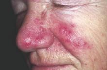
An increased focus on neurologic symptoms in patients with rosacea may be warranted, according to Danish researchers, who found a significantly increased risk of glioma associated with rosacea, in a nationwide study of Danish citizens.
The observational study followed 5,484,910 Danish adults from January 1997 through December 2011; 68,372 were diagnosed with rosacea, and the remaining 5,416,538 were the reference group. The incidence rate of glioma per 10,000 person-years (adjusted for age, sex, and socioeconomic status) was 3.34 in the reference population, but was 4.99 among those with rosacea, reported Dr. Alexander Egeberg of the department of dermatoallergology, Herlev and Gentofte University Hospital, University of Copenhagen, Hellerup, and his coauthors (JAMA Dermatol. 2016 Jan 27. doi: 10.1001/jamadermatol.2015.5549).
The adjusted incidence rate ratio (IRR) of glioma in patients with rosacea was 1.36 (P less than .001). When the researchers limited the analysis to patients who had been diagnosed by a hospital dermatologist, the adjusted IRR was 1.82. The results remained significant after sensitivity analyses and after adjustment for potential confounders.
Among the patients with rosacea, men had an increased risk of glioma, compared with women (an incidence rate per 10,000 person-years of 6.45 vs. 4.30), although “gliomas and rosacea were generally more common among women,” the authors reported.
The association might be partially mediated by mechanisms dependent on matrix metalloproteinases (MMPs), the authors said, referring to studies indicating that MMPs, in particular MMP-9, “play a pivotal role in rosacea and regulation of the invasiveness of malignant glioma cells.” While speculative, “mechanisms dependent on MMPs may contribute to the link between rosacea and the risk for glioma,” the investigators added.
An increased focus on neurologic symptoms such as headaches, memory loss, visual symptoms, cognitive decline, and personality changes in patients with rosacea “and timely referral to relevant specialists may be warranted,” they concluded.
Limitations of the study included the observational design, which cannot establish causation, the authors noted. Dr. Egeberg reported being a former employee of Pfizer; one coauthor reported receiving consultancy and/or speaker honoraria from Galderma. The study was supported by an unrestricted grant from the LEO Foundation and the Lundbeck Foundation and an unrestricted research scholarship from the Novo Nordisk Foundation.
FROM JAMA DERMATOLOGY
Key clinical point: Clinicians should be mindful of neurologic symptoms in patients with rosacea because of a significant association between glioma and rosacea.
Major finding: The incidence rate ratio of glioma per 10,000 person-years was 3.34 in the reference population and 4.99 in patients with rosacea.
Data source: A nationwide cohort study that followed 5,484 910 Danish adults from 1997 through 2011.
Disclosures: Dr. Egeberg reported being a former employee of Pfizer; one coauthor reported receiving consultancy and/or speaker honoraria from Galderma. The study was supported by an unrestricted grant from the LEO Foundation and the Lundbeck Foundation and an unrestricted research scholarship from the Novo Nordisk Foundation.
Simple SpA screening tool works in U.S. population
People under the age of 45 with chronic back pain for more than 3 months can be reliably identified as having axial spondyloarthritis (axSpA) if they have one or more of three SpA disease features, researchers report in Arthritis & Rheumatology.
The research team, led by rheumatologist Dr. Atul Deodhar from Oregon Health & Science University in Portland, noted that results of the German MASTER study indicated that among undiagnosed patients with chronic back pain starting before the age of 45 the presence of inflammatory back pain, human leukocyte antigen B27 (HLA-B27), and/or sacroiliitis on imaging was a reliable screening method for axSpA.
However, the authors said, there is limited information on the epidemiology of axSpA, which encompasses ankylosing spondylitis (AS) and nonradiographic axSpA (nr-axSpA), in the United States. Both AS and nr-axSpA typically go undiagnosed for many years, but AS is more easily identified by the presence of sacroiliitis on radiographs.
In order to determine if the German research finding applied to the U.S. population, the researchers conducted the Prevalence of Axial SpA (PROSpA) trial, involving 751 patients from 68 rheumatology centers who were either existing patients in rheumatology practices, new referrals, or were self-referred.
Participants were required to have chronic back pain for 3 or more months beginning at less than 45 years of age and have one or more of three SpA features: 1. positive HLA-B27; 2. current inflammatory back pain; and 3. MRI/x-ray evidence of sacroiliitis, and no prior SpA diagnosis.
Medical history/physical exam, pelvic x-ray, MRI of sacroiliac joints, C-reactive protein, and HLA-B27 were collected and rheumatologists were asked if a clinical diagnosis of axSpA could be made based upon results.
Results showed that out of a total of 697 patients, 319 (46%) were given a clinical diagnosis of axSpA by the rheumatologist (Arthritis Rheumatol. 2016 Jan 27. doi: 10.1002/art.39612).
Of 744 patients, 348 (47%) fulfilled Assessment of SpondyloArthritis International Society (ASAS) criteria. Of these, 238 were classified as nr-axSpA, and 108 were classified as having AS based on fulfillment of the modified New York criteria. (Two patients had missing data.)
Additionally, 238 (32%) patients were categorized as having nr-axSpA, and 396 patients did not fulfill ASAS criteria or the modified New York criteria for AS.
The specificity and sensitivity of the ASAS criteria are reported at 84% and 83%. About 80% of the patients who received a clinical diagnosis of axSpA also fulfilled the ASAS axSpA criteria. The specificity and sensitivity of the criteria in this study were 79% (95% confidence interval, 75%-83%) and 81% (95% CI, 77%-85%), respectively.
The researchers noted that the majority of patients who received a diagnosis from a rheumatologist fulfilled the imaging arm of the ASAS criteria, whereas those who did not receive a diagnosis fulfilled the clinical arm.
“This observation highlights the need for accurate interpretation of MRI images in clinical practice given the importance of MRI imaging for evaluation of patients for axial SpA,” they wrote.
Overall, the findings emphasized the need to improve the identification and diagnosis of both AS and nr-axSpA among patients already receiving care in rheumatology practices and those newly referred to rheumatologists, the researchers said.
“These patients experience similar burden of disease and can remain undiagnosed, and therefore, untreated, for many years,” they wrote.
Indeed, the data indicated that some of the patients included in the study had symptoms for an average of 14 years, they said.
FROM ARTHRITIS & RHEUMATOLOGY
Key clinical point: Many existing rheumatology patients in the United States with axSpA are undiagnosed. A simple screening tool can help identify these patients.
Major finding: Using screening tool criteria, 319 (46%) of 697 patients were given a clinical diagnosis of axSpA by a rheumatologist. Using the diagnosis as the standard, specificity, and sensitivity of the criteria were 79% (95% CI, 75%-83%) and 81% (95% CI, 77%-85%), respectively.
Data source: Multicenter, non–drug treatment, single-visit trial involving 68 rheumatology centers and 751 patients seen in rheumatology practices.
Disclosures: AbbVie funded the study.
Potential etanercept response biomarker identified
Researchers have identified DNA methylation as a potential biomarker of response to etanercept in an epigenome-wide association study of a longitudinal cohort of patients with rheumatoid arthritis .
Selecting the right therapy the first time for patients with RA is a research priority because very good disease control is only achieved with etanercept (Enbrel) in 30% of patients and other biologic therapies targeting other pathways besides tumor necrosis factor are available, wrote researchers led by Dr. Darren Plant of the National Institute for Health Research Manchester Musculoskeletal Biomedical Research Unit at the Manchester (England) Academy of Health Sciences (Arthritis Rheumatol. 2016 Jan 27. doi: 10.1002/art.39590).
Their study selected 72 patients from the Biologics in Rheumatoid Arthritis Genetics and Genomics Study Syndicate (BRAGGSS) longitudinal cohort based on having an extreme response phenotype after 3 months of treatment with etanercept. DNA from each patient was sampled before therapy was initiated.
A total of 36 patients were very good responders to etanercept and in clinical remission, defined by having a 28-joint Disease Activity Score (DAS28) of less than 2.6.
The 36 patients classified as nonresponders to etanercept had a DAS28 improvement of less than 0.6 or an endpoint DAS28 of greater than 5.1.
Following bisulfite conversion of the DNA, the research team used the HumanMethylation450 BeadChip to determine unmethylated versus methylated cytosines in the DNA and found five differentially methylated sites associated with response to etanercept that passed the 5% false discovery rate threshold.
The most compelling evidence for differential methylation was observed in the LRPAP1 gene located on chromosome 4. This gene encodes a chaperone of low density lipoprotein receptor-related protein 1 (LRP1), which is known to influence TGF-beta activity.
Four CpG sites within exon 7 of the LRPAP1 gene were more methylated in nonresponders, results showed.
“The results indicate that DNA methylation profiling may provide a new biomarker of response to etanercept in patients with RA,” concluded the researchers, who said that the study is the first to investigate the influence of epigenetic variation on response to biologic therapies. Validation in a larger independent cohort is needed before transfer to clinical practice would be possible, they said.
FROM ARTHRITIS & RHEUMATOLOGY
Key clinical point: Researchers have identified a potential biomarker of response to etanercept in patients with rheumatoid arthritis.
Major finding: The most compelling evidence for differential methylation was observed in etanercept patients mapped to the LRPAP1 gene that is known to influence TGF-beta activity.
Data source: An epigenome-wide association study involving 72 selected patients from the Biologics in Rheumatoid Arthritis Genetics and Genomics Study Syndicate (BRAGGSS) longitudinal cohort.
Disclosures: The study was supported by the NIHR Manchester Musculoskeletal Biomedical Research Unit and Arthritis Research UK.


