User login
The fast-changing world of lower-limb atherectomy
Decisions in popliteal or below-knee atherectomy can be complicated by a wide array of devices and lesion types.
Limited data on the long-term durability of interventions, or direct comparisons of approaches, can also complicate the decision-making process, as do cost concerns.
In his Sunday, September 18 talk at VIVA, titled “Popliteal and Below-the-Knee Atherectomy – Which Tool in Which Circumstance and When Not to Bother,” James F. McKinsey, MD, aims to help clinicians navigate this quickly changing field, with updates on emerging technologies.
Directional atherectomy and laser devices continue to undergo innovation, with new devices introduced almost annually. The changing device picture can be confusing, acknowledged Dr. McKinsey of Mt. Sinai Health System in New York. “I am well versed with many of them because I have a high volume. But people with just a few cases a month may not be,” he said.
Lower volume practitioners “need to find at least one, if not two, devices that they are going to be comfortable with,” Dr. McKinsey said, noting that each is associated with a special technique and may require additional support or set-up costs, such as a laser box or a generator. “And it becomes a question of how many different things can you have on the shelf?”
Dr. McKinsey said his talk is aimed at helping practitioners decide which lesions to treat, and with which device – with close attention to the morphological characteristics of lesions. “It’s almost like an algorithm,” he said.
Increasingly, he noted, lower-limb atherectomy is being approached with more than one technique. There is a strong practice trend toward combining atherectomy with drug-coated balloon therapy, he said. “I think the idea of leaving nothing behind [in the vessel] before you do a drug-coated balloon has gotten much more support. People are coming in now and saying they want to prepare the artery by debulking it, then come back and do DCB.” A new rotational laser device, he says, has particular promise in combination with DCBs.
But combining approaches means cost increases at a time when “reimbursement is going down and the product costs and associated expenses are going up,” he said.
Also on the horizon is another, potentially game-changing technology: bioabsorbable stents. While these fall outside the scope of his talk, Dr. McKinsey said he’s assisted a number of lower-limb procedures in Europe this year using them. The technology is especially promising for “more complicated, more calcified lesions,” he said. “In Europe it is being used fairly extensively,” he noted, and likely to come online in the United States within a year or so.
As with the combined approaches, the introduction of drug-eluting bioabsorbable stents into the treatment of lower-limb lesions is also likely to incur high costs, Dr. McKinsey noted. What’s needed are longer-term studies that follow patients up to 5 years, to understand whether high upfront costs are offset by later benefits.
“We have to look at is not necessarily the cost of doing a case, but the cost of treating that patient. We may have a greater upfront cost, but if the intervention has greater durability, and the patient doesn’t have a repeat procedure, then society and healthcare providers do better,” he said.
Early days of IVF marked by competition, innovation
In 1978, when England’s Louise Brown became the world’s first baby born through in vitro fertilization, physicians at academic centers all over the United States scrambled to figure out how they, too, could provide IVF to the thousands of infertile couples for whom nothing else had worked.
Interest in IVF was strong even before British physiologist Robert Edwards and gynecologist Patrick Steptoe announced their success. “We knew that IVF was being developed, that it had been accomplished in animals, and ultimately we knew it was going to succeed in humans,” said reproductive endocrinologist Zev Rosenwaks, MD, of the Weill Cornell Center for Reproductive Medicine in New York.
In the late 1970s, “we were able to help only about two-thirds of couples with infertility, either with tubal surgery, insemination – often with donor sperm – or ovulation induction. A full third could not be helped. We predicted that IVF would allow us to treat virtually everyone,” Dr. Rosenwaks said.
But even after the first IVF birth, information on the revolutionary procedure remained frustratingly scarce.
“Edwards and Steptoe would talk to nobody,” said Richard Marrs, MD, a reproductive endocrinologist and infertility specialist in Los Angeles.
And federal research support for “test-tube babies,” as IVF was known in the media then, was nil thanks to a ban on government-funded human embryo research that persists to this day.
The U.S. physicians who took part in the rush to achieve an IVF birth – most of them young fellows at the time – recall a period of improvisation, collaboration, shoestring budgets, and surprise findings.
“People who just started 10 or even 20 years ago don’t realize what it took for us to learn how to go about doing IVF,” said Dr. Rosenwaks, who in the first years of IVF worked closely with Dr. Howard Jones and Dr. Georgeanna Jones, the first team in the U.S. to announce an IVF baby.
Labs in closets
In the late 1970s, Dr. Marrs, then a fellow at the University of Southern California, was focused on surgical methods to treat infertility – and demand was sky-high. Intrauterine devices used in the 1970s left many women with severe scarring and inflammation of the fallopian tubes.
“I was very surgically oriented,” Dr. Marrs said. “I thought I could fix any disaster in the pelvis that was put in front of me, especially with microsurgery.”
After the news of IVF success in England, Dr. Marrs threw himself into a side project at a nearby cancer center, working on single-cell cultures. “I thought if I could grow tumor cells, I could one day grow embryos,” he said.
A year later, Dr. Marrs set up the first IVF lab at USC – in a storage closet. “I sterilized the place and that was our first IVF lab, literally a closet with an incubator and a microscope.” Its budget was accordingly thin, as the director at the time felt certain that IVF was a dead end. To fund his work, Dr. Marrs asked IVF candidate patients for research donations in lieu of payment.
But before Dr. Marrs attempted to perform his first IVF, two centers in Australia announced their own IVF babies. “I decided I really needed to go see someone who had had a baby,” he said. He used his vacation time to fly to Melbourne, shuttling between two competing clinics that were “four blocks apart and wouldn’t even talk to each other,” he recalled.
Over 6 weeks, “I learned how to stimulate, how to time ovulation. I watched the PhDs in the lab – how they handled the eggs and the sperm, what the conditions were, the incubator settings,” he said.
The first IVF babies in the United States were born only months apart: The first, in December 1981, was at the Jones Institute for Reproductive Medicine in Norfolk, Va., where Dr. Rosenwaks served as the first director.
The second baby born was at USC. After that, “we had 4,000 women on a waiting list, all under age 35,” Dr. Marrs said. The Jones Institute reportedly had 5,000.
As demand soared and more IVF babies arrived, the cloak of secrecy surrounding the procedure started to lift. British, Australian, and U.S. clinicians started getting together regularly. “We would pick a spot in the world, present our data: what we’d done, how many cycles, what we used for stimulation, when we took the eggs out,” Dr. Marrs said. “I don’t know how many hundreds of thousands of miles I flew in the first years of IVF, because it was the only way I could get information. We would literally stay up all night talking.”
Answering safety questions
Alan H. DeCherney, MD, currently an infertility researcher at the National Institutes of Health, started Yale University’s IVF program at around the same time Dr. Marrs and the Joneses were starting theirs. Yale already had a large infertility practice, and only academic centers had the laboratory resources and skilled staff needed to attempt IVF in those years.
In 1983, when Yale announced the birth of its first IVF baby – the fifth in the United States – Dr. DeCherney was starting to think about measuring outcomes, as there was concern over the potential for congenital anomalies related to IVF. “This was such a change in the way conception occurred, people were afraid that all kinds of crazy things would happen,” he said.
One concern was about ovarian stimulation with fertility drugs or gonadotropins. The earliest efforts – including by Dr. Steptoe and Dr. Edwards – used no drugs, instead trying to pinpoint the moment of natural egg release by measuring a woman’s hormone levels constantly, but these proved disappointing. Use of clomiphene citrate and human menopausal gonadotropin allowed for more control over timing, and for multiple mature eggs to be harvested at once.
But there were still many unanswered questions related to these agents’ safety and dosing, both for women and for babies.
When the NIH refused to fund a study of IVF outcomes, Dr. DeCherney and Dr. Marrs collaborated on a registry funded by a gonadotropin maker. “The drug company didn’t want to be associated with some terrible abnormal outcomes,” Dr. DeCherney recalled, though by then, “there were 10, maybe even 20 babies around the world, and they seemed to be fine,” he said.
The first registry results affirmed no changes in the rate of congenital abnormalities. (Larger, more recent studies have shown a small but significant elevation in birth defect risk associated with IVF.) A few years later, ovarian stimulation was adjusted to correspond with ovarian reserve, reducing the risk of ovarian hyperstimulation syndrome.
But even by the late 1980s, success rates for IVF per attempted cycle were still low overall, leading many critics, even within the profession, to accuse practitioners of misleading couples. Charles E. Miller, MD, an infertility specialist in Chicago, recalled an early investigation by a major newspaper “that looked at all the IVF clinics in Chicago and found the chances of having a baby was under 3%.”
It was true, Dr. Miller acknowledged – “the rates were dismal. But remember that IVF at the time was still considered a procedure of last resort.” Complex diagnostic testing to determine the cause of infertility, surgery, and fertility drugs all came first.
Some important innovations would soon change that and turn IVF into a mainstay of infertility treatment that could help women not only with damaged tubes but also with ovarian failure, low ovarian reserve, or dense pelvic adhesions. Even some types of male factor infertility would find an answer in IVF, by way of intracytoplasmic sperm transfer.
Eggs without surgery
Laparoscopic egg retrieval was the norm in the first decade of IVF. “We went through the belly button, allowing us to directly visualize the ovary and see whether ovulation had already occurred or we had to retrieve it by introducing a needle into the follicle,” Dr. Rosenwaks recalled.
“Some of us were doing 6 or even 10 laparoscopies a day, and it was physically quite challenging,” he said. “There were no video screens in those days. You had to bend over the scope.” And it was worse still for patients, who had to endure multiple surgeries.
Though egg and embryo cryopreservation were already being worked on, it would be years before these techniques were optimized, giving women more chances from a single retrieval of oocytes.
Finding a less invasive means of retrieving eggs was crucial.
Maria Bustillo, MD, an infertility specialist in Miami, recalled being criticized by peers when she and her then-colleagues at the Genetics & IVF Institute in Fairfax, Va., began retrieving eggs via a needle placed in the vagina, using abdominal ultrasound as a guide.
While the technique was far less invasive than laparoscopy, “we were doing it semi-blindly, and were told it was dangerous,” Dr. Bustillo said.
But these freehand ultrasound retrievals paved the way for what would become a revolutionary advance – the vaginal ultrasound probe, which by the end of the 1980s made nonsurgical extraction of eggs the norm.
Dr. Marrs recalled receiving a prototype of a vaginal ultrasound probe, in the mid-1980s, and finding patients unwilling to use it, except one who relented only because she had an empty bladder. Abdominal ultrasonography required a full bladder to work.
“It was as though somebody had removed the cloud cover,” he said. “I couldn’t believe it. I could see everything: her ovaries, tiny follicles, the uterus.”
Later probes were fitted with a needle and aspirator to retrieve eggs. Multiple IVF cycles no longer meant multiple surgeries, and the less-invasive procedure helped in recruiting egg donors, allowing women with ovarian disease or low ovarian reserves, including older women, to receive IVF.
“It didn’t make sense for a volunteer to go through a surgery, especially back in the early ’80s when the results were not all that great,” Dr. Bustillo said.
Improving ‘home brews’
The culture media in which embryos were grown was another strong factor limiting the success rates of early IVF. James Toner, MD, PhD, an IVF specialist in Atlanta, called the early media “home brews.”
“Everyone made them themselves,” said Dr. Toner, who spent 15 years at the Jones Institute. “You had to do a hamster or mouse embryo test on every batch to make sure embryos would grow.” And often they did not.
Poor success rates resulted in the emergence of alternative procedures: GIFT (gamete intrafallopian transfer) and ZIFT (zygote intrafallopian transfer). Both aimed to get embryos back into the patient as soon as possible, with the thought that the natural environment offered a better chance for success.
But advances in culture media allowed more time for embryos to be observed. With longer development, “you could do a better job selecting the ones that had a chance, and de-selecting those with no chance,” Dr. Toner said.
This also meant fewer embryos could be transferred back into patients, lowering the likelihood of multiples. Ultimately, for young women, single-embryo transfer would become the norm. “The problem of multiple pregnancy that we used to have no longer exists for IVF,” Dr. Toner said.
Allowing embryos to reach the blastocyst stage – day 5 or 6 – opened other, previously unthinkable possibilities: placing embryos directly into the uterus, without surgery, and pre-implementation genetic screening for abnormalities.
“As the cell number went up, the idea that you could do a genetic test with minimal impact on the embryo eventually became true,” Dr. Toner said.
A genetic revolution?
While many important IVF innovations were achieved in countries with staunch government support, one of the remarkable things about IVF’s evolution in the United States is that so many occurred with virtually none.
By the mid-1990s, most of the early practitioners had moved from academic settings into private practice, though they continued to publish. “After a while it didn’t help to be in academics. It just sort of slowed you down. Because you weren’t going to get any [government] money anyway, you might as well be in a place that’s a little more nimble,” Dr. Toner said.
At the same time, he said, IVF remains a costly, usually unreimbursed procedure – limiting patients’ willingness to take part in randomized trials. “IVF research is built more on cohort studies.”
Most of the current research focus in IVF is on possibilities for genetic screening. Dr. Miller said that rapid DNA sequencing is allowing specialists to “look at more, pick up more abnormalities. That will continue to improve so that we will be able to see virtually everything.”
But he cautioned there is still much to be done in IVF apart from the genetics – he’s concerned, he said, that the field has moved too far from its surgical origins, and is working with the academic societies to encourage more surgical training.
“We don’t do the same work we did before on fallopian tubes, which is good,” Dr. Miller said, noting that there have been many advances, particularly minimally invasive surgeries in the uterus or ovaries, that have occurred parallel to IVF and can improve success rates. “I think we have a better understanding of what kind of patients require surgical treatments and what kind of surgeries can help enhance fertility, and also what not to do.”
Dr. Bustillo said that “cytogenetics is wonderful, but not everything. You have embryos that are genetically normal and still don’t implant. There’s a lot of work to be done on the interaction between the mother and the embryo.”
Dr. Marrs said that even safety questions related to stimulation have yet to be fully answered. “I’ve always been a big believer that lower is better, but we need to know whether stimulation creates genetic abnormalities and whether less stimulation produces fewer – and we need more data to prove it,” he said. Dr. Marrs is an investigator on a national randomized trial comparing outcomes from IVF with standard-dose and ultra-low dose stimulation.
Access, income, and age
The IVF pioneers agree broadly that access to IVF is nowhere near what it should be in the United States, where only 15 states mandate any insurance coverage for infertility.
“Our limited access to care is a crime,” Dr. Toner said. “People who, through no fault of their own, find themselves infertile are asked to write a check for $15,000 to get pregnant. That’s not fair.”
Dr. DeCherney called access “an ethical issue, because who gets IVF? People with higher incomes. And if IVF allows you to select better embryos – whatever that means – it gives that group another advantage.”
Dr. Toner warned that the push toward genetic testing of embryos, especially in the absence of known hereditary disease, could create new problems for the profession – not unlike in the early days of IVF, when the Jones Institute and other clinics were picketed over the specter of “test tube babies.”
“It’s one thing to say this embryo does not have the right number of chromosomes and couldn’t possibly be a child, so let’s not use it, but what about looking for traits? Sex selection? We have this privileged position in which the government does not really interfere in what we do, but to retain this status we need to stay within the bounds that our society accepts,” Dr. Toner said.
In recent years, IVF uptake has been high among women of advanced reproductive age, which poses its own set of challenges. Outcomes in older women using their own eggs become progressively poorer with age, though donor eggs drastically improve their chances, and egg freezing offers the possibility of preserving quality eggs for later pregnancies.
“We could make this situation better by promoting social freezing, doing more work for women early in their lives to get out their own eggs and store them,” Dr. Miller said. “But again, you still face the issue of access.”
Regardless of what technologies are available or become available in assisted reproduction, doctors and women alike need to be better educated on their options and chances early, with a clearer understanding of what happens as they age, Dr. Bustillo said.
“This is not to pressure them, but just so they understand that when they get to be 42 and are just thinking about reproducing, it’s not a major surprise when I tell them this could be a problem,” she said.
Throughout 2016, Ob.Gyn. News is celebrating its 50th anniversary with exclusive articles looking at the evolution of the specialty, including the history of contraception, changes in gynecologic surgery, and the transformation of the well-woman visit.
Pneumonitis with nivolumab treatment shows common radiographic patterns
A study of cancer patients enrolled in trials of the programmed cell death-1 inhibiting medicine nivolumab found that among a minority who developed pneumonitis during treatment, distinct radiographic patterns were significantly associated with the level of pneumonitis severity.
Investigators found that cryptic organizing pneumonia pattern (COP) was the most common, though not the most severe. Led by Mizuki Nishino, MD, of Brigham and Women’s Hospital, Boston, the researchers looked at the 20 patients out of a cohort of 170 (11.8%) who had developed pneumonitis, and found that radiologic patterns indicating acute interstitial pneumonia/acute respiratory distress syndrome (n = 2) had the highest severity grade on a scale of 1-5 (median 3), followed by those with COP pattern (n = 13, median grade 2), hypersensitivity pneumonitis (n = 2, median grade 1), and nonspecific interstitial pneumonia (n = 3, median grade 1). The pattern was significantly associated with severity (P = .0006).
The study cohort included patients being treated with nivolumab for lung cancer, melanoma, and lymphoma; the COP patten was the most common across tumor types and observed in patients receiving monotherapy and combination therapy alike. Therapy with nivolumab was suspended for all 20 pneumonitis patients, and most (n = 17) received treatment for pneumonitis with corticosteroids with or without infliximab, for a median treatment time of 6 weeks. Seven patients were able to restart nivolumab, though pneumonitis recurred in two, the investigators reported (Clin Cancer Res. 2016 Aug 17. doi: 10.1158/1078-0432.CCR-16-1320).
“Time from initiation of therapy to the development of pneumonitis had a wide range (0.5-11.5 months), indicating an importance of careful observation and follow-up for signs and symptoms of pneumonitis throughout treatment,” Dr. Nishino and colleagues wrote in their analysis, adding that shorter times were observed for lung cancer patients, possibly because of their higher pulmonary burden, a lower threshold for performing chest scans in these patients, or both. “In most patients, clinical and radiographic improvements were noted after treatment, indicating that [PD-1 inhibitor-related pneumonitis], although potentially serious, is treatable if diagnosed and managed appropriately. The observation emphasizes the importance of timely recognition, accurate diagnosis, and early intervention.”
The lead author and several coauthors disclosed funding from Bristol-Myers Squibb, which sponsored the trial, as well as from other manufacturers.
FROM CLINICAL CANCER RESEARCH
Key clinical point: Pneumonitis related to treatment with PD-1 inhibitors showed distinct radiographic patterns associated with severity; most cases resolved with corticosteroid treatment.
Major finding: Of 20 patients in nivolumab trials who developed pneumonitis, a COP pattern was seen in 13, and other patterns in 7; different patterns were significantly associated with pneumonitis severity (P = .006).
Data source: 170 patients with melanoma, lung cancer or lymphoma enrolled in single-site open-label clinical trial of nivolumab.
Disclosures: The lead author and several coauthors disclosed funding from Bristol-Myers Squibb, which sponsored the trial, as well as from other manufacturers.
Guillain-Barré incidence rose with Zika across Americas
Increased incidence of Guillain-Barré syndrome corresponded closely with patterns of Zika virus disease incidence in Central and South America from April 2015 through March 2016, according to results from a new temporal and graphic analysis.
The findings show Guillain-Barré syndrome (GBS) cases increasing from 100% to nearly 900% above previously recorded baseline rates during periods of Zika virus transmission in El Salvador, the Dominican Republic, Colombia, Honduras, Suriname, Venezuela, and the Brazilian state of Bahia (N Engl J Med. 2016 Aug 31. doi: 10.1056/NEJMc1609015).
The analysis of the yearlong period also revealed that declines in GBS incidence accompanied declines in Zika virus disease when and where transmission began to wane. The researchers, led by Marcos A. Espinal, MD, DrPH, of the Pan American Health Organization in Washington, did not find significant associations between co-circulation of dengue virus and GBS incidence. The study, which looked at 164,237 confirmed and suspected cases of Zika virus disease and 1,474 cases of GBS, found a 75% higher Zika virus disease incidence rate in women, which Dr. Espinal and colleagues said might be attributable to differences in health care–seeking behavior. GBS incidence, meanwhile, was 28% higher among males. The higher rate of GBS in men, the authors said, was consistent with findings from previous epidemiological studies of GBS.
While the new results did not show that Zika virus causes GBS, Dr. Espinal and colleagues wrote, they argued that they were indicative of a strong association, adding that GBS “could serve as a sentinel for Zika virus disease and other neurological disorders linked to Zika virus,” including microcephaly.
Most of the study authors worked for the Pan American Health Organization or for national health agencies in the data-contributing countries. None declared conflicts of interest.
FROM NEW ENGLAND JOURNAL OF MEDICINE
Guidelines add two new heart failure treatments
Optimal use of two recently approved medications for heart failure has been detailed by the major heart societies in a guideline update.
The American College of Cardiology, the American Heart Association, and the Heart Failure Society of America issued joint recommendations May 20 on the two new medicines for stage C heart failure patients with a reduced ejection fraction.
Valsartan/sacubitril (Entresto, Novartis), is a combination angiotensin receptor–neprilysin inhibitor, the first in a novel class of drugs slugged ARNIs. Ivabradine (Corlanor, Amgen), is a sinoatrial node modulator. Both medicines were approved by the Food and Drug Administration in 2015, though ivabradine has been licensed for a decade in Europe.
Although a comprehensive update to ACC/AHA/HSFA heart failure guidelines is still being developed, the focused update is intended to coincide with the release of new European Society of Cardiology heart failure guidelines, “in order to minimize confusion and improve the care of patients with heart failure,” the societies said in a statement May 20. The recommendations were published online simultaneously in Circulation and the Journal of Cardiac Failure.
The guideline authors, led by Dr. Clyde W. Yancy of Northwestern University in Chicago, recommend that the ARNI replace an ACE inhibitor or an angiotensin II receptor blocker (ARB) for patients who have been tolerating these therapies alongside standard care with a beta-blocker and, for some patients, an aldosterone antagonist as well. The guidelines caution against combining an ARNI with an ACE inhibitor, and against using ARNIs in patients with a history of angioedema.
For patients not suited to treatment with an ARNI, continued use of an ACE inhibitor is recommended. In patients for whom an ACE inhibitor or an ARNI is inappropriate, use of an ARB remains advised. The authors noted that head-to-head comparisons of an ARB versus an ARNI for heart failure do not exist; however, in a randomized, controlled trial in heart failure patients, treatment with valsartan/sacubitril plus standard care reduced cardiovascular death or heart failure hospitalization by 20%, compared with treatment with an ACE inhibitor plus standard care.
Ivabradine, meanwhile, has shown benefit in reducing heart failure hospitalizations in patients with symptomatic, stable, chronic heart failure with reduced ejection fraction who are receiving standard treatment including a beta-blocker, and who are in sinus rhythm with a heart rate of 70 beats per minute or greater at rest.
The new therapies, “when applied judiciously, complement established pharmacological and device-based therapies, representing milestones in the evolution of care for patients with heart failure,” wrote Dr. Elliott M. Antman of Brigham and Women’s Hospital and Harvard Medical School in Boston, Mass., in an editorial accompanying the guidelines.
About half the guideline writing committee members and guideline reviewers disclosed financial relationships with pharmaceutical companies or device manufacturers. Dr. Yancy disclosed no conflicts of interest.
FROM CIRCULATION
SFA-Popliteal Claudication: What’s Appropriate Care?
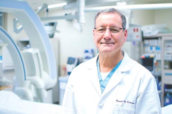
In past years the Crawford Critical Issues Symposium has served as a forum in which nonclinical issues took center stage. At the 2016 Vascular Annual Meeting, Dr. Ron Fairman of the University of Pennsylvania Health System will take the symposium in a clinical direction with an issue that, he says, highlights critical questions about appropriateness of care. Titled “In Search of Clarity,” speakers will talk about interventions for claudication in the superficial femoral-popliteal arteries. This year’s lineup will address the role of medical management and exercise, along with the choice, cost, and durability of common interventions.
Speakers will be:
Dr. Mary McDermott of Northwestern University, on exercise;
Dr. Elizabeth Ratchford, Johns Hopkins University on medical management;
Dr. Michael Conte, of the University of California San Francisco, on understanding data sets, durability, and guidelines;
Dr. Peter Schneider of Kaiser Permanente on how claudication is approached in a system outside the traditional fee-for-service model;
Dr. Dennis Gable of Texas Vascular Associates on approaches to claudication in the community practice setting; and
Dr. Robert Zwolak of Dartmouth-Hitchcock Medical Center on the financial aspects, including hidden costs.
VC: Why did you choose claudication as this year’s key theme?
Dr. Fairman: There is probably no other subject that touches more on appropriateness of care than interventions for claudication, and if you look at how patients are managed across the country, there probably is no greater area of disparity. One of the most significant aspects of treating patients with expensive interventions for claudication is frequently the lack of durability. We often see patients in the office who present for a second opinion who already have undergone multiple invasive interventions over a 1- to 2-year period, all resulting in short-term improvement followed by – frighteningly – a much more serious set of symptoms than they started with. And this is fostered by a reimbursement system that demonstrates a willingness to reimburse physicians and hospitals for procedures that have little durability.
VC: Is there too much of a leap in the vascular community to invasive interventions?
Dr. Fairman: I believe that the members of our society understand that before a patient is considered for an invasive intervention for claudication, they should undergo exercise training and optimization of their medical status – but there are challenges associated with that. How do you do get a patient connected to a training regimen? Have all the patient’s risk factors been effectively addressed? Have they quit smoking? Is their blood sugar under control? We view ourselves as a unique specialty that offers comprehensive care of patients with vascular disease. We don’t just do surgery and procedures, but we have the ability to offer the entire package. We also understand by virtue of our training the natural history of the anatomic lesions we are treating. It is essential that physicians look at the whole patient before planning an intervention, and this forum should help us think more about that.
VC: What’s missing from a research perspective that might shed light on when intervention is needed and which interventions to perform?
Dr. Fairman: We need studies comparing durability of interventions with longer follow-up; 1 year is not enough. Dr. Conte will address what the available large data sets show in terms of durability. We need real-world data that compare technologies for claudication. We all have biases about technology based on our own experiences, but many of us struggle in selecting from the very many interventional options available to treat our patients. In addition, physician reimbursement typically increases from simple balloon angioplasty, to placement of a stent, to atherectomy.
VC: What other cost and reimbursement issues do you expect to deal with during the symposium?
I’m interested in Dr. Schneider’s perspective practicing in a health system that is a non–fee-for-service model. How does he make day-to-day decisions about using technology? Dr. Zwolek will talk about the financial side, not only in terms of physician reimbursement, but how physicians should view the expensive disposable inventory that they use when treating patients with claudication. This relates to hospital or facility reimbursement and issues related to margin. Regrettably, given the time constraints of this program, we were unable to secure speakers representing the viewpoints and perspectives of CMS [the Centers for Medicare and Medicaid Services] and the FDA [Food and Drug Administration].
Clozapine REMS still plagued by problems
A consolidated registry system designed to increase access to the second-generation antipsychotic clozapine is plagued with glitches, delays, and excess red tape more than 8 months after its initial launch, according to clinicians and pharmacists who are attempting to use it.
The problems include psychiatrists receiving information from the registry on patients not in their care, a breach of Health Insurance Portability and Accountability Act privacy rules. And some said they fear that clozapine, the only Food and Drug Administration–approved drug for treatment-resistant schizophrenia, will end up underprescribed as a result of difficulties complying with the new registry’s demands.
The psychiatric community lauded the FDA’s decision in September 2015 to change the monitoring requirements for clozapine. One rare but dangerous adverse effect of the drug is severe neutropenia, and patients on clozapine must be monitored through regular blood screening. This means that every clozapine script requires careful coordination among the prescriber, patient, laboratory, and pharmacy.
Now, instead of looking at white blood cell counts as before, the FDA said, absolute neutrophil count (ANC) will be the lab measure used to determine whether a patient can be started or continued on clozapine, and new lowered ANC thresholds will allow more patients to be initiated. The new lowered ANC thresholds may pertain to people of African and Middle Eastern heritage with a naturally lower ANC called “benign ethnic neutropenia” and who previously might not have been able to receive the drug.
At the same time it announced the new monitoring rules, the FDA also said the six existing manufacturer registries of clozapine would be consolidated into one, called the Clozapine Risk Evaluation and Mitigation Strategies, or Clozapine REMS. The REMS is managed jointly by the manufacturers. Prescribers and pharmacies dealing with clozapine must become certified under the REMS if they wish patients to receive the drug. Though certification was supposed to have been completed by this month, the agency now is saying providers have until year-end.
REMS applauded early on
Originally, psychiatrists welcomed the news of the REMS, as it promised to make it easier for a patient to continue on clozapine even if the drug supplier changed – when transitioning from an inpatient to outpatient setting, for example.
However, when it launched in the fall of 2015, the Clozapine REMS website was full of glitches, and calls to the toll-free number weren’t picked up. “I couldn’t even get onto [the registry], as they wouldn’t answer the phone,” said Dr. Ira D. Glick, professor emeritus of psychiatry and behavioral sciences and psychopharmacology at Stanford (Calif.) University, who has prescribed clozapine for decades. More recently, he said, the response time has improved.
The FDA has acknowledged providers’ complaints, and mainly characterizes the registry’s problems as growing pains. “As with any new IT system, and large data migration and reconciliation effort, there are challenges,” an agency spokesperson said in an interview about the REMS. “Merging six previous clozapine registries was a huge undertaking for the manufacturers that encompassed merging data from over 50,000 prescribers, 28,000 pharmacies, and 200,000 patient records.”
The FDA has been working “to address the issues identified when the Clozapine REMS Program was initially implemented,” the representative said. “We believe that most of the issues have been resolved.” The FDA did not directly respond to questions about confidentiality breaches.
Providers said in interviews, however, that the issues continue, including mix-ups of confidential health information.
“I’m now registered as a provider designee for several clozapine prescribers here at my hospital,” said Megan Maroney, Pharm.D., of Monmouth Medical Center in Long Branch, N.J. “One of our prescribers also has a private practice, and those patients are popping up on my list, but they’re not my patients.” Dr. Maroney said she contacted the Clozapine REMS about the problem. “They haven’t yet come up with a good way to deal with it,” she said.
Dr. Jean-Pierre Lindenmayer, clinical professor in the department of psychiatry at New York University, said he, too, receives information about patients who are not his. “I’m still getting faxed notifications on patients I have no idea who they are, saying they’re due for a blood test. It’s incredible that they keep doing this.” The REMS never responded to his complaints about privacy breaches, he said.
Problems ‘causing confusion’
Providers say the Clozapine REMS website remains filled with glitches and that deadlines are being extended until these can be resolved. “We’re trying to follow the intended rules with the understanding that if issues come up, we’re supposed to use our clinical judgment and not delay care to the patient. But it’s causing a lot of confusion among my staff, because whenever we try to use it, we encounter some kind of glitch,” Dr. Maroney said.
Another of the providers’ key concerns is the extra red-tape burdens imposed by the REMS, which, they say, have the potential to dampen the prescribing of clozapine – the exact opposite of its intended effect.
By mandating that only registered prescribers can write for clozapine, the REMS can cause problems for hospitals. “If a patient comes in for a medical reason and happens to be on clozapine, it’s impossible for us to get our internal medicine physicians registered just so they can prescribe it for their one patient who comes through,” Dr. Maroney said. Her workaround has been to make sure all the hospital’s psychiatrists are registered and that patients on clozapine have a psych consult, regardless of the reason they’re hospitalized. This, she acknowledged, could drive up costs.
“Here at my hospital, I tend to work out the issues for my prescribers so that they can start patients and continue them, and I think we’ve been doing a pretty good job. But I wonder about places without the manpower to do that or that don’t have a psych pharmacist who can work with them,” she said.
The REMS “is a major obstacle, and it’s more complicated now than it was before,” said Dr. Lindenmayer, who also is affiliated with the Manhattan Psychiatric Center in New York, an institution that manages about 100 patients on clozapine. “The excessive registry demands, and sending doctors letters about all the potential terrible side effects, will discourage providers. Clozapine is already difficult to prescribe: You have to have a pharmacy lined up, you have to have a lab lined up, you have to have a patient that gets the prescription and the blood test in a timely manner, and you are not being reimbursed at any higher rate by having a clozapine patient.”
Dr. Glick agreed. “Clozapine is one of our best drugs, but the most difficult to manage – as it takes a lot of time and effort. This is one more step making it more complicated.”
Dr. Maroney said that at her institution, she’s already seen a chilling effect from the REMS. Recently, she said, “my prescribers and I were going over all the changes and some of them said, ‘Just forget it, I’ll put [patients] on something else.’ And I said, ‘No – the whole point of a lot of the changes was to make [clozapine] more accessible.’ ”
Dr. Lindenmayer said he considers the REMS – or at least the extra layers of bureaucracy and certification it imposes – to have been a misguided move by the FDA. “I am not aware that there have been more deaths recently due to clozapine prescribing, and haven’t seen any upsurge of morbidity and mortality in the literature, which I follow closely.” Relatively few prescribers use clozapine, he said, and those who do “are fairly careful and knowledgeable about what they’re doing. So they’re preaching to the choir,” he said.
Yet Dr. Maroney said she remains optimistic that the REMS and the providers will be able to reach common ground – eventually: “The College of Psychiatric and Neurologic Pharmacists has been communicating with the FDA to hammer out these issues. I think it should get better. I just don’t know when that will occur and how many of these issues will be completely addressed.”
The FDA spokesperson confirmed that the agency was seeking provider input to improve the Clozapine REMS, and that several changes already had been made in response to provider concerns.
CDC confirms Zika virus as a cause of microcephaly
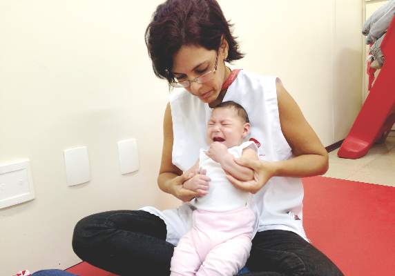
Officials at the Centers for Disease Control and Prevention have determined that Zika virus infection is a cause of microcephaly in babies born to infected mothers, following a systematic review of the latest research on Zika virus.
The CDC released findings from that review in a peer-reviewed special report published online April 13 in the New England Journal of Medicine (2016. doi: 10.1056/NEJMsr1604338). The report, CDC officials said, incorporated evidence from as recently as the past week.
In a press conference in April, CDC director Tom Frieden said there was “no longer any doubt” that Zika virus causes microcephaly. Dr. Frieden’s statements reflect what appears to be a growing scientific consensus. On March 31, the World Health Organization reported that there was a “strong” consensus that Zika virus can cause microcephaly, Guillain-Barré syndrome, and other neurological disorders. New microcephaly cases in Colombia – with 32 reported by the end of March – are among the findings cited by both the WHO and the CDC.
Dr. Frieden stressed that no single piece of evidence was seen to provide conclusive proof of causation, but that the CDC scientists’ conclusions were based on “a thorough review of the best available evidence” subjected to established criteria.
For its review published in the New England Journal of Medicine, CDC scientists led by Dr. Sonja Rasmussen subjected available evidence to two separate criteria to determine the relationship of Zika virus to the spikes in microcephaly cases seen in countries where Zika is spreading. Shepard’s criteria were used to prove teratogenicity, and the Bradford Hill criteria were used to show evidence of causation.
The CDC has not made any changes to its guidance on Zika virus prevention, which includes advising pregnant women to avoid travel to regions where Zika transmission is occurring, take steps to prevent infection if they live in areas where Zika virus is present, and use condoms to prevent sexual transmission of Zika from a partner. On April 13, the CDC added St. Lucia to its growing list of countries to be avoided by pregnant women.
The full spectrum of defects caused by congenital Zika infection is still poorly understood. Additional studies are underway at CDC, Dr. Frieden said, to better understand the severe phenotype of microcephaly seen in babies born to Zika-infected mothers and “to determine whether children who have microcephaly born to mothers infected by the Zika virus is the tip of the iceberg of what we could see in damaging effects on the brain.”
FROM A CDC PRESS CONFERENCE
Cardiac cath lab work carries multiple risks
Workers performing fluoroscopically guided cardiovascular procedures are at significantly higher risk of a diverse array of health disorders compared with nonexposed subjects, according to a survey.
Though cancers and ocular, skin, and thyroid disorders are recognized risks of working with low-dose radiation, as are orthopedic problems associated with wearing heavy lead aprons, the new study, published online April 12 in Circulation: Cardiovascular Interventions, adds weight to emerging concerns about lesser-known risks.
In addition to higher prevalence of skin lesions, cataracts, thyroid disorders, cancer, and orthopedic problems, exposed workers saw significantly higher prevalence of hypertension, hypercholesterolemia, and depression or anxiety, compared with controls.
Maria Grazia Andreassi, Ph.D., of the CNR Institute of Clinical Physiology, in Pisa, Italy, led the study, which used questionnaires to collect health, exposure, and demographic information from 466 workers in cardiac catheterization laboratories in Italy: 218 interventional cardiologists and electrophysiologists, 191 nurses, and 57 technicians. Radiation exposure was estimated based on subjects’ occupational radiological risk score (ORRS), which takes into consideration years of work, caseloads, and proximity to the radiation source in the lab.
The researchers administered the same questionnaires to 280 health workers who did not have occupational radiation exposure. The two groups were well matched for age, sex, education level, alcohol use, and body mass index, though current smoking differed significantly (27% of lab staff vs. 23% of unexposed workers).
As expected, orthopedic injuries were markedly higher among the lab workers, with 30% reporting disorders compared with 5% of controls (P less than .001). Nearly 5% of the lab workers had cataracts, compared with less than 1% of the nonexposed (P = .003), and risk increased for subjects with more years in the lab. Cancer was higher among exposed participants (2.6% vs. 0.7%), a difference that did not reach statistical significance. However, a significant trend was seen for cancer risk increasing across duration of exposure (Circ Cardiovasc Interv. 2016;9:e003273).
In the composite endpoint of cataract and cancer, a surrogate for disease associated with occupational exposure to radiation, the researchers found that the interventional cardiologists and electrophysiologists had a significantly higher prevalence than did the nurses and technicians (69% vs. 22% and 9%, respectively; P = .03).
Furthermore, the cancer in physicians occurred more often in the left side, when laterality was applicable, in 67% of cases, whereas the malignancy was left-sided in 33% of cancers occurring in nurses and technicians.
No significant differences in cardiovascular events were seen between the groups. However, hypertension and high cholesterol were more prevalent in cath lab workers, with adjusted odds ratios of 1.7 (95% confidence interval, 1-3; P = .05) and 2.9 (CI, 1-5; P = .004), respectively, for highly exposed subjects, compared with controls.
Last year, in a separate study, Dr. Andreassi’s group reported that exposure to low-dose radiation over time may increase carotid intima-media thickness, an early indicator of vascular injury, among cath lab workers (J Am Coll Cardiol Intv. 2015;8:616-27).
Anxiety and depression occurred in 12% of exposed subjects, compared with 2% of controls. The finding could reflect high occupational stress, the researchers wrote in their analysis, or may be an effect of radiation, “which is especially relevant on the unprotected head of the first operator and at chronic low doses may impact detrimentally on hippocampal neurogenesis and neuronal plasticity.”
The researchers acknowledged the possibility of selection bias in their study, favoring “a possible disproportionate contribution of respondents with existing health issues, who may reasonably think that their complaints are occupationally related,” they wrote. They also noted that the study was not designed to directly assess radiation dose, in part because dosimeters were not regularly worn in some of the settings where subjects worked. Finally, the radiation-exposed group had more cardiovascular risk factors. Nonetheless, the findings highlight the need to “spread the culture of safety” in cardiac cath labs, the researchers wrote.
There is now “more than enough information for us to conclude that the interventional catheterization laboratory is not a healthy workplace,” Dr. Lloyd W. Klein and Dr. Mugurel Bazavan of Advocate Illinois Masonic Medical Center, and Rush Medical College in Chicago, wrote in an accompanying editorial.
Finding “the courage to change business practice,” they wrote, “is the only path to innovative cath laboratory design and shielding techniques that can prevent these occupational hazards” (Circ Cardiovasc Interv. 2016;9:e003742).
Dr. Andreassi and colleagues disclosed no conflicts of interest related to their study, and Dr. Klein and Dr. Bazavan disclosed no conflicts related to their editorial.
The field of interventional cardiology is tremendously exciting, and yet the long-term risks are not as well defined as they should be for the protection of cath lab personnel.
 |
Dr. Charles E. Chambers |
This study shows strikingly that both physicians and nonphysicians are at increased risk of a diverse group of medical conditions. Quantifying the contribution of radiation exposure to CV risk and separating it from smoking, dietary, and other factors, will require studies using dosimetry data in lieu of the estimated exposure measures seen in this study.
However, as these findings underscore, there is no doubt that we must be concerned about radiation and that manufacturers, hospitals, and providers need to come to the table to address these urgent questions of personal liability and workplace safety, particularly with so many young people entering the field.
Dr. Charles E. Chambers is professor of medicine and radiology at Pennsylvania State University, Hershey, and past president of the Society for Cardiovascular Angiography and Interventions. He made these comments in an interview, and reports no conflicts of interest.
The field of interventional cardiology is tremendously exciting, and yet the long-term risks are not as well defined as they should be for the protection of cath lab personnel.
 |
Dr. Charles E. Chambers |
This study shows strikingly that both physicians and nonphysicians are at increased risk of a diverse group of medical conditions. Quantifying the contribution of radiation exposure to CV risk and separating it from smoking, dietary, and other factors, will require studies using dosimetry data in lieu of the estimated exposure measures seen in this study.
However, as these findings underscore, there is no doubt that we must be concerned about radiation and that manufacturers, hospitals, and providers need to come to the table to address these urgent questions of personal liability and workplace safety, particularly with so many young people entering the field.
Dr. Charles E. Chambers is professor of medicine and radiology at Pennsylvania State University, Hershey, and past president of the Society for Cardiovascular Angiography and Interventions. He made these comments in an interview, and reports no conflicts of interest.
The field of interventional cardiology is tremendously exciting, and yet the long-term risks are not as well defined as they should be for the protection of cath lab personnel.
 |
Dr. Charles E. Chambers |
This study shows strikingly that both physicians and nonphysicians are at increased risk of a diverse group of medical conditions. Quantifying the contribution of radiation exposure to CV risk and separating it from smoking, dietary, and other factors, will require studies using dosimetry data in lieu of the estimated exposure measures seen in this study.
However, as these findings underscore, there is no doubt that we must be concerned about radiation and that manufacturers, hospitals, and providers need to come to the table to address these urgent questions of personal liability and workplace safety, particularly with so many young people entering the field.
Dr. Charles E. Chambers is professor of medicine and radiology at Pennsylvania State University, Hershey, and past president of the Society for Cardiovascular Angiography and Interventions. He made these comments in an interview, and reports no conflicts of interest.
Workers performing fluoroscopically guided cardiovascular procedures are at significantly higher risk of a diverse array of health disorders compared with nonexposed subjects, according to a survey.
Though cancers and ocular, skin, and thyroid disorders are recognized risks of working with low-dose radiation, as are orthopedic problems associated with wearing heavy lead aprons, the new study, published online April 12 in Circulation: Cardiovascular Interventions, adds weight to emerging concerns about lesser-known risks.
In addition to higher prevalence of skin lesions, cataracts, thyroid disorders, cancer, and orthopedic problems, exposed workers saw significantly higher prevalence of hypertension, hypercholesterolemia, and depression or anxiety, compared with controls.
Maria Grazia Andreassi, Ph.D., of the CNR Institute of Clinical Physiology, in Pisa, Italy, led the study, which used questionnaires to collect health, exposure, and demographic information from 466 workers in cardiac catheterization laboratories in Italy: 218 interventional cardiologists and electrophysiologists, 191 nurses, and 57 technicians. Radiation exposure was estimated based on subjects’ occupational radiological risk score (ORRS), which takes into consideration years of work, caseloads, and proximity to the radiation source in the lab.
The researchers administered the same questionnaires to 280 health workers who did not have occupational radiation exposure. The two groups were well matched for age, sex, education level, alcohol use, and body mass index, though current smoking differed significantly (27% of lab staff vs. 23% of unexposed workers).
As expected, orthopedic injuries were markedly higher among the lab workers, with 30% reporting disorders compared with 5% of controls (P less than .001). Nearly 5% of the lab workers had cataracts, compared with less than 1% of the nonexposed (P = .003), and risk increased for subjects with more years in the lab. Cancer was higher among exposed participants (2.6% vs. 0.7%), a difference that did not reach statistical significance. However, a significant trend was seen for cancer risk increasing across duration of exposure (Circ Cardiovasc Interv. 2016;9:e003273).
In the composite endpoint of cataract and cancer, a surrogate for disease associated with occupational exposure to radiation, the researchers found that the interventional cardiologists and electrophysiologists had a significantly higher prevalence than did the nurses and technicians (69% vs. 22% and 9%, respectively; P = .03).
Furthermore, the cancer in physicians occurred more often in the left side, when laterality was applicable, in 67% of cases, whereas the malignancy was left-sided in 33% of cancers occurring in nurses and technicians.
No significant differences in cardiovascular events were seen between the groups. However, hypertension and high cholesterol were more prevalent in cath lab workers, with adjusted odds ratios of 1.7 (95% confidence interval, 1-3; P = .05) and 2.9 (CI, 1-5; P = .004), respectively, for highly exposed subjects, compared with controls.
Last year, in a separate study, Dr. Andreassi’s group reported that exposure to low-dose radiation over time may increase carotid intima-media thickness, an early indicator of vascular injury, among cath lab workers (J Am Coll Cardiol Intv. 2015;8:616-27).
Anxiety and depression occurred in 12% of exposed subjects, compared with 2% of controls. The finding could reflect high occupational stress, the researchers wrote in their analysis, or may be an effect of radiation, “which is especially relevant on the unprotected head of the first operator and at chronic low doses may impact detrimentally on hippocampal neurogenesis and neuronal plasticity.”
The researchers acknowledged the possibility of selection bias in their study, favoring “a possible disproportionate contribution of respondents with existing health issues, who may reasonably think that their complaints are occupationally related,” they wrote. They also noted that the study was not designed to directly assess radiation dose, in part because dosimeters were not regularly worn in some of the settings where subjects worked. Finally, the radiation-exposed group had more cardiovascular risk factors. Nonetheless, the findings highlight the need to “spread the culture of safety” in cardiac cath labs, the researchers wrote.
There is now “more than enough information for us to conclude that the interventional catheterization laboratory is not a healthy workplace,” Dr. Lloyd W. Klein and Dr. Mugurel Bazavan of Advocate Illinois Masonic Medical Center, and Rush Medical College in Chicago, wrote in an accompanying editorial.
Finding “the courage to change business practice,” they wrote, “is the only path to innovative cath laboratory design and shielding techniques that can prevent these occupational hazards” (Circ Cardiovasc Interv. 2016;9:e003742).
Dr. Andreassi and colleagues disclosed no conflicts of interest related to their study, and Dr. Klein and Dr. Bazavan disclosed no conflicts related to their editorial.
FROM CIRCULATION: CARDIOVASCULAR INTERVENTIONS
Key clinical point: Workers in cardiac catheterization labs exposed to ionizing radiation are at risk of a wide array of health disorders.
Major finding: Prevalences of skin lesions, orthopedic illness, cataract, hypertension, hypercholesterolemia, and anxiety/depression were significantly higher in exposed cath lab workers, compared with other health care workers.
Data source: A multicenter, controlled study of 746 Italian physicians and staff (466 exposed to radiation and 280 matched unexposed controls) completing self-administered questionnaires.
Disclosures: None.
High-dose vitamin D improves heart structure, function in chronic heart failure
High-dose oral vitamin D supplements taken for 1 year significantly improved cardiac structure and function in patients with chronic heart failure secondary to left ventricular systolic dysfunction, according to results from a new study.
However, the same study. led by Dr. Klaus Witte of the University of Leeds (England), found that 6-minute walk distance – the study’s primary outcome measure – was not improved after a year’s supplementation with vitamin D.
It is unclear why vitamin D deficiency co-occurs in a majority of people with chronic heart failure (CHF) due to left ventricular systolic dysfunction (LVSD) or to what degree reversing it can improve outcomes. However, vitamin D deficiency is thought to interfere with calcium transport in cardiac cells, and may contribute to cardiac fibrosis and inflammation, leading to faster progression to heart failure following damage to cardiac muscle.
The new VINDICATE study randomized 223 patients with CHF due to LVSD and vitamin D deficiency to 1 year’s treatment with 4,000 IU of 25(OH) vitamin D3 daily, or placebo, Dr. Witte and associates concluded at the annual meeting of the American College of Cardiology. The results were published online April 4 in JACC (doi: 10.1016/j.jacc.2016.03.508).
Of these patients, 163 completed follow-up at 12 months, and 6-minute walk distance (MWT) and echocardiography findings were recorded at baseline and follow-up.
Dr. Witte and colleagues found significant evidence of improved function in the vitamin D–treated patients as measured by left ventricular ejection fraction +6.07% (95% confidence interval 3.20, 8.95; P less than .0001); and a reversal of left ventricular remodeling (left ventricular end diastolic diameter –2.49 mm (95% CI –4.09, –0.90; P equal to .002) and left ventricular end systolic diameter –2.09 mm (95% CI –4.11; –0.06; P equal to .043).
The researchers also drew blood at 3-month intervals to check for serum calcium concentration, renal function, and vitamin D levels. Treatment was well tolerated, and no patients suffered hypervitaminosis or required a dose adjustment.
“There was no effect of vitamin D supplementation on the primary endpoint of 6 MWT distance but there were statistically significant, and prognostically and clinically relevant improvements in the secondary outcomes of left ventricular ejection fraction, dimensions, and volumes, suggesting that vitamin D is leading to beneficial reverse remodeling,” the investigators wrote in their analysis.
The study’s failure to meet its primary endpoint despite significant results from its secondary endpoints led Dr. Witte and colleagues to say that its design led to underpowering.
“Variability in the walk distance measure at baseline was much greater than predicted from our pilot study such that our sample size only had 7% post hoc power to detect a difference between the groups,” meaning it was underpowered to detect a clinically relevant change in walk distance. The findings “have implications for future studies using 6-minute walk distance as an outcome measure,” they wrote.
The investigators championed the addition of vitamin D3 to CHF treatment regimens.
As new therapies for CHF are “often expensive, increasingly technical, and frequently fail to meet the rigorous demands of large phase III clinical trials,” Dr. Witte and colleagues wrote, vitamin D “might be a cheap and safe additional option for CHF patients and may have beneficial effects on multiple features of the syndrome.”
The U.K.’s National Institute for Health Research supported the study, and none of its authors declared conflicts of interest.
FROM ACC16
Key clinical point: Oral supplementation of high-dose vitamin D3 led to significantly improved left ventricular function and structure in a cohort of vitamin-deficient patients.
Major finding: Treated patients had significantly improved left ventricular ejection fraction of +6.07% vs. nontreated patients at 1 year, and significant reversal of left ventricular remodeling (left ventricular end diastolic diameter –2.49 mm and left ventricular end systolic diameter –2.09 mm).
Data source: A single-site randomized trial in which 229 patients with LV CHF received high-dose vitamin D or placebo for 12 months.
Disclosures: The U.K.’s National Institute for Health Research supported the study, and none of its authors declared conflicts of interest.


