User login
Hypoxia of obstructive sleep apnea aggravates NAFLD, NASH in adolescents
A new study has found that a strong association exists in adolescents who have obstructive sleep apnea, and their risks of developing more highly progressed forms of nonalcoholic fatty liver disease (NAFLD) or nonalcoholic steatohepatitis (NASH).
“Substantial evidence suggests oxidative stress is a central mediator in NAFLD pathogenesis and progression, although the specific trigger for reactive oxygen species (ROS) generation has not been clearly delineated,” wrote the authors, led by Shikha S. Sundaram, MD of the University of Colorado at Denver, Aurora, adding that “Emerging evidence demonstrates that obesity-related obstructive sleep apnea (OSA) and intermittent nocturnal hypoxia are associated with NAFLD progression.”
Dr. Sundaram and her coinvestigators looked at patients admitted to the Children’s Hospital Colorado Pediatric Liver Center from June 2009 through January 2014. Subjects included were children ages 8-18 years, male and female, who were classified as Tanner stage 2-4 with liver biopsy evidence of NAFLD.
“In our center, a clinical liver biopsy for suspected NAFLD is performed in overweight or obese children (body mass index greater than 85% for age and gender) with chronically elevated aminotransferases in whom a diagnosis is unclear based on serologic testing,” Dr. Sundaram and her coauthors clarified regarding the screening process.
Additionally, age-matched “lean” children, that is, those with a body mass index lower than 85%, were also enrolled as controls; these subjects were included if they had no evidence of hepatomegaly or liver disease – translated to AST and ALT levels of 640 IU/L – and were also Tanner stage 2-4. The authors explained that this Tanner stage range was chosen in order to “minimize variations in insulin sensitivity that may confound the interpretation of potential associations between OSA/hypoxia and NAFLD.”
Ultimately, 36 NAFLD adolescent subjects and 14 controls completed the study. A total of 25 of the 36 NAFLD subjects (69.4%) had OSA and/or nocturnal hypoxia; of these, 15 were classified as having isolated OSA, 9 had both OSA and hypoxia, and 1 had isolated hypoxia. Polysomnograms found that all NAFLD subjects spent more than 12% of their total time asleep in REM sleep, which was deemed adequate enough to consider the findings valid.
Based on liver histology scoring, laboratory testing, urine F2-isoprostanes, and 4-hydroxynonenal liver immunohistochemistry tests that were conducted on all subjects, Dr. Sundaram and her coinvestigators found that subjects with OSA or hypoxia had more severe fibrosis than did those without. While the latter cohort were 100% stage 0-2, only 64% of those with OSA/hypoxia were stage 0-2, while the remaining 36% were stage 3 (P = .03). Additionally, higher F2-isoprostanes – used to measure lipid peroxidation – correlated with apnea/hypoxia index (r = 0.39; P = .03), and the most severe OSA/hypoxia occurred in subjects that had the greatest 4-hydroxynonenal staining (P = .03). Furthermore, an increase in both F2-isoprostanes and 4-hydroxynonenal hepatic staining was shown to lead to a higher risk of worse steatosis: r = 0.32 and r = 0.47, respectively (P = .04 and P = .007).
“These data support sleep disordered breathing as an important trigger of oxidative stress that promotes progression of pediatric NAFLD to NASH,” the authors concluded, adding that “this study confirms that OSA/hypoxia is common in pediatric NAFLD and that more severe OSA/hypoxia is associated with elevated aminotransferases, hepatic steatosis, inflammation, NAS [NAFLD activity score], and fibrosis.”
Dr. Sundaram and her coauthors call for further research to examine if “prevention or reversal of NASH following effective therapy of OSA and nocturnal hypoxia in obese patients” is viable.
This study was supported by funding from the National Institutes of Health. Dr. Sundaram and her coinvestigators did not report any relevant financial disclosures.
FROM THE JOURNAL OF HEPATOLOGY
Key clinical point: Adolescents with obstructive sleep apnea have a higher risk for nonalcoholic fatty liver disease, because of liver tissue scarring.
Major finding: The cohort of subjects with OSA had more severe fibrosis (64%, stages 0-2; 36% stage 3) than those without OSA (100%, stages 0-2) (P = .03).
Data source: Prospective cohort study of 36 adolescents with NAFLD and 14 lean controls.
Disclosures: Funding provided by the NIH. Authors reported no relevant financial disclosures.
Hospitalist surgical comanagement program reduced complications, costs
The comanagement of surgical patients by a team of surgeons and hospitalists yielded improvements in rates of medical complications, 30-day readmissions, consultant involvement, length of hospital stay, and total cost of care per patient, according to a study published in the Annals of Surgery.
“Traditionally, hospitals have utilized a consultation model of care for surgical patients in which the medicine consultants are involved ‘as needed’ [but] this model, though common, may not be the best approach to care for surgical patients,” wrote the investigators, led by Nidhi Rohatgi, MD, MS, of Stanford (Calif.) University. “To address these limitations in the care of surgical patients, we transitioned from the consultation model to surgical comanagement (SCM) model in Orthopedic and Neurosurgery at our institution in August 2012.”
Dr. Rohatgi and her coinvestigators enrolled 16,930 patients admitted for orthopedic surgery or neurosurgery into the SCM intervention cohort, which was further divided into a preintervention cohort and a postintervention cohort. The former were admitted from January 2009 to July 2012, and the latter from September 2012 to September 2013. A further 3,695 patients were enrolled in a control group, also made up of orthopedic surgery and neurosurgery patients, which was divided into two cohorts of pre- and postcontrol over the same time periods. The two primary outcomes were the proportion of patients that had at least one medical complication – defined as sepsis, pneumonia, urinary tract infections, delirium, acute kidney injury, atrial fibrillation, or ileus – and the proportion of patients with a hospital stay of at least 5 days. Other outcomes included 30-day readmissions, costs, two or more medical consultants, and satisfaction of both patients and providers.
The findings
Overall, the SCM model was associated with a decrease in the proportion of patients with at least one complication (odds ratio, 0.86; 95% confidence interval, 0.74-0.96; P = .008) in the intervention group, compared with the control group. Similarly, there was a reduction in the proportion of patients with a hospital stay of at least 5 days (OR, 0.75; 95% CI, 0.67-0.84; P less than .001). In addition, the SCM model was associated with a reduction in 30-day readmission rates caused by medical complications (OR, 0.67; 95% CI, 0.52-0.81; P less than .001) and a drop in the proportion of patients with two or more medical consultants (OR, 0.55; 95% CI, 0.49-0.63; P less than .001).
The overall rate of provider satisfaction in the SCM cohort was 88.3%, but there was no significant change in patient satisfaction between the pre- and post-intervention cohorts (OR, 1.08; 95% CI, 0.87-1.33; P = .507). Furthermore, Dr. Rohatgi and her coinvestigators estimated that there was at least a $2,642 cost savings per patient, with that number also estimated as being as high as $4,303.
The SCM mode work flow
The SCM model consists of a structured work flow that hospitalists adhere to. On weekdays, an orthopedic and a neurosurgery hospitalist would screen all patients admitted for each respective specialty during the day and would consult with staff after hours to discuss any potential issues they were having with these patients. On weekends, one SCM hospitalist would remain on call to screen patients and consult for both specialities. If a medical intervention was deemed necessary, the SCM on call would formally see the patient to prevent medical complications from occurring.
The SCM would also go on rounds for select patients with the rest of the surgical team, making an effort to keep communication consistent throughout the day. These rounds would be in addition to the daily, multidisciplinary rounds that would include the “surgical team, case managers, social workers, unit nurses, physical and occupational therapists, pharmacist, [and] dietitian.” Additionally, while the surgical team would conduct the discharge summaries, the SCM hospitalists would be the ones response for discharge education, medication reconciliation, and tracking patients every 2 weeks via an electronic tracking system set up by the Stanford Health Care’s Quality and Patient Safety department.
“We postulate the effect of SCM on the quality of patient care has been underestimated due to methodological shortcomings of prior studies,” the authors noted, adding that while “the initial cost of setting up SCM might be perceived as a deterrent, [our] SCM model provided a meaningful return on investment with cost savings.”
No source of funding was provided for this study. Dr. Rohatgi and her coauthors did not report any relevant financial disclosures.
FROM THE ANNALS OF SURGERY
Key clinical point: Surgical comanagement (SCM) of hospital patients can mitigate medical complications, length of stay, and total cost of care.
Major finding: SCM was associated with a reduction of the proportion of patients with at least one medical complication, LOS of at least 5 days, 30-day readmissions, and those requiring at least two medical consultants.
Data source: A retrospective study of 16,930 patients receiving SCM intervention and 3,695 control patients between January 2009 and September 2013.
Disclosures: No funding source disclosed. Authors did not report any relevant financial disclosures.
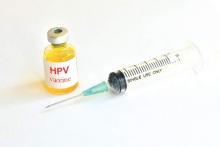
Two-dose HPV vaccines at younger age are noninferior to standard three-dose vaccine
“The licensed vaccines HPV-16/18 AS04–adjuvanted (Cervarix; GSK Vaccines) and HPV-6/11/16/18 (Gardasil; Merck) were first approved as regimen including three doses (3D) with administration over a 6-month period,” reported Thanyawee Puthanakit, MD, of Chulalongkorn University in Bangkok, and her associates. “Reduced dose schedules could make vaccination easier to implement and more affordable, creating the potential for higher vaccination coverage and improved cervical cancer protection (J Infect Dis. 2016;214:525-36).”
The investigators enrolled girls aged 9-25 years starting in June 2011, from 33 clinical sites in Canada, Germany, Italy, Taiwan, and Thailand. The study lasted through January 2013, during which time 1,447 girls were enrolled and 1,428 were randomized into one of three cohorts: one receiving the two-dose, 6-month regimen; one receiving the two-dose, 12-month regime; and one receiving the three-dose, 6-month regimen. In the former two cohorts, girls aged 9-14 years would receive their doses at 0 months and either 6 or 12 months, depending on the cohort. In the three-dose regimen, girls aged 15-25 years received doses at 0, 1, and 6 months. At enrollment, 550 girls were randomized into the two-dose, 6-month regimen; 415 girls into the two-dose, 12-month regimen; and 482 girls into the three-dose, 6-month regimen. Follow-up occurred at 1 month after they received the final dose for all three cohorts.
Ultimately, 534 girls in the two-dose, 6-month cohort; 394 in the two-dose, 12-month cohort; and 427 in the three-dose, 6-month cohort completed the study through the 1-month follow-up. Serologic testing indicated that girls in both the two-dose cohorts were not immunologically worse off than those who were in the three-dose cohort, with seroconversion differences of 0.00 when comparing HPV-16 and HPV-18 antibodies between any two of the three regimens. Furthermore, geometric mean antibody titer ratios for HPV-16 and HPV-18 were 1.09 (95% confidence interval, 0.97-1.22) and 0.85 (95% CI, 0.76-0.95) for the 6-month two-dose regimen vs. three-dose, and 0.89 (95% CI, 0.79-1.01) and 0.75 (95% CI, 0.67-0.85) for 12-month two-dose regimen vs.three-dose, indicating little statistically significant difference.
“The flexibility around the timing of the second dose, giving the option of annual vaccination over 2 consecutive years, is an added benefit [because] reduced dose schedules of HPV vaccines may facilitate vaccination implementation and reduce cost, allowing for higher vaccination coverage and potentially more girls being protected from cervical cancer,” the investigators noted.
The study was supported by GlaxoSmithKline Biologicals SA. Dr. Puthanakit received a grant from GSK through her institution. Some of her many coauthors disclosed relationships with GSK and/or other pharmaceutical companies.
FROM THE JOURNAL OF INFECTIOUS DISEASES
Key clinical point: Girls receiving 2-dose HPV-16/18 AS04-adjuvanted vaccine at age 9-14 years over a 6- or 12-month timespan appear to be equally protected against HPV than those receiving 3-dose HPV vaccine schedules at age 15-25 years.
Major finding: Geometric mean antibody titer ratios for HPV-16 and HPV-18 were 1.09 (95% CI, 0.97-1.22) and 0.85 (95% CI, 0.76-0.95) for 6-month two-dose regimen vs. three-dose, and 0.89 (95% CI 0.79-1.01) and 0.75 (95% CI, 0.67-0.85) for 12-month two-dose regimen vs. three-dose.
Data source: A randomized, open trial of 1,447 vaccinated girls aged 9-25 years.
Disclosures: Study supported by GlaxoSmithKline Biologicals SA. Dr. Puthanakit received a grant from GSK through her institution. Some of her many coauthors disclosed relationships with GSK and/or other pharmaceutical companies.
Blood viral RNA may indicate severity of MERS coronavirus clinical course
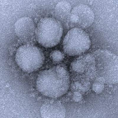
The presence of blood viral RNA in patients presenting with possible Middle East respiratory syndrome coronavirus (MERS-CoV) may be a very reliable indicator of the severity of the infection’s clinical course, according to a new study published in Emerging Infectious Diseases.
“Our study aimed to evaluate the diagnostic utility of blood specimens for MERS-CoV infection by using large numbers of patients with a single viral origin and to determine the relationship between blood viral detection and clinical characteristics,” wrote the authors, led by So Yeon Kim, MD, of the National Medical Center in Seoul, South Korea.
The investigators recruited 21 MERS-CoV patients within South Korea, all of whom had been previously diagnosed by the Korea Centers for Disease Control and Prevention via respiratory samples and were of “a single viral origin.” All subjects contributed ethylenediaminetetraacetic acid (EDTA)-treated whole blood and serum specimens, from which viral RNA was extracted (Emerg Infect Dis. 2016 Oct 15;22[10]. doi: 10.3201/eid2210.160218).
Viral RNA was detected in 6 of 21 whole blood samples and 6 of 21 serum samples at hospital admission. However, because two patients were viral positive in either specimen subtype of EDTA-treated whole blood or serum, the overall detection rate for the population was 7 of 21 (33%). Being positive for blood viral RNA at admission was found to be associated with a fever of higher than 37.5 °C (99.5 °F) on the date of sample collection (P = .007), being placed on mechanical ventilation at some point during the clinical course (P = .003), extracorporeal membrane oxygenation (P = .025), and death (P = .025).
However, “between the blood viral RNA-positive and -negative groups, we found no differences in age, duration from symptom onset to diagnosis of MERS-CoV infection, or an invasive procedure before the specimens were obtained,” the investigators noted.
The takeaway, the authors underscore, is that although early blood viral RNA presence may not be a useful diagnostic tool, it “might be a good prognostic indicator of severe outcome” due to its high association with worse clinical course.
The research was funded by the National Medical Center Research Institute.
FROM EMERGING INFECTIOUS DISEASES
Key clinical point: Checking for blood viral RNA at hospital admission may be a reliable indicator of the severity of MERS coronavirus infection clinical course.
Major finding: Blood viral RNA positivity at admission was associated with fever higher than 37.5 °C on the sampling date (P = .007), requirement for mechanical ventilation during the following clinical course (P = .003), extracorporeal membrane oxygenation (P = .025), and patient death (P = .025).
Data source: Prospective analysis of 21 patients with Middle East respiratory syndrome coronavirus (MERS-CoV).
Disclosures: The research was funded by the National Medical Center Research Institute.
Small Study: Placebo Bests Chondroitin, Glucosamine Combo for Knee Osteoarthritis
Combination therapy involving chondroitin sulfate and glucosamine sulfate proved no better than placebo for the management of pain and functional impairment brought on by knee osteoarthritis, in a double-blind, randomized trial.
“This is the first RCT sponsored by a pharmaceutical company to evaluate the efficacy of a combination of CS [chondroitin sulfate] plus GS [glucosamine sulfate] and including a [Data and Safety Monitoring Board] composed of independent experts charged with ensuring participant safety and accurate, bias-free data,” wrote the authors, led by Jorge A. Roman-Blas, MD, of Fundación Jiménez Díaz in Madrid (Arthritis Rheumatol. 2016 Jul 31. doi: 10.1002/art.39819).
Dr. Roman-Blas and his coinvestigators recruited patients with Kellgren-Lawrence stages II-III knee osteoarthritis from one orthopedic surgery and nine rheumatology centers across Spain. The study was split into two parts; the first part would have half of the patients complete the study in 6 months, followed by an interim analysis and evaluation of the data. If deemed okay by the Data and Safety Monitoring Board, the second part of the study would proceed, in which half of the patients would again complete the study in 6 months.
Ultimately, the study comprised 164 patients. They were randomized in a 1:1 ratio into cohorts receiving either a placebo or a regimen of 1,200 mg CS derived from bovine tracheal cartilage plus 1,500 mg of crystalline GS derived from crustaceans. The primary endpoint was reduction in pain from baseline evaluation, which was based on the visual analog scale (VAS) of 1-100 mm.
Patients in the combination therapy cohort had a lower average reduction in pain at 6 months than did those taking the placebo. In the modified intention-to-treat population, those taking CS+GS therapy had an average reduction of 11.8 ± 2.4 mm, or 19%, compared with baseline. On the other hand, those in the placebo cohort recorded a 20.5 ± 2.4 mm, or 33%, reduction in pain (P = .029). Based on this analysis, the trial was stopped early at 6 months to prevent “overexposure of additional patients to placebo treatment,” the investigators said.
However, pain measured by VAS did not differ between the groups in a per-protocol analysis. Furthermore, when taking Western Ontario and McMaster Universities Arthritis Index (WOMAC) scores into account, there was no significant difference in improvement between CS+GS therapy and placebo in pain and function, for both intention-to-treat and per-protocol subjects.
Although placebo was more effective in pain reduction in the modified intent-to-treat analysis than was CS plus GS combination treatment, the effect size of placebo was small and the effect size of CS plus GS in this study was smaller than in previous studies, the investigators noted. “These findings may be related to the fact that GS may interfere with the absorption of CS and therefore reduce its local effect. Eventually, our patients may have had less severe pain than those in other studies, which would justify a lesser effect after the pharmacological intervention,” the authors wrote. “Furthermore, when the main outcome studied is particularly subjective, the placebo effect can overcome the effect of the active drug.”
Tedec Meiji Farma SA of Madrid funded the study. Dr. Roman-Blas and his coauthors did not report any relevant financial disclosures.
FROM ARTHRITIS & RHEUMATOLOGY
Small study: Placebo bests chondroitin, glucosamine combo for knee osteoarthritis
Combination therapy involving chondroitin sulfate and glucosamine sulfate proved no better than placebo for the management of pain and functional impairment brought on by knee osteoarthritis, in a double-blind, randomized trial.
“This is the first RCT sponsored by a pharmaceutical company to evaluate the efficacy of a combination of CS [chondroitin sulfate] plus GS [glucosamine sulfate] and including a [Data and Safety Monitoring Board] composed of independent experts charged with ensuring participant safety and accurate, bias-free data,” wrote the authors, led by Jorge A. Roman-Blas, MD, of Fundación Jiménez Díaz in Madrid (Arthritis Rheumatol. 2016 Jul 31. doi: 10.1002/art.39819).
Dr. Roman-Blas and his coinvestigators recruited patients with Kellgren-Lawrence stages II-III knee osteoarthritis from one orthopedic surgery and nine rheumatology centers across Spain. The study was split into two parts; the first part would have half of the patients complete the study in 6 months, followed by an interim analysis and evaluation of the data. If deemed okay by the Data and Safety Monitoring Board, the second part of the study would proceed, in which half of the patients would again complete the study in 6 months.
Ultimately, the study comprised 164 patients. They were randomized in a 1:1 ratio into cohorts receiving either a placebo or a regimen of 1,200 mg CS derived from bovine tracheal cartilage plus 1,500 mg of crystalline GS derived from crustaceans. The primary endpoint was reduction in pain from baseline evaluation, which was based on the visual analog scale (VAS) of 1-100 mm.
Patients in the combination therapy cohort had a lower average reduction in pain at 6 months than did those taking the placebo. In the modified intention-to-treat population, those taking CS+GS therapy had an average reduction of 11.8 ± 2.4 mm, or 19%, compared with baseline. On the other hand, those in the placebo cohort recorded a 20.5 ± 2.4 mm, or 33%, reduction in pain (P = .029). Based on this analysis, the trial was stopped early at 6 months to prevent “overexposure of additional patients to placebo treatment,” the investigators said.
However, pain measured by VAS did not differ between the groups in a per-protocol analysis. Furthermore, when taking Western Ontario and McMaster Universities Arthritis Index (WOMAC) scores into account, there was no significant difference in improvement between CS+GS therapy and placebo in pain and function, for both intention-to-treat and per-protocol subjects.
Although placebo was more effective in pain reduction in the modified intent-to-treat analysis than was CS plus GS combination treatment, the effect size of placebo was small and the effect size of CS plus GS in this study was smaller than in previous studies, the investigators noted. “These findings may be related to the fact that GS may interfere with the absorption of CS and therefore reduce its local effect. Eventually, our patients may have had less severe pain than those in other studies, which would justify a lesser effect after the pharmacological intervention,” the authors wrote. “Furthermore, when the main outcome studied is particularly subjective, the placebo effect can overcome the effect of the active drug.”
Tedec Meiji Farma SA of Madrid funded the study. Dr. Roman-Blas and his coauthors did not report any relevant financial disclosures.
FROM ARTHRITIS & RHEUMATOLOGY
Key clinical point: Chondroitin sulfate plus glucosamine sulfate is ineffective at reducing pain and functional impairment in patients with knee osteoarthritis.
Major finding: CS plus GS treatment registered a 19% reduction in joint pain in the modified intention-to-treat population, versus the 33% reduction brought on by the placebo (P = .029).
Data source: A multicenter, randomized, double-blind, placebo-controlled study of 164 patients with Kellgren-Lawrence stages II-III knee osteoarthritis.
Disclosures: Tedec Meiji Farma SA of Madrid funded the study. The authors reported no relevant financial disclosures.
MCR-1 gene a growing concern for antibiotic resistance
The MCR-1 gene is quickly emerging as a powerful roadblock in the fight against antibiotic-resistant bacteria, according to experts from the Centers for Disease Control and Prevention.
Alex J. Kallen, MD, of the CDC in Atlanta, said in an Aug. 2 webinar – cohosted by CDC and the Partnership for Quality Care – that the CDC is closely monitoring the emergence of antibiotic resistant bacteria in the United States. The prevailing message of Dr. Kallen and his CDC colleague Arjun Srinivasan, MD was that the importance of the MCR-1 gene should not be underestimated.
First discovered in a human patient last year, the presence of the MCR-1 gene makes bacteria resistant to colistin, an antibiotic used often as a last resort to “treat patients with multidrug-resistant infections,” according to the CDC. Because the MCR-1 gene exists on a plasmid, or a small piece of DNA, it is easily transferable among bacteria, making it a problem for health care providers treating a patient infected with bacteria that has the gene.
“As of this year, all state and some local health departments will be funded to respond to [antibiotic resistance] threats, including emerging threats like [MCR-1], within their jurisdiction,” Dr. Kallen said. This funding could be used to support technical assistance, contact investigations, and laboratory testing, among other things.
In order to help prevent the spread of MCR-1 and mitigate cases of novel antimicrobial resistance, health care providers and facilities should institute recommended intensive care precautions as soon as resistance is identified, along with alerting their local public health office. Isolates should be saved, and prospective and retrospective surveillance should be implemented to “identify isolates with similar phenotypes.” If an infected patient is being transferred to another facility, that facility should be notified ahead of time about the patient and what protocols to follow.
Because antibiotic-resistant organisms do not spread through the air like influenza, the risk that these organisms pose to health care workers is relatively low, Dr. Srinivasan explained. However, he stressed that a multifaceted, team-based approach to antibiotic stewardship and decontamination of health care facilities is absolutely necessary to achieve the best results for both patients and staff.
“We are now beginning to recognize that the contamination of surfaces and items in our health care environment is increasingly a problem in the transmission of these drug-resistant organisms,” Dr. Srinivasan explained. He said that, given the complexity of a typical health care environment, such as a hospital room or operating theater, it’s perhaps not so surprising that keeping everything clean is not the top priority.
In addition to making sure hospital rooms and other areas are properly cleaned, simple things like washing hands and keeping surfaces clean are just as important. Dr. Srinivasan pointed to a 2006 study by Philip C. Carling, MD, and his associates which showed that educational interventions can lead to substantial increases in hygiene and cleanliness, and that training of new staff is also important. Furthermore, giving staff enough time to properly clean rooms could significantly contribute to curtailing MCR-1 bacteria from spreading, he said.
“The other thing to keep in mind is that if we lose effective therapy, if we lose antibiotic therapy, the infections that are currently very treatable and are seldom deadly could again become very, very serious threats to life,” Dr. Srinivasan warned, specifically citing Escherichia coli, which is the leading cause of urinary tract infections. If community strains of E.coli become resistant to typical antibiotic treatment, these cases could become “difficult, if not impossible to treat.”
According to data shared by Dr. Srinivasan, in 2013 there were 2,049,422 illnesses in the United States attributed to antibiotic resistance and 23,000 fatalities.
AGA resource
The AGA Center for Gut Microbiome Research and Education was created to serve as a virtual “home” for AGA activities related to the gut microbiome. The center is focused on advancing gut microbiome research, educating AGA members and other stakeholders on the latest microbiome breakthroughs, and working with FDA and others to ensure that emerging microbiome-based treatments are safe and appropriately evaluated. Learn more here.
MCR-1 gene a growing concern for antibiotic resistance
The MCR-1 gene is quickly emerging as a powerful roadblock in the fight against antibiotic-resistant bacteria, according to experts from the Centers for Disease Control and Prevention.
Alex J. Kallen, MD, of the CDC in Atlanta, said in an Aug. 2 webinar – cohosted by CDC and the Partnership for Quality Care – that the CDC is closely monitoring the emergence of antibiotic resistant bacteria in the United States. The prevailing message of Dr. Kallen and his CDC colleague Arjun Srinivasan, MD was that the importance of the MCR-1 gene should not be underestimated.
First discovered in a human patient last year, the presence of the MCR-1 gene makes bacteria resistant to colistin, an antibiotic used often as a last resort to “treat patients with multidrug-resistant infections,” according to the CDC. Because the MCR-1 gene exists on a plasmid, or a small piece of DNA, it is easily transferable among bacteria, making it a problem for health care providers treating a patient infected with bacteria that has the gene.
“As of this year, all state and some local health departments will be funded to respond to [antibiotic resistance] threats, including emerging threats like [MCR-1], within their jurisdiction,” Dr. Kallen said. This funding could be used to support technical assistance, contact investigations, and laboratory testing, among other things.
In order to help prevent the spread of MCR-1 and mitigate cases of novel antimicrobial resistance, health care providers and facilities should institute recommended intensive care precautions as soon as resistance is identified, along with alerting their local public health office. Isolates should be saved, and prospective and retrospective surveillance should be implemented to “identify isolates with similar phenotypes.” If an infected patient is being transferred to another facility, that facility should be notified ahead of time about the patient and what protocols to follow.
Because antibiotic-resistant organisms do not spread through the air like influenza, the risk that these organisms pose to health care workers is relatively low, Dr. Srinivasan explained. However, he stressed that a multifaceted, team-based approach to antibiotic stewardship and decontamination of health care facilities is absolutely necessary to achieve the best results for both patients and staff.
“We are now beginning to recognize that the contamination of surfaces and items in our health care environment is increasingly a problem in the transmission of these drug-resistant organisms,” Dr. Srinivasan explained. He said that, given the complexity of a typical health care environment, such as a hospital room or operating theater, it’s perhaps not so surprising that keeping everything clean is not the top priority.
In addition to making sure hospital rooms and other areas are properly cleaned, simple things like washing hands and keeping surfaces clean are just as important. Dr. Srinivasan pointed to a 2006 study by Philip C. Carling, MD, and his associates which showed that educational interventions can lead to substantial increases in hygiene and cleanliness, and that training of new staff is also important. Furthermore, giving staff enough time to properly clean rooms could significantly contribute to curtailing MCR-1 bacteria from spreading, he said.
“The other thing to keep in mind is that if we lose effective therapy, if we lose antibiotic therapy, the infections that are currently very treatable and are seldom deadly could again become very, very serious threats to life,” Dr. Srinivasan warned, specifically citing Escherichia coli, which is the leading cause of urinary tract infections. If community strains of E.coli become resistant to typical antibiotic treatment, these cases could become “difficult, if not impossible to treat.”
According to data shared by Dr. Srinivasan, in 2013 there were 2,049,422 illnesses in the United States attributed to antibiotic resistance and 23,000 fatalities.
FDA grants fast track status to volixibat
The Food and Drug Administration has granted fast track status to volixibat, an investigational treatment manufactured by Shire.
“The FDA’s fast track is a process designed to facilitate the development, and expedite the review, of drugs to treat serious conditions and fill an unmet medical need,” the Dublin-based pharmaceutical company explained in a statement. “However, it does not guarantee that the FDA will ultimately approve [volixibat] for NASH [nonalcoholic steatohepatitis] or the timing of any such approval.”
Volixibat, also known as SHP626, is meant to treat NASH with liver fibrosis in adult patients via an apical sodium-dependent bile acid transporter inhibitor, which is a protein that recycles bile acids from the intestine and into the liver. This orally administered, once-daily treatment would be the first ever treatment available for NASH.
“This fast track designation is further recognition of the critical need to develop new, effective therapeutic options for patients with this serious condition,” said Philip J. Vickers, PhD, head of research and development for Shire, in a statement.
Preclinical and phase I studies have already been completed, the results of which contributed to the FDA granting volixibat fast track status. A randomized, placebo-controlled, double-blind phase II trial is set to get underway shortly at centers in the United States, Canada, and the United Kingdom, which will examine safety, tolerability, and efficacy of a three-dose volixibat regimen administered over the course of 48 weeks.
UTIs not caused by E. coli more likely in certain children
Certain children are more highly predisposed to contracting a urinary tract infection caused by a pathogen other than Escherichia coli, which is typically the most common cause of UTIs, a study showed.
“It may be clinically important to predict which children have UTIs caused by organisms other than E. coli because these organisms differ in their patterns of antimicrobial susceptibility,” wrote the study authors led by Nader Shaikh, MD, of the University of Pittsburgh. “Furthermore, some guidelines have suggested that screening for vesicoureteral reflux (VUR) with a voiding cystourethrogram (VCUG) should, at least in part, be based on whether an organism other than E. coli is recovered,” they wrote.
Dr. Shaikh and his coinvestigators examined the medical records of children in the Randomized Intervention for Children With Vesicoureteral Reflux (RIVUR) trial and the Careful Urinary Tract Infection Evaluation (CUTIE), both of which were prospective multicenter studies. Children included in both studies were 2-71 months of age; RIVUR subjects had VUR grades 1-4 and presented with either a first or second febrile or symptomatic UTI, while CUTIE subjects presented with either their first or second UTI but not VUR (Ped Inf Dis J. 2016. doi:10.1097/INF.0000000000001301).
In total, 769 children from 19 centers were included from both studies, of which 703 (91%) were female and 596 (78%) were white. Nine percent of all the children had UTIs that were not caused by E. coli. Circumcised males had the highest odds ratio associated with non–E. coli UTIs, with an OR of 5.5 (95% CI, 1.18-17.1; P = .003). significantly higher than the 1.6 odds ratio for uncircumcised males (95% CI, 0.6-4.6; P = .35).
Hispanic children also had a higher risk (OR = 2.3; 95% CI, 1.1-4.6; P = .02) than either non-Hispanic children or females, which were reference cohorts. Other groups found to be at higher-than-normal risk for non–E. coli UTIs were children without fever (OR = 2.8; 95% CI, 1.2-6.6; P = .02) and children with VUR grade 3 or 4 (OR = 2.2; 95% CI, 1.2-4.1; P = .01).
While more than 90% of children’s UTIs were caused by E. coli, the most common pathogens causing UTIs, causative organisms in the other 70 children were Proteus species (21 children, 30%), Klebsiella species (16, 23%), Enterococcus species (14, 20%), Enterobacter species (8, 11%), and “other species” (11, 16%).
“Children with UTIs caused by organisms other than E. coli were twice as likely to have high-grade VUR (grade 3 and 4), which is consistent with prior studies,” Dr. Shaikh and his coauthors noted, adding, “the association between Hispanic ethnicity and non-E. coli pathogens is novel and may be due to differences in genes involved with susceptibility to UTIs.”
There were no disclosures or sources of funding provided.
In this study of almost 800 children in the Pittsburgh area, the investigators sought to identify children at risk for urinary tract infections that more likely would have a bacterial organism not susceptible to standard first-line empiric antibiotic treatment.
They found that circumcised males, children with grade 3-4 vesicoureteral reflux, Hispanic children, and children without fever were more likely to have a UTI caused by organisms other than Escherichia coli and, therefore, less likely to respond to standard first-line antibiotic therapy. These investigators are the preeminent authorities in UTI management for children, so their findings should be viewed in that light.
 |
Dr. Michael E. Pichichero |
The advance from the study is not a major one because all children with a suspected UTI should have a suitable culture specimen obtained before starting antibiotics, and the treatment choice continued or changed based on culture results. So really, the findings apply only to a decision about initial empiric treatment while awaiting culture results.
As a guide, if a clinician were to consider the diagnosis of UTI based on history, examination, and urinalysis, and the child was a circumcised male, a child with known grade 3 or 4 vesicoureteral reflux, Hispanic, or without fever, then the empiric antibiotic selected should be broader spectrum while awaiting urine culture results.
Michael E. Pichichero, MD, a specialist in pediatric infectious diseases, is director of the Research Institute, Rochester (N.Y.) General Hospital. He is also a pediatrician at Legacy Pediatrics in Rochester. Dr. Pichichero said he had no relevant financial disclosures.
In this study of almost 800 children in the Pittsburgh area, the investigators sought to identify children at risk for urinary tract infections that more likely would have a bacterial organism not susceptible to standard first-line empiric antibiotic treatment.
They found that circumcised males, children with grade 3-4 vesicoureteral reflux, Hispanic children, and children without fever were more likely to have a UTI caused by organisms other than Escherichia coli and, therefore, less likely to respond to standard first-line antibiotic therapy. These investigators are the preeminent authorities in UTI management for children, so their findings should be viewed in that light.
 |
Dr. Michael E. Pichichero |
The advance from the study is not a major one because all children with a suspected UTI should have a suitable culture specimen obtained before starting antibiotics, and the treatment choice continued or changed based on culture results. So really, the findings apply only to a decision about initial empiric treatment while awaiting culture results.
As a guide, if a clinician were to consider the diagnosis of UTI based on history, examination, and urinalysis, and the child was a circumcised male, a child with known grade 3 or 4 vesicoureteral reflux, Hispanic, or without fever, then the empiric antibiotic selected should be broader spectrum while awaiting urine culture results.
Michael E. Pichichero, MD, a specialist in pediatric infectious diseases, is director of the Research Institute, Rochester (N.Y.) General Hospital. He is also a pediatrician at Legacy Pediatrics in Rochester. Dr. Pichichero said he had no relevant financial disclosures.
In this study of almost 800 children in the Pittsburgh area, the investigators sought to identify children at risk for urinary tract infections that more likely would have a bacterial organism not susceptible to standard first-line empiric antibiotic treatment.
They found that circumcised males, children with grade 3-4 vesicoureteral reflux, Hispanic children, and children without fever were more likely to have a UTI caused by organisms other than Escherichia coli and, therefore, less likely to respond to standard first-line antibiotic therapy. These investigators are the preeminent authorities in UTI management for children, so their findings should be viewed in that light.
 |
Dr. Michael E. Pichichero |
The advance from the study is not a major one because all children with a suspected UTI should have a suitable culture specimen obtained before starting antibiotics, and the treatment choice continued or changed based on culture results. So really, the findings apply only to a decision about initial empiric treatment while awaiting culture results.
As a guide, if a clinician were to consider the diagnosis of UTI based on history, examination, and urinalysis, and the child was a circumcised male, a child with known grade 3 or 4 vesicoureteral reflux, Hispanic, or without fever, then the empiric antibiotic selected should be broader spectrum while awaiting urine culture results.
Michael E. Pichichero, MD, a specialist in pediatric infectious diseases, is director of the Research Institute, Rochester (N.Y.) General Hospital. He is also a pediatrician at Legacy Pediatrics in Rochester. Dr. Pichichero said he had no relevant financial disclosures.
Certain children are more highly predisposed to contracting a urinary tract infection caused by a pathogen other than Escherichia coli, which is typically the most common cause of UTIs, a study showed.
“It may be clinically important to predict which children have UTIs caused by organisms other than E. coli because these organisms differ in their patterns of antimicrobial susceptibility,” wrote the study authors led by Nader Shaikh, MD, of the University of Pittsburgh. “Furthermore, some guidelines have suggested that screening for vesicoureteral reflux (VUR) with a voiding cystourethrogram (VCUG) should, at least in part, be based on whether an organism other than E. coli is recovered,” they wrote.
Dr. Shaikh and his coinvestigators examined the medical records of children in the Randomized Intervention for Children With Vesicoureteral Reflux (RIVUR) trial and the Careful Urinary Tract Infection Evaluation (CUTIE), both of which were prospective multicenter studies. Children included in both studies were 2-71 months of age; RIVUR subjects had VUR grades 1-4 and presented with either a first or second febrile or symptomatic UTI, while CUTIE subjects presented with either their first or second UTI but not VUR (Ped Inf Dis J. 2016. doi:10.1097/INF.0000000000001301).
In total, 769 children from 19 centers were included from both studies, of which 703 (91%) were female and 596 (78%) were white. Nine percent of all the children had UTIs that were not caused by E. coli. Circumcised males had the highest odds ratio associated with non–E. coli UTIs, with an OR of 5.5 (95% CI, 1.18-17.1; P = .003). significantly higher than the 1.6 odds ratio for uncircumcised males (95% CI, 0.6-4.6; P = .35).
Hispanic children also had a higher risk (OR = 2.3; 95% CI, 1.1-4.6; P = .02) than either non-Hispanic children or females, which were reference cohorts. Other groups found to be at higher-than-normal risk for non–E. coli UTIs were children without fever (OR = 2.8; 95% CI, 1.2-6.6; P = .02) and children with VUR grade 3 or 4 (OR = 2.2; 95% CI, 1.2-4.1; P = .01).
While more than 90% of children’s UTIs were caused by E. coli, the most common pathogens causing UTIs, causative organisms in the other 70 children were Proteus species (21 children, 30%), Klebsiella species (16, 23%), Enterococcus species (14, 20%), Enterobacter species (8, 11%), and “other species” (11, 16%).
“Children with UTIs caused by organisms other than E. coli were twice as likely to have high-grade VUR (grade 3 and 4), which is consistent with prior studies,” Dr. Shaikh and his coauthors noted, adding, “the association between Hispanic ethnicity and non-E. coli pathogens is novel and may be due to differences in genes involved with susceptibility to UTIs.”
There were no disclosures or sources of funding provided.
FROM THE PEDIATRIC INFECTIOUS DISEASE JOURNAL
Key clinical point: Non–Escherichia coli urinary tract infections are more likely to occur in children who are uncircumcised, are Hispanic, have no fever, or have grade 3-4 vesicoureteral reflux.
Major finding: Circumcised males had an odds ratio of 5.5 (95% CI, 1.8-17.1; P = .003) of infection by pathogens other than E. coli; the odds ratio for Hispanic children (OR = 2.3; 95% CI, 1.1-4.6; P = .02), children without fever (OR = 2.8; 95% CI, 1.2-6.6; P = .02), and children with grade 3-4 VUR (OR = 2.2; 95% CI, 1.2-4.1; P = .01) also were relatively high.
Data source: A review of data from two prospective multicenter studies involving 769 children with a UTI aged 2-71 months .
Disclosures: Funding sources and individual disclosures were not provided.
