User login
Once-monthly aripiprazole drastically cut psychiatric hospitalizations
BERLIN– Switching patients with schizophrenia from various oral antipsychotics to long-acting injectable aripiprazole at 400 mg once monthly resulted in a dramatic reduction in their psychiatric hospitalization rate in a phase IIIb study.
This robust 90% reduction in psychiatric hospitalizations during the first 3 months on long-acting aripiprazole, compared with participants’ last 3 months prior to making the switch, suggests that injectable aripiprazole might offer significant cost savings to the health care system, Dr. Timothy S. Peters-Strickland observed at the annual congress of the European College of Neuropsychopharmacology.
He presented the findings of a phase IIIb, multicenter, open-label, mirror-image clinical trial involving 433 schizophrenia patients. The study, conducted in a naturalistic community setting, was dubbed a mirror-image study, because it retrospectively looked back at the psychiatric hospitalization rate of participants when they were on oral antipsychotics as well as conducting a prospective assessment of participants’ psychiatric hospitalizations after they made the switch to once-monthly injectable aripiprazole. Thus, patients served as their own controls.
Of note, at enrollment all participants required a change away from their current oral antipsychotic for various reasons, including lack of efficacy, side effects, or poor compliance. This might well have been a factor in some of the psychiatric hospitalizations occurring during the period before the switch.
As part of the study protocol, all participants on an oral antipsychotic other than aripiprazole (Abilify) were converted to oral aripiprazole over the course of 1-4 weeks of cross titration before making the switch to injectable aripiprazole. The duration of the cross-titration period was left to physician discretion.
A key observation regarding this cross-titration phase is that longer proved to be better. That is, the rate of discontinuation because of adverse events during the conversion process was substantially higher – 10.4% – in patients whose cross-titration period lasted 1 week than the 2.9% rate in patients whose cross-titration continued for more than 1 week and up to 4 weeks before introducing long-acting injectable therapy, reported Dr. Peters-Strickland of Otsuka in Princeton, N.J.
The primary study endpoint was the psychiatric hospitalization rate during the last 3 months on oral antipsychotic therapy prior to cross titration, compared with the rate during the first 3 months after the switch to long-acting aripiprazole. The rate on oral therapy was 27.1%, an order of magnitude greater than the 2.7% rate once the same patients had made the switch.
A hefty 32% of patients discontinued long-acting aripiprazole within the first 6 months. A total of 9% of subjects did so because of adverse events, 8% were lost to follow-up, and 2% quit because of lack of efficacy. The most common treatment-emergent adverse events were insomnia, akathisia, and emergence of a psychotic disorder.
The psychiatric hospitalization rate during the last 6 months that patients were on oral antipsychotic therapy was 38.1%, compared with an 8.8% rate during the first 6 months after they started on injectable aripiprazole, whether they stayed on the long-acting medication for the full 6 months or not.
This mirror-image study was sponsored by Lundbeck and Otsuka. Dr. Peters-Strickland is an Otsuka employee.
AT THE ECNP CONGRESS
Key clinical point: The psychiatric hospitalization rate was reduced 10-fold after schizophrenia patients were switched from various oral antipsychotic agents to long-acting injectable aripiprazole.
Major finding: The psychiatric hospitalization rate during the last 3 months patients were on oral antipsychotic therapy was 27%, compared with a 2.7% rate in the 3 months following a switch to long-acting intramuscular aripiprazole.
Data source: This prospective, open-label, mirror-image, multicenter study included 433 patients with schizophrenia who were switched from oral antipsychotic therapy with a variety of agents to once-monthly injectable aripiprazole at 400 mg.
Disclosures: This study was supported by H. Lundbeck and Otsuka. The presenter is an Otsuka employee.
Lurasidone favorably affects metabolic syndrome risk
BERLIN – The prevalence of metabolic syndrome remained stable in outpatients with schizophrenia during 12 months of double-blind treatment with lurasidone, while the rate climbed significantly in those randomized to risperidone in a multicenter head-to head comparative study.
The study included 611 outpatients with clinically stable schizophrenia who were randomized double-blind 2:1 to 12 months of flexibly dosed oral lurasidone (Latuda) at 37-111 mg/day or to oral risperidone (Risperdal) at 2-6 mg/day.
The prevalence of metabolic syndrome at enrollment as defined by International Diabetes Federation criteria was 32.5% in the lurasidone group and virtually identical at 32.7% in the risperidone group. After 1 year of double-blind treatment, 37% of those with baseline metabolic syndrome in the lurasidone group no longer met criteria for the metabolic derangement, compared with a 28% metabolic syndrome reversal rate in the risperidone group, Dr. John W. Newcomer reported at the annual congress of the European College of Neuropsychopharmacology.
Among subjects without metabolic syndrome at baseline, the incidence of new-onset metabolic syndrome during 12 months of therapy was 16% with lurasidone, compared with 27% in those on risperidone. Thus, the 1-year on-treatment risk of developing metabolic syndrome was reduced by 48% in patients treated with lurasidone – as compared to risperidone-treated patients – observed Dr. Newcomer, a psychiatrist who serves as executive vice dean and professor of clinical biomedical science at the Florida Atlantic University College of Medicine in Boca Raton.
Patients on risperidone gained a mean of 2.6 kg over the course of the year, and those on lurasidone lost 0.9 kg. Mean waist circumference grew by 2.6 cm in the risperidone group, compared with a 0.4-cm decrease in the lurasidone group. Blood pressure remained unchanged over time in both treatment arms.
At the end of 12 months of double-blind therapy, 223 study participants continued on for a 6-month open-label extension phase on flexibly dosed lurasidone. Among patients who had been on 12 months of double-blind risperidone, the prevalence of metabolic syndrome went from 42.9% at the outset of the double-blind phase to 48.4% 12 months later, thereafter dropping to 38.5% after 6 months of open-label lurasidone. During their 6 months on lurasidone, the patients formerly on risperidone lost a mean of 2.9 kg in body weight and 1.6 cm in waist circumference.
Meanwhile, among patients on lurasidone for the entire 18 months, the prevalence of metabolic syndrome decreased from 33.3% at enrollment to 26.6% after 18 months of continuous treatment. This benefit was driven by a mean 1.7-kg weight loss and a 1.5-cm reduction in waist circumference.
This head-to-head comparative study was sponsored by Sunovion Pharmaceuticals and Takeda Pharmaceuticals. Dr. Newcomer reported having served as an independent scientific member on data safety monitoring boards for studies sponsored by a handful of other companies while having no financial relationship with the cosponsors of the current study.
AT THE ECNP CONGRESS
Key clinical point: Lurasidone was associated with a significantly lower metabolic syndrome rate than risperidone in schizophrenia outpatients.
Major finding: Among 157 patients without metabolic syndrome at baseline who were randomized to once-daily lurasidone or risperidone in a head-to-head comparative study, the risk of developing metabolic syndrome over 12 months of therapy was 48% less in the lurasidone group.
Data source: This was a post hoc analysis of a double-blind, multicenter study in which 611 clinically stable outpatients with schizophrenia were randomized 2:1 to flexibly dosed lurasidone or risperidone, following which 174 continued on for a 6-month open-label extension phase.
Disclosures: The study was sponsored by Sunovion Pharmaceuticals and Takeda Pharmaceuticals. Dr. Newcomer reported having no relevant financial relationships.
Phase III outcomes bode well for novel hyperkalemia therapies
LAS VEGAS – Positive phase III results for two novel oral therapies for hyperkalemia received an enthusiastic reception at the annual meeting of the Heart Failure Society of America.
That’s because even though spironolactone and eplerenone (Inspra) have a well-established mortality benefit and a class I indication for the treatment of heart failure with reduced ejection fraction, these renin-angiotensin-aldosterone system (RAAS) inhibitors and mineralocorticoid receptor antagonists remain greatly underutilized on account of their associated substantial risk of hyperkalemia.
“I think the availability of new and safe therapies to treat hyperkalemia and maintain patients on a RAAS inhibitor opens up the possibility of further reductions of cardiovascular events and reductions in health care costs in these very high-risk patients,” said Dr. Bertram Pitt, professor of internal medicine at the University of Michigan, Ann Arbor.
The data we have so far is quite promising,” agreed Dr. Gregg C. Fonarow. “There is a large and unmet need for better treatments for hyperkalemia. Many patients end up on RAAS inhibitor doses below those identified in randomized controlled trials and the major guidelines as the target dose.”
“Moreover, I’m impressed that in the United States we find that two-thirds of patients who have a class I recommendation for a RAAS inhibitor according to the guidelines are not being treated with any dose of these agents at all. So I see a large potential benefit in removing that concern and providing a reliable way of greatly reducing the risk of hyperkalemia. That would really meet a very important unmet need,” according to Dr. Fonarow, professor of medicine, director of the Ahmanson-UCLA Cardiomyopathy Center, and cochief of the cardiology division at the University of California, Los Angeles.
The sole approved agent at present for treating hyperkalemia – sodium polystyrene sulfonate (Kayexalate) – is problematic. Approved by the Food and Drug Administration back in 1958 based upon weak data, this agent is nonselective, unpopular, and poorly tolerated, with loose stools or diarrhea a common complaint.
The two new agents with positive phase III results reported at the meeting are patiromer and sodium zirconium cyclosilicate, also known as ZS-9. Patiromer is a nonabsorbed polymer that exchanges calcium for potassium. Its site of action is the lumen of the colon, where potassium concentration is highest. ZS-9 is a highly stable, inert, insoluble, nonabsorbed, inorganic polymer whose selectivity for potassium is 80- to 125-fold greater than that of sodium polystyrene sulfonate.
Patiromer
Dr. Pitt presented a prespecified subgroup analysis of the pivotal, multinational, phase III randomized trial of patiromer. This analysis, intended for an audience of heart failure specialists, was restricted to patients with chronic kidney disease and hyperkalemia who were on a RAAS inhibitor. In all, 102 of them had heart failure; 141 did not. Chronic kidney disease was severe in 45% of subjects, as evident in their estimated glomerular filtration rate of less than 30 mL/min.
Mean serum potassium dropped by about 1 mEq/L over the course of 4 weeks of therapy, regardless of whether a patient had heart failure or not. The benefit occurred quickly, with a mean drop of 0.5 mEq/L seen during the first 3 days of therapy. At the 4-week mark, three-quarters of patients had a normal-range serum potassium of 3.8 mEq/L to less than 5.1 mEq/L. They were then randomized single-blind to continued patiromer or a switch to placebo for another 4 weeks. Serum potassium climbed in placebo-treated controls by an average of 0.8 mEq/L while remaining unchanged in patients on patiromer. Fifty-two percent of heart failure patients on placebo developed hyperkalemia as defined by a serum potassium level of 5.5 mEq/L or more, compared with 8% on patiromer.
The safety profile of patiromer was essentially that of placebo in the pivotal trial, with two exceptions: 3% of patients on active therapy developed mild, transient hypokalemia which was easily corrected, and 7% experienced mild nausea or diarrhea, Dr. Pitt reported.
Relypsa, the company developing the drug, announced that it submitted a New Drug Application to the Food and Drug Administration for patiromer in October, seeking an indication for hyperkalemia.
ZS-9
The ZS-9 phase III study involved 753 hyperkalemic patients, making it the largest-ever trial of a hyperkalemia therapy. Dr. Mohamed El-Shahawy presented a prespecified subgroup analysis focusing on the 300 participants with heart failure, although he noted that the results were no different in the larger group without heart failure.
During the acute phase of the study, when participants were randomized double-blind to one of four t.i.d. doses of ZS-9 or placebo, serum potassium fell in patients on ZS-9 in a dose-dependent fashion. A reduction in serum potassium was observed as early as 1 hour after the first dose. In the highest-dose group, at 10 g three times daily, mean serum potassium fell from 5.3 mEq/L to 4.5 mEq/L.
In the subsequent extended phase of the study, patients on ZS-9 t.i.d. in the acute phase were rerandomized to once-daily ZS-9 or placebo for 12 days. Patients on ZS-9 at 5 g or 10 g once daily maintained normokalemia, while those switched to placebo became hyperkalemic once again by day 12, according to Dr. El-Shahawy of the Academic Medical Research Institute, Los Angeles.
The side-effect profile of ZS-9 was similar to that of placebo, with no significant hypokalemia or GI side effects being noted, he added.
A large, long-term study of ZS-9 for maintenance of normokalemia is ongoing.
Dr. Fonarow noted that another, entirely different approach to reducing the risk of hyperkalemia involves the development of novel, nonsteroidal, cardioselective mineralocorticoid receptor antagonists.
He cited a promising phase II study of one such agent, known for now as BAY 94-8862. That study was conducted in patients with heart failure and mild to moderate chronic kidney disease. Patients assigned to the novel agent had significantly smaller rises in serum potassium than those randomized to spironolactone. Yet BAY 94-8862 reduced levels of B-type natriuretic peptide, amino-terminal brain natriuretic peptide, and albuminuria at least as much as did spironolactone, and with significantly lower rates of worsening renal failure (Eur. Heart J. 2013;34:2453-63).
Once new and better therapies for hyperkalemia enter clinical practice, Dr. Fonarow said, the priorities will be to get patients on RAAS inhibitor therapy who should be on it but now aren’t, as well as to augment dosing up to target levels in those now on suboptimal doses. But access to improved treatments for hyperkalemia also opens the door to another intriguing possibility.
“Could the ability to raise doses of RAAS inhibitors even higher than current target doses without causing hyperkalemia further enhance remodeling and improve clinical outcomes? That could indeed be the case. It’s something we’ll want to test in prospective trials,” he said.
Dr. Fonarow reported serving as a consultant to and/or advisory board member for Medtronic, Novartis, Johnson & Johnson, Bayer, and Amgen. Dr. El-Shahawy is on an advisory board for ZS Pharma and has received research grants from that company as well as Amgen, GlaxoSmithKline, Celgene, and Abbvie. Dr. Pitt reported serving as a consultant to Pfizer, Bayer, Lilly, Relypsa, and Sarfez.
EXPERT ANALYSIS FROM THE HFSA ANNUAL SCIENTIFIC MEETING
Who will get cancer treatment-induced cardiotoxicity?
LAS VEGAS – The search is on for predictors of which cancer patients will experience treatment-induced cardiotoxicity, and an initial report from the PREDICT study has identified several.
One predictor is the patient’s type of cancer. In PREDICT, patients with lymphoma had a twofold greater risk of developing treatment-related cardiotoxicity, compared with those with breast cancer. Moreover, those with a cancer diagnosis other than lymphoma or breast cancer had a fivefold greater risk than breast cancer patients, Dr. Daniel J. Lenihan reported at the annual meeting of the Heart Failure Society of America.

“It’s important to know that many of the studies on cardiotoxicity risk have been done in breast cancer patients. There are a lot of other cancer patients out there,” observed Dr. Lenihan, professor of medicine and director of clinical research in the division of cardiovascular medicine at Vanderbilt University in Nashville.
PREDICT is a prospective, community-based study of 597 cancer patients undergoing anthracycline-based chemotherapy in 24 community oncology programs. It is primarily a study of the effectiveness of using cardiac biomarkers to predict cardiotoxicity, along with an analysis of the results of various forms of treatment of the cardiotoxicity.
During up to 12 months of follow-up 11% of PREDICT participants experienced a cardiac event, most commonly symptomatic heart failure or a greater than 10% drop in left ventricular ejection fraction, which took a patient from normal range to below normal. Another impressive finding was the substantial burden of conventional cardiovascular risk factors present at baseline in patients scheduled for anthracycline-based chemotherapy. In a multivariate logistic regression analysis, the higher a cancer patient’s cardiovascular risk factor level, the greater the likelihood of chemotherapy-related cardiotoxicity.
A baseline B-type natriuretic peptide (BNP) level in excess of 100 pg/mL was a powerful predictor of a chemotherapy-related cardiac event, with an associated 2.1-fold increased risk. As a predictor of cardiotoxicity during the study period, baseline BNP had a sensitivity of 35%, a specificity of 85%, a positive predictive value of 22%, and – most importantly – a negative predictive value of 92%, according to Dr. Lenihan.
Similarly, using as cutoffs either a baseline BNP greater than 100 pg/mL or a troponin greater than 0.05 ng/mL had a sensitivity of 60%, a specificity of 50%, a positive predictive value of 13%, and a negative predictive value of 91%, he continued.
Myocardial imaging as a tool for predicting which patients will develop cardiotoxicity during or after cancer therapy is another active area of investigation. Other investigators have shown that myocardial strain imaging holds considerable promise (J. Am. Coll. Cardiol. 2014;63:2751-68); however, it’s not terribly practical because many echocardiography laboratories balk at the idea of routinely performing serial strain imaging studies in all cancer patients, Dr. Lenihan said.
Practical predictors of increased risk for cancer therapy-related cardiotoxicity are sorely needed in order to identify candidates for prophylaxis with an agent such as dexrazoxane, which has been shown in a meta-analysis to reduce the risk of clinical or subclinical heart failure by 71% (Cochrane Database Syst. Review 2008; April 16:CD003917).
Another promising preventive approach was displayed in the Spanish OVERCOME trial, involving 90 patients undergoing intensive chemotherapy for malignant hemopathies. Those randomized to combined prophylaxis with enalapril plus carvedilol had a 6.7% rate of the composite endpoint of death, heart failure, or an LVEF below 45% at 6 months, compared with 24.4% in controls (J. Am. Coll. Cardiol. 2013;61:2355-62).
Also, predictors of increased risk are helpful in identifying cancer therapy–related cardiotoxicity early in its course, when aggressive treatment with standard heart failure medications such as beta blockers and ACE inhibitors is most likely to be beneficial.
“Everybody in cardiology is used to the concept that time is muscle: don’t let ischemia persist. Have a strategy to resolve it as soon as possible. That paradigm really can also apply to chemotherapy-related injury: the longer you leave it alone, the more permanent it becomes. Being able to detect it at its earliest stage is critically important,” according to Dr. Lenihan.
He cited as an example a study of 201 consecutive patients with anthracycline-induced cardiomyopathy conducted at the European Institute of Oncology in Milan. The conventional dogma is that anthracycline-induced cardiomyopathy is typically permanent, but this study showed that’s not true.
When treatment with enalapril and carvedilol was initiated within the first couple of months following the end of chemotherapy, 64% of patients experienced complete recovery of their LVEF. When the heart failure medications were commenced 3-4 months after completing chemotherapy, the LVEF recovery rate dropped to 28%. No complete recovery of LVEF occurred in patients who began enalapril plus carvedilol after 6 months (J. Am. Coll. Cardiol. 2010;55:213-20).
“There is a real opportunity here to improve the care of cancer patients and prevent heart failure,” Dr. Lenihan concluded.
He reported receiving research support from Singulex, Millenium, and Acorda as well as serving as a consultant to Onyx and Roche.
LAS VEGAS – The search is on for predictors of which cancer patients will experience treatment-induced cardiotoxicity, and an initial report from the PREDICT study has identified several.
One predictor is the patient’s type of cancer. In PREDICT, patients with lymphoma had a twofold greater risk of developing treatment-related cardiotoxicity, compared with those with breast cancer. Moreover, those with a cancer diagnosis other than lymphoma or breast cancer had a fivefold greater risk than breast cancer patients, Dr. Daniel J. Lenihan reported at the annual meeting of the Heart Failure Society of America.
“It’s important to know that many of the studies on cardiotoxicity risk have been done in breast cancer patients. There are a lot of other cancer patients out there,” observed Dr. Lenihan, professor of medicine and director of clinical research in the division of cardiovascular medicine at Vanderbilt University in Nashville.
PREDICT is a prospective, community-based study of 597 cancer patients undergoing anthracycline-based chemotherapy in 24 community oncology programs. It is primarily a study of the effectiveness of using cardiac biomarkers to predict cardiotoxicity, along with an analysis of the results of various forms of treatment of the cardiotoxicity.
During up to 12 months of follow-up 11% of PREDICT participants experienced a cardiac event, most commonly symptomatic heart failure or a greater than 10% drop in left ventricular ejection fraction, which took a patient from normal range to below normal. Another impressive finding was the substantial burden of conventional cardiovascular risk factors present at baseline in patients scheduled for anthracycline-based chemotherapy. In a multivariate logistic regression analysis, the higher a cancer patient’s cardiovascular risk factor level, the greater the likelihood of chemotherapy-related cardiotoxicity.
A baseline B-type natriuretic peptide (BNP) level in excess of 100 pg/mL was a powerful predictor of a chemotherapy-related cardiac event, with an associated 2.1-fold increased risk. As a predictor of cardiotoxicity during the study period, baseline BNP had a sensitivity of 35%, a specificity of 85%, a positive predictive value of 22%, and – most importantly – a negative predictive value of 92%, according to Dr. Lenihan.
Similarly, using as cutoffs either a baseline BNP greater than 100 pg/mL or a troponin greater than 0.05 ng/mL had a sensitivity of 60%, a specificity of 50%, a positive predictive value of 13%, and a negative predictive value of 91%, he continued.
Myocardial imaging as a tool for predicting which patients will develop cardiotoxicity during or after cancer therapy is another active area of investigation. Other investigators have shown that myocardial strain imaging holds considerable promise (J. Am. Coll. Cardiol. 2014;63:2751-68); however, it’s not terribly practical because many echocardiography laboratories balk at the idea of routinely performing serial strain imaging studies in all cancer patients, Dr. Lenihan said.
Practical predictors of increased risk for cancer therapy-related cardiotoxicity are sorely needed in order to identify candidates for prophylaxis with an agent such as dexrazoxane, which has been shown in a meta-analysis to reduce the risk of clinical or subclinical heart failure by 71% (Cochrane Database Syst. Review 2008; April 16:CD003917).
Another promising preventive approach was displayed in the Spanish OVERCOME trial, involving 90 patients undergoing intensive chemotherapy for malignant hemopathies. Those randomized to combined prophylaxis with enalapril plus carvedilol had a 6.7% rate of the composite endpoint of death, heart failure, or an LVEF below 45% at 6 months, compared with 24.4% in controls (J. Am. Coll. Cardiol. 2013;61:2355-62).
Also, predictors of increased risk are helpful in identifying cancer therapy–related cardiotoxicity early in its course, when aggressive treatment with standard heart failure medications such as beta blockers and ACE inhibitors is most likely to be beneficial.
“Everybody in cardiology is used to the concept that time is muscle: don’t let ischemia persist. Have a strategy to resolve it as soon as possible. That paradigm really can also apply to chemotherapy-related injury: the longer you leave it alone, the more permanent it becomes. Being able to detect it at its earliest stage is critically important,” according to Dr. Lenihan.
He cited as an example a study of 201 consecutive patients with anthracycline-induced cardiomyopathy conducted at the European Institute of Oncology in Milan. The conventional dogma is that anthracycline-induced cardiomyopathy is typically permanent, but this study showed that’s not true.
When treatment with enalapril and carvedilol was initiated within the first couple of months following the end of chemotherapy, 64% of patients experienced complete recovery of their LVEF. When the heart failure medications were commenced 3-4 months after completing chemotherapy, the LVEF recovery rate dropped to 28%. No complete recovery of LVEF occurred in patients who began enalapril plus carvedilol after 6 months (J. Am. Coll. Cardiol. 2010;55:213-20).
“There is a real opportunity here to improve the care of cancer patients and prevent heart failure,” Dr. Lenihan concluded.
He reported receiving research support from Singulex, Millenium, and Acorda as well as serving as a consultant to Onyx and Roche.
AT THE HFSA ANNUAL SCIENTIFIC MEETING
Key clinical point: An elevated baseline B-type natriuretic peptide or troponin level identifies cancer patients at increased risk of cardiotoxicity during anthracycline-based chemotherapy.
Major finding: Cancer patients with a BNP level greater than 100 pg/mL prior to going on anthracycline-based chemotherapy were 2.1-fold more likely to develop treatment-related cardiotoxicity.
Data source: The prospective PREDICT study includes roughly 600 patients undergoing anthracycline-based chemotherapy at 24 community cancer centers.
Disclosures: The study is sponsored by M.D. Anderson Cancer Center. The presenter reported serving as a consultant to Onyx and Roche.

Cancer treatment–induced cardiotoxicity greatly underappreciated
LAS VEGAS – When it comes to discussing the cardiotoxicity of cancer pharmacotherapies, it often seems that oncologists and cardiologists are on different planets.
“You will see wildly different toxicity numbers. … One trial will report 3% cardiotoxicity; another studying the same regimen will report 30%. It’s usually because they’re using vastly different definitions of toxicity,” Dr. Ronald M. Witteles said at the annual meeting of the Heart Failure Society of America.
“The typical clinical trial definition of a cardiac event in a cancer treatment trial is new symptomatic NYHA class III or IV heart failure or cardiac death; that’s it,” added Dr. Witteles, a cardiologist at Stanford (Calif.) University.
The consensus within the emerging field of cardio-oncology is that that simply isn’t good enough. A reasonable definition of cancer treatment–related cardiotoxicity must be far broader, encompassing a drop in left ventricular ejection fraction (LVEF) of greater than 10% from normal-range pretreatment to below the threshold of normal, regardless of whether that decline is accompanied by symptoms, as well as the development of an acute coronary syndrome or symptomatic arrhythmia, according to Dr. Daniel J. Lenihan, president of the International Cardioncology Society, an organization devoted to closer collaboration between the two medical specialties.
“Development of cardiomyopathy during cancer therapy is a lot more common than most people think,” emphasized Dr. Lenihan, professor of medicine and director of clinical research in the division of cardiovascular medicine at Vanderbilt University, Nashville, Tenn.
Dr. Witteles noted that the product labeling for doxorubicin quotes a preposterously low 1%-2% probability of developing impaired myocardial function at a total cumulative dose of 300 mg/m2, which is what’s typically used in treating lymphoma. That’s because the primary source of that estimate is 35-year-old data in which there was no routine measurement of LVEF and the diagnosis of impaired myocardial function was made by the treating oncologist on the basis of clinical signs and symptoms.
There are far better data available, but they’re not in the product label. Dr. Witteles cited as an example a more recent study featuring routine LVEF monitoring, where the incidence of a greater than 10% fall from normal to below normal at a cumulative dose of 300 mg/m2 wasn’t 1%-2%; it was 16% (Cancer 2003;97:2869-79).
He added that current evidence indicates that a fall in LVEF of more than 10%, taking a patient from normal range to below normal, occurs in 7%-8% of patients on an anthracycline such as doxorubicin at a cumulative dose of 240 mg/m2, as is typical in treating breast cancer. The incidence with trastuzumab (Herceptin) when prescribed after completion of anthracycline-based chemotherapy is about 25%; however, when trastuzumab is given in an anthracycline-free regimen, the rate drops to 9%-10%. A study by Dr. Witteles and his coworkers pegged the rate in cancer patients treated with sunitinib (Sutent) or other tyrosine kinase inhibitors at about 15% (JACC Heart Fail. 2013;1:72-8).
“Any way you slice it, that’s a whole lot of people,” he commented.
Dr. Lenihan reported receiving research support from Singulex, Millenium, and Acorda as well as serving as a consultant to Onyx and Roche. Dr. Witteles declared having no financial conflicts.
EXPERT ANALYSIS FROM THE HFSA ANNUAL SCIENTIFIC MEETING
Oral curcumin shown effective in psoriasis
AMSTERDAM – Oral curcumin proved safe and effective as adjunctive therapy in patients on topical corticosteroids for mild to moderate psoriasis vulgaris in a 12-week, randomized, placebo-controlled clinical trial.
This agent helps fill an unmet need in psoriasis, Dr. Emiliano Antiga observed in presenting the study findings at the annual congress of the European Academy of Dermatology and Venereology.

That’s because by far most of the action in the development of new treatments for psoriasis focuses on biologics and other extremely costly agents targeting patients at the moderate to severe end of the disease spectrum. But psoriasis is a chronic condition, and the many patients with milder disease require long-term therapies that are nontoxic and won’t break the bank. Enter curcumin.
“Oral curcumin is effective, safe, and it is cheap,” declared Dr. Antiga, a dermatologist at the University of Florence (Italy).
Moreover, it has a biologically plausible mechanism of benefit in psoriasis, as was demonstrated in his 60-patient randomized trial. Serum levels of the proinflammatory cytokine interleukin-22 were cut in half in the group assigned to 12 weeks of daily oral curcumin while remaining unchanged in the control group.
Curcumin is derived from turmeric, the dried rhizome of a plant, Curcuma longa. Turmeric is a yellowish Indian spice used in curries. But curcumin has long been used therapeutically in traditional Indian and Chinese medicine. Studies have shown that curcumin has antiproliferative, antiangiogenic, and anti-inflammatory effects. In a small study by other investigators, topical turmeric not only successfully cleared psoriasis lesions, it also suppressed phosphorylase kinase activity, which is important to keratinocyte proliferation (Br. J. Dermatol. 2000;143:937-49).
Dr. Antiga presented a study of 60 patients with mild to moderate psoriasis vulgaris as defined by a baseline median Psoriasis Area and Severity Index (PASI) score of 5.5 who were randomly assigned to 12 weeks of treatment with topical corticosteroids plus 3 g per day of oral curcumin or topical steroids plus placebo. The active-treatment capsules contained curcumin embedded in nanoparticle liposomes to enhance bioavailability.
Forty-nine patients completed the study. The primary endpoint was reduction in PASI values over 12 weeks. Both groups showed improvement – after all, the controls were on active treatment with topical steroids – but the change in PASI scores was significantly greater in the curcumin-treated patients.
Adverse events in the curcumin group were limited to one case of diarrhea. There was one case of nausea and one papular eruption in the control group.
Although IL-22 levels at 12 weeks were halved in the curcumin group and unchanged in controls, levels of the inflammatory cytokines IL-10 and -17 and transforming growth factor–beta remained unchanged in both groups over time.
Dr. Antiga reported having no financial conflicts regarding this study.
AMSTERDAM – Oral curcumin proved safe and effective as adjunctive therapy in patients on topical corticosteroids for mild to moderate psoriasis vulgaris in a 12-week, randomized, placebo-controlled clinical trial.
This agent helps fill an unmet need in psoriasis, Dr. Emiliano Antiga observed in presenting the study findings at the annual congress of the European Academy of Dermatology and Venereology.
That’s because by far most of the action in the development of new treatments for psoriasis focuses on biologics and other extremely costly agents targeting patients at the moderate to severe end of the disease spectrum. But psoriasis is a chronic condition, and the many patients with milder disease require long-term therapies that are nontoxic and won’t break the bank. Enter curcumin.
“Oral curcumin is effective, safe, and it is cheap,” declared Dr. Antiga, a dermatologist at the University of Florence (Italy).
Moreover, it has a biologically plausible mechanism of benefit in psoriasis, as was demonstrated in his 60-patient randomized trial. Serum levels of the proinflammatory cytokine interleukin-22 were cut in half in the group assigned to 12 weeks of daily oral curcumin while remaining unchanged in the control group.
Curcumin is derived from turmeric, the dried rhizome of a plant, Curcuma longa. Turmeric is a yellowish Indian spice used in curries. But curcumin has long been used therapeutically in traditional Indian and Chinese medicine. Studies have shown that curcumin has antiproliferative, antiangiogenic, and anti-inflammatory effects. In a small study by other investigators, topical turmeric not only successfully cleared psoriasis lesions, it also suppressed phosphorylase kinase activity, which is important to keratinocyte proliferation (Br. J. Dermatol. 2000;143:937-49).
Dr. Antiga presented a study of 60 patients with mild to moderate psoriasis vulgaris as defined by a baseline median Psoriasis Area and Severity Index (PASI) score of 5.5 who were randomly assigned to 12 weeks of treatment with topical corticosteroids plus 3 g per day of oral curcumin or topical steroids plus placebo. The active-treatment capsules contained curcumin embedded in nanoparticle liposomes to enhance bioavailability.
Forty-nine patients completed the study. The primary endpoint was reduction in PASI values over 12 weeks. Both groups showed improvement – after all, the controls were on active treatment with topical steroids – but the change in PASI scores was significantly greater in the curcumin-treated patients.
Adverse events in the curcumin group were limited to one case of diarrhea. There was one case of nausea and one papular eruption in the control group.
Although IL-22 levels at 12 weeks were halved in the curcumin group and unchanged in controls, levels of the inflammatory cytokines IL-10 and -17 and transforming growth factor–beta remained unchanged in both groups over time.
Dr. Antiga reported having no financial conflicts regarding this study.
AT THE EADV CONGRESS
Key clinical point: A common ingredient in Indian curry spice mixes is safe and effective as adjuvant therapy for mild-to-moderate psoriasis.
Major finding: Daily oral curcumin capsules plus topical corticosteroids resulted in a PASI 75 improvement rate of 48% compared with a 12% rate in patients who got topical steroids plus placebo.
Data source: A prospective randomized 12-week clinical trial involving 60 patients with mild to moderate psoriasis vulgaris.
Disclosures: The presenter reported no financial conflicts regarding this study, conducted free of commercial support.

Annual echo an option for cardiac allograft vasculopathy screening
LAS VEGAS – The experience at one major heart transplantation center indicates that annual screening dobutamine stress echocardiography to detect cardiac allograft vasculopathy renders annual coronary angiography unnecessary.
“This noninvasive method has very good specificity and is associated with a negative predictive value of 94%-97%. It can be used in our experience in lieu of annual invasive coronary angiography,” Dr. Jerry D. Estep declared at the annual meeting of the Heart Failure Society of America.

Cardiac allograft vasculopathy (CAV) is a unique, highly aggressive form of CAD. After 3 years post transplant it becomes the No. 1 cause of cardiac retransplantation and mortality. Guidelines recommend consideration of annual screening coronary angiography to detect it early to institute aggressive countermeasures. That’s the practice at most transplant centers.
However, at Houston Methodist Hospital, where Dr. Estep is medical director of the heart transplant and LVAD program, annual dobutamine stress echocardiography (DSE) is used instead. Because there is a scarcity of published data on this noninvasive alternative approach, he presented a retrospective study of the Houston transplant center’s experience over a recent 5-year period.
The study included 144 heart transplant recipients who underwent screening DSE for CAV annually for the first 4 years post transplant and coronary angiography at year 5.
During years 1-4, DSE detected CAV in 19% of patients. They didn’t differ in terms of baseline characteristics from those who remained free of this serious complication.
The good news: Ninety-four percent of patients with normal DSEs during years 1-4 had no CAV upon angiography at year 5. Moreover, the 5% who did have CAV at year 5 after earlier negative DSEs had mild to moderate disease.
The investigators documented the performance of annual screening DSE in predicting the development of major adverse cardiac events, defined as readmission for acute coronary syndrome, heart failure, revascularization, repeat heart transplantation, or cardiac death.
Dr. Estep reported having no financial conflicts regarding this study.
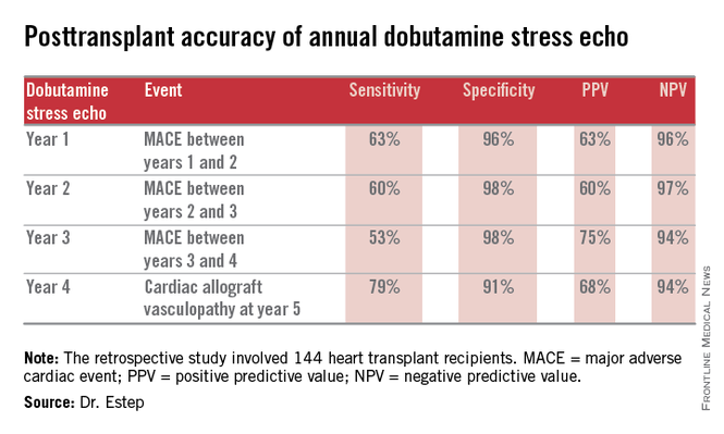
Dr. Hossein Almassi, FCCP, comments: Among solid organ transplants, cardiac transplant is rather unique in its need for invasive biopsy and angiography in following up the cardiac allograft. The search for noninvasive monitoring tools has been ongoing for a number of years. The report by the Houston group is a positive development in the right direction awaiting further confirmation by other cardiac transplant centers.
Dr. Almassi specializes in cardiothoracic surgery at the Medical College of Wisconsin in Milwaukee, Wisconsin.
LAS VEGAS – The experience at one major heart transplantation center indicates that annual screening dobutamine stress echocardiography to detect cardiac allograft vasculopathy renders annual coronary angiography unnecessary.
“This noninvasive method has very good specificity and is associated with a negative predictive value of 94%-97%. It can be used in our experience in lieu of annual invasive coronary angiography,” Dr. Jerry D. Estep declared at the annual meeting of the Heart Failure Society of America.
Cardiac allograft vasculopathy (CAV) is a unique, highly aggressive form of CAD. After 3 years post transplant it becomes the No. 1 cause of cardiac retransplantation and mortality. Guidelines recommend consideration of annual screening coronary angiography to detect it early to institute aggressive countermeasures. That’s the practice at most transplant centers.
However, at Houston Methodist Hospital, where Dr. Estep is medical director of the heart transplant and LVAD program, annual dobutamine stress echocardiography (DSE) is used instead. Because there is a scarcity of published data on this noninvasive alternative approach, he presented a retrospective study of the Houston transplant center’s experience over a recent 5-year period.
The study included 144 heart transplant recipients who underwent screening DSE for CAV annually for the first 4 years post transplant and coronary angiography at year 5.
During years 1-4, DSE detected CAV in 19% of patients. They didn’t differ in terms of baseline characteristics from those who remained free of this serious complication.
The good news: Ninety-four percent of patients with normal DSEs during years 1-4 had no CAV upon angiography at year 5. Moreover, the 5% who did have CAV at year 5 after earlier negative DSEs had mild to moderate disease.
The investigators documented the performance of annual screening DSE in predicting the development of major adverse cardiac events, defined as readmission for acute coronary syndrome, heart failure, revascularization, repeat heart transplantation, or cardiac death.
Dr. Estep reported having no financial conflicts regarding this study.
AT THE HFSA ANNUAL SCIENTIFIC MEETING
Key clinical point: Annual dobutamine stress echocardiography to screen heart transplant recipients for cardiac allograft vasculopathy is an excellent noninvasive alternative to the widely used practice of annual screening coronary angiography.
Major finding: Annual screening dobutamine stress echo during years 1-4 after heart transplant had a 94% negative predictive value for cardiac allograft vasculopathy at year 5.
Data source: A retrospective study of 144 heart transplant recipients at a major transplant center where screening for cardiac allograft vasculopathy is done noninvasively by annual dobutamine stress echocardiography rather than angiography, which is widely used elsewhere.
Disclosures: The presenter reported having no conflicts relevant to the study, which was free of commercial support.

Riociguat benefits subgroup of pulmonary hypertension patients
LAS VEGAS – Patients with pulmonary hypertension due to systolic left ventricular dysfunction show marked hemodynamic and quality of life improvements in response to oral riociguat if they have low baseline pulmonary vascular resistance, but not if their baseline pulmonary vascular resistance exceeds 240 dyn s/cm5, Dr. Marc J. Semigran reported at the annual meeting of the Heart Failure Society of America.
This was the key finding of a prespecified secondary analysis of the Left Ventricular Systolic Dysfunction Association with Pulmonary Hypertension Riociguat Trial (LEPHT).
LEPHT was a phase-IIb, double-blind, placebo-controlled study in which 201 affected patients were randomized to 16 weeks of oral riociguat or placebo. This previously reported study (Circulation 2013;128:502-11) did not meet its primary endpoint because patients on riociguat at 2 mg t.i.d. did not experience a significantly greater decrease in mean pulmonary artery pressure than with placebo. However, the investigators hypothesized that the response to riociguat would differ based upon baseline pulmonary vasomotor tone, and this hypothesis was testable in the secondary analysis, explained Dr. Semigran, medical director of the heart failure and transplantation program at Massachusetts General Hospital, Boston.
Riociguat (Adempas) is a first-in-class oral stimulator of soluble guanylate cyclase. It is approved for two indications: chronic thromboembolic pulmonary hypertension and pulmonary arterial hypertension, both of which are uncommon conditions. The LEPHT study was part of an ongoing program to win an additional indication for a much more common condition: pulmonary hypertension caused by systolic left ventricular dysfunction.
For the new secondary analysis, Dr. Semigran and coworkers divided the LEPHT population into two subgroups: the 92 patients with a low baseline pulmonary vascular resistance (PVR) of 240 dyn s/cm5, and 109 others with a PVR above that threshold. The two groups were well balanced in terms of baseline characteristics with two exceptions: ischemic etiology of heart failure and diabetes mellitus were both more common in the high PVR group.
While both groups showed a significant reduction in systemic vascular resistance over 16 weeks in response to riociguat at 2 mg t.i.d., only patients in the low baseline PVR group experienced significant reductions in pulmonary capillary wedge pressure (–6.55 mm Hg) and mean arterial pressure (–8.8 mm Hg). Moreover, improvements in stroke volume (+17 mL) and cardiac index (+0.6 L/min/m2) in response to riociguat were confined to the low-baseline-PVR group.
“The increase in cardiac index and decrease in pulmonary capillary wedge pressure seen in the low PVR group may reflect balanced pulmonary and systemic vasodilation, although a direct myocardial effect can’t be excluded based on the data at hand,” the cardiologist said.
Also, riociguat-treated patients in the low PVR group showed a mean 12-point improvement on the Minnesota Living With Heart Failure Questionnaire, while those with high baseline PVR didn’t show a significant change.
“I think a question for future consideration is, do the patients in the lower baseline PVR group have less advanced disease, making them better candidates for soluble guanylate cyclase stimulator therapy?” Dr. Semigran concluded.
The LEPHT study was sponsored by Bayer. As principal investigator in the trial, Dr. Semigran received a research grant from the company.
AT THE HFSA ANNUAL SCIENTIFIC MEETING
Thumbs-up on laparoscopic splenectomy for malignancies
LAS VEGAS – Laparoscopic splenectomy for hematologic malignancies involving the spleen is an effective surgical strategy, although the operator must expect a higher rate of conversion to laparotomy than when laparoscopic splenectomy is performed for benign hematologic diseases, Dr. Roberta Gelmini said at the annual Minimally Invasive Surgery Week.
The rate of postoperative complications, including wound infection, abdominal abscess, hemiperitoneum, and splenic vein thrombosis, is low and similar to that associated with laparoscopic splenectomy for benign hematologic diseases. The one exception is the complication of pleural effusion, which is seven- to eightfold more frequent following laparoscopic splenectomy for malignancies, probably reflecting affected patients’ larger spleen size and higher conversion rate, observed Dr. Gelmini, professor of surgery at the University of Modena and Reggio Emilia (Italy).
She presented a retrospective analysis of a series of 126 consecutive elective laparoscopic splenectomies performed in adults. Fifty-five were performed for malignant diseases, 71 for benign hematologic conditions. The two groups were well matched except that the patients with benign hematologic diseases were younger and thinner, with a mean age of 39 and a body mass index of 24.4 kg/m2, as compared with 55 years and 25.9 kg/m2 in patients undergoing laparoscopic splenectomy for hematologic malignancies.
The No. 1 indication for splenectomy in patients with benign disease was idiopathic thrombocytopenic purpura, accounting for 61% of cases. In patients with malignancies, non-Hodgkin’s lymphoma was the indication for splenectomy in two-thirds of cases.
As part of their preoperative work-up, all patients underwent ultrasound to establish spleen size and vessel diameter. Bipolar splenic length was significantly greater in the group with malignancies: a mean of 17 cm, compared with 13.4 cm in patients with benign disease.
With regard to key intraoperative findings, the mean 148-minute operative time in patients with malignancies was 22 minutes longer than in patients with benign disease. The conversion rate was 18.2% in patients with malignancies, compared with 5.6% when laparoscopic splenectomy was undertaken for benign disease. But the incidence of intraoperative blood loss greater than 500 mL was similar in the two groups, as was the transfusion rate, Dr. Gelmini reported at the meeting presented by the Society of Laparoscopic Surgeons and affiliated societies.
Four patients underwent conversion to laparotomy or mini-laparotomy for malignancies because of a difficult splenic hilum dissection.
Pleural effusion occurred in 7% of patients operated upon for benign disease, compared with 56% of those with hematologic malignancies. Otherwise the complication rates in the two groups were similar.
Time to refeeding was similar in the two patient groups. Mean postoperative length of hospital stay was 5.3 days in patients who underwent laparoscopic splenectomy for benign disease and similar at 5.8 days for those with malignancies.
“Our data suggest that the nature of the disease does not significantly influence the postoperative outcome,” the surgeon observed.
She added that the results of her series were quite similar to those in an earlier report by surgeons at the University of Genoa (Italy) in terms of conversion rate and complications in 38 patients who underwent laparoscopic splenectomy for benign hematologic diseases and 25 with hematologic malignancies (Tumori 2004;90:229-32).
Dr. Gelmini reported having no financial conflicts regarding her presentation.
EXPERT ANALYSIS FROM MINIMALLY INVASIVE SURGERY WEEK
Vagal stimulation advances for heart failure
LAS VEGAS – A novel device-based approach to the treatment of heart failure via vagal nerve stimulation resulted in improvements in objectively measurable cardiac function as well as in subjective heart failure symptoms in the ANTHEM-HF trial.
The improvements seen in ANTHEM-HF (Autonomic Neural Regulation Therapy to Enhance Myocardial Function in Heart Failure) were similar regardless of whether participants were randomized to stimulation of the left or right vagal nerve. That’s an important finding because left-sided vagal nerve stimulation (VNS) technology could readily be combined with implantable cardioverter-defibrillators and cardiac resynchronization therapy devices, which are routinely placed on the left side of the thorax, Dr. Inder S. Anand observed in presenting the study findings at the annual meeting of the Heart Failure Society of America.
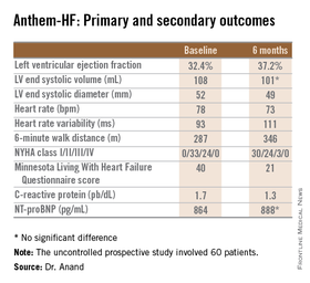
“We believe that if this technology pans out and proves effective, it will be introduced into devices very easily. That’s the advantage of left-sided stimulation,” explained Dr. Anand, professor of medicine at the University of Minnesota, Minneapolis.
He was quick to add, however, that “There needs to be a lot more work before autonomic regulation therapy is ready for prime time.” That’s because ANTHEM-HF was a relatively small prospective study – just 60 patients – and it was uncontrolled and unblinded.
Left-sided VNS is a well-established treatment for epilepsy, with more than 100,000 patients having received devices in the last several decades. It’s also seeing increasing use in refractory depression.
The approach is still investigational for heart failure, where autonomic imbalance with increased sympathetic activity and decreased parasympathetic tone is associated with heart failure progression and worse clinical outcomes, the cardiologist explained.
In ANTHEM-HF, 60 patients on optimal medical therapy for heart failure with reduced ejection fraction were randomized to left- or right-sided implantation of the Cyberonics VNS device. Once activated, the VNS intensity was titrated over 10 weeks to the maximum tolerable current that remained below the threshold of heart rate change, side effects, and patient sensation. The chronic intermittent stimulation to the vagus nerve was delivered at 10 Hz, a 250 microsec pulse width, and an average stimulation current of 2.0 mA. The stimulation cycle was 14 seconds on, 66 seconds off.

The primary study endpoint was change in LVEF over the course of 6 months. From a baseline of 32.4% it improved significantly to 37.2%, which Dr. Anand deemed “very impressive” for just 6 months of therapy. The improvement was similar regardless of whether the VNS was left or right sided.
He said the study didn’t raise any safety concerns. The only serious treatment-related adverse event was a fatal perioperative embolic stroke in a recipient of a left-sided device.
The most common adverse events were hoarseness or other voice alterations, which occurred in 19 patients. Thirteen patients reported a new cough. These side effects mirror the experience in patients treated with the VNS device for epilepsy or depression.
Discussant Jagmeet P. Singh emphasized that “it’s important to interpret the ANTHEM-HF results with some degree of caution.” That’s because the usual variability within LVEF measurements can be in the 5% range, and the 4.8% improvement seen in this study wasn’t accompanied by a significant improvement in LV end systolic volume. Moreover, the improvements seen in secondary endpoints – 6-minute walk distance, NYHA class, and the Minnesota Living With Heart Failure qualify-of-life score – are all potentially open to bias since neither patients nor physicians were blinded.
Dr. Singh noted that the NECTAR-HF study, a blinded, sham-controlled clinical trial of VNS presented earlier at the annual congress of the European Society of Cardiology, was a negative study that showed no improvement in functional measures. While NECTAR-HF caused some skeptics to question the whole concept of VNS as a useful therapeutic strategy, the stimulation protocols used in NECTAR-HF and ANTHEM-HF were quite different, and it’s likely that the stimulation amplitude employed in NECTAR-HF wasn’t sufficient to affect the target subset of vagal nerve fibers, according to Dr. Singh, director of the cardiac resynchronization therapy program and the Holter laboratory at Massachusetts General Hospital, Boston.
He considers the ANTHEM-HF finding that left-sided VNS is comparable to right to be an important advance that “really moves the field forward.” Yet many critical questions about autonomic regulation therapy for heart failure remain unanswered. These include whether it truly is safe and effective, and if so, the optimal target dose and frequency of stimulation. Answers should be forthcoming from the ongoing INOVATE-HF study, a randomized, controlled, 650-patient study, sponsored by BioControl Medical. The trial features hard clinical endpoints and higher target-dose stimulation protocols than in prior studies.
“This trial will probably put an end to the debate as to whether vagal nerve stimulation has an impact on heart failure or not,” the cardiologist predicted.
“Strategies to nonpharmacologically modulate the autonomic nervous system is an area of immense interest and investigation, and it’s going to be extended,” he observed. “I think that this is the autonomic era, and so it’s really important that we get it right. I think we’ve learned from the renal denervation experience that we need to be really careful in selecting patients for any form of autonomic manipulation. I don’t think we should select patients just off their LVEF. We should have some parameter that can quantify autonomic dysregulation prior to initiation of therapy.”
It will also be important to be able to individualize VNS therapy, just as physicians now do with beta blockers and other pharmacotherapies, he added.
The ANTHEM-HF trial was sponsored by Cyberonics. Dr. Anand reported serving as a consultant to and/or recipient of research grants from Cyberonics, Amgen, Critical Diagnostics, Novartis, and Zensun. Dr. Singh has received research support and/or served on speakers bureaus for Boston Scientific, St. Jude Medical, and the Sorin Group.
LAS VEGAS – A novel device-based approach to the treatment of heart failure via vagal nerve stimulation resulted in improvements in objectively measurable cardiac function as well as in subjective heart failure symptoms in the ANTHEM-HF trial.
The improvements seen in ANTHEM-HF (Autonomic Neural Regulation Therapy to Enhance Myocardial Function in Heart Failure) were similar regardless of whether participants were randomized to stimulation of the left or right vagal nerve. That’s an important finding because left-sided vagal nerve stimulation (VNS) technology could readily be combined with implantable cardioverter-defibrillators and cardiac resynchronization therapy devices, which are routinely placed on the left side of the thorax, Dr. Inder S. Anand observed in presenting the study findings at the annual meeting of the Heart Failure Society of America.
“We believe that if this technology pans out and proves effective, it will be introduced into devices very easily. That’s the advantage of left-sided stimulation,” explained Dr. Anand, professor of medicine at the University of Minnesota, Minneapolis.
He was quick to add, however, that “There needs to be a lot more work before autonomic regulation therapy is ready for prime time.” That’s because ANTHEM-HF was a relatively small prospective study – just 60 patients – and it was uncontrolled and unblinded.
Left-sided VNS is a well-established treatment for epilepsy, with more than 100,000 patients having received devices in the last several decades. It’s also seeing increasing use in refractory depression.
The approach is still investigational for heart failure, where autonomic imbalance with increased sympathetic activity and decreased parasympathetic tone is associated with heart failure progression and worse clinical outcomes, the cardiologist explained.
In ANTHEM-HF, 60 patients on optimal medical therapy for heart failure with reduced ejection fraction were randomized to left- or right-sided implantation of the Cyberonics VNS device. Once activated, the VNS intensity was titrated over 10 weeks to the maximum tolerable current that remained below the threshold of heart rate change, side effects, and patient sensation. The chronic intermittent stimulation to the vagus nerve was delivered at 10 Hz, a 250 microsec pulse width, and an average stimulation current of 2.0 mA. The stimulation cycle was 14 seconds on, 66 seconds off.
The primary study endpoint was change in LVEF over the course of 6 months. From a baseline of 32.4% it improved significantly to 37.2%, which Dr. Anand deemed “very impressive” for just 6 months of therapy. The improvement was similar regardless of whether the VNS was left or right sided.
He said the study didn’t raise any safety concerns. The only serious treatment-related adverse event was a fatal perioperative embolic stroke in a recipient of a left-sided device.
The most common adverse events were hoarseness or other voice alterations, which occurred in 19 patients. Thirteen patients reported a new cough. These side effects mirror the experience in patients treated with the VNS device for epilepsy or depression.
Discussant Jagmeet P. Singh emphasized that “it’s important to interpret the ANTHEM-HF results with some degree of caution.” That’s because the usual variability within LVEF measurements can be in the 5% range, and the 4.8% improvement seen in this study wasn’t accompanied by a significant improvement in LV end systolic volume. Moreover, the improvements seen in secondary endpoints – 6-minute walk distance, NYHA class, and the Minnesota Living With Heart Failure qualify-of-life score – are all potentially open to bias since neither patients nor physicians were blinded.
Dr. Singh noted that the NECTAR-HF study, a blinded, sham-controlled clinical trial of VNS presented earlier at the annual congress of the European Society of Cardiology, was a negative study that showed no improvement in functional measures. While NECTAR-HF caused some skeptics to question the whole concept of VNS as a useful therapeutic strategy, the stimulation protocols used in NECTAR-HF and ANTHEM-HF were quite different, and it’s likely that the stimulation amplitude employed in NECTAR-HF wasn’t sufficient to affect the target subset of vagal nerve fibers, according to Dr. Singh, director of the cardiac resynchronization therapy program and the Holter laboratory at Massachusetts General Hospital, Boston.
He considers the ANTHEM-HF finding that left-sided VNS is comparable to right to be an important advance that “really moves the field forward.” Yet many critical questions about autonomic regulation therapy for heart failure remain unanswered. These include whether it truly is safe and effective, and if so, the optimal target dose and frequency of stimulation. Answers should be forthcoming from the ongoing INOVATE-HF study, a randomized, controlled, 650-patient study, sponsored by BioControl Medical. The trial features hard clinical endpoints and higher target-dose stimulation protocols than in prior studies.
“This trial will probably put an end to the debate as to whether vagal nerve stimulation has an impact on heart failure or not,” the cardiologist predicted.
“Strategies to nonpharmacologically modulate the autonomic nervous system is an area of immense interest and investigation, and it’s going to be extended,” he observed. “I think that this is the autonomic era, and so it’s really important that we get it right. I think we’ve learned from the renal denervation experience that we need to be really careful in selecting patients for any form of autonomic manipulation. I don’t think we should select patients just off their LVEF. We should have some parameter that can quantify autonomic dysregulation prior to initiation of therapy.”
It will also be important to be able to individualize VNS therapy, just as physicians now do with beta blockers and other pharmacotherapies, he added.
The ANTHEM-HF trial was sponsored by Cyberonics. Dr. Anand reported serving as a consultant to and/or recipient of research grants from Cyberonics, Amgen, Critical Diagnostics, Novartis, and Zensun. Dr. Singh has received research support and/or served on speakers bureaus for Boston Scientific, St. Jude Medical, and the Sorin Group.
AT THE HFSA ANNUAL SCIENTIFIC MEETING
Key clinical point: Autonomic regulation therapy via a vagal nerve stimulation device shows promise for the treatment of heart failure with reduced ejection fraction.
Major finding: During 6 months of vagal nerve stimulation, patients’ mean LVEF rose significantly from 32.4% to 37.2%, with similar results regardless of whether the stimulation was left- or right-sided.
Data source: The ANTHEM-HF study was a prospective, unblinded, and uncontrolled study involving 60 patients who received vagal nerve stimulation for 6 months.
Disclosures: The study was sponsored by Cyberonics. The presenter has received research grants from and served as a consultant to that and other companies.