User login
Anticoagulation Hub contains news and clinical review articles for physicians seeking the most up-to-date information on the rapidly evolving treatment options for preventing stroke, acute coronary events, deep vein thrombosis, and pulmonary embolism in at-risk patients. The Anticoagulation Hub is powered by Frontline Medical Communications.
Impaired kidney function no problem for dabigatran reversal
ORLANDO – Idarucizumab, the reversal agent for the anticoagulant dabigatran, appeared as effective in quickly reversing dabigatran’s effects in patients with severe renal dysfunction as in patients with normally working kidneys, in a post hoc analysis of data collected in the drug’s pivotal trial.
A standard dose of idarucizumab “works just as well in patients with bad kidney function as it does in patients with preserved kidney function,” John W. Eikelboom, MD, said at the annual meeting of the American College of Cardiology. “The time to cessation of bleeding and the degree of normal hemostasis achieved was consistent” across the entire range of renal function examined, from severe renal dysfunction, with a creatinine clearance rate of less than 30 mL/min, to normal function, with an estimated rate of 80 mL/min or greater.
The ability of idarucizumab (Praxbind), conditionally approved by the Food and Drug Administration in 2015 and then fully approved in April 2018, to work in patients with impaired renal function has been an open question and concern because dabigatran (Pradaxa) is excreted renally, so it builds to unusually high levels in patients with poor kidney function. “Plasma dabigatran levels might be sky high, so a standard dose of idarucizumab might not work. That’s been a fear of clinicians,” explained Dr. Eikelboom, a hematologist at McMaster University in Hamilton, Ont.
To examine whether idarucizumab’s activity varied by renal function he used data from the patients enrolled in the RE-VERSE AD (Reversal Effects of Idarucizumab on Active Dabigatran) study, the pivotal dataset that led to idarucizumab’s U.S. approval. The new, post hoc analysis divided patients into four subgroups based on their kidney function, and focused on the 489 patients for whom renal data were available out of the 503 patients in the study (N Engl J Med. 2017 Aug 3;377[5]:431-41). The subgroups included 91 patients with severe dysfunction with a creatinine clearance rate of less than 30 mL/min; 127 with moderate dysfunction and a clearance rate of 30-49 mL/min; 163 with mild dysfunction and a clearance rate of 50-79 mL/min; and 108 with normal function and a creatinine clearance of at least 80 mL/min.
Patients in the subgroup with severe renal dysfunction had the worst clinical profile overall, and as predicted, had a markedly elevated average plasma level of dabigatran, 231 ng/mL, nearly five times higher than the 47-ng/mL average level in patients with normal renal function.
The ability of a single, standard dose of idarucizumab to reverse the anticoagulant effects of dabigatran were essentially identical across the four strata of renal activity, with 98% of patients in both the severely impaired subgroup and the normal subgroup having 100% reversal within 4 hours of treatment, Dr. Eikelboom reported. Every patient included in the analysis had more than 50% reversal.
The study followed patients to 12-24 hours after they received idarucizumab, and 55% of patients with severe renal dysfunction showed a plasma dabigatran level that crept back toward a clinically meaningful level and so might need a second idarucizumab dose. In contrast, this happened in 8% of patients with normal renal function.
In patients with severe renal dysfunction given idarucizumab, “be alert for a recurrent bleed,” which could require a second dose of idarucizumab, Dr. Eikelboom suggested.
SOURCE: Eikelboom JW et al. ACC 18, Abstract 1231M-11.
REPORTING FROM ACC 18
Key clinical point: Renal function had no impact on idarucizumab’s efficacy for dabigatran reversal.
Major finding: Complete dabigatran reversal occurred in 98% of patients with severe renal dysfunction who received idarucizumab.
Study details: Post hoc analysis of data from RE-VERSE AD, idarucizumab’s pivotal trial with 503 patients.
Disclosures: RE-VERSE AD was funded by Boehringer Ingelheim, the company that markets idarucizumab (Praxbind) and dabigatran (Pradaxa). Dr. Eikelboom has been a consultant to and has received research support from Boehringer Ingelheim, as well as from Bayer, Bristol-Myers Squibb, Daiichi-Sankyo, Janssen, and Pfizer.
Source: Eikelboom JW et al. ACC 18, Abstract 1231M-11.
VTE risk after bariatric surgery should be assessed
SEATTLE – Preop thromboelastometry can identify patients who need extra according to a prospective investigation of 40 patients at Conemaugh Memorial Medical Center in Johnstown, Pa.
Enoxaparin 40 mg twice daily just wasn’t enough for people who were hypercoagulable before surgery. The goal of the study was to find the best way to prevent venous thromboembolism (VTE) after weight loss surgery. At present, there’s no consensus on prophylaxis dosing, timing, duration, or even what agent to use for these patients. Conemaugh uses postop enoxaparin, a low-molecular-weight heparin. Among many other options, some hospitals opt for preop dosing with traditional heparin, which is less expensive.
The Conemaugh team turned to thromboelastometry (TEM) to look at the question of VTE risk in bariatric surgery patients. The test gauges coagulation status by measuring elasticity as a small blood sample clots over a few minutes. The investigators found that patients who were hypercoagulable before surgery were likely to be hypercoagulable afterwards. The finding argues for baseline TEM testing to guide postop anticoagulation.
The problem is that bariatric services don’t often have access to TEM equipment, and insurance doesn’t cover the $60 test. In this instance, the Lake Erie College of Osteopathic Medicine in Erie, Pa., had the equipment and covered the testing for the study.
The patients had TEM at baseline and then received 40 mg of enoxaparin about 4 hours after surgery – mostly laparoscopic gastric bypasses – and a second dose about 12 hours after the first. TEM was repeated about 2 hours after the second dose.
At baseline, 2 (5%) of the patients were hypocoagulable, 15 (37.5%) were normal, and 23 (57.5%) were hypercoagulable. On postop TEM, 17 patients (42.5%) were normal and 23 (57.5%) were hypercoagulable: “These 23 were inadequately anticoagulated,” said lead investigator Daniel Urias, MD, a general surgery resident at the medical center.
“There was an association between being normal at baseline and being normal postop, and being hypercoagulable at baseline and hypercoagulable postop. We didn’t anticipate finding such similarity between the numbers. Our suspicion that baseline status plays a major role is holding true,” Dr. Urias said at the World Congress of Endoscopic Surgery hosted by SAGES & CAGS.
When patients test hypercoagulable at baseline, “we are [now] leaning towards [enoxaparin] 60 mg twice daily,” he said.
Ultimately, anticoagulation TEM could be used to titrate patients into the normal range. For best outcomes, it’s likely that “obese patients require goal-directed therapy instead of weight-based or fixed dosing,” he said, but nothing is going to happen until insurance steps up.
The patients did not have underlying coagulopathies, and 33 (82.5%) were women; the average age was 44 years and average body mass index was 43.6 kg/m2. The mean preop Caprini score was 4, indicating moderate VTE risk. Surgery lasted about 200 minutes. Patients were out of bed and walking on postop day 0.
The investigators had no relevant disclosures.
SOURCE: Urias D et al. World Congress of Endoscopic Surgery hosted by SAGES & CAGS abstract S023.
REPORTING FROM SAGES 2018
Key clinical point: Preoperative thromboelastometry identifies patients who need extra anticoagulation against venous thromboembolism following bariatric surgery.
Major finding: Baseline and postop coagulation were similar: 37.5% vs. 42.5% were normal and 57.5% vs 57.5% were hypercoagulable.
Study details: Prospective study of 40 bariatric surgery patients.
Disclosures: The investigators did not have any relevant disclosures. The Lake Erie College of Osteopathic Medicine paid for the testing.
Source: Urias D et al. World Congress of Endoscopic Surgery hosted by SAGES & CAGS abstract S023.
VIDEO: Screening ECG patch boosts AF diagnoses ninefold
ORLANDO – An ECG patch worn twice for a total of about 24 days produced a nearly ninefold increase in the number of high-risk people diagnosed with atrial fibrillation, compared with those followed with usual care in a randomized trial with 2,655 people.
During 4 months of follow-up, 1,364 high-risk people assigned to ECG patch screening had a 5.1% rate of new atrial fibrillation (AF) diagnoses, compared with a 0.6% rate among 1,291 controls who wore the patch but were identified with new-onset AF using standard follow-up that did not take the patch data into account. This was a statistically significant difference for the study’s primary endpoint, Steven R. Steinhubl, MD, said at the annual meeting of the American College of Cardiology.
The video associated with this article is no longer available on this site. Please view all of our videos on the MDedge YouTube channel
In addition to proving that the ECG patch can better identify asymptomatic people who have AF than conventional means – usually waiting until a stroke occurs or for symptoms to appear – the noninvasive and relatively low-cost patch also gives researchers a new way to try to address the more fundamental medical question created by this line of investigation: How clinically important are relatively brief, asymptomatic episodes of atrial fibrillation, and are patient outcomes improved by treatments begun in this early phase?
The study results “show we can look beyond implantable devices with a less expensive, noninvasive way” to identify patients with asymptomatic AF and determine its natural history and need for intervention, Dr. Steinhubl said in a video interview.
The mSToP (mHealth Screening to Prevent Strokes) trial ran at Scripps and began by identifying more than 359,000 U.S. residents with Aetna health insurance who met the study’s definition of having high AF risk, either by being at least 75 years old, or at least 55 years old and male or at least 65 years old and female. To qualify as high risk those younger than 75 years also had to have at least one clinical risk factor, which could include a prior cerebrovascular event, heart failure, hypertension plus diabetes, obstructive sleep apnea, or any of six other comorbidities. The researchers also excluded potential participants because of several factors, including a history of atrial fibrillation or flutter, current treatment with an anticoagulant, end-stage renal disease, and patients with an implanted pacemaker or defibrillator.
They invited more than 100,000 of these qualifying Aetna beneficiaries to participate, and 2,655 agreed and received by mail a pair of ECG measurement patches (Zio) with instructions to wear one for 2 weeks at the start of the study and to wear the second during the final 2 weeks of the 4-month study period. The participants averaged 73 years of age, and their average CHA2DS2-VASc score was 3.
All patients in the study were told to wear their patches and mail them in, but the researchers used the collected ECG data for diagnosing AF in only the 1,364 patients randomized to the active arm. The ECG findings for the 1,291 controls wasn’t provided to their physicians, and so any new-onset AF had to be found either by symptom onset or incidentally. About one-third of the people assigned to each of the study arms never wore their patches. Those who wore their patches did so for an average of nearly 12 days each. Diagnosis of new-onset AF was based on finding either at least one AF episode recorded by the patches that lasted at least 30 seconds or an AF diagnosis appearing in the patient’s record. The average AF burden – the percentage of time a person with incident AF had an abnormal sinus rhythm – was 0.9%.
Even though many patients did not use their patches, the investigators assessed the primary endpoint of new AF diagnoses during the 4-month study period on an intention-to-treat basis. Their analysis showed an 8.8-fold higher rate of new AF diagnoses among people in the intervention arm whose patch data were used for immediate diagnosis, reported Dr. Steinhubl, an interventional cardiologist and director of digital medicine at the Scripps Translational Science Institute in La Jolla, Ca.
As a secondary endpoint, the researchers merged the entire group of 1,738 participants who had sent in patches with ECG data and compared their 1-year incidence of diagnosed AF against 3,476 matched controls from the Aetna database. After 1 year, the rate of new AF diagnoses was 6.3% in those with patch information and 2.3% among the controls, a threefold difference in diagnosis rates after adjustment for potential confounders.
“The clinical significance of the short AF episodes” manifested by many patch users identified with AF “requires greater clarity, especially in terms of stroke risk,” Dr. Steinhubl said. But he added, “I like to think that, as we learn more, we can look at more than just anticoagulation” as intervention options. For example, if a morbidly obese patient has asymptomatic AF found by patch screening, it might strengthen the case for bariatric surgery if it’s eventually shown that weight loss after bariatric surgery slows AF progression. The same holds true for more aggressive sleep apnea intervention in patients with sleep apnea and asymptomatic AF, as well as for patients with asymptomatic AF and another type of associated comorbidity.
SOURCE: Steinhubl S. ACC 18, Abstract 402-19.
Results from several studies have now shown that some kind of monitoring for AF in asymptomatic, at-risk people results in an increased diagnosis of subclinical AF. Study results also suggest that, in general, people diagnosed with subclinical AF are at a lower risk of stroke than patients with symptomatic AF. As of now, no prospective study has evaluated the efficacy of anticoagulant therapy in people diagnosed with subclinical AF, although such studies are now in progress. Until we have these results, the question of how to manage patients with subclinical AF remains unanswered.
N.A. Mark Estes, MD , is professor of medicine and director of the New England Cardiac Arrhythmia Center at Tufts Medical Center in Boston. He has been a consultant to Boston Scientific, Medtronic, and St. Jude. He made these comments as designated discussant for the mSToPS report.
ORLANDO – An ECG patch worn twice for a total of about 24 days produced a nearly ninefold increase in the number of high-risk people diagnosed with atrial fibrillation, compared with those followed with usual care in a randomized trial with 2,655 people.
During 4 months of follow-up, 1,364 high-risk people assigned to ECG patch screening had a 5.1% rate of new atrial fibrillation (AF) diagnoses, compared with a 0.6% rate among 1,291 controls who wore the patch but were identified with new-onset AF using standard follow-up that did not take the patch data into account. This was a statistically significant difference for the study’s primary endpoint, Steven R. Steinhubl, MD, said at the annual meeting of the American College of Cardiology.
The video associated with this article is no longer available on this site. Please view all of our videos on the MDedge YouTube channel
In addition to proving that the ECG patch can better identify asymptomatic people who have AF than conventional means – usually waiting until a stroke occurs or for symptoms to appear – the noninvasive and relatively low-cost patch also gives researchers a new way to try to address the more fundamental medical question created by this line of investigation: How clinically important are relatively brief, asymptomatic episodes of atrial fibrillation, and are patient outcomes improved by treatments begun in this early phase?
The study results “show we can look beyond implantable devices with a less expensive, noninvasive way” to identify patients with asymptomatic AF and determine its natural history and need for intervention, Dr. Steinhubl said in a video interview.
The mSToP (mHealth Screening to Prevent Strokes) trial ran at Scripps and began by identifying more than 359,000 U.S. residents with Aetna health insurance who met the study’s definition of having high AF risk, either by being at least 75 years old, or at least 55 years old and male or at least 65 years old and female. To qualify as high risk those younger than 75 years also had to have at least one clinical risk factor, which could include a prior cerebrovascular event, heart failure, hypertension plus diabetes, obstructive sleep apnea, or any of six other comorbidities. The researchers also excluded potential participants because of several factors, including a history of atrial fibrillation or flutter, current treatment with an anticoagulant, end-stage renal disease, and patients with an implanted pacemaker or defibrillator.
They invited more than 100,000 of these qualifying Aetna beneficiaries to participate, and 2,655 agreed and received by mail a pair of ECG measurement patches (Zio) with instructions to wear one for 2 weeks at the start of the study and to wear the second during the final 2 weeks of the 4-month study period. The participants averaged 73 years of age, and their average CHA2DS2-VASc score was 3.
All patients in the study were told to wear their patches and mail them in, but the researchers used the collected ECG data for diagnosing AF in only the 1,364 patients randomized to the active arm. The ECG findings for the 1,291 controls wasn’t provided to their physicians, and so any new-onset AF had to be found either by symptom onset or incidentally. About one-third of the people assigned to each of the study arms never wore their patches. Those who wore their patches did so for an average of nearly 12 days each. Diagnosis of new-onset AF was based on finding either at least one AF episode recorded by the patches that lasted at least 30 seconds or an AF diagnosis appearing in the patient’s record. The average AF burden – the percentage of time a person with incident AF had an abnormal sinus rhythm – was 0.9%.
Even though many patients did not use their patches, the investigators assessed the primary endpoint of new AF diagnoses during the 4-month study period on an intention-to-treat basis. Their analysis showed an 8.8-fold higher rate of new AF diagnoses among people in the intervention arm whose patch data were used for immediate diagnosis, reported Dr. Steinhubl, an interventional cardiologist and director of digital medicine at the Scripps Translational Science Institute in La Jolla, Ca.
As a secondary endpoint, the researchers merged the entire group of 1,738 participants who had sent in patches with ECG data and compared their 1-year incidence of diagnosed AF against 3,476 matched controls from the Aetna database. After 1 year, the rate of new AF diagnoses was 6.3% in those with patch information and 2.3% among the controls, a threefold difference in diagnosis rates after adjustment for potential confounders.
“The clinical significance of the short AF episodes” manifested by many patch users identified with AF “requires greater clarity, especially in terms of stroke risk,” Dr. Steinhubl said. But he added, “I like to think that, as we learn more, we can look at more than just anticoagulation” as intervention options. For example, if a morbidly obese patient has asymptomatic AF found by patch screening, it might strengthen the case for bariatric surgery if it’s eventually shown that weight loss after bariatric surgery slows AF progression. The same holds true for more aggressive sleep apnea intervention in patients with sleep apnea and asymptomatic AF, as well as for patients with asymptomatic AF and another type of associated comorbidity.
SOURCE: Steinhubl S. ACC 18, Abstract 402-19.
REPORTING FROM ACC 18
Key clinical point: .
Major finding: After 4 months, new AF diagnoses occurred in 5.1% of patch users and 0.6% of usual-care controls.
Study details: mSToPS, a single-center, randomized study with 2,655 people at high risk for developing AF.
Disclosures: mSToPS received support from Aetna, Janssen, and iRhythm. Dr. Steinhubl has been an advisor to Airstrip, DynoSense, EasyG, FocusMotion, LifeWatch, MyoKardia, Novartis, and Spry Health, he serves on the board of Celes Health, and he has received research support from Janssen and Novartis.
Source: Steinhubl S. ACC 18, Abstract 402-19.
Few acutely ill hospitalized patients receive VTE prophylaxis
SAN DIEGO – Among patients hospitalized for acute medical illnesses, the risk of venous thromboembolism (VTE) remained elevated 30-40 days after discharge, results from a large analysis of national data showed.
Moreover, only 7% of at-risk patients received VTE prophylaxis in both the inpatient and outpatient setting.
“The results of this real-world study imply that there is a significantly unmet medical need for effective VTE prophylaxis in both the inpatient and outpatient continuum of care among patients hospitalized for acute medical illnesses,” researchers led by Alpesh Amin, MD, wrote in a poster presented at the biennial summit of the Thrombosis & Hemostasis Societies of North America.
According to Dr. Amin, who chairs the department of medicine at the University of California, Irvine, hospitalized patients with acute medical illnesses face an increased risk for VTE during hospital discharge, mainly within 40 days following hospital admission. However, the treatment patterns of VTE prophylaxis in this patient population have not been well studied in the “real-world” setting. In an effort to improve this area of clinical practice, the researchers used the Marketscan database between Jan. 1, 2012, and June 30, 2015, to identify acutely ill hospitalized patients, such as those with heart failure, respiratory diseases, ischemic stroke, cancer, infectious diseases, and rheumatic diseases. The key outcomes of interest were the proportion of patients receiving inpatient and outpatient VTE prophylaxis and the proportion of patients with VTE events during and after the index hospitalization. They used Kaplan-Meier analysis to examine the risk for VTE events after the index inpatient admission.
The mean age of the 17,895 patients was 58 years, 55% were female, and most (77%) were from the Southern area of the United States. Their mean Charlson Comborbidity Index score prior to hospitalization was 2.2. Nearly all hospitals (87%) were urban based, nonteaching (95%), and large, with 68% having at least 300 beds. Nearly three-quarters of patients (72%) were hospitalized for infectious and respiratory diseases, and the mean length of stay was 5 days.
Dr. Amin and his associates found that 59% of hospitalized patients did not receive any VTE prophylaxis, while only 7% received prophylaxis in both the inpatient and outpatient continuum of care. At the same time, cumulative VTE rates within 40 days of index admission were highest among patients hospitalized for infectious diseases and cancer (3.4% each), followed by those with heart failure (3.1%), respiratory diseases (2%), ischemic stroke (1.5%), and rheumatic diseases (1.3%). The cumulative VTE event rate for the overall study population within 40 days from index hospitalization was nearly 3%, with 60% of VTE events having occurred within 40 days.
The study was funded by Portola Pharmaceuticals. Dr. Amin reported having no financial disclosures.
SOURCE: Amin A et al. THSNA 2018, Poster 51.
REPORTING FROM THSNA 2018
Key clinical point: There is a significant unmet medical need for VTE prophylaxis in the continuum of care of patients hospitalized for acute medical illnesses.
Major finding: Of the overall study population, only 7% received both inpatient and outpatient VTE prophylaxis.
Study details: An analysis of national data from 17,895 acutely ill hospitalized patients.
Disclosures: The study was funded by Portola Pharmaceuticals. The presenter reported having no financial conflicts.
Source: Amin A et al. THSNA 2018, Poster 51.
ODYSSEY Outcomes trial redefines secondary cardiovascular prevention
ORLANDO – In what was hailed as a major advance in preventive cardiology, the ODYSSEY Outcomes trial has shown that adding the PCSK9 inhibitor alirocumab on top of intensive statin therapy reduced major adverse cardiovascular events and all-cause mortality significantly more than placebo plus intensive statin therapy in patients with a recent acute coronary syndrome and an elevated on-statin LDL cholesterol level.
ODYSSEY Outcomes was a double-blind trial in which 18,924 patients at 1,315 sites in 57 countries were randomized to alirocumab (Praluent) or placebo plus background high-intensity statin therapy starting a median of 2.5 months after an acute coronary syndrome. All participants had to have a baseline LDL cholesterol level of 70 mg/dL or higher despite intensive statin therapy. Alirocumab was titrated to maintain a target LDL of 25-50 mg/dL. An LDL of 15-25 mg/dL was deemed acceptable, but if the level dropped below 15 mg/dL on two consecutive measurements the patient was blindly switched to placebo, as occurred in 7.7% of the alirocumab group.
The primary study endpoint was a composite outcome comprised of CHD (coronary heart disease) death, nonfatal MI, ischemic stroke, or unstable angina requiring hospitalization. During a median 2.8 years of follow-up, this outcome occurred in 9.5% of the overall population randomized to alirocumab and 11.1% of those on placebo, for a statistically significant and clinically meaningful 15% reduction in relative risk. The CHD death rates in the two study arms were similar; however, the other three components of the primary endpoint occurred significantly less often in the alirocumab group: The risk of nonfatal MI was 14% less (6.6% vs. 7.6%), ischemic stroke was 27% less (1.2 vs. 1.6%), and unstable angina was 39% less (0.4% vs. 0.6%).
All-cause mortality occurred in 3.5% of patients receiving alirocumab and 4.1% on placebo, once again for a statistically significant 15% reduction in risk. This was a major achievement, since even statins haven’t shown a mortality benefit in the post-ACS setting, observed Dr. Steg, cochair of the study.
The greatest benefits were seen in the 5,629 participants with a baseline LDL of 100 mg/dL or more on high-intensity statin therapy. In this large subgroup at highest baseline risk, alirocumab resulted in an absolute 3.4% risk reduction and a 24% reduction in relative risk of MACE. All-cause mortality decreased by an absolute 1.7%, translating to a 29% relative risk reduction. The number-needed-to-treat (NNT) for the duration of the study in order to prevent one additional MACE event in this group was 29, with an NNT to prevent one additional death of 60, added Dr. Steg, professor of cardiology at the University of Paris and chief of cardiology at Bichat Hospital.
“The risk/benefit for alirocumab is extraordinarily favorable. There was almost no risk over the course of the trial. There was no increase in neurocognitive disorders, new-onset or worsening diabetes, cataracts, or hemorrhagic stroke,” the cardiologist said.
Indeed, the sole adverse event that occurred more frequently in the alirocumab group was mild local injection site reactions, which occurred in 3.8% of the alirocumab group and 2.1% of controls.
There was a tendency for LDL to creep upward in both the alirocumab and placebo arms over the course of follow-up. Dr. Steg attributed this to down-titration or cessation of alirocumab as per protocol along with the inability of a substantial proportion of patients to tolerate intensive statin therapy. Most study participants had never been on a statin until their ACS.
A year ago at ACC 2017, other investigators presented the results of FOURIER, a large clinical outcomes trial of evolocumab (Repatha), another PCSK9 (proprotein convertase subtilisin/kexin type 9) inhibitor. FOURIER also showed a 15% relative risk reduction in major adverse cardiovascular events, but unlike in ODYSSEY Outcomes, there was no significant impact upon mortality. Dr. Steg attributed this to several key differences between the two trials.
The post-ACS population of ODYSSEY Outcomes was on average higher-risk than FOURIER participants, who had stable atherosclerotic cardiovascular disease. The background statin therapy was more intensive in ODYSSEY, and the average follow-up was close to 8 months longer, too.
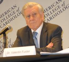
The study population is representative of an enormous number of patients seen in clinical practice, added Dr. Fuster, professor of medicine and physician-in-chief at Mount Sinai Hospital in New York. He estimated that one-third of patients who experience ACS can’t subsequently get their LDL down to the 70 mg/dL range on statin therapy, generally because of drug intolerance.
He voiced a concern: “Up until now, the feasibility and affordability of using this type of drug has been extremely difficult. I hope this particular study is a trigger – a catalyzer – for making this drug much more available to people who need it.”
The study met with an enthusiastic audience reception. Prior to presentation of the results at the meeting’s opening session, 79% of the audience of more than 4,000 in the main arena indicated they either don’t prescribe PCSK9 inhibitors or do so only a handful of times per year.
Immediately after seeing the data, 62% of the audience said their practice will change as a result of the study findings.
ODYSSEY Outcomes was funded by Sanofi and Regeneron Pharmaceuticals. Dr. Steg reported serving as a consultant to and receiving research grants from those pharmaceutical companies and numerous others.
bjancin@frontlinemedcom.com
SOURCE: Steg GP.
REPORTING FROM ACC 2018
Key clinical point: Alirocumab reduced both all-cause mortality and major adverse cardiovascular events in high-risk patients with a recent acute coronary syndrome.
Major finding: Alirocumab reduced MACE by 15% and all-cause mortality by an equal margin compared with placebo in patients with a recent acute coronary syndrome and elevated LDL cholesterol despite intensive statin therapy alone.
Study details: The ODYSSEY Outcomes trial was a double-blind, randomized trial of nearly 19,000 patients with a recent acute coronary syndrome and an LDL cholesterol of 70 mg/dL or more despite intensive statin therapy.
Disclosures: ODYSSEY Outcomes was funded by Sanofi and Regeneron Pharmaceuticals. The presenter reported serving as a consultant to and receiving research grants from those pharmaceutical companies and numerous others.
Source: Steig, GP.
Post-ACS death lowered in ODYSSEY Outcomes
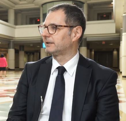
ORLANDO – should set the stage for broader use of the PCSK9-inhibitor in certain high-risk patients, Gabriel Steg, MD, said in a video interview at the annual meeting of the American College of Cardiology.
The findings from the 3-year trial are threefold: First, the trial met its primary goal, significantly lower major adverse cardiovascular events and 15% lower mortality in alirocumab-treated patients compared with those on placebo; second, the effect was greater in patients who started with an LDL level above 100 mg/dL; and third, alirocumab was remarkably safe, said Dr. Steg, director of the coronary care unit of Bichat Hospital in Paris.
“We now have good reason to target a lower LDL range of at least less than 50 mg/dL, and possibly even lower, using PCSK9 inhibitors to get the benefits we’re seeing in the trial applied to broader groups of patients,” he added.
SOURCE: Steg G ACC 18.
REPORTING FROM ACC 18
Young diabetics are at sevenfold increased risk of sudden cardiac death
ANAHEIM, CALIF. – Children and young adults with type 1 or type 2 diabetes have a 7.4-fold increased risk of sudden cardiac death, compared with nondiabetic, age-matched controls, according to a first-of-its-kind Danish national study.
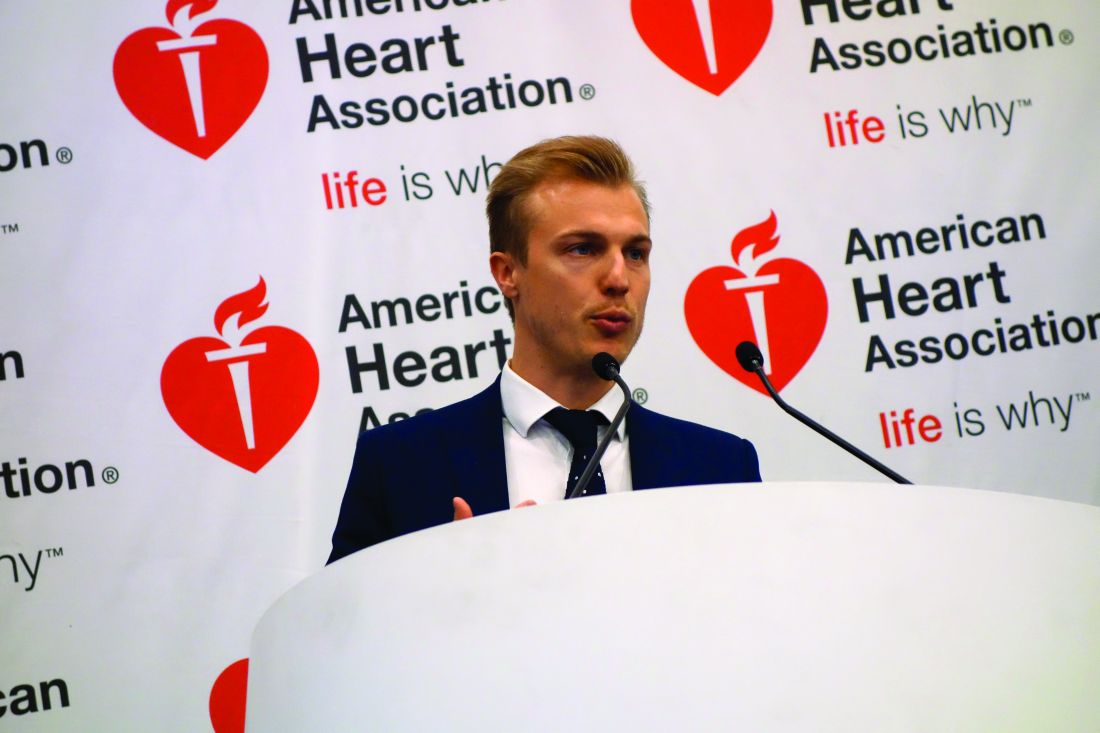
“Luckily the absolute risk is low, but we hope these data will make [young patients with diabetes] think more about taking their insulin and following their physicians’ recommendations about treatment,” Jesper Svane said in presenting the study findings at the American Heart Association scientific sessions.
Sudden cardiac death (SCD) was the No. 1 cause of mortality in the diabetic cohort with a rate of 34.8 deaths/100,000 person-years, compared with 4.7 deaths/100,000 person-years in an age-matched nondiabetic cohort. That translates to a 7.4-fold increased risk of SCD in the diabetic cohort, noted Mr. Svane, a medical student at Copenhagen University.
When deaths caused by SCD were combined with those from other cardiac diseases, the young diabetic cohort had a rate of 68.3 deaths/100,000 person-years versus 8.2 deaths/100,000 person-years in age-matched controls, for an 8.3-fold increased risk. This observation underscores an important point: Monitoring cardiovascular risk needs to start early in young patients with diabetes.
“It’s important to pay attention when a young person with diabetes comes in complaining of chest pain or syncope,” Mr. Svane said.
The second and third most common causes of death in young Danish diabetic patients were pulmonary and endocrine diseases, which occurred at rates 7.2- and 79.2-fold greater in these patients, respectively, than in controls.
All-cause mortality occurred at a rate of 234.9 deaths/100,000 person-years in young patients with diabetes versus 50.9 deaths/100,000 person-years in matched controls, for a 4.6-fold increased risk.
Autopsies showed that the most common cause of death in diabetic persons aged 36-49 years was coronary artery disease, while in those up to age 35, it was sudden arrhythmic death syndrome, a label bestowed when a medical examiner can’t find an apparent cause of death.
Session moderator Robert H. Eckle, MD, a past AHA president, cautioned against routinely attributing the deaths without apparent cause at autopsy in young people with diabetes to sudden arrhythmic death syndrome. Given that the Danish registry data don’t include data on diabetic patients’ degree of metabolic control, it seems likely that an uncertain number of those deaths were really caused by hypoglycemia, observed Dr. Eckle, professor of medicine at the University of Colorado at Denver, Aurora.
Mr. Svane reported having no financial conflicts of interest.
SOURCE: Svane J. 2017 AHA Sessions.
REPORTING FROM THE AHA SCIENTIFIC SESSIONS
Key clinical point: Young persons with diabetes are 8.3-fold more likely to die of cardiac causes.
Major finding: In particular, the risk of sudden cardiac death in Danish children and young adults with diabetes was 7.4-fold greater than in age-matched controls.
Study details: This was a Danish national registry study of more than 14,000 children and young adults with diabetes and their rate of sudden cardiac death.
Disclosures: The study presenter reported having no financial conflicts.
Source: Svane J. 2017 AHA Sessions.
Sex-triggered sudden cardiac arrest extremely rare
ANAHEIM, CALIF. – Patients with heart disease can safely be reassured that sexual intercourse as a trigger for sudden cardiac death is extremely rare, Aapo Aro, MD, said at the American Heart Association scientific sessions.
He presented an analysis from the ongoing Oregon Sudden Unexpected Death Study, a population-based case-control study that captures all cases of sudden cardiac arrest (SCA) in the Portland, Ore., area.
This was the first-ever study to examine the burden of SCA triggered by sexual activity. Of 4,557 adjudicated cases of SCA in adults during 2002-2015, a mere 34, or 0.7%, happened during or within 1 hour of sexual intercourse.
Thirty-two of the 34 cases occurred in men. That works out to 1% of SCAs in men being related to sexual activity. In women, the rate was 10-fold lower, at 0.1%, noted Dr. Aro, a cardiologist at Helsinki University Hospital who was at Cedars-Sinai Medical Center in Los Angeles at the time he conducted this research.
Many of the Oregonians who experienced SCA had known heart disease at the time, regardless of whether the event occurred during sexual activity or at another time. Of note, however, SCA during sexual activity presented with ventricular fibrillation/ventricular tachycardia in 76% of cases, versus a 45% rate in individuals whose SCA was not associated with sexual intercourse.
“The data are very reassuring,” Dr. Aro said in an interview. “Many of these patients had known cardiac disease, but still the absolute numbers of events are very small. Our take home message from this study is that sexual activity can be regarded as safe even in cardiac patients.”
The Oregon Sudden Unexpected Death Study is funded by the National Heart, Lung, and Blood Institute and the American Heart Association. Dr. Aro reported having no financial conflicts of interest.
REPORTING FROM THE AHA SCIENTIFIC SESSIONS
Key clinical point: Patients with cardiac disease needn’t shy away from sexual intercourse because it might trigger cardiac arrest.
Major finding: Only 0.7% of 4,557 adjudicated sudden cardiac arrests occurred in association with sexual intercourse.
Study details: This population-based case-control study examined 4,557 sudden cardiac arrests in the Portland, Ore., area.
Disclosures: The Oregon Sudden Unexpected Death Study is funded by the National Heart, Lung and Blood Institute and American Heart Association. The presenter reported having no financial conflicts.
Overweight and obese individuals face greater cardiovascular morbidity
.
In a report published online Feb. 28 in JAMA Cardiology, researchers presented an analysis of pooled data from 190,672 participants and 3.2 million person-years of follow-up in 10 prospective cohort studies, including the Framingham Heart Study, the Multi-Ethnic Study of Atherosclerosis, and the Atherosclerosis Risk in Communities Study.
Incident cardiovascular events occurred in 37% of overweight middle-aged men and 28% of overweight middle-aged women. In obese middle-aged men and women, those figures were 47% and 39%, respectively, and in the morbidly obese they were 65% and 48%. By comparison, incident cardiovascular events occurred in 32% of middle-aged men of normal BMI, and 22% of women.
Across all the studies, there were 7,136 fatal or nonfatal myocardial infarctions, 3,733 fatal or nonfatal strokes, 4,614 diagnoses of heart failure, and 13,457 cardiovascular disease events during 856,532 person-years of follow-up in middle-aged adults.
After adjustment for age, ethnicity, and smoking status, the competing hazard ratios for experiencing a cardiovascular disease event compared to a noncardiovascular disease death were greater in the higher-BMI categories, and greatest among morbidly obese middle-aged men and women, largely because of a greater proportion of coronary heart disease and heart failure events.
“In addition, greater all-cause mortality in higher-BMI categories occurred at the expense of a greater proportion of deaths from cardiovascular causes in middle-aged men and women who are overweight and obese,” wrote Sadiya S. Khan, MD, MSc, of Northwestern University, Chicago, and her coauthors.
The research suggested that for each increasing unit of BMI in middle-aged men and women, the adjusted competing hazard ratios of incident cardiovascular disease events increased by a significant 5%.
The study found that middle-aged men in the normal and overweight BMI group enjoyed more years free from cardiovascular disease than did obese middle-aged men. In middle-aged women, those who were in the normal BMI range had significantly more years lived free of cardiovascular disease than did overweight or obese women.
The incidence of cardiovascular disease was significantly delayed by an average of 7.5 years in middle-aged men of normal BMI and 7.1 years in middle-aged women of normal BMI, compared with those with morbid obesity.
In terms of longevity, men and women with normal BMI lived on average 5.6 years and 2 years longer, respectively, than did men and women with morbid obesity.
“The results of this study build on prior research from the Cardiovascular Disease Lifetime Risk Pooling Project highlighting marked differences in lifetime risks of CVD and further highlight the importance of consideration of BMI as a risk factor for diminished healthy longevity and greater overall CVD morbidity and mortality,” the authors wrote.
The study was intended to address recent controversy over the health implications of overweight, with some evidence suggesting that overweight individuals have all-cause mortality similar to or lower than that of normal-weight groups.
“While we do observe evidence of the well-described overweight and obesity paradox, in which heavier individuals appear to live longer on average after diagnosis of CVD compared with individuals with normal BMI, our data when following up individuals prior to the onset of CVD indicate that this occurs because of a trend toward earlier onset of disease in individuals who are overweight and obese,” they wrote.
The study did not account for change in BMI over the course of follow-up, nor did it use data on fat distribution or the degree of visceral adiposity, the researchers noted.
“Additional important outcomes of obesity-related morbidity, such as atrial fibrillation, sleep-disordered breathing, and chronic liver disease, were not ascertained routinely in our cohort studies, and we likely underestimated the overall comorbidity burden of excess weight.”
The National Heart, Lung, and Blood Institute supported the study. No conflicts of interest were declared.
SOURCE: Khan SS et al. JAMA Cardiol. 2018 Feb 28. doi: 10.1001/jamacardio.2018.0022.
FROM JAMA CARDIOLOGY
Key clinical point: Obese individuals have shorter life spans and spend significantly more time dealing with the burden of cardiovascular morbidity than do normal-weight individuals.
Major finding: Overweight and obese middle-aged individuals have a significantly higher incidence of cardiovascular events and mortality compared with normal-weight middle-aged individuals.
Data source: Analysis of pooled data from 190,672 participants and 3.2 million person-years of follow-up in 10 prospective cohort studies.
Disclosures: The National Heart, Lung, and Blood Institute supported the study. No conflicts of interest were declared.
Source: Khan SS et al. JAMA Cardiol. 2018 Feb 28. doi: 10.1001/jamacardio.2018.0022
Thrombectomy’s success treating strokes prompts rethinking of selection criteria
LOS ANGELES – CT or magnetic resonance brain imaging of acute ischemic stroke patients was the key triage tool in two groundbreaking thrombectomy trials, DAWN and DEFUSE; results from both showed that patients found by imaging to have limited infarcted cores could safely benefit from endovascular thrombectomy, even when they are more than 6 hours out from their stroke onset, breaking the 6-hour barrier created 3 years ago by the first wave of thrombectomy trials.
But some stroke neurologists studying thrombectomy are now convinced that imaging is not needed and may actually harm acute ischemic stroke patients early on by introducing an unnecessary time delay when they present within the first 6 hours after stroke onset.
This new thinking on how to best use brain imaging in acute ischemic stroke patients is part of the rapid evolution of acute stroke management as experts process new data and refine their approach both within 6 hours of stroke onset and during the new treatment window of 6-24 hours post onset. The dramatic success achieved with thrombectomy in highly selected late-window patients prompted researchers to promote pushing the boundaries further to find less-selected late patients who could also potentially benefit from thrombectomy.
The downside of early imaging
“Early on, we have a mix of fast and slow progressors. Slow progressors are about a third to half the patients, so there is a lot of potential for [late] treatment, but the majority of patients during the first 6 hours after onset are fast progressors,” patients who won’t benefit from thrombectomy delivery beyond 6 hours, said Dr. Jovin, an interventional neurologist and director of the Stroke Institute of the University of Pittsburgh Medical Center.
“Time is very precious in the 0- to 6-hour window. When we’re dealing with a lot of fast progressors, we pay a price [in added time to treatment] for any imaging we do. We need to understand that this is a real price we pay, even when CT takes perhaps 24 minutes, and MRI adds about 12 minutes. It’s not the case in all patients that doing CT angiography just adds 5 minutes. It can take 15, 20 minutes,” especially at centers that don’t treat these types of stroke patients day in and day out. “There is no question that imaging slows us down,” Dr. Jovin said.
He highlighted that “the main role of imaging is to exclude patients from treatment, a treatment that has unbelievable effects.” Imaging can rule out patients who have a hemorrhage, lack an occlusion, have a large infarcted core, or have none of the brain at risk or just a small amount, he noted. “Excluding hemorrhage is reasonable, but we can do that in the angiography suite, when the patient is on the table. The main benefit from advanced imaging is to more precisely define the core,” but for most patients the size of their core is not important because the vast majority of acute ischemic stroke patients seen within 6 hours of onset have cores smaller than 70 mL.
“Is this much ado about nothing – especially because we punish all the other patients [with smaller cores] who need to be treated [quickly] when we do additional imaging?” asked Dr. Jovin, who was one of the two lead investigators for the Clinical Mismatch in the Triage of Wake Up and Late Presenting Strokes Undergoing Neurointervention With Trevo (DAWN) trial (N Engl J Med. 2018 Jan 4;378[1]:11-21). Another factor undercutting the value of imaging and determining core size is registry results that show patients who undergo thrombectomy with a large infarcted core are not harmed by treatment.
Current practice often uses imaging “to exclude the 20% of patients who may not have a large vessel occlusion or may not get benefit but whom we are unlikely to harm. But we harm the other 80% by delaying treatment by 30 minutes because of imaging. That’s why we need to rethink imaging and minimize its use.”
Dr. Jovin noted that at his center in Pittsburgh, the stroke institute staff began in 2013 to take patients transported from other stroke facilities and already diagnosed with a large vessel occlusion directly to the angiography suite, bypassing further brain imaging. By doing this, they cut their average door-to-groin puncture time down to 22 minutes from what had been an average of 81 minutes with imaging (Stroke. 2017 July;48[7]:1884-9). Right now the door-to-groin time is even lower, he added.
“Don’t waste time imaging,” said Dr. Nogueira, a stroke neurologist who is director of the neuroendovascular division of the Marcus Stroke and Neuroscience Center and professor of neurology, neurosurgery, and radiology at Emory University, both in Atlanta, as well as the second lead investigator for DAWN. “Time is crucial and trumps patient selection. Most selection criteria are informative rather than truly selective. It is important to understand that in every time window, we do not yet know who doesn’t benefit from treatment. Select faster, select less, and treat more” during the 0- to 6-hour window, he told his colleagues.
Expanding thrombectomy 6-24 hours after stroke onset
While Dr. Jovin and Dr. Nogueira called for more aggressive and less selective use of thrombectomy in patients who present within 6 hours of their stroke onset, they acknowledged that for patients who present during the 6- to 24-hour window, selection for thrombectomy should follow the rules applied in DAWN and in the more inclusive Endovascular Therapy Following Imaging Evaluation for Ischemic Stroke (DEFUSE 3) trial (N Engl J Med. 2018 Feb 22;378:708-18). That means using imaging to confirm that the patient’s infarcted core is within the enrollment ceiling, but both neurologists also downplayed the need for the more sophisticated imaging approaches that were often used in both trials as well as in current routine practice at comprehensive stroke centers. They agreed that noncontrast CT, a widely available method, seems adequate for patient selection, based on the admittedly limited data available right now.
Dr. Nogueira cited data from the Trevo stent retriever registry, collected from nearly 1,000 U.S. acute ischemic stroke patients who underwent thrombectomy with this device. (Researchers at the conference reported updated registry data with nearly 2,000 patients with findings similar to what Dr. Nogueira referenced.) Although these patients underwent treatment before results of DAWN and DEFUSE 3 came out and before release of the new U.S. acute stroke management guidelines (Stroke. 2018 Jan 24; doi: 10.1161/STR.0000000000000158) that endorsed targeted thrombectomy for patients 6-24 hours out from their stroke onset, 278 (28%) of the registry patients underwent thrombectomy treatment during the 6- to 24-hour time window. In this subgroup, 34 patients underwent noncontrast CT to assess their infarcted core prior to thrombectomy, while the other patients underwent perfusion CT, MRI, or both. The noncontrast CT patients had recanalization rates, adverse event rates, and 90-day modified Rankin Scale (mRS) scores comparable with those of patients assessed with more advanced imaging.
Based on this, “just looking at CT only seems reasonable. Noncontrast CT is a pretty valid way to select patients,” Dr. Nogueira said.
“This is the direction we should follow to simplify the paradigm for treating beyond 6 hours,” agreed Dr. Jovin, who also called for further advances in patient selection to expand the pool of patients who qualify for thrombectomy during the 6- to 24-hour postonset period.
“We want the DAWN results to be generalizable, to be simpler. We are exploring some more easily generalizable inclusion criteria that would allow us to treat more patients in more parts of the world,” Dr. Jovin added.
Both clinicians cited the remarkably low number needed to treat found in both DAWN and DEFUSE 3 of roughly three patients to produce one additional patient with a statistically significant and clinically meaningful improvement in their 90-day mRS score, compared with controls, as an unmistakable sign that the treatment in both trials was too targeted.
“When we planned the DAWN and DEFUSE 3 trials we didn’t expect how powerful the treatment effect would be. There are probably other patients who could also benefit, so how low can we go? How liberal can we be in our inclusion criteria and still get benefit?” Dr. Jovin asked.
“When intravenous TPA [tissue plasminogen activator] first came out, we went by the book [for patient selection], but as we got to know the treatment and became more comfortable with it, we began to bend the rules. Now we’re at the point of getting comfortable with endovascular treatment, and we need to figure out where to bend the rules by building the database. There is no doubt that the rules need bending because of the treatment effect that we’ve seen. We need to get our patients to endovascular treatment,” she said in her presentation at the conference.
But these physicians realize that for the time being, standard of care will follow the imaging and data processing primarily used in DAWN and DEFUSE 3, which not only involved perfusion CT or MRI but also a proprietary, automated image processing software, RAPID, that takes imaging data and calculates the amount and ratio of infarcted core and hypoperfused, ischemic brain tissue.
“I asked our imaging experts [at the University of Cincinnati] what should my threshold be [for mismatch between the infarcted core and ischemic tissue], and they said, ‘Use the automated software,’ ” Dr. Khatri said. If centers managing acute ischemic stroke patients don’t already have this software, “they need it. I think there is no way around that. It’s the only way we’ll be able to do this,” she commented. Most U.S. community hospitals that admit stroke patients currently lack this software, largely because of its high cost, she added.
“We’re struggling because it is very difficult to get some community hospitals – primary stroke centers – to invest in the software, but that’s really the only way we’ll be able to do this. There are issues of cost, and of getting technicians trained,” she noted.
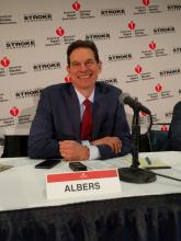
That’s because DEFUSE 3 enrolled patients with a baseline National Institutes of Health Stroke Scale (NIHSS) score of at least 6, while patients in DAWN required a score of at least 10, a loosening that allowed inclusion of 31 patients with scores of 6-9 in DEFUSE 3. Another, less restrictive criterion was the patient’s prestroke mRS score, which could have been 0-2 in DEFUSE 3 but could be only 0-1 in DAWN. Thirteen of the DEFUSE 3 patients had prestroke mRS scores of 2. DEFUSE 3 also had somewhat more liberal criteria for the size of a patient’s infarcted core at enrollment, with 41 patients who would have been excluded from DAWN because of an overly large infarcted core, Dr. Albers said in his presentation at the conference.
Currently, for patients presenting more than 16 hours following their stroke onset but within 24 hours, the DAWN enrollment criteria exclusively determine which patients should get thrombectomy.
One area where these rules could be bent is by a more thoughtful approach to the prestroke mRS score rule out, such as patients with orthopedic or rheumatic complications that limit mobility and give them an mRS score of 3. “We don’t have data for patients with back pain who can’t walk. I currently take these patients to endovascular therapy, and I’m sure many others do, too,” Dr. Khatri said.
Another potential way to grow the inclusion criteria is to investigate thrombectomy in patients with larger infarcted cores than were enrolled in DAWN and DEFUSE 3, but assessing this will require a new prospective study, Dr. Albers said.
Running the 6- to 24-hour numbers
Adoption of the 6- to 24-hour time window for endovascular intervention in selected patients means that suddenly the U.S. acute stroke infrastructure needs to accommodate a significantly increased number of patients. Just how many added patients this means is uncertain for the time being, and will vary from region to region and center to center. Dr. Albers roughly guessed that the new late time window might double the number of stroke patients undergoing thrombectomy at his center in Stanford. Dr. Khatri put together a more data-driven but still very speculative estimate that at the University of Cincinnati, it will mean about 40% more stroke patients going to thrombectomy. She shared the numbers behind this estimate in a report she gave at the conference.
To calculate the incremental change produced by the late time window, she used data collected on 2,297 acute ischemic stroke patients from the Greater Cincinnati/Northern Kentucky region who were seen at the University of Cincinnati during 2010. Prior analysis by Dr. Khatri and her associates showed that 159 of these patients presented quickly enough and with an appropriate stroke to qualify for thrombolytic therapy, and that 29 patients would have qualified for thrombectomy performed during the 0- to 6-hour time window.
In the new analysis Dr. Khatri calculated that 791 patients presented at 5-23 hours, and of these 34 had other features that would have made them eligible for enrollment in DAWN. Because no imaging data existed for these 2010 patients, she applied an estimate that 22% of these patients would qualify by the size of their infarcted core and ischemic penumbra, resulting in seven additional thrombectomy-eligible patients. Accounting for patients who would qualify by the more liberal DEFUSE 3 criteria added another 5 patients for a total increment of 12 patients during 2010 who would have been eligible for thrombectomy, about 40% of the number from the 0- to 6-hour window.
She noted that “this is likely an underestimate,” and “too small a sample to project to national estimates,” but concluded that “resources must be adapted to account for this increased volume in endovascular treatment.”
Dr. Khatri acknowledged that the new 6- to 24-hour window for endovascular therapy, and concerns about imaging delays in 0- to 6-hour patients, raise challenging issues regarding the message to give U.S. clinicians about treating acute ischemic stroke patients.
“We have a mandate to figure it out in every region. There is no doubt that stroke patients need access to this care. We need to become a lot more aggressive with endovascular treatment. It’s so gratifying to see the outcomes that we’re seeing,” Dr. Khatri said. “A lot of work is needed to accommodate endovascular therapy–eligible patients in an extended time window. We need more refined prehospital triage tools, we need to adequately implement imaging software, and we need increased capacity to perform endovascular treatment with additional procedure suites, operators, and ICU beds.”
Dr. Jovin has been a consultant to Anaconda Biomed, Blockade Medical, Cerenovus, FreeOx Biotech, and Silk Road Medical. Dr. Nogueira has received travel expense reimbursement from Stryker. Dr. Khatri has been a consultant to Biogen, Medpace/Novartis, and St. Jude; has received travel support from Neuravi and EmstoPA; and has received research support from Genentech, Lumosa, and Neurospring. Dr. Albers has an ownership interest in iSchemaView, the company that markets the RAPID imaging software, and is a consultant to iSchemaView and Medtronic.
EXPERT ANALYSIS FROM ISC 2018
