User login
Milk interferes with levothyroxine absorption
ORLANDO – Consuming milk and levothyroxine at the same time reduced the absorption of the thyroid hormone replacement, according to Deborah Chon, MD, speaking at a press briefing at the annual meeting of the Endocrine Society.
This finding comes from a small study of 10 healthy adults with normal TSH concentration at baseline. The study participants had a mean age of 34 years and 6 were men.
The total serum T4 absorption over 6 hours, calculated as area under the curve, was significantly lower when participants took levothyroxine and milk concurrently, compared with taking it alone (67.26 vs. 73.48; P equals .02).
The best interval between taking levothyroxine and drinking milk has yet to be established, according to Dr. Chon, who is on the faculty of the University of California, Los Angeles. Findings from earlier research showed that use of elemental calcium supplements interfere with absorption of levothyroxine.
In 2014, levothyroxine became the most commonly prescribed drug in the United States, according to a survey by the IMS Institute for Healthcare Informatics, now QuintilesIMS. Patients who need to take it because of Hashimoto’s thyroiditis or after thyroidectomy are often unhappy with how they feel. Dose adjustments are common as endocrinologists struggle to improve patients’ quality of life. It may be that a simple strategy of not taking the thyroid replacement at the same time as milk might leave patients feeling better.
Dr. Chon reported having no relevant financial conflicts of interest.
This article was updated 4/10/17.
AT ENDO 2017
Key clinical point:
Major finding: Calculated as an area under the curve, simultaneous consumption of levothyroxine and milk reduced absorption of the supplement by a mean of 67.26 vs. 73.48 when taken without milk.
Data source: A pharmacokinetic study of 10 healthy subjects.
Disclosures: Dr. Chon reported having no relevant conflicts of interest.
Quality of life in hypothyroidism is key, but no treatment guide exists
ORLANDO – The quality of life of someone with treated hypothyroidism is worse than that of the general population and requires endocrinologists to think “outside the pill box,” said Anna M. Sawka, MD, PhD, of the University of Toronto, at a spring symposium presented by the American Thyroid Association.
While issues such as whether monotherapy with T4 is the treatment of choice or whether there is a subset of patients who might benefit from treatment with either T4/T3 in combination or even with extract of dessicated thyroid, many patients with treated hypothyroidism continue to feel bad, judging from discussion during the symposium.
As defined by the CDC, health-related quality of life can be energy level, mood, health risks and condition, functional and socioeconomic status. Here, at this meeting, the focus is on treatment. But QoL is so much more than that. It is recorded by the patient; it’s their perceptions. Of the available disease-specific–measures of QoL, many have not been validated.
One group of researchers assessed four QoL assessment questionnaires and gave top rating to the ThyPRO (thyroid-specific patient reported outcome) questionnaire for use in patients with hypothyroidism due to Hashimoto’s disease or thyroidectomy (J Clin Epidemiol. 2016 Oct;78:63-72).
The ThyPRO looks at two factors that are key to QoL in hypothyroidism: satisfaction with thyroid hormone and symptom control.
Looking at hypothyroidism from the perspective of the patient is catching on in endocrinology. Dr. Sawka reviewed data from 6 studies of QoL in people with treated hypothyroidism. The years of the studies ranged from 2002 to 2017 and the number of patients ranged from 58 to 591. While only one study used the ThyPRO, all found that QoL was worse in patients with treated hypothyroidism than in the general population.
Findings from one study based on in-depth interviews with 16 patients, of whom 5 had hypothyroidism and 11 had hyperthyroidism, showed that a key trait of either condition was that of losing control over their physical and mental states, with the hypothyroid group reporting feeling drained while the others said they felt sped up. The signs of hypothyroidism can be attributed to other things, such as aging or stress, and that ambiguity frustrated the patients in the study. Also key was the experience of having to negotiate their disease, with a keen awareness that their endocrinologist did not validate their experience, that they had to look to lab tests to tell them the state of their disease, and they had to deal with functional limitations (Qual Health Research. 2015 Jul;25[7]:945-53).
Dr. Sawka reported that she has no financial disclosures.
AT AN ATA SYMPOSIUM
Moderate exercise benefits hypertrophic cardiomyopathy patients
WASHINGTON – A regimen of moderately intense exercise for 16 weeks produced no adverse effects and led to a clinically meaningful improvement in exercise capacity in a multicenter, randomized trial involving 136 patients with hypertrophic cardiomyopathy.
While finding that regular exercise produced a statistically significant improvement, compared with control patients fulfilled the study’s primary endpoint, it was perhaps as important that patients with hypertrophic cardiomyopathy (HCM) randomized to the exercise arm experienced no ill effects, a finding that bucked conventional wisdom that, once diagnosed, HCM patients must adhere to a largely inactive lifestyle.
“I see patients with HCM who tell me that, when they were first diagnosed, they were told by their physician not to do anything at all,” said Dr. Saberi, a cardiologist at the University of Michigan in Arbor. Although she cautioned that the findings from this modestly sized trial cannot prove that exercise is safe for HMC patients – something that would require randomizing more than 2,000 patients and following them for about 3 years – the findings provide some level of reassurance that regular, moderately intense exercise as simple as walking can benefit patients and likely not cause any problems. “We saw no signal of harm,” she stressed in an interview. “I would recommend moderate-intensity, regular exercise. Physicians can feel comfortable using the exercise prescription” used in the study, she advised.
Concurrently with Dr. Saberi’s report at the meeting, the results also appeared in an article published online (JAMA. 2017 Mar 17. doi: 10.1001/jama.2017.2503).
The Study of Exercise Training in Hypertrophic Cardiomyopathy (RESET-HCM) ran during 2010-2015 at the University of Michigan and Stanford University, and enrolled 136 patients 18-80 years old diagnosed with HCM but without more severe manifestations such as a history of exercise-induced syncope or ventricular arrhythmia, a left ventricular ejection fraction less than 55%, recent clinical decompensation, or a history of a severe hypotensive response to exercise. Enrolled patients averaged about 50 years old, their baseline peak oxygen consumption, VO2, was about 22 mL/kg per min, and their maximal ventricular wall thickness was about 21 mm.
Following a comprehensive baseline heart function and imaging assessment, including a cardiopulmonary exercise test, the 67 patients in the intervention arm received an individualized exercise prescription based on their heart rate reserve. At a minimum, they started with three exercise sessions per week lasting at least 20 minutes on their own, a regimen designed to bring them to 60% of their heart rate reserve. Their prescription gradually increased to four to seven exercise periods weekly lasting up to 60 minutes and designed to bring them to 70% of their heart rate reserve. Although most patients accomplished this through walking, some engaged in activities that included cycling, swimming, jogging, or elliptical training. The researchers told the 69 control patients to continue doing their usual activities. Among the patients who entered the study, 57 in the exercise group and 56 controls remained for the full 16 weeks and had a complete final assessment.
After 16 weeks, the average change in peak VO2, was 1.35 mL/kg per min in the intervention group and 0.08 mL/kg per min in the control patients, a statistically significant difference for the primary endpoint and a 6% increase in exercise capacity, compared with baseline for the intervention group. Dr. Saberi noted that, in a prior study of patients with chronic heart failure, a 6% increase in peak VO2 linked with an 8% decrease in cardiovascular mortality of heart failure hospitalization.
This result is “a glimmer of hope for using exercise in HCM patients,” she said. “There are no other disease-modifying treatments for HCM, so this is the first ‘treatment’ to have any impact on the disease.”
The patients who exercised had no identified major adverse effects or changes in cardiac morphology after 16 weeks. Assessment of physical function quality of life using the SF-36v2 showed an average 8-point improvement in the patients who exercised, compared with the controls. In addition, “a lot of patients in the exercise group told us that they felt better,” Dr. Saberi said.
Because patients with HCM are often advised to limit their activity, the consequence is “we increasingly see HCM patients who are obese and have complications such as sleep apnea and atrial fibrillation. Patients are even fearful of going up and down stairs.” She stressed the simplicity and ease of the intervention she and her colleagues tested, which can involve nothing more than regular, daily walks of 20 minutes or more. Dr. Saberi advised tailoring the intensity and frequency of the exercise intervention to each patient and avoiding having the patient push beyond what they find comfortable.
A multicenter U.S. registry, the Lifestyle and Exercise in Hypertrophic Cardiomyopathy Study (LIVE-HCM), is now enrolling HCM patients that should eventually follow enough patients for a long enough period of time to definitely prove whether moderate exercise is safe for HCM patients, but the results will not be available for several more years, Dr. Saberi said. A recent analysis estimated that one in every 200 adults has HCM (J Am Coll Cardiol. 2015 Mar 31;65[12]:1249-54), which means the disease likely affects more than one million Americans.
mzoler@frontlinemedcom.com
On Twitter @mitchelzoler
AT ACC 17
Key clinical point:
Major finding: Average exercise capacity, peak VO2, increased by 1.27 mL/kg per min in exercising patients compared with nonexercising controls.
Data source: RESET-HCM, a multicenter, randomized trial with 136 hypertrophic cardiomyopathy patients.
Disclosures: Dr. Saberi had no disclosures.
EBUS scope, EUS-FNA similarly effective
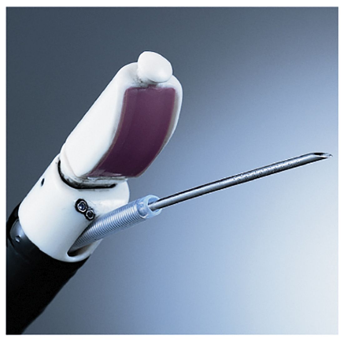
When assessing a patient for lung cancer, a procedure involving the insertion of an EBUS scope in the esophagus – EUS-B-FNA – can achieve similarly accurate results as endoscopic ultrasound guided–fine-needle aspiration (EUS-FNA), according to a new study.
This finding could lead patients to choose EUS-B-FNA over EUS-FNA – the standard of care for analyzing potential metastasis of the left adrenal glands (LAGs) – resulting in both time and cost savings for patients.
“A recent report showed that LAG visualization using the EBUS scope was possible in 85% of patients,” according to the authors of this study, including Prof. Jouke T. Annema, MD of the University of Amsterdam. Prior to this new research, it was unknown to what extent a single EBUS scope adequately assess and sample the LAGs and how its performance related to the use of a conventional endoscopic ultrasound–guided scope (Lung Cancer. 2017. doi: org/10.1016/j.lungcan.2017.02.011).
Dr. Annema and his coauthors recruited patients from four centers – three in the Netherlands, one in Poland – and followed them prospectively. Patients with “(suspected) lung cancer [who] had an indication for both mediastinal lymph node and LAG sampling” were recruited for the study. The researchers followed 44 patients through final diagnosis to determine if they ultimately had lung cancer.
Subjects first received complete mediastinal and hilar staging of lung cancer and any present tumors via an EBUS and EUS-B procedure. Following an EBUS examination of the mediastinum, the EBUS scope was retracted from the trachea and positioned into the esophagus for an examination of the mediastinal nodes. Then, the EBUS scope was advanced into the stomach for identification of the LAG. Afterward, the routine EUS-FNA was performed. LAG analysis across both methods involved visualizing the LAG and collecting an adequate tissue sample for testing.
“In short, in order to locate the LAG, a structured three step approach was used according to the EUS assessment tool (EUS-AT): identification of the liver, abdominal aorta, coeliac trunk, left kidney, and LAG,” the authors noted. “By turning the EBUS scope clockwise from the liver, the abdominal aorta and coeliac trun[k] are identified. By subsequently turning the EBUS scope gently in caudal direction, the left kidney and LAG are identified.”
Endoscopists then evaluated both procedures in each subject according to feasibility and practicability to determine if the findings of the experimental procedure were usable. Finally, a cytologic exam was conducted, using Giemsa or Papanicolaou staining to determine if any present cancer had metastasized, and a final diagnosis was made.
LAG analysis had a success rate of 89% (39/44; 95% confidence interval, 76-95%) for EUS-B-FNA, compared with 93% (41/44; 95% CI, 82-98%) for EUS-FNA. Similarly, when looking at the rate of sensitivity for LAG metastases, EUS-B had a rate of sensitivity for LAG metastases of at least 87% (95% CI, 65-97%), while EUS-FNA was found to be at least 83% (95% CI, 62-95%. Endoscopists were equally satisfied with both procedures in the “majority” of cases in this study.
“In [five] cases (11%), the EUS-B-FNA procedure was unsuccessful, due to the inability to make good contact of the ultrasound transducer and the stomach wall,” the authors explained. “The conventional EUS scope is more stable as a result of the increased tube diameter. Another advantage of the conventional echo-endoscope is its wider scanning angle. ... The conventional EUS scope is also longer than the EBUS scope, [but that] does not seem to be the limiting factor.”
No funding source was disclosed for this study. The authors reported no relevant financial disclosures.
FROM LUNG CANCER
Key clinical point:
Major finding: EUS-B-FNA had a success rate of 89%, versus 93% for EUS-FNA, while sensitivity for LAG metastases were 87% and 83%, respectively.
Data source: Multicenter, prospective study of 44 consecutive suspected lung cancer patients.
Disclosures: No funding source disclosed. Authors reported no relevant financial disclosures.
Three signs predict hypercalcemic crisis in hyperparathyroid patients
LAS VEGAS – A triad of signs – elevated serum calcium, elevated parathyroid hormone, and a history of kidney stones – can predict hypercalcemic crisis among patients with hyperparathyroidism, a study showed.
Patients who present with the trifecta should be considered for expedited parathyroidectomy, Andrew Lowell said at the Association for Academic Surgery/Society of University Surgeons Academic Surgical Congress.
The model was based on a retrospective analysis of 183 patients with hyperparathyroidism who were hospitalized and treated for hypercalcemia. These were divided into two groups: those who developed a hypercalcemic crisis (29) and those who did not (154).
There were no significant differences in age, sex, alcohol or tobacco use, body mass index, or Charlson comorbidity score. However, those who developed a crisis were significantly more likely to have had kidney stones (31% vs. 14%). Their preoperative serum calcium level was also significantly higher (median, 13.8 vs. 12.4 mg/dL), and they had significantly higher parathyroid hormone (PTH) levels (median, 318 vs. 160 pg/mL). Their preoperative vitamin D level was also significantly lower (median, 16 vs. 26 ng/mL).
Parathyroidectomy was equally effective in both groups, but twice as many patients with crisis needed a multigland resection (24% vs. 12%).
Mr. Lowell conducted a univariate, and then a multivariate, analysis to determine independent risk factors for hypercalcemic crisis. This revealed that a higher preoperative calcium level, an elevated PTH level, and a history of kidney stones were significantly associated with crisis.
Hypercalcemia developed in:
• 91% of those with a serum calcium higher than 13.25 mg/dL and 6% of those with a lower serum calcium level.
• 60% of those with a PTH of 394 pg/mL or higher and 19% of those with a PTH less than 394 pg/mL.
• 31% of those with a history of kidney stones and 14% of those without such a history.
The investigators created a decision tree that begins with a calcium level greater than 13.25 mg/dL, a PTH level higher than 394 pg/mL, and a Charlson comorbidity index of 4 or greater. The model carried an overall predictive accuracy of 90% and a positive predictive value of 76%, Mr. Lowell said.
Session moderator Benjamin Poulose, MD, of Vanderbilt University, Nashville, Tenn., said the model looks very good on paper, but might be challenging to implement when assessing emergent patients.
Mr. Lowell suggested that it would be better employed in an outpatient setting.
“I think this would be more useful in the situation of a physician who knows that patient’s comorbidities, in the context of counseling, to determine” the need for and timing of surgery, he said.
He had no relevant financial disclosures.
On Twitter @Alz_Gal
AT THE ACADEMIC SURGICAL CONGRESS
Key clinical point: .
Major finding: A model based on the biomarkers and comorbidity status had a predictive accuracy of 90% and a positive predictive value of 76%.
Data source: A study cohort consisting of 183 patients.
Disclosures: Mr. Lowell had no relevant financial disclosures.
Pheochromocytoma linked to higher risk of postop complications
LAS VEGAS – Patients with pheochromocytoma are likely to have preoperative comorbidities that predispose them to postoperative cardiopulmonary complications, leading to a longer length of stay and greater hospital charges.
A 5-year national database review found high rates of chronic lung disease and malignant hypertension among these patients, Punam P. Parikh, MD, said at the Association for Academic Surgery/Society of University Surgeons Academic Surgical Congress.
“They are also at an increased risk for vascular injury during surgery, perhaps because these tumors are so vascular in nature, and associated intraoperative blood transfusion,” said Dr. Parikh of the University of Miami. Postoperatively, patients with pheochromocytoma are twice as likely to experience respiratory complications and almost eight times as likely to experience cardiac complications as patients with other hormonally active adrenal tumors.
Dr. Parikh queried the National Inpatient Sample to find patients who underwent adrenalectomy for the rare adrenal tumor from 2006 to 2011. Of 27,312 patients who had adrenalectomy during the 5-year period, 22% had hormonally active adrenal tumors. Of these, just 1.4% (85) were pheochromocytoma. Other hormonally active adrenal tumors were Conn’s syndrome (65%) and Cushing’s syndrome (33%).
A number of comorbidities were significantly more common among pheochromocytoma patients than among those with Conn’s and Cushing’s syndromes, including congestive heart failure (12% vs. 4% in the other syndromes) and malignant hypertension (5% vs. 3% and 0.3%, respectively). A third of pheochromocytoma patients also had diabetes.
The rate of intraoperative complications was significantly higher in these patients (22%) than in those with Conn’s and Cushing’s (11% and 17%). Vascular injury occurred in 6% vs. 2% and 4%, respectively. Almost a quarter of pheochromocytoma patients (21%) needed an intraoperative transfusion, compared with 2% of Conn’s patients and 3% of Cushing’s patients.
There were also more postoperative complications among pheochromocytoma patients than Conn’s or Cushing’s patients, including cardiac (6% vs. 0.4% and 0.6%) and pulmonary complications (17% vs. 6% and 9%).
Not surprisingly, Dr. Parikh said, pheochromocytoma patients had longer hospital stays (5 days), compared with patients with the other tumors (3 days). Hospital charges were also higher for those with pheochromocytoma ($50,000) than those with Conn’s or Cushing’s ($35,500 and $46,334, respectively).
A multivariate analysis concluded that pheochromocytoma was an independent risk factor for intraoperative blood transfusion (odds ratio, 4.2), postoperative cardiac complications (OR, 7.6), and postoperative respiratory complications (OR, 1.9).
Dr. Parikh suggested that patients with pheochromocytoma could benefit from some preoperative preparation.
“Because of these issues, these high-risk patients should undergo appropriate preoperative medical optimization in preparation for their adrenalectomy,” she noted.
She had no financial disclosures.
msullivan@frontlinemedcom.com
On Twitter @Alz_Gal
AT THE ACADEMIC SURGICAL CONGRESS
Key clinical point: that predispose them to postoperative complications and prolonged hospital stays.
Major finding: Pheochromocytoma patients had more postoperative complications than Conn’s or Cushing’s patients, including cardiac (6% vs. 0.4% and 0.6%) and pulmonary complications (17% vs. 6% and 9%).
Data source: The database review comprised more than 27,000 patients with adrenal tumors.
Disclosures: Dr. Parikh had no financial disclosures.
One-third of micropapillary thyroid cancer found to be multifocal
LAS VEGAS – Micropapillary thyroid carcinoma may not be as indolent as generally thought, according to the findings of a retrospective study of thyroidectomy cases.
A review of 213 patients diagnosed with the cancer found that 34% of them had multifocal disease, and 14%, metastatic disease, Maggie Bosley reported at the Association for Academic Surgery/Society of University Surgeons Academic Surgical Congress.
Ms. Bosley presented a review of 213 consecutive patients who underwent thyroidectomy from 2007 to 2015, and were found to have micropapillary thyroid cancer. She reviewed the pathology reports for tumor size, presence or absence of metastases in the central and lateral node basins, and multifocality.
Most of the patients (88%) were women, with an average age of 56 years, although the range was wide (18-89 years).
About a third of the patients (73; 34%) had multifocal disease. This was bilateral in 21 (29%). Metastasis to the central nodes was present in 31 patients (14%); 4 of these patients also had positive lateral neck node metastases (2%).
“Approximately 13% of patients with node metastasis also required selective lateral neck dissections,” Ms. Bosley said.
She noted that, in 2015, the American Thyroid Association published a set of guidelines for diagnosing and treating micropapillary cancer. The guidelines suggest that most of these cancers can be safely followed with ultrasound exams, if there is no extrathyroid extension or nodal metastasis.
“However, ultrasound surveillance [quality] is very operator dependent,” Ms. Bosley said. Technician skill “could potentially impact the quality of surveillance.”
She had no relevant financial declarations.
msullivan@frontlinemedcom.com
On Twitter @Alz_Gal
AT THE ACADEMIC SURGICAL CONGRESS
Key clinical point:
Major finding: Micropapillary thyroid cancer was metastatic in 14% of cases.
Data source: A review involving 213 patients.
Disclosures: Ms. Bosley had no relevant financial disclosures.
Intraoperative PTH spikes may mean multigland disease
SAN DIEGO – Intraoperative spikes of parathyroid hormone don’t predict a failed parathyroidectomy, according to a retrospective study of patients who had the surgery for hyperparathyroidism.
They should, however, raise the suspicion of multigland disease, Richard Teo said at the Association for Academic Surgery/Society of University Surgeons Academic Surgical Congress.
“Significantly more patients with intraoperative spikes didn’t achieve this drop, and they had a higher rate of multigland disease requiring bilateral neck exploration,” he said. “But although spikes did increase the suspicion of multigland disease, they did not affect the operative success rate in this study.”
He presented a retrospective analysis of 683 patients who underwent parathyroidectomy for hyperparathyroidism. These patients were largely female (76%). Those who had the intraoperative spikes were older (60 vs. 58 years) and had higher preoperative calcium than patients without spikes. There were no differences in parathyroid hormone (PTH) or creatinine levels.
Operative success – described as normocalcemia at least 6 months after surgery – occurred in 98% of the entire group. The operative failure rate was 0.9%, and the recurrence rate was 1%. About 5% of the entire group had multigland disease.
Intraoperative PTH spikes occurred in 224 patients (33%). Compared with those without spikes, patients with spikes were significantly less likely to achieve the PTH decrease of 50% or greater at 10 minutes after gland excision (70% vs. 90%).
Bilateral neck explorations were significantly more common among those with spikes (10% vs. 5%), as was multigland disease (8% vs. 3%). There was no significant difference in operative time (54 vs. 59 minutes).
Postoperative outcomes were similar. At last follow-up, calcium levels were identical (9.3 mg/dL) in the group with and the group without a spike in PTH. In addition, the PTH levels were not significantly different (47 vs. 57 pg/mL).
Operative success was achieved in 98% of both groups, with a 2% failure rate in both groups. Recurrence was slightly, though not significantly, less in the spike group (0.4% vs. 1.3%).
“We were able to show that intraoperative PTH spikes don’t predict a poor outcome of parathyroidectomy,” Mr. Teo said. “We also feel this study reaffirms the clinical utility of the 50% or greater intraoperative PTH drop as a predictor of the successful removal of all hypersecreting parathyroid tissue during parathyroidectomy guided by intraoperative PTH monitoring.”
He had no financial disclosures.
msullivan@frontlinemedcom.com
On Twitter @alz_gal
Key clinical point: Major finding: Intraoperative PTH spikes occurred in 33% of parathyroidectomy patients, and 8% of patients with spikes had multigland disease.
Data source: The retrospective study comprised 683 patients.
Disclosures: He had no financial disclosures.
Thyroid disease does not affect primary biliary cholangitis complications
While associations are known to exist between primary biliary cholangitis (PBC) and many different types of thyroid disease (TD), a new study shows that the mere presence of thyroid disease does not have any bearing on the hepatic complications or progression of PBC.
“The prevalence of TD in PBC reportedly ranges between 7.24% and 14.4%, the most often encountered thyroid dysfunction being Hashimoto’s thyroiditis,” wrote the study’s authors, led by Annarosa Floreani, MD, of the University of Padua (Italy).
Of the 921 total patients enrolled, 150 (16.3%) had TD. The most common TD patients had were Hashimoto’s thyroiditis, which 94 (10.2%) individuals had; Graves’ disease, found in 15 (1.6%) patients; multinodular goiter, which 22 (2.4%) patients had; thyroid cancer, which was found in 7 (0.8%); and “other thyroid conditions,” which affected 12 (1.3%) patients. Patients from Padua had significantly more Graves’ disease and thyroid cancer than those from Barcelona: 11 (15.7%) versus 4 (5.0%) for Graves’ (P = .03), and 6 (8.6%) versus 1 (1.3%) for thyroid cancer (P = .03), respectively. However, no significant differences were found in PBC patients who had TD and those who did not, when it came to comparing the histologic stages at which they were diagnosed with PBC, hepatic decompensation events, occurrence of hepatocellular carcinoma, or liver transplantation rate. Furthermore, TD was not found to affect PBC survival rates, either positively or negatively.
“The results of our study confirm that TDs are often associated with PBC, especially Hashimoto’s thyroiditis, which shares an autoimmune etiology with PBC,” the authors concluded, adding that “More importantly … the clinical characteristics and natural history of PBC were much the same in the two cohorts, as demonstrated by the absence of significant differences regarding histological stage at diagnosis (the only exception being more patients in stage III in the Italian cohort); biochemical data; response to UDCA [ursodeoxycholic acid]; the association with other extrahepatic autoimmune disorders; the occurrence of clinical events; and survival.”
No funding source was reported for this study. Dr. Floreani and her coauthors did not report any financial disclosures relevant to this study.
Key clinical point:
Major finding: 150 of 921 PBC patients had TD (16.3%), but there was no correlation between PBC patients who had TD and their histologic stage either at diagnosis, hepatic decompensation events, occurrence of hepatocelluler carcinoma, or liver transplantation rates.
Data source: Prospective study of 921 PBC patients in Padua and Barcelona from 1975 to 2015.
Disclosures: No funding source was disclosed; authors reported no relevant financial disclosures.
ATA’s risk assessment guidelines for thyroid nodules using sonography patterns validated
DENVER – The malignancy risk of thyroid nodules can be assessed with reassuring accuracy using ultrasound and the guidelines developed by the American Thyroid Association.
Ultrasound assessment is the first step of the evaluation of any patient with one or more thyroid nodules. “Maybe it shouldn’t be, but, for now, it is,” noted David L. Steward, MD, at the annual meeting of the American Thyroid Association.
The ATA guidelines categorize thyroid nodules on the basis of their ultrasound patterns, with the high risk of malignancy being in nodules that are taller than they are wide and /or have microcalcifications, irregular margins, hypoechoic areas, extrathyroidal extension, interrupted rim calcification with soft tissue extrusion, and suspicious lymph nodes. Between 70% and 90% of thyroids with such patterns will contain malignancy, according to the ATA guidelines. Lesions with an intermediate risk of malignancy have such sonographic findings as hypoechoic solid tissue and regular margins; between 10% and 20% of these are malignant. The third category in the ATA’s guidelines are those that are of low suspicion, with hyperechoic solid tissue, isoechoic solid tissue, partially cystic with eccentric solid area, and regular margins; 5%-10% of these are malignant. Thyroid nodules with a very-low risk of malignancy (less than 3%) are spongiform or partially cystic with no suspicious findings. Finally, benign nodules, of which less than 1% contain malignancy, are cysts, he said.
“We found that the size of the nodule on ultrasound that underwent fine needle aspiration was inversely correlated with malignancy risk: The lower risk nodules were larger,” he said.
Using the ATA’s system, 9 (4%) of the nodules were high risk, 64 (31%) were intermediate risk, 79 (38%) were low risk, 54 (26%) were very-low risk, and none were benign. Five of the nodules were not included in the results presented.
There was good correlation between the Bethesda and ATA classification systems. Of the lesions that were malignant or suspicious for malignancy in the Bethesda system, 77% were very-high risk for malignancy on ultrasound according to the ATA. Of the lesions that were atypia of undetermined significance (AUS)/follicular lesion of undetermined significance (FLUS), 22% were very high risk according to the ATA. Neither of the systems classified as malignant any of the lesions as follicular/Hurthle cell cancer, benign, or nondiagnostic.
The AUS/FLUS nodules “tend to be all over the map,” he noted. Looking at just the AUS/FLUS nodules, malignancy was found on pathology in 100% classified by the ATA system as being high risk; in 21% of those called intermediate risk; in 17% of those called low risk; and in 12% of the very-low risk group.
The study was funded by the University of Cincinnati. Dr. Steward said his only disclosure is that he was a member of the ATA committee that wrote the guidelines under evaluation in this study.
Key clinical point:
Major finding: Of the lesions that were malignant or suspicious for malignancy in the Bethesda system, 77% were very-high risk for malignancy on ultrasound, according to the ATA.
Data source: Prospective validation of the ATA’s ultrasound risk assessment guidelines on 211 thyroid nodules excised from 199 patients.
Disclosures: The study was funded by the University of Cincinnati. Dr. Steward said his only disclosure is that he was a member of the ATA committee that wrote the guidelines under evaluation in this study.


