User login
Compartment Syndrome in Children: Diagnosis and Management
Compartment syndrome (CS) is one of the true orthopedic emergencies. Identifying the high-risk patient, making a prompt diagnosis, and initiating effective treatment are the crucial steps in avoiding a poor outcome. A physician’s inability to communicate with young children can interfere with diagnosing CS in a timely fashion. Many young patients in hospitals are admitted to pediatric floors where routine orthopedic care is not the norm and staff are unfamiliar with the signs and symptoms of evolving CS. As orthopedic surgeons are often involved in caring for these patients, they should be aware of the aspects of CS that are unique to children and should be able to identify patients who are at risk and would benefit from close monitoring. In addition, given the consequences of late diagnosis, early diagnosis is important from a medicolegal standpoint. Only 44% of cases of adult and pediatric CS are decided in favor of treating physicians, compared with 75% of cases in other orthopedic malpractice claims.1,2
Risk Factors for Posttraumatic Compartment Syndrome
Supracondylar Humeral Fracture
CS is a well-described complication of this injury. CS develops in 0.1% to 0.3% of children who present with supracondylar humeral fracture.3,4 Casted elbow flexion beyond 90° and concomitant vascular injury put these children at increased risk for CS. Mubarak and Carroll5 reported 9 cases of CS in the volar compartment of the forearm after an extension-type supracondylar humeral fracture and attributed 8 of them to elbow flexion beyond 90° after closed reduction. In 29 children with supracondylar humeral fracture,Battaglia and colleagues3 found the highest compartment pressure in the deep volar compartment, especially near the fracture site, as well as a significant increase in pressure with the elbow flexed beyond 90°.
In a study of children with supracondylar humeral fracture, Choi and colleagues6 found 2 cases of CS among 9 patients who presented with a pulseless, poorly perfused hand and no cases of CS among 24 patients who presented with a pulseless but well-perfused hand.
Studies have found that a treatment delay of 8 to 12 hours did not increase the rate of CS in Gartland type 2 and type 3 fractures.7-10 The investigators in these studies did not recommend delaying treatment of patients with neurologic deficit and absent radial pulse. Ramachandran and colleagues4 reported 11 cases of CS in patients with low-energy supracondylar humeral fracture and intact radial pulse at presentation. The patients who developed CS presented with severe swelling, and their mean treatment delay was 22 hours (range, 6-64 hours). Given the data, we do not recommend delayed treatment for children with supracondylar humeral fracture and neurologic deficit or absent pulse. We do recommend close inpatient preoperative monitoring of patients with severe swelling.
CS after supracondylar humeral fracture is mostly seen in the volar compartment of the forearm, but it has also been reported in the mobile wad, the anterior arm compartment, and the posterior arm compartment.11,12
Floating Elbow
CS has been reported in children with ipsilateral humeral and forearm fractures. Blakemore and colleagues13 reported a 33% rate of CS in children with displaced distal humeral and forearm fractures. A retrospective review of 16 cases of floating elbow treated at Boston Children’s Hospital found CS in 2 patients and incipient CS in 4 of 10 patients with forearm fractures treated with closed reduction and plaster casting. There were no signs of CS in 6 patients with distal humeral and forearm fractures stabilized with Kirschner wires.14 Given the data, we do not recommend circumferential casting for forearm fractures in children with floating elbow.
Forearm Fracture
Haasbeek and Cole15 reported CS in 5 (11%) of 46 children with open forearm fracture. Yuan and colleagues16 reported CS in 3 (6%) of 50 open forearm fractures and 3 of 30 closed fractures treated with closed reduction and intramedullary nailing. They found increased risk for CS in patients with longer operative time, indicating prolonged closed manipulation of these fractures as a risk factor for CS. They did not find any cases of CS among 205 forearm fractures treated with closed reduction and casting.
Flynn and colleagues17 reported CS in 2 of 30 patients treated with intramedullary nailing within 24 hours of injury and in 0 of 73 patients treated after 24 hours.
Blackman and colleagues18 reported CS in 3 (7.7%) of 39 open forearm fractures and 0 of 74 closed fractures treated operatively. In their series, a small incision was made to facilitate reduction in 38 (51.4%) of 74 closed fractures to decrease closed manipulation and operative time. The rate of CS after intramedullary nailing of closed forearm fractures was lower in this series than in similar reports in the literature.
Reported data indicate increased risk for CS in children with open forearm fractures and fractures treated with closed reduction and intramedullary nailing, especially performed within 24 hours of injury, and prolonged closed manipulation performed during surgery. We recommend close monitoring of all children with operatively treated forearm fractures and, in particular, children with the risk factors mentioned.
Femoral Fracture
Although CS after femoral shaft fractures is not common, CS has been reported after 90/90 spica casting of femoral shaft fractures in children. Mubarak and colleagues19 reported on 9 children who developed calf CS after treatment of femoral shaft fracture in 90/90 spica casts. The technique used in 7 of the 9 reported cases involved initial application of a short leg cast and then traction applied to the leg—believed to cause impinging of the cast on the posterior compartment of the leg. The authors recommended an alternative method of applying spica casts, which is beyond the scope of this review.
Tibial Fracture
Children with tibial fracture, especially a fracture sustained in a motor vehicle accident, are at risk for CS. Hope and Cole20 found CS in 4 (4%) of 92 children with open tibial fracture.
Children with tibial tubercle fracture are at increased risk for CS because of concomitant vascular injury. Pandya and colleagues21 reported CS or vascular compromise in 4 of 40 patients with tibial tubercle fracture. We recommend close monitoring for signs of impending CS in children who present with high-energy tibial shaft fracture and tibial tubercle fracture.
Flynn and colleagues22 reported outcomes of 43 cases of acute CS of the leg in children treated at 2 pediatric trauma centers. Mean time from injury to fasciotomy was 20.5 hours (range, 3.9-118 hours). Functional outcome was excellent at time of follow-up; 41 of 43 cases had no sequelae, and the 2 patients who lost function underwent fasciotomy more than 80 hours after injury. Despite the long interval between injury and surgery, excellent results were achieved with fasciotomy, suggesting an increased potential for recovery in the pediatric population.
Mubarak23 reported on 6 cases of distal tibial physis fracture in patients who presented with severe pain and swelling of the ankle, hyposthesia of the first web space, weakness of the extensor hallucis longus and extensor digitorum communis, and pain on passive flexion of the toes. In all these patients, intramuscular pressure was more than 40 mm Hg beneath the extensor retinaculum and less than 20 mm Hg in the anterior compartment. All patients experienced prompt relief of pain and improved sensation and strength within 24 hours after release of the superior extensor retinaculum and fracture stabilization.
Miscellaneous and Nontraumatic Causes of Compartment Syndrome
Neonatal CS is very rare, and diagnosis is often missed. Neonatal CS is thought to be caused by a combination of low neonatal blood pressure and birth trauma.24 Ragland and colleagues25 reported on 24 cases of neonatal CS; in only 1 case was the diagnosis made within 24 hours.They described a “sentinel skin lesion” on the forearm of each patient as the sign of neonatal CS. Late diagnosis results in contracture and growth arrest of the involved extremity. In their series, only 1 patient underwent fasciotomy within 24 hours, and it resulted in a good functional outcome. High clinical suspicion is the key to early diagnosis and treatment of this rare pathology.
Medical problems that cause intracompartmental bleeding (hepatic failure, renal failure, leukemia, hemophilia) have been cited as causing CS.26-28 CS may be the first symptom of occult hemophilia29 Correction of the coagulation defect may take priority over surgical treatment in these cases, though the decision should be made on a case-by-case basis.26
CS in children can also be caused by snakebites. Shaw and Hosalkar30 reported on successful use of antivenin in preventing the need for surgical treatment in 16 of 19 patients with rattlesnake bites. Two patients had limited surgical débridement, and 1 underwent fasciotomy for CS. The authors recommended using antivenin to prevent CS in children with snakebites.30
Prasarn and colleagues2 reported on 12 cases of upper extremity CS in children in the absence of fractures. Of the 12 patients, 10 were managed in an intensive care unit and had an obtunded sensorium. Etiology in 7 (58%) of the 12 cases was iatrogenic (intravenous infiltration, retained phlebotomy tourniquet). In this series, 4 amputations were performed on affected extremities.
Diagnosis
Identification of evolving CS in a child is difficult because of the child’s limited ability to communicate and anxiety about being examined by a stranger. Orthopedists are trained to look for the 5 Ps (pain, paresthesia, paralysis, pallor, pulselessness) associated with CS. Examining an anxious, frightened young child is difficult, and documenting the degree of pain is not practical in a child who may not be able or willing to communicate effectively.
In a series of 33 children with CS, Bae and colleagues31 found that the 5 Ps were relatively unreliable in making a timely diagnosis. The authors also found that increased analgesic use was documented a mean of 7.3 hours before a change in vascular status and that it was a more sensitive indicator of CS in children. The resulting recommendation is that children at risk for CS be closely monitored for the 3 As (increasing analgesic requirement, anxiety, agitation).32
Regional anesthesia is used to control postoperative pain in adults and children.33,34 Injudicious use may mask the primary symptom (pain) of CS.32,35-38 Use of regional anesthesia in patients at high risk for CS is highly discouraged.
Although CS is a clinical diagnosis, compartment pressure measurements can be useful in making decisions in certain clinical scenarios. In an obtunded child or in a child with severe mental and communication disability, such a measurement can help confirm or rule out the diagnosis.
Normal compartment pressures are higher in children than in adults. Staudt and colleagues39 compared pressures in 4 lower leg compartments of 20 healthy children and 20 healthy adults. Mean pressure varied from 13.3 mm Hg to 16.6 mm Hg in children and from 5.2 mm Hg to 9.7 mm Hg in adults—indicating higher normal pressure in lower leg compartments in children.
Compartment pressures were reported highest within 5 cm of the fracture site.40 When clinically indicated, they should be measured in that area in an injured extremity. The pressure threshold that requires fasciotomy is debatable. Intracompartmental pressures of 30 to 45 mm Hg, or measurements less than 30 mm Hg of diastolic blood pressure (pressure change = diastolic blood pressure – compartment pressure), have been recommended as cutoffs by some authors.41-44 As resting normal compartment pressures are higher in children, these cutoffs cannot be used as reliably in children as in adults. Direct measurement of intracompartmental pressure is invasive and can be difficult in an agitated, awake child. The potential utility of near-infrared spectroscopy in the diagnosis of increased compartment pressure has been reported.45,46 This method uses differential light absorption properties of oxygenated hemoglobin to measure tissue ischemia—similar to the method used in pulse oximetry. Compared with pulse oximetry, near-infrared spectroscopy can sample deeper tissue (3 cm below skin level). Shuler and colleagues45 reported near-infrared spectroscopy findings for 14 adults with acute CS. Lower tissue oxygenation levels correlated with increased intracompartmental pressures, but the authors could not define a cutoff for which near-infrared spectroscopy measurements would indicate significant tissue ischemia. Use of this method in diagnosing CS in children was described in a case report.46
CS remains a clinical diagnosis. Informing family and staff about the signs and symptoms of this syndrome and closely monitoring analgesic use in these patients are crucial. Compartment pressure measurements can be used when the diagnosis is unclear, particularly in noncommunicative patients, but these values should be interpreted with caution.
Treatment
Once CS is diagnosed, emergent fasciotomy and decompression are indicated. Surgeons planning fasciotomy should be aware of the definitive treatment of the CS etiology. Treatment of clotting deficiency in cases caused by excessive bleeding, fracture fixation, and vascular repair may be indicated during fasciotomy and decompression.
Summary
Increased need for analgesics is often the first sign of CS in children and should be considered the sentinel alarm for ongoing tissue necrosis. CS remains a clinical diagnosis, and compartment pressure should be measured only as a confirmatory test in noncommunicative patients or when the diagnosis is unclear. Children with supracondylar humeral fractures, forearm fractures, tibial fractures, and medical risk factors for coagulopathy are at increased risk and should be monitored closely. When the diagnosis is made promptly and the condition is treated with fasciotomy, good long-term clinical results can be expected.
1. Bhattacharyya T, Vrahas MS. The medical-legal aspects of compartment syndrome. J Bone Joint Surg Am. 2004;86(4):864-868.
2. Prasarn ML, Ouellette EA, Livingstone A, Giuffrida AY. Acute pediatric upper extremity compartment syndrome in the absence of fracture. J Pediatr Orthop. 2009;29(3):263-268.
3. Battaglia TC, Armstrong DG, Schwend RM. Factors affecting forearm compartment pressures in children with supracondylar fractures of the humerus. J Pediatr Orthop. 2002;22(4):431-439.
4. Ramachandran M, Skaggs DL, Crawford HA, et al. Delaying treatment of supracondylar fractures in children: has the pendulum swung too far? J Bone Joint Surg Br. 2008;90(9):1228-1233.
5. Mubarak SJ, Carroll NC. Volkmann’s contracture in children: aetiology and prevention. J Bone Joint Surg Br. 1979;61(3):285-293.
6. Choi PD, Melikian R, Skaggs DL. Risk factors for vascular repair and compartment syndrome in the pulseless supracondylar humerus fracture in children. J Pediatr Orthop. 2010;30(1):50-56.
7. Gupta N, Kay RM, Leitch K, Femino JD, Tolo VT, Skaggs DL. Effect of surgical delay on perioperative complications and need for open reduction in supracondylar humerus fractures in children. J Pediatr Orthop. 2004;24(3):245-248.
8. Iyengar SR, Hoffinger SA, Townsend DR. Early versus delayed reduction and pinning of type III displaced supracondylar fractures of the humerus in children: a comparative study. J Orthop Trauma. 1999;13(1):51-55.
9. Leet AI, Frisancho J, Ebramzadeh E. Delayed treatment of type 3 supracondylar humerus fractures in children. J Pediatr Orthop. 2002;22(2):203-207.
10. Mehlman CT, Strub WM, Roy DR, Wall EJ, Crawford AH. The effect of surgical timing on the perioperative complications of treatment of supracondylar humeral fractures in children. J Bone Joint Surg Am. 2001;83(3):323-327.
11. Diesselhorst MM, Deck JW, Davey JP. Compartment syndrome of the upper arm after closed reduction and percutaneous pinning of a supracondylar humerus fracture. J Pediatr Orthop. 2014;34(2):e1-e4.
12. Mai MC, Beck R, Gabriel K, Singh KA. Posterior arm compartment syndrome after a combined supracondylar humeral and capitellar fractures in an adolescent: a case report. J Pediatr Orthop. 2011;31(3):e16-e19.
13. Blakemore LC, Cooperman DR, Thompson GH, Wathey C, Ballock RT. Compartment syndrome in ipsilateral humerus and forearm fractures in children. Clin Orthop Relat Res. 2000;(376):32-38.
14. Ring D, Waters PM, Hotchkiss RN, Kasser JR. Pediatric floating elbow. J Pediatr Orthop. 2001;21(4):456-459.
15. Haasbeek JF, Cole WG. Open fractures of the arm in children. J Bone Joint Surg Br. 1995;77(4):576-581.
16. Yuan PS, Pring ME, Gaynor TP, Mubarak SJ, Newton PO. Compartment syndrome following intramedullary fixation of pediatric forearm fractures. J Pediatr Orthop. 2004;24(4):370-375.
17. Flynn JM, Jones KJ, Garner MR, Goebel J. Eleven years experience in the operative management of pediatric forearm fractures. J Pediatr Orthop. 2010;30(4):313-319.
18. Blackman AJ, Wall LB, Keeler KA, et al. Acute compartment syndrome after intramedullary nailing of isolated radius and ulna fractures in children. J Pediatr Orthop. 2014;34(1):50-54.
19. Mubarak SJ, Frick S, Sink E, Rathjen K, Noonan KJ. Volkmann contracture and compartment syndromes after femur fractures in children treated with 90/90 spica casts. J Pediatr Orthop. 2006;26(5):567-572.
20. Hope PG, Cole WG. Open fractures of the tibia in children. J Bone Joint Surg Br. 1992;74(4):546-553.
21. Pandya NK, Edmonds EK, Roocroft JH, Mubarak SJ. Tibial tubercle fractures: complications, classification, and the need for intra-articular assessment. J Pediatr Orthop. 2012;32(8):749-759.
22. Flynn JM, Bashyal RK, Yeger-McKeever M, Garner MR, Launay F, Sponseller PD. Acute traumatic compartment syndrome of the leg in children: diagnosis and outcome. J Bone Joint Surg Am. 2011;93(10):937-941.
23. Mubarak SJ. Extensor retinaculum syndrome of the ankle after injury to the distal tibial physis. J Bone Joint Surg Br. 2002;84(1):11-14.
24. Macer GA Jr. Forearm compartment syndrome in the newborn. J Hand Surg Am. 2006;31(9):1550.
25. Ragland R 3rd, Moukoko D, Ezaki M, Carter PR, Mills J. Forearm compartment syndrome in the newborn: report of 24 cases. J Hand Surg Am. 2005;30(5):997-1003.
26. Alioglu B, Avci Z, Baskin E, Ozcay F, Tuncay IC, Ozbek N. Successful use of recombinant factor VIIa (NovoSeven) in children with compartment syndrome: two case reports. J Pediatr Orthop. 2006;26(6):815-817.
27. Lee DK, Jeong WK, Lee DH, Lee SH. Multiple compartment syndrome in a pediatric patient with CML. J Pediatr Orthop. 2011;31(8):889-892.
28. Dumontier C, Sautet A, Man M, Bennani M, Apoil A. Entrapment and compartment syndromes of the upper limb in haemophilia. J Hand Surg Br. 1994;19(4):427-429.
29. Jones G, Thompson K, Johnson M. Acute compartment syndrome after minor trauma in a patient with undiagnosed mild haemophilia B. Lancet. 2013;382(9905):1678.
30. Shaw BA, Hosalkar HS. Rattlesnake bites in children: antivenin treatment and surgical indications. J Bone Joint Surg Am. 2002;84(9):1624-1629.
31. Bae DS, Kadiyala RK, Waters PM. Acute compartment syndrome in children: contemporary diagnosis, treatment, and outcome. J Pediatr Orthop. 2001;21(5):680-688.
32. Noonan KJ, McCarthy JJ. Compartment syndromes in the pediatric patient. J Pediatr Orthop. 2010;30(2 suppl):S96-S101.
33. Dalens B. Some current controversies in paediatric regional anaesthesia. Curr Opin Anaesthesiol. 2006;19(3):301-308.
34. Wedel DJ. Regional anesthesia and pain management: reviewing the past decade and predicting the future. Anesth Analg. 2000;90(5):1244-1245.
35. Mubarak SJ. Wilton NC. Compartment syndromes and epidural analgesia. J Pediatr Orthop. 1997;17(3):282-284.
36. Price C, Ribeiro J, Kinnebrew T. Compartment syndromes associated with postoperative epidural analgesia. A case report. J Bone Joint Surg Am. 1996;78(4):597-599.
37. Thonse R, Ashford RU, Williams TI, Harrington P. Differences in attitudes to analgesia in post-operative limb surgery put patients at risk of compartment syndrome. Injury. 2004;35(3):290-295.
38. Whitesides TE Jr. Pain: friend or foe? J Bone Joint Surg Am. 2001;83(9):1424-1425.
39. Staudt JM, Smeulders MJ, van der Horst CM. Normal compartment pressures of the lower leg in children. J Bone Joint Surg Br. 2008;90(2):215-219.
40. Heckman MM, Whitesides TE Jr, Grewe SR, Rooks MD. Compartment pressure in association with closed tibial fractures. The relationship between tissue pressure, compartment, and the distance from the site of the fracture. J Bone Joint Surg Am. 1994;76(9):1285-1292.
41. Hargens AR, Schmidt DA, Evans KL, et al. Quantitation of skeletal-muscle necrosis in a model compartment syndrome. J Bone Joint Surg Am. 1981;63(4):631-636.
42. Heppenstall RB, Sapega AA, Scott R, et al. The compartment syndrome. An experimental and clinical study of muscular energy metabolism using phosphorus nuclear magnetic resonance spectroscopy. Clin Orthop Relat Res. 1988;(226):138-155.
43. McQueen MM, Court-Brown CM. Compartment monitoring in tibial fractures. The pressure threshold for decompression. J Bone Joint Surg Br. 1996;78(1):99-104.
44. Rorabeck CH. The treatment of compartment syndromes of the leg. J Bone Joint Surg Br. 1984;66(1):93-97.
45. Shuler MS, Reisman WM, Kinsey TL, et al. Correlation between muscle oxygenation and compartment pressures in acute compartment syndrome of the leg. J Bone Joint Surg Am. 2010;92(4):863-870.
46. Tobias JD, Hoernschemeyer DG. Near-infrared spectroscopy identifies compartment syndrome in an infant. J Pediatr Orthop. 2007;27(3):311-313.
Compartment syndrome (CS) is one of the true orthopedic emergencies. Identifying the high-risk patient, making a prompt diagnosis, and initiating effective treatment are the crucial steps in avoiding a poor outcome. A physician’s inability to communicate with young children can interfere with diagnosing CS in a timely fashion. Many young patients in hospitals are admitted to pediatric floors where routine orthopedic care is not the norm and staff are unfamiliar with the signs and symptoms of evolving CS. As orthopedic surgeons are often involved in caring for these patients, they should be aware of the aspects of CS that are unique to children and should be able to identify patients who are at risk and would benefit from close monitoring. In addition, given the consequences of late diagnosis, early diagnosis is important from a medicolegal standpoint. Only 44% of cases of adult and pediatric CS are decided in favor of treating physicians, compared with 75% of cases in other orthopedic malpractice claims.1,2
Risk Factors for Posttraumatic Compartment Syndrome
Supracondylar Humeral Fracture
CS is a well-described complication of this injury. CS develops in 0.1% to 0.3% of children who present with supracondylar humeral fracture.3,4 Casted elbow flexion beyond 90° and concomitant vascular injury put these children at increased risk for CS. Mubarak and Carroll5 reported 9 cases of CS in the volar compartment of the forearm after an extension-type supracondylar humeral fracture and attributed 8 of them to elbow flexion beyond 90° after closed reduction. In 29 children with supracondylar humeral fracture,Battaglia and colleagues3 found the highest compartment pressure in the deep volar compartment, especially near the fracture site, as well as a significant increase in pressure with the elbow flexed beyond 90°.
In a study of children with supracondylar humeral fracture, Choi and colleagues6 found 2 cases of CS among 9 patients who presented with a pulseless, poorly perfused hand and no cases of CS among 24 patients who presented with a pulseless but well-perfused hand.
Studies have found that a treatment delay of 8 to 12 hours did not increase the rate of CS in Gartland type 2 and type 3 fractures.7-10 The investigators in these studies did not recommend delaying treatment of patients with neurologic deficit and absent radial pulse. Ramachandran and colleagues4 reported 11 cases of CS in patients with low-energy supracondylar humeral fracture and intact radial pulse at presentation. The patients who developed CS presented with severe swelling, and their mean treatment delay was 22 hours (range, 6-64 hours). Given the data, we do not recommend delayed treatment for children with supracondylar humeral fracture and neurologic deficit or absent pulse. We do recommend close inpatient preoperative monitoring of patients with severe swelling.
CS after supracondylar humeral fracture is mostly seen in the volar compartment of the forearm, but it has also been reported in the mobile wad, the anterior arm compartment, and the posterior arm compartment.11,12
Floating Elbow
CS has been reported in children with ipsilateral humeral and forearm fractures. Blakemore and colleagues13 reported a 33% rate of CS in children with displaced distal humeral and forearm fractures. A retrospective review of 16 cases of floating elbow treated at Boston Children’s Hospital found CS in 2 patients and incipient CS in 4 of 10 patients with forearm fractures treated with closed reduction and plaster casting. There were no signs of CS in 6 patients with distal humeral and forearm fractures stabilized with Kirschner wires.14 Given the data, we do not recommend circumferential casting for forearm fractures in children with floating elbow.
Forearm Fracture
Haasbeek and Cole15 reported CS in 5 (11%) of 46 children with open forearm fracture. Yuan and colleagues16 reported CS in 3 (6%) of 50 open forearm fractures and 3 of 30 closed fractures treated with closed reduction and intramedullary nailing. They found increased risk for CS in patients with longer operative time, indicating prolonged closed manipulation of these fractures as a risk factor for CS. They did not find any cases of CS among 205 forearm fractures treated with closed reduction and casting.
Flynn and colleagues17 reported CS in 2 of 30 patients treated with intramedullary nailing within 24 hours of injury and in 0 of 73 patients treated after 24 hours.
Blackman and colleagues18 reported CS in 3 (7.7%) of 39 open forearm fractures and 0 of 74 closed fractures treated operatively. In their series, a small incision was made to facilitate reduction in 38 (51.4%) of 74 closed fractures to decrease closed manipulation and operative time. The rate of CS after intramedullary nailing of closed forearm fractures was lower in this series than in similar reports in the literature.
Reported data indicate increased risk for CS in children with open forearm fractures and fractures treated with closed reduction and intramedullary nailing, especially performed within 24 hours of injury, and prolonged closed manipulation performed during surgery. We recommend close monitoring of all children with operatively treated forearm fractures and, in particular, children with the risk factors mentioned.
Femoral Fracture
Although CS after femoral shaft fractures is not common, CS has been reported after 90/90 spica casting of femoral shaft fractures in children. Mubarak and colleagues19 reported on 9 children who developed calf CS after treatment of femoral shaft fracture in 90/90 spica casts. The technique used in 7 of the 9 reported cases involved initial application of a short leg cast and then traction applied to the leg—believed to cause impinging of the cast on the posterior compartment of the leg. The authors recommended an alternative method of applying spica casts, which is beyond the scope of this review.
Tibial Fracture
Children with tibial fracture, especially a fracture sustained in a motor vehicle accident, are at risk for CS. Hope and Cole20 found CS in 4 (4%) of 92 children with open tibial fracture.
Children with tibial tubercle fracture are at increased risk for CS because of concomitant vascular injury. Pandya and colleagues21 reported CS or vascular compromise in 4 of 40 patients with tibial tubercle fracture. We recommend close monitoring for signs of impending CS in children who present with high-energy tibial shaft fracture and tibial tubercle fracture.
Flynn and colleagues22 reported outcomes of 43 cases of acute CS of the leg in children treated at 2 pediatric trauma centers. Mean time from injury to fasciotomy was 20.5 hours (range, 3.9-118 hours). Functional outcome was excellent at time of follow-up; 41 of 43 cases had no sequelae, and the 2 patients who lost function underwent fasciotomy more than 80 hours after injury. Despite the long interval between injury and surgery, excellent results were achieved with fasciotomy, suggesting an increased potential for recovery in the pediatric population.
Mubarak23 reported on 6 cases of distal tibial physis fracture in patients who presented with severe pain and swelling of the ankle, hyposthesia of the first web space, weakness of the extensor hallucis longus and extensor digitorum communis, and pain on passive flexion of the toes. In all these patients, intramuscular pressure was more than 40 mm Hg beneath the extensor retinaculum and less than 20 mm Hg in the anterior compartment. All patients experienced prompt relief of pain and improved sensation and strength within 24 hours after release of the superior extensor retinaculum and fracture stabilization.
Miscellaneous and Nontraumatic Causes of Compartment Syndrome
Neonatal CS is very rare, and diagnosis is often missed. Neonatal CS is thought to be caused by a combination of low neonatal blood pressure and birth trauma.24 Ragland and colleagues25 reported on 24 cases of neonatal CS; in only 1 case was the diagnosis made within 24 hours.They described a “sentinel skin lesion” on the forearm of each patient as the sign of neonatal CS. Late diagnosis results in contracture and growth arrest of the involved extremity. In their series, only 1 patient underwent fasciotomy within 24 hours, and it resulted in a good functional outcome. High clinical suspicion is the key to early diagnosis and treatment of this rare pathology.
Medical problems that cause intracompartmental bleeding (hepatic failure, renal failure, leukemia, hemophilia) have been cited as causing CS.26-28 CS may be the first symptom of occult hemophilia29 Correction of the coagulation defect may take priority over surgical treatment in these cases, though the decision should be made on a case-by-case basis.26
CS in children can also be caused by snakebites. Shaw and Hosalkar30 reported on successful use of antivenin in preventing the need for surgical treatment in 16 of 19 patients with rattlesnake bites. Two patients had limited surgical débridement, and 1 underwent fasciotomy for CS. The authors recommended using antivenin to prevent CS in children with snakebites.30
Prasarn and colleagues2 reported on 12 cases of upper extremity CS in children in the absence of fractures. Of the 12 patients, 10 were managed in an intensive care unit and had an obtunded sensorium. Etiology in 7 (58%) of the 12 cases was iatrogenic (intravenous infiltration, retained phlebotomy tourniquet). In this series, 4 amputations were performed on affected extremities.
Diagnosis
Identification of evolving CS in a child is difficult because of the child’s limited ability to communicate and anxiety about being examined by a stranger. Orthopedists are trained to look for the 5 Ps (pain, paresthesia, paralysis, pallor, pulselessness) associated with CS. Examining an anxious, frightened young child is difficult, and documenting the degree of pain is not practical in a child who may not be able or willing to communicate effectively.
In a series of 33 children with CS, Bae and colleagues31 found that the 5 Ps were relatively unreliable in making a timely diagnosis. The authors also found that increased analgesic use was documented a mean of 7.3 hours before a change in vascular status and that it was a more sensitive indicator of CS in children. The resulting recommendation is that children at risk for CS be closely monitored for the 3 As (increasing analgesic requirement, anxiety, agitation).32
Regional anesthesia is used to control postoperative pain in adults and children.33,34 Injudicious use may mask the primary symptom (pain) of CS.32,35-38 Use of regional anesthesia in patients at high risk for CS is highly discouraged.
Although CS is a clinical diagnosis, compartment pressure measurements can be useful in making decisions in certain clinical scenarios. In an obtunded child or in a child with severe mental and communication disability, such a measurement can help confirm or rule out the diagnosis.
Normal compartment pressures are higher in children than in adults. Staudt and colleagues39 compared pressures in 4 lower leg compartments of 20 healthy children and 20 healthy adults. Mean pressure varied from 13.3 mm Hg to 16.6 mm Hg in children and from 5.2 mm Hg to 9.7 mm Hg in adults—indicating higher normal pressure in lower leg compartments in children.
Compartment pressures were reported highest within 5 cm of the fracture site.40 When clinically indicated, they should be measured in that area in an injured extremity. The pressure threshold that requires fasciotomy is debatable. Intracompartmental pressures of 30 to 45 mm Hg, or measurements less than 30 mm Hg of diastolic blood pressure (pressure change = diastolic blood pressure – compartment pressure), have been recommended as cutoffs by some authors.41-44 As resting normal compartment pressures are higher in children, these cutoffs cannot be used as reliably in children as in adults. Direct measurement of intracompartmental pressure is invasive and can be difficult in an agitated, awake child. The potential utility of near-infrared spectroscopy in the diagnosis of increased compartment pressure has been reported.45,46 This method uses differential light absorption properties of oxygenated hemoglobin to measure tissue ischemia—similar to the method used in pulse oximetry. Compared with pulse oximetry, near-infrared spectroscopy can sample deeper tissue (3 cm below skin level). Shuler and colleagues45 reported near-infrared spectroscopy findings for 14 adults with acute CS. Lower tissue oxygenation levels correlated with increased intracompartmental pressures, but the authors could not define a cutoff for which near-infrared spectroscopy measurements would indicate significant tissue ischemia. Use of this method in diagnosing CS in children was described in a case report.46
CS remains a clinical diagnosis. Informing family and staff about the signs and symptoms of this syndrome and closely monitoring analgesic use in these patients are crucial. Compartment pressure measurements can be used when the diagnosis is unclear, particularly in noncommunicative patients, but these values should be interpreted with caution.
Treatment
Once CS is diagnosed, emergent fasciotomy and decompression are indicated. Surgeons planning fasciotomy should be aware of the definitive treatment of the CS etiology. Treatment of clotting deficiency in cases caused by excessive bleeding, fracture fixation, and vascular repair may be indicated during fasciotomy and decompression.
Summary
Increased need for analgesics is often the first sign of CS in children and should be considered the sentinel alarm for ongoing tissue necrosis. CS remains a clinical diagnosis, and compartment pressure should be measured only as a confirmatory test in noncommunicative patients or when the diagnosis is unclear. Children with supracondylar humeral fractures, forearm fractures, tibial fractures, and medical risk factors for coagulopathy are at increased risk and should be monitored closely. When the diagnosis is made promptly and the condition is treated with fasciotomy, good long-term clinical results can be expected.
Compartment syndrome (CS) is one of the true orthopedic emergencies. Identifying the high-risk patient, making a prompt diagnosis, and initiating effective treatment are the crucial steps in avoiding a poor outcome. A physician’s inability to communicate with young children can interfere with diagnosing CS in a timely fashion. Many young patients in hospitals are admitted to pediatric floors where routine orthopedic care is not the norm and staff are unfamiliar with the signs and symptoms of evolving CS. As orthopedic surgeons are often involved in caring for these patients, they should be aware of the aspects of CS that are unique to children and should be able to identify patients who are at risk and would benefit from close monitoring. In addition, given the consequences of late diagnosis, early diagnosis is important from a medicolegal standpoint. Only 44% of cases of adult and pediatric CS are decided in favor of treating physicians, compared with 75% of cases in other orthopedic malpractice claims.1,2
Risk Factors for Posttraumatic Compartment Syndrome
Supracondylar Humeral Fracture
CS is a well-described complication of this injury. CS develops in 0.1% to 0.3% of children who present with supracondylar humeral fracture.3,4 Casted elbow flexion beyond 90° and concomitant vascular injury put these children at increased risk for CS. Mubarak and Carroll5 reported 9 cases of CS in the volar compartment of the forearm after an extension-type supracondylar humeral fracture and attributed 8 of them to elbow flexion beyond 90° after closed reduction. In 29 children with supracondylar humeral fracture,Battaglia and colleagues3 found the highest compartment pressure in the deep volar compartment, especially near the fracture site, as well as a significant increase in pressure with the elbow flexed beyond 90°.
In a study of children with supracondylar humeral fracture, Choi and colleagues6 found 2 cases of CS among 9 patients who presented with a pulseless, poorly perfused hand and no cases of CS among 24 patients who presented with a pulseless but well-perfused hand.
Studies have found that a treatment delay of 8 to 12 hours did not increase the rate of CS in Gartland type 2 and type 3 fractures.7-10 The investigators in these studies did not recommend delaying treatment of patients with neurologic deficit and absent radial pulse. Ramachandran and colleagues4 reported 11 cases of CS in patients with low-energy supracondylar humeral fracture and intact radial pulse at presentation. The patients who developed CS presented with severe swelling, and their mean treatment delay was 22 hours (range, 6-64 hours). Given the data, we do not recommend delayed treatment for children with supracondylar humeral fracture and neurologic deficit or absent pulse. We do recommend close inpatient preoperative monitoring of patients with severe swelling.
CS after supracondylar humeral fracture is mostly seen in the volar compartment of the forearm, but it has also been reported in the mobile wad, the anterior arm compartment, and the posterior arm compartment.11,12
Floating Elbow
CS has been reported in children with ipsilateral humeral and forearm fractures. Blakemore and colleagues13 reported a 33% rate of CS in children with displaced distal humeral and forearm fractures. A retrospective review of 16 cases of floating elbow treated at Boston Children’s Hospital found CS in 2 patients and incipient CS in 4 of 10 patients with forearm fractures treated with closed reduction and plaster casting. There were no signs of CS in 6 patients with distal humeral and forearm fractures stabilized with Kirschner wires.14 Given the data, we do not recommend circumferential casting for forearm fractures in children with floating elbow.
Forearm Fracture
Haasbeek and Cole15 reported CS in 5 (11%) of 46 children with open forearm fracture. Yuan and colleagues16 reported CS in 3 (6%) of 50 open forearm fractures and 3 of 30 closed fractures treated with closed reduction and intramedullary nailing. They found increased risk for CS in patients with longer operative time, indicating prolonged closed manipulation of these fractures as a risk factor for CS. They did not find any cases of CS among 205 forearm fractures treated with closed reduction and casting.
Flynn and colleagues17 reported CS in 2 of 30 patients treated with intramedullary nailing within 24 hours of injury and in 0 of 73 patients treated after 24 hours.
Blackman and colleagues18 reported CS in 3 (7.7%) of 39 open forearm fractures and 0 of 74 closed fractures treated operatively. In their series, a small incision was made to facilitate reduction in 38 (51.4%) of 74 closed fractures to decrease closed manipulation and operative time. The rate of CS after intramedullary nailing of closed forearm fractures was lower in this series than in similar reports in the literature.
Reported data indicate increased risk for CS in children with open forearm fractures and fractures treated with closed reduction and intramedullary nailing, especially performed within 24 hours of injury, and prolonged closed manipulation performed during surgery. We recommend close monitoring of all children with operatively treated forearm fractures and, in particular, children with the risk factors mentioned.
Femoral Fracture
Although CS after femoral shaft fractures is not common, CS has been reported after 90/90 spica casting of femoral shaft fractures in children. Mubarak and colleagues19 reported on 9 children who developed calf CS after treatment of femoral shaft fracture in 90/90 spica casts. The technique used in 7 of the 9 reported cases involved initial application of a short leg cast and then traction applied to the leg—believed to cause impinging of the cast on the posterior compartment of the leg. The authors recommended an alternative method of applying spica casts, which is beyond the scope of this review.
Tibial Fracture
Children with tibial fracture, especially a fracture sustained in a motor vehicle accident, are at risk for CS. Hope and Cole20 found CS in 4 (4%) of 92 children with open tibial fracture.
Children with tibial tubercle fracture are at increased risk for CS because of concomitant vascular injury. Pandya and colleagues21 reported CS or vascular compromise in 4 of 40 patients with tibial tubercle fracture. We recommend close monitoring for signs of impending CS in children who present with high-energy tibial shaft fracture and tibial tubercle fracture.
Flynn and colleagues22 reported outcomes of 43 cases of acute CS of the leg in children treated at 2 pediatric trauma centers. Mean time from injury to fasciotomy was 20.5 hours (range, 3.9-118 hours). Functional outcome was excellent at time of follow-up; 41 of 43 cases had no sequelae, and the 2 patients who lost function underwent fasciotomy more than 80 hours after injury. Despite the long interval between injury and surgery, excellent results were achieved with fasciotomy, suggesting an increased potential for recovery in the pediatric population.
Mubarak23 reported on 6 cases of distal tibial physis fracture in patients who presented with severe pain and swelling of the ankle, hyposthesia of the first web space, weakness of the extensor hallucis longus and extensor digitorum communis, and pain on passive flexion of the toes. In all these patients, intramuscular pressure was more than 40 mm Hg beneath the extensor retinaculum and less than 20 mm Hg in the anterior compartment. All patients experienced prompt relief of pain and improved sensation and strength within 24 hours after release of the superior extensor retinaculum and fracture stabilization.
Miscellaneous and Nontraumatic Causes of Compartment Syndrome
Neonatal CS is very rare, and diagnosis is often missed. Neonatal CS is thought to be caused by a combination of low neonatal blood pressure and birth trauma.24 Ragland and colleagues25 reported on 24 cases of neonatal CS; in only 1 case was the diagnosis made within 24 hours.They described a “sentinel skin lesion” on the forearm of each patient as the sign of neonatal CS. Late diagnosis results in contracture and growth arrest of the involved extremity. In their series, only 1 patient underwent fasciotomy within 24 hours, and it resulted in a good functional outcome. High clinical suspicion is the key to early diagnosis and treatment of this rare pathology.
Medical problems that cause intracompartmental bleeding (hepatic failure, renal failure, leukemia, hemophilia) have been cited as causing CS.26-28 CS may be the first symptom of occult hemophilia29 Correction of the coagulation defect may take priority over surgical treatment in these cases, though the decision should be made on a case-by-case basis.26
CS in children can also be caused by snakebites. Shaw and Hosalkar30 reported on successful use of antivenin in preventing the need for surgical treatment in 16 of 19 patients with rattlesnake bites. Two patients had limited surgical débridement, and 1 underwent fasciotomy for CS. The authors recommended using antivenin to prevent CS in children with snakebites.30
Prasarn and colleagues2 reported on 12 cases of upper extremity CS in children in the absence of fractures. Of the 12 patients, 10 were managed in an intensive care unit and had an obtunded sensorium. Etiology in 7 (58%) of the 12 cases was iatrogenic (intravenous infiltration, retained phlebotomy tourniquet). In this series, 4 amputations were performed on affected extremities.
Diagnosis
Identification of evolving CS in a child is difficult because of the child’s limited ability to communicate and anxiety about being examined by a stranger. Orthopedists are trained to look for the 5 Ps (pain, paresthesia, paralysis, pallor, pulselessness) associated with CS. Examining an anxious, frightened young child is difficult, and documenting the degree of pain is not practical in a child who may not be able or willing to communicate effectively.
In a series of 33 children with CS, Bae and colleagues31 found that the 5 Ps were relatively unreliable in making a timely diagnosis. The authors also found that increased analgesic use was documented a mean of 7.3 hours before a change in vascular status and that it was a more sensitive indicator of CS in children. The resulting recommendation is that children at risk for CS be closely monitored for the 3 As (increasing analgesic requirement, anxiety, agitation).32
Regional anesthesia is used to control postoperative pain in adults and children.33,34 Injudicious use may mask the primary symptom (pain) of CS.32,35-38 Use of regional anesthesia in patients at high risk for CS is highly discouraged.
Although CS is a clinical diagnosis, compartment pressure measurements can be useful in making decisions in certain clinical scenarios. In an obtunded child or in a child with severe mental and communication disability, such a measurement can help confirm or rule out the diagnosis.
Normal compartment pressures are higher in children than in adults. Staudt and colleagues39 compared pressures in 4 lower leg compartments of 20 healthy children and 20 healthy adults. Mean pressure varied from 13.3 mm Hg to 16.6 mm Hg in children and from 5.2 mm Hg to 9.7 mm Hg in adults—indicating higher normal pressure in lower leg compartments in children.
Compartment pressures were reported highest within 5 cm of the fracture site.40 When clinically indicated, they should be measured in that area in an injured extremity. The pressure threshold that requires fasciotomy is debatable. Intracompartmental pressures of 30 to 45 mm Hg, or measurements less than 30 mm Hg of diastolic blood pressure (pressure change = diastolic blood pressure – compartment pressure), have been recommended as cutoffs by some authors.41-44 As resting normal compartment pressures are higher in children, these cutoffs cannot be used as reliably in children as in adults. Direct measurement of intracompartmental pressure is invasive and can be difficult in an agitated, awake child. The potential utility of near-infrared spectroscopy in the diagnosis of increased compartment pressure has been reported.45,46 This method uses differential light absorption properties of oxygenated hemoglobin to measure tissue ischemia—similar to the method used in pulse oximetry. Compared with pulse oximetry, near-infrared spectroscopy can sample deeper tissue (3 cm below skin level). Shuler and colleagues45 reported near-infrared spectroscopy findings for 14 adults with acute CS. Lower tissue oxygenation levels correlated with increased intracompartmental pressures, but the authors could not define a cutoff for which near-infrared spectroscopy measurements would indicate significant tissue ischemia. Use of this method in diagnosing CS in children was described in a case report.46
CS remains a clinical diagnosis. Informing family and staff about the signs and symptoms of this syndrome and closely monitoring analgesic use in these patients are crucial. Compartment pressure measurements can be used when the diagnosis is unclear, particularly in noncommunicative patients, but these values should be interpreted with caution.
Treatment
Once CS is diagnosed, emergent fasciotomy and decompression are indicated. Surgeons planning fasciotomy should be aware of the definitive treatment of the CS etiology. Treatment of clotting deficiency in cases caused by excessive bleeding, fracture fixation, and vascular repair may be indicated during fasciotomy and decompression.
Summary
Increased need for analgesics is often the first sign of CS in children and should be considered the sentinel alarm for ongoing tissue necrosis. CS remains a clinical diagnosis, and compartment pressure should be measured only as a confirmatory test in noncommunicative patients or when the diagnosis is unclear. Children with supracondylar humeral fractures, forearm fractures, tibial fractures, and medical risk factors for coagulopathy are at increased risk and should be monitored closely. When the diagnosis is made promptly and the condition is treated with fasciotomy, good long-term clinical results can be expected.
1. Bhattacharyya T, Vrahas MS. The medical-legal aspects of compartment syndrome. J Bone Joint Surg Am. 2004;86(4):864-868.
2. Prasarn ML, Ouellette EA, Livingstone A, Giuffrida AY. Acute pediatric upper extremity compartment syndrome in the absence of fracture. J Pediatr Orthop. 2009;29(3):263-268.
3. Battaglia TC, Armstrong DG, Schwend RM. Factors affecting forearm compartment pressures in children with supracondylar fractures of the humerus. J Pediatr Orthop. 2002;22(4):431-439.
4. Ramachandran M, Skaggs DL, Crawford HA, et al. Delaying treatment of supracondylar fractures in children: has the pendulum swung too far? J Bone Joint Surg Br. 2008;90(9):1228-1233.
5. Mubarak SJ, Carroll NC. Volkmann’s contracture in children: aetiology and prevention. J Bone Joint Surg Br. 1979;61(3):285-293.
6. Choi PD, Melikian R, Skaggs DL. Risk factors for vascular repair and compartment syndrome in the pulseless supracondylar humerus fracture in children. J Pediatr Orthop. 2010;30(1):50-56.
7. Gupta N, Kay RM, Leitch K, Femino JD, Tolo VT, Skaggs DL. Effect of surgical delay on perioperative complications and need for open reduction in supracondylar humerus fractures in children. J Pediatr Orthop. 2004;24(3):245-248.
8. Iyengar SR, Hoffinger SA, Townsend DR. Early versus delayed reduction and pinning of type III displaced supracondylar fractures of the humerus in children: a comparative study. J Orthop Trauma. 1999;13(1):51-55.
9. Leet AI, Frisancho J, Ebramzadeh E. Delayed treatment of type 3 supracondylar humerus fractures in children. J Pediatr Orthop. 2002;22(2):203-207.
10. Mehlman CT, Strub WM, Roy DR, Wall EJ, Crawford AH. The effect of surgical timing on the perioperative complications of treatment of supracondylar humeral fractures in children. J Bone Joint Surg Am. 2001;83(3):323-327.
11. Diesselhorst MM, Deck JW, Davey JP. Compartment syndrome of the upper arm after closed reduction and percutaneous pinning of a supracondylar humerus fracture. J Pediatr Orthop. 2014;34(2):e1-e4.
12. Mai MC, Beck R, Gabriel K, Singh KA. Posterior arm compartment syndrome after a combined supracondylar humeral and capitellar fractures in an adolescent: a case report. J Pediatr Orthop. 2011;31(3):e16-e19.
13. Blakemore LC, Cooperman DR, Thompson GH, Wathey C, Ballock RT. Compartment syndrome in ipsilateral humerus and forearm fractures in children. Clin Orthop Relat Res. 2000;(376):32-38.
14. Ring D, Waters PM, Hotchkiss RN, Kasser JR. Pediatric floating elbow. J Pediatr Orthop. 2001;21(4):456-459.
15. Haasbeek JF, Cole WG. Open fractures of the arm in children. J Bone Joint Surg Br. 1995;77(4):576-581.
16. Yuan PS, Pring ME, Gaynor TP, Mubarak SJ, Newton PO. Compartment syndrome following intramedullary fixation of pediatric forearm fractures. J Pediatr Orthop. 2004;24(4):370-375.
17. Flynn JM, Jones KJ, Garner MR, Goebel J. Eleven years experience in the operative management of pediatric forearm fractures. J Pediatr Orthop. 2010;30(4):313-319.
18. Blackman AJ, Wall LB, Keeler KA, et al. Acute compartment syndrome after intramedullary nailing of isolated radius and ulna fractures in children. J Pediatr Orthop. 2014;34(1):50-54.
19. Mubarak SJ, Frick S, Sink E, Rathjen K, Noonan KJ. Volkmann contracture and compartment syndromes after femur fractures in children treated with 90/90 spica casts. J Pediatr Orthop. 2006;26(5):567-572.
20. Hope PG, Cole WG. Open fractures of the tibia in children. J Bone Joint Surg Br. 1992;74(4):546-553.
21. Pandya NK, Edmonds EK, Roocroft JH, Mubarak SJ. Tibial tubercle fractures: complications, classification, and the need for intra-articular assessment. J Pediatr Orthop. 2012;32(8):749-759.
22. Flynn JM, Bashyal RK, Yeger-McKeever M, Garner MR, Launay F, Sponseller PD. Acute traumatic compartment syndrome of the leg in children: diagnosis and outcome. J Bone Joint Surg Am. 2011;93(10):937-941.
23. Mubarak SJ. Extensor retinaculum syndrome of the ankle after injury to the distal tibial physis. J Bone Joint Surg Br. 2002;84(1):11-14.
24. Macer GA Jr. Forearm compartment syndrome in the newborn. J Hand Surg Am. 2006;31(9):1550.
25. Ragland R 3rd, Moukoko D, Ezaki M, Carter PR, Mills J. Forearm compartment syndrome in the newborn: report of 24 cases. J Hand Surg Am. 2005;30(5):997-1003.
26. Alioglu B, Avci Z, Baskin E, Ozcay F, Tuncay IC, Ozbek N. Successful use of recombinant factor VIIa (NovoSeven) in children with compartment syndrome: two case reports. J Pediatr Orthop. 2006;26(6):815-817.
27. Lee DK, Jeong WK, Lee DH, Lee SH. Multiple compartment syndrome in a pediatric patient with CML. J Pediatr Orthop. 2011;31(8):889-892.
28. Dumontier C, Sautet A, Man M, Bennani M, Apoil A. Entrapment and compartment syndromes of the upper limb in haemophilia. J Hand Surg Br. 1994;19(4):427-429.
29. Jones G, Thompson K, Johnson M. Acute compartment syndrome after minor trauma in a patient with undiagnosed mild haemophilia B. Lancet. 2013;382(9905):1678.
30. Shaw BA, Hosalkar HS. Rattlesnake bites in children: antivenin treatment and surgical indications. J Bone Joint Surg Am. 2002;84(9):1624-1629.
31. Bae DS, Kadiyala RK, Waters PM. Acute compartment syndrome in children: contemporary diagnosis, treatment, and outcome. J Pediatr Orthop. 2001;21(5):680-688.
32. Noonan KJ, McCarthy JJ. Compartment syndromes in the pediatric patient. J Pediatr Orthop. 2010;30(2 suppl):S96-S101.
33. Dalens B. Some current controversies in paediatric regional anaesthesia. Curr Opin Anaesthesiol. 2006;19(3):301-308.
34. Wedel DJ. Regional anesthesia and pain management: reviewing the past decade and predicting the future. Anesth Analg. 2000;90(5):1244-1245.
35. Mubarak SJ. Wilton NC. Compartment syndromes and epidural analgesia. J Pediatr Orthop. 1997;17(3):282-284.
36. Price C, Ribeiro J, Kinnebrew T. Compartment syndromes associated with postoperative epidural analgesia. A case report. J Bone Joint Surg Am. 1996;78(4):597-599.
37. Thonse R, Ashford RU, Williams TI, Harrington P. Differences in attitudes to analgesia in post-operative limb surgery put patients at risk of compartment syndrome. Injury. 2004;35(3):290-295.
38. Whitesides TE Jr. Pain: friend or foe? J Bone Joint Surg Am. 2001;83(9):1424-1425.
39. Staudt JM, Smeulders MJ, van der Horst CM. Normal compartment pressures of the lower leg in children. J Bone Joint Surg Br. 2008;90(2):215-219.
40. Heckman MM, Whitesides TE Jr, Grewe SR, Rooks MD. Compartment pressure in association with closed tibial fractures. The relationship between tissue pressure, compartment, and the distance from the site of the fracture. J Bone Joint Surg Am. 1994;76(9):1285-1292.
41. Hargens AR, Schmidt DA, Evans KL, et al. Quantitation of skeletal-muscle necrosis in a model compartment syndrome. J Bone Joint Surg Am. 1981;63(4):631-636.
42. Heppenstall RB, Sapega AA, Scott R, et al. The compartment syndrome. An experimental and clinical study of muscular energy metabolism using phosphorus nuclear magnetic resonance spectroscopy. Clin Orthop Relat Res. 1988;(226):138-155.
43. McQueen MM, Court-Brown CM. Compartment monitoring in tibial fractures. The pressure threshold for decompression. J Bone Joint Surg Br. 1996;78(1):99-104.
44. Rorabeck CH. The treatment of compartment syndromes of the leg. J Bone Joint Surg Br. 1984;66(1):93-97.
45. Shuler MS, Reisman WM, Kinsey TL, et al. Correlation between muscle oxygenation and compartment pressures in acute compartment syndrome of the leg. J Bone Joint Surg Am. 2010;92(4):863-870.
46. Tobias JD, Hoernschemeyer DG. Near-infrared spectroscopy identifies compartment syndrome in an infant. J Pediatr Orthop. 2007;27(3):311-313.
1. Bhattacharyya T, Vrahas MS. The medical-legal aspects of compartment syndrome. J Bone Joint Surg Am. 2004;86(4):864-868.
2. Prasarn ML, Ouellette EA, Livingstone A, Giuffrida AY. Acute pediatric upper extremity compartment syndrome in the absence of fracture. J Pediatr Orthop. 2009;29(3):263-268.
3. Battaglia TC, Armstrong DG, Schwend RM. Factors affecting forearm compartment pressures in children with supracondylar fractures of the humerus. J Pediatr Orthop. 2002;22(4):431-439.
4. Ramachandran M, Skaggs DL, Crawford HA, et al. Delaying treatment of supracondylar fractures in children: has the pendulum swung too far? J Bone Joint Surg Br. 2008;90(9):1228-1233.
5. Mubarak SJ, Carroll NC. Volkmann’s contracture in children: aetiology and prevention. J Bone Joint Surg Br. 1979;61(3):285-293.
6. Choi PD, Melikian R, Skaggs DL. Risk factors for vascular repair and compartment syndrome in the pulseless supracondylar humerus fracture in children. J Pediatr Orthop. 2010;30(1):50-56.
7. Gupta N, Kay RM, Leitch K, Femino JD, Tolo VT, Skaggs DL. Effect of surgical delay on perioperative complications and need for open reduction in supracondylar humerus fractures in children. J Pediatr Orthop. 2004;24(3):245-248.
8. Iyengar SR, Hoffinger SA, Townsend DR. Early versus delayed reduction and pinning of type III displaced supracondylar fractures of the humerus in children: a comparative study. J Orthop Trauma. 1999;13(1):51-55.
9. Leet AI, Frisancho J, Ebramzadeh E. Delayed treatment of type 3 supracondylar humerus fractures in children. J Pediatr Orthop. 2002;22(2):203-207.
10. Mehlman CT, Strub WM, Roy DR, Wall EJ, Crawford AH. The effect of surgical timing on the perioperative complications of treatment of supracondylar humeral fractures in children. J Bone Joint Surg Am. 2001;83(3):323-327.
11. Diesselhorst MM, Deck JW, Davey JP. Compartment syndrome of the upper arm after closed reduction and percutaneous pinning of a supracondylar humerus fracture. J Pediatr Orthop. 2014;34(2):e1-e4.
12. Mai MC, Beck R, Gabriel K, Singh KA. Posterior arm compartment syndrome after a combined supracondylar humeral and capitellar fractures in an adolescent: a case report. J Pediatr Orthop. 2011;31(3):e16-e19.
13. Blakemore LC, Cooperman DR, Thompson GH, Wathey C, Ballock RT. Compartment syndrome in ipsilateral humerus and forearm fractures in children. Clin Orthop Relat Res. 2000;(376):32-38.
14. Ring D, Waters PM, Hotchkiss RN, Kasser JR. Pediatric floating elbow. J Pediatr Orthop. 2001;21(4):456-459.
15. Haasbeek JF, Cole WG. Open fractures of the arm in children. J Bone Joint Surg Br. 1995;77(4):576-581.
16. Yuan PS, Pring ME, Gaynor TP, Mubarak SJ, Newton PO. Compartment syndrome following intramedullary fixation of pediatric forearm fractures. J Pediatr Orthop. 2004;24(4):370-375.
17. Flynn JM, Jones KJ, Garner MR, Goebel J. Eleven years experience in the operative management of pediatric forearm fractures. J Pediatr Orthop. 2010;30(4):313-319.
18. Blackman AJ, Wall LB, Keeler KA, et al. Acute compartment syndrome after intramedullary nailing of isolated radius and ulna fractures in children. J Pediatr Orthop. 2014;34(1):50-54.
19. Mubarak SJ, Frick S, Sink E, Rathjen K, Noonan KJ. Volkmann contracture and compartment syndromes after femur fractures in children treated with 90/90 spica casts. J Pediatr Orthop. 2006;26(5):567-572.
20. Hope PG, Cole WG. Open fractures of the tibia in children. J Bone Joint Surg Br. 1992;74(4):546-553.
21. Pandya NK, Edmonds EK, Roocroft JH, Mubarak SJ. Tibial tubercle fractures: complications, classification, and the need for intra-articular assessment. J Pediatr Orthop. 2012;32(8):749-759.
22. Flynn JM, Bashyal RK, Yeger-McKeever M, Garner MR, Launay F, Sponseller PD. Acute traumatic compartment syndrome of the leg in children: diagnosis and outcome. J Bone Joint Surg Am. 2011;93(10):937-941.
23. Mubarak SJ. Extensor retinaculum syndrome of the ankle after injury to the distal tibial physis. J Bone Joint Surg Br. 2002;84(1):11-14.
24. Macer GA Jr. Forearm compartment syndrome in the newborn. J Hand Surg Am. 2006;31(9):1550.
25. Ragland R 3rd, Moukoko D, Ezaki M, Carter PR, Mills J. Forearm compartment syndrome in the newborn: report of 24 cases. J Hand Surg Am. 2005;30(5):997-1003.
26. Alioglu B, Avci Z, Baskin E, Ozcay F, Tuncay IC, Ozbek N. Successful use of recombinant factor VIIa (NovoSeven) in children with compartment syndrome: two case reports. J Pediatr Orthop. 2006;26(6):815-817.
27. Lee DK, Jeong WK, Lee DH, Lee SH. Multiple compartment syndrome in a pediatric patient with CML. J Pediatr Orthop. 2011;31(8):889-892.
28. Dumontier C, Sautet A, Man M, Bennani M, Apoil A. Entrapment and compartment syndromes of the upper limb in haemophilia. J Hand Surg Br. 1994;19(4):427-429.
29. Jones G, Thompson K, Johnson M. Acute compartment syndrome after minor trauma in a patient with undiagnosed mild haemophilia B. Lancet. 2013;382(9905):1678.
30. Shaw BA, Hosalkar HS. Rattlesnake bites in children: antivenin treatment and surgical indications. J Bone Joint Surg Am. 2002;84(9):1624-1629.
31. Bae DS, Kadiyala RK, Waters PM. Acute compartment syndrome in children: contemporary diagnosis, treatment, and outcome. J Pediatr Orthop. 2001;21(5):680-688.
32. Noonan KJ, McCarthy JJ. Compartment syndromes in the pediatric patient. J Pediatr Orthop. 2010;30(2 suppl):S96-S101.
33. Dalens B. Some current controversies in paediatric regional anaesthesia. Curr Opin Anaesthesiol. 2006;19(3):301-308.
34. Wedel DJ. Regional anesthesia and pain management: reviewing the past decade and predicting the future. Anesth Analg. 2000;90(5):1244-1245.
35. Mubarak SJ. Wilton NC. Compartment syndromes and epidural analgesia. J Pediatr Orthop. 1997;17(3):282-284.
36. Price C, Ribeiro J, Kinnebrew T. Compartment syndromes associated with postoperative epidural analgesia. A case report. J Bone Joint Surg Am. 1996;78(4):597-599.
37. Thonse R, Ashford RU, Williams TI, Harrington P. Differences in attitudes to analgesia in post-operative limb surgery put patients at risk of compartment syndrome. Injury. 2004;35(3):290-295.
38. Whitesides TE Jr. Pain: friend or foe? J Bone Joint Surg Am. 2001;83(9):1424-1425.
39. Staudt JM, Smeulders MJ, van der Horst CM. Normal compartment pressures of the lower leg in children. J Bone Joint Surg Br. 2008;90(2):215-219.
40. Heckman MM, Whitesides TE Jr, Grewe SR, Rooks MD. Compartment pressure in association with closed tibial fractures. The relationship between tissue pressure, compartment, and the distance from the site of the fracture. J Bone Joint Surg Am. 1994;76(9):1285-1292.
41. Hargens AR, Schmidt DA, Evans KL, et al. Quantitation of skeletal-muscle necrosis in a model compartment syndrome. J Bone Joint Surg Am. 1981;63(4):631-636.
42. Heppenstall RB, Sapega AA, Scott R, et al. The compartment syndrome. An experimental and clinical study of muscular energy metabolism using phosphorus nuclear magnetic resonance spectroscopy. Clin Orthop Relat Res. 1988;(226):138-155.
43. McQueen MM, Court-Brown CM. Compartment monitoring in tibial fractures. The pressure threshold for decompression. J Bone Joint Surg Br. 1996;78(1):99-104.
44. Rorabeck CH. The treatment of compartment syndromes of the leg. J Bone Joint Surg Br. 1984;66(1):93-97.
45. Shuler MS, Reisman WM, Kinsey TL, et al. Correlation between muscle oxygenation and compartment pressures in acute compartment syndrome of the leg. J Bone Joint Surg Am. 2010;92(4):863-870.
46. Tobias JD, Hoernschemeyer DG. Near-infrared spectroscopy identifies compartment syndrome in an infant. J Pediatr Orthop. 2007;27(3):311-313.
Poor sportsmanship
I try to avoid revisiting a subject I have pontificated on in the recent past, but when I encounter a situation in which scientists are behaving unscientifically it is hard to remain silent. In 2002, a Pittsburgh neuropathologist named Bennet Omalu performed an autopsy on Mike Webster, a former National Football League (NFL) lineman who had died in his 50s. Webster had been exhibiting bizarre behaviors and was developing dementia. What Dr. Omalu found in Webster’s brain was a collection of changes that have become known as chronic traumatic encephalopathy (CTE).
In the decade following the publication of Dr. Omalu’s findings in the journal Neurosurgery in 2005, there has been some unsavory back and forths between the NFL’s Mild Traumatic Brain Injury Committee and Dr. Omalu that I learned about in the Wall Street Journal (“The Doctor the NFL Tried to Silence,” by Jeanne Marie Laskas, Nov 24, 2015). The doctor’s side of the story has been published in a book, “Concussion” (New York: Penguin Random House, 2015). “Concussion,” the movie based on the book, was slated for release in December.

The tangle of he said – our experts don’t agree has involved the University of Michigan and Boston University, and the smell of conflict of interest hangs over the NFL’s choice of experts and its decisions to publish or not publish the results of various studies. It now appears that Dr. Omalu’s discovery was the tip of an iceberg of undetermined size. As happens far too often, assumptions and attributions have been made in haste based on scanty evidence from small studies that have surely failed to control for all of the possible contributors.
Considering the results of the autopsies on a few NFL players, it is probably reasonable to suspect that there is something in the culture surrounding professional football that makes some of the players vulnerable to central nervous system damage. And blows to the head are likely to be one of those factors. However, leaping to the conclusion that parents shouldn’t allow their young children to play football is another story. But that is just what Dr. Omalu has done in an op-ed piece that has appeared in the New York Times (“Don’t Let Kids Play Football,” Dec 7, 2015).
Relying heavily on the analogy with cumulative effects of cigarette smoking, Dr. Omalu continues to fan the flame that he ignited with his initial autopsy finding. The timing of the piece is interesting in light of the movie’s release date of Dec. 25. While his discovery of CTE in a professional player is important, Dr. Omalu’s case for prohibiting children from playing football is rife with half-truths and unwarranted conclusions.
For example, he states that in his 30 years as a neuropathologist he has yet to see a “neuron that naturally creates a new neuron to regenerate itself.” True, but he fails to report that there is new evidence that the long-held dictum that neurons can’t heal themselves may be wrong.
Dr. Omalu observes that “if a child who plays football is subjected to advanced radiological and neurocognitive studies during the season and several months after there can be evidence of brain damage at the cellular level even if there were no documented concussions or reported symptoms.” It took some time, but I eventually found the study to which I assume he is referring, by Dr. Christopher T. Whitlow of Wake Forest University, Winston-Salem, N.C., presented at the Radiological Society of North America meeting in December of 2014. Its lead author is careful to state that conclusions should not be drawn from this small preliminary study and observes, “it is unclear whether or not these effects will be associated with any long-term consequences.” However, Dr. Omalu asserts that “If that child continues to play over many seasons, these cellular injuries accumulate to cause irreversible brain damage.” He states this as fact without any supporting evidence.
Fortunately, the American Academy of Pediatrics has presented a more balanced perspective on allowing children to participate in football in light of what we are learning about the health of professional players (“Tackling in Youth Football” [Pediatrics. 2015;136(5)e1419-31]). Dr. William P. Meehan III and Dr. Gregory L. Landry, speaking for the Council on Sports Medicine and Fitness, point out that serious head and neck injury in young football players is very unlikely, and that by teaching proper tackling technique, these injuries can be further decreased.
The real solution to the problem that Dr. Omalu first brought to light in 2002 lies with zero tolerance for the practice of tackling headfirst at all levels of football. Although the NFL has made some feeble attempts to discipline its teams, there is still more that should be done. Every professional and college football game is being video recorded, often from multiple angles. Retrospective analysis of these images should be used to discipline players whose injury-threatening tactics have not been detected by the officials during the game. Multiple game suspensions meted out promptly, and without possibility of appeal, would go a long way to return football to being the safer sport it was when leather helmets discouraged players from using their heads as lethal weapons.
Dr. Wilkoff practiced primary care pediatrics in Brunswick, Maine, for nearly 40 years. He has authored several books on behavioral pediatrics including “How to Say No to Your Toddler.”
I try to avoid revisiting a subject I have pontificated on in the recent past, but when I encounter a situation in which scientists are behaving unscientifically it is hard to remain silent. In 2002, a Pittsburgh neuropathologist named Bennet Omalu performed an autopsy on Mike Webster, a former National Football League (NFL) lineman who had died in his 50s. Webster had been exhibiting bizarre behaviors and was developing dementia. What Dr. Omalu found in Webster’s brain was a collection of changes that have become known as chronic traumatic encephalopathy (CTE).
In the decade following the publication of Dr. Omalu’s findings in the journal Neurosurgery in 2005, there has been some unsavory back and forths between the NFL’s Mild Traumatic Brain Injury Committee and Dr. Omalu that I learned about in the Wall Street Journal (“The Doctor the NFL Tried to Silence,” by Jeanne Marie Laskas, Nov 24, 2015). The doctor’s side of the story has been published in a book, “Concussion” (New York: Penguin Random House, 2015). “Concussion,” the movie based on the book, was slated for release in December.
The tangle of he said – our experts don’t agree has involved the University of Michigan and Boston University, and the smell of conflict of interest hangs over the NFL’s choice of experts and its decisions to publish or not publish the results of various studies. It now appears that Dr. Omalu’s discovery was the tip of an iceberg of undetermined size. As happens far too often, assumptions and attributions have been made in haste based on scanty evidence from small studies that have surely failed to control for all of the possible contributors.
Considering the results of the autopsies on a few NFL players, it is probably reasonable to suspect that there is something in the culture surrounding professional football that makes some of the players vulnerable to central nervous system damage. And blows to the head are likely to be one of those factors. However, leaping to the conclusion that parents shouldn’t allow their young children to play football is another story. But that is just what Dr. Omalu has done in an op-ed piece that has appeared in the New York Times (“Don’t Let Kids Play Football,” Dec 7, 2015).
Relying heavily on the analogy with cumulative effects of cigarette smoking, Dr. Omalu continues to fan the flame that he ignited with his initial autopsy finding. The timing of the piece is interesting in light of the movie’s release date of Dec. 25. While his discovery of CTE in a professional player is important, Dr. Omalu’s case for prohibiting children from playing football is rife with half-truths and unwarranted conclusions.
For example, he states that in his 30 years as a neuropathologist he has yet to see a “neuron that naturally creates a new neuron to regenerate itself.” True, but he fails to report that there is new evidence that the long-held dictum that neurons can’t heal themselves may be wrong.
Dr. Omalu observes that “if a child who plays football is subjected to advanced radiological and neurocognitive studies during the season and several months after there can be evidence of brain damage at the cellular level even if there were no documented concussions or reported symptoms.” It took some time, but I eventually found the study to which I assume he is referring, by Dr. Christopher T. Whitlow of Wake Forest University, Winston-Salem, N.C., presented at the Radiological Society of North America meeting in December of 2014. Its lead author is careful to state that conclusions should not be drawn from this small preliminary study and observes, “it is unclear whether or not these effects will be associated with any long-term consequences.” However, Dr. Omalu asserts that “If that child continues to play over many seasons, these cellular injuries accumulate to cause irreversible brain damage.” He states this as fact without any supporting evidence.
Fortunately, the American Academy of Pediatrics has presented a more balanced perspective on allowing children to participate in football in light of what we are learning about the health of professional players (“Tackling in Youth Football” [Pediatrics. 2015;136(5)e1419-31]). Dr. William P. Meehan III and Dr. Gregory L. Landry, speaking for the Council on Sports Medicine and Fitness, point out that serious head and neck injury in young football players is very unlikely, and that by teaching proper tackling technique, these injuries can be further decreased.
The real solution to the problem that Dr. Omalu first brought to light in 2002 lies with zero tolerance for the practice of tackling headfirst at all levels of football. Although the NFL has made some feeble attempts to discipline its teams, there is still more that should be done. Every professional and college football game is being video recorded, often from multiple angles. Retrospective analysis of these images should be used to discipline players whose injury-threatening tactics have not been detected by the officials during the game. Multiple game suspensions meted out promptly, and without possibility of appeal, would go a long way to return football to being the safer sport it was when leather helmets discouraged players from using their heads as lethal weapons.
Dr. Wilkoff practiced primary care pediatrics in Brunswick, Maine, for nearly 40 years. He has authored several books on behavioral pediatrics including “How to Say No to Your Toddler.”
I try to avoid revisiting a subject I have pontificated on in the recent past, but when I encounter a situation in which scientists are behaving unscientifically it is hard to remain silent. In 2002, a Pittsburgh neuropathologist named Bennet Omalu performed an autopsy on Mike Webster, a former National Football League (NFL) lineman who had died in his 50s. Webster had been exhibiting bizarre behaviors and was developing dementia. What Dr. Omalu found in Webster’s brain was a collection of changes that have become known as chronic traumatic encephalopathy (CTE).
In the decade following the publication of Dr. Omalu’s findings in the journal Neurosurgery in 2005, there has been some unsavory back and forths between the NFL’s Mild Traumatic Brain Injury Committee and Dr. Omalu that I learned about in the Wall Street Journal (“The Doctor the NFL Tried to Silence,” by Jeanne Marie Laskas, Nov 24, 2015). The doctor’s side of the story has been published in a book, “Concussion” (New York: Penguin Random House, 2015). “Concussion,” the movie based on the book, was slated for release in December.
The tangle of he said – our experts don’t agree has involved the University of Michigan and Boston University, and the smell of conflict of interest hangs over the NFL’s choice of experts and its decisions to publish or not publish the results of various studies. It now appears that Dr. Omalu’s discovery was the tip of an iceberg of undetermined size. As happens far too often, assumptions and attributions have been made in haste based on scanty evidence from small studies that have surely failed to control for all of the possible contributors.
Considering the results of the autopsies on a few NFL players, it is probably reasonable to suspect that there is something in the culture surrounding professional football that makes some of the players vulnerable to central nervous system damage. And blows to the head are likely to be one of those factors. However, leaping to the conclusion that parents shouldn’t allow their young children to play football is another story. But that is just what Dr. Omalu has done in an op-ed piece that has appeared in the New York Times (“Don’t Let Kids Play Football,” Dec 7, 2015).
Relying heavily on the analogy with cumulative effects of cigarette smoking, Dr. Omalu continues to fan the flame that he ignited with his initial autopsy finding. The timing of the piece is interesting in light of the movie’s release date of Dec. 25. While his discovery of CTE in a professional player is important, Dr. Omalu’s case for prohibiting children from playing football is rife with half-truths and unwarranted conclusions.
For example, he states that in his 30 years as a neuropathologist he has yet to see a “neuron that naturally creates a new neuron to regenerate itself.” True, but he fails to report that there is new evidence that the long-held dictum that neurons can’t heal themselves may be wrong.
Dr. Omalu observes that “if a child who plays football is subjected to advanced radiological and neurocognitive studies during the season and several months after there can be evidence of brain damage at the cellular level even if there were no documented concussions or reported symptoms.” It took some time, but I eventually found the study to which I assume he is referring, by Dr. Christopher T. Whitlow of Wake Forest University, Winston-Salem, N.C., presented at the Radiological Society of North America meeting in December of 2014. Its lead author is careful to state that conclusions should not be drawn from this small preliminary study and observes, “it is unclear whether or not these effects will be associated with any long-term consequences.” However, Dr. Omalu asserts that “If that child continues to play over many seasons, these cellular injuries accumulate to cause irreversible brain damage.” He states this as fact without any supporting evidence.
Fortunately, the American Academy of Pediatrics has presented a more balanced perspective on allowing children to participate in football in light of what we are learning about the health of professional players (“Tackling in Youth Football” [Pediatrics. 2015;136(5)e1419-31]). Dr. William P. Meehan III and Dr. Gregory L. Landry, speaking for the Council on Sports Medicine and Fitness, point out that serious head and neck injury in young football players is very unlikely, and that by teaching proper tackling technique, these injuries can be further decreased.
The real solution to the problem that Dr. Omalu first brought to light in 2002 lies with zero tolerance for the practice of tackling headfirst at all levels of football. Although the NFL has made some feeble attempts to discipline its teams, there is still more that should be done. Every professional and college football game is being video recorded, often from multiple angles. Retrospective analysis of these images should be used to discipline players whose injury-threatening tactics have not been detected by the officials during the game. Multiple game suspensions meted out promptly, and without possibility of appeal, would go a long way to return football to being the safer sport it was when leather helmets discouraged players from using their heads as lethal weapons.
Dr. Wilkoff practiced primary care pediatrics in Brunswick, Maine, for nearly 40 years. He has authored several books on behavioral pediatrics including “How to Say No to Your Toddler.”

Orchestrated protocols for treating CLABSI decreases hospitalizations
Hospitals and health centers that participate in orchestrated testing protocols for diagnosis and treatment of central line–associated bloodstream infections (CLABSI) have lower rates of hospital admissions, according to a new study published in Pediatrics.
“The purpose of the SLUG Bug (Standardizing Line Care Under Guideline Recommendations) collaborative was to provide potentially better practice recommendations for neonatal health care professionals in care and maintenance of CVCs [central venous catheters] and to prevent CLABSI [central line–associated bloodstream infections],” wrote Dr. Anthony J. Piazza of the department of pediatrics at Emory University, Atlanta, and his colleagues.
The researchers examined 17 health care centers, from which eight test groups were identified and evaluated via orchestrated testing methods. Each center was surveyed to compare CLABSI rates before and after implementation of SLUG Bug protocols, and to determine each center’s compliance rate on procedural aspects such as tubing change, hub care, and sterilization. The measurable target Dr. Piazza and his colleagues had in mind was to lower CLABSI rates by 15% – deemed “a clinically meaningful target” – over the course of 12 months (Pediatrics. 2016 Dec. 22. doi:10.1542/peds.2014-3642).
The mean CLABSI rate across centers declined from 1.333 per 1,000 line-days at baseline to 1.076 per 1,000 line-days at 12 months, a reduction of 19.28%, meeting the benchmark set by the investigators. Furthermore, all but 1 of the 17 centers included in the study recorded a compliance rate of 75% or higher. Of the eight study groups, only two did not see a reduction in CLABSI rates, with changes of 0.512 and 0.444; all other groups saw CLABSI rates decrease anywhere from –0.157 to –1.860.
“The CLABSI Clinical Practice Recommendation is generalizable to other settings in which prolonged CVC use is medically necessary,” the authors wrote, adding that the “results support strong consideration for the use of sterile [tubing change] in conjunction with [hub care] compliance monitoring to further reduce NICU rates of CLABSI.”
The study did not receive any outside funding. Dr. Piazza did not report any relevant financial disclosures, but several coauthors reported potential conflicts of their own.
Hospitals and health centers that participate in orchestrated testing protocols for diagnosis and treatment of central line–associated bloodstream infections (CLABSI) have lower rates of hospital admissions, according to a new study published in Pediatrics.
“The purpose of the SLUG Bug (Standardizing Line Care Under Guideline Recommendations) collaborative was to provide potentially better practice recommendations for neonatal health care professionals in care and maintenance of CVCs [central venous catheters] and to prevent CLABSI [central line–associated bloodstream infections],” wrote Dr. Anthony J. Piazza of the department of pediatrics at Emory University, Atlanta, and his colleagues.
The researchers examined 17 health care centers, from which eight test groups were identified and evaluated via orchestrated testing methods. Each center was surveyed to compare CLABSI rates before and after implementation of SLUG Bug protocols, and to determine each center’s compliance rate on procedural aspects such as tubing change, hub care, and sterilization. The measurable target Dr. Piazza and his colleagues had in mind was to lower CLABSI rates by 15% – deemed “a clinically meaningful target” – over the course of 12 months (Pediatrics. 2016 Dec. 22. doi:10.1542/peds.2014-3642).
The mean CLABSI rate across centers declined from 1.333 per 1,000 line-days at baseline to 1.076 per 1,000 line-days at 12 months, a reduction of 19.28%, meeting the benchmark set by the investigators. Furthermore, all but 1 of the 17 centers included in the study recorded a compliance rate of 75% or higher. Of the eight study groups, only two did not see a reduction in CLABSI rates, with changes of 0.512 and 0.444; all other groups saw CLABSI rates decrease anywhere from –0.157 to –1.860.
“The CLABSI Clinical Practice Recommendation is generalizable to other settings in which prolonged CVC use is medically necessary,” the authors wrote, adding that the “results support strong consideration for the use of sterile [tubing change] in conjunction with [hub care] compliance monitoring to further reduce NICU rates of CLABSI.”
The study did not receive any outside funding. Dr. Piazza did not report any relevant financial disclosures, but several coauthors reported potential conflicts of their own.
Hospitals and health centers that participate in orchestrated testing protocols for diagnosis and treatment of central line–associated bloodstream infections (CLABSI) have lower rates of hospital admissions, according to a new study published in Pediatrics.
“The purpose of the SLUG Bug (Standardizing Line Care Under Guideline Recommendations) collaborative was to provide potentially better practice recommendations for neonatal health care professionals in care and maintenance of CVCs [central venous catheters] and to prevent CLABSI [central line–associated bloodstream infections],” wrote Dr. Anthony J. Piazza of the department of pediatrics at Emory University, Atlanta, and his colleagues.
The researchers examined 17 health care centers, from which eight test groups were identified and evaluated via orchestrated testing methods. Each center was surveyed to compare CLABSI rates before and after implementation of SLUG Bug protocols, and to determine each center’s compliance rate on procedural aspects such as tubing change, hub care, and sterilization. The measurable target Dr. Piazza and his colleagues had in mind was to lower CLABSI rates by 15% – deemed “a clinically meaningful target” – over the course of 12 months (Pediatrics. 2016 Dec. 22. doi:10.1542/peds.2014-3642).
The mean CLABSI rate across centers declined from 1.333 per 1,000 line-days at baseline to 1.076 per 1,000 line-days at 12 months, a reduction of 19.28%, meeting the benchmark set by the investigators. Furthermore, all but 1 of the 17 centers included in the study recorded a compliance rate of 75% or higher. Of the eight study groups, only two did not see a reduction in CLABSI rates, with changes of 0.512 and 0.444; all other groups saw CLABSI rates decrease anywhere from –0.157 to –1.860.
“The CLABSI Clinical Practice Recommendation is generalizable to other settings in which prolonged CVC use is medically necessary,” the authors wrote, adding that the “results support strong consideration for the use of sterile [tubing change] in conjunction with [hub care] compliance monitoring to further reduce NICU rates of CLABSI.”
The study did not receive any outside funding. Dr. Piazza did not report any relevant financial disclosures, but several coauthors reported potential conflicts of their own.
FROM PEDIATRICS
Key clinical point: Multicenter orchestrated testing for central line–associated bloodstream infection (CLABSI) leads to significantly decreased NICU admission rates.
Major finding: CLABSI rates decreased from 1.333 to 1.076 per 1,000 line-days (19.28%), with 16 of 17 centers achieving at least 75% compliance rates.
Data source: Cohort study of 17 hospital centers.
Disclosures: The study received no external funding. Several coauthors reported individual potential conflicts of interest.
Study: Toddlers use and understand touch screens
Toddlers are able to meaningfully engage with touch screen devices in various ways, suggests a study conducted in Ireland.
The researchers’ data consisted of questionnaire responses, collected over a 5-month period, from parents of children aged 12 months to 3 years. Parents were asked about their children’s access to touch screen devices and ability to perform common forms of interaction on them. Medical staff recruited the study participants in both inpatient and outpatient settings at a university hospital.

The researchers analyzed responses from the parents of 82 children. “Toddlers at high risk of or under investigation for developmental delay were excluded” from the study, reported Dr. Caroline Ahearne and her colleagues.
Among the 67 parents who owned a touch screen device, 58 reported having allowed the child to play with the device for a median of 15 minutes per day. Thirty-six of the 58 parents who gave their children access to a touch screen device took the additional step of downloading applications specifically for the child. Among the same group of 58 parents, 53 reported that the child could swipe across a touch screen, 29 said the child was able to unlock a touch screen, 37 reported that they thought the child actively looked at touch screen features, and 42 reported that they thought the child was able to identify and use touch screen features “and that this skill significantly improved with age.”
At a median age of 29 months, 19 of the 58 toddlers could engage with a touch screen in all four of the ways mentioned, according to their parents.
This study shows that a touch screen testing platform could be “feasible and acceptable” for “assessment of development and early intervention” in high-risk toddlers. “However, further testing is required in a variety of populations both typically developing and at risk of developmental delay to explore the trajectory of the development of touch screen skills and the effects of pathology on the process,” wrote the researchers, who reported no conflicts of interest.
Read the full the study in Archives of Disease in Childhood (2015 Dec 21. doi: 10.1136/archdischild-2015-309278).
Toddlers are able to meaningfully engage with touch screen devices in various ways, suggests a study conducted in Ireland.
The researchers’ data consisted of questionnaire responses, collected over a 5-month period, from parents of children aged 12 months to 3 years. Parents were asked about their children’s access to touch screen devices and ability to perform common forms of interaction on them. Medical staff recruited the study participants in both inpatient and outpatient settings at a university hospital.
The researchers analyzed responses from the parents of 82 children. “Toddlers at high risk of or under investigation for developmental delay were excluded” from the study, reported Dr. Caroline Ahearne and her colleagues.
Among the 67 parents who owned a touch screen device, 58 reported having allowed the child to play with the device for a median of 15 minutes per day. Thirty-six of the 58 parents who gave their children access to a touch screen device took the additional step of downloading applications specifically for the child. Among the same group of 58 parents, 53 reported that the child could swipe across a touch screen, 29 said the child was able to unlock a touch screen, 37 reported that they thought the child actively looked at touch screen features, and 42 reported that they thought the child was able to identify and use touch screen features “and that this skill significantly improved with age.”
At a median age of 29 months, 19 of the 58 toddlers could engage with a touch screen in all four of the ways mentioned, according to their parents.
This study shows that a touch screen testing platform could be “feasible and acceptable” for “assessment of development and early intervention” in high-risk toddlers. “However, further testing is required in a variety of populations both typically developing and at risk of developmental delay to explore the trajectory of the development of touch screen skills and the effects of pathology on the process,” wrote the researchers, who reported no conflicts of interest.
Read the full the study in Archives of Disease in Childhood (2015 Dec 21. doi: 10.1136/archdischild-2015-309278).
Toddlers are able to meaningfully engage with touch screen devices in various ways, suggests a study conducted in Ireland.
The researchers’ data consisted of questionnaire responses, collected over a 5-month period, from parents of children aged 12 months to 3 years. Parents were asked about their children’s access to touch screen devices and ability to perform common forms of interaction on them. Medical staff recruited the study participants in both inpatient and outpatient settings at a university hospital.
The researchers analyzed responses from the parents of 82 children. “Toddlers at high risk of or under investigation for developmental delay were excluded” from the study, reported Dr. Caroline Ahearne and her colleagues.
Among the 67 parents who owned a touch screen device, 58 reported having allowed the child to play with the device for a median of 15 minutes per day. Thirty-six of the 58 parents who gave their children access to a touch screen device took the additional step of downloading applications specifically for the child. Among the same group of 58 parents, 53 reported that the child could swipe across a touch screen, 29 said the child was able to unlock a touch screen, 37 reported that they thought the child actively looked at touch screen features, and 42 reported that they thought the child was able to identify and use touch screen features “and that this skill significantly improved with age.”
At a median age of 29 months, 19 of the 58 toddlers could engage with a touch screen in all four of the ways mentioned, according to their parents.
This study shows that a touch screen testing platform could be “feasible and acceptable” for “assessment of development and early intervention” in high-risk toddlers. “However, further testing is required in a variety of populations both typically developing and at risk of developmental delay to explore the trajectory of the development of touch screen skills and the effects of pathology on the process,” wrote the researchers, who reported no conflicts of interest.
Read the full the study in Archives of Disease in Childhood (2015 Dec 21. doi: 10.1136/archdischild-2015-309278).
FROM ARCHIVES OF DISEASE IN CHILDHOOD

USPSTF proposes two updated recs: CVD prevention and lipid screening in kids
A draft statement from the U.S. Preventive Services Task Force gives a B-level recommendation for the use of statins to prevent cardiovascular disease in certain patients at increased risk. In a separate draft, the task force indicated that the current evidence does not support – or oppose – lipid disorder screening in patients under age 20 years. The drafts were posted online Dec. 21.
Insufficient evidence was found to recommend statin use for primary prevention in adults older than 75 years.
“In the age range in which statins have been studied for primary prevention, universal screening for elevated lipid levels is required to make this determination. Therefore, the screening framework used in the last USPSTF recommendation statement is no longer relevant and has been replaced by a preventive medication framework,” according to the draft recommendation.
Regarding screening lipid screening in children and adolescents, two systematic reviews were commissioned, one to assess familial hypercholesterolemia and one to assess multifactoral dyslipidemia. Neither review found enough evidence to successfully ascertain the comparative benefits and harms of lipid disorder screening in children and adolescents less than 20 years old, which is a similar conclusion to the 2007 USPSTF recommendation.
“The task force recognizes the importance of cardiovascular health for young people and calls for more research on the benefits and harms of screening and treating young people for high cholesterol,” Dr. Douglas K. Owens, a member of the task force, said in a statement.
The draft recommendations are available for public comment on the USPSTF website. Comments will be accepted until Jan. 25, 2016.
A draft statement from the U.S. Preventive Services Task Force gives a B-level recommendation for the use of statins to prevent cardiovascular disease in certain patients at increased risk. In a separate draft, the task force indicated that the current evidence does not support – or oppose – lipid disorder screening in patients under age 20 years. The drafts were posted online Dec. 21.
Insufficient evidence was found to recommend statin use for primary prevention in adults older than 75 years.
“In the age range in which statins have been studied for primary prevention, universal screening for elevated lipid levels is required to make this determination. Therefore, the screening framework used in the last USPSTF recommendation statement is no longer relevant and has been replaced by a preventive medication framework,” according to the draft recommendation.
Regarding screening lipid screening in children and adolescents, two systematic reviews were commissioned, one to assess familial hypercholesterolemia and one to assess multifactoral dyslipidemia. Neither review found enough evidence to successfully ascertain the comparative benefits and harms of lipid disorder screening in children and adolescents less than 20 years old, which is a similar conclusion to the 2007 USPSTF recommendation.
“The task force recognizes the importance of cardiovascular health for young people and calls for more research on the benefits and harms of screening and treating young people for high cholesterol,” Dr. Douglas K. Owens, a member of the task force, said in a statement.
The draft recommendations are available for public comment on the USPSTF website. Comments will be accepted until Jan. 25, 2016.
A draft statement from the U.S. Preventive Services Task Force gives a B-level recommendation for the use of statins to prevent cardiovascular disease in certain patients at increased risk. In a separate draft, the task force indicated that the current evidence does not support – or oppose – lipid disorder screening in patients under age 20 years. The drafts were posted online Dec. 21.
Insufficient evidence was found to recommend statin use for primary prevention in adults older than 75 years.
“In the age range in which statins have been studied for primary prevention, universal screening for elevated lipid levels is required to make this determination. Therefore, the screening framework used in the last USPSTF recommendation statement is no longer relevant and has been replaced by a preventive medication framework,” according to the draft recommendation.
Regarding screening lipid screening in children and adolescents, two systematic reviews were commissioned, one to assess familial hypercholesterolemia and one to assess multifactoral dyslipidemia. Neither review found enough evidence to successfully ascertain the comparative benefits and harms of lipid disorder screening in children and adolescents less than 20 years old, which is a similar conclusion to the 2007 USPSTF recommendation.
“The task force recognizes the importance of cardiovascular health for young people and calls for more research on the benefits and harms of screening and treating young people for high cholesterol,” Dr. Douglas K. Owens, a member of the task force, said in a statement.
The draft recommendations are available for public comment on the USPSTF website. Comments will be accepted until Jan. 25, 2016.
Complete Closing Wedge Osteotomy for Correction of Blount Disease (Tibia Vara): A Technique
Blount disease (tibia vara) is an angular tibia deformity that includes varus, increased posterior slope, and internal rotation. This deformity was first described in 1922 by Erlacher1 in Germany. In 1937, Walter Blount2 reported on it in the United States. It is the most common cause of pathologic genu varum in adolescence and childhood.
An oblique incomplete closing wedge osteotomy of the proximal tibial metaphysis was described by Wagner3 for the treatment of unicompartmental osteoarthrosis of the knee in adults. Laurencin and colleagues4 applied this technique to the treatment of pediatric tibia vara with favorable results. They spared the medial cortex of the tibia in their incomplete closing wedge osteotomy technique. In each of the 9 cases we treated and describe here, we accidentally completed the tibial osteotomy when attempting the Laurencin technique. Given that the osteotomy was completed, we modified the Laurencin technique by using a 6-hole, 4.5-mm compression plate rather than a 5-hole semitubular plate, and added a large oblique screw from the medial side to compress the osteotomy site and to protect the plate from fracture. In addition, in 2 patients who weighed more than 250 pounds, we used an external fixator for additional stability. In this article, we report the outcomes of correcting adolescent tibia vara with a complete closing wedge tibial osteotomy and an oblique fibular osteotomy.
Materials and Methods
This study was approved by the Institutional Review Board at Pennsylvania State University. Between 2009 and 2012, we performed 9 complete oblique proximal tibial lateral closing wedge osteotomies on 8 patients (2 girls, 6 boys). In each case, the primary diagnosis was Blount disease. One patient also had renal dysplasia and was receiving dialysis. Mean age at time of operation was 15 years (range, 13-17 years). Mean preoperative weight was 215 pounds (range, 119-317 lb). Mean weight gain at follow-up was 4.39 pounds (range, –10 to 19 lb). Mean body mass index (BMI) was 38 (range, 25-48) (Table). All patients had varus angulation of the proximal tibia before surgery. Mean preoperative varus on standing films was 22° (range, 10°-36°). Because of the patients’ size, we used standing long-leg radiographs, on individual cassettes, for each leg.
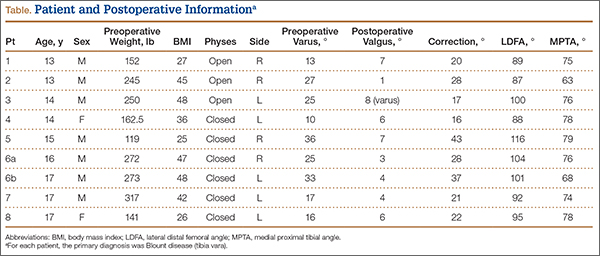
Surgical Technique
Before surgery, we use paper cutouts to template the osteotomy wedge. We also use perioperative antibiotics and a standard time-out. For visualization of the entire leg for accurate correction, we prepare and drape the entire leg. A sterile tourniquet is used. At the midshaft of the fibula, a 4-cm incision is made, and dissection is carefully carried down to the fibula. Subperiosteal dissection is performed about the fibula, allowing adequate clearance for an oblique osteotomy. The osteotomy removes about 1 cm of fibula, which is to be used as bone graft for the tibial osteotomy. In addition, a lateral compartment fasciotomy is performed to prevent swelling-related complications. The wound is irrigated and injected with bupivacaine and closed in routine fashion.
We then make an inverted hockey-stick incision over the proximal tibia, centered down to the tibial tubercle. After dissecting down to the anterior compartment, we perform a fasciotomy of about 8 cm to accommodate swelling. Subperiosteal dissection is then performed around the proximal tibia. The medial soft tissues are left attached to increase blood supply and healing. During subperiosteal dissection, soft elevators are used to gently retract the lateral soft tissues along with the inferior and posterior structures. We use fluoroscopic imaging to guide the osteotomy as well as screw and plate placement. We use a 6-hole, 4.5-mm compression plate and screws for fixation. The 2 proximal screws of the plate are predrilled in place to allow for application of the plate after completion of the osteotomy. The plate is then rotated out of position on 1 screw, and the osteotomy is identified under fluoroscopy with the appropriate position distal to the second hole of the 6-hole plate.
An oscillating saw and osteotomes are used to perform the oblique osteotomy. The pre-estimated bone wedge is removed. Wedge size is adjusted, if needed. The bone wedge is morselized for bone graft. The osteotomy is then closed, correcting both varus and internal tibial torsion. Our goal is 5° valgus. After correction is obtained, the plate is placed, and the proximal screw is snugly seated. Three cortical screws are placed distally to hold the plate in place under compression mode, and a cancellous screw is placed superiorly at the proximal portion of the plate for additional fixation. The screw placed proximal to the osteotomy site is a fully threaded cortical screw with excellent compression. Correction and proper placement of hardware are verified with fluoroscopy.
The wound is irrigated and injected with bupivacaine. Bone graft is then placed at the osteotomy site. Additional bone graft is placed posteriorly between the osteotomy site and the muscle mass to stimulate additional healing. Another screw is placed obliquely from the medial side across the osteotomy site to provide additional fixation (Figure 1).
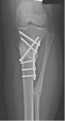
A deep drain is placed and connected to bulb suction for 24 hours after surgery. The wound is then closed in routine fashion. In 2 patients who weighed more than 250 pounds, we used an external fixator for additional stability (Figure 2).
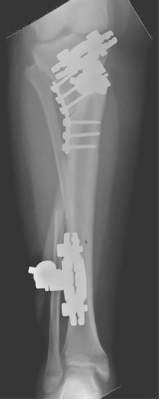
Postoperative Care
The incisions are dressed with antibiotic ointment and 4×4-in bandages and then wrapped with sterile cotton under-cast padding. The leg is placed into a well-padded cylinder cast with the knee flexed 10°. The leg is aligned to about 5° valgus. The cast is then split on the side and spread to allow for swelling and to prevent compartment syndrome.5 We also use a drain hooked to bulb suction, which is removed 24 hours after surgery. Toe-touch weight-bearing with crutches is allowed immediately after surgery. The cast is removed at 6 weeks, and a hinged range-of-motion knee brace is worn for another 6 weeks. All patients are allowed to resume normal activity after 4 months. In our 2 external-fixator cases, a cast was not used, and toe-touch weight-bearing and knee motion were allowed immediately. The external fixators were removed at about 10 weeks.
Results
Mean postoperative mechanical femoral-tibial angle was 3°, and mean correction was 26° (range, 16°-43°) (Table). Lateral distal femoral angle did not show significant femoral deformity in our sample. Mean medial proximal tibial angle was 74° (range, 63°-79°). In each case, the varus deformity was primarily in the tibia. Mean tourniquet time was 88 minutes (range, 50-119 min). Our complication rate was 11% (1 knee). In our first case, in which we did not use an extra medial screw, the 4.5-mm plate fractured at the osteotomy site 2.5 months after surgery. The 250-pound patient subsequently lost 17° of correction, and valgus alignment was not achieved. Preoperative varus was 25°, and postoperative alignment was 8° varus. This plate fracture led us to use an extra medial screw for additional stability in all subsequent cases and to consider using an external fixator for patients weighing more than 250 pounds. After the first case, there were no other plate fractures. A potential problem with closing wedge osteotomy is shortening, but varus correction restores some length. Mean postoperative leg-length difference was 10 mm (range, 0-16 mm). No patient complained of leg-length difference during the postoperative follow-up.
Eight and a half months after surgery, 1 patient had hardware removed, at the family’s request. No patient experienced perioperative infection or neurovascular damage. Our overall patient population was obese—mean BMI was 38 (range, 25-48), and mean postoperative weight was 219 pounds. Three of our 8 patients were overweight (BMI, 25-30), and 5 were obese (BMI, >30). For prevention of plate failure, we recommend using an extra oblique screw in all patients and considering an external fixator for patients who weigh more than 250 pounds.
Discussion
Correction of adolescent tibia vara can be challenging because of patient obesity. The technique described here—a modification of the technique of Laurencin and colleagues4—is practical and reproducible in this population. The goals in performing osteotomy are to correct the deformity, restore joint alignment, preserve leg length, and prevent recurrent deformity and other complications, such as neurovascular injury, nonunion, and infection.3,6-8 Our technique minimizes the risk for these complications. For example, the fasciotomy provides excellent decompression of the anterior and lateral compartments, minimizing neurovascular ischemia and the risk for compartment syndrome. During cast placement, splitting and spreading reduce the risk for compartment syndrome as well.5
Wagner3,9 demonstrated the utility of a closing wedge proximal tibial osteotomy in adults. Laurencin and colleagues4 showed this technique is effective in correcting tibia vara in a pediatric population. However, they did not specify patient weight and used a small semitubular plate for fixation, and some of their patients had infantile Blount disease. We modified the technique in 3 ways. First, we performed a complete osteotomy. Second, because our patients were adolescents and very large, we used a 6-hole, 4.5-mm compression plate and screws. Third, we used an external fixator for increased stability in patients who weighed more than 250 pounds.
The reported technique, using an oblique metaphyseal closing wedge osteotomy with internal fixation in obese patients, is practical, safe, and reliable. This technique is a useful alternative to an external fixator. We used it on 9 knees with tibia vara, and it was completely successful in 8 cases and partially successful in 1 (hardware breakage occurred). An external fixator was used to prevent hardware breakage in 2 patients who weighed more than 250 pounds. This technique is a valuable treatment option for surgical correction, especially in obese patients.
1. Erlacher P. Deformierende Prozesse der Epiphysengegend bei Kindem. Archiv Orthop Unfall-Chir. 1922;20:81-96.
2. Blount WP. Tibia vara. J Bone Joint Surg. 1937;29:1-28.
3. Wagner H. Principles of corrective osteotomies in osteoarthrosis of the knee. In: Weal UH, ed. Joint Preserving Procedures of the Lower Extremity. New York, NY: Springer; 1980:77-102.
4. Laurencin CT, Ferriter PJ, Millis MB. Oblique proximal tibial osteotomy for the correction of tibia vara in the young. Clin Orthop Relat Res. 1996;(327):218-224.
5. Garfin SR, Mubarak SJ, Evans KL, Hargens AR, Akeson WH. Quantification of intracompartmental pressure and volume under plaster casts. J Bone Joint Surg Am. 1981;63(3):449-453.
6. Mycoskie PJ. Complications of osteotomies about the knee in children. Orthopedics. 1981;4(9):1005-1015.
7. Matsen FA, Staheli LT. Neurovascular complications following tibial osteotomy in children. A case report. Clin Orthop Relat Res. 1975;(110):210-214.
8. Steel HH, Sandrew RE, Sullivan PD. Complications of tibial osteotomy in children for genu varum or valgum. Evidence that neurological changes are due to ischemia. J Bone Joint Surg Am. 1971;53(8):1629-1635.
9. Wagner H. The displacement osteotomy as a correction principle. In: Heirholzer G, Muller KH, eds. Corrective Osteotomies of the Lower Extremity After Trauma. Berlin, Germany: Springer; 1985:141-150.
Blount disease (tibia vara) is an angular tibia deformity that includes varus, increased posterior slope, and internal rotation. This deformity was first described in 1922 by Erlacher1 in Germany. In 1937, Walter Blount2 reported on it in the United States. It is the most common cause of pathologic genu varum in adolescence and childhood.
An oblique incomplete closing wedge osteotomy of the proximal tibial metaphysis was described by Wagner3 for the treatment of unicompartmental osteoarthrosis of the knee in adults. Laurencin and colleagues4 applied this technique to the treatment of pediatric tibia vara with favorable results. They spared the medial cortex of the tibia in their incomplete closing wedge osteotomy technique. In each of the 9 cases we treated and describe here, we accidentally completed the tibial osteotomy when attempting the Laurencin technique. Given that the osteotomy was completed, we modified the Laurencin technique by using a 6-hole, 4.5-mm compression plate rather than a 5-hole semitubular plate, and added a large oblique screw from the medial side to compress the osteotomy site and to protect the plate from fracture. In addition, in 2 patients who weighed more than 250 pounds, we used an external fixator for additional stability. In this article, we report the outcomes of correcting adolescent tibia vara with a complete closing wedge tibial osteotomy and an oblique fibular osteotomy.
Materials and Methods
This study was approved by the Institutional Review Board at Pennsylvania State University. Between 2009 and 2012, we performed 9 complete oblique proximal tibial lateral closing wedge osteotomies on 8 patients (2 girls, 6 boys). In each case, the primary diagnosis was Blount disease. One patient also had renal dysplasia and was receiving dialysis. Mean age at time of operation was 15 years (range, 13-17 years). Mean preoperative weight was 215 pounds (range, 119-317 lb). Mean weight gain at follow-up was 4.39 pounds (range, –10 to 19 lb). Mean body mass index (BMI) was 38 (range, 25-48) (Table). All patients had varus angulation of the proximal tibia before surgery. Mean preoperative varus on standing films was 22° (range, 10°-36°). Because of the patients’ size, we used standing long-leg radiographs, on individual cassettes, for each leg.
Surgical Technique
Before surgery, we use paper cutouts to template the osteotomy wedge. We also use perioperative antibiotics and a standard time-out. For visualization of the entire leg for accurate correction, we prepare and drape the entire leg. A sterile tourniquet is used. At the midshaft of the fibula, a 4-cm incision is made, and dissection is carefully carried down to the fibula. Subperiosteal dissection is performed about the fibula, allowing adequate clearance for an oblique osteotomy. The osteotomy removes about 1 cm of fibula, which is to be used as bone graft for the tibial osteotomy. In addition, a lateral compartment fasciotomy is performed to prevent swelling-related complications. The wound is irrigated and injected with bupivacaine and closed in routine fashion.
We then make an inverted hockey-stick incision over the proximal tibia, centered down to the tibial tubercle. After dissecting down to the anterior compartment, we perform a fasciotomy of about 8 cm to accommodate swelling. Subperiosteal dissection is then performed around the proximal tibia. The medial soft tissues are left attached to increase blood supply and healing. During subperiosteal dissection, soft elevators are used to gently retract the lateral soft tissues along with the inferior and posterior structures. We use fluoroscopic imaging to guide the osteotomy as well as screw and plate placement. We use a 6-hole, 4.5-mm compression plate and screws for fixation. The 2 proximal screws of the plate are predrilled in place to allow for application of the plate after completion of the osteotomy. The plate is then rotated out of position on 1 screw, and the osteotomy is identified under fluoroscopy with the appropriate position distal to the second hole of the 6-hole plate.
An oscillating saw and osteotomes are used to perform the oblique osteotomy. The pre-estimated bone wedge is removed. Wedge size is adjusted, if needed. The bone wedge is morselized for bone graft. The osteotomy is then closed, correcting both varus and internal tibial torsion. Our goal is 5° valgus. After correction is obtained, the plate is placed, and the proximal screw is snugly seated. Three cortical screws are placed distally to hold the plate in place under compression mode, and a cancellous screw is placed superiorly at the proximal portion of the plate for additional fixation. The screw placed proximal to the osteotomy site is a fully threaded cortical screw with excellent compression. Correction and proper placement of hardware are verified with fluoroscopy.
The wound is irrigated and injected with bupivacaine. Bone graft is then placed at the osteotomy site. Additional bone graft is placed posteriorly between the osteotomy site and the muscle mass to stimulate additional healing. Another screw is placed obliquely from the medial side across the osteotomy site to provide additional fixation (Figure 1).
A deep drain is placed and connected to bulb suction for 24 hours after surgery. The wound is then closed in routine fashion. In 2 patients who weighed more than 250 pounds, we used an external fixator for additional stability (Figure 2).
Postoperative Care
The incisions are dressed with antibiotic ointment and 4×4-in bandages and then wrapped with sterile cotton under-cast padding. The leg is placed into a well-padded cylinder cast with the knee flexed 10°. The leg is aligned to about 5° valgus. The cast is then split on the side and spread to allow for swelling and to prevent compartment syndrome.5 We also use a drain hooked to bulb suction, which is removed 24 hours after surgery. Toe-touch weight-bearing with crutches is allowed immediately after surgery. The cast is removed at 6 weeks, and a hinged range-of-motion knee brace is worn for another 6 weeks. All patients are allowed to resume normal activity after 4 months. In our 2 external-fixator cases, a cast was not used, and toe-touch weight-bearing and knee motion were allowed immediately. The external fixators were removed at about 10 weeks.
Results
Mean postoperative mechanical femoral-tibial angle was 3°, and mean correction was 26° (range, 16°-43°) (Table). Lateral distal femoral angle did not show significant femoral deformity in our sample. Mean medial proximal tibial angle was 74° (range, 63°-79°). In each case, the varus deformity was primarily in the tibia. Mean tourniquet time was 88 minutes (range, 50-119 min). Our complication rate was 11% (1 knee). In our first case, in which we did not use an extra medial screw, the 4.5-mm plate fractured at the osteotomy site 2.5 months after surgery. The 250-pound patient subsequently lost 17° of correction, and valgus alignment was not achieved. Preoperative varus was 25°, and postoperative alignment was 8° varus. This plate fracture led us to use an extra medial screw for additional stability in all subsequent cases and to consider using an external fixator for patients weighing more than 250 pounds. After the first case, there were no other plate fractures. A potential problem with closing wedge osteotomy is shortening, but varus correction restores some length. Mean postoperative leg-length difference was 10 mm (range, 0-16 mm). No patient complained of leg-length difference during the postoperative follow-up.
Eight and a half months after surgery, 1 patient had hardware removed, at the family’s request. No patient experienced perioperative infection or neurovascular damage. Our overall patient population was obese—mean BMI was 38 (range, 25-48), and mean postoperative weight was 219 pounds. Three of our 8 patients were overweight (BMI, 25-30), and 5 were obese (BMI, >30). For prevention of plate failure, we recommend using an extra oblique screw in all patients and considering an external fixator for patients who weigh more than 250 pounds.
Discussion
Correction of adolescent tibia vara can be challenging because of patient obesity. The technique described here—a modification of the technique of Laurencin and colleagues4—is practical and reproducible in this population. The goals in performing osteotomy are to correct the deformity, restore joint alignment, preserve leg length, and prevent recurrent deformity and other complications, such as neurovascular injury, nonunion, and infection.3,6-8 Our technique minimizes the risk for these complications. For example, the fasciotomy provides excellent decompression of the anterior and lateral compartments, minimizing neurovascular ischemia and the risk for compartment syndrome. During cast placement, splitting and spreading reduce the risk for compartment syndrome as well.5
Wagner3,9 demonstrated the utility of a closing wedge proximal tibial osteotomy in adults. Laurencin and colleagues4 showed this technique is effective in correcting tibia vara in a pediatric population. However, they did not specify patient weight and used a small semitubular plate for fixation, and some of their patients had infantile Blount disease. We modified the technique in 3 ways. First, we performed a complete osteotomy. Second, because our patients were adolescents and very large, we used a 6-hole, 4.5-mm compression plate and screws. Third, we used an external fixator for increased stability in patients who weighed more than 250 pounds.
The reported technique, using an oblique metaphyseal closing wedge osteotomy with internal fixation in obese patients, is practical, safe, and reliable. This technique is a useful alternative to an external fixator. We used it on 9 knees with tibia vara, and it was completely successful in 8 cases and partially successful in 1 (hardware breakage occurred). An external fixator was used to prevent hardware breakage in 2 patients who weighed more than 250 pounds. This technique is a valuable treatment option for surgical correction, especially in obese patients.
Blount disease (tibia vara) is an angular tibia deformity that includes varus, increased posterior slope, and internal rotation. This deformity was first described in 1922 by Erlacher1 in Germany. In 1937, Walter Blount2 reported on it in the United States. It is the most common cause of pathologic genu varum in adolescence and childhood.
An oblique incomplete closing wedge osteotomy of the proximal tibial metaphysis was described by Wagner3 for the treatment of unicompartmental osteoarthrosis of the knee in adults. Laurencin and colleagues4 applied this technique to the treatment of pediatric tibia vara with favorable results. They spared the medial cortex of the tibia in their incomplete closing wedge osteotomy technique. In each of the 9 cases we treated and describe here, we accidentally completed the tibial osteotomy when attempting the Laurencin technique. Given that the osteotomy was completed, we modified the Laurencin technique by using a 6-hole, 4.5-mm compression plate rather than a 5-hole semitubular plate, and added a large oblique screw from the medial side to compress the osteotomy site and to protect the plate from fracture. In addition, in 2 patients who weighed more than 250 pounds, we used an external fixator for additional stability. In this article, we report the outcomes of correcting adolescent tibia vara with a complete closing wedge tibial osteotomy and an oblique fibular osteotomy.
Materials and Methods
This study was approved by the Institutional Review Board at Pennsylvania State University. Between 2009 and 2012, we performed 9 complete oblique proximal tibial lateral closing wedge osteotomies on 8 patients (2 girls, 6 boys). In each case, the primary diagnosis was Blount disease. One patient also had renal dysplasia and was receiving dialysis. Mean age at time of operation was 15 years (range, 13-17 years). Mean preoperative weight was 215 pounds (range, 119-317 lb). Mean weight gain at follow-up was 4.39 pounds (range, –10 to 19 lb). Mean body mass index (BMI) was 38 (range, 25-48) (Table). All patients had varus angulation of the proximal tibia before surgery. Mean preoperative varus on standing films was 22° (range, 10°-36°). Because of the patients’ size, we used standing long-leg radiographs, on individual cassettes, for each leg.
Surgical Technique
Before surgery, we use paper cutouts to template the osteotomy wedge. We also use perioperative antibiotics and a standard time-out. For visualization of the entire leg for accurate correction, we prepare and drape the entire leg. A sterile tourniquet is used. At the midshaft of the fibula, a 4-cm incision is made, and dissection is carefully carried down to the fibula. Subperiosteal dissection is performed about the fibula, allowing adequate clearance for an oblique osteotomy. The osteotomy removes about 1 cm of fibula, which is to be used as bone graft for the tibial osteotomy. In addition, a lateral compartment fasciotomy is performed to prevent swelling-related complications. The wound is irrigated and injected with bupivacaine and closed in routine fashion.
We then make an inverted hockey-stick incision over the proximal tibia, centered down to the tibial tubercle. After dissecting down to the anterior compartment, we perform a fasciotomy of about 8 cm to accommodate swelling. Subperiosteal dissection is then performed around the proximal tibia. The medial soft tissues are left attached to increase blood supply and healing. During subperiosteal dissection, soft elevators are used to gently retract the lateral soft tissues along with the inferior and posterior structures. We use fluoroscopic imaging to guide the osteotomy as well as screw and plate placement. We use a 6-hole, 4.5-mm compression plate and screws for fixation. The 2 proximal screws of the plate are predrilled in place to allow for application of the plate after completion of the osteotomy. The plate is then rotated out of position on 1 screw, and the osteotomy is identified under fluoroscopy with the appropriate position distal to the second hole of the 6-hole plate.
An oscillating saw and osteotomes are used to perform the oblique osteotomy. The pre-estimated bone wedge is removed. Wedge size is adjusted, if needed. The bone wedge is morselized for bone graft. The osteotomy is then closed, correcting both varus and internal tibial torsion. Our goal is 5° valgus. After correction is obtained, the plate is placed, and the proximal screw is snugly seated. Three cortical screws are placed distally to hold the plate in place under compression mode, and a cancellous screw is placed superiorly at the proximal portion of the plate for additional fixation. The screw placed proximal to the osteotomy site is a fully threaded cortical screw with excellent compression. Correction and proper placement of hardware are verified with fluoroscopy.
The wound is irrigated and injected with bupivacaine. Bone graft is then placed at the osteotomy site. Additional bone graft is placed posteriorly between the osteotomy site and the muscle mass to stimulate additional healing. Another screw is placed obliquely from the medial side across the osteotomy site to provide additional fixation (Figure 1).
A deep drain is placed and connected to bulb suction for 24 hours after surgery. The wound is then closed in routine fashion. In 2 patients who weighed more than 250 pounds, we used an external fixator for additional stability (Figure 2).
Postoperative Care
The incisions are dressed with antibiotic ointment and 4×4-in bandages and then wrapped with sterile cotton under-cast padding. The leg is placed into a well-padded cylinder cast with the knee flexed 10°. The leg is aligned to about 5° valgus. The cast is then split on the side and spread to allow for swelling and to prevent compartment syndrome.5 We also use a drain hooked to bulb suction, which is removed 24 hours after surgery. Toe-touch weight-bearing with crutches is allowed immediately after surgery. The cast is removed at 6 weeks, and a hinged range-of-motion knee brace is worn for another 6 weeks. All patients are allowed to resume normal activity after 4 months. In our 2 external-fixator cases, a cast was not used, and toe-touch weight-bearing and knee motion were allowed immediately. The external fixators were removed at about 10 weeks.
Results
Mean postoperative mechanical femoral-tibial angle was 3°, and mean correction was 26° (range, 16°-43°) (Table). Lateral distal femoral angle did not show significant femoral deformity in our sample. Mean medial proximal tibial angle was 74° (range, 63°-79°). In each case, the varus deformity was primarily in the tibia. Mean tourniquet time was 88 minutes (range, 50-119 min). Our complication rate was 11% (1 knee). In our first case, in which we did not use an extra medial screw, the 4.5-mm plate fractured at the osteotomy site 2.5 months after surgery. The 250-pound patient subsequently lost 17° of correction, and valgus alignment was not achieved. Preoperative varus was 25°, and postoperative alignment was 8° varus. This plate fracture led us to use an extra medial screw for additional stability in all subsequent cases and to consider using an external fixator for patients weighing more than 250 pounds. After the first case, there were no other plate fractures. A potential problem with closing wedge osteotomy is shortening, but varus correction restores some length. Mean postoperative leg-length difference was 10 mm (range, 0-16 mm). No patient complained of leg-length difference during the postoperative follow-up.
Eight and a half months after surgery, 1 patient had hardware removed, at the family’s request. No patient experienced perioperative infection or neurovascular damage. Our overall patient population was obese—mean BMI was 38 (range, 25-48), and mean postoperative weight was 219 pounds. Three of our 8 patients were overweight (BMI, 25-30), and 5 were obese (BMI, >30). For prevention of plate failure, we recommend using an extra oblique screw in all patients and considering an external fixator for patients who weigh more than 250 pounds.
Discussion
Correction of adolescent tibia vara can be challenging because of patient obesity. The technique described here—a modification of the technique of Laurencin and colleagues4—is practical and reproducible in this population. The goals in performing osteotomy are to correct the deformity, restore joint alignment, preserve leg length, and prevent recurrent deformity and other complications, such as neurovascular injury, nonunion, and infection.3,6-8 Our technique minimizes the risk for these complications. For example, the fasciotomy provides excellent decompression of the anterior and lateral compartments, minimizing neurovascular ischemia and the risk for compartment syndrome. During cast placement, splitting and spreading reduce the risk for compartment syndrome as well.5
Wagner3,9 demonstrated the utility of a closing wedge proximal tibial osteotomy in adults. Laurencin and colleagues4 showed this technique is effective in correcting tibia vara in a pediatric population. However, they did not specify patient weight and used a small semitubular plate for fixation, and some of their patients had infantile Blount disease. We modified the technique in 3 ways. First, we performed a complete osteotomy. Second, because our patients were adolescents and very large, we used a 6-hole, 4.5-mm compression plate and screws. Third, we used an external fixator for increased stability in patients who weighed more than 250 pounds.
The reported technique, using an oblique metaphyseal closing wedge osteotomy with internal fixation in obese patients, is practical, safe, and reliable. This technique is a useful alternative to an external fixator. We used it on 9 knees with tibia vara, and it was completely successful in 8 cases and partially successful in 1 (hardware breakage occurred). An external fixator was used to prevent hardware breakage in 2 patients who weighed more than 250 pounds. This technique is a valuable treatment option for surgical correction, especially in obese patients.
1. Erlacher P. Deformierende Prozesse der Epiphysengegend bei Kindem. Archiv Orthop Unfall-Chir. 1922;20:81-96.
2. Blount WP. Tibia vara. J Bone Joint Surg. 1937;29:1-28.
3. Wagner H. Principles of corrective osteotomies in osteoarthrosis of the knee. In: Weal UH, ed. Joint Preserving Procedures of the Lower Extremity. New York, NY: Springer; 1980:77-102.
4. Laurencin CT, Ferriter PJ, Millis MB. Oblique proximal tibial osteotomy for the correction of tibia vara in the young. Clin Orthop Relat Res. 1996;(327):218-224.
5. Garfin SR, Mubarak SJ, Evans KL, Hargens AR, Akeson WH. Quantification of intracompartmental pressure and volume under plaster casts. J Bone Joint Surg Am. 1981;63(3):449-453.
6. Mycoskie PJ. Complications of osteotomies about the knee in children. Orthopedics. 1981;4(9):1005-1015.
7. Matsen FA, Staheli LT. Neurovascular complications following tibial osteotomy in children. A case report. Clin Orthop Relat Res. 1975;(110):210-214.
8. Steel HH, Sandrew RE, Sullivan PD. Complications of tibial osteotomy in children for genu varum or valgum. Evidence that neurological changes are due to ischemia. J Bone Joint Surg Am. 1971;53(8):1629-1635.
9. Wagner H. The displacement osteotomy as a correction principle. In: Heirholzer G, Muller KH, eds. Corrective Osteotomies of the Lower Extremity After Trauma. Berlin, Germany: Springer; 1985:141-150.
1. Erlacher P. Deformierende Prozesse der Epiphysengegend bei Kindem. Archiv Orthop Unfall-Chir. 1922;20:81-96.
2. Blount WP. Tibia vara. J Bone Joint Surg. 1937;29:1-28.
3. Wagner H. Principles of corrective osteotomies in osteoarthrosis of the knee. In: Weal UH, ed. Joint Preserving Procedures of the Lower Extremity. New York, NY: Springer; 1980:77-102.
4. Laurencin CT, Ferriter PJ, Millis MB. Oblique proximal tibial osteotomy for the correction of tibia vara in the young. Clin Orthop Relat Res. 1996;(327):218-224.
5. Garfin SR, Mubarak SJ, Evans KL, Hargens AR, Akeson WH. Quantification of intracompartmental pressure and volume under plaster casts. J Bone Joint Surg Am. 1981;63(3):449-453.
6. Mycoskie PJ. Complications of osteotomies about the knee in children. Orthopedics. 1981;4(9):1005-1015.
7. Matsen FA, Staheli LT. Neurovascular complications following tibial osteotomy in children. A case report. Clin Orthop Relat Res. 1975;(110):210-214.
8. Steel HH, Sandrew RE, Sullivan PD. Complications of tibial osteotomy in children for genu varum or valgum. Evidence that neurological changes are due to ischemia. J Bone Joint Surg Am. 1971;53(8):1629-1635.
9. Wagner H. The displacement osteotomy as a correction principle. In: Heirholzer G, Muller KH, eds. Corrective Osteotomies of the Lower Extremity After Trauma. Berlin, Germany: Springer; 1985:141-150.
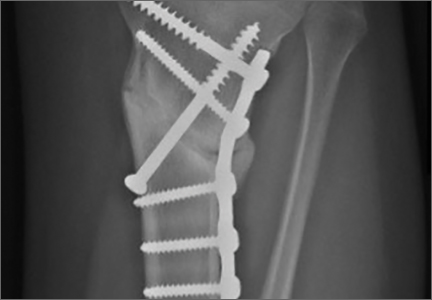
The Changing Face of Pediatric Orthopedics
In my 16 years of practice, there has been tremendous change in the field of pediatric orthopedics in both demographics and scope of practice. Because of scientific and technological advances, efforts of the Pediatric Orthopaedic Society of North America (POSNA), and a changing workforce, the nature of pediatric orthopedics is changing dramatically and will continue to do so.
In the late 1990s, a “typical” pediatric orthopedic surgeon was treating fractures, developmental dysplasia of the hip, clubfeet, and other congenital deformities. Surgery for adolescent idiopathic scoliosis was moving toward anterior instrumentation and correction of the spine. The concepts of early-onset scoliosis and thoracic insufficiency syndrome were in their infancy. Children with anterior cruciate ligament tears were treated with braces until skeletal maturity, often leading to life-altering meniscal pathology. Essential medical treatments for genetic conditions, including bisphosphonates for osteogenesis imperfecta and corticosteroids for Duchenne muscular dystrophy, were considered experimental.
The field itself also was at a crossroads. In 1993, there were 410 active members in POSNA (vs 653 in 2014), and the vast majority were male.1 In the late 1990s, there were approximately 30 pediatric fellowship spots and 10 fellows being trained per year. Simultaneously, approximately 20 to 30 active POSNA members were retiring annually, leading to a projected shortage of pediatric orthopedic surgeons.1 A 2007 American Orthopaedic Association survey found that 59% of members believed that pediatric orthopedics was the most underserved specialty for a variety of reasons, including perceived lower reimbursement, higher volume of nonoperative treatment, and lifestyle issues (such as on-call burden).2
Owing in part to efforts of POSNA in resident/fellow education and mentorship, the practice of pediatric orthopedics in 2016 is dramatically different from a decade ago. The number of fellowship programs has increased to 44 programs, offering a total of 71 fellowship spots, of which 60 were filled by US applicants in 2014. Interestingly, the current active membership of POSNA is 19% female, and the 2014 fellowship class was 34% female. This is in contrast to the 4.4% of all AAOS members who are female. If current trends continue, POSNA could be 40% female by 2025 as senior, predominantly male members retire.1
Pediatric orthopedic practice in 2016 is also dramatically different owing to the development of subspecialization in areas of pediatric sports medicine, hand surgery, trauma, and the treatment of adolescent hip pathology. In fact, a recent survey of fellowship graduates showed that 30% of graduating fellows were going to do a second fellowship.3
While technological advances have driven the care of many pediatric orthopedic conditions such as spinal deformity and sports injuries, there also has been a resurgence of interest in the nonoperative treatment of clubfeet using the Ponseti method and of early-onset scoliosis using Mehta casting. Children with clubfeet even a decade ago were being treated with wide comprehensive releases and capsulotomies, leading to stiff painful feet as young adults. Now comprehensive releases are rarely used. Owing to advances in posterior spinal instrumentation as well as studies showing some decline in pulmonary function after thoracotomy and anterior spinal fusion, the treatment of adolescent scoliosis is predominantly done through the posterior approach. Advances in screening have led to a dramatic decrease in the surgical treatment of hip dysplasia. Medical treatment, such as corticosteroids for Duchenne muscular dystrophy, has prolonged length of life and improved quality of life as well as decreased the number of spinal fusions performed. Recombinant factor replacement for hemophilia has almost eliminated the horrible morbidity associated with hemophilic arthropathy and the need for synovectomy, arthrodesis, and arthroplasty, as well as the infectious issues, such as human immunodeficiency virus (HIV) and hepatitis, associated with the use of pooled blood products. The use of growth-friendly spinal implants, such as the Vertical Expandable Prosthetic Titanium Rib (VEPTR; DePuy Synthes), magnetically driven growing rods (MAGEC; Ellipse), and spinal tethers have improved pulmonary outcomes and presumably life expectancy in young patients with early-onset scoliosis who a decade ago may have had an in situ spinal fusion. These are just a few examples, and there are many more.
The articles in this issue highlight some of these changes. Tibial osteotomy and deformity correction, as described in the article by Burton and Hennrikus (pages 16-18), are classic techniques used by pediatric orthopedists over the past decades and will continue to be useful. The article by Hosseinzadeh and Talwalkar (pages 19-22) reviews unique aspects of pediatric compartment syndrome. While the basic concepts of compartment syndrome have not changed, the signs of compartment syndrome, the 5 Ps we all learned a decade ago (pain, paresthesia, paralysis, pallor, and pulselessness) have now been replaced in children with the 3 As (increasing analgesia, anxiety, and agitation). Finally, the article by Sferopoulos (pages 38-41) describing a case of a giant bone island in a child reminds us that we have a lot more to learn as pediatric orthopedists regarding the molecular nature and cause of disease.
The next few years will continue to be an exciting and dynamic time in the field of pediatric orthopedics. Not only is the workforce itself changing and growing, but so are the definitions of what a pediatric orthopedic surgeon is and does. While subspecialization is the trend in most aspects of medicine, it will be important to continue to monitor<hl name="1"/> this trend to ensure that pediatric orthopedics does not become too highly specialized. With the tremendous inflow of new talent, ideas, and technology, the future for pediatric orthopedics has never looked brighter.
References
1. Sawyer JR, Jones KC, Copley LA, Chambers S; POSNA Practice Management Committee. Pediatric orthopaedic workforce in 2014: current workforce and projections for the future [published online ahead of print October 30, 2015]. J Pediatr Orthop.
2. Salsberg ES, Grover A, Simon MA, Frick SL, Kuremsky MA, Goodman DC. An AOA critical issue. Future physician workforce requirements: implications for orthopaedic surgery education. J Bone Joint Surg Am. 2008;90(5):1143-1159.
3. Glotzbecker MP, Shore BJ, Fletcher ND, Larson AN, Hydorn CR, Sawyer JR; Practice Management Committee of the Pediatric Orthopaedic Society of North America. Early career experience of pediatric orthopaedic fellows: what to expect and need for their services [published online ahead of print March 3, 2015]. J Pediatr Orthop.
In my 16 years of practice, there has been tremendous change in the field of pediatric orthopedics in both demographics and scope of practice. Because of scientific and technological advances, efforts of the Pediatric Orthopaedic Society of North America (POSNA), and a changing workforce, the nature of pediatric orthopedics is changing dramatically and will continue to do so.
In the late 1990s, a “typical” pediatric orthopedic surgeon was treating fractures, developmental dysplasia of the hip, clubfeet, and other congenital deformities. Surgery for adolescent idiopathic scoliosis was moving toward anterior instrumentation and correction of the spine. The concepts of early-onset scoliosis and thoracic insufficiency syndrome were in their infancy. Children with anterior cruciate ligament tears were treated with braces until skeletal maturity, often leading to life-altering meniscal pathology. Essential medical treatments for genetic conditions, including bisphosphonates for osteogenesis imperfecta and corticosteroids for Duchenne muscular dystrophy, were considered experimental.
The field itself also was at a crossroads. In 1993, there were 410 active members in POSNA (vs 653 in 2014), and the vast majority were male.1 In the late 1990s, there were approximately 30 pediatric fellowship spots and 10 fellows being trained per year. Simultaneously, approximately 20 to 30 active POSNA members were retiring annually, leading to a projected shortage of pediatric orthopedic surgeons.1 A 2007 American Orthopaedic Association survey found that 59% of members believed that pediatric orthopedics was the most underserved specialty for a variety of reasons, including perceived lower reimbursement, higher volume of nonoperative treatment, and lifestyle issues (such as on-call burden).2
Owing in part to efforts of POSNA in resident/fellow education and mentorship, the practice of pediatric orthopedics in 2016 is dramatically different from a decade ago. The number of fellowship programs has increased to 44 programs, offering a total of 71 fellowship spots, of which 60 were filled by US applicants in 2014. Interestingly, the current active membership of POSNA is 19% female, and the 2014 fellowship class was 34% female. This is in contrast to the 4.4% of all AAOS members who are female. If current trends continue, POSNA could be 40% female by 2025 as senior, predominantly male members retire.1
Pediatric orthopedic practice in 2016 is also dramatically different owing to the development of subspecialization in areas of pediatric sports medicine, hand surgery, trauma, and the treatment of adolescent hip pathology. In fact, a recent survey of fellowship graduates showed that 30% of graduating fellows were going to do a second fellowship.3
While technological advances have driven the care of many pediatric orthopedic conditions such as spinal deformity and sports injuries, there also has been a resurgence of interest in the nonoperative treatment of clubfeet using the Ponseti method and of early-onset scoliosis using Mehta casting. Children with clubfeet even a decade ago were being treated with wide comprehensive releases and capsulotomies, leading to stiff painful feet as young adults. Now comprehensive releases are rarely used. Owing to advances in posterior spinal instrumentation as well as studies showing some decline in pulmonary function after thoracotomy and anterior spinal fusion, the treatment of adolescent scoliosis is predominantly done through the posterior approach. Advances in screening have led to a dramatic decrease in the surgical treatment of hip dysplasia. Medical treatment, such as corticosteroids for Duchenne muscular dystrophy, has prolonged length of life and improved quality of life as well as decreased the number of spinal fusions performed. Recombinant factor replacement for hemophilia has almost eliminated the horrible morbidity associated with hemophilic arthropathy and the need for synovectomy, arthrodesis, and arthroplasty, as well as the infectious issues, such as human immunodeficiency virus (HIV) and hepatitis, associated with the use of pooled blood products. The use of growth-friendly spinal implants, such as the Vertical Expandable Prosthetic Titanium Rib (VEPTR; DePuy Synthes), magnetically driven growing rods (MAGEC; Ellipse), and spinal tethers have improved pulmonary outcomes and presumably life expectancy in young patients with early-onset scoliosis who a decade ago may have had an in situ spinal fusion. These are just a few examples, and there are many more.
The articles in this issue highlight some of these changes. Tibial osteotomy and deformity correction, as described in the article by Burton and Hennrikus (pages 16-18), are classic techniques used by pediatric orthopedists over the past decades and will continue to be useful. The article by Hosseinzadeh and Talwalkar (pages 19-22) reviews unique aspects of pediatric compartment syndrome. While the basic concepts of compartment syndrome have not changed, the signs of compartment syndrome, the 5 Ps we all learned a decade ago (pain, paresthesia, paralysis, pallor, and pulselessness) have now been replaced in children with the 3 As (increasing analgesia, anxiety, and agitation). Finally, the article by Sferopoulos (pages 38-41) describing a case of a giant bone island in a child reminds us that we have a lot more to learn as pediatric orthopedists regarding the molecular nature and cause of disease.
The next few years will continue to be an exciting and dynamic time in the field of pediatric orthopedics. Not only is the workforce itself changing and growing, but so are the definitions of what a pediatric orthopedic surgeon is and does. While subspecialization is the trend in most aspects of medicine, it will be important to continue to monitor<hl name="1"/> this trend to ensure that pediatric orthopedics does not become too highly specialized. With the tremendous inflow of new talent, ideas, and technology, the future for pediatric orthopedics has never looked brighter.
References
In my 16 years of practice, there has been tremendous change in the field of pediatric orthopedics in both demographics and scope of practice. Because of scientific and technological advances, efforts of the Pediatric Orthopaedic Society of North America (POSNA), and a changing workforce, the nature of pediatric orthopedics is changing dramatically and will continue to do so.
In the late 1990s, a “typical” pediatric orthopedic surgeon was treating fractures, developmental dysplasia of the hip, clubfeet, and other congenital deformities. Surgery for adolescent idiopathic scoliosis was moving toward anterior instrumentation and correction of the spine. The concepts of early-onset scoliosis and thoracic insufficiency syndrome were in their infancy. Children with anterior cruciate ligament tears were treated with braces until skeletal maturity, often leading to life-altering meniscal pathology. Essential medical treatments for genetic conditions, including bisphosphonates for osteogenesis imperfecta and corticosteroids for Duchenne muscular dystrophy, were considered experimental.
The field itself also was at a crossroads. In 1993, there were 410 active members in POSNA (vs 653 in 2014), and the vast majority were male.1 In the late 1990s, there were approximately 30 pediatric fellowship spots and 10 fellows being trained per year. Simultaneously, approximately 20 to 30 active POSNA members were retiring annually, leading to a projected shortage of pediatric orthopedic surgeons.1 A 2007 American Orthopaedic Association survey found that 59% of members believed that pediatric orthopedics was the most underserved specialty for a variety of reasons, including perceived lower reimbursement, higher volume of nonoperative treatment, and lifestyle issues (such as on-call burden).2
Owing in part to efforts of POSNA in resident/fellow education and mentorship, the practice of pediatric orthopedics in 2016 is dramatically different from a decade ago. The number of fellowship programs has increased to 44 programs, offering a total of 71 fellowship spots, of which 60 were filled by US applicants in 2014. Interestingly, the current active membership of POSNA is 19% female, and the 2014 fellowship class was 34% female. This is in contrast to the 4.4% of all AAOS members who are female. If current trends continue, POSNA could be 40% female by 2025 as senior, predominantly male members retire.1
Pediatric orthopedic practice in 2016 is also dramatically different owing to the development of subspecialization in areas of pediatric sports medicine, hand surgery, trauma, and the treatment of adolescent hip pathology. In fact, a recent survey of fellowship graduates showed that 30% of graduating fellows were going to do a second fellowship.3
While technological advances have driven the care of many pediatric orthopedic conditions such as spinal deformity and sports injuries, there also has been a resurgence of interest in the nonoperative treatment of clubfeet using the Ponseti method and of early-onset scoliosis using Mehta casting. Children with clubfeet even a decade ago were being treated with wide comprehensive releases and capsulotomies, leading to stiff painful feet as young adults. Now comprehensive releases are rarely used. Owing to advances in posterior spinal instrumentation as well as studies showing some decline in pulmonary function after thoracotomy and anterior spinal fusion, the treatment of adolescent scoliosis is predominantly done through the posterior approach. Advances in screening have led to a dramatic decrease in the surgical treatment of hip dysplasia. Medical treatment, such as corticosteroids for Duchenne muscular dystrophy, has prolonged length of life and improved quality of life as well as decreased the number of spinal fusions performed. Recombinant factor replacement for hemophilia has almost eliminated the horrible morbidity associated with hemophilic arthropathy and the need for synovectomy, arthrodesis, and arthroplasty, as well as the infectious issues, such as human immunodeficiency virus (HIV) and hepatitis, associated with the use of pooled blood products. The use of growth-friendly spinal implants, such as the Vertical Expandable Prosthetic Titanium Rib (VEPTR; DePuy Synthes), magnetically driven growing rods (MAGEC; Ellipse), and spinal tethers have improved pulmonary outcomes and presumably life expectancy in young patients with early-onset scoliosis who a decade ago may have had an in situ spinal fusion. These are just a few examples, and there are many more.
The articles in this issue highlight some of these changes. Tibial osteotomy and deformity correction, as described in the article by Burton and Hennrikus (pages 16-18), are classic techniques used by pediatric orthopedists over the past decades and will continue to be useful. The article by Hosseinzadeh and Talwalkar (pages 19-22) reviews unique aspects of pediatric compartment syndrome. While the basic concepts of compartment syndrome have not changed, the signs of compartment syndrome, the 5 Ps we all learned a decade ago (pain, paresthesia, paralysis, pallor, and pulselessness) have now been replaced in children with the 3 As (increasing analgesia, anxiety, and agitation). Finally, the article by Sferopoulos (pages 38-41) describing a case of a giant bone island in a child reminds us that we have a lot more to learn as pediatric orthopedists regarding the molecular nature and cause of disease.
The next few years will continue to be an exciting and dynamic time in the field of pediatric orthopedics. Not only is the workforce itself changing and growing, but so are the definitions of what a pediatric orthopedic surgeon is and does. While subspecialization is the trend in most aspects of medicine, it will be important to continue to monitor<hl name="1"/> this trend to ensure that pediatric orthopedics does not become too highly specialized. With the tremendous inflow of new talent, ideas, and technology, the future for pediatric orthopedics has never looked brighter.
References
1. Sawyer JR, Jones KC, Copley LA, Chambers S; POSNA Practice Management Committee. Pediatric orthopaedic workforce in 2014: current workforce and projections for the future [published online ahead of print October 30, 2015]. J Pediatr Orthop.
2. Salsberg ES, Grover A, Simon MA, Frick SL, Kuremsky MA, Goodman DC. An AOA critical issue. Future physician workforce requirements: implications for orthopaedic surgery education. J Bone Joint Surg Am. 2008;90(5):1143-1159.
3. Glotzbecker MP, Shore BJ, Fletcher ND, Larson AN, Hydorn CR, Sawyer JR; Practice Management Committee of the Pediatric Orthopaedic Society of North America. Early career experience of pediatric orthopaedic fellows: what to expect and need for their services [published online ahead of print March 3, 2015]. J Pediatr Orthop.
1. Sawyer JR, Jones KC, Copley LA, Chambers S; POSNA Practice Management Committee. Pediatric orthopaedic workforce in 2014: current workforce and projections for the future [published online ahead of print October 30, 2015]. J Pediatr Orthop.
2. Salsberg ES, Grover A, Simon MA, Frick SL, Kuremsky MA, Goodman DC. An AOA critical issue. Future physician workforce requirements: implications for orthopaedic surgery education. J Bone Joint Surg Am. 2008;90(5):1143-1159.
3. Glotzbecker MP, Shore BJ, Fletcher ND, Larson AN, Hydorn CR, Sawyer JR; Practice Management Committee of the Pediatric Orthopaedic Society of North America. Early career experience of pediatric orthopaedic fellows: what to expect and need for their services [published online ahead of print March 3, 2015]. J Pediatr Orthop.

Parental monitoring buffers peer influence on kids’ substance use
Middle schoolers who socialize with friends engaged in risky or deviant behaviors are more likely to use alcohol, tobacco, or drugs early, but increased parental monitoring mediates this effect, according to a recent study.
“The peer selection pathway describes how children who use or intend to use alcohol, tobacco, or drugs select peers who will facilitate alcohol, tobacco, or drug use,” reported Thomas J. Schofield, Ph.D., of Iowa State University, Ames, and his associates. “In contrast, the peer socialization pathway describes how children with deviant peers become more likely to use alcohol, tobacco, or drugs because of peer influence. In the current investigation, we found support for both of these pathways over time among a sample of Mexican-origin children,” they wrote (Drug Alcohol Depend. 2015 Dec 1;157:129-35).

The researchers tracked 674 California fifth graders, all of Mexican origin, for 2 years, collecting data during fifth and seventh grades on the kids’ social relationships, their intent to use alcohol, tobacco, or other drugs, and their use of those substances in the previous year. Families were also video-recorded during a 20-minute structured interaction task between the mother and child and, if possible, father and child, at the fifth grade assessment so trained observers could rate parents’ interactions with their children.
“The monitoring variable was operationalized as the degree to which parents accurately track the behaviors, activities, and social involvements of the child, as well as parents’ specific knowledge about the child’s life and activities,” the researchers wrote.
Questions about social relationships specifically asked the children how many of their friends engaged in various risky or deviant behaviors, such as using alcohol to get drunk, hanging out with a gang, and using substances to get high.
In fifth grade, less than 1% of the kids had tried cigarettes, and 92% had no intention of doing so; 3.4% had tried beer while 89.6% had no plans to use alcohol. No children reported trying street drugs in fifth grade, and 93.2% reported no intention of doing so. Similarly high numbers of kids had no intentions of smoking or using drugs or alcohol in seventh grade.
No data emerged to suggest children’s gender or generational status influenced their substance use, and the proportion of children interacting with deviant peers, intending to use substances or already using substances remained stable from fifth to seventh grades. However, interaction with deviant peers increased kids’ likelihood of using substances or intending to.
“Despite almost no use or intent to use alcohol, tobacco, and other drugs in fifth grade, adolescents who did entertain the idea of using alcohol, tobacco, and other drugs over the next year were already more likely to be associating with deviant peers in fifth grade,” the authors wrote. But analyses of parent interactions revealed that “parental monitoring significantly moderated the pathway from deviant peers in fifth grade to later alcohol, tobacco, and other drug use as well as the pathway from alcohol, tobacco, and other drug use in fifth grade to later associations with deviant peers.”
The only personality feature that reduced children’s likelihood of socializing with deviant peers was being shy. Parents’ use of alcohol predicted an increase in children’s alcohol, tobacco, and other drug use.
Parental monitoring may reduce kids’ likelihood of using substances because a close relationship may help the child identify with the parent values more or children may have fewer opportunities to use with closer monitoring, the authors propose.
The research was funded by the National Institute on Drug Abuse. The authors reported no disclosures.
Middle schoolers who socialize with friends engaged in risky or deviant behaviors are more likely to use alcohol, tobacco, or drugs early, but increased parental monitoring mediates this effect, according to a recent study.
“The peer selection pathway describes how children who use or intend to use alcohol, tobacco, or drugs select peers who will facilitate alcohol, tobacco, or drug use,” reported Thomas J. Schofield, Ph.D., of Iowa State University, Ames, and his associates. “In contrast, the peer socialization pathway describes how children with deviant peers become more likely to use alcohol, tobacco, or drugs because of peer influence. In the current investigation, we found support for both of these pathways over time among a sample of Mexican-origin children,” they wrote (Drug Alcohol Depend. 2015 Dec 1;157:129-35).
The researchers tracked 674 California fifth graders, all of Mexican origin, for 2 years, collecting data during fifth and seventh grades on the kids’ social relationships, their intent to use alcohol, tobacco, or other drugs, and their use of those substances in the previous year. Families were also video-recorded during a 20-minute structured interaction task between the mother and child and, if possible, father and child, at the fifth grade assessment so trained observers could rate parents’ interactions with their children.
“The monitoring variable was operationalized as the degree to which parents accurately track the behaviors, activities, and social involvements of the child, as well as parents’ specific knowledge about the child’s life and activities,” the researchers wrote.
Questions about social relationships specifically asked the children how many of their friends engaged in various risky or deviant behaviors, such as using alcohol to get drunk, hanging out with a gang, and using substances to get high.
In fifth grade, less than 1% of the kids had tried cigarettes, and 92% had no intention of doing so; 3.4% had tried beer while 89.6% had no plans to use alcohol. No children reported trying street drugs in fifth grade, and 93.2% reported no intention of doing so. Similarly high numbers of kids had no intentions of smoking or using drugs or alcohol in seventh grade.
No data emerged to suggest children’s gender or generational status influenced their substance use, and the proportion of children interacting with deviant peers, intending to use substances or already using substances remained stable from fifth to seventh grades. However, interaction with deviant peers increased kids’ likelihood of using substances or intending to.
“Despite almost no use or intent to use alcohol, tobacco, and other drugs in fifth grade, adolescents who did entertain the idea of using alcohol, tobacco, and other drugs over the next year were already more likely to be associating with deviant peers in fifth grade,” the authors wrote. But analyses of parent interactions revealed that “parental monitoring significantly moderated the pathway from deviant peers in fifth grade to later alcohol, tobacco, and other drug use as well as the pathway from alcohol, tobacco, and other drug use in fifth grade to later associations with deviant peers.”
The only personality feature that reduced children’s likelihood of socializing with deviant peers was being shy. Parents’ use of alcohol predicted an increase in children’s alcohol, tobacco, and other drug use.
Parental monitoring may reduce kids’ likelihood of using substances because a close relationship may help the child identify with the parent values more or children may have fewer opportunities to use with closer monitoring, the authors propose.
The research was funded by the National Institute on Drug Abuse. The authors reported no disclosures.
Middle schoolers who socialize with friends engaged in risky or deviant behaviors are more likely to use alcohol, tobacco, or drugs early, but increased parental monitoring mediates this effect, according to a recent study.
“The peer selection pathway describes how children who use or intend to use alcohol, tobacco, or drugs select peers who will facilitate alcohol, tobacco, or drug use,” reported Thomas J. Schofield, Ph.D., of Iowa State University, Ames, and his associates. “In contrast, the peer socialization pathway describes how children with deviant peers become more likely to use alcohol, tobacco, or drugs because of peer influence. In the current investigation, we found support for both of these pathways over time among a sample of Mexican-origin children,” they wrote (Drug Alcohol Depend. 2015 Dec 1;157:129-35).
The researchers tracked 674 California fifth graders, all of Mexican origin, for 2 years, collecting data during fifth and seventh grades on the kids’ social relationships, their intent to use alcohol, tobacco, or other drugs, and their use of those substances in the previous year. Families were also video-recorded during a 20-minute structured interaction task between the mother and child and, if possible, father and child, at the fifth grade assessment so trained observers could rate parents’ interactions with their children.
“The monitoring variable was operationalized as the degree to which parents accurately track the behaviors, activities, and social involvements of the child, as well as parents’ specific knowledge about the child’s life and activities,” the researchers wrote.
Questions about social relationships specifically asked the children how many of their friends engaged in various risky or deviant behaviors, such as using alcohol to get drunk, hanging out with a gang, and using substances to get high.
In fifth grade, less than 1% of the kids had tried cigarettes, and 92% had no intention of doing so; 3.4% had tried beer while 89.6% had no plans to use alcohol. No children reported trying street drugs in fifth grade, and 93.2% reported no intention of doing so. Similarly high numbers of kids had no intentions of smoking or using drugs or alcohol in seventh grade.
No data emerged to suggest children’s gender or generational status influenced their substance use, and the proportion of children interacting with deviant peers, intending to use substances or already using substances remained stable from fifth to seventh grades. However, interaction with deviant peers increased kids’ likelihood of using substances or intending to.
“Despite almost no use or intent to use alcohol, tobacco, and other drugs in fifth grade, adolescents who did entertain the idea of using alcohol, tobacco, and other drugs over the next year were already more likely to be associating with deviant peers in fifth grade,” the authors wrote. But analyses of parent interactions revealed that “parental monitoring significantly moderated the pathway from deviant peers in fifth grade to later alcohol, tobacco, and other drug use as well as the pathway from alcohol, tobacco, and other drug use in fifth grade to later associations with deviant peers.”
The only personality feature that reduced children’s likelihood of socializing with deviant peers was being shy. Parents’ use of alcohol predicted an increase in children’s alcohol, tobacco, and other drug use.
Parental monitoring may reduce kids’ likelihood of using substances because a close relationship may help the child identify with the parent values more or children may have fewer opportunities to use with closer monitoring, the authors propose.
The research was funded by the National Institute on Drug Abuse. The authors reported no disclosures.
FROM DRUG AND ALCOHOL DEPENDENCE
Key clinical point: Parental monitoring can reduce children’s likelihood of tobacco, alcohol, or other drug use.
Major finding: Kids’ socialization with deviant peers increases their risk of using or intending to use tobacco, alcohol, and other drugs.
Data source: The findings are based on a 2-year cohort study of 674 California middle schoolers of Mexican origin.
Disclosures: The research was funded by the National Institute on Drug Abuse. The authors reported no disclosures.

WDC: Disease-modifying immunotherapy ‘the future of diabetes care’
VANCOUVER – Perhaps in the not-too-distant future, people at risk for type 1 diabetes will be screened for antibodies against beta cells, and, if two or more are present, started on immunotherapy to prevent beta cell destruction and clinical disease.
The vision is to treat “islet autoimmunity” as we do hypertension and other silent diseases to prevent problems down the road.

In the everyday medical world, that’s mostly science fiction for now – treatment starts when symptoms emerge – but researchers around the world are working hard to make it a reality, including Dr. Carla J. Greenbaum, director of the Diabetes Research Program at the Benaroya Research Institute in Seattle.
It’s known now that when babies have two or more antibodies against beta cells, about 11% per year will develop clinical diabetes, and all of them eventually. Adults with two or more antibodies will also develop diabetes, although at a slower rate than children.
“The take-home message is that type 1 diabetes starts when you have two antibodies. We need to change from symptom management to disease-modifying therapy, and that will require immunotherapy; it will be the future of diabetes care. Endocrinologists need to learn about immunotherapy,” Dr. Greenbaum said at the World Diabetes Congress.
So far, studies of four immunotherapies – teplizumab (Diabetes. 2013 Nov;62[11]:3766-74), rituximab (N Engl J Med. 2009 Nov 26;361[22]:2143-52), abatacept (Diabetes Care. 2014 Apr;37[4]:1069-75), and, most recently, alefacept (J Clin Invest. 2015 Aug 3;125[8]:3285-96) – have shown positive outcomes in preserving beta cell function after diagnosis of type 1. “The positive results are largely driven by children,” she said, suggesting that they might benefit most from a screen-and-treat approach to islet autoimmunity.
Teplizumab, oral insulin, and abatacept are now being tested in antibody-positive patients who haven’t developed symptoms. The trials aim to reduce the risk of clinical disease by 40%. If successful, treating 100 people with islet autoimmunity would prevent 14 from getting clinical type 1 diabetes, said Dr. Greenbaum, who is involved in the work.
A lot of questions need to be answered if the results pan out. Who should be screened, for instance, and how, and what immunotherapies should be used in different patient groups? What’s the right balance between risks and benefits?
There’s also a big question about how to “bring disease-modifying therapies to the clinic. As endocrinologists, we don’t use immunotherapies, but it’s important to recognize that millions of people for many decades have been using them safely to change the course of disease. We need to learn how to do this,” Dr. Greenbaum said.
“I often get people saying immunotherapy doesn’t really work, but that’s not true.” The effect sizes are small – maybe 20% – but it’s the same case in multiple sclerosis and rheumatoid arthritis. “The issue is if you have a weak joint, and you are 20% better, you feel it, but if beta cells are doing better, you might not notice,” she said.
Dr. Greenbaum disclosed research support from Novo Nordisk.
VANCOUVER – Perhaps in the not-too-distant future, people at risk for type 1 diabetes will be screened for antibodies against beta cells, and, if two or more are present, started on immunotherapy to prevent beta cell destruction and clinical disease.
The vision is to treat “islet autoimmunity” as we do hypertension and other silent diseases to prevent problems down the road.
In the everyday medical world, that’s mostly science fiction for now – treatment starts when symptoms emerge – but researchers around the world are working hard to make it a reality, including Dr. Carla J. Greenbaum, director of the Diabetes Research Program at the Benaroya Research Institute in Seattle.
It’s known now that when babies have two or more antibodies against beta cells, about 11% per year will develop clinical diabetes, and all of them eventually. Adults with two or more antibodies will also develop diabetes, although at a slower rate than children.
“The take-home message is that type 1 diabetes starts when you have two antibodies. We need to change from symptom management to disease-modifying therapy, and that will require immunotherapy; it will be the future of diabetes care. Endocrinologists need to learn about immunotherapy,” Dr. Greenbaum said at the World Diabetes Congress.
So far, studies of four immunotherapies – teplizumab (Diabetes. 2013 Nov;62[11]:3766-74), rituximab (N Engl J Med. 2009 Nov 26;361[22]:2143-52), abatacept (Diabetes Care. 2014 Apr;37[4]:1069-75), and, most recently, alefacept (J Clin Invest. 2015 Aug 3;125[8]:3285-96) – have shown positive outcomes in preserving beta cell function after diagnosis of type 1. “The positive results are largely driven by children,” she said, suggesting that they might benefit most from a screen-and-treat approach to islet autoimmunity.
Teplizumab, oral insulin, and abatacept are now being tested in antibody-positive patients who haven’t developed symptoms. The trials aim to reduce the risk of clinical disease by 40%. If successful, treating 100 people with islet autoimmunity would prevent 14 from getting clinical type 1 diabetes, said Dr. Greenbaum, who is involved in the work.
A lot of questions need to be answered if the results pan out. Who should be screened, for instance, and how, and what immunotherapies should be used in different patient groups? What’s the right balance between risks and benefits?
There’s also a big question about how to “bring disease-modifying therapies to the clinic. As endocrinologists, we don’t use immunotherapies, but it’s important to recognize that millions of people for many decades have been using them safely to change the course of disease. We need to learn how to do this,” Dr. Greenbaum said.
“I often get people saying immunotherapy doesn’t really work, but that’s not true.” The effect sizes are small – maybe 20% – but it’s the same case in multiple sclerosis and rheumatoid arthritis. “The issue is if you have a weak joint, and you are 20% better, you feel it, but if beta cells are doing better, you might not notice,” she said.
Dr. Greenbaum disclosed research support from Novo Nordisk.
VANCOUVER – Perhaps in the not-too-distant future, people at risk for type 1 diabetes will be screened for antibodies against beta cells, and, if two or more are present, started on immunotherapy to prevent beta cell destruction and clinical disease.
The vision is to treat “islet autoimmunity” as we do hypertension and other silent diseases to prevent problems down the road.
In the everyday medical world, that’s mostly science fiction for now – treatment starts when symptoms emerge – but researchers around the world are working hard to make it a reality, including Dr. Carla J. Greenbaum, director of the Diabetes Research Program at the Benaroya Research Institute in Seattle.
It’s known now that when babies have two or more antibodies against beta cells, about 11% per year will develop clinical diabetes, and all of them eventually. Adults with two or more antibodies will also develop diabetes, although at a slower rate than children.
“The take-home message is that type 1 diabetes starts when you have two antibodies. We need to change from symptom management to disease-modifying therapy, and that will require immunotherapy; it will be the future of diabetes care. Endocrinologists need to learn about immunotherapy,” Dr. Greenbaum said at the World Diabetes Congress.
So far, studies of four immunotherapies – teplizumab (Diabetes. 2013 Nov;62[11]:3766-74), rituximab (N Engl J Med. 2009 Nov 26;361[22]:2143-52), abatacept (Diabetes Care. 2014 Apr;37[4]:1069-75), and, most recently, alefacept (J Clin Invest. 2015 Aug 3;125[8]:3285-96) – have shown positive outcomes in preserving beta cell function after diagnosis of type 1. “The positive results are largely driven by children,” she said, suggesting that they might benefit most from a screen-and-treat approach to islet autoimmunity.
Teplizumab, oral insulin, and abatacept are now being tested in antibody-positive patients who haven’t developed symptoms. The trials aim to reduce the risk of clinical disease by 40%. If successful, treating 100 people with islet autoimmunity would prevent 14 from getting clinical type 1 diabetes, said Dr. Greenbaum, who is involved in the work.
A lot of questions need to be answered if the results pan out. Who should be screened, for instance, and how, and what immunotherapies should be used in different patient groups? What’s the right balance between risks and benefits?
There’s also a big question about how to “bring disease-modifying therapies to the clinic. As endocrinologists, we don’t use immunotherapies, but it’s important to recognize that millions of people for many decades have been using them safely to change the course of disease. We need to learn how to do this,” Dr. Greenbaum said.
“I often get people saying immunotherapy doesn’t really work, but that’s not true.” The effect sizes are small – maybe 20% – but it’s the same case in multiple sclerosis and rheumatoid arthritis. “The issue is if you have a weak joint, and you are 20% better, you feel it, but if beta cells are doing better, you might not notice,” she said.
Dr. Greenbaum disclosed research support from Novo Nordisk.
EXPERT ANALYSIS AT THE WORLD DIABETES CONGRESS

WDC: When diagnosed young, type 2 diabetes is more lethal than type 1
VANCOUVER – When diabetes is diagnosed from ages 15 to 30 years, type 2 patients are less likely than are type 1 patients to reach their 50th birthday, according to investigators from the University of Sydney.
Even just a few years after diagnosis, young type 2 patients have a worse cardiovascular profile and worse cardiovascular disease, which leads to an earlier death.
“We pay less attention to type 2 than type 1 in young people. They’re not as sick, and don’t develop [diabetic ketoacidosis] if they miss a treatment. But young type 2 is not a milder form of diabetes in young people, and its detrimental impact occurs early. Before we know what’s going on, patients are middle age, and it’s too late,” said investigator Maria Constantino, a Ph.D. candidate, nurse, and diabetes researcher and educator at the University of Sydney.

Screening for type 2 diabetes “should start at a young age in at-risk groups,” and diabetes cardiovascular risk criteria should be reconsidered so that younger type 2 patients aren’t overlooked because of their age. Current criteria likely “lead to delay in preventive treatment” in the young, she said.
The conclusions come from a review of diabetes patients treated since 1990 at the Royal Prince Alfred Hospital in Sydney (Diabetes Care. 2013 Dec; 36[12]:3863-9).
The investigators compared 354 type 2 patients with 470 type 1 patients diagnosed from age 15-30 years. By around the age of 40 years, 11% of the type 2 patients had died, vs. 6.8% with type 1 (hazard ratio, 2.0; 95% confidence interval, 1.2-3.2; P = .003). Strokes, coronary artery disease, and other macrovascular complications were also far more common in the type 2 group, and they had worse hypertension and dyslipidemia despite taking more drugs to combat both. Type 2 patients also had more albuminuria and neuropathy.
The differences occurred despite the fact that type 2 patients smoked less, had a slightly shorter duration of disease (11.6 vs. 14.7 years), and equivalent glycemic control with their type 1 counterparts, with a mean hemoglobin A1c of 8.1% in both groups. They were heavier, however, with a mean body mass index of 32.2 kg/m2 vs. 25.6 kg/m2 in type 1 patients.
In short, young-onset type 2 is a “more lethal phenotype of diabetes. We are not saying one type of diabetes is more important; we need to conquer both.” But in young type 2 patients, “we need to focus on more than just glycemic control.” Cardiovascular risk factors are “detectable early, and treatable,” Ms. Constantino said.
The investigators also compared their 354 young-onset type 2 patients with 1,062 patients diagnosed from age 40-50 years.
By the time they were about 50 years old, young-onset patients were 6.5 times more likely to have died than were their age-matched peers without diabetes in the general Australian population. The peak in excess mortality for those diagnosed in their 5th decade came at about age of 65 years, with a risk of death about 2.5 times higher than nondiabetic peers in the general population.
“The impact of type 2 is much higher the younger a person is. You can argue that in our enthusiasm to diagnose diabetes, we are casting our screening net wider and wider, and take pride in diagnosing many elderly patients with diabetes, but we should not lose sight of the fact that finding and treating an elderly person with diabetes has much less impact than finding and treating one in a young age group,” she said.
Ms. Constantino has no conflicts of interest.
VANCOUVER – When diabetes is diagnosed from ages 15 to 30 years, type 2 patients are less likely than are type 1 patients to reach their 50th birthday, according to investigators from the University of Sydney.
Even just a few years after diagnosis, young type 2 patients have a worse cardiovascular profile and worse cardiovascular disease, which leads to an earlier death.
“We pay less attention to type 2 than type 1 in young people. They’re not as sick, and don’t develop [diabetic ketoacidosis] if they miss a treatment. But young type 2 is not a milder form of diabetes in young people, and its detrimental impact occurs early. Before we know what’s going on, patients are middle age, and it’s too late,” said investigator Maria Constantino, a Ph.D. candidate, nurse, and diabetes researcher and educator at the University of Sydney.
Screening for type 2 diabetes “should start at a young age in at-risk groups,” and diabetes cardiovascular risk criteria should be reconsidered so that younger type 2 patients aren’t overlooked because of their age. Current criteria likely “lead to delay in preventive treatment” in the young, she said.
The conclusions come from a review of diabetes patients treated since 1990 at the Royal Prince Alfred Hospital in Sydney (Diabetes Care. 2013 Dec; 36[12]:3863-9).
The investigators compared 354 type 2 patients with 470 type 1 patients diagnosed from age 15-30 years. By around the age of 40 years, 11% of the type 2 patients had died, vs. 6.8% with type 1 (hazard ratio, 2.0; 95% confidence interval, 1.2-3.2; P = .003). Strokes, coronary artery disease, and other macrovascular complications were also far more common in the type 2 group, and they had worse hypertension and dyslipidemia despite taking more drugs to combat both. Type 2 patients also had more albuminuria and neuropathy.
The differences occurred despite the fact that type 2 patients smoked less, had a slightly shorter duration of disease (11.6 vs. 14.7 years), and equivalent glycemic control with their type 1 counterparts, with a mean hemoglobin A1c of 8.1% in both groups. They were heavier, however, with a mean body mass index of 32.2 kg/m2 vs. 25.6 kg/m2 in type 1 patients.
In short, young-onset type 2 is a “more lethal phenotype of diabetes. We are not saying one type of diabetes is more important; we need to conquer both.” But in young type 2 patients, “we need to focus on more than just glycemic control.” Cardiovascular risk factors are “detectable early, and treatable,” Ms. Constantino said.
The investigators also compared their 354 young-onset type 2 patients with 1,062 patients diagnosed from age 40-50 years.
By the time they were about 50 years old, young-onset patients were 6.5 times more likely to have died than were their age-matched peers without diabetes in the general Australian population. The peak in excess mortality for those diagnosed in their 5th decade came at about age of 65 years, with a risk of death about 2.5 times higher than nondiabetic peers in the general population.
“The impact of type 2 is much higher the younger a person is. You can argue that in our enthusiasm to diagnose diabetes, we are casting our screening net wider and wider, and take pride in diagnosing many elderly patients with diabetes, but we should not lose sight of the fact that finding and treating an elderly person with diabetes has much less impact than finding and treating one in a young age group,” she said.
Ms. Constantino has no conflicts of interest.
VANCOUVER – When diabetes is diagnosed from ages 15 to 30 years, type 2 patients are less likely than are type 1 patients to reach their 50th birthday, according to investigators from the University of Sydney.
Even just a few years after diagnosis, young type 2 patients have a worse cardiovascular profile and worse cardiovascular disease, which leads to an earlier death.
“We pay less attention to type 2 than type 1 in young people. They’re not as sick, and don’t develop [diabetic ketoacidosis] if they miss a treatment. But young type 2 is not a milder form of diabetes in young people, and its detrimental impact occurs early. Before we know what’s going on, patients are middle age, and it’s too late,” said investigator Maria Constantino, a Ph.D. candidate, nurse, and diabetes researcher and educator at the University of Sydney.
Screening for type 2 diabetes “should start at a young age in at-risk groups,” and diabetes cardiovascular risk criteria should be reconsidered so that younger type 2 patients aren’t overlooked because of their age. Current criteria likely “lead to delay in preventive treatment” in the young, she said.
The conclusions come from a review of diabetes patients treated since 1990 at the Royal Prince Alfred Hospital in Sydney (Diabetes Care. 2013 Dec; 36[12]:3863-9).
The investigators compared 354 type 2 patients with 470 type 1 patients diagnosed from age 15-30 years. By around the age of 40 years, 11% of the type 2 patients had died, vs. 6.8% with type 1 (hazard ratio, 2.0; 95% confidence interval, 1.2-3.2; P = .003). Strokes, coronary artery disease, and other macrovascular complications were also far more common in the type 2 group, and they had worse hypertension and dyslipidemia despite taking more drugs to combat both. Type 2 patients also had more albuminuria and neuropathy.
The differences occurred despite the fact that type 2 patients smoked less, had a slightly shorter duration of disease (11.6 vs. 14.7 years), and equivalent glycemic control with their type 1 counterparts, with a mean hemoglobin A1c of 8.1% in both groups. They were heavier, however, with a mean body mass index of 32.2 kg/m2 vs. 25.6 kg/m2 in type 1 patients.
In short, young-onset type 2 is a “more lethal phenotype of diabetes. We are not saying one type of diabetes is more important; we need to conquer both.” But in young type 2 patients, “we need to focus on more than just glycemic control.” Cardiovascular risk factors are “detectable early, and treatable,” Ms. Constantino said.
The investigators also compared their 354 young-onset type 2 patients with 1,062 patients diagnosed from age 40-50 years.
By the time they were about 50 years old, young-onset patients were 6.5 times more likely to have died than were their age-matched peers without diabetes in the general Australian population. The peak in excess mortality for those diagnosed in their 5th decade came at about age of 65 years, with a risk of death about 2.5 times higher than nondiabetic peers in the general population.
“The impact of type 2 is much higher the younger a person is. You can argue that in our enthusiasm to diagnose diabetes, we are casting our screening net wider and wider, and take pride in diagnosing many elderly patients with diabetes, but we should not lose sight of the fact that finding and treating an elderly person with diabetes has much less impact than finding and treating one in a young age group,” she said.
Ms. Constantino has no conflicts of interest.
AT THE WORLD DIABETES CONGRESS
Key clinical point: Screening for type 2 diabetes should start at a young age in at-risk groups, and diabetes cardiovascular risk criteria should be reconsidered so that younger type 2 patients aren’t overlooked because of their age.
Major finding: Among diabetics diagnosed at ages 15 to 30 years, 11% of type 2 patients, but only 6.8% of type 1 patients, were dead by age 40 years.
Data source: Review of diabetes patients treated since 1990 at the Royal Prince Alfred Hospital in Sydney.
Disclosures: The investigators have no disclosures.
