User login
Vitamin D supplementation cuts dust mite atopy
BALTIMORE – Three months of daily, oral treatment with a relatively high but safe dosage of a vitamin D supplement to pregnant mothers during late gestation followed by continued oral supplementation to their neonates during the first 6 months of life led to a significant reduction in the prevalence of dust-mite skin reactivity in those children once they reached 18 months old in a randomized, controlled trial with 259 mothers and infants.
And in a preliminary assessment that tallied the number of children who required primary care office visits for asthma through age 18 months, children who had received the highest vitamin D supplementation also showed a statistically significant reduction of these visits, compared with the placebo control children, Dr. Cameron C. Grant reported at the annual meeting of the Pediatric Academic Societies.

This suggestion that the vitamin D intervention could cut asthma development is not completely certain because in 18-month-old children, diagnosis of asthma is “very insecure,” noted Dr. Grant, a pediatrician at the University of Auckland, New Zealand and at Starship Children’s Hospital, also in Auckland. In addition, a limitation of the observed effect on dust mite atopy on skin-test challenge was that this follow-up occurred in only 186 (72%) of the 259 infants who participated in the study.
The study’s premise was that vitamin D is an immune system modulator, and that New Zealand provides an excellent setting to test the hypothesis that normalized vitamin D levels can help prevent development of atopy and asthma because many of the country’s residents are vitamin D deficient due to their diet and sun avoidance to prevent skin cancers. Results from prior studies had shown that 57% of New Zealand neonates have inadequate levels of vitamin D at birth, defined as a serum level of 25-hydroxyvitamin D of less than 20 ng/ml (less than 50 nmol/L), Dr. Grant noted.
“I think this intervention will only work in populations that are vitamin D deficient,” Dr. Grant said in an interview. In his study, the average serum level of 25-hydroxyvitamin D among control neonates was 38 nmol/L (about 15 ng/mL). In contrast, neonates born to mothers who had received a daily, higher-dose vitamin D supplement during the third trimester had serum measures that were roughly twice that level.
The study enrolled 260 pregnant women from the Auckland area with a single pregnancy at 26-30 weeks’ gestation; average gestational age at baseline was 27 weeks. Dr. Grant and his associates randomized the mothers to receive 1,000 IU oral vitamin D daily, 2,000 oral vitamin D daily, or placebo. The women delivered 259 infants. Infants born to women on the lower dosage supplement then received 400 IU vitamin daily for 6 months, those born to mothers on the higher level supplement received 800 IU vitamin D daily for 6 months, and those born to mothers in the placebo group received placebo supplements daily for 6 months.
Both supplement regimens led to statistically significant increases in serum levels of 25-hydroxyvitamin D in maternal serum at 36 weeks’ gestation, in cord blood at delivery, in the neonates’ serum at ages 2 months and 4 months, and in infant serum in the higher dosage group at 6 months of age, compared with similar measures taken at all these time points in the placebo group.
In addition, the neonates in the higher dosage group had significantly higher serum levels at 2, 4, and 6 months, compared with the lower dosage group. When measured a final time at 18-month follow-up, a year after the end of vitamin D supplementation, average serum levels of 25-hydroxyvitamin D in an three subgroups of children were virtually identical and similar to maternal serum levels at baseline. Dr. Grant and his associates had previously reported these findings and also had documented the safety of both the low and high levels of vitamin D supplements for both mothers and their children (Pediatrics. 2014 Jan;133[1]:e143-53).
The new findings reported by Dr. Grant focused on clinical outcomes at 18 months. He and his colleagues ran skin-prick testing on 186 of the 259 (72%) children in the study (the remaining children weren’t available for this follow-up assessment). They tested three aeroallergens: cat, pollen, and house dust mite. They saw no significant differences in the prevalence of positive skin-prick reactions among the three study groups to cat and pollen, but prevalence levels of positive reactions to dust mite were 9% in the controls, 3% of children in the low-dosage group, and none in the high dosage group. The difference between the controls and high dosage groups was statistically significant; the difference between the controls and the low dosage group was not significant, Dr. Grant said. Additional testing of specific IgE responses to four different dust mite antigens showed statistically significant reductions in responses to each of the four antigens among the high dosage children, compared with the controls and with the low dosage children.
The researchers also tallied the number of acute, primary care office visits during the first 18 months of life among the children in each of the three subgroups for a variety of respiratory diagnoses. The three groups showed no significant differences in total number of office visits for most of these diagnoses, including colds, otitis media, croup, and bronchitis. However, about 12% of children in the control group had been seen in a primary care office for a diagnosis of asthma, compared with none of the children in the low dosage group and about 4% in the high-dosage group. The differences between the two intervention groups and the control group were statistically significant. Dr. Grant cautioned that this finding is very preliminary and that any conclusions about the impact of vitamin D supplements on asthma incidence must await studies with larger numbers of children who are followed to an older age.
Dr. Grant had no disclosures.
On Twitter @mitchelzoler
BALTIMORE – Three months of daily, oral treatment with a relatively high but safe dosage of a vitamin D supplement to pregnant mothers during late gestation followed by continued oral supplementation to their neonates during the first 6 months of life led to a significant reduction in the prevalence of dust-mite skin reactivity in those children once they reached 18 months old in a randomized, controlled trial with 259 mothers and infants.
And in a preliminary assessment that tallied the number of children who required primary care office visits for asthma through age 18 months, children who had received the highest vitamin D supplementation also showed a statistically significant reduction of these visits, compared with the placebo control children, Dr. Cameron C. Grant reported at the annual meeting of the Pediatric Academic Societies.
This suggestion that the vitamin D intervention could cut asthma development is not completely certain because in 18-month-old children, diagnosis of asthma is “very insecure,” noted Dr. Grant, a pediatrician at the University of Auckland, New Zealand and at Starship Children’s Hospital, also in Auckland. In addition, a limitation of the observed effect on dust mite atopy on skin-test challenge was that this follow-up occurred in only 186 (72%) of the 259 infants who participated in the study.
The study’s premise was that vitamin D is an immune system modulator, and that New Zealand provides an excellent setting to test the hypothesis that normalized vitamin D levels can help prevent development of atopy and asthma because many of the country’s residents are vitamin D deficient due to their diet and sun avoidance to prevent skin cancers. Results from prior studies had shown that 57% of New Zealand neonates have inadequate levels of vitamin D at birth, defined as a serum level of 25-hydroxyvitamin D of less than 20 ng/ml (less than 50 nmol/L), Dr. Grant noted.
“I think this intervention will only work in populations that are vitamin D deficient,” Dr. Grant said in an interview. In his study, the average serum level of 25-hydroxyvitamin D among control neonates was 38 nmol/L (about 15 ng/mL). In contrast, neonates born to mothers who had received a daily, higher-dose vitamin D supplement during the third trimester had serum measures that were roughly twice that level.
The study enrolled 260 pregnant women from the Auckland area with a single pregnancy at 26-30 weeks’ gestation; average gestational age at baseline was 27 weeks. Dr. Grant and his associates randomized the mothers to receive 1,000 IU oral vitamin D daily, 2,000 oral vitamin D daily, or placebo. The women delivered 259 infants. Infants born to women on the lower dosage supplement then received 400 IU vitamin daily for 6 months, those born to mothers on the higher level supplement received 800 IU vitamin D daily for 6 months, and those born to mothers in the placebo group received placebo supplements daily for 6 months.
Both supplement regimens led to statistically significant increases in serum levels of 25-hydroxyvitamin D in maternal serum at 36 weeks’ gestation, in cord blood at delivery, in the neonates’ serum at ages 2 months and 4 months, and in infant serum in the higher dosage group at 6 months of age, compared with similar measures taken at all these time points in the placebo group.
In addition, the neonates in the higher dosage group had significantly higher serum levels at 2, 4, and 6 months, compared with the lower dosage group. When measured a final time at 18-month follow-up, a year after the end of vitamin D supplementation, average serum levels of 25-hydroxyvitamin D in an three subgroups of children were virtually identical and similar to maternal serum levels at baseline. Dr. Grant and his associates had previously reported these findings and also had documented the safety of both the low and high levels of vitamin D supplements for both mothers and their children (Pediatrics. 2014 Jan;133[1]:e143-53).
The new findings reported by Dr. Grant focused on clinical outcomes at 18 months. He and his colleagues ran skin-prick testing on 186 of the 259 (72%) children in the study (the remaining children weren’t available for this follow-up assessment). They tested three aeroallergens: cat, pollen, and house dust mite. They saw no significant differences in the prevalence of positive skin-prick reactions among the three study groups to cat and pollen, but prevalence levels of positive reactions to dust mite were 9% in the controls, 3% of children in the low-dosage group, and none in the high dosage group. The difference between the controls and high dosage groups was statistically significant; the difference between the controls and the low dosage group was not significant, Dr. Grant said. Additional testing of specific IgE responses to four different dust mite antigens showed statistically significant reductions in responses to each of the four antigens among the high dosage children, compared with the controls and with the low dosage children.
The researchers also tallied the number of acute, primary care office visits during the first 18 months of life among the children in each of the three subgroups for a variety of respiratory diagnoses. The three groups showed no significant differences in total number of office visits for most of these diagnoses, including colds, otitis media, croup, and bronchitis. However, about 12% of children in the control group had been seen in a primary care office for a diagnosis of asthma, compared with none of the children in the low dosage group and about 4% in the high-dosage group. The differences between the two intervention groups and the control group were statistically significant. Dr. Grant cautioned that this finding is very preliminary and that any conclusions about the impact of vitamin D supplements on asthma incidence must await studies with larger numbers of children who are followed to an older age.
Dr. Grant had no disclosures.
On Twitter @mitchelzoler
BALTIMORE – Three months of daily, oral treatment with a relatively high but safe dosage of a vitamin D supplement to pregnant mothers during late gestation followed by continued oral supplementation to their neonates during the first 6 months of life led to a significant reduction in the prevalence of dust-mite skin reactivity in those children once they reached 18 months old in a randomized, controlled trial with 259 mothers and infants.
And in a preliminary assessment that tallied the number of children who required primary care office visits for asthma through age 18 months, children who had received the highest vitamin D supplementation also showed a statistically significant reduction of these visits, compared with the placebo control children, Dr. Cameron C. Grant reported at the annual meeting of the Pediatric Academic Societies.
This suggestion that the vitamin D intervention could cut asthma development is not completely certain because in 18-month-old children, diagnosis of asthma is “very insecure,” noted Dr. Grant, a pediatrician at the University of Auckland, New Zealand and at Starship Children’s Hospital, also in Auckland. In addition, a limitation of the observed effect on dust mite atopy on skin-test challenge was that this follow-up occurred in only 186 (72%) of the 259 infants who participated in the study.
The study’s premise was that vitamin D is an immune system modulator, and that New Zealand provides an excellent setting to test the hypothesis that normalized vitamin D levels can help prevent development of atopy and asthma because many of the country’s residents are vitamin D deficient due to their diet and sun avoidance to prevent skin cancers. Results from prior studies had shown that 57% of New Zealand neonates have inadequate levels of vitamin D at birth, defined as a serum level of 25-hydroxyvitamin D of less than 20 ng/ml (less than 50 nmol/L), Dr. Grant noted.
“I think this intervention will only work in populations that are vitamin D deficient,” Dr. Grant said in an interview. In his study, the average serum level of 25-hydroxyvitamin D among control neonates was 38 nmol/L (about 15 ng/mL). In contrast, neonates born to mothers who had received a daily, higher-dose vitamin D supplement during the third trimester had serum measures that were roughly twice that level.
The study enrolled 260 pregnant women from the Auckland area with a single pregnancy at 26-30 weeks’ gestation; average gestational age at baseline was 27 weeks. Dr. Grant and his associates randomized the mothers to receive 1,000 IU oral vitamin D daily, 2,000 oral vitamin D daily, or placebo. The women delivered 259 infants. Infants born to women on the lower dosage supplement then received 400 IU vitamin daily for 6 months, those born to mothers on the higher level supplement received 800 IU vitamin D daily for 6 months, and those born to mothers in the placebo group received placebo supplements daily for 6 months.
Both supplement regimens led to statistically significant increases in serum levels of 25-hydroxyvitamin D in maternal serum at 36 weeks’ gestation, in cord blood at delivery, in the neonates’ serum at ages 2 months and 4 months, and in infant serum in the higher dosage group at 6 months of age, compared with similar measures taken at all these time points in the placebo group.
In addition, the neonates in the higher dosage group had significantly higher serum levels at 2, 4, and 6 months, compared with the lower dosage group. When measured a final time at 18-month follow-up, a year after the end of vitamin D supplementation, average serum levels of 25-hydroxyvitamin D in an three subgroups of children were virtually identical and similar to maternal serum levels at baseline. Dr. Grant and his associates had previously reported these findings and also had documented the safety of both the low and high levels of vitamin D supplements for both mothers and their children (Pediatrics. 2014 Jan;133[1]:e143-53).
The new findings reported by Dr. Grant focused on clinical outcomes at 18 months. He and his colleagues ran skin-prick testing on 186 of the 259 (72%) children in the study (the remaining children weren’t available for this follow-up assessment). They tested three aeroallergens: cat, pollen, and house dust mite. They saw no significant differences in the prevalence of positive skin-prick reactions among the three study groups to cat and pollen, but prevalence levels of positive reactions to dust mite were 9% in the controls, 3% of children in the low-dosage group, and none in the high dosage group. The difference between the controls and high dosage groups was statistically significant; the difference between the controls and the low dosage group was not significant, Dr. Grant said. Additional testing of specific IgE responses to four different dust mite antigens showed statistically significant reductions in responses to each of the four antigens among the high dosage children, compared with the controls and with the low dosage children.
The researchers also tallied the number of acute, primary care office visits during the first 18 months of life among the children in each of the three subgroups for a variety of respiratory diagnoses. The three groups showed no significant differences in total number of office visits for most of these diagnoses, including colds, otitis media, croup, and bronchitis. However, about 12% of children in the control group had been seen in a primary care office for a diagnosis of asthma, compared with none of the children in the low dosage group and about 4% in the high-dosage group. The differences between the two intervention groups and the control group were statistically significant. Dr. Grant cautioned that this finding is very preliminary and that any conclusions about the impact of vitamin D supplements on asthma incidence must await studies with larger numbers of children who are followed to an older age.
Dr. Grant had no disclosures.
On Twitter @mitchelzoler
AT THE PAS ANNUAL MEETING
Key clinical point: Maternal treatment to achieve adequate vitamin D levels during late gestation followed by neonatal vitamin D supplementation significantly cut dust mite atopy at 18 months of age, along with a suggestion of reduced asthma incidence.
Major finding: Dust mite reactivity at 18 months occurred in no children treated with higher vitamin D supplementation and in 9% of controls.
Data source: A randomized, controlled, single-center study with 260 pregnant women who delivered 259 infants.
Disclosures: Dr. Grant had no disclosures.

Pediatric self-administration drives cough and cold drug mishaps
BALTIMORE – The vast majority of reported U.S. episodes of cough and cold medication serious adverse event episodes in young children occurred by an accidental, self-administration overdose, according to a review of all pediatric episodes collected by the designated national surveillance system during 2008-2014.
This pattern highlights the continued need for diligent education of families about the potential danger posed by these largely OTC products as well as a possible additional need for further improvement in protective packaging, Dr. G. Sam Wang said at the annual meeting of the Pediatric Academic Societies.

Although the manufacturers of these products voluntarily changed their labeling in 2007 to say “do not use” in children younger than 4 years old, the continued vulnerability of very young children and the high rate of self-administration suggests that labeling restrictions alone are “unlikely to have a significant impact” on the problem, he said. “What could be better is storage and [packaging] engineering controls to prevent the accidental ingestions that seem to represent the majority of cases,” Dr. Wang said in an interview.
The good news was that the 4,250 reported U.S. cases during 2008-2014 in children younger than age 12 years and judged by an expert review panel to be at least potentially related to cold and cough medications represents a significant decline, compared with earlier periods, and is also “quite low” when compared with the millions of units in annual U.S. sales.
“The overall adverse event rate compared with the volume sold is in the single digits per million of products sold, and the rate has been declining,” said Dr. Wang, a pediatric toxicologist at the University of Colorado in Denver and a consultant to the Rocky Mountain Poison & Drug Center, also in Denver, the group that maintains and reviews this registry, begun in January 2008. “I think we’re making progress,” but diligent education by physicians and other health care providers about the dangers posed by these drugs must continue, he said.
The analysis also identified that two drugs were by far the top culprits in causing pediatric adverse reactions to cough and cold medications, diphenhydramine and dextromethorphan. Diphenhydramine played a role in 53% of the 4,224 nonfatal adverse reaction cases and 54% of the 26 fatal cases identified by the registry panel as at least potentially related to a cough and cold medication, while dextromethorphan was responsible for 41% of the nonfatal and 19% of the fatal cases. In a majority of cases, these drugs were in products with a single active ingredient, although products with combined ingredients also played a role for some cases. Most often these drugs were in OTC formulations and in pediatric formulations.
Dr. Wang called it unlikely that manufacturers would formulate cold and cough medications without diphenhydramine or dextromethorphan because these drugs have the antitussive and sedative properties that consumers seek from cough and cold medications. He also noted that the addition of bittering agents to formulations have not had a history of reducing accidental self-administrations by children, but added “a good taste doesn’t help.”
During 2008-2014 U.S. surveillance by the registry review panel identified a total of 5,342 unique case reports of serious adverse events in children less than 12 years old and believed related to any of eight drugs commonly found in cold and cough medications. The reports came from any of five sources: the National Poison Data System, the Food and Drug Administration’s adverse event reporting system, safety reports to manufacturers, and through surveillance of the medical literature, and the news media. The panel winnowed these down to 4,250 cases at least potentially related to these drugs.
Among the 26 fatal cases, 16 (62%) occurred in children less than 2 years old and an additional four (15%) were in children aged 2 years to less than 4 years. Nine of these cases (35%) involved parental administration, with only two cases (8%) involving self-administration. An additional nine cases (35%) had no reported source of administration, and the remaining six (23%) cases involved other sources of administration. Seven of the 26 fatalities involved confirmed overdoses, with the dose unknown for the remaining 19 cases, Dr. Wang reported.
Among the 4,224 nonfatal cases, 15% occurred in children less than 2 years, 46% in children ages 2 years to less than 4 years, 19% in children 4 years to less than 6 years and 20% in children 6 years to less than 12 years. These cases involved a confirmed overdose in 73% of cases, a therapeutic range dose in 7%, with the remainder involving a dose of unknown size. Self-administration occurred 75% of the time.
On Twitter @mitchelzoler
BALTIMORE – The vast majority of reported U.S. episodes of cough and cold medication serious adverse event episodes in young children occurred by an accidental, self-administration overdose, according to a review of all pediatric episodes collected by the designated national surveillance system during 2008-2014.
This pattern highlights the continued need for diligent education of families about the potential danger posed by these largely OTC products as well as a possible additional need for further improvement in protective packaging, Dr. G. Sam Wang said at the annual meeting of the Pediatric Academic Societies.
Although the manufacturers of these products voluntarily changed their labeling in 2007 to say “do not use” in children younger than 4 years old, the continued vulnerability of very young children and the high rate of self-administration suggests that labeling restrictions alone are “unlikely to have a significant impact” on the problem, he said. “What could be better is storage and [packaging] engineering controls to prevent the accidental ingestions that seem to represent the majority of cases,” Dr. Wang said in an interview.
The good news was that the 4,250 reported U.S. cases during 2008-2014 in children younger than age 12 years and judged by an expert review panel to be at least potentially related to cold and cough medications represents a significant decline, compared with earlier periods, and is also “quite low” when compared with the millions of units in annual U.S. sales.
“The overall adverse event rate compared with the volume sold is in the single digits per million of products sold, and the rate has been declining,” said Dr. Wang, a pediatric toxicologist at the University of Colorado in Denver and a consultant to the Rocky Mountain Poison & Drug Center, also in Denver, the group that maintains and reviews this registry, begun in January 2008. “I think we’re making progress,” but diligent education by physicians and other health care providers about the dangers posed by these drugs must continue, he said.
The analysis also identified that two drugs were by far the top culprits in causing pediatric adverse reactions to cough and cold medications, diphenhydramine and dextromethorphan. Diphenhydramine played a role in 53% of the 4,224 nonfatal adverse reaction cases and 54% of the 26 fatal cases identified by the registry panel as at least potentially related to a cough and cold medication, while dextromethorphan was responsible for 41% of the nonfatal and 19% of the fatal cases. In a majority of cases, these drugs were in products with a single active ingredient, although products with combined ingredients also played a role for some cases. Most often these drugs were in OTC formulations and in pediatric formulations.
Dr. Wang called it unlikely that manufacturers would formulate cold and cough medications without diphenhydramine or dextromethorphan because these drugs have the antitussive and sedative properties that consumers seek from cough and cold medications. He also noted that the addition of bittering agents to formulations have not had a history of reducing accidental self-administrations by children, but added “a good taste doesn’t help.”
During 2008-2014 U.S. surveillance by the registry review panel identified a total of 5,342 unique case reports of serious adverse events in children less than 12 years old and believed related to any of eight drugs commonly found in cold and cough medications. The reports came from any of five sources: the National Poison Data System, the Food and Drug Administration’s adverse event reporting system, safety reports to manufacturers, and through surveillance of the medical literature, and the news media. The panel winnowed these down to 4,250 cases at least potentially related to these drugs.
Among the 26 fatal cases, 16 (62%) occurred in children less than 2 years old and an additional four (15%) were in children aged 2 years to less than 4 years. Nine of these cases (35%) involved parental administration, with only two cases (8%) involving self-administration. An additional nine cases (35%) had no reported source of administration, and the remaining six (23%) cases involved other sources of administration. Seven of the 26 fatalities involved confirmed overdoses, with the dose unknown for the remaining 19 cases, Dr. Wang reported.
Among the 4,224 nonfatal cases, 15% occurred in children less than 2 years, 46% in children ages 2 years to less than 4 years, 19% in children 4 years to less than 6 years and 20% in children 6 years to less than 12 years. These cases involved a confirmed overdose in 73% of cases, a therapeutic range dose in 7%, with the remainder involving a dose of unknown size. Self-administration occurred 75% of the time.
On Twitter @mitchelzoler
BALTIMORE – The vast majority of reported U.S. episodes of cough and cold medication serious adverse event episodes in young children occurred by an accidental, self-administration overdose, according to a review of all pediatric episodes collected by the designated national surveillance system during 2008-2014.
This pattern highlights the continued need for diligent education of families about the potential danger posed by these largely OTC products as well as a possible additional need for further improvement in protective packaging, Dr. G. Sam Wang said at the annual meeting of the Pediatric Academic Societies.
Although the manufacturers of these products voluntarily changed their labeling in 2007 to say “do not use” in children younger than 4 years old, the continued vulnerability of very young children and the high rate of self-administration suggests that labeling restrictions alone are “unlikely to have a significant impact” on the problem, he said. “What could be better is storage and [packaging] engineering controls to prevent the accidental ingestions that seem to represent the majority of cases,” Dr. Wang said in an interview.
The good news was that the 4,250 reported U.S. cases during 2008-2014 in children younger than age 12 years and judged by an expert review panel to be at least potentially related to cold and cough medications represents a significant decline, compared with earlier periods, and is also “quite low” when compared with the millions of units in annual U.S. sales.
“The overall adverse event rate compared with the volume sold is in the single digits per million of products sold, and the rate has been declining,” said Dr. Wang, a pediatric toxicologist at the University of Colorado in Denver and a consultant to the Rocky Mountain Poison & Drug Center, also in Denver, the group that maintains and reviews this registry, begun in January 2008. “I think we’re making progress,” but diligent education by physicians and other health care providers about the dangers posed by these drugs must continue, he said.
The analysis also identified that two drugs were by far the top culprits in causing pediatric adverse reactions to cough and cold medications, diphenhydramine and dextromethorphan. Diphenhydramine played a role in 53% of the 4,224 nonfatal adverse reaction cases and 54% of the 26 fatal cases identified by the registry panel as at least potentially related to a cough and cold medication, while dextromethorphan was responsible for 41% of the nonfatal and 19% of the fatal cases. In a majority of cases, these drugs were in products with a single active ingredient, although products with combined ingredients also played a role for some cases. Most often these drugs were in OTC formulations and in pediatric formulations.
Dr. Wang called it unlikely that manufacturers would formulate cold and cough medications without diphenhydramine or dextromethorphan because these drugs have the antitussive and sedative properties that consumers seek from cough and cold medications. He also noted that the addition of bittering agents to formulations have not had a history of reducing accidental self-administrations by children, but added “a good taste doesn’t help.”
During 2008-2014 U.S. surveillance by the registry review panel identified a total of 5,342 unique case reports of serious adverse events in children less than 12 years old and believed related to any of eight drugs commonly found in cold and cough medications. The reports came from any of five sources: the National Poison Data System, the Food and Drug Administration’s adverse event reporting system, safety reports to manufacturers, and through surveillance of the medical literature, and the news media. The panel winnowed these down to 4,250 cases at least potentially related to these drugs.
Among the 26 fatal cases, 16 (62%) occurred in children less than 2 years old and an additional four (15%) were in children aged 2 years to less than 4 years. Nine of these cases (35%) involved parental administration, with only two cases (8%) involving self-administration. An additional nine cases (35%) had no reported source of administration, and the remaining six (23%) cases involved other sources of administration. Seven of the 26 fatalities involved confirmed overdoses, with the dose unknown for the remaining 19 cases, Dr. Wang reported.
Among the 4,224 nonfatal cases, 15% occurred in children less than 2 years, 46% in children ages 2 years to less than 4 years, 19% in children 4 years to less than 6 years and 20% in children 6 years to less than 12 years. These cases involved a confirmed overdose in 73% of cases, a therapeutic range dose in 7%, with the remainder involving a dose of unknown size. Self-administration occurred 75% of the time.
On Twitter @mitchelzoler
AT THE PAS ANNUAL MEETING
Key clinical point: Serious adverse events in U.S. children caused by cough and cold medications most commonly occur from self-administration in children younger than 4 years old.
Major finding: Three-quarters of serious adverse events occurred by self-administration, with 61% of episodes in children younger than 4 years old.
Data source: Review of 5,342 reported U.S. cough and cold medication serious adverse event episodes in children during 2008-2014.
Disclosures: Dr. Wang had no disclosures.

Youth Concussion Symptoms Vary Across Competition Levels
High school football athletes have more concussion-related symptoms than either college or youth football athletes, and have the highest proportion of concussions for which athletes are kept from play for at least 30 days, according to injury surveillance data published online May 2 in JAMA Pediatrics.
Researchers analyzed data on 1,429 sports-related concussions, collected by three injury surveillance programs, finding that high school football players with concussions showed the highest mean number of reported symptoms (5.60), followed by college athletes (5.56) and youth (4.76).

“Our findings suggest an association between level of competition and these outcomes, and highlight the need for future research to continue exploring how the epidemiology of sports-related concussion varies across the lifespan,” wrote Zachary Y. Kerr, Ph.D., of the Datalys Center for Sports Injury Research and Prevention, Indianapolis, and his coauthors.
Overall, 15.5% of concussions resulted in the athlete being returned to play after at least 30 days, and 3.1% of athletes with concussion returned to play less than 24 hours after the concussion.
“In youth football, 1 in10 athletes with concussions returned to play less than 24 hours after injury,” the authors reported. “Such findings may be the result of athletes presenting with delayed concussion symptoms, disagreement between athletic trainers and physicians, or difficulty of youth in reporting symptoms.”
The odds of return to play at least 30 days after injury were significantly higher in high school athletes (odd ratio, 2.89) and youth athletes (OR, 2.75), compared with college athletes (JAMA Pediatrics. 2016 May 2. doi: 10.1001/jamapediatrics.2016.0073).
The mean number of concussion symptoms reported was 5.48, with the most common symptoms being headache (94.3%), dizziness (75.0%), and difficulty concentrating (60.5%). The authors noted that very few concussions in any level of play resulted in loss of consciousness, pointing out that this further highlighted the limited utility of that symptom in diagnosis.
Neurocognitive symptoms such as difficulty concentrating and excess drowsiness were more common in high school than in youth athletes, while both high school and college athletes reported a higher mean number of sleep symptoms than did youth athletes.
“These data provide the most common symptoms across levels of play, providing clinicians some guidance as to which symptoms should be minimally included in an assessment,” the authors wrote.
The study was supported by USA Football, the National Athletic Trainers’ Association Research and Education Foundation, BioCrossroads in partnership with the Central Indiana Corporate Partnership Foundation, and the National Collegiate Athletic Association. No financial conflicts of interest were reported.
High school football athletes have more concussion-related symptoms than either college or youth football athletes, and have the highest proportion of concussions for which athletes are kept from play for at least 30 days, according to injury surveillance data published online May 2 in JAMA Pediatrics.
Researchers analyzed data on 1,429 sports-related concussions, collected by three injury surveillance programs, finding that high school football players with concussions showed the highest mean number of reported symptoms (5.60), followed by college athletes (5.56) and youth (4.76).
“Our findings suggest an association between level of competition and these outcomes, and highlight the need for future research to continue exploring how the epidemiology of sports-related concussion varies across the lifespan,” wrote Zachary Y. Kerr, Ph.D., of the Datalys Center for Sports Injury Research and Prevention, Indianapolis, and his coauthors.
Overall, 15.5% of concussions resulted in the athlete being returned to play after at least 30 days, and 3.1% of athletes with concussion returned to play less than 24 hours after the concussion.
“In youth football, 1 in10 athletes with concussions returned to play less than 24 hours after injury,” the authors reported. “Such findings may be the result of athletes presenting with delayed concussion symptoms, disagreement between athletic trainers and physicians, or difficulty of youth in reporting symptoms.”
The odds of return to play at least 30 days after injury were significantly higher in high school athletes (odd ratio, 2.89) and youth athletes (OR, 2.75), compared with college athletes (JAMA Pediatrics. 2016 May 2. doi: 10.1001/jamapediatrics.2016.0073).
The mean number of concussion symptoms reported was 5.48, with the most common symptoms being headache (94.3%), dizziness (75.0%), and difficulty concentrating (60.5%). The authors noted that very few concussions in any level of play resulted in loss of consciousness, pointing out that this further highlighted the limited utility of that symptom in diagnosis.
Neurocognitive symptoms such as difficulty concentrating and excess drowsiness were more common in high school than in youth athletes, while both high school and college athletes reported a higher mean number of sleep symptoms than did youth athletes.
“These data provide the most common symptoms across levels of play, providing clinicians some guidance as to which symptoms should be minimally included in an assessment,” the authors wrote.
The study was supported by USA Football, the National Athletic Trainers’ Association Research and Education Foundation, BioCrossroads in partnership with the Central Indiana Corporate Partnership Foundation, and the National Collegiate Athletic Association. No financial conflicts of interest were reported.
High school football athletes have more concussion-related symptoms than either college or youth football athletes, and have the highest proportion of concussions for which athletes are kept from play for at least 30 days, according to injury surveillance data published online May 2 in JAMA Pediatrics.
Researchers analyzed data on 1,429 sports-related concussions, collected by three injury surveillance programs, finding that high school football players with concussions showed the highest mean number of reported symptoms (5.60), followed by college athletes (5.56) and youth (4.76).
“Our findings suggest an association between level of competition and these outcomes, and highlight the need for future research to continue exploring how the epidemiology of sports-related concussion varies across the lifespan,” wrote Zachary Y. Kerr, Ph.D., of the Datalys Center for Sports Injury Research and Prevention, Indianapolis, and his coauthors.
Overall, 15.5% of concussions resulted in the athlete being returned to play after at least 30 days, and 3.1% of athletes with concussion returned to play less than 24 hours after the concussion.
“In youth football, 1 in10 athletes with concussions returned to play less than 24 hours after injury,” the authors reported. “Such findings may be the result of athletes presenting with delayed concussion symptoms, disagreement between athletic trainers and physicians, or difficulty of youth in reporting symptoms.”
The odds of return to play at least 30 days after injury were significantly higher in high school athletes (odd ratio, 2.89) and youth athletes (OR, 2.75), compared with college athletes (JAMA Pediatrics. 2016 May 2. doi: 10.1001/jamapediatrics.2016.0073).
The mean number of concussion symptoms reported was 5.48, with the most common symptoms being headache (94.3%), dizziness (75.0%), and difficulty concentrating (60.5%). The authors noted that very few concussions in any level of play resulted in loss of consciousness, pointing out that this further highlighted the limited utility of that symptom in diagnosis.
Neurocognitive symptoms such as difficulty concentrating and excess drowsiness were more common in high school than in youth athletes, while both high school and college athletes reported a higher mean number of sleep symptoms than did youth athletes.
“These data provide the most common symptoms across levels of play, providing clinicians some guidance as to which symptoms should be minimally included in an assessment,” the authors wrote.
The study was supported by USA Football, the National Athletic Trainers’ Association Research and Education Foundation, BioCrossroads in partnership with the Central Indiana Corporate Partnership Foundation, and the National Collegiate Athletic Association. No financial conflicts of interest were reported.
FROM JAMA PEDIATRICS

Youth concussion symptoms vary across competition levels
High school football athletes have more concussion-related symptoms than either college or youth football athletes, and have the highest proportion of concussions for which athletes are kept from play for at least 30 days, according to injury surveillance data published online May 2 in JAMA Pediatrics.
Researchers analyzed data on 1,429 sports-related concussions, collected by three injury surveillance programs, finding that high school football players with concussions showed the highest mean number of reported symptoms (5.60), followed by college athletes (5.56) and youth (4.76).

“Our findings suggest an association between level of competition and these outcomes, and highlight the need for future research to continue exploring how the epidemiology of sports-related concussion varies across the lifespan,” wrote Zachary Y. Kerr, Ph.D., of the Datalys Center for Sports Injury Research and Prevention, Indianapolis, and his coauthors.
Overall, 15.5% of concussions resulted in the athlete being returned to play after at least 30 days, and 3.1% of athletes with concussion returned to play less than 24 hours after the concussion.
“In youth football, 1 in10 athletes with concussions returned to play less than 24 hours after injury,” the authors reported. “Such findings may be the result of athletes presenting with delayed concussion symptoms, disagreement between athletic trainers and physicians, or difficulty of youth in reporting symptoms.”
The odds of return to play at least 30 days after injury were significantly higher in high school athletes (odd ratio, 2.89) and youth athletes (OR, 2.75), compared with college athletes (JAMA Pediatrics. 2016 May 2. doi: 10.1001/jamapediatrics.2016.0073).
The mean number of concussion symptoms reported was 5.48, with the most common symptoms being headache (94.3%), dizziness (75.0%), and difficulty concentrating (60.5%). The authors noted that very few concussions in any level of play resulted in loss of consciousness, pointing out that this further highlighted the limited utility of that symptom in diagnosis.
Neurocognitive symptoms such as difficulty concentrating and excess drowsiness were more common in high school than in youth athletes, while both high school and college athletes reported a higher mean number of sleep symptoms than did youth athletes.
“These data provide the most common symptoms across levels of play, providing clinicians some guidance as to which symptoms should be minimally included in an assessment,” the authors wrote.
The study was supported by USA Football, the National Athletic Trainers’ Association Research and Education Foundation, BioCrossroads in partnership with the Central Indiana Corporate Partnership Foundation, and the National Collegiate Athletic Association. No financial conflicts of interest were reported.
High school football athletes have more concussion-related symptoms than either college or youth football athletes, and have the highest proportion of concussions for which athletes are kept from play for at least 30 days, according to injury surveillance data published online May 2 in JAMA Pediatrics.
Researchers analyzed data on 1,429 sports-related concussions, collected by three injury surveillance programs, finding that high school football players with concussions showed the highest mean number of reported symptoms (5.60), followed by college athletes (5.56) and youth (4.76).
“Our findings suggest an association between level of competition and these outcomes, and highlight the need for future research to continue exploring how the epidemiology of sports-related concussion varies across the lifespan,” wrote Zachary Y. Kerr, Ph.D., of the Datalys Center for Sports Injury Research and Prevention, Indianapolis, and his coauthors.
Overall, 15.5% of concussions resulted in the athlete being returned to play after at least 30 days, and 3.1% of athletes with concussion returned to play less than 24 hours after the concussion.
“In youth football, 1 in10 athletes with concussions returned to play less than 24 hours after injury,” the authors reported. “Such findings may be the result of athletes presenting with delayed concussion symptoms, disagreement between athletic trainers and physicians, or difficulty of youth in reporting symptoms.”
The odds of return to play at least 30 days after injury were significantly higher in high school athletes (odd ratio, 2.89) and youth athletes (OR, 2.75), compared with college athletes (JAMA Pediatrics. 2016 May 2. doi: 10.1001/jamapediatrics.2016.0073).
The mean number of concussion symptoms reported was 5.48, with the most common symptoms being headache (94.3%), dizziness (75.0%), and difficulty concentrating (60.5%). The authors noted that very few concussions in any level of play resulted in loss of consciousness, pointing out that this further highlighted the limited utility of that symptom in diagnosis.
Neurocognitive symptoms such as difficulty concentrating and excess drowsiness were more common in high school than in youth athletes, while both high school and college athletes reported a higher mean number of sleep symptoms than did youth athletes.
“These data provide the most common symptoms across levels of play, providing clinicians some guidance as to which symptoms should be minimally included in an assessment,” the authors wrote.
The study was supported by USA Football, the National Athletic Trainers’ Association Research and Education Foundation, BioCrossroads in partnership with the Central Indiana Corporate Partnership Foundation, and the National Collegiate Athletic Association. No financial conflicts of interest were reported.
High school football athletes have more concussion-related symptoms than either college or youth football athletes, and have the highest proportion of concussions for which athletes are kept from play for at least 30 days, according to injury surveillance data published online May 2 in JAMA Pediatrics.
Researchers analyzed data on 1,429 sports-related concussions, collected by three injury surveillance programs, finding that high school football players with concussions showed the highest mean number of reported symptoms (5.60), followed by college athletes (5.56) and youth (4.76).
“Our findings suggest an association between level of competition and these outcomes, and highlight the need for future research to continue exploring how the epidemiology of sports-related concussion varies across the lifespan,” wrote Zachary Y. Kerr, Ph.D., of the Datalys Center for Sports Injury Research and Prevention, Indianapolis, and his coauthors.
Overall, 15.5% of concussions resulted in the athlete being returned to play after at least 30 days, and 3.1% of athletes with concussion returned to play less than 24 hours after the concussion.
“In youth football, 1 in10 athletes with concussions returned to play less than 24 hours after injury,” the authors reported. “Such findings may be the result of athletes presenting with delayed concussion symptoms, disagreement between athletic trainers and physicians, or difficulty of youth in reporting symptoms.”
The odds of return to play at least 30 days after injury were significantly higher in high school athletes (odd ratio, 2.89) and youth athletes (OR, 2.75), compared with college athletes (JAMA Pediatrics. 2016 May 2. doi: 10.1001/jamapediatrics.2016.0073).
The mean number of concussion symptoms reported was 5.48, with the most common symptoms being headache (94.3%), dizziness (75.0%), and difficulty concentrating (60.5%). The authors noted that very few concussions in any level of play resulted in loss of consciousness, pointing out that this further highlighted the limited utility of that symptom in diagnosis.
Neurocognitive symptoms such as difficulty concentrating and excess drowsiness were more common in high school than in youth athletes, while both high school and college athletes reported a higher mean number of sleep symptoms than did youth athletes.
“These data provide the most common symptoms across levels of play, providing clinicians some guidance as to which symptoms should be minimally included in an assessment,” the authors wrote.
The study was supported by USA Football, the National Athletic Trainers’ Association Research and Education Foundation, BioCrossroads in partnership with the Central Indiana Corporate Partnership Foundation, and the National Collegiate Athletic Association. No financial conflicts of interest were reported.
FROM JAMA PEDIATRICS
Key clinical point: High school football athletes have more concussion-related symptoms than either college or youth football athletes.
Major finding: High school football athletes with concussion have a mean of 5.6 symptoms, compared with 4.76 symptoms in youth athletes.
Data source: An analysis of injury surveillance data for 1,429 sports-related concussions in youth, high school, and college football athletes.
Disclosures: The study was supported by USA Football, the National Athletic Trainers’ Association Research and Education Foundation, BioCrossroads in partnership with the Central Indiana Corporate Partnership Foundation, and the National Collegiate Athletic Association. No financial conflicts of interest were reported.

mHealth shows promise in getting teens moving
BALTIMORE – More than two-thirds of teens consistently wore and synced a digital activity tracker in a small study of an integrating mobile health (mHealth) approach for teens who had or were at risk to acquire obesity.
The devices were well accepted by the adolescent participants, whose activity increased for several months after beginning the study. Of the teens who participated for the full 9 months of the study, 85% of them had maintained or decreased their body mass index.

Set in a high school in St. Petersburg, Fla., an intervention called “Teens Tracking for Health,” or TT4H, examined 9th graders with a BMI greater than the 85th percentile, Dr. Raquel Hernandez said at the annual meeting of the Pediatric Academic Societies. The study population was identified through a schoolwide health assessment.
“You wouldn’t be surprised to learn that physical activity exposure was quite minimal” at the participants’ high school, said Dr. Hernandez, professor of pediatrics at Johns Hopkins University, based at All Children’s Hospital in St. Petersburg. “We were also not surprised, but concerned, to learn that the prevalence of morbid obesity in this student body was over 9%, which is twice what is observed nationally.”
Nutrition and fitness sessions were conducted within the school day during lunch hours. Motivational counseling was offered twice a month after school.
Each student received individual tailored texts, as well as group texts reinforcing the content delivered in group sessions. Investigators created Instagram and Twitter accounts to help the teens connect as well.
All participants received a digital tracking device – a Fitbit Flex – that was used for the duration of the study period. The step goal for participants was set at 8,000 steps per day.
Participants were also asked to track food intake via MyFitnessPal.
The 18 participants were mostly African American (11, 61%) and female (11, 61%), and most (11, 61%) participated in the free and reduced lunch program.
“There were significant fluctuations in activity of the entire group based on seasonality,” noted Dr. Hernandez. Dips in activity were seen during schoolwide standardized testing, during the winter holiday, and at the beginning of summer.
The greatest activity and consistency in syncing devices was seen in the first 20 weeks of the 9-month program.
“We noted an increase in activity in our male participants of about 1,000 steps early in the study; that, unfortunately, waned as we followed the teens,” said Dr. Hernandez. Female participants also had a progressive decline in activity over the course of the study. Overall, females were substantially less active than males in the study; data analysis is ongoing to quantify the difference and to identify associations with the lower activity level.
Data from individual activity monitors were exported into the research database whenever a device was synced, allowing for real time monitoring and feedback for participants in the form of individualized texts.
Messaging in the texts sent to students was congratulatory and encouraging, rather than nagging, Dr. Hernandez said in an interview. “So if a participant was 2,000 steps away from their goal for the day, we could send them a text cheering them on and encouraging them to be a little more active for the rest of the day,” she said.
Dietary tracking was “problematic” among the adolescent participants. “Teens had many reasons for not logging their food, primarily confusion and uncertainty in terms of logging their food in the app,” said Dr. Hernandez. Efforts at further education were unsuccessful in improving the rate of diet tracking.
The study was funded by a Building Healthy Communities Grant from the Florida Blue Foundation.
BALTIMORE – More than two-thirds of teens consistently wore and synced a digital activity tracker in a small study of an integrating mobile health (mHealth) approach for teens who had or were at risk to acquire obesity.
The devices were well accepted by the adolescent participants, whose activity increased for several months after beginning the study. Of the teens who participated for the full 9 months of the study, 85% of them had maintained or decreased their body mass index.
Set in a high school in St. Petersburg, Fla., an intervention called “Teens Tracking for Health,” or TT4H, examined 9th graders with a BMI greater than the 85th percentile, Dr. Raquel Hernandez said at the annual meeting of the Pediatric Academic Societies. The study population was identified through a schoolwide health assessment.
“You wouldn’t be surprised to learn that physical activity exposure was quite minimal” at the participants’ high school, said Dr. Hernandez, professor of pediatrics at Johns Hopkins University, based at All Children’s Hospital in St. Petersburg. “We were also not surprised, but concerned, to learn that the prevalence of morbid obesity in this student body was over 9%, which is twice what is observed nationally.”
Nutrition and fitness sessions were conducted within the school day during lunch hours. Motivational counseling was offered twice a month after school.
Each student received individual tailored texts, as well as group texts reinforcing the content delivered in group sessions. Investigators created Instagram and Twitter accounts to help the teens connect as well.
All participants received a digital tracking device – a Fitbit Flex – that was used for the duration of the study period. The step goal for participants was set at 8,000 steps per day.
Participants were also asked to track food intake via MyFitnessPal.
The 18 participants were mostly African American (11, 61%) and female (11, 61%), and most (11, 61%) participated in the free and reduced lunch program.
“There were significant fluctuations in activity of the entire group based on seasonality,” noted Dr. Hernandez. Dips in activity were seen during schoolwide standardized testing, during the winter holiday, and at the beginning of summer.
The greatest activity and consistency in syncing devices was seen in the first 20 weeks of the 9-month program.
“We noted an increase in activity in our male participants of about 1,000 steps early in the study; that, unfortunately, waned as we followed the teens,” said Dr. Hernandez. Female participants also had a progressive decline in activity over the course of the study. Overall, females were substantially less active than males in the study; data analysis is ongoing to quantify the difference and to identify associations with the lower activity level.
Data from individual activity monitors were exported into the research database whenever a device was synced, allowing for real time monitoring and feedback for participants in the form of individualized texts.
Messaging in the texts sent to students was congratulatory and encouraging, rather than nagging, Dr. Hernandez said in an interview. “So if a participant was 2,000 steps away from their goal for the day, we could send them a text cheering them on and encouraging them to be a little more active for the rest of the day,” she said.
Dietary tracking was “problematic” among the adolescent participants. “Teens had many reasons for not logging their food, primarily confusion and uncertainty in terms of logging their food in the app,” said Dr. Hernandez. Efforts at further education were unsuccessful in improving the rate of diet tracking.
The study was funded by a Building Healthy Communities Grant from the Florida Blue Foundation.
BALTIMORE – More than two-thirds of teens consistently wore and synced a digital activity tracker in a small study of an integrating mobile health (mHealth) approach for teens who had or were at risk to acquire obesity.
The devices were well accepted by the adolescent participants, whose activity increased for several months after beginning the study. Of the teens who participated for the full 9 months of the study, 85% of them had maintained or decreased their body mass index.
Set in a high school in St. Petersburg, Fla., an intervention called “Teens Tracking for Health,” or TT4H, examined 9th graders with a BMI greater than the 85th percentile, Dr. Raquel Hernandez said at the annual meeting of the Pediatric Academic Societies. The study population was identified through a schoolwide health assessment.
“You wouldn’t be surprised to learn that physical activity exposure was quite minimal” at the participants’ high school, said Dr. Hernandez, professor of pediatrics at Johns Hopkins University, based at All Children’s Hospital in St. Petersburg. “We were also not surprised, but concerned, to learn that the prevalence of morbid obesity in this student body was over 9%, which is twice what is observed nationally.”
Nutrition and fitness sessions were conducted within the school day during lunch hours. Motivational counseling was offered twice a month after school.
Each student received individual tailored texts, as well as group texts reinforcing the content delivered in group sessions. Investigators created Instagram and Twitter accounts to help the teens connect as well.
All participants received a digital tracking device – a Fitbit Flex – that was used for the duration of the study period. The step goal for participants was set at 8,000 steps per day.
Participants were also asked to track food intake via MyFitnessPal.
The 18 participants were mostly African American (11, 61%) and female (11, 61%), and most (11, 61%) participated in the free and reduced lunch program.
“There were significant fluctuations in activity of the entire group based on seasonality,” noted Dr. Hernandez. Dips in activity were seen during schoolwide standardized testing, during the winter holiday, and at the beginning of summer.
The greatest activity and consistency in syncing devices was seen in the first 20 weeks of the 9-month program.
“We noted an increase in activity in our male participants of about 1,000 steps early in the study; that, unfortunately, waned as we followed the teens,” said Dr. Hernandez. Female participants also had a progressive decline in activity over the course of the study. Overall, females were substantially less active than males in the study; data analysis is ongoing to quantify the difference and to identify associations with the lower activity level.
Data from individual activity monitors were exported into the research database whenever a device was synced, allowing for real time monitoring and feedback for participants in the form of individualized texts.
Messaging in the texts sent to students was congratulatory and encouraging, rather than nagging, Dr. Hernandez said in an interview. “So if a participant was 2,000 steps away from their goal for the day, we could send them a text cheering them on and encouraging them to be a little more active for the rest of the day,” she said.
Dietary tracking was “problematic” among the adolescent participants. “Teens had many reasons for not logging their food, primarily confusion and uncertainty in terms of logging their food in the app,” said Dr. Hernandez. Efforts at further education were unsuccessful in improving the rate of diet tracking.
The study was funded by a Building Healthy Communities Grant from the Florida Blue Foundation.
AT THE PAS ANNUAL MEETING
Key clinical point: More than two-thirds of teens consistently wore and synced a digital activity tracker in a pilot study.
Major finding: Body mass index was maintained or decreased in 85% of participating teens over a 9-month period.
Data source: A pilot study of 18 at-risk 9th graders who participated in a school-based program that integrated face-to-face counseling with mobile health (mHealth) approaches.
Disclosures: The study was funded by a grant from the Florida Blue Foundation.

Kawasaki disease and infections aren’t mutually exclusive
CHICAGO – Kawasaki disease and concurrent bacterial or viral infection are by no means mutually exclusive, Dr. Cathie-Kim Le cautioned at the annual meeting of the American College of Cardiology.
“Recognizing that both can coexist will ensure timely IVIg [intravenous immunoglobulin] treatment and appropriate containment of coronary artery complications,” said Dr. Le of Sainte-Justine University Hospital in Montreal.
She presented a retrospective study of 128 patients, mean age 3.4 years, admitted to the pediatric academic tertiary care center with a discharge diagnosis of Kawasaki disease. During their hospitalization all of them underwent a work-up for bacterial and viral infectious diseases, which proved positive in 33% of cases. Roughly 40% of subjects had incomplete Kawasaki disease, meaning they lacked a sufficient number of signs of mucocutaneous inflammation to fulfill the epidemiologic case definition; however, their prevalence of concomitant infection was similar to that of the group with classic Kawasaki disease.
Among the most common types of infections in patients with Kawasaki disease were otitis media, which accounted for 17% of the infections; upper respiratory infections, 21%; and group A streptococcal pharyngitis and pneumonia, which accounted for 14% each.
There were no differences in clinical presentation or laboratory values between children with or without concomitant infection. Nor was myocardial profiling useful in differentiating patients with concomitant infection from those without: Ventricular shortening fraction scores and N-terminal probrain natriuretic peptide levels were similar in the two groups of Kawasaki disease patients.
Acute coronary dilatation occurred in 26% of Kawasaki disease patients with concomitant infection and similarly in 30% of those without infection. Coronary aneurysm, the most serious complication of Kawasaki disease, occurred in 14% of patients with infection and an identical proportion of the uninfected.
Almost all patients received IVIg. Resistance to the effects of IVIg occurred in 36% of patients with concomitant infection, a rate twice that seen in the group without infection. Coronary aneurysms and dilatations were significantly more common in IVIg-resistant patients, regardless of their infection status.
Dr. Le reported having no relevant financial conflicts.
CHICAGO – Kawasaki disease and concurrent bacterial or viral infection are by no means mutually exclusive, Dr. Cathie-Kim Le cautioned at the annual meeting of the American College of Cardiology.
“Recognizing that both can coexist will ensure timely IVIg [intravenous immunoglobulin] treatment and appropriate containment of coronary artery complications,” said Dr. Le of Sainte-Justine University Hospital in Montreal.
She presented a retrospective study of 128 patients, mean age 3.4 years, admitted to the pediatric academic tertiary care center with a discharge diagnosis of Kawasaki disease. During their hospitalization all of them underwent a work-up for bacterial and viral infectious diseases, which proved positive in 33% of cases. Roughly 40% of subjects had incomplete Kawasaki disease, meaning they lacked a sufficient number of signs of mucocutaneous inflammation to fulfill the epidemiologic case definition; however, their prevalence of concomitant infection was similar to that of the group with classic Kawasaki disease.
Among the most common types of infections in patients with Kawasaki disease were otitis media, which accounted for 17% of the infections; upper respiratory infections, 21%; and group A streptococcal pharyngitis and pneumonia, which accounted for 14% each.
There were no differences in clinical presentation or laboratory values between children with or without concomitant infection. Nor was myocardial profiling useful in differentiating patients with concomitant infection from those without: Ventricular shortening fraction scores and N-terminal probrain natriuretic peptide levels were similar in the two groups of Kawasaki disease patients.
Acute coronary dilatation occurred in 26% of Kawasaki disease patients with concomitant infection and similarly in 30% of those without infection. Coronary aneurysm, the most serious complication of Kawasaki disease, occurred in 14% of patients with infection and an identical proportion of the uninfected.
Almost all patients received IVIg. Resistance to the effects of IVIg occurred in 36% of patients with concomitant infection, a rate twice that seen in the group without infection. Coronary aneurysms and dilatations were significantly more common in IVIg-resistant patients, regardless of their infection status.
Dr. Le reported having no relevant financial conflicts.
CHICAGO – Kawasaki disease and concurrent bacterial or viral infection are by no means mutually exclusive, Dr. Cathie-Kim Le cautioned at the annual meeting of the American College of Cardiology.
“Recognizing that both can coexist will ensure timely IVIg [intravenous immunoglobulin] treatment and appropriate containment of coronary artery complications,” said Dr. Le of Sainte-Justine University Hospital in Montreal.
She presented a retrospective study of 128 patients, mean age 3.4 years, admitted to the pediatric academic tertiary care center with a discharge diagnosis of Kawasaki disease. During their hospitalization all of them underwent a work-up for bacterial and viral infectious diseases, which proved positive in 33% of cases. Roughly 40% of subjects had incomplete Kawasaki disease, meaning they lacked a sufficient number of signs of mucocutaneous inflammation to fulfill the epidemiologic case definition; however, their prevalence of concomitant infection was similar to that of the group with classic Kawasaki disease.
Among the most common types of infections in patients with Kawasaki disease were otitis media, which accounted for 17% of the infections; upper respiratory infections, 21%; and group A streptococcal pharyngitis and pneumonia, which accounted for 14% each.
There were no differences in clinical presentation or laboratory values between children with or without concomitant infection. Nor was myocardial profiling useful in differentiating patients with concomitant infection from those without: Ventricular shortening fraction scores and N-terminal probrain natriuretic peptide levels were similar in the two groups of Kawasaki disease patients.
Acute coronary dilatation occurred in 26% of Kawasaki disease patients with concomitant infection and similarly in 30% of those without infection. Coronary aneurysm, the most serious complication of Kawasaki disease, occurred in 14% of patients with infection and an identical proportion of the uninfected.
Almost all patients received IVIg. Resistance to the effects of IVIg occurred in 36% of patients with concomitant infection, a rate twice that seen in the group without infection. Coronary aneurysms and dilatations were significantly more common in IVIg-resistant patients, regardless of their infection status.
Dr. Le reported having no relevant financial conflicts.
AT ACC 16
Key clinical point: One-third of Kawasaki patients have a concomitant common infection that may delay timely diagnosis.
Major finding: Coronary aneurysm, the most serious complication of Kawasaki disease, occurred in 14% of patients with an infection and in 14% of the uninfected.
Data source: A retrospective study of 128 children with Kawasaki disease.
Disclosures: The study presenter reported having no relevant financial conflicts.
Arboviral and other vector-borne diseases
May has arrived, and for the majority of your patients it signals the end of the school year and the beginning of summer vacation. Zika virus is on the minds of most people since its arrival to the Western Hemisphere in March 2015. With the fluidity of this outbreak and almost daily news updates and recommendations, many parents have voiced or will be voicing concerns regarding summer travel destinations.
Many concerns about Zika virus have been previously addressed in this column (“Zika virus: More questions than answers?” by Dr. Kristina Bryant). However, if the decision is to avoid international travel because of the ongoing Zika outbreak, it doesn’t mean your patients get a free pass and will not have to be concerned about acquiring any infectious diseases. They still need to be vigilant about avoiding those pesky vectors that transmit arboviruses and other vector-borne diseases that occur in the United States.

Arboviruses are transmitted by mosquitoes, ticks, or fleas. Most infections are subclinical. If symptoms develop, they are manifested by a generalized febrile illness including fever, headache, myalgia, arthralgia, and rash. Hemorrhagic fever (dengue) or neuroinvasive disease can include aseptic meningitis, encephalitis, or acute flaccid paralysis. Neuroinvasive disease rarely occurs with dengue, Colorado tick fever, and chikungunya infections.
While more than 100 arboviruses can cause infection, some of the more common arboviruses associated with human disease include West Nile, first detected in the United States in 1999 and chikungunya, first reported in the Americas in 2013 with local transmission documented in Florida, Puerto Rico, and the U.S. Virgin Islands in 2014. It is estimated that dengue causes over 100 million cases worldwide annually. Almost 40% of the world’s inhabitants live in endemic areas. The majority of cases on the U.S. mainland are imported. However, it is endemic in all U.S. territories including Guam, American Samoa, the U.S. Virgin Islands, and Puerto Rico. Between September 2015 and March 2016, Hawaii experienced a dengue outbreak involving 264 individuals including 46 children. As of April 16, 2016, there were no infectious individuals on the island.
Other domestic arboviruses causing disease include St. Louis, Eastern, and Western Equine encephalitis, La Crosse encephalitis, Colorado tick fever, and Powassan virus. All are transmitted by mosquitoes with the exception of Powassan and Colorado tick fever, which are transmitted by ticks. The numbers of cases nationally are much lower for these diseases, compared with West Nile, dengue, and chikungunya. National and state-specific information is available for domestic arboviruses at diseasemaps.usgs.gov/mapviewer. Data is compiled by ArboNET, a national arboviral surveillance system that is managed by the Centers for Disease Control and Prevention (CDC) in conjunction with state health departments. Not only is human disease monitored, but it also maintains data on viremic blood donors, dead birds, mosquitoes, veterinary disease cases, and sentinel animals.
Spring and summer are the most active seasons for ticks. Bacterial and spirochetal diseases transmitted by them include rickettsial diseases such as Rocky Mountain Spotted Fever, ehrlichiosis, and anaplasmosis. Tularemia in addition to Lyme and tick-borne relapsing fever are also transmitted by ticks. Babesiosis, which is due to a parasite, and southern tick-associated rash illness (STARI), whose causative agent is yet to be determined, are two additional tick-related diagnoses.
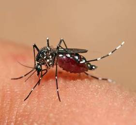
Of note, dengue, chikungunya, and Zika are all transmitted by infected Aedes mosquitoes. There is no enzootic cycle. Just human-mosquito-human transmission. In contrast, West Nile virus is transmitted by Culex mosquitoes in an enzootic cycle between an avian reservoir and humans.
Treatment
There is no specific treatment for arboviral infections. The primary goal is relief of symptoms with fluids, bed rest, and analgesics. For bacterial vector-borne diseases, antibiotic therapy is indicated and is based on the specific pathogen. Doxycycline is the drug of choice for treatment of suspected and confirmed Rocky Mountain Spotted Fever, ehrlichiosis, and anaplasmosis even in children less than 8 years of age. Delay in initiation of antimicrobial therapy pending definitive diagnosis may lead to an adverse outcome. It is also the drug of choice for tick-borne relapsing fever.
Lyme disease is also responsive to antibiotic treatment. Therapy is based on the disease category. (Lyme disease in “Red Book: 2015 Report of the Committee on Infectious Diseases,” [Elk Grove Village, Ill.: American Academy of Pediatrics, 2015, pp. 516-25]).
STARI clinically presents with a lesion that resembles erythema migrans in southern and southeastern states. However, it has not been associated with any of the complications reported with disseminated Lyme disease. Treatment is not recommended.
Tularemia and babesiosis are both responsive to antimicrobial therapy and would best be managed in consultation with an infectious disease physician.
A handy, concise, up to date reference guide about all of the tick-borne diseases including photographs is available at the App Store. The Tickborne Diseases App was developed by the CDC and it is free!
Prevention
The cornerstone of disease prevention is avoidance of mosquito and tick bites, in addition to eliminating mosquito breeding sites. Ticks are generally found near the ground, in brushy or wooded areas. They usually wait for a potential host to brush against them. When this happens, they climb onto the host and find a site to attach.
Is there a role for antimicrobial prophylaxis once a tick has been discovered? There is no data to support antimicrobial prophylaxis to prevent Rocky Mountain spotted fever, ehrlichiosis, and anaplasmosis. Prophylaxis with doxycycline or ciprofloxacin is recommended for children and adults after exposure to an intentional release of tularemia and for laboratory workers after inadvertent exposure. For prevention of Lyme disease, a single dose of doxycycline (4 mg/kg, max dose 200 mg) may be offered under limited conditions: The patient is at least 8 years of age, resides in an area where Lyme is highly endemic, the tick removed was engorged, therapy can be initiated within 72 hours after tick removal, and the estimated time of attachment was at least 36 hours. There is inadequate data on the use of amoxicillin.
Remember, not all mosquitoes are alike. Those that transmit chikungunya, dengue, and Zika (Aedes mosquitoes) are primarily daytime mosquitoes, but also can bite at night. West Nile is transmitted by Culex mosquitoes, which feed from dusk to dawn.
Here are some tips to share with your patients that should decrease their chances of acquiring a mosquito or tick-borne disease:
• Apply mosquito repellent only to intact exposed skin when outdoors. Most repellents can be safely used on children at least 2 months of age and older. Avoid applying repellent directly on the child’s hand. Use at least a 20% DEET (N,N-diethyl-meta-toluamide) containing product. Other Environmental Protection Agency–registered repellents are an alternative (Additional information is available at http://www2.epa.gov/insect-repellents). Products containing oil of lemon eucalyptus (OLE) or p-Menthane-3,8-diol (PMD) should not be used on children under 3 years of age.
• Apply permethrin to clothing, hats, boots, and so on. It is designed to repel mosquitoes and ticks. It can last for several washings. It is ideal to spray over nets covering carriers in children younger than 2 months of age.
• Wear long-sleeved shirts and long pants tucked inside of socks when hiking.
• Check for ticks daily, especially under the arms, behind the ears, around the waist, behind the knees, and inside belly buttons after outdoor activities.
• Have your patients learn how to effectively remove a tick. With a fine tipped tweezer, grasp the tick as close to the skin as possible and pull straight up with even pressure. Do not twist or jerk the tick. Do not squash the tick. Place it in a bag and dispose of it. Clean the site after removal with alcohol, iodine, or soap and water.
• Encourage families to mosquito proof their home by using screens on windows and doors, and using air conditioning when available.
• Empty and scrub all items that contain water such as birdbaths, planters, or wading pools around the outside of the home at least weekly because mosquitoes lay eggs in or near free standing water.
• Dogs and cats should be treated for ticks as recommended by the veterinarian.
The impact of the ongoing Zika virus outbreak is uncertain. While it may have an impact on those planning international travel now and in the near future, several arboviral and vector-borne diseases currently exist in the United States. Encouraging our patients to practice interventions to prevent mosquito and tick bites now will also serve to protect them if Zika virus becomes established in the Aedes mosquitoes here in the future and/or if they have plans for international travel. For up to date information on Zika virus for yourself and your patients, visit www.cdc.gov/zika.
Bonnie M. Word, M.D., is a pediatric infectious disease specialist and director of the Houston Travel Medicine Clinic. She said she had no relevant financial disclosures. Email Dr. Word at pdnews@frontlinemedcom.com.
May has arrived, and for the majority of your patients it signals the end of the school year and the beginning of summer vacation. Zika virus is on the minds of most people since its arrival to the Western Hemisphere in March 2015. With the fluidity of this outbreak and almost daily news updates and recommendations, many parents have voiced or will be voicing concerns regarding summer travel destinations.
Many concerns about Zika virus have been previously addressed in this column (“Zika virus: More questions than answers?” by Dr. Kristina Bryant). However, if the decision is to avoid international travel because of the ongoing Zika outbreak, it doesn’t mean your patients get a free pass and will not have to be concerned about acquiring any infectious diseases. They still need to be vigilant about avoiding those pesky vectors that transmit arboviruses and other vector-borne diseases that occur in the United States.
Arboviruses are transmitted by mosquitoes, ticks, or fleas. Most infections are subclinical. If symptoms develop, they are manifested by a generalized febrile illness including fever, headache, myalgia, arthralgia, and rash. Hemorrhagic fever (dengue) or neuroinvasive disease can include aseptic meningitis, encephalitis, or acute flaccid paralysis. Neuroinvasive disease rarely occurs with dengue, Colorado tick fever, and chikungunya infections.
While more than 100 arboviruses can cause infection, some of the more common arboviruses associated with human disease include West Nile, first detected in the United States in 1999 and chikungunya, first reported in the Americas in 2013 with local transmission documented in Florida, Puerto Rico, and the U.S. Virgin Islands in 2014. It is estimated that dengue causes over 100 million cases worldwide annually. Almost 40% of the world’s inhabitants live in endemic areas. The majority of cases on the U.S. mainland are imported. However, it is endemic in all U.S. territories including Guam, American Samoa, the U.S. Virgin Islands, and Puerto Rico. Between September 2015 and March 2016, Hawaii experienced a dengue outbreak involving 264 individuals including 46 children. As of April 16, 2016, there were no infectious individuals on the island.
Other domestic arboviruses causing disease include St. Louis, Eastern, and Western Equine encephalitis, La Crosse encephalitis, Colorado tick fever, and Powassan virus. All are transmitted by mosquitoes with the exception of Powassan and Colorado tick fever, which are transmitted by ticks. The numbers of cases nationally are much lower for these diseases, compared with West Nile, dengue, and chikungunya. National and state-specific information is available for domestic arboviruses at diseasemaps.usgs.gov/mapviewer. Data is compiled by ArboNET, a national arboviral surveillance system that is managed by the Centers for Disease Control and Prevention (CDC) in conjunction with state health departments. Not only is human disease monitored, but it also maintains data on viremic blood donors, dead birds, mosquitoes, veterinary disease cases, and sentinel animals.
Spring and summer are the most active seasons for ticks. Bacterial and spirochetal diseases transmitted by them include rickettsial diseases such as Rocky Mountain Spotted Fever, ehrlichiosis, and anaplasmosis. Tularemia in addition to Lyme and tick-borne relapsing fever are also transmitted by ticks. Babesiosis, which is due to a parasite, and southern tick-associated rash illness (STARI), whose causative agent is yet to be determined, are two additional tick-related diagnoses.
Of note, dengue, chikungunya, and Zika are all transmitted by infected Aedes mosquitoes. There is no enzootic cycle. Just human-mosquito-human transmission. In contrast, West Nile virus is transmitted by Culex mosquitoes in an enzootic cycle between an avian reservoir and humans.
Treatment
There is no specific treatment for arboviral infections. The primary goal is relief of symptoms with fluids, bed rest, and analgesics. For bacterial vector-borne diseases, antibiotic therapy is indicated and is based on the specific pathogen. Doxycycline is the drug of choice for treatment of suspected and confirmed Rocky Mountain Spotted Fever, ehrlichiosis, and anaplasmosis even in children less than 8 years of age. Delay in initiation of antimicrobial therapy pending definitive diagnosis may lead to an adverse outcome. It is also the drug of choice for tick-borne relapsing fever.
Lyme disease is also responsive to antibiotic treatment. Therapy is based on the disease category. (Lyme disease in “Red Book: 2015 Report of the Committee on Infectious Diseases,” [Elk Grove Village, Ill.: American Academy of Pediatrics, 2015, pp. 516-25]).
STARI clinically presents with a lesion that resembles erythema migrans in southern and southeastern states. However, it has not been associated with any of the complications reported with disseminated Lyme disease. Treatment is not recommended.
Tularemia and babesiosis are both responsive to antimicrobial therapy and would best be managed in consultation with an infectious disease physician.
A handy, concise, up to date reference guide about all of the tick-borne diseases including photographs is available at the App Store. The Tickborne Diseases App was developed by the CDC and it is free!
Prevention
The cornerstone of disease prevention is avoidance of mosquito and tick bites, in addition to eliminating mosquito breeding sites. Ticks are generally found near the ground, in brushy or wooded areas. They usually wait for a potential host to brush against them. When this happens, they climb onto the host and find a site to attach.
Is there a role for antimicrobial prophylaxis once a tick has been discovered? There is no data to support antimicrobial prophylaxis to prevent Rocky Mountain spotted fever, ehrlichiosis, and anaplasmosis. Prophylaxis with doxycycline or ciprofloxacin is recommended for children and adults after exposure to an intentional release of tularemia and for laboratory workers after inadvertent exposure. For prevention of Lyme disease, a single dose of doxycycline (4 mg/kg, max dose 200 mg) may be offered under limited conditions: The patient is at least 8 years of age, resides in an area where Lyme is highly endemic, the tick removed was engorged, therapy can be initiated within 72 hours after tick removal, and the estimated time of attachment was at least 36 hours. There is inadequate data on the use of amoxicillin.
Remember, not all mosquitoes are alike. Those that transmit chikungunya, dengue, and Zika (Aedes mosquitoes) are primarily daytime mosquitoes, but also can bite at night. West Nile is transmitted by Culex mosquitoes, which feed from dusk to dawn.
Here are some tips to share with your patients that should decrease their chances of acquiring a mosquito or tick-borne disease:
• Apply mosquito repellent only to intact exposed skin when outdoors. Most repellents can be safely used on children at least 2 months of age and older. Avoid applying repellent directly on the child’s hand. Use at least a 20% DEET (N,N-diethyl-meta-toluamide) containing product. Other Environmental Protection Agency–registered repellents are an alternative (Additional information is available at http://www2.epa.gov/insect-repellents). Products containing oil of lemon eucalyptus (OLE) or p-Menthane-3,8-diol (PMD) should not be used on children under 3 years of age.
• Apply permethrin to clothing, hats, boots, and so on. It is designed to repel mosquitoes and ticks. It can last for several washings. It is ideal to spray over nets covering carriers in children younger than 2 months of age.
• Wear long-sleeved shirts and long pants tucked inside of socks when hiking.
• Check for ticks daily, especially under the arms, behind the ears, around the waist, behind the knees, and inside belly buttons after outdoor activities.
• Have your patients learn how to effectively remove a tick. With a fine tipped tweezer, grasp the tick as close to the skin as possible and pull straight up with even pressure. Do not twist or jerk the tick. Do not squash the tick. Place it in a bag and dispose of it. Clean the site after removal with alcohol, iodine, or soap and water.
• Encourage families to mosquito proof their home by using screens on windows and doors, and using air conditioning when available.
• Empty and scrub all items that contain water such as birdbaths, planters, or wading pools around the outside of the home at least weekly because mosquitoes lay eggs in or near free standing water.
• Dogs and cats should be treated for ticks as recommended by the veterinarian.
The impact of the ongoing Zika virus outbreak is uncertain. While it may have an impact on those planning international travel now and in the near future, several arboviral and vector-borne diseases currently exist in the United States. Encouraging our patients to practice interventions to prevent mosquito and tick bites now will also serve to protect them if Zika virus becomes established in the Aedes mosquitoes here in the future and/or if they have plans for international travel. For up to date information on Zika virus for yourself and your patients, visit www.cdc.gov/zika.
Bonnie M. Word, M.D., is a pediatric infectious disease specialist and director of the Houston Travel Medicine Clinic. She said she had no relevant financial disclosures. Email Dr. Word at pdnews@frontlinemedcom.com.
May has arrived, and for the majority of your patients it signals the end of the school year and the beginning of summer vacation. Zika virus is on the minds of most people since its arrival to the Western Hemisphere in March 2015. With the fluidity of this outbreak and almost daily news updates and recommendations, many parents have voiced or will be voicing concerns regarding summer travel destinations.
Many concerns about Zika virus have been previously addressed in this column (“Zika virus: More questions than answers?” by Dr. Kristina Bryant). However, if the decision is to avoid international travel because of the ongoing Zika outbreak, it doesn’t mean your patients get a free pass and will not have to be concerned about acquiring any infectious diseases. They still need to be vigilant about avoiding those pesky vectors that transmit arboviruses and other vector-borne diseases that occur in the United States.
Arboviruses are transmitted by mosquitoes, ticks, or fleas. Most infections are subclinical. If symptoms develop, they are manifested by a generalized febrile illness including fever, headache, myalgia, arthralgia, and rash. Hemorrhagic fever (dengue) or neuroinvasive disease can include aseptic meningitis, encephalitis, or acute flaccid paralysis. Neuroinvasive disease rarely occurs with dengue, Colorado tick fever, and chikungunya infections.
While more than 100 arboviruses can cause infection, some of the more common arboviruses associated with human disease include West Nile, first detected in the United States in 1999 and chikungunya, first reported in the Americas in 2013 with local transmission documented in Florida, Puerto Rico, and the U.S. Virgin Islands in 2014. It is estimated that dengue causes over 100 million cases worldwide annually. Almost 40% of the world’s inhabitants live in endemic areas. The majority of cases on the U.S. mainland are imported. However, it is endemic in all U.S. territories including Guam, American Samoa, the U.S. Virgin Islands, and Puerto Rico. Between September 2015 and March 2016, Hawaii experienced a dengue outbreak involving 264 individuals including 46 children. As of April 16, 2016, there were no infectious individuals on the island.
Other domestic arboviruses causing disease include St. Louis, Eastern, and Western Equine encephalitis, La Crosse encephalitis, Colorado tick fever, and Powassan virus. All are transmitted by mosquitoes with the exception of Powassan and Colorado tick fever, which are transmitted by ticks. The numbers of cases nationally are much lower for these diseases, compared with West Nile, dengue, and chikungunya. National and state-specific information is available for domestic arboviruses at diseasemaps.usgs.gov/mapviewer. Data is compiled by ArboNET, a national arboviral surveillance system that is managed by the Centers for Disease Control and Prevention (CDC) in conjunction with state health departments. Not only is human disease monitored, but it also maintains data on viremic blood donors, dead birds, mosquitoes, veterinary disease cases, and sentinel animals.
Spring and summer are the most active seasons for ticks. Bacterial and spirochetal diseases transmitted by them include rickettsial diseases such as Rocky Mountain Spotted Fever, ehrlichiosis, and anaplasmosis. Tularemia in addition to Lyme and tick-borne relapsing fever are also transmitted by ticks. Babesiosis, which is due to a parasite, and southern tick-associated rash illness (STARI), whose causative agent is yet to be determined, are two additional tick-related diagnoses.
Of note, dengue, chikungunya, and Zika are all transmitted by infected Aedes mosquitoes. There is no enzootic cycle. Just human-mosquito-human transmission. In contrast, West Nile virus is transmitted by Culex mosquitoes in an enzootic cycle between an avian reservoir and humans.
Treatment
There is no specific treatment for arboviral infections. The primary goal is relief of symptoms with fluids, bed rest, and analgesics. For bacterial vector-borne diseases, antibiotic therapy is indicated and is based on the specific pathogen. Doxycycline is the drug of choice for treatment of suspected and confirmed Rocky Mountain Spotted Fever, ehrlichiosis, and anaplasmosis even in children less than 8 years of age. Delay in initiation of antimicrobial therapy pending definitive diagnosis may lead to an adverse outcome. It is also the drug of choice for tick-borne relapsing fever.
Lyme disease is also responsive to antibiotic treatment. Therapy is based on the disease category. (Lyme disease in “Red Book: 2015 Report of the Committee on Infectious Diseases,” [Elk Grove Village, Ill.: American Academy of Pediatrics, 2015, pp. 516-25]).
STARI clinically presents with a lesion that resembles erythema migrans in southern and southeastern states. However, it has not been associated with any of the complications reported with disseminated Lyme disease. Treatment is not recommended.
Tularemia and babesiosis are both responsive to antimicrobial therapy and would best be managed in consultation with an infectious disease physician.
A handy, concise, up to date reference guide about all of the tick-borne diseases including photographs is available at the App Store. The Tickborne Diseases App was developed by the CDC and it is free!
Prevention
The cornerstone of disease prevention is avoidance of mosquito and tick bites, in addition to eliminating mosquito breeding sites. Ticks are generally found near the ground, in brushy or wooded areas. They usually wait for a potential host to brush against them. When this happens, they climb onto the host and find a site to attach.
Is there a role for antimicrobial prophylaxis once a tick has been discovered? There is no data to support antimicrobial prophylaxis to prevent Rocky Mountain spotted fever, ehrlichiosis, and anaplasmosis. Prophylaxis with doxycycline or ciprofloxacin is recommended for children and adults after exposure to an intentional release of tularemia and for laboratory workers after inadvertent exposure. For prevention of Lyme disease, a single dose of doxycycline (4 mg/kg, max dose 200 mg) may be offered under limited conditions: The patient is at least 8 years of age, resides in an area where Lyme is highly endemic, the tick removed was engorged, therapy can be initiated within 72 hours after tick removal, and the estimated time of attachment was at least 36 hours. There is inadequate data on the use of amoxicillin.
Remember, not all mosquitoes are alike. Those that transmit chikungunya, dengue, and Zika (Aedes mosquitoes) are primarily daytime mosquitoes, but also can bite at night. West Nile is transmitted by Culex mosquitoes, which feed from dusk to dawn.
Here are some tips to share with your patients that should decrease their chances of acquiring a mosquito or tick-borne disease:
• Apply mosquito repellent only to intact exposed skin when outdoors. Most repellents can be safely used on children at least 2 months of age and older. Avoid applying repellent directly on the child’s hand. Use at least a 20% DEET (N,N-diethyl-meta-toluamide) containing product. Other Environmental Protection Agency–registered repellents are an alternative (Additional information is available at http://www2.epa.gov/insect-repellents). Products containing oil of lemon eucalyptus (OLE) or p-Menthane-3,8-diol (PMD) should not be used on children under 3 years of age.
• Apply permethrin to clothing, hats, boots, and so on. It is designed to repel mosquitoes and ticks. It can last for several washings. It is ideal to spray over nets covering carriers in children younger than 2 months of age.
• Wear long-sleeved shirts and long pants tucked inside of socks when hiking.
• Check for ticks daily, especially under the arms, behind the ears, around the waist, behind the knees, and inside belly buttons after outdoor activities.
• Have your patients learn how to effectively remove a tick. With a fine tipped tweezer, grasp the tick as close to the skin as possible and pull straight up with even pressure. Do not twist or jerk the tick. Do not squash the tick. Place it in a bag and dispose of it. Clean the site after removal with alcohol, iodine, or soap and water.
• Encourage families to mosquito proof their home by using screens on windows and doors, and using air conditioning when available.
• Empty and scrub all items that contain water such as birdbaths, planters, or wading pools around the outside of the home at least weekly because mosquitoes lay eggs in or near free standing water.
• Dogs and cats should be treated for ticks as recommended by the veterinarian.
The impact of the ongoing Zika virus outbreak is uncertain. While it may have an impact on those planning international travel now and in the near future, several arboviral and vector-borne diseases currently exist in the United States. Encouraging our patients to practice interventions to prevent mosquito and tick bites now will also serve to protect them if Zika virus becomes established in the Aedes mosquitoes here in the future and/or if they have plans for international travel. For up to date information on Zika virus for yourself and your patients, visit www.cdc.gov/zika.
Bonnie M. Word, M.D., is a pediatric infectious disease specialist and director of the Houston Travel Medicine Clinic. She said she had no relevant financial disclosures. Email Dr. Word at pdnews@frontlinemedcom.com.

Risk of arthritis in children with Down syndrome higher than previously reported
GLASGOW – Children with Down syndrome are at increased risk for arthritis that often goes unrecognized and leads to treatment delays and potential chronic disability.
Research presented at the British Society for Rheumatology annual conference highlighted how Down arthropathy is not only more prevalent than idiopathic juvenile arthritis (JIA), but also has distinct clinical and radiographic features.
“Our research to date has shown that there is a significant increased risk of arthritis in children with trisomy 21, and higher than that previously reported,” said Dr. Charlene Foley, a research fellow at Our Lady’s Children’s Hospital in Dublin.

“There is a significant delay in diagnosis, which may be a cause of the x-ray changes at diagnosis, or it may be in fact that Down arthropathy is more aggressive,” than other childhood forms of arthritis, she observed.
Dr. Foley noted that Down arthropathy was first reported in the medical literature about 30 years ago and crude estimates suggested a prevalence of around 8.7 cases per 1,000 children versus 1 per 1,000 for JIA. However, the research she presented put the crude point prevalence at 18-21 per 1,000 children.
Dr. Foley presented the findings of an observational study conducted in the Republic of Ireland in which children with trisomy 21 and their families were identified from a variety of sources and invited to participate. After completion of a screening questionnaire and an appointment local to the participants, children who were suspected of having arthritis were invited to attend a consultant appointment. They underwent a clinical management pathway developed for JIA because no specific pathway had been developed for the children at that time, with follow-up appointments held every 3-6 months depending on the child’s needs.
Over an 18-month period, 503 children with trisomy 21 and a mean age of 8 years were screened. They had a range of musculoskeletal anomalies, the most common of which were flat feet in almost all the children (91.1%), inflammatory arthritis in 7.1%, and scoliosis in 4.8%. Many other problems occurred, with an incidence of 1.5% or less for each.
A total of 22 new cases of Down arthropathy have been identified to date, in addition to 11 at the clinic who predated the start of the study. About 75% have come through the screening clinics and the rest through pediatricians’ referral.
“It is a challenging disease both in terms of diagnosis and management,” Dr. Foley said. Of all the identified children, 91% had poor language skills or nonverbal communication and 15% had autism spectrum disorder.
On average, the time to diagnosis of the arthropathy was 1.7 years versus 0.74 years for a control group of 33 children with JIA. This is likely an underestimation, however, as 42% of the children or parents in the Down arthropathy cohort were unable to give a date on which symptoms had started.
Dr. Foley reported that the majority of trisomy 21 children had presented with polyarticular arthritis, mostly involving the proximal interphalangeal joints of the hands (78.6% of cases), or the wrists (53.6% of cases). There was significant small joint involvement (88% vs. 43% of the JIA cohort), and higher restricted joint counts (4.5 vs. 2.0). There were also differences in erythrocyte sedimentation rate and C-reactive protein at diagnosis, with these being “barely raised” in children with Down arthropathy versus children with JIA, so unlikely to aid a diagnosis. Children were also found to be rheumatoid factor negative.
Two-thirds of Down arthropathy cases had x-ray changes at presentation versus 24% of the JIA group, of which 29% versus 9.5% were erosive.
Treatment is complicated by drug-related side effects, with many children unable to tolerate methotrexate, Dr. Foley said. In the Irish cohort, treatment with methotrexate led to nausea in 75%, compared with 7.1% of the JIA children. Although reports are limited, methotrexate intolerance has been shown in children with trisomy 21, so there could be a genetic or metabolic reason behind this. Dr. Foley noted that they manage this problem by starting methotrexate on the lowest possible doses (10 mg/m2) and co-administering the antiemetic ondansetron. They have a low threshold for switching to an anti-TNF drug if needed, and have also started giving biologic drugs to some newly diagnosed children.
“The take-home message is to think outside of the Down syndrome box and don’t just blame everything on Down syndrome,” Dr. Foley said. As it may be challenging to examine a child, she suggested looking at the hands first because they are the most likely to be affected.
“We feel that a musculoskeletal assessment should be part of the annual surveillance for all children with Down syndrome,” she concluded.
As for who should conduct such an assessment, Dr. Foley suggested that general pediatricians who are regularly seeing these children for other health checks should perform it. However, as one delegate observed, nonrheumatology professionals may need a little training and guidance, as musculoskeletal assessments can be difficult. Looking only at the hands, and potentially the feet, may be one solution.
The study has raised a number of questions and future research will be needed to further characterize the arthritis and to determine how best to diagnose and treat it, noted Dr. Foley, who indicated that she had no conflicts of interest.
GLASGOW – Children with Down syndrome are at increased risk for arthritis that often goes unrecognized and leads to treatment delays and potential chronic disability.
Research presented at the British Society for Rheumatology annual conference highlighted how Down arthropathy is not only more prevalent than idiopathic juvenile arthritis (JIA), but also has distinct clinical and radiographic features.
“Our research to date has shown that there is a significant increased risk of arthritis in children with trisomy 21, and higher than that previously reported,” said Dr. Charlene Foley, a research fellow at Our Lady’s Children’s Hospital in Dublin.
“There is a significant delay in diagnosis, which may be a cause of the x-ray changes at diagnosis, or it may be in fact that Down arthropathy is more aggressive,” than other childhood forms of arthritis, she observed.
Dr. Foley noted that Down arthropathy was first reported in the medical literature about 30 years ago and crude estimates suggested a prevalence of around 8.7 cases per 1,000 children versus 1 per 1,000 for JIA. However, the research she presented put the crude point prevalence at 18-21 per 1,000 children.
Dr. Foley presented the findings of an observational study conducted in the Republic of Ireland in which children with trisomy 21 and their families were identified from a variety of sources and invited to participate. After completion of a screening questionnaire and an appointment local to the participants, children who were suspected of having arthritis were invited to attend a consultant appointment. They underwent a clinical management pathway developed for JIA because no specific pathway had been developed for the children at that time, with follow-up appointments held every 3-6 months depending on the child’s needs.
Over an 18-month period, 503 children with trisomy 21 and a mean age of 8 years were screened. They had a range of musculoskeletal anomalies, the most common of which were flat feet in almost all the children (91.1%), inflammatory arthritis in 7.1%, and scoliosis in 4.8%. Many other problems occurred, with an incidence of 1.5% or less for each.
A total of 22 new cases of Down arthropathy have been identified to date, in addition to 11 at the clinic who predated the start of the study. About 75% have come through the screening clinics and the rest through pediatricians’ referral.
“It is a challenging disease both in terms of diagnosis and management,” Dr. Foley said. Of all the identified children, 91% had poor language skills or nonverbal communication and 15% had autism spectrum disorder.
On average, the time to diagnosis of the arthropathy was 1.7 years versus 0.74 years for a control group of 33 children with JIA. This is likely an underestimation, however, as 42% of the children or parents in the Down arthropathy cohort were unable to give a date on which symptoms had started.
Dr. Foley reported that the majority of trisomy 21 children had presented with polyarticular arthritis, mostly involving the proximal interphalangeal joints of the hands (78.6% of cases), or the wrists (53.6% of cases). There was significant small joint involvement (88% vs. 43% of the JIA cohort), and higher restricted joint counts (4.5 vs. 2.0). There were also differences in erythrocyte sedimentation rate and C-reactive protein at diagnosis, with these being “barely raised” in children with Down arthropathy versus children with JIA, so unlikely to aid a diagnosis. Children were also found to be rheumatoid factor negative.
Two-thirds of Down arthropathy cases had x-ray changes at presentation versus 24% of the JIA group, of which 29% versus 9.5% were erosive.
Treatment is complicated by drug-related side effects, with many children unable to tolerate methotrexate, Dr. Foley said. In the Irish cohort, treatment with methotrexate led to nausea in 75%, compared with 7.1% of the JIA children. Although reports are limited, methotrexate intolerance has been shown in children with trisomy 21, so there could be a genetic or metabolic reason behind this. Dr. Foley noted that they manage this problem by starting methotrexate on the lowest possible doses (10 mg/m2) and co-administering the antiemetic ondansetron. They have a low threshold for switching to an anti-TNF drug if needed, and have also started giving biologic drugs to some newly diagnosed children.
“The take-home message is to think outside of the Down syndrome box and don’t just blame everything on Down syndrome,” Dr. Foley said. As it may be challenging to examine a child, she suggested looking at the hands first because they are the most likely to be affected.
“We feel that a musculoskeletal assessment should be part of the annual surveillance for all children with Down syndrome,” she concluded.
As for who should conduct such an assessment, Dr. Foley suggested that general pediatricians who are regularly seeing these children for other health checks should perform it. However, as one delegate observed, nonrheumatology professionals may need a little training and guidance, as musculoskeletal assessments can be difficult. Looking only at the hands, and potentially the feet, may be one solution.
The study has raised a number of questions and future research will be needed to further characterize the arthritis and to determine how best to diagnose and treat it, noted Dr. Foley, who indicated that she had no conflicts of interest.
GLASGOW – Children with Down syndrome are at increased risk for arthritis that often goes unrecognized and leads to treatment delays and potential chronic disability.
Research presented at the British Society for Rheumatology annual conference highlighted how Down arthropathy is not only more prevalent than idiopathic juvenile arthritis (JIA), but also has distinct clinical and radiographic features.
“Our research to date has shown that there is a significant increased risk of arthritis in children with trisomy 21, and higher than that previously reported,” said Dr. Charlene Foley, a research fellow at Our Lady’s Children’s Hospital in Dublin.
“There is a significant delay in diagnosis, which may be a cause of the x-ray changes at diagnosis, or it may be in fact that Down arthropathy is more aggressive,” than other childhood forms of arthritis, she observed.
Dr. Foley noted that Down arthropathy was first reported in the medical literature about 30 years ago and crude estimates suggested a prevalence of around 8.7 cases per 1,000 children versus 1 per 1,000 for JIA. However, the research she presented put the crude point prevalence at 18-21 per 1,000 children.
Dr. Foley presented the findings of an observational study conducted in the Republic of Ireland in which children with trisomy 21 and their families were identified from a variety of sources and invited to participate. After completion of a screening questionnaire and an appointment local to the participants, children who were suspected of having arthritis were invited to attend a consultant appointment. They underwent a clinical management pathway developed for JIA because no specific pathway had been developed for the children at that time, with follow-up appointments held every 3-6 months depending on the child’s needs.
Over an 18-month period, 503 children with trisomy 21 and a mean age of 8 years were screened. They had a range of musculoskeletal anomalies, the most common of which were flat feet in almost all the children (91.1%), inflammatory arthritis in 7.1%, and scoliosis in 4.8%. Many other problems occurred, with an incidence of 1.5% or less for each.
A total of 22 new cases of Down arthropathy have been identified to date, in addition to 11 at the clinic who predated the start of the study. About 75% have come through the screening clinics and the rest through pediatricians’ referral.
“It is a challenging disease both in terms of diagnosis and management,” Dr. Foley said. Of all the identified children, 91% had poor language skills or nonverbal communication and 15% had autism spectrum disorder.
On average, the time to diagnosis of the arthropathy was 1.7 years versus 0.74 years for a control group of 33 children with JIA. This is likely an underestimation, however, as 42% of the children or parents in the Down arthropathy cohort were unable to give a date on which symptoms had started.
Dr. Foley reported that the majority of trisomy 21 children had presented with polyarticular arthritis, mostly involving the proximal interphalangeal joints of the hands (78.6% of cases), or the wrists (53.6% of cases). There was significant small joint involvement (88% vs. 43% of the JIA cohort), and higher restricted joint counts (4.5 vs. 2.0). There were also differences in erythrocyte sedimentation rate and C-reactive protein at diagnosis, with these being “barely raised” in children with Down arthropathy versus children with JIA, so unlikely to aid a diagnosis. Children were also found to be rheumatoid factor negative.
Two-thirds of Down arthropathy cases had x-ray changes at presentation versus 24% of the JIA group, of which 29% versus 9.5% were erosive.
Treatment is complicated by drug-related side effects, with many children unable to tolerate methotrexate, Dr. Foley said. In the Irish cohort, treatment with methotrexate led to nausea in 75%, compared with 7.1% of the JIA children. Although reports are limited, methotrexate intolerance has been shown in children with trisomy 21, so there could be a genetic or metabolic reason behind this. Dr. Foley noted that they manage this problem by starting methotrexate on the lowest possible doses (10 mg/m2) and co-administering the antiemetic ondansetron. They have a low threshold for switching to an anti-TNF drug if needed, and have also started giving biologic drugs to some newly diagnosed children.
“The take-home message is to think outside of the Down syndrome box and don’t just blame everything on Down syndrome,” Dr. Foley said. As it may be challenging to examine a child, she suggested looking at the hands first because they are the most likely to be affected.
“We feel that a musculoskeletal assessment should be part of the annual surveillance for all children with Down syndrome,” she concluded.
As for who should conduct such an assessment, Dr. Foley suggested that general pediatricians who are regularly seeing these children for other health checks should perform it. However, as one delegate observed, nonrheumatology professionals may need a little training and guidance, as musculoskeletal assessments can be difficult. Looking only at the hands, and potentially the feet, may be one solution.
The study has raised a number of questions and future research will be needed to further characterize the arthritis and to determine how best to diagnose and treat it, noted Dr. Foley, who indicated that she had no conflicts of interest.
AT RHEUMATOLOGY 2016
Key clinical point:A musculoskeletal assessment should be part of the annual surveillance for all children with Down syndrome to look for arthritis.
Major finding: Arthritis in children with Down syndrome typically presents as polyarticular inflammation in the hands and wrists.
Data source: Observational study of 33 children with Down arthropathy and 33 with juvenile idiopathic arthritis living in Ireland.
Disclosures: Dr. Foley had no conflicts of interest.

Norovirus costs $60 billion worldwide, making inroads in U.S.
The global economic impact of norovirus is roughly $60 billion in societal costs and $4 billion in direct health care costs per year, according to an simulation model analysis.
Most of the costs (84%-99%, varying by region) resulted from lost productivity. The total societal cost was highest in children younger than 5 years, nearly $40 billion, compared with $20 billion for all other age groups combined, but the cost per illness was highest among adults aged 55 years and older.
Disease incidence was similar across all countries in the simulation model that included 233 countries stratified by World Health Organization region and globally but high-income countries accounted for 62% of the global health system costs, wrote Sarah M. Bartsch of Johns Hopkins University, Baltimore, and colleagues (PLoS ONE 2016. doi: 10.1371/journal.pone.0151219).
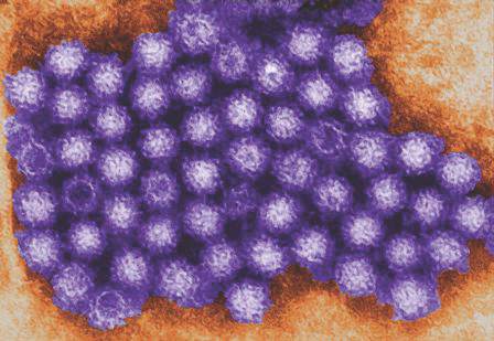
In the United States, norovirus was the most common pathogen identified in cases of acute gastroenteritis (AGE, 6%), followed by rotavirus and sapovirus (2% for both) and astrovirus (1%), according to data from a population-based study, including 1,099 stool specimens collected between August 2012 and September 2013. These findings were published in a separate study (PLoS ONE 2016. doi: 10.1371/journal.pone.0148395).
Overall, U.S. children younger than 5 years of age had the highest prevalence of norovirus; outpatient incidence and community incidences were 25.6 and 152.2 per 1,000 person-years, respectively. The next highest norovirus prevalence occurred in adults older than 65 years; outpatient and community incidence were 7.8 and 7.8 per 1,000 person-years, respectively. The study provides “the first U.S. laboratory-based age-specific norovirus AGE incidence rates in both community and outpatient settings,” wrote Scott P. Grytdal of the Centers for Disease Control and Prevention in Atlanta, and colleagues.
The global economic impact of norovirus is roughly $60 billion in societal costs and $4 billion in direct health care costs per year, according to an simulation model analysis.
Most of the costs (84%-99%, varying by region) resulted from lost productivity. The total societal cost was highest in children younger than 5 years, nearly $40 billion, compared with $20 billion for all other age groups combined, but the cost per illness was highest among adults aged 55 years and older.
Disease incidence was similar across all countries in the simulation model that included 233 countries stratified by World Health Organization region and globally but high-income countries accounted for 62% of the global health system costs, wrote Sarah M. Bartsch of Johns Hopkins University, Baltimore, and colleagues (PLoS ONE 2016. doi: 10.1371/journal.pone.0151219).
In the United States, norovirus was the most common pathogen identified in cases of acute gastroenteritis (AGE, 6%), followed by rotavirus and sapovirus (2% for both) and astrovirus (1%), according to data from a population-based study, including 1,099 stool specimens collected between August 2012 and September 2013. These findings were published in a separate study (PLoS ONE 2016. doi: 10.1371/journal.pone.0148395).
Overall, U.S. children younger than 5 years of age had the highest prevalence of norovirus; outpatient incidence and community incidences were 25.6 and 152.2 per 1,000 person-years, respectively. The next highest norovirus prevalence occurred in adults older than 65 years; outpatient and community incidence were 7.8 and 7.8 per 1,000 person-years, respectively. The study provides “the first U.S. laboratory-based age-specific norovirus AGE incidence rates in both community and outpatient settings,” wrote Scott P. Grytdal of the Centers for Disease Control and Prevention in Atlanta, and colleagues.
The global economic impact of norovirus is roughly $60 billion in societal costs and $4 billion in direct health care costs per year, according to an simulation model analysis.
Most of the costs (84%-99%, varying by region) resulted from lost productivity. The total societal cost was highest in children younger than 5 years, nearly $40 billion, compared with $20 billion for all other age groups combined, but the cost per illness was highest among adults aged 55 years and older.
Disease incidence was similar across all countries in the simulation model that included 233 countries stratified by World Health Organization region and globally but high-income countries accounted for 62% of the global health system costs, wrote Sarah M. Bartsch of Johns Hopkins University, Baltimore, and colleagues (PLoS ONE 2016. doi: 10.1371/journal.pone.0151219).
In the United States, norovirus was the most common pathogen identified in cases of acute gastroenteritis (AGE, 6%), followed by rotavirus and sapovirus (2% for both) and astrovirus (1%), according to data from a population-based study, including 1,099 stool specimens collected between August 2012 and September 2013. These findings were published in a separate study (PLoS ONE 2016. doi: 10.1371/journal.pone.0148395).
Overall, U.S. children younger than 5 years of age had the highest prevalence of norovirus; outpatient incidence and community incidences were 25.6 and 152.2 per 1,000 person-years, respectively. The next highest norovirus prevalence occurred in adults older than 65 years; outpatient and community incidence were 7.8 and 7.8 per 1,000 person-years, respectively. The study provides “the first U.S. laboratory-based age-specific norovirus AGE incidence rates in both community and outpatient settings,” wrote Scott P. Grytdal of the Centers for Disease Control and Prevention in Atlanta, and colleagues.
FROM PLOS ONE
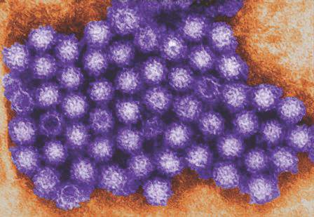
Smartphones, smart parents
In the age of technology, parents denying their teens a smartphone is blatant child abuse, at least in the eyes of the teen. Between Twitter, Instagram, and Snapchat, a teen’s entire life revolves around minute-to-minute check-ins. Smartphones have opened the door for sexual predators, bullying, and complete withdrawal from the world that surrounds them. But, with every bad there is a good and it’s knowing how to make the technology takeover work for you.

The smartphone is the best bargaining chip ever created. A teen would sooner die than lose his or her phone. Parents need to use this upper hand to get just about anything done: “You will get your phone back when XYZ is done.” Teens should understand that a phone is a privilege and not a right, so if they don’t want to cooperate, then there is a consequence.
Second, there is no greater source of information than a teen’s phone. From information in text, to locations, to the dreaded selfies, teens cannot help themselves when it comes to sharing every aspect of their lives. There are countless stories of teens getting busted because they posted a picture on Instagram with the person they were not supposed to be with or from a place they were not supposed to be. There are several great apps that allow parents to see deleted texts, and track locations and websites visited. These same apps allow parents to add controls that block X-rated websites, and notify them when the teen leaves a location or signs up for social media apps.
Accidents are the leading cause of death among teens and smartphones have only increased that. A recent study showed that 34% admitted to texting and driving. Another study reported that 11 teens die per day* because of distracted driver accidents. Not only are teens distracted, they also are inexperienced and are increasing their risk of injury. Teens are 23 times more likely to be in an accident as a result of distracted driving.

Now, there are apps that will alert parents when the teen is driving above the speed limit or has left the restricted area designated by the parent. These apps can silence incoming texts and prevent texts from being sent if the teen is in motion. Some of the apps will read the text out loud and respond with an automated response, letting the caller know that they are unavailable. Canary, My Mobile Watchdog, and Drivesafe.ly are examples, but both Sprint and Verizon have similar apps available. This is an excellent way for parents to monitor teen driving habits. The cost varies from $7.99/month to $99/month but the information provided is priceless.
Whether we like it or not, smartphones are here and have totally changed how teens interact and give them limitless exposure. Many parents, even if they own a smartphone, only use it for its basic functions and may have no idea these types of controls exist. Educating parents that they can use the phone as a tool to monitor and protect their teens is an important part of the well visit, and could very well save a life.
Dr. Pearce is a pediatrician in Frankfort, Ill. Email her at pdnews@frontlinemedcom.com.
*Correction, 6/1/2016: The frequency of teen deaths due to distracted driver accidents was misquoted.
In the age of technology, parents denying their teens a smartphone is blatant child abuse, at least in the eyes of the teen. Between Twitter, Instagram, and Snapchat, a teen’s entire life revolves around minute-to-minute check-ins. Smartphones have opened the door for sexual predators, bullying, and complete withdrawal from the world that surrounds them. But, with every bad there is a good and it’s knowing how to make the technology takeover work for you.
The smartphone is the best bargaining chip ever created. A teen would sooner die than lose his or her phone. Parents need to use this upper hand to get just about anything done: “You will get your phone back when XYZ is done.” Teens should understand that a phone is a privilege and not a right, so if they don’t want to cooperate, then there is a consequence.
Second, there is no greater source of information than a teen’s phone. From information in text, to locations, to the dreaded selfies, teens cannot help themselves when it comes to sharing every aspect of their lives. There are countless stories of teens getting busted because they posted a picture on Instagram with the person they were not supposed to be with or from a place they were not supposed to be. There are several great apps that allow parents to see deleted texts, and track locations and websites visited. These same apps allow parents to add controls that block X-rated websites, and notify them when the teen leaves a location or signs up for social media apps.
Accidents are the leading cause of death among teens and smartphones have only increased that. A recent study showed that 34% admitted to texting and driving. Another study reported that 11 teens die per day* because of distracted driver accidents. Not only are teens distracted, they also are inexperienced and are increasing their risk of injury. Teens are 23 times more likely to be in an accident as a result of distracted driving.
Now, there are apps that will alert parents when the teen is driving above the speed limit or has left the restricted area designated by the parent. These apps can silence incoming texts and prevent texts from being sent if the teen is in motion. Some of the apps will read the text out loud and respond with an automated response, letting the caller know that they are unavailable. Canary, My Mobile Watchdog, and Drivesafe.ly are examples, but both Sprint and Verizon have similar apps available. This is an excellent way for parents to monitor teen driving habits. The cost varies from $7.99/month to $99/month but the information provided is priceless.
Whether we like it or not, smartphones are here and have totally changed how teens interact and give them limitless exposure. Many parents, even if they own a smartphone, only use it for its basic functions and may have no idea these types of controls exist. Educating parents that they can use the phone as a tool to monitor and protect their teens is an important part of the well visit, and could very well save a life.
Dr. Pearce is a pediatrician in Frankfort, Ill. Email her at pdnews@frontlinemedcom.com.
*Correction, 6/1/2016: The frequency of teen deaths due to distracted driver accidents was misquoted.
In the age of technology, parents denying their teens a smartphone is blatant child abuse, at least in the eyes of the teen. Between Twitter, Instagram, and Snapchat, a teen’s entire life revolves around minute-to-minute check-ins. Smartphones have opened the door for sexual predators, bullying, and complete withdrawal from the world that surrounds them. But, with every bad there is a good and it’s knowing how to make the technology takeover work for you.
The smartphone is the best bargaining chip ever created. A teen would sooner die than lose his or her phone. Parents need to use this upper hand to get just about anything done: “You will get your phone back when XYZ is done.” Teens should understand that a phone is a privilege and not a right, so if they don’t want to cooperate, then there is a consequence.
Second, there is no greater source of information than a teen’s phone. From information in text, to locations, to the dreaded selfies, teens cannot help themselves when it comes to sharing every aspect of their lives. There are countless stories of teens getting busted because they posted a picture on Instagram with the person they were not supposed to be with or from a place they were not supposed to be. There are several great apps that allow parents to see deleted texts, and track locations and websites visited. These same apps allow parents to add controls that block X-rated websites, and notify them when the teen leaves a location or signs up for social media apps.
Accidents are the leading cause of death among teens and smartphones have only increased that. A recent study showed that 34% admitted to texting and driving. Another study reported that 11 teens die per day* because of distracted driver accidents. Not only are teens distracted, they also are inexperienced and are increasing their risk of injury. Teens are 23 times more likely to be in an accident as a result of distracted driving.
Now, there are apps that will alert parents when the teen is driving above the speed limit or has left the restricted area designated by the parent. These apps can silence incoming texts and prevent texts from being sent if the teen is in motion. Some of the apps will read the text out loud and respond with an automated response, letting the caller know that they are unavailable. Canary, My Mobile Watchdog, and Drivesafe.ly are examples, but both Sprint and Verizon have similar apps available. This is an excellent way for parents to monitor teen driving habits. The cost varies from $7.99/month to $99/month but the information provided is priceless.
Whether we like it or not, smartphones are here and have totally changed how teens interact and give them limitless exposure. Many parents, even if they own a smartphone, only use it for its basic functions and may have no idea these types of controls exist. Educating parents that they can use the phone as a tool to monitor and protect their teens is an important part of the well visit, and could very well save a life.
Dr. Pearce is a pediatrician in Frankfort, Ill. Email her at pdnews@frontlinemedcom.com.
*Correction, 6/1/2016: The frequency of teen deaths due to distracted driver accidents was misquoted.