User login
Cancer risk six times higher in children with congenital heart defects
NEW ORLEANS – Children with congenital heart defects have a five- to sixfold increased risk of developing pediatric cancer, Matthew Oster, MD, reported at the American Heart Association scientific sessions.
“The absolute risk is small, but it’s still a five- to sixfold risk compared to the general pediatric population, and it does warrant monitoring and a high index of suspicion in our children with congenital heart defects,” said Dr. Oster of Children’s Healthcare of Atlanta.

The increased cancer risk in adults with congenital heart defects has recently been the focus of research attention. However, little is known about the risk of cancer during the childhood of patients with congenital heart defects.
This was the impetus for Dr. Oster’s nationwide retrospective study of 6.1 million children and adolescents continuously enrolled in private, employer-sponsored health insurance plans during 2009-2015. The data came from the Truven Health MarketScan administrative database.
Children with Down syndrome were excluded from the study because their condition is known to be associated with increased rates of both congenital heart defects and pediatric cancers.
Among 88,493 individuals under age 18 with a diagnosed congenital heart defect, the incidence of any neoplasm diagnosed at least 30 days after diagnosis of the heart defect was 3.91/1,000, compared with 0.79/1,000 in more than 6 million children and adolescents without congenital heart disease.
Thus, children with a congenital heart defect were at a 4.9-fold increased risk for developing a childhood cancer. The risk for bone tumors was 11.2-fold greater than in the general pediatric population, and their neuroblastoma risk was 9.8-fold greater. Their risks of lymphoma and leukemia were increased 5.2- and 2.8-fold, respectively. Of note, they had no increased risk of brain tumors.
To confirm their results, Dr. Oster and his coinvestigators also conducted a sensitivity analysis limited to the 55,079 children and adolescents with at least two outpatient or one inpatient ICD-9 diagnostic code for congenital heart disease. Here the incidence of childhood malignancies was 5.1/1,000 patients, for an overall 6.4-fold increased relative risk.
“Bedside-to-bench work is now needed to determine potential mechanisms,” he said. “We believe the increased risk is related more to a common genetic pathway than to an exposure or treatment pathway because it occurs so early. But there may be some impact of exposures or treatment as well. We think that radiation exposure is more of a longer-term risk.”
He and his coinvestigators next plan to look at the impact on pediatric cancer risk of specific types of congenital heart defects.
Dr. Oster reported having no financial conflicts of interest regarding this study, which received Centers for Disease Control and Prevention funding.
NEW ORLEANS – Children with congenital heart defects have a five- to sixfold increased risk of developing pediatric cancer, Matthew Oster, MD, reported at the American Heart Association scientific sessions.
“The absolute risk is small, but it’s still a five- to sixfold risk compared to the general pediatric population, and it does warrant monitoring and a high index of suspicion in our children with congenital heart defects,” said Dr. Oster of Children’s Healthcare of Atlanta.
The increased cancer risk in adults with congenital heart defects has recently been the focus of research attention. However, little is known about the risk of cancer during the childhood of patients with congenital heart defects.
This was the impetus for Dr. Oster’s nationwide retrospective study of 6.1 million children and adolescents continuously enrolled in private, employer-sponsored health insurance plans during 2009-2015. The data came from the Truven Health MarketScan administrative database.
Children with Down syndrome were excluded from the study because their condition is known to be associated with increased rates of both congenital heart defects and pediatric cancers.
Among 88,493 individuals under age 18 with a diagnosed congenital heart defect, the incidence of any neoplasm diagnosed at least 30 days after diagnosis of the heart defect was 3.91/1,000, compared with 0.79/1,000 in more than 6 million children and adolescents without congenital heart disease.
Thus, children with a congenital heart defect were at a 4.9-fold increased risk for developing a childhood cancer. The risk for bone tumors was 11.2-fold greater than in the general pediatric population, and their neuroblastoma risk was 9.8-fold greater. Their risks of lymphoma and leukemia were increased 5.2- and 2.8-fold, respectively. Of note, they had no increased risk of brain tumors.
To confirm their results, Dr. Oster and his coinvestigators also conducted a sensitivity analysis limited to the 55,079 children and adolescents with at least two outpatient or one inpatient ICD-9 diagnostic code for congenital heart disease. Here the incidence of childhood malignancies was 5.1/1,000 patients, for an overall 6.4-fold increased relative risk.
“Bedside-to-bench work is now needed to determine potential mechanisms,” he said. “We believe the increased risk is related more to a common genetic pathway than to an exposure or treatment pathway because it occurs so early. But there may be some impact of exposures or treatment as well. We think that radiation exposure is more of a longer-term risk.”
He and his coinvestigators next plan to look at the impact on pediatric cancer risk of specific types of congenital heart defects.
Dr. Oster reported having no financial conflicts of interest regarding this study, which received Centers for Disease Control and Prevention funding.
NEW ORLEANS – Children with congenital heart defects have a five- to sixfold increased risk of developing pediatric cancer, Matthew Oster, MD, reported at the American Heart Association scientific sessions.
“The absolute risk is small, but it’s still a five- to sixfold risk compared to the general pediatric population, and it does warrant monitoring and a high index of suspicion in our children with congenital heart defects,” said Dr. Oster of Children’s Healthcare of Atlanta.
The increased cancer risk in adults with congenital heart defects has recently been the focus of research attention. However, little is known about the risk of cancer during the childhood of patients with congenital heart defects.
This was the impetus for Dr. Oster’s nationwide retrospective study of 6.1 million children and adolescents continuously enrolled in private, employer-sponsored health insurance plans during 2009-2015. The data came from the Truven Health MarketScan administrative database.
Children with Down syndrome were excluded from the study because their condition is known to be associated with increased rates of both congenital heart defects and pediatric cancers.
Among 88,493 individuals under age 18 with a diagnosed congenital heart defect, the incidence of any neoplasm diagnosed at least 30 days after diagnosis of the heart defect was 3.91/1,000, compared with 0.79/1,000 in more than 6 million children and adolescents without congenital heart disease.
Thus, children with a congenital heart defect were at a 4.9-fold increased risk for developing a childhood cancer. The risk for bone tumors was 11.2-fold greater than in the general pediatric population, and their neuroblastoma risk was 9.8-fold greater. Their risks of lymphoma and leukemia were increased 5.2- and 2.8-fold, respectively. Of note, they had no increased risk of brain tumors.
To confirm their results, Dr. Oster and his coinvestigators also conducted a sensitivity analysis limited to the 55,079 children and adolescents with at least two outpatient or one inpatient ICD-9 diagnostic code for congenital heart disease. Here the incidence of childhood malignancies was 5.1/1,000 patients, for an overall 6.4-fold increased relative risk.
“Bedside-to-bench work is now needed to determine potential mechanisms,” he said. “We believe the increased risk is related more to a common genetic pathway than to an exposure or treatment pathway because it occurs so early. But there may be some impact of exposures or treatment as well. We think that radiation exposure is more of a longer-term risk.”
He and his coinvestigators next plan to look at the impact on pediatric cancer risk of specific types of congenital heart defects.
Dr. Oster reported having no financial conflicts of interest regarding this study, which received Centers for Disease Control and Prevention funding.
AT THE AHA SCIENTIFIC SESSIONS
Key clinical point:
Major finding: Children with congenital heart defects are at a roughly sixfold increased risk of developing a pediatric cancer.
Data source: A retrospective analysis of administrative health insurance plan data on 6.1 million U.S. subjects under age 18 years, more than 88,000 of whom had congenital heart defects.
Disclosures: The presenter reported having no financial conflicts of interest regarding this study, which received Centers for Disease Control and Prevention funding.
Delayed cord clamping reduces anemia risk
Photo by Meutia Chaerani
and Indradi Soemardjan
Delaying umbilical cord clamping by a few minutes can reduce the risk of anemia several months after birth, according to research published in JAMA Pediatrics.
A randomized clinical trial showed that delaying cord clamping by 3 minutes or more after birth—rather than clamping within 1 minute of birth—reduced the prevalence of anemia, iron deficiency, and iron deficiency anemia in infants at 8 months and 12 months of age.
Ola Andersson, MD, PhD, of Uppsala University in Uppsala, Sweden, and his colleagues conducted this research in Nepal, a country with a high prevalence of anemia.
The study included 540 infants—281 boys and 259 girls—with a mean gestational age of 39.2 weeks. Half of the subjects were randomized to delayed cord clamping (3 minutes or more after birth) or early cord clamping (within 1 minute of birth).
At 8 months of age, 78.5% of infants from the delayed clamping group and 69.6% from the early clamping group returned for blood sampling.
Results showed that infants in the delayed clamping group had higher levels of hemoglobin than infants in the early clamping group—10.4 g/dL and 10.2 g/dL, respectively (P=0.008).
Infants in the delayed clamping group also had a lower incidence of:
- Anemia (hemoglobin level <11.0 g/dL)—73.0% and 82.2%, respectively (P=0.01)
- Iron deficiency—22.2% and 38.1%, respectively (P<0.001)
- Iron deficiency anemia—19.3% and 33.3%, respectively (P<0.001).
The relative risk (RR) of anemia was 0.89, the RR of iron deficiency was 0.58, and the RR of iron deficiency anemia was 0.58.
Results were similar when the infants reached 12 months of age, although all the between-group differences were not statistically significant.
Again, infants in the delayed clamping group had higher levels of hemoglobin than infants in the early clamping group—10.3 g/dL and 10.1 g/dL, respectively (P=0.02).
And infants in the delayed clamping group had a lower incidence of:
- Anemia—77.8% and 85.9%, respectively (P=0.02)
- Iron deficiency—35.6% and 43%, respectively (P=0.09)
- Iron deficiency anemia—30.4% and 37.8%, respectively (P=0.08).
The RR of anemia was 0.91, the RR of iron deficiency was 0.83, and the RR of iron deficiency anemia was 0.80.
The researchers said this study shows that delayed cord clamping was an effective intervention to reduce anemia in a high-risk population, with minimal cost and without apparent adverse effects.
The team believes that, if this intervention were implemented on a global scale, this could translate to 5 million fewer infants with anemia at 8 months of age. 
Photo by Meutia Chaerani
and Indradi Soemardjan
Delaying umbilical cord clamping by a few minutes can reduce the risk of anemia several months after birth, according to research published in JAMA Pediatrics.
A randomized clinical trial showed that delaying cord clamping by 3 minutes or more after birth—rather than clamping within 1 minute of birth—reduced the prevalence of anemia, iron deficiency, and iron deficiency anemia in infants at 8 months and 12 months of age.
Ola Andersson, MD, PhD, of Uppsala University in Uppsala, Sweden, and his colleagues conducted this research in Nepal, a country with a high prevalence of anemia.
The study included 540 infants—281 boys and 259 girls—with a mean gestational age of 39.2 weeks. Half of the subjects were randomized to delayed cord clamping (3 minutes or more after birth) or early cord clamping (within 1 minute of birth).
At 8 months of age, 78.5% of infants from the delayed clamping group and 69.6% from the early clamping group returned for blood sampling.
Results showed that infants in the delayed clamping group had higher levels of hemoglobin than infants in the early clamping group—10.4 g/dL and 10.2 g/dL, respectively (P=0.008).
Infants in the delayed clamping group also had a lower incidence of:
- Anemia (hemoglobin level <11.0 g/dL)—73.0% and 82.2%, respectively (P=0.01)
- Iron deficiency—22.2% and 38.1%, respectively (P<0.001)
- Iron deficiency anemia—19.3% and 33.3%, respectively (P<0.001).
The relative risk (RR) of anemia was 0.89, the RR of iron deficiency was 0.58, and the RR of iron deficiency anemia was 0.58.
Results were similar when the infants reached 12 months of age, although all the between-group differences were not statistically significant.
Again, infants in the delayed clamping group had higher levels of hemoglobin than infants in the early clamping group—10.3 g/dL and 10.1 g/dL, respectively (P=0.02).
And infants in the delayed clamping group had a lower incidence of:
- Anemia—77.8% and 85.9%, respectively (P=0.02)
- Iron deficiency—35.6% and 43%, respectively (P=0.09)
- Iron deficiency anemia—30.4% and 37.8%, respectively (P=0.08).
The RR of anemia was 0.91, the RR of iron deficiency was 0.83, and the RR of iron deficiency anemia was 0.80.
The researchers said this study shows that delayed cord clamping was an effective intervention to reduce anemia in a high-risk population, with minimal cost and without apparent adverse effects.
The team believes that, if this intervention were implemented on a global scale, this could translate to 5 million fewer infants with anemia at 8 months of age.
Photo by Meutia Chaerani
and Indradi Soemardjan
Delaying umbilical cord clamping by a few minutes can reduce the risk of anemia several months after birth, according to research published in JAMA Pediatrics.
A randomized clinical trial showed that delaying cord clamping by 3 minutes or more after birth—rather than clamping within 1 minute of birth—reduced the prevalence of anemia, iron deficiency, and iron deficiency anemia in infants at 8 months and 12 months of age.
Ola Andersson, MD, PhD, of Uppsala University in Uppsala, Sweden, and his colleagues conducted this research in Nepal, a country with a high prevalence of anemia.
The study included 540 infants—281 boys and 259 girls—with a mean gestational age of 39.2 weeks. Half of the subjects were randomized to delayed cord clamping (3 minutes or more after birth) or early cord clamping (within 1 minute of birth).
At 8 months of age, 78.5% of infants from the delayed clamping group and 69.6% from the early clamping group returned for blood sampling.
Results showed that infants in the delayed clamping group had higher levels of hemoglobin than infants in the early clamping group—10.4 g/dL and 10.2 g/dL, respectively (P=0.008).
Infants in the delayed clamping group also had a lower incidence of:
- Anemia (hemoglobin level <11.0 g/dL)—73.0% and 82.2%, respectively (P=0.01)
- Iron deficiency—22.2% and 38.1%, respectively (P<0.001)
- Iron deficiency anemia—19.3% and 33.3%, respectively (P<0.001).
The relative risk (RR) of anemia was 0.89, the RR of iron deficiency was 0.58, and the RR of iron deficiency anemia was 0.58.
Results were similar when the infants reached 12 months of age, although all the between-group differences were not statistically significant.
Again, infants in the delayed clamping group had higher levels of hemoglobin than infants in the early clamping group—10.3 g/dL and 10.1 g/dL, respectively (P=0.02).
And infants in the delayed clamping group had a lower incidence of:
- Anemia—77.8% and 85.9%, respectively (P=0.02)
- Iron deficiency—35.6% and 43%, respectively (P=0.09)
- Iron deficiency anemia—30.4% and 37.8%, respectively (P=0.08).
The RR of anemia was 0.91, the RR of iron deficiency was 0.83, and the RR of iron deficiency anemia was 0.80.
The researchers said this study shows that delayed cord clamping was an effective intervention to reduce anemia in a high-risk population, with minimal cost and without apparent adverse effects.
The team believes that, if this intervention were implemented on a global scale, this could translate to 5 million fewer infants with anemia at 8 months of age.
Children in United States vaccinated for polio elsewhere may require revaccination
Poliovirus vaccinations for children in the United States who may have been vaccinated elsewhere must meet Advisory Committee on Immunization Practices recommendations to ensure protection against all three poliovirus types, according to guidance published in the Centers for Disease Control and Prevention’s Morbidity and Mortality Weekly Report.
Although the United States continues to use an inactivated polio vaccine (IPV) that contains all three types, other countries using an oral polio vaccine (OPV) switched from a trivalent oral poliovirus vaccine (tOPV) to a bivalent version (bOPV) after the World Health Organization declared type 2 polio virus eradicated in September 2015.
The researchers note that only written, dated records are acceptable evidence of complete vaccination, and documentation of polio vaccination outside of the United States should specify that the child has been vaccinated against all three types. “If both tOPV and IPV were administered as part of a series, the total number of doses needed to complete the series is the same as that recommended for the U.S. IPV schedule,” they wrote. That is, all infants and children should receive four doses of IPV at ages 2 months, 4 months, 6-18 months, and at 4-6 years. The minimum interval between the third and fourth doses should be 6 months.
Children younger than 18 years lacking written, dated documentation of poliovirus vaccination should be vaccinated or revaccinated according to the U.S. IPV schedule for their age, according to the guidelines.
Serologic testing, once used to confirm immunity to polio, is not recommended as a measure of immunity in the United States because antibody testing against type 2 poliovirus often is not available, the researchers added.
The full guidelines are available at MMWR. 2017 Jan 13. doi: 10.15585/mmwr.mm6601a6.
Poliovirus vaccinations for children in the United States who may have been vaccinated elsewhere must meet Advisory Committee on Immunization Practices recommendations to ensure protection against all three poliovirus types, according to guidance published in the Centers for Disease Control and Prevention’s Morbidity and Mortality Weekly Report.
Although the United States continues to use an inactivated polio vaccine (IPV) that contains all three types, other countries using an oral polio vaccine (OPV) switched from a trivalent oral poliovirus vaccine (tOPV) to a bivalent version (bOPV) after the World Health Organization declared type 2 polio virus eradicated in September 2015.
The researchers note that only written, dated records are acceptable evidence of complete vaccination, and documentation of polio vaccination outside of the United States should specify that the child has been vaccinated against all three types. “If both tOPV and IPV were administered as part of a series, the total number of doses needed to complete the series is the same as that recommended for the U.S. IPV schedule,” they wrote. That is, all infants and children should receive four doses of IPV at ages 2 months, 4 months, 6-18 months, and at 4-6 years. The minimum interval between the third and fourth doses should be 6 months.
Children younger than 18 years lacking written, dated documentation of poliovirus vaccination should be vaccinated or revaccinated according to the U.S. IPV schedule for their age, according to the guidelines.
Serologic testing, once used to confirm immunity to polio, is not recommended as a measure of immunity in the United States because antibody testing against type 2 poliovirus often is not available, the researchers added.
The full guidelines are available at MMWR. 2017 Jan 13. doi: 10.15585/mmwr.mm6601a6.
Poliovirus vaccinations for children in the United States who may have been vaccinated elsewhere must meet Advisory Committee on Immunization Practices recommendations to ensure protection against all three poliovirus types, according to guidance published in the Centers for Disease Control and Prevention’s Morbidity and Mortality Weekly Report.
Although the United States continues to use an inactivated polio vaccine (IPV) that contains all three types, other countries using an oral polio vaccine (OPV) switched from a trivalent oral poliovirus vaccine (tOPV) to a bivalent version (bOPV) after the World Health Organization declared type 2 polio virus eradicated in September 2015.
The researchers note that only written, dated records are acceptable evidence of complete vaccination, and documentation of polio vaccination outside of the United States should specify that the child has been vaccinated against all three types. “If both tOPV and IPV were administered as part of a series, the total number of doses needed to complete the series is the same as that recommended for the U.S. IPV schedule,” they wrote. That is, all infants and children should receive four doses of IPV at ages 2 months, 4 months, 6-18 months, and at 4-6 years. The minimum interval between the third and fourth doses should be 6 months.
Children younger than 18 years lacking written, dated documentation of poliovirus vaccination should be vaccinated or revaccinated according to the U.S. IPV schedule for their age, according to the guidelines.
Serologic testing, once used to confirm immunity to polio, is not recommended as a measure of immunity in the United States because antibody testing against type 2 poliovirus often is not available, the researchers added.
The full guidelines are available at MMWR. 2017 Jan 13. doi: 10.15585/mmwr.mm6601a6.
FROM MMWR
PCV effective against HNIs, pHNIs that require hospitalization in immunized children
Pneumococcal conjugate vaccines (PCV) in immunized children show effectiveness against head and neck infections (HNIs), especially against pneumococcal HNIs (pHNIs) that require hospitalization, according to Tal Marom, MD, of Tel Aviv University, Zerifin, Israel, and associates.
In a retrospective study, researchers identified children aged 0-16 years who were hospitalized with HNIs between Jan. 1, 2007 and Dec. 31, 2014. HNIs accounted for 2.5%-4.7% of admissions to the pediatric department, and there was a downward trend in the incidence in the post-PCV years, compared with previous years. Of the 820 children admitted with HNIs, 11% children were identified with pHNIs, 5% with acute otitis media, 4% with acute mastoiditis, and 2% with meningitis; there were no cases of pneumococcal acute bacterial sinusitis or pneumococcal head and neck abscesses. In 2009-2010 (considered as the “transition years,” in which both PCV7 and PCV13 were implemented in Israel), pHNIs incidence sharply decreased, from 7/1,000 to 1.74/1,000 hospitalized children per year, because of a 55% reduction of pneumococcal acute otitis media episodes. There also was an additional decrease observed in the post-PCV years of 2012-2014 (1.62/1,000 hospitalized children per year).
It was noted that unimmunized children were more likely to suffer from pneumococcal infections than immunized children (P = .001). There were no significant differences in the clinical presentation of these two groups, except for the need for surgery, which was significantly greater in immunized children (P = .042).
“The findings of the current study provide further evidence of PCV effectiveness against HNIs in PCV immunized children, and particularly against pHNI which required hospitalization,” Dr. Marom and associates concluded. “A substantial reduction in the all-cause HNIs incidence, and more specifically in pHNIs, in PCV immunized children who required hospitalization, was evident in the present study. This was also followed by a significant reduction in [acute otitis media] rates and to a much lesser extent, in [acute mastoiditis] and meningitis rates.”
Find the full study in the Pediatric Infectious Disease Journal (2016. doi: 10.1097/INF.0000000000001425).
Pneumococcal conjugate vaccines (PCV) in immunized children show effectiveness against head and neck infections (HNIs), especially against pneumococcal HNIs (pHNIs) that require hospitalization, according to Tal Marom, MD, of Tel Aviv University, Zerifin, Israel, and associates.
In a retrospective study, researchers identified children aged 0-16 years who were hospitalized with HNIs between Jan. 1, 2007 and Dec. 31, 2014. HNIs accounted for 2.5%-4.7% of admissions to the pediatric department, and there was a downward trend in the incidence in the post-PCV years, compared with previous years. Of the 820 children admitted with HNIs, 11% children were identified with pHNIs, 5% with acute otitis media, 4% with acute mastoiditis, and 2% with meningitis; there were no cases of pneumococcal acute bacterial sinusitis or pneumococcal head and neck abscesses. In 2009-2010 (considered as the “transition years,” in which both PCV7 and PCV13 were implemented in Israel), pHNIs incidence sharply decreased, from 7/1,000 to 1.74/1,000 hospitalized children per year, because of a 55% reduction of pneumococcal acute otitis media episodes. There also was an additional decrease observed in the post-PCV years of 2012-2014 (1.62/1,000 hospitalized children per year).
It was noted that unimmunized children were more likely to suffer from pneumococcal infections than immunized children (P = .001). There were no significant differences in the clinical presentation of these two groups, except for the need for surgery, which was significantly greater in immunized children (P = .042).
“The findings of the current study provide further evidence of PCV effectiveness against HNIs in PCV immunized children, and particularly against pHNI which required hospitalization,” Dr. Marom and associates concluded. “A substantial reduction in the all-cause HNIs incidence, and more specifically in pHNIs, in PCV immunized children who required hospitalization, was evident in the present study. This was also followed by a significant reduction in [acute otitis media] rates and to a much lesser extent, in [acute mastoiditis] and meningitis rates.”
Find the full study in the Pediatric Infectious Disease Journal (2016. doi: 10.1097/INF.0000000000001425).
Pneumococcal conjugate vaccines (PCV) in immunized children show effectiveness against head and neck infections (HNIs), especially against pneumococcal HNIs (pHNIs) that require hospitalization, according to Tal Marom, MD, of Tel Aviv University, Zerifin, Israel, and associates.
In a retrospective study, researchers identified children aged 0-16 years who were hospitalized with HNIs between Jan. 1, 2007 and Dec. 31, 2014. HNIs accounted for 2.5%-4.7% of admissions to the pediatric department, and there was a downward trend in the incidence in the post-PCV years, compared with previous years. Of the 820 children admitted with HNIs, 11% children were identified with pHNIs, 5% with acute otitis media, 4% with acute mastoiditis, and 2% with meningitis; there were no cases of pneumococcal acute bacterial sinusitis or pneumococcal head and neck abscesses. In 2009-2010 (considered as the “transition years,” in which both PCV7 and PCV13 were implemented in Israel), pHNIs incidence sharply decreased, from 7/1,000 to 1.74/1,000 hospitalized children per year, because of a 55% reduction of pneumococcal acute otitis media episodes. There also was an additional decrease observed in the post-PCV years of 2012-2014 (1.62/1,000 hospitalized children per year).
It was noted that unimmunized children were more likely to suffer from pneumococcal infections than immunized children (P = .001). There were no significant differences in the clinical presentation of these two groups, except for the need for surgery, which was significantly greater in immunized children (P = .042).
“The findings of the current study provide further evidence of PCV effectiveness against HNIs in PCV immunized children, and particularly against pHNI which required hospitalization,” Dr. Marom and associates concluded. “A substantial reduction in the all-cause HNIs incidence, and more specifically in pHNIs, in PCV immunized children who required hospitalization, was evident in the present study. This was also followed by a significant reduction in [acute otitis media] rates and to a much lesser extent, in [acute mastoiditis] and meningitis rates.”
Find the full study in the Pediatric Infectious Disease Journal (2016. doi: 10.1097/INF.0000000000001425).
FROM THE PEDIATRIC INFECTIOUS DISEASE JOURNAL
Delayed cord clamping cuts anemia for 1 year
Delaying the clamping of the umbilical cord for 3 minutes after delivery decreased anemia for as long as 8-12 months in a population at high risk for the disorder, according to a report published online Jan. 17 in JAMA Pediatrics.
If early (within 1 minute of birth) cord clamping is avoided after delivery, fetoplacental blood moves into the newborn, augmenting his or her blood volume by 30%-40%. This is known to increase iron stores and prevent iron deficiency for at least 6 months, but until now there has been little evidence of how long this beneficial effect persists, said Ashish KC, MD, PhD, of International Maternal and Child Health, Uppsala (Sweden) University and the United Nations Children’s Fund, Lalitpur, Nepal, and associates.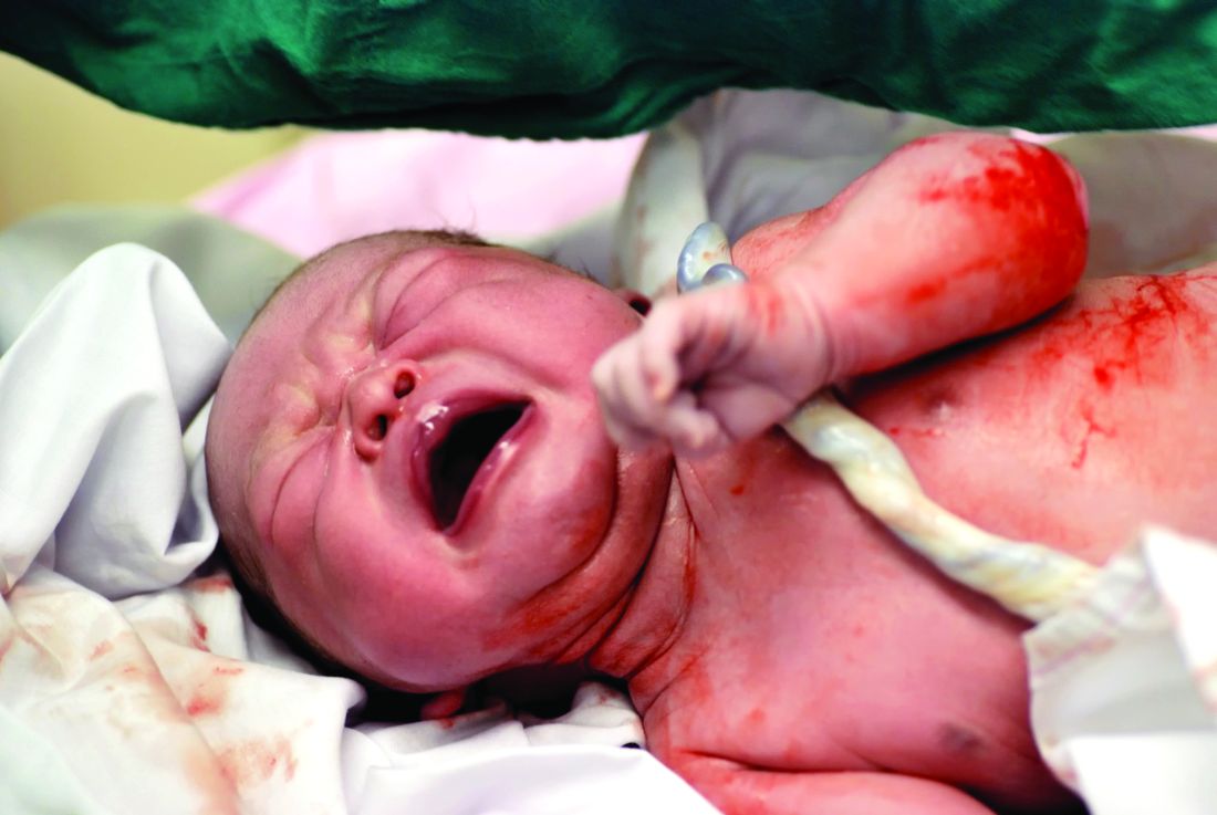
The primary outcome measure – the hemoglobin level at 8 months of age – was a significant 0.2 g/dL higher after delayed clamping. Also, anemia was significantly less prevalent with delayed cord clamping (73.0% vs. 82.2%). This represents an 11% reduction in the risk of anemia and a 42% reduction in the risk of iron deficiency. “The relative risk for having iron deficiency anemia was 0.58, with a number needed to treat of 7,” Dr. KC and associates said (JAMA Ped. 2017 Jan 17. doi: 10.1001/jamapediatrics.2016.3971).
The benefits of delayed cord clamping persisted at 12 months of age; the mean hemoglobin was 0.3 g/dL higher in the delayed group. Anemia was less prevalent in the delayed clamping group; the relative risk was 0.91, the investigators said.
If this intervention were implemented worldwide, “this could translate to 5 million fewer infants with anemia at 8 months of age, with particular public health significance in South Asia and sub-Saharan Africa, where the prevalence of anemia is highest,” the investigators said. This study was supported by the Midwifery Society of Nepal, the Swedish Society of Medicine, the Little Child’s Foundation, the Swedish Society of Medical Research, and the United Nations Children’s Fund. Dr. KC and associates reported having no relevant financial disclosures.
Delaying the clamping of the umbilical cord for 3 minutes after delivery decreased anemia for as long as 8-12 months in a population at high risk for the disorder, according to a report published online Jan. 17 in JAMA Pediatrics.
If early (within 1 minute of birth) cord clamping is avoided after delivery, fetoplacental blood moves into the newborn, augmenting his or her blood volume by 30%-40%. This is known to increase iron stores and prevent iron deficiency for at least 6 months, but until now there has been little evidence of how long this beneficial effect persists, said Ashish KC, MD, PhD, of International Maternal and Child Health, Uppsala (Sweden) University and the United Nations Children’s Fund, Lalitpur, Nepal, and associates.
The primary outcome measure – the hemoglobin level at 8 months of age – was a significant 0.2 g/dL higher after delayed clamping. Also, anemia was significantly less prevalent with delayed cord clamping (73.0% vs. 82.2%). This represents an 11% reduction in the risk of anemia and a 42% reduction in the risk of iron deficiency. “The relative risk for having iron deficiency anemia was 0.58, with a number needed to treat of 7,” Dr. KC and associates said (JAMA Ped. 2017 Jan 17. doi: 10.1001/jamapediatrics.2016.3971).
The benefits of delayed cord clamping persisted at 12 months of age; the mean hemoglobin was 0.3 g/dL higher in the delayed group. Anemia was less prevalent in the delayed clamping group; the relative risk was 0.91, the investigators said.
If this intervention were implemented worldwide, “this could translate to 5 million fewer infants with anemia at 8 months of age, with particular public health significance in South Asia and sub-Saharan Africa, where the prevalence of anemia is highest,” the investigators said. This study was supported by the Midwifery Society of Nepal, the Swedish Society of Medicine, the Little Child’s Foundation, the Swedish Society of Medical Research, and the United Nations Children’s Fund. Dr. KC and associates reported having no relevant financial disclosures.
Delaying the clamping of the umbilical cord for 3 minutes after delivery decreased anemia for as long as 8-12 months in a population at high risk for the disorder, according to a report published online Jan. 17 in JAMA Pediatrics.
If early (within 1 minute of birth) cord clamping is avoided after delivery, fetoplacental blood moves into the newborn, augmenting his or her blood volume by 30%-40%. This is known to increase iron stores and prevent iron deficiency for at least 6 months, but until now there has been little evidence of how long this beneficial effect persists, said Ashish KC, MD, PhD, of International Maternal and Child Health, Uppsala (Sweden) University and the United Nations Children’s Fund, Lalitpur, Nepal, and associates.
The primary outcome measure – the hemoglobin level at 8 months of age – was a significant 0.2 g/dL higher after delayed clamping. Also, anemia was significantly less prevalent with delayed cord clamping (73.0% vs. 82.2%). This represents an 11% reduction in the risk of anemia and a 42% reduction in the risk of iron deficiency. “The relative risk for having iron deficiency anemia was 0.58, with a number needed to treat of 7,” Dr. KC and associates said (JAMA Ped. 2017 Jan 17. doi: 10.1001/jamapediatrics.2016.3971).
The benefits of delayed cord clamping persisted at 12 months of age; the mean hemoglobin was 0.3 g/dL higher in the delayed group. Anemia was less prevalent in the delayed clamping group; the relative risk was 0.91, the investigators said.
If this intervention were implemented worldwide, “this could translate to 5 million fewer infants with anemia at 8 months of age, with particular public health significance in South Asia and sub-Saharan Africa, where the prevalence of anemia is highest,” the investigators said. This study was supported by the Midwifery Society of Nepal, the Swedish Society of Medicine, the Little Child’s Foundation, the Swedish Society of Medical Research, and the United Nations Children’s Fund. Dr. KC and associates reported having no relevant financial disclosures.
FROM JAMA PEDIATRICS
Key clinical point: Delaying the clamping of the umbilical cord for 3 minutes decreased anemia for as long as 8-12 months in a population at high risk for the disorder.
Major finding: Delayed cord clamping yielded an 11% reduction in the risk of anemia and a 42% reduction in the risk of iron deficiency, compared with early cord clamping.
Data source: A prospective randomized trial involving 540 term and late preterm infants born in Nepal during a 7-week period
Disclosures: This study was supported by the Midwifery Society of Nepal, the Swedish Society of Medicine, the Little Child’s Foundation, the Swedish Society of Medical Research, and the United Nations Children’s Fund. Dr. KC and associates reported having no relevant financial disclosures.
Assay testing accurate in distinguishing bacterial from viral respiratory tract infections
An assay designed to distinguish between bacterial and viral infections of the lower respiratory tract appears effective and shows promise for helping hospital physicians reduce overprescribing of antibiotics to children, a study showed.
“It is often not possible to differentiate between bacterial and nonbacterial disease on the basis of clinical judgment alone, [so] antibiotics are prescribed almost twice as often as required in children with acute respiratory tract infections in the USA,” wrote Chantal B. van Houten of the University Medical Centre Utrecht (the Netherlands) and associates in a study published in the Lancet Infectious Diseases.
The assay in question is called ImmunoXpert, which uses three biomarkers – tumor necrosis factor–related apoptosis-inducing ligand (TRAIL), interferon-gamma–induced protein-10 (IP-10), and C-reactive protein (CRP) – to determine if a lower respiratory tract infection has a viral or bacterial origin. A total of 777 subjects, aged 2-60 months, were recruited from four hospitals in the Netherlands and two hospitals in Israel between October 16, 2013, and March 1, 2015 (Lancet Inf Dis. 2016 Dec. doi: 10.1016/S1473-3099(16)30519-9).
The patients all had fevers with unidentified sources when they presented, and had a follow-up assessment carried out 28 days after baseline. Blood samples and nasal swabs were collected within 24 hours of presentation for assay analysis. Additionally, every subject was diagnosed as “bacterial” or “viral” by a three-member panel of pediatricians, whose diagnoses were based on the data available from the follow-up assessment and from clinical and laboratory data. The panel diagnosis for each subject was used as the reference standard.
Of the 777 subjects initially recruited, 200 were excluded from the final analysis for various reasons. Of the 577 who remained, the panel diagnosed 435 as having a viral infection and 71 as having a bacterial infection; 71 were deemed “inconclusive.” The panel was unanimous in 354 of these cases, and a majority of the panel (two of the three experts) agreed in 443 of these cases. In unanimous cases, the sensitivity of distinguishing between viral and bacterial cases correctly was 87.8%, with a specificity of 93.0%. The panel’s positive and negative predictive value were 62.1% and 98.3%, respectively.
The assay’s sensitivity rate in distinguishing between viral and bacterial infections was very close: 86.7%, with a specificity of 91.1%, which the authors noted was “promising diagnostic accuracy.” The positive predictive value of the assay was 60.5%, while the negative predictive value was found to be 97.8%.
Regarding the 71 cases that were deemed “inconclusive,” Dr. van Houten and coauthors acknowledged that “such inconclusive cases are inherent to studies without a gold standard, and this was taken into account when calculating the sample size.” Additionally, they noted that follow-up studies should take into consideration the costs of utilizing assay testing like ImmunoXpert, in order to better assess the financial implications that adopting the technology would have on a health care facility.
Nevertheless, the investigators concluded, “our findings [support] the need for implementation research to examine the added clinical utility of ImmunoXpert to diagnose bacterial infection in clinical care for children with lower respiratory tract infection and fever without source presenting at the hospital.”
Funding for this study was provided by MeMed Diagnostics. Dr. van Houten and coauthors did not report any relevant financial disclosures.
“A study by Chantal van Houten and colleagues in this issue of the Lancet Infectious Diseases tested the combined measurement of CRP, TRAIL, and IP-10, and found that this test distinguished bacterial from viral infections with a sensitivity of 86.7% and a specificity of 91.1%. This assay is significantly more effective than procalcitonin determinations in identifying the cause of infection because it improves the diagnostic classification of bacterial infections by 6.3% and of viral infections by 5.4%. However, by comparison with CRP, the CRP, TRAIL, and IP-10 combined assay is as effective at classifying bacterial cases, although it does improve the identification of patients with viral infections by 8.6%. Furthermore, it still has some limitations that currently preclude its routine use in clinical practice.
“First, the test requires advanced laboratory techniques and cannot be used outside hospitals. Second, the collected data came from a relatively small number of children, none of whom had an underlying disease that might modify host response to infection. Third – as in the case of all of the studies that have tried to differentiate bacterial and viral infection – the definition of cause of infection used in these studies varies. Finally, respiratory infections are frequently classified on the basis of clinical and radiological findings, and the results of a microbiological assessment of nasopharyngeal swabs.
“However, it is well known that the investigation into upper respiratory secretions in children can be confounding and lead to the erroneous classification of a lower respiratory disease, and that bacteria and viruses can simply be carried and could have no association with the cause of a disease. This means that future studies should confirm the results of host protein-based assays in larger study populations with various characteristics, and consider their cost to benefit ratios in relation to their real effect on reducing antibiotic use.”
Susanna Esposito, MD, and Nicola Principi, MD, are with the University of Milan. Their opinions are excerpted from a commentary on the article by Dr. van Houten et al. (Lancet Inf Dis. 2016 Dec. doi: 10.1016/S1473-3099(16)30536-9). They had no relevant financial disclosures.
“A study by Chantal van Houten and colleagues in this issue of the Lancet Infectious Diseases tested the combined measurement of CRP, TRAIL, and IP-10, and found that this test distinguished bacterial from viral infections with a sensitivity of 86.7% and a specificity of 91.1%. This assay is significantly more effective than procalcitonin determinations in identifying the cause of infection because it improves the diagnostic classification of bacterial infections by 6.3% and of viral infections by 5.4%. However, by comparison with CRP, the CRP, TRAIL, and IP-10 combined assay is as effective at classifying bacterial cases, although it does improve the identification of patients with viral infections by 8.6%. Furthermore, it still has some limitations that currently preclude its routine use in clinical practice.
“First, the test requires advanced laboratory techniques and cannot be used outside hospitals. Second, the collected data came from a relatively small number of children, none of whom had an underlying disease that might modify host response to infection. Third – as in the case of all of the studies that have tried to differentiate bacterial and viral infection – the definition of cause of infection used in these studies varies. Finally, respiratory infections are frequently classified on the basis of clinical and radiological findings, and the results of a microbiological assessment of nasopharyngeal swabs.
“However, it is well known that the investigation into upper respiratory secretions in children can be confounding and lead to the erroneous classification of a lower respiratory disease, and that bacteria and viruses can simply be carried and could have no association with the cause of a disease. This means that future studies should confirm the results of host protein-based assays in larger study populations with various characteristics, and consider their cost to benefit ratios in relation to their real effect on reducing antibiotic use.”
Susanna Esposito, MD, and Nicola Principi, MD, are with the University of Milan. Their opinions are excerpted from a commentary on the article by Dr. van Houten et al. (Lancet Inf Dis. 2016 Dec. doi: 10.1016/S1473-3099(16)30536-9). They had no relevant financial disclosures.
“A study by Chantal van Houten and colleagues in this issue of the Lancet Infectious Diseases tested the combined measurement of CRP, TRAIL, and IP-10, and found that this test distinguished bacterial from viral infections with a sensitivity of 86.7% and a specificity of 91.1%. This assay is significantly more effective than procalcitonin determinations in identifying the cause of infection because it improves the diagnostic classification of bacterial infections by 6.3% and of viral infections by 5.4%. However, by comparison with CRP, the CRP, TRAIL, and IP-10 combined assay is as effective at classifying bacterial cases, although it does improve the identification of patients with viral infections by 8.6%. Furthermore, it still has some limitations that currently preclude its routine use in clinical practice.
“First, the test requires advanced laboratory techniques and cannot be used outside hospitals. Second, the collected data came from a relatively small number of children, none of whom had an underlying disease that might modify host response to infection. Third – as in the case of all of the studies that have tried to differentiate bacterial and viral infection – the definition of cause of infection used in these studies varies. Finally, respiratory infections are frequently classified on the basis of clinical and radiological findings, and the results of a microbiological assessment of nasopharyngeal swabs.
“However, it is well known that the investigation into upper respiratory secretions in children can be confounding and lead to the erroneous classification of a lower respiratory disease, and that bacteria and viruses can simply be carried and could have no association with the cause of a disease. This means that future studies should confirm the results of host protein-based assays in larger study populations with various characteristics, and consider their cost to benefit ratios in relation to their real effect on reducing antibiotic use.”
Susanna Esposito, MD, and Nicola Principi, MD, are with the University of Milan. Their opinions are excerpted from a commentary on the article by Dr. van Houten et al. (Lancet Inf Dis. 2016 Dec. doi: 10.1016/S1473-3099(16)30536-9). They had no relevant financial disclosures.
An assay designed to distinguish between bacterial and viral infections of the lower respiratory tract appears effective and shows promise for helping hospital physicians reduce overprescribing of antibiotics to children, a study showed.
“It is often not possible to differentiate between bacterial and nonbacterial disease on the basis of clinical judgment alone, [so] antibiotics are prescribed almost twice as often as required in children with acute respiratory tract infections in the USA,” wrote Chantal B. van Houten of the University Medical Centre Utrecht (the Netherlands) and associates in a study published in the Lancet Infectious Diseases.
The assay in question is called ImmunoXpert, which uses three biomarkers – tumor necrosis factor–related apoptosis-inducing ligand (TRAIL), interferon-gamma–induced protein-10 (IP-10), and C-reactive protein (CRP) – to determine if a lower respiratory tract infection has a viral or bacterial origin. A total of 777 subjects, aged 2-60 months, were recruited from four hospitals in the Netherlands and two hospitals in Israel between October 16, 2013, and March 1, 2015 (Lancet Inf Dis. 2016 Dec. doi: 10.1016/S1473-3099(16)30519-9).
The patients all had fevers with unidentified sources when they presented, and had a follow-up assessment carried out 28 days after baseline. Blood samples and nasal swabs were collected within 24 hours of presentation for assay analysis. Additionally, every subject was diagnosed as “bacterial” or “viral” by a three-member panel of pediatricians, whose diagnoses were based on the data available from the follow-up assessment and from clinical and laboratory data. The panel diagnosis for each subject was used as the reference standard.
Of the 777 subjects initially recruited, 200 were excluded from the final analysis for various reasons. Of the 577 who remained, the panel diagnosed 435 as having a viral infection and 71 as having a bacterial infection; 71 were deemed “inconclusive.” The panel was unanimous in 354 of these cases, and a majority of the panel (two of the three experts) agreed in 443 of these cases. In unanimous cases, the sensitivity of distinguishing between viral and bacterial cases correctly was 87.8%, with a specificity of 93.0%. The panel’s positive and negative predictive value were 62.1% and 98.3%, respectively.
The assay’s sensitivity rate in distinguishing between viral and bacterial infections was very close: 86.7%, with a specificity of 91.1%, which the authors noted was “promising diagnostic accuracy.” The positive predictive value of the assay was 60.5%, while the negative predictive value was found to be 97.8%.
Regarding the 71 cases that were deemed “inconclusive,” Dr. van Houten and coauthors acknowledged that “such inconclusive cases are inherent to studies without a gold standard, and this was taken into account when calculating the sample size.” Additionally, they noted that follow-up studies should take into consideration the costs of utilizing assay testing like ImmunoXpert, in order to better assess the financial implications that adopting the technology would have on a health care facility.
Nevertheless, the investigators concluded, “our findings [support] the need for implementation research to examine the added clinical utility of ImmunoXpert to diagnose bacterial infection in clinical care for children with lower respiratory tract infection and fever without source presenting at the hospital.”
Funding for this study was provided by MeMed Diagnostics. Dr. van Houten and coauthors did not report any relevant financial disclosures.
An assay designed to distinguish between bacterial and viral infections of the lower respiratory tract appears effective and shows promise for helping hospital physicians reduce overprescribing of antibiotics to children, a study showed.
“It is often not possible to differentiate between bacterial and nonbacterial disease on the basis of clinical judgment alone, [so] antibiotics are prescribed almost twice as often as required in children with acute respiratory tract infections in the USA,” wrote Chantal B. van Houten of the University Medical Centre Utrecht (the Netherlands) and associates in a study published in the Lancet Infectious Diseases.
The assay in question is called ImmunoXpert, which uses three biomarkers – tumor necrosis factor–related apoptosis-inducing ligand (TRAIL), interferon-gamma–induced protein-10 (IP-10), and C-reactive protein (CRP) – to determine if a lower respiratory tract infection has a viral or bacterial origin. A total of 777 subjects, aged 2-60 months, were recruited from four hospitals in the Netherlands and two hospitals in Israel between October 16, 2013, and March 1, 2015 (Lancet Inf Dis. 2016 Dec. doi: 10.1016/S1473-3099(16)30519-9).
The patients all had fevers with unidentified sources when they presented, and had a follow-up assessment carried out 28 days after baseline. Blood samples and nasal swabs were collected within 24 hours of presentation for assay analysis. Additionally, every subject was diagnosed as “bacterial” or “viral” by a three-member panel of pediatricians, whose diagnoses were based on the data available from the follow-up assessment and from clinical and laboratory data. The panel diagnosis for each subject was used as the reference standard.
Of the 777 subjects initially recruited, 200 were excluded from the final analysis for various reasons. Of the 577 who remained, the panel diagnosed 435 as having a viral infection and 71 as having a bacterial infection; 71 were deemed “inconclusive.” The panel was unanimous in 354 of these cases, and a majority of the panel (two of the three experts) agreed in 443 of these cases. In unanimous cases, the sensitivity of distinguishing between viral and bacterial cases correctly was 87.8%, with a specificity of 93.0%. The panel’s positive and negative predictive value were 62.1% and 98.3%, respectively.
The assay’s sensitivity rate in distinguishing between viral and bacterial infections was very close: 86.7%, with a specificity of 91.1%, which the authors noted was “promising diagnostic accuracy.” The positive predictive value of the assay was 60.5%, while the negative predictive value was found to be 97.8%.
Regarding the 71 cases that were deemed “inconclusive,” Dr. van Houten and coauthors acknowledged that “such inconclusive cases are inherent to studies without a gold standard, and this was taken into account when calculating the sample size.” Additionally, they noted that follow-up studies should take into consideration the costs of utilizing assay testing like ImmunoXpert, in order to better assess the financial implications that adopting the technology would have on a health care facility.
Nevertheless, the investigators concluded, “our findings [support] the need for implementation research to examine the added clinical utility of ImmunoXpert to diagnose bacterial infection in clinical care for children with lower respiratory tract infection and fever without source presenting at the hospital.”
Funding for this study was provided by MeMed Diagnostics. Dr. van Houten and coauthors did not report any relevant financial disclosures.
FROM THE LANCET INFECTIOUS DISEASES
Key clinical point:
Major finding: The assay distinguished between bacterial and viral infections with 86.7% sensitivity, compared with unanimous panel diagnosis, which did so with 87.8% sensitivity.
Data source: A double-blind, multicenter, validation study of 577 children aged 2-60 months from October 2013 through March 2015.
Disclosures: The study was funded by MeMed Diagnostics. The authors reported no relevant financial disclosures.
Genetic studies link JIA subtypes to adult diseases, show uniqueness of systemic disease
Two new studies of the genetic relationships between the seven designated categories of juvenile idiopathic arthritis (JIA) provide compelling support for reclassification of the categories, particularly for systemic JIA, and give evidence that some of the categories have clear equivalents in the realm of adult-onset diseases.
The International League of Associations for Rheumatology (ILAR) classification system (J Rheumatol. 2004;31:390-2) that defined the seven juvenile idiopathic arthritis (JIA) categories – systemic arthritis, oligoarticular arthritis, rheumatoid factor (RF)–negative polyarticular arthritis, RF-positive polyarticular arthritis, psoriatic arthritis (PsA), enthesitis-related arthritis (ERA), and undifferentiated arthritis – is problematic because the long-term outcome and response to treatment varies not only between subtypes but also within the subtypes, suggesting that these subgroups do not yet represent uniform groups of patients,” Wendy Thomson, PhD, professor of genetic epidemiology at the University of Manchester (England) and a senior author on both studies, explained in an interview.
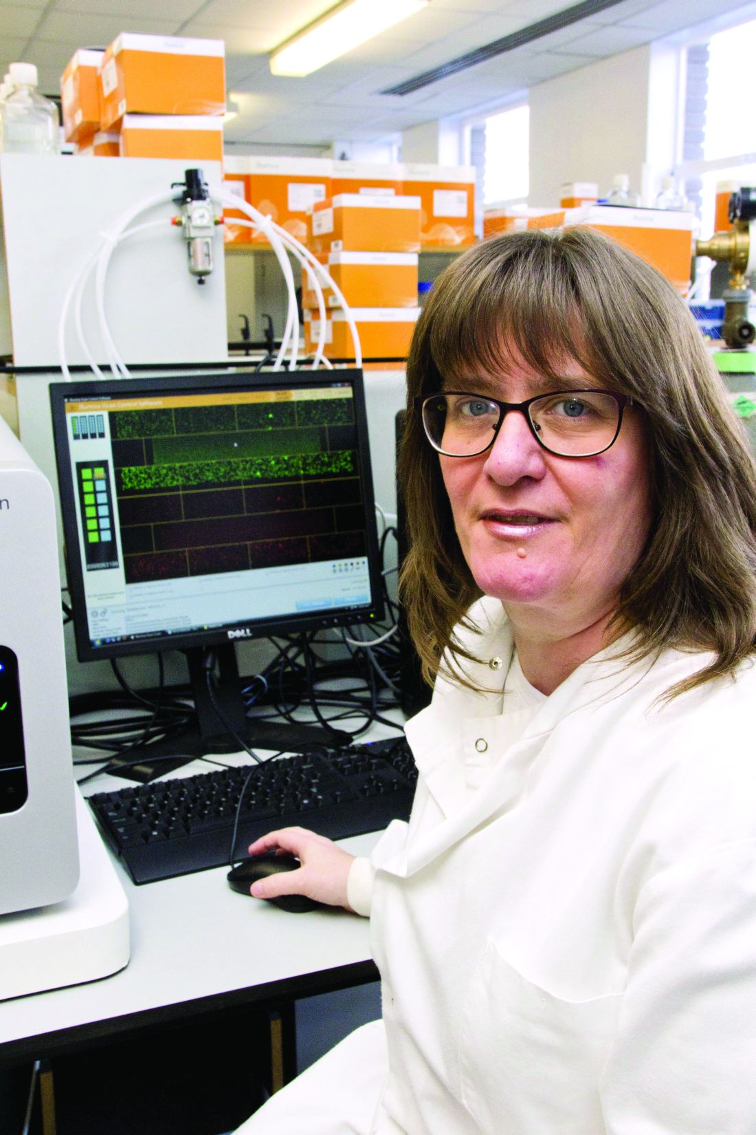
“Despite the differences between the subtypes, current treatments of this disease often involve using the same drugs across all subtypes of JIA,” said Dr. Thomson, who is also deputy director of the Arthritis Research UK Centre for Genetics and Genomics at the university. “Understanding the genetic basis of the subtypes of this disease could help understand the cause of this disease and identify more appropriate treatment options [because] the current classification is largely based on clinical data and it is proposed that the addition of genetic data could improve classification.”
Interrelationships between JIA categories and adult disease
The first of these studies found that particular alleles in the human leukocyte antigen (HLA) region that have been associated with the different JIA categories strongly correlate some of the categories with one another, and that each JIA category potentially has an adult-onset counterpart based on shared HLA associations. It is the largest investigation of association of the HLA region with JIA and its categories to date, according to the researchers (Ann Rheum Dis. 2016 Dec 20. doi: 10.1136/annrheumdis-2016-210025).
In particular, the investigators demonstrated for the first time that RF-negative polyarticular JIA and oligoarticular JIA are genetically similar in their HLA associations. They also reported that RF-positive polyarthritis shares an association with adult seropositive rheumatoid arthritis and that combined data for oligoarthritis and RF-negative polyarthritis share the same association with adult seronegative RA. In addition, the researchers generated support for genetic associations between the particular JIA categories that are most commonly thought to have adult counterparts, such as ERA and adult ankylosing spondylitis (AS) as well as juvenile PsA and adult PsA.
Dr. Thomson and her coinvestigators used ImmunoChip genotype array data for 191,494 single nucleotide polymorphism (SNP) markers from the HLA region of 5,737 JIA patients and 16,403 controls in the United States, United Kingdom, Canada, Norway, and Germany to impute classical HLA alleles as well as specific amino acid positions within HLA alleles that may play an important functional role. After quality-control measures for the data were put in place, comparisons between 5,043 JIA cases and 14,390 controls showed that oligoarthritis and RF-negative polyarthritis were the most common and homogeneous JIA categories investigated (2,409 and 1,525 patients, respectively), and that they share a significant association across the HLA spectrum, meaning that they are genetically similar. When combined, the data from these two JIA categories correspond with seronegative rheumatoid arthritis in adults, while RF-positive polyarthritis on its own has an association with seropositive rheumatoid arthritis involving histidine at position 13 of the HLA-DRB1 allele. As expected, the most significant association between the ERA category and AS was for HLA-B*27. For juvenile PsA, no associations reached genome-wide level of significance, although HLA-C*0602 was modestly associated with juvenile PsA, and it is known to be associated with adult-onset PsA and is the primary HLA association in psoriasis.
“The results of this study have important implications for understanding disease pathogenesis, etiology, and potential future therapeutic strategies for JIA categories,” Dr. Thomson and her coauthors wrote, adding that “heterogeneity of JIA remains a key challenge to pediatric rheumatologists; however, these results may inform the debate on classification and help define a more biological-driven and molecular-driven classification system.”
Uniqueness of systemic JIA
In the second of the two studies, investigators from many different childhood arthritis study groups focused on systemic JIA (sJIA), also known in the past as Still’s disease. According to the authors, it is the first large-scale genomic study of sJIA ever published (Ann Rheum Dis. 2016 Dec 7. doi: 10.1136/annrheumdis-2016-210324).
“[sJIA] is characterized by prominent systemic inflammation and has a rare adult-onset counterpart; and undifferentiated arthritis includes arthritis that does not fit into any single category,” the authors wrote, adding that the “unique clinical characteristics of sJIA suggest that it is distinct from other forms of JIA, leading to the contention by some that sJIA should be separated from other forms of JIA and labeled as an autoinflammatory disease.”
The investigators carried out SNP genotyping on 982 children with sJIA in the United States, United Kingdom, Germany, Turkey, Italy, Brazil, Argentina, Canada, and Spain, along with 431 healthy children without sJIA who came from the same countries. A total of 7,579 control patients also underwent genotyping. Ultimately, 770 sJIA and 6,947 control subjects completed the study. sJIA was then compared with other JIA subtypes by using weighted genetic risk scores.
Comparison of the data sets for sJIA with those for the combined categories of RF-negative polyarticular JIA and oligoarticular JIA, as well as for the individual category of RF-positive polyarticular JIA, showed that sJIA has a distinct genetic architecture that separates it from those JIA categories, providing further evidence that sJIA should be classified separately from other forms of JIA and, potentially, treated differently as well.
According to Dr. Thomson, the finding “provides important evidence that sJIA should be considered a unique disease with its own specific disease mechanisms.” Dr. Thomson explained that “knowing more about the genetic risk factors of this disease might give a greater understanding of the disease processes involved in this condition and ultimately lead to novel therapies for this severely disabling disease.”
Funding for the first study was provided by the Wellcome Trust, the National Institutes of Health, the Doris Duke Charitable Foundation, the Medical Research Council, the Canadian Institutes of Health Research, the Liaison Committee between the Central Norway Regional Health Authority and the Norwegian University of Science and Technology, the Juvenile Diabetes Research Foundation International, and the Texas Scottish Rite Hospital for Children. Funding for the second study was provided by intramural research programs of the National Institute of Arthritis and Musculoskeletal and Skin Diseases and the National Human Genome Research Institute, individual NIH grants, and grants from charitable foundations, advocacy organizations, and the governments of individual researchers’ countries. Dr. Thomson did not report any relevant financial disclosures; however, a number of coauthors reported potential conflicts of interest.
These two excellent multi-institutional genetic studies demonstrate once and for all the error promulgated by the International League of Associations for Rheumatology (ILAR) criteria formulated in the 1990s. Despite contrary voices, the ILAR committee grouped all childhood arthritis that was not due to sepsis or trauma as “juvenile idiopathic arthritis.” And despite the lack of clarity of the subtypes and the many cases that are “unclassifiable,” pediatric rheumatologists have continued to use these criteria in both drug trials and scientific studies.

Thomas Lehman, MD, is chief of the division of pediatric rheumatology at the Hospital for Special Surgery in New York. He has no relevant financial disclosures.
These two excellent multi-institutional genetic studies demonstrate once and for all the error promulgated by the International League of Associations for Rheumatology (ILAR) criteria formulated in the 1990s. Despite contrary voices, the ILAR committee grouped all childhood arthritis that was not due to sepsis or trauma as “juvenile idiopathic arthritis.” And despite the lack of clarity of the subtypes and the many cases that are “unclassifiable,” pediatric rheumatologists have continued to use these criteria in both drug trials and scientific studies.
Thomas Lehman, MD, is chief of the division of pediatric rheumatology at the Hospital for Special Surgery in New York. He has no relevant financial disclosures.
These two excellent multi-institutional genetic studies demonstrate once and for all the error promulgated by the International League of Associations for Rheumatology (ILAR) criteria formulated in the 1990s. Despite contrary voices, the ILAR committee grouped all childhood arthritis that was not due to sepsis or trauma as “juvenile idiopathic arthritis.” And despite the lack of clarity of the subtypes and the many cases that are “unclassifiable,” pediatric rheumatologists have continued to use these criteria in both drug trials and scientific studies.
Thomas Lehman, MD, is chief of the division of pediatric rheumatology at the Hospital for Special Surgery in New York. He has no relevant financial disclosures.
Two new studies of the genetic relationships between the seven designated categories of juvenile idiopathic arthritis (JIA) provide compelling support for reclassification of the categories, particularly for systemic JIA, and give evidence that some of the categories have clear equivalents in the realm of adult-onset diseases.
The International League of Associations for Rheumatology (ILAR) classification system (J Rheumatol. 2004;31:390-2) that defined the seven juvenile idiopathic arthritis (JIA) categories – systemic arthritis, oligoarticular arthritis, rheumatoid factor (RF)–negative polyarticular arthritis, RF-positive polyarticular arthritis, psoriatic arthritis (PsA), enthesitis-related arthritis (ERA), and undifferentiated arthritis – is problematic because the long-term outcome and response to treatment varies not only between subtypes but also within the subtypes, suggesting that these subgroups do not yet represent uniform groups of patients,” Wendy Thomson, PhD, professor of genetic epidemiology at the University of Manchester (England) and a senior author on both studies, explained in an interview.
“Despite the differences between the subtypes, current treatments of this disease often involve using the same drugs across all subtypes of JIA,” said Dr. Thomson, who is also deputy director of the Arthritis Research UK Centre for Genetics and Genomics at the university. “Understanding the genetic basis of the subtypes of this disease could help understand the cause of this disease and identify more appropriate treatment options [because] the current classification is largely based on clinical data and it is proposed that the addition of genetic data could improve classification.”
Interrelationships between JIA categories and adult disease
The first of these studies found that particular alleles in the human leukocyte antigen (HLA) region that have been associated with the different JIA categories strongly correlate some of the categories with one another, and that each JIA category potentially has an adult-onset counterpart based on shared HLA associations. It is the largest investigation of association of the HLA region with JIA and its categories to date, according to the researchers (Ann Rheum Dis. 2016 Dec 20. doi: 10.1136/annrheumdis-2016-210025).
In particular, the investigators demonstrated for the first time that RF-negative polyarticular JIA and oligoarticular JIA are genetically similar in their HLA associations. They also reported that RF-positive polyarthritis shares an association with adult seropositive rheumatoid arthritis and that combined data for oligoarthritis and RF-negative polyarthritis share the same association with adult seronegative RA. In addition, the researchers generated support for genetic associations between the particular JIA categories that are most commonly thought to have adult counterparts, such as ERA and adult ankylosing spondylitis (AS) as well as juvenile PsA and adult PsA.
Dr. Thomson and her coinvestigators used ImmunoChip genotype array data for 191,494 single nucleotide polymorphism (SNP) markers from the HLA region of 5,737 JIA patients and 16,403 controls in the United States, United Kingdom, Canada, Norway, and Germany to impute classical HLA alleles as well as specific amino acid positions within HLA alleles that may play an important functional role. After quality-control measures for the data were put in place, comparisons between 5,043 JIA cases and 14,390 controls showed that oligoarthritis and RF-negative polyarthritis were the most common and homogeneous JIA categories investigated (2,409 and 1,525 patients, respectively), and that they share a significant association across the HLA spectrum, meaning that they are genetically similar. When combined, the data from these two JIA categories correspond with seronegative rheumatoid arthritis in adults, while RF-positive polyarthritis on its own has an association with seropositive rheumatoid arthritis involving histidine at position 13 of the HLA-DRB1 allele. As expected, the most significant association between the ERA category and AS was for HLA-B*27. For juvenile PsA, no associations reached genome-wide level of significance, although HLA-C*0602 was modestly associated with juvenile PsA, and it is known to be associated with adult-onset PsA and is the primary HLA association in psoriasis.
“The results of this study have important implications for understanding disease pathogenesis, etiology, and potential future therapeutic strategies for JIA categories,” Dr. Thomson and her coauthors wrote, adding that “heterogeneity of JIA remains a key challenge to pediatric rheumatologists; however, these results may inform the debate on classification and help define a more biological-driven and molecular-driven classification system.”
Uniqueness of systemic JIA
In the second of the two studies, investigators from many different childhood arthritis study groups focused on systemic JIA (sJIA), also known in the past as Still’s disease. According to the authors, it is the first large-scale genomic study of sJIA ever published (Ann Rheum Dis. 2016 Dec 7. doi: 10.1136/annrheumdis-2016-210324).
“[sJIA] is characterized by prominent systemic inflammation and has a rare adult-onset counterpart; and undifferentiated arthritis includes arthritis that does not fit into any single category,” the authors wrote, adding that the “unique clinical characteristics of sJIA suggest that it is distinct from other forms of JIA, leading to the contention by some that sJIA should be separated from other forms of JIA and labeled as an autoinflammatory disease.”
The investigators carried out SNP genotyping on 982 children with sJIA in the United States, United Kingdom, Germany, Turkey, Italy, Brazil, Argentina, Canada, and Spain, along with 431 healthy children without sJIA who came from the same countries. A total of 7,579 control patients also underwent genotyping. Ultimately, 770 sJIA and 6,947 control subjects completed the study. sJIA was then compared with other JIA subtypes by using weighted genetic risk scores.
Comparison of the data sets for sJIA with those for the combined categories of RF-negative polyarticular JIA and oligoarticular JIA, as well as for the individual category of RF-positive polyarticular JIA, showed that sJIA has a distinct genetic architecture that separates it from those JIA categories, providing further evidence that sJIA should be classified separately from other forms of JIA and, potentially, treated differently as well.
According to Dr. Thomson, the finding “provides important evidence that sJIA should be considered a unique disease with its own specific disease mechanisms.” Dr. Thomson explained that “knowing more about the genetic risk factors of this disease might give a greater understanding of the disease processes involved in this condition and ultimately lead to novel therapies for this severely disabling disease.”
Funding for the first study was provided by the Wellcome Trust, the National Institutes of Health, the Doris Duke Charitable Foundation, the Medical Research Council, the Canadian Institutes of Health Research, the Liaison Committee between the Central Norway Regional Health Authority and the Norwegian University of Science and Technology, the Juvenile Diabetes Research Foundation International, and the Texas Scottish Rite Hospital for Children. Funding for the second study was provided by intramural research programs of the National Institute of Arthritis and Musculoskeletal and Skin Diseases and the National Human Genome Research Institute, individual NIH grants, and grants from charitable foundations, advocacy organizations, and the governments of individual researchers’ countries. Dr. Thomson did not report any relevant financial disclosures; however, a number of coauthors reported potential conflicts of interest.
Two new studies of the genetic relationships between the seven designated categories of juvenile idiopathic arthritis (JIA) provide compelling support for reclassification of the categories, particularly for systemic JIA, and give evidence that some of the categories have clear equivalents in the realm of adult-onset diseases.
The International League of Associations for Rheumatology (ILAR) classification system (J Rheumatol. 2004;31:390-2) that defined the seven juvenile idiopathic arthritis (JIA) categories – systemic arthritis, oligoarticular arthritis, rheumatoid factor (RF)–negative polyarticular arthritis, RF-positive polyarticular arthritis, psoriatic arthritis (PsA), enthesitis-related arthritis (ERA), and undifferentiated arthritis – is problematic because the long-term outcome and response to treatment varies not only between subtypes but also within the subtypes, suggesting that these subgroups do not yet represent uniform groups of patients,” Wendy Thomson, PhD, professor of genetic epidemiology at the University of Manchester (England) and a senior author on both studies, explained in an interview.
“Despite the differences between the subtypes, current treatments of this disease often involve using the same drugs across all subtypes of JIA,” said Dr. Thomson, who is also deputy director of the Arthritis Research UK Centre for Genetics and Genomics at the university. “Understanding the genetic basis of the subtypes of this disease could help understand the cause of this disease and identify more appropriate treatment options [because] the current classification is largely based on clinical data and it is proposed that the addition of genetic data could improve classification.”
Interrelationships between JIA categories and adult disease
The first of these studies found that particular alleles in the human leukocyte antigen (HLA) region that have been associated with the different JIA categories strongly correlate some of the categories with one another, and that each JIA category potentially has an adult-onset counterpart based on shared HLA associations. It is the largest investigation of association of the HLA region with JIA and its categories to date, according to the researchers (Ann Rheum Dis. 2016 Dec 20. doi: 10.1136/annrheumdis-2016-210025).
In particular, the investigators demonstrated for the first time that RF-negative polyarticular JIA and oligoarticular JIA are genetically similar in their HLA associations. They also reported that RF-positive polyarthritis shares an association with adult seropositive rheumatoid arthritis and that combined data for oligoarthritis and RF-negative polyarthritis share the same association with adult seronegative RA. In addition, the researchers generated support for genetic associations between the particular JIA categories that are most commonly thought to have adult counterparts, such as ERA and adult ankylosing spondylitis (AS) as well as juvenile PsA and adult PsA.
Dr. Thomson and her coinvestigators used ImmunoChip genotype array data for 191,494 single nucleotide polymorphism (SNP) markers from the HLA region of 5,737 JIA patients and 16,403 controls in the United States, United Kingdom, Canada, Norway, and Germany to impute classical HLA alleles as well as specific amino acid positions within HLA alleles that may play an important functional role. After quality-control measures for the data were put in place, comparisons between 5,043 JIA cases and 14,390 controls showed that oligoarthritis and RF-negative polyarthritis were the most common and homogeneous JIA categories investigated (2,409 and 1,525 patients, respectively), and that they share a significant association across the HLA spectrum, meaning that they are genetically similar. When combined, the data from these two JIA categories correspond with seronegative rheumatoid arthritis in adults, while RF-positive polyarthritis on its own has an association with seropositive rheumatoid arthritis involving histidine at position 13 of the HLA-DRB1 allele. As expected, the most significant association between the ERA category and AS was for HLA-B*27. For juvenile PsA, no associations reached genome-wide level of significance, although HLA-C*0602 was modestly associated with juvenile PsA, and it is known to be associated with adult-onset PsA and is the primary HLA association in psoriasis.
“The results of this study have important implications for understanding disease pathogenesis, etiology, and potential future therapeutic strategies for JIA categories,” Dr. Thomson and her coauthors wrote, adding that “heterogeneity of JIA remains a key challenge to pediatric rheumatologists; however, these results may inform the debate on classification and help define a more biological-driven and molecular-driven classification system.”
Uniqueness of systemic JIA
In the second of the two studies, investigators from many different childhood arthritis study groups focused on systemic JIA (sJIA), also known in the past as Still’s disease. According to the authors, it is the first large-scale genomic study of sJIA ever published (Ann Rheum Dis. 2016 Dec 7. doi: 10.1136/annrheumdis-2016-210324).
“[sJIA] is characterized by prominent systemic inflammation and has a rare adult-onset counterpart; and undifferentiated arthritis includes arthritis that does not fit into any single category,” the authors wrote, adding that the “unique clinical characteristics of sJIA suggest that it is distinct from other forms of JIA, leading to the contention by some that sJIA should be separated from other forms of JIA and labeled as an autoinflammatory disease.”
The investigators carried out SNP genotyping on 982 children with sJIA in the United States, United Kingdom, Germany, Turkey, Italy, Brazil, Argentina, Canada, and Spain, along with 431 healthy children without sJIA who came from the same countries. A total of 7,579 control patients also underwent genotyping. Ultimately, 770 sJIA and 6,947 control subjects completed the study. sJIA was then compared with other JIA subtypes by using weighted genetic risk scores.
Comparison of the data sets for sJIA with those for the combined categories of RF-negative polyarticular JIA and oligoarticular JIA, as well as for the individual category of RF-positive polyarticular JIA, showed that sJIA has a distinct genetic architecture that separates it from those JIA categories, providing further evidence that sJIA should be classified separately from other forms of JIA and, potentially, treated differently as well.
According to Dr. Thomson, the finding “provides important evidence that sJIA should be considered a unique disease with its own specific disease mechanisms.” Dr. Thomson explained that “knowing more about the genetic risk factors of this disease might give a greater understanding of the disease processes involved in this condition and ultimately lead to novel therapies for this severely disabling disease.”
Funding for the first study was provided by the Wellcome Trust, the National Institutes of Health, the Doris Duke Charitable Foundation, the Medical Research Council, the Canadian Institutes of Health Research, the Liaison Committee between the Central Norway Regional Health Authority and the Norwegian University of Science and Technology, the Juvenile Diabetes Research Foundation International, and the Texas Scottish Rite Hospital for Children. Funding for the second study was provided by intramural research programs of the National Institute of Arthritis and Musculoskeletal and Skin Diseases and the National Human Genome Research Institute, individual NIH grants, and grants from charitable foundations, advocacy organizations, and the governments of individual researchers’ countries. Dr. Thomson did not report any relevant financial disclosures; however, a number of coauthors reported potential conflicts of interest.
FROM ANNALS OF THE RHEUMATIC DISEASES
NAS linked to poor and deteriorating school performance
A neonatal diagnostic code of neonatal abstinence syndrome (NAS) is strongly associated with poor and deteriorating school performance, according to Ju Lee Oei, MD, of the University of New South Wales, Sydney, and her associates.
In a study of 604,829 children born in 2000-2006 in New South Wales, linkage rates were similar between matched controls (77.6% of 4,330) and other NSW children (77.4% of 598,265; P = .83) but were significantly lower in children with NAS (75.6% of 2,234; P = .03). The controls were matched for gender,gestation, and socioeconomic status.
The children with NAS had significantly lower scores than the controls and other NSW children in every grade and every domain of testing (reading, writing, numeracy, spelling, and grammar/punctuation). By grade 7, 38% of children with NAS did not meet National Minimum Standard (NMS) in one or more domains (versus 18.4% of controls and 14.5% of other NSW children). The mean serial composite scores also were lower in children with NAS from grades 3 to 7, compared with the other two groups; the difference was progressive, the investigators said. And by grade 7, the scores for children with NAS were lower than other children’s scores in grade 5.
It was noted that children with NAS, indigenous status (adjusted odds ratio, 1.7), male gender (aOR, 1.3), and having a primary parent without a grade 9 education (aOR, 1.3) increased the risk of failure to meet NMS. Overall, NAS (aOR, 2.5), indigenous status (aOR, 2.2), male gender (aOR, 1.3), and prematurity (less than 37 weeks’ gestation [aOR, 1.2]) increased the risk of failure to meet NMS.
“To date these are the only data demonstrating long-term school outcomes for children with a history of NAS. Similar data for children born from the current opioid epidemic gripping much of the Northern Hemisphere, assuming linkage is possible, will be available only in 7-10 years,” researchers concluded. “Although this study was conducted in Australia, the high risk of poor academic performance in this vulnerable group of children is applicable to all countries, and strategies to address this risk and prevent poor adult outcomes and intergenerational vulnerability must be urgently addressed.”
Read the full study in Pediatrics (doi: 10.1542/peds.2016-2651).
A neonatal diagnostic code of neonatal abstinence syndrome (NAS) is strongly associated with poor and deteriorating school performance, according to Ju Lee Oei, MD, of the University of New South Wales, Sydney, and her associates.
In a study of 604,829 children born in 2000-2006 in New South Wales, linkage rates were similar between matched controls (77.6% of 4,330) and other NSW children (77.4% of 598,265; P = .83) but were significantly lower in children with NAS (75.6% of 2,234; P = .03). The controls were matched for gender,gestation, and socioeconomic status.
The children with NAS had significantly lower scores than the controls and other NSW children in every grade and every domain of testing (reading, writing, numeracy, spelling, and grammar/punctuation). By grade 7, 38% of children with NAS did not meet National Minimum Standard (NMS) in one or more domains (versus 18.4% of controls and 14.5% of other NSW children). The mean serial composite scores also were lower in children with NAS from grades 3 to 7, compared with the other two groups; the difference was progressive, the investigators said. And by grade 7, the scores for children with NAS were lower than other children’s scores in grade 5.
It was noted that children with NAS, indigenous status (adjusted odds ratio, 1.7), male gender (aOR, 1.3), and having a primary parent without a grade 9 education (aOR, 1.3) increased the risk of failure to meet NMS. Overall, NAS (aOR, 2.5), indigenous status (aOR, 2.2), male gender (aOR, 1.3), and prematurity (less than 37 weeks’ gestation [aOR, 1.2]) increased the risk of failure to meet NMS.
“To date these are the only data demonstrating long-term school outcomes for children with a history of NAS. Similar data for children born from the current opioid epidemic gripping much of the Northern Hemisphere, assuming linkage is possible, will be available only in 7-10 years,” researchers concluded. “Although this study was conducted in Australia, the high risk of poor academic performance in this vulnerable group of children is applicable to all countries, and strategies to address this risk and prevent poor adult outcomes and intergenerational vulnerability must be urgently addressed.”
Read the full study in Pediatrics (doi: 10.1542/peds.2016-2651).
A neonatal diagnostic code of neonatal abstinence syndrome (NAS) is strongly associated with poor and deteriorating school performance, according to Ju Lee Oei, MD, of the University of New South Wales, Sydney, and her associates.
In a study of 604,829 children born in 2000-2006 in New South Wales, linkage rates were similar between matched controls (77.6% of 4,330) and other NSW children (77.4% of 598,265; P = .83) but were significantly lower in children with NAS (75.6% of 2,234; P = .03). The controls were matched for gender,gestation, and socioeconomic status.
The children with NAS had significantly lower scores than the controls and other NSW children in every grade and every domain of testing (reading, writing, numeracy, spelling, and grammar/punctuation). By grade 7, 38% of children with NAS did not meet National Minimum Standard (NMS) in one or more domains (versus 18.4% of controls and 14.5% of other NSW children). The mean serial composite scores also were lower in children with NAS from grades 3 to 7, compared with the other two groups; the difference was progressive, the investigators said. And by grade 7, the scores for children with NAS were lower than other children’s scores in grade 5.
It was noted that children with NAS, indigenous status (adjusted odds ratio, 1.7), male gender (aOR, 1.3), and having a primary parent without a grade 9 education (aOR, 1.3) increased the risk of failure to meet NMS. Overall, NAS (aOR, 2.5), indigenous status (aOR, 2.2), male gender (aOR, 1.3), and prematurity (less than 37 weeks’ gestation [aOR, 1.2]) increased the risk of failure to meet NMS.
“To date these are the only data demonstrating long-term school outcomes for children with a history of NAS. Similar data for children born from the current opioid epidemic gripping much of the Northern Hemisphere, assuming linkage is possible, will be available only in 7-10 years,” researchers concluded. “Although this study was conducted in Australia, the high risk of poor academic performance in this vulnerable group of children is applicable to all countries, and strategies to address this risk and prevent poor adult outcomes and intergenerational vulnerability must be urgently addressed.”
Read the full study in Pediatrics (doi: 10.1542/peds.2016-2651).
FROM PEDIATRICS
HAIs in kids are most common among infants, study shows

Photo by Bertrand Devouard
Data from hospitals in 29 European countries suggest that healthcare-associated infections (HAIs) reported in children occur most often in infants younger than 12 months of age.
And the prevalence of infection is highest for children in intensive care units (ICUs).
Walter Zingg, MD, of Imperial College London in the UK, and his colleagues conducted this research and detailed the results in The Lancet Infectious Diseases.
The researchers analyzed data from the European Centre for Disease Prevention and Control (ECDC) point prevalence survey of HAIs and antimicrobial use in European acute care hospitals.
The data encompassed 17,273 children and adolescents (ages 0 to 18) treated at 1149 hospitals in 29 European countries* from May 2011 to November 2012.
During that time, there were 770 infections reported in 726 children and adolescents.
The researchers found that most HAIs (77%) occurred in infants younger than 12 months. And the prevalence of infections was highest in pediatric ICUs and neonatal ICUs—15.5% and 10.7%, respectively.
Bloodstream infections were the most common type (45%), followed by lower respiratory tract infections (22%); gastrointestinal infections (8%); eye, ear, nose, and throat infections (7%); urinary tract infections (5%); and surgical-site infections (4%).
Analyses suggested that independent risk factors for infection included being younger than 12 months, having a fatal disease, a prolonged length of hospital stay, and receiving treatment with invasive medical devices.
The researchers said a pan-European program is urgently needed to prevent and reduce the unacceptably high rates of HAIs in children in Europe, with a focus in neonatal and pediatric ICUs and addressing the issues related to healthcare-associated bloodstream infections.
The team said this is the largest multinational study describing HAIs in children thus far.
A second point prevalence survey is ongoing in Europe, and its results are expected to be published by the ECDC after 2017.
*Austria, Belgium, Bulgaria, Croatia, Cyprus, Czech Republic, Denmark, Estonia, Finland, France, Germany, Greece, Hungary, Iceland, Ireland, Italy, Latvia, Lithuania, Luxembourg, Malta, Netherlands, Norway, Poland, Portugal, Romania, Slovakia, Slovenia, Spain, and the UK.
Photo by Bertrand Devouard
Data from hospitals in 29 European countries suggest that healthcare-associated infections (HAIs) reported in children occur most often in infants younger than 12 months of age.
And the prevalence of infection is highest for children in intensive care units (ICUs).
Walter Zingg, MD, of Imperial College London in the UK, and his colleagues conducted this research and detailed the results in The Lancet Infectious Diseases.
The researchers analyzed data from the European Centre for Disease Prevention and Control (ECDC) point prevalence survey of HAIs and antimicrobial use in European acute care hospitals.
The data encompassed 17,273 children and adolescents (ages 0 to 18) treated at 1149 hospitals in 29 European countries* from May 2011 to November 2012.
During that time, there were 770 infections reported in 726 children and adolescents.
The researchers found that most HAIs (77%) occurred in infants younger than 12 months. And the prevalence of infections was highest in pediatric ICUs and neonatal ICUs—15.5% and 10.7%, respectively.
Bloodstream infections were the most common type (45%), followed by lower respiratory tract infections (22%); gastrointestinal infections (8%); eye, ear, nose, and throat infections (7%); urinary tract infections (5%); and surgical-site infections (4%).
Analyses suggested that independent risk factors for infection included being younger than 12 months, having a fatal disease, a prolonged length of hospital stay, and receiving treatment with invasive medical devices.
The researchers said a pan-European program is urgently needed to prevent and reduce the unacceptably high rates of HAIs in children in Europe, with a focus in neonatal and pediatric ICUs and addressing the issues related to healthcare-associated bloodstream infections.
The team said this is the largest multinational study describing HAIs in children thus far.
A second point prevalence survey is ongoing in Europe, and its results are expected to be published by the ECDC after 2017.
*Austria, Belgium, Bulgaria, Croatia, Cyprus, Czech Republic, Denmark, Estonia, Finland, France, Germany, Greece, Hungary, Iceland, Ireland, Italy, Latvia, Lithuania, Luxembourg, Malta, Netherlands, Norway, Poland, Portugal, Romania, Slovakia, Slovenia, Spain, and the UK.
Photo by Bertrand Devouard
Data from hospitals in 29 European countries suggest that healthcare-associated infections (HAIs) reported in children occur most often in infants younger than 12 months of age.
And the prevalence of infection is highest for children in intensive care units (ICUs).
Walter Zingg, MD, of Imperial College London in the UK, and his colleagues conducted this research and detailed the results in The Lancet Infectious Diseases.
The researchers analyzed data from the European Centre for Disease Prevention and Control (ECDC) point prevalence survey of HAIs and antimicrobial use in European acute care hospitals.
The data encompassed 17,273 children and adolescents (ages 0 to 18) treated at 1149 hospitals in 29 European countries* from May 2011 to November 2012.
During that time, there were 770 infections reported in 726 children and adolescents.
The researchers found that most HAIs (77%) occurred in infants younger than 12 months. And the prevalence of infections was highest in pediatric ICUs and neonatal ICUs—15.5% and 10.7%, respectively.
Bloodstream infections were the most common type (45%), followed by lower respiratory tract infections (22%); gastrointestinal infections (8%); eye, ear, nose, and throat infections (7%); urinary tract infections (5%); and surgical-site infections (4%).
Analyses suggested that independent risk factors for infection included being younger than 12 months, having a fatal disease, a prolonged length of hospital stay, and receiving treatment with invasive medical devices.
The researchers said a pan-European program is urgently needed to prevent and reduce the unacceptably high rates of HAIs in children in Europe, with a focus in neonatal and pediatric ICUs and addressing the issues related to healthcare-associated bloodstream infections.
The team said this is the largest multinational study describing HAIs in children thus far.
A second point prevalence survey is ongoing in Europe, and its results are expected to be published by the ECDC after 2017.
*Austria, Belgium, Bulgaria, Croatia, Cyprus, Czech Republic, Denmark, Estonia, Finland, France, Germany, Greece, Hungary, Iceland, Ireland, Italy, Latvia, Lithuania, Luxembourg, Malta, Netherlands, Norway, Poland, Portugal, Romania, Slovakia, Slovenia, Spain, and the UK.
Rhinovirus most often caused HA-VRIs in two hospitals
Health care–associated viral respiratory infections (HA-VRIs) were common in two pediatric hospitals, with rhinovirus the most frequent cause of the infections in a 3-year analysis.
The incidence rate of laboratory-confirmed HA-VRIs was 1.29/1,000 patient-days in an examination of the hospitals’ patient data. Forty-eight percent of all 323 HA-VRI cases were caused by rhinovirus, with an overall incidence rate of 0.72/1,000 patient-days. Additionally, rhinovirus was the most frequently identified virus in cases of HA-VRI in all units of both hospitals, followed by parainfluenza virus and respiratory syncytial virus. An exception was the medical/surgical ward of Steven and Alexandra Cohen Children’s Medical Center (CCMC) of New York; in this unit of the CCMC, the incidence rate of parainfluenza virus was higher than that of rhinovirus (0.21/1,000 patient-days vs. 0.15/1,000 patient-days) (J Ped Inf Dis. 2016. doi: 10.1093/jpids/piw072).
The researchers used infection prevention and control surveillance databases from Montreal Children’s Hospital (MCH) in Quebec and the CCMC to identify HA-VRIs that occurred between April 1, 2010, and March 31, 2013, In both hospitals, HAIs were attributed to the unit to which the patient was admitted at the time of transmission. Both hospitals used a multiplex nucleic acid amplification test for respiratory virus detection on nasopharyngeal swabs or aspirates.
“An HA-VRI with an onset of symptoms after hospital discharge would be detected and included only for patients who presented to the emergency department or were readmitted for VRI and tested,” according to Caroline Quach, MD, of the Montreal Children’s Hospital, McGill University Health Centre, Quebec, and her colleagues.
The HA-VRI rate was 1.91/1,000 patient-days at Montreal Children’s Hospital, compared with 0.80/1,000 patient-days at the CCMC (P less than .0001). At the CCMC, the HA-VRI incidence rate was lowest in the neonatal ICU, but at Montgomery Children’s Hospital, the hematology/oncology ward had the lowest rate of HA-VRI.
Having less than 50% single rooms in a given unit was associated with a statistically significantly higher rate of HA-VRI, after the investigators adjusted for unit type and took the correlation of HA-VRI rates within a hospital into consideration. The study authors’ model predicted that units with less than 50% single rooms have 1.33 times higher HA-VRI rates than units with at least 50% single rooms, regardless of unit type.
Dr. Quach has received funding from GlaxoSmithKline, Pfizer, Sage, and AbbVie for an unrelated research project, while the other authors disclosed no financial relationships.
Health care–associated viral respiratory infections (HA-VRIs) were common in two pediatric hospitals, with rhinovirus the most frequent cause of the infections in a 3-year analysis.
The incidence rate of laboratory-confirmed HA-VRIs was 1.29/1,000 patient-days in an examination of the hospitals’ patient data. Forty-eight percent of all 323 HA-VRI cases were caused by rhinovirus, with an overall incidence rate of 0.72/1,000 patient-days. Additionally, rhinovirus was the most frequently identified virus in cases of HA-VRI in all units of both hospitals, followed by parainfluenza virus and respiratory syncytial virus. An exception was the medical/surgical ward of Steven and Alexandra Cohen Children’s Medical Center (CCMC) of New York; in this unit of the CCMC, the incidence rate of parainfluenza virus was higher than that of rhinovirus (0.21/1,000 patient-days vs. 0.15/1,000 patient-days) (J Ped Inf Dis. 2016. doi: 10.1093/jpids/piw072).
The researchers used infection prevention and control surveillance databases from Montreal Children’s Hospital (MCH) in Quebec and the CCMC to identify HA-VRIs that occurred between April 1, 2010, and March 31, 2013, In both hospitals, HAIs were attributed to the unit to which the patient was admitted at the time of transmission. Both hospitals used a multiplex nucleic acid amplification test for respiratory virus detection on nasopharyngeal swabs or aspirates.
“An HA-VRI with an onset of symptoms after hospital discharge would be detected and included only for patients who presented to the emergency department or were readmitted for VRI and tested,” according to Caroline Quach, MD, of the Montreal Children’s Hospital, McGill University Health Centre, Quebec, and her colleagues.
The HA-VRI rate was 1.91/1,000 patient-days at Montreal Children’s Hospital, compared with 0.80/1,000 patient-days at the CCMC (P less than .0001). At the CCMC, the HA-VRI incidence rate was lowest in the neonatal ICU, but at Montgomery Children’s Hospital, the hematology/oncology ward had the lowest rate of HA-VRI.
Having less than 50% single rooms in a given unit was associated with a statistically significantly higher rate of HA-VRI, after the investigators adjusted for unit type and took the correlation of HA-VRI rates within a hospital into consideration. The study authors’ model predicted that units with less than 50% single rooms have 1.33 times higher HA-VRI rates than units with at least 50% single rooms, regardless of unit type.
Dr. Quach has received funding from GlaxoSmithKline, Pfizer, Sage, and AbbVie for an unrelated research project, while the other authors disclosed no financial relationships.
Health care–associated viral respiratory infections (HA-VRIs) were common in two pediatric hospitals, with rhinovirus the most frequent cause of the infections in a 3-year analysis.
The incidence rate of laboratory-confirmed HA-VRIs was 1.29/1,000 patient-days in an examination of the hospitals’ patient data. Forty-eight percent of all 323 HA-VRI cases were caused by rhinovirus, with an overall incidence rate of 0.72/1,000 patient-days. Additionally, rhinovirus was the most frequently identified virus in cases of HA-VRI in all units of both hospitals, followed by parainfluenza virus and respiratory syncytial virus. An exception was the medical/surgical ward of Steven and Alexandra Cohen Children’s Medical Center (CCMC) of New York; in this unit of the CCMC, the incidence rate of parainfluenza virus was higher than that of rhinovirus (0.21/1,000 patient-days vs. 0.15/1,000 patient-days) (J Ped Inf Dis. 2016. doi: 10.1093/jpids/piw072).
The researchers used infection prevention and control surveillance databases from Montreal Children’s Hospital (MCH) in Quebec and the CCMC to identify HA-VRIs that occurred between April 1, 2010, and March 31, 2013, In both hospitals, HAIs were attributed to the unit to which the patient was admitted at the time of transmission. Both hospitals used a multiplex nucleic acid amplification test for respiratory virus detection on nasopharyngeal swabs or aspirates.
“An HA-VRI with an onset of symptoms after hospital discharge would be detected and included only for patients who presented to the emergency department or were readmitted for VRI and tested,” according to Caroline Quach, MD, of the Montreal Children’s Hospital, McGill University Health Centre, Quebec, and her colleagues.
The HA-VRI rate was 1.91/1,000 patient-days at Montreal Children’s Hospital, compared with 0.80/1,000 patient-days at the CCMC (P less than .0001). At the CCMC, the HA-VRI incidence rate was lowest in the neonatal ICU, but at Montgomery Children’s Hospital, the hematology/oncology ward had the lowest rate of HA-VRI.
Having less than 50% single rooms in a given unit was associated with a statistically significantly higher rate of HA-VRI, after the investigators adjusted for unit type and took the correlation of HA-VRI rates within a hospital into consideration. The study authors’ model predicted that units with less than 50% single rooms have 1.33 times higher HA-VRI rates than units with at least 50% single rooms, regardless of unit type.
Dr. Quach has received funding from GlaxoSmithKline, Pfizer, Sage, and AbbVie for an unrelated research project, while the other authors disclosed no financial relationships.
FROM THE JOURNAL OF THE PEDIATRIC INFECTIOUS DISEASES SOCIETY
Key clinical point:
Major finding: The incidence rate of HA-VRIs was 1.29/1,000 patient-days in an examination of two pediatric hospitals’ patient data between April 1, 2010, and March 31, 2013.
Data source: A retrospective comparison of two hospitals’ 3 years of infection prevention and control surveillance data.
Disclosures: Dr. Quach has received funding from GlaxoSmithKline, Pfizer, Sage, and AbbVie for an unrelated research project, while the other authors disclosed no relevant financial relationships.