User login
Neuronal Protein Could Be a Blood Biomarker of MS
LONDON—Higher levels of a neuronal protein were found in the blood of patients with relapsing-remitting multiple sclerosis (RRMS) than in healthy subjects in a proof-of-concept study reported at the 32nd Annual Congress of the European Committee for Treatment and Research in Multiple Sclerosis (ECTRIMS).
Blood levels of neurofilament light chain (NfL) were 28.1 and 12.5 pg/mL, respectively, and were also found to be higher in patients with RRMS with greater disease activity seen on MRI.
As the number of gadolinium-enhancing (Gd+) lesions increased, so did the blood concentration of NfL, which was 23.9 pg/mL in patients with no Gd+ lesions, 26.7 pg/mL in those with one Gd+ lesion, 33.4 pg/mL in those with two to three Gd+ lesions, and 55.9 pg/mL in those with more than three Gd+ lesions.
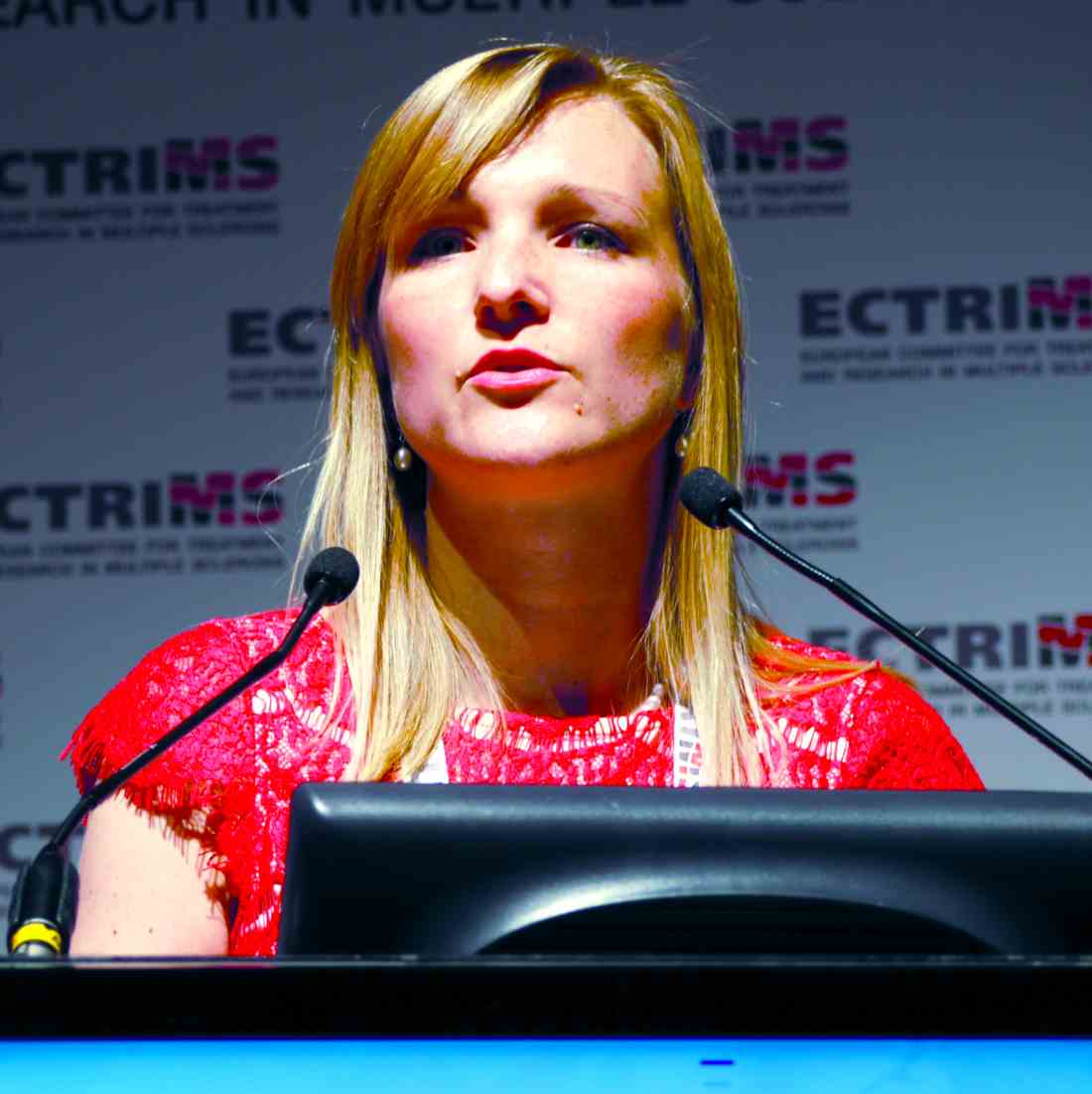
“These findings support a role for NfL as a peripheral biomarker for MS,” said Jens Kuhle, MD, of University Hospital Basel in Switzerland. “There is an urgent unmet need for reliable biomarkers of neurodegeneration, besides efforts that are being done in MRI, optical coherence tomography, and evoked potentials.

“NfL is an exclusively neuronal protein. It is expressed in the cytosol of neurons, it is released upon cell injury into the CSF, and obviously it also appears in the blood circulation.” NfL in the CSF thus reflects nerve damage and had been seen in patients with MS and several other neurologic diseases, including Alzheimer’s disease, Parkinson’s disease, and ALS.
Until recently, however, it was not possible to detect NfL in the blood, as levels are around 50 to 100 times lower than in the CSF, but Dr. Kuhle and his associates have shown that an electrochemiluminescence immunoassay could detect increasing NfL levels with increasing disease activity. In the current study, Dr. Kuhle and colleagues used a Single Molecule Array Immunoassay (Quanterix). This test is based on an enzyme-linked immunoassay with more than 280,000 wells and was developed to be an ultrasensitive diagnostic platform to measure minute quantities of individual proteins. Dr. Kuhle noted that it had “significantly increased sensitivity to measure NfL in blood,” when compared with conventional enzyme-linked immunosorbent assay (ELISA) or electrochemiluminescence.
Two to three consecutive blood samples taken from 149 patients with RRMS participating in the phase III FREEDOMS trial were obtained and compared with samples from 29 similarly aged healthy individuals without MS obtained from a separate biobank.
Patients with two or more relapses in the previous 24 months had significantly higher NfL levels than those with one relapse or no relapses. Serum NfL also significantly increased with the Expanded Disability Status Scale score recorded at the time the blood samples were taken.
Furthermore, “blood NfL levels predicted subsequent brain atrophy rates,” Dr. Kuhle reported, with NfL levels at six months being highly predictive of brain volume changes at 24 months.
And, in this preliminary dataset, patients who had been treated with fingolimod versus placebo during the FREEDOMS trial had lower NfL levels at six, 12, and 24 months. Dr. Kuhle observed that the findings of this study were corroborated by other study data presented at the ECTRIMS meeting.
The FREEDOMS trial was sponsored by Novartis. Dr. Kuhle received research support and consulting fees from Biogen, Novartis, and Protagen. He also disclosed receiving speaker fees and travel expenses from Novartis and several other pharmaceutical companies. Several of Dr. Kuhle’s coinvestigators were employees of Novartis.
—Sara Freeman
LONDON—Higher levels of a neuronal protein were found in the blood of patients with relapsing-remitting multiple sclerosis (RRMS) than in healthy subjects in a proof-of-concept study reported at the 32nd Annual Congress of the European Committee for Treatment and Research in Multiple Sclerosis (ECTRIMS).
Blood levels of neurofilament light chain (NfL) were 28.1 and 12.5 pg/mL, respectively, and were also found to be higher in patients with RRMS with greater disease activity seen on MRI.
As the number of gadolinium-enhancing (Gd+) lesions increased, so did the blood concentration of NfL, which was 23.9 pg/mL in patients with no Gd+ lesions, 26.7 pg/mL in those with one Gd+ lesion, 33.4 pg/mL in those with two to three Gd+ lesions, and 55.9 pg/mL in those with more than three Gd+ lesions.
“These findings support a role for NfL as a peripheral biomarker for MS,” said Jens Kuhle, MD, of University Hospital Basel in Switzerland. “There is an urgent unmet need for reliable biomarkers of neurodegeneration, besides efforts that are being done in MRI, optical coherence tomography, and evoked potentials.
“NfL is an exclusively neuronal protein. It is expressed in the cytosol of neurons, it is released upon cell injury into the CSF, and obviously it also appears in the blood circulation.” NfL in the CSF thus reflects nerve damage and had been seen in patients with MS and several other neurologic diseases, including Alzheimer’s disease, Parkinson’s disease, and ALS.
Until recently, however, it was not possible to detect NfL in the blood, as levels are around 50 to 100 times lower than in the CSF, but Dr. Kuhle and his associates have shown that an electrochemiluminescence immunoassay could detect increasing NfL levels with increasing disease activity. In the current study, Dr. Kuhle and colleagues used a Single Molecule Array Immunoassay (Quanterix). This test is based on an enzyme-linked immunoassay with more than 280,000 wells and was developed to be an ultrasensitive diagnostic platform to measure minute quantities of individual proteins. Dr. Kuhle noted that it had “significantly increased sensitivity to measure NfL in blood,” when compared with conventional enzyme-linked immunosorbent assay (ELISA) or electrochemiluminescence.
Two to three consecutive blood samples taken from 149 patients with RRMS participating in the phase III FREEDOMS trial were obtained and compared with samples from 29 similarly aged healthy individuals without MS obtained from a separate biobank.
Patients with two or more relapses in the previous 24 months had significantly higher NfL levels than those with one relapse or no relapses. Serum NfL also significantly increased with the Expanded Disability Status Scale score recorded at the time the blood samples were taken.
Furthermore, “blood NfL levels predicted subsequent brain atrophy rates,” Dr. Kuhle reported, with NfL levels at six months being highly predictive of brain volume changes at 24 months.
And, in this preliminary dataset, patients who had been treated with fingolimod versus placebo during the FREEDOMS trial had lower NfL levels at six, 12, and 24 months. Dr. Kuhle observed that the findings of this study were corroborated by other study data presented at the ECTRIMS meeting.
The FREEDOMS trial was sponsored by Novartis. Dr. Kuhle received research support and consulting fees from Biogen, Novartis, and Protagen. He also disclosed receiving speaker fees and travel expenses from Novartis and several other pharmaceutical companies. Several of Dr. Kuhle’s coinvestigators were employees of Novartis.
—Sara Freeman

Choosing Wisely Initiative Helps Physicians Provide Appropriate Care
HILTON HEAD, SC—Physicians sometimes order unnecessary medical tests and procedures for their patients, which results in wasteful spending and inappropriate care. Following medical associations’ practice recommendations, which have been collected by the Choosing Wisely initiative, can help physicians reduce waste in the health care system and provide appropriate treatment for patients, according to an overview presented at the 39th Annual Contemporary Clinical Neurology Symposium.
Avoiding Unnecessary Treatments and Tests
The American Board of Internal Medicine Foundation created the Choosing Wisely website to encourage dialogue between physicians and patients about the overuse of treatments and tests. An additional goal was to empower patients to make informed treatment decisions. More than 70 societies, including the American Academy of Neurology (AAN) and the American Headache Society, submitted recommendations to advise patients and clinicians about proper healthcare. “You can find a list of all the organizations that contributed on the Choosing Wisely website, and each one was asked to contribute five different topics for Choosing Wisely,” said Peter Donofrio, MD, Professor of Neurology at Vanderbilt University in Nashville.

The AAN recommends that clinicians not perform an EEG for headaches. In addition, the organization recommends that physicians not perform imaging of the carotid arteries for simple syncope without other neurologic symptoms. For patients with migraine, opioids or butalbital treatment should be a last resort. The AAN also recommends that doctors not prescribe interferon-beta or glatiramer acetate for patients with disability resulting from progressive, nonrelapsing forms of multiple sclerosis, because the drugs are ineffective. Finally, it advises doctors not to recommend carotid endarterectomy for asymptomatic carotid stenosis unless the complication rate from surgery is less than 3%.
The American Association of Neuromuscular and Electrodiagnostic Medicine recommends that physicians not perform MRI scans of the brain or spine for patients with peripheral neuropathy without signs of cerebral or spinal cord disease. In addition, the association discourages physicians from performing nerve conduction studies (NCSs) without a needle EMG for radiculopathy assessment. It also recommends that physicians not order or perform four-limb EMG/NCS testing for neck or back pain after trauma.
Treating Acute Low Back Pain and Headache
Other medical associations have made recommendations regarding the assessment and treatment of acute low back pain and headache. The North American Spine Society does not recommend advanced imaging of the spine within the first six weeks in patients with nonspecific acute low back pain in the absence of red flags.
The American Headache Society (AHS) recommends that physicians avoid advising prolonged or frequent use of over-the-counter pain medications for headache. The organization also discourages physicians from prescribing opioid or butalbital-containing medications as first-line treatment for recurrent headache disorders. Furthermore, it does not recommend surgical deactivation of migraine trigger points outside of a clinical trial. In addition, the society advises physicians not to perform CT imaging for headache when an MRI is available, except in emergency settings. The society also recommends that physicians should not perform neuroimaging studies for patients with stable headaches that meet migraine criteria.
When CT Scans Are Unnecessary in Children
The American Academy of Pediatrics (AAP) advises that CT scans and MRI scans are not necessary in a child with simple febrile seizure. The AAP also does not recommend CT scans for the immediate evaluation of minor head injuries; clinical observation and Pediatric Emergency Care Applied Research Network (PECARN) criteria should be used to determine whether imaging is indicated.
Treating Insomnia and Sleep Disorders
The American Academy of Sleep Medicine advises doctors not to prescribe medication for childhood insomnia, which usually arises from parent–child interactions and responds to behavioral intervention. In addition, the academy does no
—Erica Tricarico
Suggested Reading
Callaghan BC, De Lott LB, Kerber KA, et al. Neurology Choosing Wisely recommendations: 74 and growing. Neurol Clin Pract. 2015;5(5):439-447.
Langer-Gould AM, Anderson WE, Armstrong MJ, et al. The American Academy of Neurology’s top five choosing wisely recommendations. Neurology. 2013;81(11):1004-1011.
Loder E, Weisenbaum E, Fishberg B, et al. Choosing wisely in headache medicine: the American Headache Society’s list of five things physicians and patients should question. Headache. 2013;53(10):1651-1659.
HILTON HEAD, SC—Physicians sometimes order unnecessary medical tests and procedures for their patients, which results in wasteful spending and inappropriate care. Following medical associations’ practice recommendations, which have been collected by the Choosing Wisely initiative, can help physicians reduce waste in the health care system and provide appropriate treatment for patients, according to an overview presented at the 39th Annual Contemporary Clinical Neurology Symposium.
Avoiding Unnecessary Treatments and Tests
The American Board of Internal Medicine Foundation created the Choosing Wisely website to encourage dialogue between physicians and patients about the overuse of treatments and tests. An additional goal was to empower patients to make informed treatment decisions. More than 70 societies, including the American Academy of Neurology (AAN) and the American Headache Society, submitted recommendations to advise patients and clinicians about proper healthcare. “You can find a list of all the organizations that contributed on the Choosing Wisely website, and each one was asked to contribute five different topics for Choosing Wisely,” said Peter Donofrio, MD, Professor of Neurology at Vanderbilt University in Nashville.
The AAN recommends that clinicians not perform an EEG for headaches. In addition, the organization recommends that physicians not perform imaging of the carotid arteries for simple syncope without other neurologic symptoms. For patients with migraine, opioids or butalbital treatment should be a last resort. The AAN also recommends that doctors not prescribe interferon-beta or glatiramer acetate for patients with disability resulting from progressive, nonrelapsing forms of multiple sclerosis, because the drugs are ineffective. Finally, it advises doctors not to recommend carotid endarterectomy for asymptomatic carotid stenosis unless the complication rate from surgery is less than 3%.
The American Association of Neuromuscular and Electrodiagnostic Medicine recommends that physicians not perform MRI scans of the brain or spine for patients with peripheral neuropathy without signs of cerebral or spinal cord disease. In addition, the association discourages physicians from performing nerve conduction studies (NCSs) without a needle EMG for radiculopathy assessment. It also recommends that physicians not order or perform four-limb EMG/NCS testing for neck or back pain after trauma.
Treating Acute Low Back Pain and Headache
Other medical associations have made recommendations regarding the assessment and treatment of acute low back pain and headache. The North American Spine Society does not recommend advanced imaging of the spine within the first six weeks in patients with nonspecific acute low back pain in the absence of red flags.
The American Headache Society (AHS) recommends that physicians avoid advising prolonged or frequent use of over-the-counter pain medications for headache. The organization also discourages physicians from prescribing opioid or butalbital-containing medications as first-line treatment for recurrent headache disorders. Furthermore, it does not recommend surgical deactivation of migraine trigger points outside of a clinical trial. In addition, the society advises physicians not to perform CT imaging for headache when an MRI is available, except in emergency settings. The society also recommends that physicians should not perform neuroimaging studies for patients with stable headaches that meet migraine criteria.
When CT Scans Are Unnecessary in Children
The American Academy of Pediatrics (AAP) advises that CT scans and MRI scans are not necessary in a child with simple febrile seizure. The AAP also does not recommend CT scans for the immediate evaluation of minor head injuries; clinical observation and Pediatric Emergency Care Applied Research Network (PECARN) criteria should be used to determine whether imaging is indicated.
Treating Insomnia and Sleep Disorders
The American Academy of Sleep Medicine advises doctors not to prescribe medication for childhood insomnia, which usually arises from parent–child interactions and responds to behavioral intervention. In addition, the academy does no
—Erica Tricarico
Suggested Reading
Callaghan BC, De Lott LB, Kerber KA, et al. Neurology Choosing Wisely recommendations: 74 and growing. Neurol Clin Pract. 2015;5(5):439-447.
Langer-Gould AM, Anderson WE, Armstrong MJ, et al. The American Academy of Neurology’s top five choosing wisely recommendations. Neurology. 2013;81(11):1004-1011.
Loder E, Weisenbaum E, Fishberg B, et al. Choosing wisely in headache medicine: the American Headache Society’s list of five things physicians and patients should question. Headache. 2013;53(10):1651-1659.

Genetic risk score for low vitamin D may affect MS relapse rate
BALTIMORE – A genetic scoring system for identifying individuals at high risk for low vitamin D levels also detected multiple sclerosis patients with an increased risk for relapse in a multicenter cohort study.
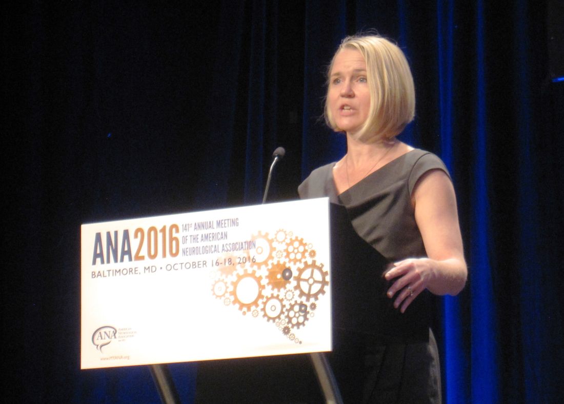
The findings could have clinical significance in multiple sclerosis (MS) treatment and patient counseling, Jennifer S. Graves, MD, PhD, of the University of California, San Francisco, said in a brief oral and poster presentation of the study at the annual meeting of the American Neurological Association.
The investigators compared the SNP profile of a discovery cohort of 181 patients with MS or high-risk clinically isolated syndrome (the discovery cohort) who were enrolled at two pediatric MS centers in California between 2006 and 2011 against a replication cohort of 110 patients of comparable age, race, and median vitamin D serum level who were enrolled at nine MS centers elsewhere in the United States from 2011 to 2015.
Three of the SNPs were strongly associated with the vitamin D levels in the discovery cohort after a statistical correction that revealed individual influences of genes among the 29 different mutations. The researchers used these three SNPs to generate risk scores for vitamin D levels. A comparison of the lowest and highest scores revealed a linear association with vitamin D levels. The highest scores were associated with serum vitamin D levels that were nearly 15 ng/mL lower in both the discovery and replication cohorts (P = .00000052 and .002, respectively).
The risk of MS relapse for individuals with the highest risk score in the discovery cohort was nearly twice as high as it was for individuals with the lowest risk score (hazard ratio, 1.94; 95% confidence interval, 1.19-3.15; P = .007).
“A genetic score of three functional SNPs captures risk of low vitamin D level and identifies those who may be at risk of relapse related to this risk factor. These findings support a causal association of vitamin D with relapse rate,” Dr. Graves said.
The study may potentially be important beyond MS. “This risk score may also have some utility in other disease states where vitamin D deficiency may be contributing to disease course,” she said.
The study was funded by The Race to Erase MS, the National Multiple Sclerosis Society, and the National Institute of Neurological Disorders and Stroke. Dr. Graves had no disclosures.
BALTIMORE – A genetic scoring system for identifying individuals at high risk for low vitamin D levels also detected multiple sclerosis patients with an increased risk for relapse in a multicenter cohort study.
The findings could have clinical significance in multiple sclerosis (MS) treatment and patient counseling, Jennifer S. Graves, MD, PhD, of the University of California, San Francisco, said in a brief oral and poster presentation of the study at the annual meeting of the American Neurological Association.
The investigators compared the SNP profile of a discovery cohort of 181 patients with MS or high-risk clinically isolated syndrome (the discovery cohort) who were enrolled at two pediatric MS centers in California between 2006 and 2011 against a replication cohort of 110 patients of comparable age, race, and median vitamin D serum level who were enrolled at nine MS centers elsewhere in the United States from 2011 to 2015.
Three of the SNPs were strongly associated with the vitamin D levels in the discovery cohort after a statistical correction that revealed individual influences of genes among the 29 different mutations. The researchers used these three SNPs to generate risk scores for vitamin D levels. A comparison of the lowest and highest scores revealed a linear association with vitamin D levels. The highest scores were associated with serum vitamin D levels that were nearly 15 ng/mL lower in both the discovery and replication cohorts (P = .00000052 and .002, respectively).
The risk of MS relapse for individuals with the highest risk score in the discovery cohort was nearly twice as high as it was for individuals with the lowest risk score (hazard ratio, 1.94; 95% confidence interval, 1.19-3.15; P = .007).
“A genetic score of three functional SNPs captures risk of low vitamin D level and identifies those who may be at risk of relapse related to this risk factor. These findings support a causal association of vitamin D with relapse rate,” Dr. Graves said.
The study may potentially be important beyond MS. “This risk score may also have some utility in other disease states where vitamin D deficiency may be contributing to disease course,” she said.
The study was funded by The Race to Erase MS, the National Multiple Sclerosis Society, and the National Institute of Neurological Disorders and Stroke. Dr. Graves had no disclosures.
AT ANA 2016
Key clinical point:
Major finding: The risk of MS relapse was 94% greater for individuals with the highest genetic risk score for low serum vitamin D level, compared with those who had the lowest risk score.
Data source: Databases of patients enrolled at two pediatric MS centers in California and nine national MS centers.
Disclosures: The study was funded by The Race to Erase MS, the National Multiple Sclerosis Society, and the National Institute of Neurological Disorders and Stroke. Dr. Graves had no disclosures.
Efficacy of Cladribine Tablets Continues After Conversion to MS
LONDON—Significant treatment effect versus placebo of cladribine tablets given to patients with clinically isolated syndrome during the initial treatment period continues to be observed in patients who convert to clinically definite multiple sclerosis (MS) and switch to treatment with a different disease-modifying drug (ie, subcutaneous interferon beta-1a), according to data presented at the 32nd Congress of the European Committee for Treatment and Research in MS (ECTRIMS).
Giancarlo Comi, MD, Professor of Neurology and Chairman of the Department of Neurology at Vita-Salute San Raffaele University in Milan, and colleagues reported that patients with clinically isolated syndrome who had been treated with cladribine tablets and who had converted to MS during the Oral Cladribine in Early MS (ORACLE-MS) initial treatment period had lower annualized relapse rates during the open-label maintenance period, relative to those patients who had received placebo during the ORACLE-MS initial treatment period.

The CLARITY (CLAdRIbine Tablets treating MS orallY) study in patients with active MS showed that annualized relapse rates and sustained disability worsening were reduced in patients treated with cladribine tablets annually for two years in short-duration courses, compared with placebo. The efficacy observed in the CLARITY study was maintained without further active treatment during the CLARITY extension study. In the ORACLE-MS study in patients with a first demyelinating event, cladribine tablets (3.5 mg/kg and 5.25 mg/kg) significantly reduced the risk of conversion to clinically definite MS, compared with placebo. If clinically definite MS occurred in the double-blinded, initial treatment period, patients were treated with subcutaneous interferon beta-1a in an open-label maintenance period.
The present study was designed to assess the annualized relapse rate during the ORACLE-MS open-label maintenance period in patients randomized to cladribine (3.5 mg/kg and 5.25 mg/kg) or placebo in the initial treatment period.
Similar to previous trials, participation in the ORACLE-MS open-label maintenance period was dependent upon the clinical course of the patient’s disease in the initial treatment period. Patients in ORACLE-MS who converted to clinically definite MS (according to Poser criteria) during the initial treatment period entered the open-label maintenance period and were treated with subcutaneous interferon beta-1a (titrated over four weeks up to the dose of 44 μg) administered three times per week.
A total of 109 patients in ORACLE-MS converted to clinically definite MS in the initial treatment period and received at least one dose of interferon beta-1a. The median time on interferon beta-1a was 56.0 weeks. Estimated annualized relapse rates in the open-label maintenance period were 0.14 for patients originally treated with cladribine 3.5 mg/kg (n = 25), 0.24 for patients originally treated with cladribine 5.25 mg/kg (n = 24), and 0.42 for patients who originally received placebo in the initial treatment period (n = 60).
According to the researchers, durable efficacy of cladribine tablets in ORACLE-MS into the open-label maintenance period is consistent with results of the CLARITY and CLARITY extension studies.
This study was sponsored by EMD Serono.
—Glenn S. Williams
Suggested Reading
Cook S, Vermersch P, Comi G, et al. Safety and tolerability of cladribine tablets in multiple sclerosis: the CLARITY (CLAdRIbine Tablets treating multiple sclerosis orallY) study. Mult Scler. 2011;17(5):578-593.
Leist TP, Comi G, Cree BA, et al. Effect of oral cladribine on time to conversion to clinically definite multiple sclerosis in patients with a first demyelinating event (ORACLE MS): a phase 3 randomised trial. Lancet Neurol. 2014;13(3):257-267.
LONDON—Significant treatment effect versus placebo of cladribine tablets given to patients with clinically isolated syndrome during the initial treatment period continues to be observed in patients who convert to clinically definite multiple sclerosis (MS) and switch to treatment with a different disease-modifying drug (ie, subcutaneous interferon beta-1a), according to data presented at the 32nd Congress of the European Committee for Treatment and Research in MS (ECTRIMS).
Giancarlo Comi, MD, Professor of Neurology and Chairman of the Department of Neurology at Vita-Salute San Raffaele University in Milan, and colleagues reported that patients with clinically isolated syndrome who had been treated with cladribine tablets and who had converted to MS during the Oral Cladribine in Early MS (ORACLE-MS) initial treatment period had lower annualized relapse rates during the open-label maintenance period, relative to those patients who had received placebo during the ORACLE-MS initial treatment period.
The CLARITY (CLAdRIbine Tablets treating MS orallY) study in patients with active MS showed that annualized relapse rates and sustained disability worsening were reduced in patients treated with cladribine tablets annually for two years in short-duration courses, compared with placebo. The efficacy observed in the CLARITY study was maintained without further active treatment during the CLARITY extension study. In the ORACLE-MS study in patients with a first demyelinating event, cladribine tablets (3.5 mg/kg and 5.25 mg/kg) significantly reduced the risk of conversion to clinically definite MS, compared with placebo. If clinically definite MS occurred in the double-blinded, initial treatment period, patients were treated with subcutaneous interferon beta-1a in an open-label maintenance period.
The present study was designed to assess the annualized relapse rate during the ORACLE-MS open-label maintenance period in patients randomized to cladribine (3.5 mg/kg and 5.25 mg/kg) or placebo in the initial treatment period.
Similar to previous trials, participation in the ORACLE-MS open-label maintenance period was dependent upon the clinical course of the patient’s disease in the initial treatment period. Patients in ORACLE-MS who converted to clinically definite MS (according to Poser criteria) during the initial treatment period entered the open-label maintenance period and were treated with subcutaneous interferon beta-1a (titrated over four weeks up to the dose of 44 μg) administered three times per week.
A total of 109 patients in ORACLE-MS converted to clinically definite MS in the initial treatment period and received at least one dose of interferon beta-1a. The median time on interferon beta-1a was 56.0 weeks. Estimated annualized relapse rates in the open-label maintenance period were 0.14 for patients originally treated with cladribine 3.5 mg/kg (n = 25), 0.24 for patients originally treated with cladribine 5.25 mg/kg (n = 24), and 0.42 for patients who originally received placebo in the initial treatment period (n = 60).
According to the researchers, durable efficacy of cladribine tablets in ORACLE-MS into the open-label maintenance period is consistent with results of the CLARITY and CLARITY extension studies.
This study was sponsored by EMD Serono.
—Glenn S. Williams
Suggested Reading
Cook S, Vermersch P, Comi G, et al. Safety and tolerability of cladribine tablets in multiple sclerosis: the CLARITY (CLAdRIbine Tablets treating multiple sclerosis orallY) study. Mult Scler. 2011;17(5):578-593.
Leist TP, Comi G, Cree BA, et al. Effect of oral cladribine on time to conversion to clinically definite multiple sclerosis in patients with a first demyelinating event (ORACLE MS): a phase 3 randomised trial. Lancet Neurol. 2014;13(3):257-267.

Paulo Fontoura, MD, PhD

Evaluation of Cortical Lesions Could Improve Diagnosis of MS
LONDON—Evaluation of cortical lesions improves the specificity of the diagnostic criteria for multiple sclerosis (MS), according to research presented at the 32nd Congress of the European Committee for Treatment and Research in MS (ECTRIMS). Assessment of cortical lesions, in concert with current McDonald criteria, also preserved a high level of diagnostic sensitivity and accuracy in a multicentric cohort of patients with clinically isolated syndrome, reported Paolo Preziosa, MD, Neuroimaging Research Unit at the Institute of Experimental Neurology and Division of Neuroscience at San Raffaele Scientific Institute, Vita-Salute San Raffaele University, Milan, Italy, and his research colleagues.

Since the publication of the 2010 revised McDonald criteria, new data regarding the application of MRI for the diagnosis of MS have become available. In a single-center study, adding the assessment of cortical lesions was shown to modify the diagnostic algorithm, resulting in higher specificity.
In the present study, Dr. Preziosa and colleagues sought to test the performance of different sets of imaging criteria, including the assessment of cortical lesions, for the development of MS in a multicentric cohort of patients with clinically isolated syndrome.
The researchers analyzed brain double inversion recovery and brain and cord T2-weighted and post-contrast T1-weighted sequences acquired from 72 patients with clinically isolated syndrome from five European centers (Barcelona, Belgrade, Mainz, Milan, and Verona) within three months and after 12 months from disease onset. Patients were followed clinically for 24 or more months or until the development of clinically defined MS. Median follow-up was 24.2 months. Sensitivity, specificity, and accuracy of the different dissemination in space MRI criteria for the development of MS were tested.
At follow-up, 65 patients (90%) had clinically and/or radiologically definite MS. The sensitivity of all criteria was high (McDonald 2005, 83%; McDonald 2010, 92%; Filippi 2010, 80%). Specificity of Filippi 2010 was higher (67%), compared with the others (50% for McDonald 2005 and 2010). The accuracy of all criteria was high (McDonald 2005, 81%; McDonald 2010, 89%; Filippi 2010, 79%).
“The detection of cortical lesions in vivo using MRI should be considered in future clinical trials,” Dr. Preziosa said.
—Glenn S. Williams
Suggested Reading
Filippi M, Rocca MA, Calabrese M, et al. Intracortical lesions: relevance for new MRI diagnostic criteria for multiple sclerosis. Neurology. 2010;75(22):1988-1994.
LONDON—Evaluation of cortical lesions improves the specificity of the diagnostic criteria for multiple sclerosis (MS), according to research presented at the 32nd Congress of the European Committee for Treatment and Research in MS (ECTRIMS). Assessment of cortical lesions, in concert with current McDonald criteria, also preserved a high level of diagnostic sensitivity and accuracy in a multicentric cohort of patients with clinically isolated syndrome, reported Paolo Preziosa, MD, Neuroimaging Research Unit at the Institute of Experimental Neurology and Division of Neuroscience at San Raffaele Scientific Institute, Vita-Salute San Raffaele University, Milan, Italy, and his research colleagues.
Since the publication of the 2010 revised McDonald criteria, new data regarding the application of MRI for the diagnosis of MS have become available. In a single-center study, adding the assessment of cortical lesions was shown to modify the diagnostic algorithm, resulting in higher specificity.
In the present study, Dr. Preziosa and colleagues sought to test the performance of different sets of imaging criteria, including the assessment of cortical lesions, for the development of MS in a multicentric cohort of patients with clinically isolated syndrome.
The researchers analyzed brain double inversion recovery and brain and cord T2-weighted and post-contrast T1-weighted sequences acquired from 72 patients with clinically isolated syndrome from five European centers (Barcelona, Belgrade, Mainz, Milan, and Verona) within three months and after 12 months from disease onset. Patients were followed clinically for 24 or more months or until the development of clinically defined MS. Median follow-up was 24.2 months. Sensitivity, specificity, and accuracy of the different dissemination in space MRI criteria for the development of MS were tested.
At follow-up, 65 patients (90%) had clinically and/or radiologically definite MS. The sensitivity of all criteria was high (McDonald 2005, 83%; McDonald 2010, 92%; Filippi 2010, 80%). Specificity of Filippi 2010 was higher (67%), compared with the others (50% for McDonald 2005 and 2010). The accuracy of all criteria was high (McDonald 2005, 81%; McDonald 2010, 89%; Filippi 2010, 79%).
“The detection of cortical lesions in vivo using MRI should be considered in future clinical trials,” Dr. Preziosa said.
—Glenn S. Williams
Suggested Reading
Filippi M, Rocca MA, Calabrese M, et al. Intracortical lesions: relevance for new MRI diagnostic criteria for multiple sclerosis. Neurology. 2010;75(22):1988-1994.
Smoking, vitamin D deficiency linked to early MS disability
LONDON – Severe vitamin D deficiency and current smoking predicted accumulated disability in patients with clinically isolated syndrome, which can be a precursor to the development of multiple sclerosis.
Prospectively collected data from the ongoing Barcelona clinically isolated syndrome (CIS) cohort, which includes more than 1,000 patients with CIS, showed that patients with a serum vitamin D below 8.0 ng/mL (9% of 503 patient samples) had more than double the risk for accumulated disability when compared with those who had higher vitamin D levels. The hazard ratio for disability with severely low vitamin D levels was 2.3 (P = .049) in an analysis adjusted for the potential confounding factors of patients’ sex and age, the number of baseline T2 lesions, receipt of disease-modifying treatment, CIS topography, and oligoclonal bands. Disability accumulation was defined as an Expanded Disability Status Scale score of 3.0 or more.

However, neither vitamin D deficiency nor current smoking predicted the conversion of CIS to clinically definite multiple sclerosis (CDMS), said María Zuluaga, MD, of the Centre d’Esclerosi Múltiple de Catalunya at Vall d’Hebron University Hospital in Barcelona.
Environmental factors such as vitamin D levels and smoking have been purported to play a role in the development of CIS to CDMS, Dr. Zuluaga explained at the annual congress of the European Committee for Treatment and Research in Multiple Sclerosis (ECTRIMS).
Blood samples collected within 6 months of a diagnosis of CIS were examined and vitamin D deficiency defined as normal, mild, moderate, or severe based on serum 25-hydroxy vitamin D of greater than 20, 16-20, 8-15, and less that 8 ng/mL.
Levels of the nicotine metabolite cotinine in the blood were used as a proxy for current smoking. Cotinine has a half-life of around 20 hours and smokers – active or passive – have a level of 14 ng/mL or more while nonsmokers have a level of less than 14 ng/mL.
The study received no commercial funding. Dr. Zuluaga reported having no conflict of interest related to the study.
LONDON – Severe vitamin D deficiency and current smoking predicted accumulated disability in patients with clinically isolated syndrome, which can be a precursor to the development of multiple sclerosis.
Prospectively collected data from the ongoing Barcelona clinically isolated syndrome (CIS) cohort, which includes more than 1,000 patients with CIS, showed that patients with a serum vitamin D below 8.0 ng/mL (9% of 503 patient samples) had more than double the risk for accumulated disability when compared with those who had higher vitamin D levels. The hazard ratio for disability with severely low vitamin D levels was 2.3 (P = .049) in an analysis adjusted for the potential confounding factors of patients’ sex and age, the number of baseline T2 lesions, receipt of disease-modifying treatment, CIS topography, and oligoclonal bands. Disability accumulation was defined as an Expanded Disability Status Scale score of 3.0 or more.
However, neither vitamin D deficiency nor current smoking predicted the conversion of CIS to clinically definite multiple sclerosis (CDMS), said María Zuluaga, MD, of the Centre d’Esclerosi Múltiple de Catalunya at Vall d’Hebron University Hospital in Barcelona.
Environmental factors such as vitamin D levels and smoking have been purported to play a role in the development of CIS to CDMS, Dr. Zuluaga explained at the annual congress of the European Committee for Treatment and Research in Multiple Sclerosis (ECTRIMS).
Blood samples collected within 6 months of a diagnosis of CIS were examined and vitamin D deficiency defined as normal, mild, moderate, or severe based on serum 25-hydroxy vitamin D of greater than 20, 16-20, 8-15, and less that 8 ng/mL.
Levels of the nicotine metabolite cotinine in the blood were used as a proxy for current smoking. Cotinine has a half-life of around 20 hours and smokers – active or passive – have a level of 14 ng/mL or more while nonsmokers have a level of less than 14 ng/mL.
The study received no commercial funding. Dr. Zuluaga reported having no conflict of interest related to the study.
Key clinical point:
Major finding: Patients with a serum 25-hydroxy vitamin D level of less than 8.0 ng/mL showed an increased risk for disability in a multivariate analysis (HR, 2.3; P = .049). Nonsmokers were significantly less likely to have disability progression (HR, 0.4; P = .002).
Data source: Barcelona CIS cohort of 1,127 individuals
Disclosures: The study received no commercial funding. Dr. Zuluaga reported having no conflict of interest related to the study.
Fluoxetine fails to slow progressive multiple sclerosis
LONDON – Contrary to expectation of a neuroprotective benefit, fluoxetine does not slow down the progressive phase of multiple sclerosis, according to the results of a randomized, double-blind, multicenter trial.
The first results of the FLUOX-PMS trial, reported by Melissa Cambron, MD, of University Hospital Brussels (Belgium), showed no statistically significant difference between fluoxetine and placebo for improving the primary endpoint of the time to confirmed disease progression.
“The progressive phase of MS remains an ill-understood part of the disease and it is a holy grail to find a drug that can stop this progression,” Dr. Cambron said at the annual congress of the European Committee for Treatment and Research in Multiple Sclerosis (ECTRIMS).
The rationale for looking at whether fluoxetine, a well-studied antidepressant drug, could be such a drug, was that it had several neuroprotective features – it has been shown to stimulate the release of brain-derived neurotrophic factor, stimulate metabolism in astrocytes, and lower glutamatergic toxicity, she said. All of these could potentially help prevent axonal degeneration.
The FLUOX-PMS (Fluoxetine in Progressive Multiple Sclerosis) trial (Trials. 2014;15:37) ran from 2012 to June 2016 and enrolled patients with primary progressive MS (PPMS) or secondary progressive MS (SPMS), as defined by the 2010 McDonald criteria. A total of 137 patients were enrolled, and 69 were randomized to treatment with fluoxetine 40 mg/day and 68 were randomized to placebo. Fluoxetine treatment was started at a dose of 20 mg and titrated to the full 40-mg dose by 12 weeks.
Patient demographics were mostly similar between the groups. Around 44% of patients in the fluoxetine and placebo groups were female; roughly 40% had PPMS and 60% had SPMS in both groups; the mean Expanded Disability Status Scale score was 5.2 in both groups; the mean age was 54 and 51 years, respectively; and the disease duration was between 18 and 20 years.
Dr. Cambron also reported that the trials’ secondary endpoints showed no advantage of using fluoxetine over placebo. The proportion of patients without sustained progression during the trial was similar among the fluoxetine- and placebo-treated patients, at a respective 69.6% and 61.8% (P = .434). The proportion of patients with a stable Hauser Ambulation Index was also similar (P = .371).
The primary and secondary endpoints were assessed every 3 months in the trial. Patients also underwent cognitive testing, completed the Beck Depression Inventory-II, and Modified Fatigue Impact Scale before treatment and at 48 and 108 weeks after treatment with fluoxetine or placebo. Brain MRI was also performed at baseline and at week 108. The results of these measurements have yet to be analyzed.
Although patients in the fluoxetine group versus the placebo arm experienced more side effects, there was no evidence of an excess of severe adverse events.
“Unfortunately, our study was inconclusive because we failed to show a statistical significant difference between the placebo arm and the fluoxetine group, although I’m convinced that there’s a trend that can certainly not be ignored,” Dr. Cambron maintained. “Probably there was not enough progression in the study and possibly the study duration was too short, she suggested, “but it remains challenging to study these patients for a long period of time, especially with a placebo-controlled design.”
The trial was funded by IWT, the Government Agency for Innovation by Science and Technology in Flanders (Belgium). Dr. Cambron had no relevant financial disclosures.
Key clinical point:
Major finding: There was no difference in the time to confirmed disease progression between fluoxetine- and placebo-treated patients (P = .07).
Data source: FLUOX-PMS, a multicenter, randomized, double-blind, placebo-controlled clinical study of 137 patients with primary or secondary progressive multiple sclerosis treated with fluoxetine or placebo.
Disclosures: The trial was funded by IWT, the Government Agency for Innovation by Science and Technology in Flanders (Belgium). Dr. Cambron had no relevant financial disclosures.
Neuronal protein could be multiple sclerosis blood biomarker
LONDON – Higher levels of a neuronal protein were found in the blood of patients with relapsing-remitting multiple sclerosis than in healthy subjects in a proof-of-concept study reported at the annual congress of the European Committee for Treatment and Research in Multiple Sclerosis (ECTRIMS).
Blood levels of neurofilament light chain (NfL) were 28.1 vs. 12.5 pg/mL, respectively (P less than .0001), and were also found to be higher in relapsing-remitting multiple sclerosis (RRMS) patients with greater disease activity seen on MRI.

“These findings support a role for neurofilament light chain (NfL) as a peripheral biomarker for MS,” said the reporting investigator Jens Kuhle, MD, of University Hospital Basel (Switzerland).
“There is an urgent unmet need for reliable biomarkers of neurodegeneration, besides efforts that are being done in MRI, optical coherence tomography, and evoked potentials,” Dr. Kuhle said.
He added: “NfL is an exclusively neuronal protein. It is expressed in the cytosol of neurons, it is released upon cell injury into the CSF [cerebral spinal fluid], and obviously it also appears in the blood circulation.” NfL in the CSF thus reflects nerve damage and had been seen in patients with MS and several other neurologic diseases, including Alzheimer’s, Parkinson’s, and amyotrophic lateral sclerosis.
Until recently, however, it was not possible to detect NfL in the blood, as levels are around 50 to 100 times lower than in the CSF, but Dr. Kuhle and his associates have shown that using an electrochemiluminescence immunoassay could detect increasing NfL levels with increasing disease activity (Mult Scler. 2016 Jan 11. doi: 10.1177/1352458515623365).
In the current study, a Single Molecule Array Immunoassay (Quanterix) was used. This is based on an enzyme-linked immunoassay with more than 280,000 wells and developed to be an ultrasensitive diagnostic platform to measure minute quantities of individual proteins. Dr. Kuhle noted that it had “significantly increased sensitivity to measure NfL in blood” when compared with conventional ELISA or electrochemiluminescence (Clin Chem Lab Med. 2016;54[10]:1655-61).
Two to three consecutive blood samples taken from 149 patients with RRMS participating in the phase III FREEDOMS trial were obtained and these were compared with samples from 29 similarly aged healthy individuals without MS obtained from a separate biobank.
Patients with two or more relapses in the past 24 months had significantly higher NfL levels than did those with one relapse or no relapses. Serum NfL also significantly increased with the Expanded Disability Status Scale score recorded at the time the blood samples were taken.
Furthermore, “blood NfL levels predicted subsequent brain atrophy rates,” Dr. Kuhle reported, with NfL levels at 6 months being highly predictive of brain volume changes at 24 months (P less than .0001).
And, in this preliminary dataset, patients who had been treated with fingolimod (Gilenya) versus placebo in the FREEDOMS trial had lower NfL levels at 6, 12, and 24 months.
Dr. Kuhle observed that the findings of this study were corroborated by other study data presented during the poster sessions at the ECTRIMS 2016 meeting.
The FREEDOMS trial was sponsored by Novartis. Dr. Kuhle disclosed receiving research support and consulting fees from Biogen, Novartis, and Protagen AG. He has also received speaker fees and travel expenses from Novartis and several other companies, and several of his coinvestigators were employees of Novartis.
LONDON – Higher levels of a neuronal protein were found in the blood of patients with relapsing-remitting multiple sclerosis than in healthy subjects in a proof-of-concept study reported at the annual congress of the European Committee for Treatment and Research in Multiple Sclerosis (ECTRIMS).
Blood levels of neurofilament light chain (NfL) were 28.1 vs. 12.5 pg/mL, respectively (P less than .0001), and were also found to be higher in relapsing-remitting multiple sclerosis (RRMS) patients with greater disease activity seen on MRI.
“These findings support a role for neurofilament light chain (NfL) as a peripheral biomarker for MS,” said the reporting investigator Jens Kuhle, MD, of University Hospital Basel (Switzerland).
“There is an urgent unmet need for reliable biomarkers of neurodegeneration, besides efforts that are being done in MRI, optical coherence tomography, and evoked potentials,” Dr. Kuhle said.
He added: “NfL is an exclusively neuronal protein. It is expressed in the cytosol of neurons, it is released upon cell injury into the CSF [cerebral spinal fluid], and obviously it also appears in the blood circulation.” NfL in the CSF thus reflects nerve damage and had been seen in patients with MS and several other neurologic diseases, including Alzheimer’s, Parkinson’s, and amyotrophic lateral sclerosis.
Until recently, however, it was not possible to detect NfL in the blood, as levels are around 50 to 100 times lower than in the CSF, but Dr. Kuhle and his associates have shown that using an electrochemiluminescence immunoassay could detect increasing NfL levels with increasing disease activity (Mult Scler. 2016 Jan 11. doi: 10.1177/1352458515623365).
In the current study, a Single Molecule Array Immunoassay (Quanterix) was used. This is based on an enzyme-linked immunoassay with more than 280,000 wells and developed to be an ultrasensitive diagnostic platform to measure minute quantities of individual proteins. Dr. Kuhle noted that it had “significantly increased sensitivity to measure NfL in blood” when compared with conventional ELISA or electrochemiluminescence (Clin Chem Lab Med. 2016;54[10]:1655-61).
Two to three consecutive blood samples taken from 149 patients with RRMS participating in the phase III FREEDOMS trial were obtained and these were compared with samples from 29 similarly aged healthy individuals without MS obtained from a separate biobank.
Patients with two or more relapses in the past 24 months had significantly higher NfL levels than did those with one relapse or no relapses. Serum NfL also significantly increased with the Expanded Disability Status Scale score recorded at the time the blood samples were taken.
Furthermore, “blood NfL levels predicted subsequent brain atrophy rates,” Dr. Kuhle reported, with NfL levels at 6 months being highly predictive of brain volume changes at 24 months (P less than .0001).
And, in this preliminary dataset, patients who had been treated with fingolimod (Gilenya) versus placebo in the FREEDOMS trial had lower NfL levels at 6, 12, and 24 months.
Dr. Kuhle observed that the findings of this study were corroborated by other study data presented during the poster sessions at the ECTRIMS 2016 meeting.
The FREEDOMS trial was sponsored by Novartis. Dr. Kuhle disclosed receiving research support and consulting fees from Biogen, Novartis, and Protagen AG. He has also received speaker fees and travel expenses from Novartis and several other companies, and several of his coinvestigators were employees of Novartis.
Key clinical point:
Major finding: Blood NfL levels were significantly higher in MS patients than in healthy individuals (28.1 vs. 12.5 pg/mL; P less than .0001); NfL levels were also increased in patients with greater disease activity seen on MRI.
Data source: Proof-of-concept study involving blood samples taken from 149 patients with relapsing-remitting MS (RRMS) participating in the phase III FREEDOMS trial and 29 healthy individuals without MS from a separate biobank.
Disclosures: The FREEDOMS trial was sponsored by Novartis. Dr. Kuhle disclosed receiving research support and consulting fees from Biogen, Novartis, and Protagen AG. He has also received speaker fees and travel expenses from Novartis and several other companies, and several of his coinvestigators were employees of Novartis.
Retinal Measurements Predict 10-Year Disability in MS
LONDON—As has been previously shown with brain atrophy and lesion volume, retinal measures can have predictive value for medium-term disability in multiple sclerosis (MS), according to research presented at the 32nd Congress of the European Committee for Treatment and Research in MS (ECTRIMS). “Our preliminary findings support the utility of optical coherence tomography (OCT) as a tool to predict neurodegeneration and disease progression over time in patients with MS,” said Alissa M. Rothman, MD, a post-doctoral research coordinator at the Johns Hopkins MS Center in Baltimore.
Measures of retinal layer thickness obtained with OCT have been shown to correlate with visual function, grey matter volume, and Expanded Disability Status Scale (EDSS) scores in MS. However, the prognostic value of retinal measurements for predicting long-term disability in patients with MS is still being evaluated. In the present study, Dr. Rothman and colleagues sought to determine whether retinal thickness, as assessed by OCT, predicts disability in MS 10 years later.
A total of 89 patients with MS were scanned on Stratus OCT between 2006 and 2007. During 2015 and 2016, these patients underwent formal, blinded EDSS determination. Average peripapillary retinal nerve fiber layer (RNFL) thickness and total macular volume (TMV) were assessed by calculating the mean value of these measures for both eyes in each subject. Patients were categorized by baseline diagnosis as relapsing remitting (RRMS), secondary progressive (SPMS), or primary progressive MS (PPMS). Mixed effects linear regression models were used to investigate whether average TMV and RNFL thickness at baseline predict EDSS score after 10 years.
The final analysis included 75 patients with RRMS, nine patients with SPMS, and five patients with PPMS. Fourteen of the 75 patients with a baseline diagnosis of RRMS transitioned to SPMS during the follow-up period. Baseline analyses revealed that the RRMS cohort was significantly younger than the SPMS and PPMS cohorts (mean difference = 21.5 years and 11.7 years, respectively) and that patients with SPMS had a longer disease duration than patients with RRMS and PPMS (mean difference = 14.2 years and 13.2 years, respectively). A history of optic neuritis (ON) was observed in the RRMS and SPMS cohorts (41% and 44%, respectively), but not in the PPMS cohorts. Adjusting for age, sex, and a history of ON, the mean TMV values at baseline predicted EDSS scores after a median follow-up of 9.3 years. On average, a 1 mm3 lower TMV value at baseline predicted a mean decrease of 2 in EDSS at follow-up. Mean baseline RNFL values did not significantly predict EDSS scores.
—Glenn S. Williams