User login
Delayed response in ipilimumab therapy
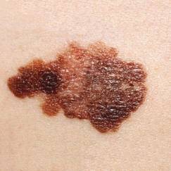
Metastatic melanoma is a deadly disease with a 5-year survival rate lower than 20%.1 In 2011, ipilimumab, a fully humanized antibody that binds to cytotoxic T-lymphocyte–associated antigen 4 (CTLA4) was approved by the US Food and Drug Administration based on improved survival in a pivotal trial.2 CTLA4 is a molecule on cytotoxic T-lymphocytes that plays a critical role in attenuating immune responses. Ipilimumab blocks the binding of B7, the ligand of CTLA4, thereby blocking the activation of CTLA4 and sustaining antitumor immune responses. The time course to response can be variable with immunotherapeutics. We report on a patient who experienced a considerable delay before responding to ipilimumab.
Click on the PDF icon at the top of this introduction to read the full article.
Melanomas were less invasive at diagnosis in patients with established dermatologist
People diagnosed with melanoma were significantly more likely to have melanoma in situ and to have thinner invasive melanomas if they had a regular dermatologist, compared with those who did not have a regular dermatologist, in a retrospective study of 388 patients diagnosed at an academic dermatology department.
These findings were statistically significant for those who had detected the melanomas themselves, but not for those whose dermatologists had detected the melanomas. Self-detected melanomas were in situ in 36 of 61 (59.0%) patients with an established dermatologist vs. 40 of 108 (37.0%) patients without an established dermatologist (P =.006), reported Michelle Cheng and her associates at the University of Pittsburgh.
The time needed to wait to see a dermatologist and the time since the last dermatologic examination were not associated with the invasiveness or the depth of the melanomas at the time of the diagnosis, the investigators said.
These findings "may be explained by a high benefit associated with a first dermatologic visit because of patient education about melanoma detection and/or having a dermatologist to call when the patient finds a suspicious lesion," they wrote (J. Am. Acad. Dermatol. 2014 March [doi:10.1016/j.jaad.2013.10.060]).
The study addressed the uncertainties about the impact of different factors on the invasiveness and depth of melanoma: having had a previous physical exam by a dermatologist before the diagnosis, the recency of that exam, and the time needed to wait for an appointment. The 388 adults (mean age, 55 years) were diagnosed with melanoma at the University of Pittsburgh Medical Center between February 2003 and December 2010. Of these patients, 51% had detected the melanoma themselves and 37% had had a dermatologic exam within the previous year at the university. Of the 388 melanomas diagnosed, 44% (171) were invasive with a mean Breslow depth of 0.96 mm. About 18% (71 patients), had a history of melanoma, and about 37% had seen a dermatologist within the previous year.
Of the 317 with no previous history of melanoma, almost 64% (103 of 162) of those with an established dermatologist were diagnosed with melanoma in situ vs. 44.5% (69 of 155) of those without an established dermatologist, a statistically significant difference (P = .001). The depths of the lesions were also significantly lower among those with an established dermatologist than among those with no dermatologist (median depth, 0.48 mm vs. 0.61 mm, respectively; P = .003).
Among the patients with self-detected melanoma, 41% of those with an established dermatologist had invasive disease, vs. 63% of those who did not have an established dermatologist (P = .006). But among the patients whose melanomas were detected by the dermatologist, 31% of those with an established dermatologist had invasive disease, vs. 40% of those with no established dermatologist, which was not statistically significant (P = .323).
When considering that a skin cancer screen is cost effective, the authors concluded, the results of this study "highlight the value of having even a single dermatologic examination and suggest that educating patients to detect their own melanomas is an important part of improving early detection of melanoma."
All of the patients were from one part of Pennsylvania and were treated at the same medical center, which was a limitation of the study, but the patients were heterogenous and were treated at four dermatology clinics by different attending dermatologists, the authors said. They could not confirm that each patient received the same level of education about melanoma at their visits, but added that most of the dermatologists in the clinics teach the ABCDEs of melanoma and provide counseling in skin self-exams and AAD skin cancer brochures.
The study included clinical research fellowship funding from the Doris Duke Charitable Foundation for one of the authors, UL1/NIH funding for another author, and funding for the statistics consultation from the National Institutes of Health. The authors declared no conflicts of interest.
FROM THE JOURNAL OF THE AMERICAN ACADEMY OF DERMATOLOGY
Major finding: In a study of 388 patients diagnosed with melanoma, those who had an established dermatologist were significantly more likely to be diagnosed with melanoma in situ than those with no dermatologist (64% vs. 44.5%; P = .001), and had thinner invasive melanoma-findings (median depth, 0.48 mm vs. 0.61 mm; P = .003).
Data source: A retrospective study of 388 patients with biopsy-confirmed melanomas at an academic medical center, which evaluated the impact of having an established dermatologist and other factors on the depth of the melanoma when diagnosed.
Disclosures: The study included Clinical Research Fellowship funding from the Doris Duke Charitable Foundation for one of the authors, UL1/NIH funding for another author, and funding for the statistics consultation from the National Institutes of Health. The authors declared no conflicts of interest.

Cell-penetrating domain improves therapeutic cancer vaccine potency
Fusing a tumor-specific antigen with a cell-penetrating domain can generate a more potent therapeutic dendritic cell–based cancer vaccine by enhancing intracellular bioavailability without altering the dendritic cell surface antigens, researchers reported online March 26 in JAMA Surgery.
The immune system can eliminate tumor cells through the generation of cytotoxic T lymphocytes, a process known as immune surveillance, but cancer cells can evade this process, and immunotherapy with dendritic cells is a possible means of reengaging the immune system.
Cancer-testis antigens are ideal candidates for tumor immunotherapy because they are restricted to gonadal germ cells and are unexpressed in healthy adult tissue. Melanoma antigen family A, 3 (MAGE-A3) is a cancer-testis antigen that has attracted attention because it is expressed in a wide variety of cancer types.
A team led by Ramesh B. Batchu, Ph.D., of Wayne State University, Detroit, cloned and purified MAGE-A3 with an amino acid sequence that can transport large proteins across the plasma membrane, to address the problem of inadequate cytoplasmic expression of tumor-specific antigens, and thus enhance production of cytotoxic T lymphocytes.
In a series of laboratory experiments, the cell-penetrating, domain-fused melanoma antigen did show more rapid and efficient penetration of the dendritic cell membrane compared with the normal antigen, Dr. Batchu and his associates reported online (JAMA Surgery 2014 March 26 [doi: 10.1001/jamasurg.2013.4113]).
"We have demonstrated for the first time, to our knowledge, that cloning and purifying MAGE-A3 with CPD [cell-penetrating domain] enhances its cytosolic bioavailability in DCs [dendritic cells] without altering cell surface antigens required for T-cell activation, potentially making it a more potent therapeutic cancer vaccine compared with existing MAGE-A3 protein and peptide vaccines," the authors wrote.
Dr. Batchu and his colleagues reported that they had no conflicts of interest.
FROM JAMA SURGERY
Major finding: A cell-penetrating, domain-fused melanoma antigen showed more rapid and efficient penetration of the dendritic cell membrane than did the normal antigen.
Data source: Laboratory experiments on dendritic cell penetration by tumor-specific MAGE-A3.
Disclosures: Dr. Batchu and his colleagues reported that they had no conflicts of interest.
In situ melanoma high risk for subsequent diagnosis
In situ melanoma patients have a significant, substantially elevated risk for subsequent invasive or in situ melanoma – nearly as high as the risk for patients presenting with more invasive disease.
The finding means that "education and continued surveillance are paramount not only for persons with an invasive melanoma but also for persons with an in situ melanoma," wrote Danny R. Youlden in JAMA Dermatology, published online March 19 (doi:10.1001/jamadermatol.2013.9852).
Mr. Youlden, a biostatistician with Cancer Council Queensland, in Brisbane, Australia, and his colleagues looked at 39,668 cases of first primary invasive melanoma, and 22,845 patients with first primary in situ melanoma, diagnosed between 1982 and 2005.
Patients were followed for a median of more than 9 years, during which time there were 5,358 subsequent primary invasive melanomas; 3,520 (66%) of these occurred in patients with previous invasive melanomas.
Compared with the general Australia population, that amounted to a standardized incidence ratio for primary invasive melanoma of 5.42 for patients with previous invasive melanoma diagnoses (95% CI, 5.23-5.61) and 4.59 for patients with previous in situ melanoma (95% CI, 4.37-4.82).
Moreover, the authors found that the body site of the second melanoma was typically the same as for the first invasive or in situ diagnosis, especially on the head and lower extremities; in particular, females with a first primary invasive melanoma on the head had a standardized incidence ratio of 13.32 for a second primary invasive melanoma of the head, compared with the general population (95% CI, 10.28-16.98).
The authors disclosed no conflicts of interest; several reported grants from the National Health and Medical Research Council.
FROM JAMA DERMATOLOGY
Major finding: In situ melanoma patients have a nearly fivefold greater risk of subsequent primary invasive melanoma, compared with the general population.
Data source: A retrospective cohort study of the Queensland Cancer Registry.
Disclosures: The authors disclosed no conflicts of interest; several reported grants from the National Health and Medical Research Council.
VIDEO: The DecisionDx-Melanoma test can predict metastasis of sentinel node-negative melanomas
DENVER – A 134-patient study of patients with stage I, II, or III cutaneous melanoma found that the DecisionDx-Melanoma test was useful for identifying a high-risk group of patients with negative sentinel lymph node biopsy results.
In a video interview, Dr. Pedram Gerami of the department of dermatology and director of melanoma research at the Northwestern University Skin Cancer Institute, Chicago, explains the best uses for the test and its patient management advantages.
The video associated with this article is no longer available on this site. Please view all of our videos on the MDedge YouTube channel
On Twitter @naseemmiller
AT THE AAD ANNUAL MEETING

Gene test predicts metastasis of sentinel node-negative melanomas
DENVER – A gene expression profile test was an independent predictor of metastasis of primary cutaneous melanomas in patients with negative sentinel lymph node biopsies.
The DecisionDx-Melanoma test is useful for identifying a high-risk group of patients with negative sentinel lymph node biopsy results, said Dr. Pedram Gerami of the department of dermatology and director of melanoma research at the Northwestern University, Chicago, Skin Cancer Institute. The test "is an independent predictor of metastasis and death, and significantly improves upon sentinel lymph node biopsy for staging melanoma patients."
The results of the DecisionDx-Melanoma test, a noninvasive 31-gene expression profile (GEP) test, were compared with the results of sentinel lymph node biopsy (SLNB) in 134 patients who had stage I, II, or III cutaneous melanoma and underwent a documented sentinel lymph node procedure. Of the 134 patients, 28 had positive sentinel lymph nodes and 91 had positive (class 2, high risk) GEP results.

Metastases developed over a subsequent 5-year period in 18 of the 28 patients with positive SLNB and in 62 of the 91 patients with positive GEP results. Metastases developed in 51 of the 106 patients with negative SLNB and in 7 of 43 patients with negative (class 1, low risk) GEP results.
While the positive predictive value of the two tests were comparable, the ability of GEP to predict negative outcomes was significantly better than that of SLNB (P less than .0001), Dr. Gerami reported at the annual meeting of the American Academy of Dermatology.
The rate of 5-year metastasis-free survival (MFS) was 55% for 106 patients with negative SLNB, compared to 37% for 28 patients with positive SLNB (P = .003). The GEP test results showed improved prognostic accuracy in these same patients with an MFS of 87% for the 43 patients with negative GEP (class 1, low risk) results and of 31% for the 91 patients with positive GEP (class 2, high risk) results (P less than .0001).
Differences in overall survival (OS) paralleled the MFS rates, with SLNB-negative patients having a 5-year OS of 67% and SLNB-positive patients having a 5-year OS of 55% (P = .024). OS for negative GEP (class 1, low risk) patients was 92% and for positive GEP (high risk, class 2) was 49% (P less than .0001).
Use of the GEP test also was analyzed in combination with SLNB status. As expected, the 20% of patients (n = 27) who had high-risk results for both tests (GEP class 2 and SLNB-positive findings) had lower survival rates (MFS, 34%; OS, 53%). Similarly, the 31% of patients (n = 42) who had low-risk results for both tests (GEP class 1 and SLNB-negative findings) had higher survival rates (MFS, 82%; OS, 92%).
Importantly, the MFS was 31% and the OS was 49% at 5 years in the 64 patients who had SLNB-negative results but class 2 GEP test results, Dr. Gerami said. Cox multivariate analysis comparing the GEP test to SLNB showed the GEP test to be the only independent and highly significant prognostic factor in this analysis (P less than .000003).
Dr. Gerami has been a consultant to Castle Biosciences. The DecisionDx-Melanoma test is a product of Castle Biosciences, the sponsor of the study. More information about the test can be found at www.skinmelanoma.com.
On Twitter @maryjodales
DENVER – A gene expression profile test was an independent predictor of metastasis of primary cutaneous melanomas in patients with negative sentinel lymph node biopsies.
The DecisionDx-Melanoma test is useful for identifying a high-risk group of patients with negative sentinel lymph node biopsy results, said Dr. Pedram Gerami of the department of dermatology and director of melanoma research at the Northwestern University, Chicago, Skin Cancer Institute. The test "is an independent predictor of metastasis and death, and significantly improves upon sentinel lymph node biopsy for staging melanoma patients."
The results of the DecisionDx-Melanoma test, a noninvasive 31-gene expression profile (GEP) test, were compared with the results of sentinel lymph node biopsy (SLNB) in 134 patients who had stage I, II, or III cutaneous melanoma and underwent a documented sentinel lymph node procedure. Of the 134 patients, 28 had positive sentinel lymph nodes and 91 had positive (class 2, high risk) GEP results.
Metastases developed over a subsequent 5-year period in 18 of the 28 patients with positive SLNB and in 62 of the 91 patients with positive GEP results. Metastases developed in 51 of the 106 patients with negative SLNB and in 7 of 43 patients with negative (class 1, low risk) GEP results.
While the positive predictive value of the two tests were comparable, the ability of GEP to predict negative outcomes was significantly better than that of SLNB (P less than .0001), Dr. Gerami reported at the annual meeting of the American Academy of Dermatology.
The rate of 5-year metastasis-free survival (MFS) was 55% for 106 patients with negative SLNB, compared to 37% for 28 patients with positive SLNB (P = .003). The GEP test results showed improved prognostic accuracy in these same patients with an MFS of 87% for the 43 patients with negative GEP (class 1, low risk) results and of 31% for the 91 patients with positive GEP (class 2, high risk) results (P less than .0001).
Differences in overall survival (OS) paralleled the MFS rates, with SLNB-negative patients having a 5-year OS of 67% and SLNB-positive patients having a 5-year OS of 55% (P = .024). OS for negative GEP (class 1, low risk) patients was 92% and for positive GEP (high risk, class 2) was 49% (P less than .0001).
Use of the GEP test also was analyzed in combination with SLNB status. As expected, the 20% of patients (n = 27) who had high-risk results for both tests (GEP class 2 and SLNB-positive findings) had lower survival rates (MFS, 34%; OS, 53%). Similarly, the 31% of patients (n = 42) who had low-risk results for both tests (GEP class 1 and SLNB-negative findings) had higher survival rates (MFS, 82%; OS, 92%).
Importantly, the MFS was 31% and the OS was 49% at 5 years in the 64 patients who had SLNB-negative results but class 2 GEP test results, Dr. Gerami said. Cox multivariate analysis comparing the GEP test to SLNB showed the GEP test to be the only independent and highly significant prognostic factor in this analysis (P less than .000003).
Dr. Gerami has been a consultant to Castle Biosciences. The DecisionDx-Melanoma test is a product of Castle Biosciences, the sponsor of the study. More information about the test can be found at www.skinmelanoma.com.
On Twitter @maryjodales
AT THE AAD ANNUAL MEETING
Major finding: Metastasis-free survival was 31% and overall survival was 49% at 5 years in the 64 patients who had negative sentinel node lymph biopsy results and high-risk, class 2 gene expression profile test results.
Data source: A study of 134 patients who had stage I, II, or III cutaneous melanoma and underwent a documented sentinel lymph node procedure.
Disclosures: Dr. Gerami has been a consultant to Castle Biosciences. The DecisionDx-Melanoma test is a product of Castle Biosciences, the study sponsor.

Blood test predicts Merkel cell carcinoma metastases
DENVER – A newly-available test based on the results of a simple blood draw has proved useful for detecting early recurrences of Merkel cell carcinomas in those patients who produce antibodies to the Merkel polyomavirus oncoprotein at initial diagnosis.
Known as AMERK, the test can be used at diagnosis to determine which patients have the oncoprotein antibodies and performed at routine followups as an indicator of recurrence in asymptomatic, antibody-positive patients.
The test already has been shown to alert clinicians to the presence of surgically manageable metastases that would have otherwise gone undetected until symptoms prompted a tomographic scan, Dr. Astrid Blom reported at the annual meeting of the American Academy of Dermatology. She presented two cases of surgically operable metastatic Merkel cell tumors, one metastatic to the kidney and the other to the pancreas; both were detected via increasing titers of oncoprotein antibodies in otherwise asymptomatic patients.
Nearly 40% of Merkel cell carcinomas recur. Performing a predictive blood test at routine followups can be a reassuring measure in those patients with negative findings. It also can be an early indicator of metastasis in otherwise asymptomatic patients who made oncoprotein antibodies at diagnosis and were successfully treated for Merkel call carcinoma, she said.
Merkel cell polyomavirus drives about 80% of the approximately 2,000 Merkel cell carcinomas that are diagnosed each year. About half of affected patients produce oncoprotein antibodies to the polyomavirus, which are detectable at diagnosis. The AMERK test is useful only for followup of those patients with oncoprotein antibodies. Patients who lack initially detectable levels of oncoprotein antibodies at diagnosis do not later produce antibodies should their disease recur, reported Dr. Blom of the University of Washington, Seattle, where the test was developed and is performed.
The clinical utility of AMERK, a 75-step assay that takes nearly 2 days to perform, was verified using 1,342 samples from 104 controls and 519 patients from around the world, with data correlating to 3,018 status updates. The data analysis was limited to the 217 patients with adequate follow-up data.
A simple blood draw was collected and sent for analysis at the University of Washington, Seattle. Oncoprotein antibodies were detected in 52% of 217 patients with incident cases of Merkel cell carcinoma and in 2% of 530 control subjects. The antibody titers in controls were barely detectable, however, unlike the levels seen in the patients. The sensitivity of the test was 82%, and the specificity was 98%; the negative predictive value was 99%, and the positive predictive value was 78%.
Levels decrease by 90% or more over the course of the year after successful surgical treatment of Merkel cell carcinomas. When the cancers recur, at least a 10-fold increase in oncoprotein antibody titers are noted.
Support for the development of the AMERK test came from the National Institutes of Health, the National Cancer Institute, and the American Cancer Society and from private donors and patients.
RESOURCES:
Information about the test is available online at http://www.merkelcell.org/sero/
A video that discusses the antibody test can be viewed here.
Dr. Kelly Paulsen, et.al. published the scientific paper that first described the test (Cancer Res. 70(21): 8388-97).
How to order the Merkel virus serology test
Physicians and staff based outside of the University of Washington system should contact their local laboratory where the patient will have their blood sample collected to initiate the process.
The local laboratory "send out test coordinator" should contact the UW Reference Laboratory Services Call Center at 206-685-6066. UW Reference Laboratory Services will set up an account for the ordering physician or clinic; collect the required billing and reporting information; and provide requirements for sample collection, processing and shipping from the local laboratory. Once this one-time administrative process is complete, patient samples can be collected routinely from that facility for the Merkel antibody test (name of test is "AMERK").
On Twitter @maryjodales
AT THE AAD ANNUAL MEETING
Major finding: Two cases of surgically operable, metastatic Merkel cell tumors, one metastatic to the kidney and the other to the pancreas, were detected via increasing serum titers of oncoprotein antibodies as part of the followup of otherwise asymptomatic patients.
Data source: Followup study of the 52% of 217 patients who had incident cases of Merkel cell carcinoma and had positive titers for oncoprotein antibodies.
Disclosures: Support for the development of the AMERK test came from the National Institutes of Health, the National Cancer Institute, the American Cancer Society, and from private donors and patients.
New tanning bed technology no safer than the old
Tanning bed technology circa the year 2000, when the industry began using lamps that emit larger doses of long-wave ultraviolet A (between 335 and 400 nm), is no safer than tanning beds pre-2000, a meta-analysis of the association between melanoma and tanning beds has shown.
Globally, people who had been exposed to tanning beds were 16% more likely to have melanoma; in the United States, where tanning bed intensity is unrestricted, the risk from exposure is 23% higher than in the general population. In Europe and in Australia and New Zealand, where the intensity is limited to an ultraviolet index of 12 and 36 respectively, the increased risk was not significant, reported investigators in the March issue of the Journal of the American Academy of Dermatology (doi:10.1016/j.jaad.2013.11.050).
Further, tanning bed use before the age of 25 years additionally increased a person’s melanoma odds to 35% compared with 11% in those who had indoor tanning exposure after the age of 25 years. Sophia Colantonia of the University of Ottawa and her coauthors also found a threshold effect of 10 or more tanning bed sessions being associated with the highest risk of melanoma.
"Assessing and communicating health risk to patients in an easily understood metric based on number of tanning bed sessions could be helpful to clinical practice," wrote the investigators.
For the meta-analysis, the investigators searched Scopus, MEDLINE, and the Cumulative Index to Nursing and Allied Health Literature for studies published before Aug. 14, 2013. In all, 31 studies that included 14,956 cases of melanoma and 233,106 controls were analyzed. In those who had ever used a tanning bed, the odds ratio (OR) for melanoma was 1.16 (95% CI, 1.05-1.28). Tanning bed use after 2000 had an OR of 1.22 (95% CI, 1.03-1.45). For individuals who had used 10 or more tanning bed sessions, the OR was 1.34 (95% CI, 1.05-1.71). North Americans were found to have higher rates of melanoma (OR, 1.23; 95% CI, 1.03-1.47) vs. Europeans and Oceanianians for whom the OR was not significant. Ten or more tanning sessions in a lifetime was associated with the highest risk of melanoma (OR, 1.34; 95% CI, 1.05-1.71).
Limitations to the meta-analysis included that not all of the studies used the same age range for their study groups, and evidence quality ranged from mediocre to poor, the investigators noted.
The investigators reported that there were no funding sources, and no conflicts of interest were declared.
FROM JOURNAL OF THE AMERICAN ACADEMY OF DERMATOLOGY
Major finding: Exposure to tanning bed technology, new or old, led to a 16% increased risk of melanoma worldwide.
Data source: A meta-analysis of 31 studies with 14,956 melanoma cases and 233,106 controls.
Disclosures: The investigators reported that there were no funding sources, and no conflicts of interest were declared.
Genomic testing refined accuracy of melanoma risk prediction
ARLINGTON, VA. – Combining test data for melanocortin-1 receptor gene variants and common genomic variants with nongenetic screening resulted in better melanoma risk prediction, Ann Cust, Ph.D., reported at the annual meeting of the American Society of Preventive Oncology.
"We’ve shown in this study that measuring genetic factors to determine who’s at high risk for melanoma can play an important role in prediction," said Dr. Cust of the University of Sydney, Australia, in an interview. "This gives us a good model for looking at whether genetic factors can improve the way we target preventive behaviors."
In the United States, melanoma accounts for only about 2% of all skin cancers, but the most skin cancer–related deaths. Fair skin, light hair, freckling, and a family history of melanoma are known risk factors for the disease. Australia, where the study was conducted, has the highest incidence of melanoma in the world.

To date, skin cancer prevention efforts in both countries have largely relied upon mass media sun protection campaigns and identifying people at high risk based on their skin and hair pigmentation, Dr. Cust said. "But some people don’t know they are at high risk, and so they aren’t taking precautions," she said.
"Some of the genetic factors for melanoma are linked to pigmentation, but there are variations in genes involved in DNA repair and other biological pathways that occur in people whose pigmentation wouldn’t suggest they are at high risk, but they are."
For the study, Dr. Cust and her colleagues genotyped common variants in 18 different genes in a study group of 552 Australians, aged 18-39 years, all of whom had confirmed cases of invasive cutaneous melanoma, and also in a control group of 405 Australians of European ancestry without melanoma. The study was a population-based, case-control family study that assessed traditional melanoma risk factors such as hair color, moles, family history of melanoma, use of indoor tanning, and tendency to sunburn. They then performed genomewide association studies to identify the 18 specific gene regions that have common genomic variants that influence melanoma risk.
The investigators found that the area under the curve (AUC) of the predicted risk in the controls went from 0.76 (95% confidence interval, 0.73-0.79) using demographic and nongenetic factors, to 0.81 (95% CI, 0.78 -0.84) when MC1R genotype and novel common genomic variant data were added. They also found that the combined contribution to the AUC of the novel common variants was similar to that of the established common variants of MC1R and CDKN2A.
By combining the genetic and nongenetic data, the quartile classification of predicted risk improved a net 17% (95% CI, 9-242), compared with the nongenetic predictive model alone.
"This is just the first step," said Dr. Cust. "The next thing we have to work out is how this knowledge of melanoma risk translates into behavior change." To that end, Dr. Cust is currently seeking a grant to study a range of implications inherent in this study.
"We need to address the ethical implications such as to do with informed consent, and the psychosocial implications, as well as the economic ones," she said. Other concerns she noted include whether it will interfere with one’s life insurance coverage.
As for the findings’ effect on practice, Dr. Cust believes that general practitioners are already caught up in the tide of genomic testing that has become part of a consumer-driven market in the last decade. "Some patients already obtain direct-to-consumer genetic tests and bring their results in to their doctors," said Dr. Cust.
Whether primary care physicians will need to learn how to administer the test and at what cost also remains to be settled.
Although she said her next study will evaluate the cost of genomics for prevention compared with the cost of intervention, there is no turning back, according to Dr. Cust. "We are part of a genomic revolution. We already use genomics for individualized treatment of cancer, so I think it’s inevitable that one day it will become part of preventive care. It’s just been slower to get into that realm."
Dr. Cust is funded by fellowships from the Australian National Health and Medical Research Council and the Cancer Institute New South Wales, Australia.
ARLINGTON, VA. – Combining test data for melanocortin-1 receptor gene variants and common genomic variants with nongenetic screening resulted in better melanoma risk prediction, Ann Cust, Ph.D., reported at the annual meeting of the American Society of Preventive Oncology.
"We’ve shown in this study that measuring genetic factors to determine who’s at high risk for melanoma can play an important role in prediction," said Dr. Cust of the University of Sydney, Australia, in an interview. "This gives us a good model for looking at whether genetic factors can improve the way we target preventive behaviors."
In the United States, melanoma accounts for only about 2% of all skin cancers, but the most skin cancer–related deaths. Fair skin, light hair, freckling, and a family history of melanoma are known risk factors for the disease. Australia, where the study was conducted, has the highest incidence of melanoma in the world.
To date, skin cancer prevention efforts in both countries have largely relied upon mass media sun protection campaigns and identifying people at high risk based on their skin and hair pigmentation, Dr. Cust said. "But some people don’t know they are at high risk, and so they aren’t taking precautions," she said.
"Some of the genetic factors for melanoma are linked to pigmentation, but there are variations in genes involved in DNA repair and other biological pathways that occur in people whose pigmentation wouldn’t suggest they are at high risk, but they are."
For the study, Dr. Cust and her colleagues genotyped common variants in 18 different genes in a study group of 552 Australians, aged 18-39 years, all of whom had confirmed cases of invasive cutaneous melanoma, and also in a control group of 405 Australians of European ancestry without melanoma. The study was a population-based, case-control family study that assessed traditional melanoma risk factors such as hair color, moles, family history of melanoma, use of indoor tanning, and tendency to sunburn. They then performed genomewide association studies to identify the 18 specific gene regions that have common genomic variants that influence melanoma risk.
The investigators found that the area under the curve (AUC) of the predicted risk in the controls went from 0.76 (95% confidence interval, 0.73-0.79) using demographic and nongenetic factors, to 0.81 (95% CI, 0.78 -0.84) when MC1R genotype and novel common genomic variant data were added. They also found that the combined contribution to the AUC of the novel common variants was similar to that of the established common variants of MC1R and CDKN2A.
By combining the genetic and nongenetic data, the quartile classification of predicted risk improved a net 17% (95% CI, 9-242), compared with the nongenetic predictive model alone.
"This is just the first step," said Dr. Cust. "The next thing we have to work out is how this knowledge of melanoma risk translates into behavior change." To that end, Dr. Cust is currently seeking a grant to study a range of implications inherent in this study.
"We need to address the ethical implications such as to do with informed consent, and the psychosocial implications, as well as the economic ones," she said. Other concerns she noted include whether it will interfere with one’s life insurance coverage.
As for the findings’ effect on practice, Dr. Cust believes that general practitioners are already caught up in the tide of genomic testing that has become part of a consumer-driven market in the last decade. "Some patients already obtain direct-to-consumer genetic tests and bring their results in to their doctors," said Dr. Cust.
Whether primary care physicians will need to learn how to administer the test and at what cost also remains to be settled.
Although she said her next study will evaluate the cost of genomics for prevention compared with the cost of intervention, there is no turning back, according to Dr. Cust. "We are part of a genomic revolution. We already use genomics for individualized treatment of cancer, so I think it’s inevitable that one day it will become part of preventive care. It’s just been slower to get into that realm."
Dr. Cust is funded by fellowships from the Australian National Health and Medical Research Council and the Cancer Institute New South Wales, Australia.
AT THE ASPO ANNUAL MEETING
Major finding: Adding genetic testing for MC1R and other novel, common genomic variants, to nongenetic screening methods improved melanoma risk prediction by 17% (95% CI, 9-24).
Data source: Combined genetic and nongenetic melanoma risk analysis of 552 Australians aged 18-39 years with confirmed invasive cutaneous melanoma, compared with that in 405 controls from an Australian, population-based, case-control family study.
Disclosures: Dr. Cust is funded by fellowships from the Australian National Health and Medical Research Council and the Cancer Institute New South Wales, Australia.

The push for smaller, smarter cancer trials
The American Society of Clinical Oncology is pressing cancer researchers to rethink the design of future clinical trials to achieve larger gains in four common cancers.
The final recommendations, which come after months of deliberations and public comment, try to hit the sweet spot between proposing guidelines that are not obtainable, and thus ignored, and having ambitious yet realistic goals.
For pancreatic cancer, for example, the experts recommended that clinical trials seek to improve median overall survival by 50%, or 4-5 months, for patients eligible for FOLFIRINOX (leucovorin, fluorouracil, irinotecan, and oxaliplatin) and by 3-4 months for those eligible for gemcitabine (Gemzar) with or without nab-paclitaxel (Abraxane).
Overall survival (OS) was selected over progression-free survival as the primary endpoint, although it was acknowledged that OS poses challenges such as the need for longer follow-up, the potential confounding effect of post-study therapies, and use of second-line therapies for secondary mutations identified after progression during first-line targeted therapy.
Ultimately, an improvement in median OS of 2.5-6 months, depending on the setting, was identified as the minimum incremental improvement over standard therapy that would define a clinically meaningful outcome.
The recommendations, published March 17 in the Journal of Clinical Oncology (J. Clin. Oncol. 2014 Mar. 17 [doi:10.1200/JCO.2013.53.8009]), also note that incremental improvements should be accompanied by little to no added toxicity over current treatments, and that a highly toxic regimen should produce the greatest OS gains to be considered clinically meaningful.
"We expect that sponsors will appreciate the need for raising the bar with regard to clinical trial goals, but that they will be conservative in their adoption of the recommendations," Dr. Lee M. Ellis, committee chair and professor of surgery at the University of Texas M.D. Anderson Cancer Center, Houston, said in an interview. "Trials designed with less ambitious goals may still be of benefit to individual patients if trial endpoints are met and if we can develop methods to identify patients most likely to benefit from the intervention."
Achieving the "smaller and smarter" trials envisioned by the committee rests on the ability to select patients for targeted therapy based on the molecular drivers of their tumors, rather than enrolling all comers. Unfortunately, in many cases, targeted agents continue to be developed without complete understanding of the drug target and, therefore, companion diagnostics to aid in patient selection, the experts observed.
"It is difficult to hit a target when it is not certain where it is or if it is valid," agreed Dr. David M. Dilts, codirector of the Center for Management Research in Healthcare, Oregon Health & Science University, Portland, in an accompanying editorial (J. Clin. Oncol. 2014 Mar. 17 [doi:10.1200/JCO.2013.54.5277]). "This, not insubstantial risk, should be ameliorated in the near future as major clinical research organizations are banking specimens, some of which are highly annotated, and as technology to analyze such specimens becomes faster, better, and cheaper."
To further this goal, the expert committee calls on trial sponsors to develop comprehensive biospecimen banks for each trial.
"Obstacles to developing these banks include cost and the willingness and ability of trial sponsors to foot the bill," Dr. Ellis said. "However, we believe the investment will pay off in increasing our ability to understand the molecular drivers of cancer and, as a result, more appropriate targeted therapies for people with cancer."
QOL
Though quality of life was a common theme that arose in all working group discussions, the recommendations lack hard targets in this area. Instead, the working groups cited the 2011 approval of the Janus kinase 1 and 2 inhibitor ruxolitinib (Jakavi) for myelofibrosis as an example of how serial assessment of specific cancer-related symptoms can define a clinically meaningful outcome for patients.
"It is not enough to just mention how important quality of life is. A clinical trial must be designed with a suite of thoughtful, feasible, validated patient-reported outcome measures that capture clinical benefit," Ms. Musa Mayer, a long-time advocate for patients with metastatic breast cancer, said in an interview. "Observed adverse events can never fully account for the lived experience of a given treatment."
Breast cancer
For breast cancer, the committee selected metastatic triple-negative breast cancer that was previously untreated for metastatic disease. They recommend clinical trials aim for an increase in OS of 4.5-6 months, although it was noted that consensus was not achieved by the breast cancer group on the magnitude of the benefit that would be considered clinically meaningful. The current median overall survival in this poor-prognosis population is 18 months.
Lung cancer
The committee addressed two lung cancer populations: nonsquamous cell carcinoma and squamous cell carcinoma. They recommend clinical trials seek to improve OS by 3.25-4 months and by 2.5-3 months, respectively. Current baseline median OS in these groups is 13 and 10 months.
Colon cancer
The recommendations for colon cancer target patients with disease progression with all prior therapies, or who are not candidates for standard second- or third-line options. Here, the goal is to improve OS by 3-5 months over the current baseline median OS of 4-6 months.
Notably, the cost of delivering the recommended targets for all four cancers was not addressed by the committee. The ASCO Value of Cancer Care Task Force, however, is already tasked with evaluating the efficacy, toxicity, and cost of specific oncology treatments.
"The working group provided thoughtful recommendations for the topics considered, although the specific recommendations were limited," Ms. Patricia Haugen, breast cancer survivor and current member and previous chair of the Department of Defense Congressionally Directed Breast Cancer Research Program Integration Panel, said in an interview.
She is hopeful that the new recommendations will be followed, but said there needs to be broad support and commitment to changes that produce more meaningful clinical benefit. "That commitment must be real and must come from all parties involved in the clinical trials process, so that clinical trials that do not meet a high bar are not considered, funded, nor implemented," she said.

Editorialist Dr. Dilts agreed that advocates from many areas are needed if the recommended goals are to be reached and suggested what might be required is "a more DARPA [Defense Advanced Research Projects Agency] approach, where answering high-risk questions are fostered and supported."
Dr. Ellis reported a consultant/advisory role with Genentech, Roche, Imclone, Eli Lilly, and Amgen. Ms. Mayer, Ms. Haugen, and Dr. Dilts reported no potential conflicts of interest.
The American Society of Clinical Oncology is pressing cancer researchers to rethink the design of future clinical trials to achieve larger gains in four common cancers.
The final recommendations, which come after months of deliberations and public comment, try to hit the sweet spot between proposing guidelines that are not obtainable, and thus ignored, and having ambitious yet realistic goals.
For pancreatic cancer, for example, the experts recommended that clinical trials seek to improve median overall survival by 50%, or 4-5 months, for patients eligible for FOLFIRINOX (leucovorin, fluorouracil, irinotecan, and oxaliplatin) and by 3-4 months for those eligible for gemcitabine (Gemzar) with or without nab-paclitaxel (Abraxane).
Overall survival (OS) was selected over progression-free survival as the primary endpoint, although it was acknowledged that OS poses challenges such as the need for longer follow-up, the potential confounding effect of post-study therapies, and use of second-line therapies for secondary mutations identified after progression during first-line targeted therapy.
Ultimately, an improvement in median OS of 2.5-6 months, depending on the setting, was identified as the minimum incremental improvement over standard therapy that would define a clinically meaningful outcome.
The recommendations, published March 17 in the Journal of Clinical Oncology (J. Clin. Oncol. 2014 Mar. 17 [doi:10.1200/JCO.2013.53.8009]), also note that incremental improvements should be accompanied by little to no added toxicity over current treatments, and that a highly toxic regimen should produce the greatest OS gains to be considered clinically meaningful.
"We expect that sponsors will appreciate the need for raising the bar with regard to clinical trial goals, but that they will be conservative in their adoption of the recommendations," Dr. Lee M. Ellis, committee chair and professor of surgery at the University of Texas M.D. Anderson Cancer Center, Houston, said in an interview. "Trials designed with less ambitious goals may still be of benefit to individual patients if trial endpoints are met and if we can develop methods to identify patients most likely to benefit from the intervention."
Achieving the "smaller and smarter" trials envisioned by the committee rests on the ability to select patients for targeted therapy based on the molecular drivers of their tumors, rather than enrolling all comers. Unfortunately, in many cases, targeted agents continue to be developed without complete understanding of the drug target and, therefore, companion diagnostics to aid in patient selection, the experts observed.
"It is difficult to hit a target when it is not certain where it is or if it is valid," agreed Dr. David M. Dilts, codirector of the Center for Management Research in Healthcare, Oregon Health & Science University, Portland, in an accompanying editorial (J. Clin. Oncol. 2014 Mar. 17 [doi:10.1200/JCO.2013.54.5277]). "This, not insubstantial risk, should be ameliorated in the near future as major clinical research organizations are banking specimens, some of which are highly annotated, and as technology to analyze such specimens becomes faster, better, and cheaper."
To further this goal, the expert committee calls on trial sponsors to develop comprehensive biospecimen banks for each trial.
"Obstacles to developing these banks include cost and the willingness and ability of trial sponsors to foot the bill," Dr. Ellis said. "However, we believe the investment will pay off in increasing our ability to understand the molecular drivers of cancer and, as a result, more appropriate targeted therapies for people with cancer."
QOL
Though quality of life was a common theme that arose in all working group discussions, the recommendations lack hard targets in this area. Instead, the working groups cited the 2011 approval of the Janus kinase 1 and 2 inhibitor ruxolitinib (Jakavi) for myelofibrosis as an example of how serial assessment of specific cancer-related symptoms can define a clinically meaningful outcome for patients.
"It is not enough to just mention how important quality of life is. A clinical trial must be designed with a suite of thoughtful, feasible, validated patient-reported outcome measures that capture clinical benefit," Ms. Musa Mayer, a long-time advocate for patients with metastatic breast cancer, said in an interview. "Observed adverse events can never fully account for the lived experience of a given treatment."
Breast cancer
For breast cancer, the committee selected metastatic triple-negative breast cancer that was previously untreated for metastatic disease. They recommend clinical trials aim for an increase in OS of 4.5-6 months, although it was noted that consensus was not achieved by the breast cancer group on the magnitude of the benefit that would be considered clinically meaningful. The current median overall survival in this poor-prognosis population is 18 months.
Lung cancer
The committee addressed two lung cancer populations: nonsquamous cell carcinoma and squamous cell carcinoma. They recommend clinical trials seek to improve OS by 3.25-4 months and by 2.5-3 months, respectively. Current baseline median OS in these groups is 13 and 10 months.
Colon cancer
The recommendations for colon cancer target patients with disease progression with all prior therapies, or who are not candidates for standard second- or third-line options. Here, the goal is to improve OS by 3-5 months over the current baseline median OS of 4-6 months.
Notably, the cost of delivering the recommended targets for all four cancers was not addressed by the committee. The ASCO Value of Cancer Care Task Force, however, is already tasked with evaluating the efficacy, toxicity, and cost of specific oncology treatments.
"The working group provided thoughtful recommendations for the topics considered, although the specific recommendations were limited," Ms. Patricia Haugen, breast cancer survivor and current member and previous chair of the Department of Defense Congressionally Directed Breast Cancer Research Program Integration Panel, said in an interview.
She is hopeful that the new recommendations will be followed, but said there needs to be broad support and commitment to changes that produce more meaningful clinical benefit. "That commitment must be real and must come from all parties involved in the clinical trials process, so that clinical trials that do not meet a high bar are not considered, funded, nor implemented," she said.
Editorialist Dr. Dilts agreed that advocates from many areas are needed if the recommended goals are to be reached and suggested what might be required is "a more DARPA [Defense Advanced Research Projects Agency] approach, where answering high-risk questions are fostered and supported."
Dr. Ellis reported a consultant/advisory role with Genentech, Roche, Imclone, Eli Lilly, and Amgen. Ms. Mayer, Ms. Haugen, and Dr. Dilts reported no potential conflicts of interest.
FROM THE JOURNAL OF CLINICAL ONCOLOGY