User login
No kidding: Adults fare better with pediatric ALL regimen
COPENHAGEN – A study from seven Nordic and Baltic countries adds to the growing body of evidence that young adults with acute lymphoblastic leukemia do better when they are treated like children – that is, with a standard pediatric chemotherapy regimen.
In a study of 221 patients from the age of 18 to 45 with acute lymphoblastic leukemia (ALL) treated with a pediatric regimen and followed for a median of 4 years, the event-free survival rate was 73%, compared with 42% for historical controls, reported Dr. Nina Toft from Herlev (Denmark) University Hospital.
“We have improved the survival for ALL patients 18 to 45 years, and we show that the cure rates are close to those of children. If you compare an adult in the standard-risk group with a child in the standard-risk group, there’s no difference,” she said at a briefing at the annual congress of the European Hematology Association (EHA).
The findings support those from an earlier study reported at the 2014 annual meeting of the American Society of Hematology. That study, conducted by Dr. Wendy Stock of the University of Chicago Medical Center and her colleagues showed that among 296 adolescents and young adults with ALL treated with an intensive pediatric chemotherapy combination regimen rather than a less-intensive adult regimen, the rate of overall survival (OS) at 2 years was 78%, and the event-free survival (EFS) rate was 66%.
In the current study, Dr. Toft and her colleagues looked at event-free survival among 1,509 children and adults (up to age 45) diagnosed with Philadelphia chromosome-negative ALL from July 2008 through December 2014.
The patients were treated in Sweden, Norway, Iceland, Finland Denmark, Lithuania, and Estonia, and all received the Nordic Society for Pediatric Hematology and Oncology (NOPHO)–ALL 2008 protocol, consisting of induction with prednisolone, vincristine, doxorubicin, and pegylated (PEG) asparaginase; consolidation for patients with standard- and intermediate-risk disease, with mercaptopurine, vincristine, PEG asparaginase, and methotrexate; and additional intensive combination chemotherapy for patients with high-risk disease, followed by maintenance with mercaptopurine and/or methotrexate, PEG asparaginase, vincristine, and dexamethasone.
Of the 1,509 patients, 1,022 were children from 1 to 9 years, 266 were preteens/adolescents (10-17 years), and 221 were adults up to age 45.
All patients were stratified by clinical, cytogenetic, and other factors into one of four risk groups: standard, intermediate, high risk, or high risk in first remission after a hematopoietic stem cell transplant, and all were treated with the NOPHO-ALL 2008 protocol.
The investigators found that in general older patients had higher-risk disease. Nonetheless, treatment intervals and severe toxicities, with the exception of osteonecrosis and thrombosis, were “almost identical” between adults and children, Dr. Toft said.
After a median of 4 years of follow-up, 16 patients, 3 of whom were adults, had died during the induction phase, and 50 patients (12 adults) had died in first remission.
There were a total of 123 relapses (36 among adults), and 1 adult and 12 children were diagnosed with a second malignancy.
Overall 5-year EFS rates were 88% for patients aged 1-9 years, 79% for those 10-17 years, and 73% for those 18-45, and these differences were significant (P less than .001). However, when EFS was stratified by risk group, EFS rates were lower only for adults with intermediate-risk disease (78% vs. 90% for 1- to 9-year-olds and 82% for 10- to 17-year-olds, P = .002).
Although the investigators did not formally report overall survival data, it was approximately 95% among all children in the cohort, and approximately 76% among the adults, Dr. Toft said in response to a reporter.
“What we need now is further cooperation, because we need to unite and get more patients so that we can show new developments faster. We also need to find new risk criteria, because of the intermediate-risk group; something is missing for the adults. We need more cytogenetics, we need something to define the patients as high risk or not,” Dr. Toft said.
“We also need new drugs, because with this method we’ve reached the limit of toxicity,” she added.
The study was sponsored by health authorities in the participating countries. Dr. Toft reported no relevant financial disclosures.
AT THE EHA CONGRESS
Key clinical point: Younger adults with acute lymphoblastic leukemia have better outcomes when treated with more intensive chemotherapy regimens designed for children with ALL.
Major finding: No significant differences were found in event-free survival between children and adults, except for a slightly lower EFS among adults with intermediate risk.
Data source: A prospective study of 1,509 children and adults with ALL in seven Nordic and Baltic countries.
Disclosures: The study was sponsored by health authorities in the participating countries. Dr. Toft reported no relevant financial disclosures.
Immunotherapy ‘outcompetes’ chemo in rel/ref B-ALL

COPENHAGEN—Interim results from the phase 3 TOWER trial suggest blinatumomab can prolong overall survival (OS) when compared to standard care in adults with Ph-negative, relapsed/refractory B-cell precursor acute lymphoblastic leukemia (B-ALL).
The median OS for patients treated with blinatumomab was nearly double the median OS of patients who received standard chemotherapy (investigator’s choice of 4 different regimens).
Based on these results, Amgen, the company sponsoring the trial, decided to stop it early.
“This was the first study to show that immunotherapy can outcompete chemotherapy,” said Max S. Topp, MD, of University Hospital of Wuerzburg in Germany.
Dr Topp presented results from this study at the 21st Congress of the European Hematology Association (abstract S149).
Patients and treatment
The TOWER trial enrolled and randomized 405 patients with Ph-negative, relapsed/refractory B-ALL, and 376 of them ultimately received treatment.
The patients received blinatumomab (n=267) or investigator’s choice of 1 of 4 protocol-defined standard chemotherapy regimens (n=109):
- FLAG (fludarabine, high-dose cytarabine, and granulocyte-colony stimulating factor), with or without an anthracycline (n=49, 45%)
- A high-dose cytarabine-based regimen (n=19, 17%)
- A high-dose methotrexate-based regimen (n=22, 20%)
- A clofarabine-based regimen (n=19, 17%).
Patients who received blinatumomab received it as a continuous infusion, 4 weeks on and 2 weeks off, at 9 µg/day for 7 days, then 28 µg/day on weeks 2-4. They received 2 cycles of induction, which was followed by 3 cycles of consolidation if they had ≤5% blasts.
If patients still had ≤5% blasts after consolidation, they received up to 12 months of blinatumomab maintenance. Maintenance was a continuous infusion, 4 weeks on and 8 weeks off, at 28 µg/day.
Patient characteristics were similar between the treatment arms. The median age was 37 in both arms (overall range, 18-80). About 40% of patients in the blinatumomab arm and 50% in the chemotherapy arm had not received any prior salvage regimens.
More than 30% of patients in both arms had undergone an allogeneic hematopoietic stem cell transplant (allo-HSCT), and about 20% were primary refractory. Roughly 30% of blinatumomab-treated patients were refractory to salvage therapy, as were more than 20% of chemotherapy-treated patients.
Response and survival
During induction, in the intent-to-treat population (n=271 in the blinatumomab arm and 134 in the chemotherapy arm), the overall response rate was 44% in the blinatumomab arm and 25% in the chemotherapy arm (P<0.001). The complete response rates were 34% and 16%, respectively.
Among patients who received their assigned treatment (n=267 in the blinatumomab arm and 109 in the chemotherapy arm), the overall response rates were 45% and 30%, respectively (P=0.007).
In the intent-to-treat population, the median OS was 7.7 months (95% CI, 5.6-9.6) in the blinatumomab arm and 4 months (95% CI, 2.9-5.3) in the chemotherapy arm (hazard ratio=0.71, P=0.012). The survival curves were similar in the as-treated population.
Dr Topp noted that the improvement in OS with blinatumomab was consistent across subgroups, regardless of age, prior salvage therapy, or prior allo-HSCT.
Dr Topp and his colleagues also considered the effect post-treatment allo-HSCT might have on OS. Sixty-five patients in the blinatumomab arm and 32 in the chemotherapy arm went on to receive an allo-HSCT (24% of patients in both arms).
When the researchers censored for post-treatment allo-HSCT, the median OS was 6.9 months in the blinatumomab arm and 3.9 months in the chemotherapy arm (hazard ratio=0.66, P=0.004).
Safety
In the as-treated population, 99% of patients in both arms experienced adverse events (AEs).
The incidence of grade 3 AEs was 37% in the blinatumomab arm and 30% in the chemotherapy arm. The incidence of grade 4 AEs was 31% and 44%, respectively. The incidence of grade 5 AEs was 19% and 17%, respectively.
Grade 3 or higher AEs of interest, according to Dr Topp, were infection (34% with blinatumomab and 52% with chemotherapy), neutropenia (38% and 58%, respectively), neurologic events (9% and 8%, respectively), and cytokine release syndrome (5% and 0%, respectively).
Seven patients—5 in the blinatumomab arm and 2 in the chemotherapy arm—who did not undergo allo-HSCT died during the study without documented relapse. ![]()
ESA benefits lower-risk MDS patients
COPENHAGEN—The erythropoiesis-stimulating agent (ESA) darbepoetin alfa can provide a clinical benefit in patients with lower-risk myelodysplastic syndromes (MDS), a phase 3 trial suggests.
In the ARCADE trial, darbepoetin alfa significantly reduced the incidence of red blood cell (RBC) transfusions in patients with low- and intermediate-1 risk myelodysplastic syndrome (MDS), when compared to placebo.
The ESA also significantly improved erythroid response.
In addition, researchers said adverse events (AEs) were generally balanced between the darbepoetin alfa and placebo arms.
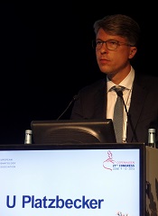
Uwe Platzbecker, MD, of University Hospital Carl Gustav Carus Dresden in Germany, presented these results at the 21st Congress of the European Hematology Association (abstract S128). The ARCADE trial was sponsored by Amgen.
Dr Platzbecker noted that, although ESAs are recommended in clinical guidelines to treat anemia in patients with lower-risk MDS, the drugs are not widely approved for this indication.
So, in the ARCADE trial, he and his colleagues assessed darbepoetin alfa in patients with low- or intermediate-1 risk MDS who had not previously taken ESAs or biologic response modifiers.
The patients had hemoglobin levels ≤10 g/dL, endogenous erythropoietin levels ≤500 mU/mL, and low transfusion burden (<4 RBC units in each of 2 consecutive 8-week periods prior to randomization).
During a 24-week period, 147 patients received either darbepoetin alfa at 500 μg (n=97) or placebo (n=49) every 3 weeks. The ESA dose was withheld if patients’ hemoglobin was >12.0 g/dL and decreased if hemoglobin increased by >1.5 g/dL in 3 weeks without transfusion.
At week 25, when the primary and key secondary endpoints were assessed, patients underwent an end-of-treatment period visit. They could then enter a 48-week active treatment period and cross over to receive darbepoetin alfa, with dose escalation allowed beginning on week 31. Treatment continued until week 72 or 73, and patients continue to be assessed every 26 weeks, for a minimum of 3 years.
Patient characteristics
Dr Platzbecker said baseline demographic and disease characteristics were generally similar between the treatment arms. All patients were Caucasian, and about 55% were male. The median age was 74 (range, 67-79). About half of patients in each treatment arm belonged to the low-risk IPSS category.
In both arms, most patients had refractory cytopenia with multilineage dysplasia (38.8% in the placebo arm and 46.4% in the darbepoetin alfa arm). Patients also had refractory anemia with excess blasts-1 (20.4% and 13.4%, respectively), refractory anemia (26.5% and 9.3%), refractory anemia with ring sideroblasts (8.2% and 17.5%), 5q deletion (4.1% and 11.3%), unclassifiable MDS (2.0% and 1.0%), and MDS of an unknown type (0% and 1.0%).
In the 16 weeks before randomization, 58.2% of all patients—53.1% in the placebo arm and 60.8% in the darbepoetin alfa arm—did not have any RBC transfusions. About 25% (24.7%)—22.4% in the placebo arm and 25.8% in the darbepoetin alfa arm—received 1 to 3 RBC units. And 17.1%—24.5% in the placebo arm and 13.4% in the darbepoetin alfa arm—received 4 or more RBC units.
Dosing
During the 24-week double-blind period of the study, 77% (37/48) of patients in the placebo arm and 79% (77/98) in the darbepoetin alfa arm received all 8 doses of treatment.
Sixteen percent (n=16) of patients in the darbepoetin alfa arm had a single dose reduction, and 2% (n=2) had 2 dose reductions. None of the patients in the placebo arm had a dose reduction.
Eleven percent of patients in the darbepoetin alfa arm had doses withheld due to increased hemoglobin. The dose was withheld once for 6 patients, twice for 4 patients, and 3 times for 1 patient. None of the placebo-treated patients had a dose withheld for this reason.
Ten percent (n=5) of placebo-treated patients and 2% (n=2) of darbepoetin alfa-treated patients had a dose withheld due to an AE. Two percent (n=1) and 3% (n=3) of patients, respectively, had a dose withheld for “other” reasons (noncompliance, investigator decision, and no investigational product on site).
During the 48-week open-label period of the study, 81% (102/126) of patients who received darbepoetin alfa increased their dose frequency from every 3 weeks to every 2 weeks. Dr Platzbecker said this suggests the optimal dose of the drug was not achieved during the 24-week double-blind period of the study.
Efficacy
During the 24-week double-blind period, there was a significant difference between the treatment arms with regard to RBC transfusions. The transfusion incidence was 59.2% (29/49) in the placebo arm and 36.1% (35/97) in the darbepoetin alfa arm (P=0.008).
During the 48-week open-label period, the incidence of RBC transfusion was 50.8% (64/126) among patients receiving darbepoetin alfa.
During the 24-week double-blind period, 11 patients (14.7%) in the darbepoetin alfa arm had an erythroid hematologic improvement (HI-E), but none of the patients in the placebo arm had such an improvement.
All 11 patients with HI-E had a baseline serum erythropoietin level less than 100 mU/mL, 1 of the patients had 2 RBC units transfused in the 16 weeks prior to randomization, but none had transfusions in the 8 weeks prior to randomization. Four of the patients had a dose withheld due to having hemoglobin levels greater than 12 g/dL.
During the 48-week open-label period, the HI-E rate was 34.7% (34/98) among patients receiving darbepoetin alfa.
Dr Platzbecker said the nature of the HI-E criteria likely underestimated the clinical benefit of darbepoetin alfa in this trial, and this was further complicated by the trial design. Specifically, hemoglobin was measured every 3 weeks, some patients may have had their doses reduced even if they were still anemic, and the optimal dose of darbepoetin alfa was likely not given during the double-blind period (as evidenced by the increase in doses during the open-label period).
For these reasons, Dr Platzbecker and his colleagues are exploring alternative response analyses to determine if there were additional patients who received a clinical benefit from darbepoetin alfa but did not meet HI-E criteria.
Safety
During the 24-week double-blind period, 4.2% (n=2) of patients in the placebo arm and 3.1% (n=3) in the darbepoetin alfa arm had AEs that led to treatment discontinuation. In the placebo arm, these events were pulmonary hypertension and renal failure. In the darbepoetin alfa arm, the events were pulmonary thrombosis, thrombocytopenia, and increased blast cell count.
The incidence of grade 3 or higher AEs was 27.1% (n=13) in the placebo arm and 15.3% (n=15) in the darbepoetin alfa arm. The incidence of grade 4 or higher AEs was 12.5% (n=6) and 5.1% (n=5), respectively. And the incidence of serious AEs was 16.7% (n=8) and 11.2% (n=11), respectively.
The incidence of fatal AEs was 4.2% (n=2) and 1% (n=1), respectively, but none of these were treatment-related. The deaths in the placebo arm were due to cardiac failure and cerebral hemorrhage, while the death in the darbepoetin alfa arm was due to hemorrhagic proctitis.
One patient in the darbepoetin alfa arm experienced a treatment-related serious AE.
AEs occurring at least 5% more frequently in the darbepoetin alfa arm than the placebo arm were fatigue (17.3% and 8.3%), pyrexia (9.2% and 2.1%), headache (7.1% and 2.1%), and myalgia (5.1% and 0%).
During the 48-week double-blind period, 7.9% (n=3) of patients formerly in the placebo arm and 3.4% (n=3) of patients formerly in the darbepoetin alfa arm had AEs that led to treatment discontinuation.
The incidence of grade 3 or higher AEs was 23.7% (n=9) and 31.0% (n=27), respectively. The incidence of grade 4 or higher AEs was 10.5% (n=4) and 10.3% (n=9), respectively. And the incidence of serious AEs was 18.4% (n=7) and 25.3% (n=22), respectively.
The incidence of fatal AEs was 2.6% (n=1) and 1.1% (n=1), respectively, but none of these were treatment-related. Two patients experienced a treatment-related serious AE—1 from each of the former treatment arms. ![]()
COPENHAGEN—The erythropoiesis-stimulating agent (ESA) darbepoetin alfa can provide a clinical benefit in patients with lower-risk myelodysplastic syndromes (MDS), a phase 3 trial suggests.
In the ARCADE trial, darbepoetin alfa significantly reduced the incidence of red blood cell (RBC) transfusions in patients with low- and intermediate-1 risk myelodysplastic syndrome (MDS), when compared to placebo.
The ESA also significantly improved erythroid response.
In addition, researchers said adverse events (AEs) were generally balanced between the darbepoetin alfa and placebo arms.
Uwe Platzbecker, MD, of University Hospital Carl Gustav Carus Dresden in Germany, presented these results at the 21st Congress of the European Hematology Association (abstract S128). The ARCADE trial was sponsored by Amgen.
Dr Platzbecker noted that, although ESAs are recommended in clinical guidelines to treat anemia in patients with lower-risk MDS, the drugs are not widely approved for this indication.
So, in the ARCADE trial, he and his colleagues assessed darbepoetin alfa in patients with low- or intermediate-1 risk MDS who had not previously taken ESAs or biologic response modifiers.
The patients had hemoglobin levels ≤10 g/dL, endogenous erythropoietin levels ≤500 mU/mL, and low transfusion burden (<4 RBC units in each of 2 consecutive 8-week periods prior to randomization).
During a 24-week period, 147 patients received either darbepoetin alfa at 500 μg (n=97) or placebo (n=49) every 3 weeks. The ESA dose was withheld if patients’ hemoglobin was >12.0 g/dL and decreased if hemoglobin increased by >1.5 g/dL in 3 weeks without transfusion.
At week 25, when the primary and key secondary endpoints were assessed, patients underwent an end-of-treatment period visit. They could then enter a 48-week active treatment period and cross over to receive darbepoetin alfa, with dose escalation allowed beginning on week 31. Treatment continued until week 72 or 73, and patients continue to be assessed every 26 weeks, for a minimum of 3 years.
Patient characteristics
Dr Platzbecker said baseline demographic and disease characteristics were generally similar between the treatment arms. All patients were Caucasian, and about 55% were male. The median age was 74 (range, 67-79). About half of patients in each treatment arm belonged to the low-risk IPSS category.
In both arms, most patients had refractory cytopenia with multilineage dysplasia (38.8% in the placebo arm and 46.4% in the darbepoetin alfa arm). Patients also had refractory anemia with excess blasts-1 (20.4% and 13.4%, respectively), refractory anemia (26.5% and 9.3%), refractory anemia with ring sideroblasts (8.2% and 17.5%), 5q deletion (4.1% and 11.3%), unclassifiable MDS (2.0% and 1.0%), and MDS of an unknown type (0% and 1.0%).
In the 16 weeks before randomization, 58.2% of all patients—53.1% in the placebo arm and 60.8% in the darbepoetin alfa arm—did not have any RBC transfusions. About 25% (24.7%)—22.4% in the placebo arm and 25.8% in the darbepoetin alfa arm—received 1 to 3 RBC units. And 17.1%—24.5% in the placebo arm and 13.4% in the darbepoetin alfa arm—received 4 or more RBC units.
Dosing
During the 24-week double-blind period of the study, 77% (37/48) of patients in the placebo arm and 79% (77/98) in the darbepoetin alfa arm received all 8 doses of treatment.
Sixteen percent (n=16) of patients in the darbepoetin alfa arm had a single dose reduction, and 2% (n=2) had 2 dose reductions. None of the patients in the placebo arm had a dose reduction.
Eleven percent of patients in the darbepoetin alfa arm had doses withheld due to increased hemoglobin. The dose was withheld once for 6 patients, twice for 4 patients, and 3 times for 1 patient. None of the placebo-treated patients had a dose withheld for this reason.
Ten percent (n=5) of placebo-treated patients and 2% (n=2) of darbepoetin alfa-treated patients had a dose withheld due to an AE. Two percent (n=1) and 3% (n=3) of patients, respectively, had a dose withheld for “other” reasons (noncompliance, investigator decision, and no investigational product on site).
During the 48-week open-label period of the study, 81% (102/126) of patients who received darbepoetin alfa increased their dose frequency from every 3 weeks to every 2 weeks. Dr Platzbecker said this suggests the optimal dose of the drug was not achieved during the 24-week double-blind period of the study.
Efficacy
During the 24-week double-blind period, there was a significant difference between the treatment arms with regard to RBC transfusions. The transfusion incidence was 59.2% (29/49) in the placebo arm and 36.1% (35/97) in the darbepoetin alfa arm (P=0.008).
During the 48-week open-label period, the incidence of RBC transfusion was 50.8% (64/126) among patients receiving darbepoetin alfa.
During the 24-week double-blind period, 11 patients (14.7%) in the darbepoetin alfa arm had an erythroid hematologic improvement (HI-E), but none of the patients in the placebo arm had such an improvement.
All 11 patients with HI-E had a baseline serum erythropoietin level less than 100 mU/mL, 1 of the patients had 2 RBC units transfused in the 16 weeks prior to randomization, but none had transfusions in the 8 weeks prior to randomization. Four of the patients had a dose withheld due to having hemoglobin levels greater than 12 g/dL.
During the 48-week open-label period, the HI-E rate was 34.7% (34/98) among patients receiving darbepoetin alfa.
Dr Platzbecker said the nature of the HI-E criteria likely underestimated the clinical benefit of darbepoetin alfa in this trial, and this was further complicated by the trial design. Specifically, hemoglobin was measured every 3 weeks, some patients may have had their doses reduced even if they were still anemic, and the optimal dose of darbepoetin alfa was likely not given during the double-blind period (as evidenced by the increase in doses during the open-label period).
For these reasons, Dr Platzbecker and his colleagues are exploring alternative response analyses to determine if there were additional patients who received a clinical benefit from darbepoetin alfa but did not meet HI-E criteria.
Safety
During the 24-week double-blind period, 4.2% (n=2) of patients in the placebo arm and 3.1% (n=3) in the darbepoetin alfa arm had AEs that led to treatment discontinuation. In the placebo arm, these events were pulmonary hypertension and renal failure. In the darbepoetin alfa arm, the events were pulmonary thrombosis, thrombocytopenia, and increased blast cell count.
The incidence of grade 3 or higher AEs was 27.1% (n=13) in the placebo arm and 15.3% (n=15) in the darbepoetin alfa arm. The incidence of grade 4 or higher AEs was 12.5% (n=6) and 5.1% (n=5), respectively. And the incidence of serious AEs was 16.7% (n=8) and 11.2% (n=11), respectively.
The incidence of fatal AEs was 4.2% (n=2) and 1% (n=1), respectively, but none of these were treatment-related. The deaths in the placebo arm were due to cardiac failure and cerebral hemorrhage, while the death in the darbepoetin alfa arm was due to hemorrhagic proctitis.
One patient in the darbepoetin alfa arm experienced a treatment-related serious AE.
AEs occurring at least 5% more frequently in the darbepoetin alfa arm than the placebo arm were fatigue (17.3% and 8.3%), pyrexia (9.2% and 2.1%), headache (7.1% and 2.1%), and myalgia (5.1% and 0%).
During the 48-week double-blind period, 7.9% (n=3) of patients formerly in the placebo arm and 3.4% (n=3) of patients formerly in the darbepoetin alfa arm had AEs that led to treatment discontinuation.
The incidence of grade 3 or higher AEs was 23.7% (n=9) and 31.0% (n=27), respectively. The incidence of grade 4 or higher AEs was 10.5% (n=4) and 10.3% (n=9), respectively. And the incidence of serious AEs was 18.4% (n=7) and 25.3% (n=22), respectively.
The incidence of fatal AEs was 2.6% (n=1) and 1.1% (n=1), respectively, but none of these were treatment-related. Two patients experienced a treatment-related serious AE—1 from each of the former treatment arms. ![]()
Acute myeloid leukemia genomic classification and prognosis
Acute myeloid leukemia (AML) consists of at least 11 disease classes that represent distinct paths in the evolution of AML and have prognostic implications, based on an analysis of somatic driver mutations in 1,540 patients.
In total, 5,234 driver mutations were identified in 76 genes or regions, with 96% of patients having at least one mutation and 86% having two or more mutations. However, nearly one-half of the cohort did not fall into one of the molecular groups defined by the World Health Organization in 2008.

“The characterization of many new leukemia genes, multiple driver mutations per patient, and complex co-mutation patterns prompted us to reevaluate genomic classification of AML from the beginning,” wrote Elli Papaemmanuil, Ph.D., a molecular geneticist at Memorial Sloan Kettering, New York, and of the Cancer Genome Project, Wellcome Trust Sanger Institute, and her colleagues (N Engl J Med. 2016 Jun 9; 374:2209-21).
The team developed a Bayesian statistical model to define 11 mutually exclusive subtypes based on patterns of co-mutations. The schema unambiguously classified 1,236 of 1,540 patients (80%) into a single subgroup and 56 (4%) into two or more groups. A subset of patients (166, 11%) remained unclassified, possibly due to mutations in genes not sequenced in the study.
NPM1-mutated AML was the largest class (27% of the cohort), followed by the chromatin-spliceosome group (18% of the cohort) that included mutations in genes regulating RNA splicing (SRSF2, SF3B1, U2AF1, and ZRSR2), chromatin (ASXL1, STAG2, BCOR, MLLPTD, EZH2, and PHF6), or transcription (RUNX1). Another subgroup consisted of mutations in TP53, as well as complex karyotype alterations, cytogenetically visible copy-number alterations (aneuploidies), or a combination. While broader than previous classifications, such as “monosomal karyotype AML” and “complex karyotype AML,” this group emerged from correlated chromosomal abnormalities and was mutually exclusive of other class-defining lesions. In general, patients in this group were older and had fewer RAS pathway mutations.
The groups had considerable differences in clinical presentation and overall survival, according to the report. The TP53-aneuploidy subgroup had poor outcomes, as previously described. Patients in the chromatin-spliceosome group had lower rates of response to induction chemotherapy, higher relapse rates, and poorer long-term outcomes, compared with other groups. Most of these patients (84%) would be classified as intermediate risk under current guidelines, but the characteristics were more similar to those of subgroups with adverse outcomes.
Overall survival was correlated with the number of driver mutations, and deleterious affects of mutations often were found to be additive. In some cases, complex gene interactions accounted for variation in outcomes, suggesting the clinical effect of some driver mutations may depend on the occurrence of co-mutations in a wider genomic context.
The study by Papaemmanuil and her colleagues offers practice-changing insights that redefine molecular classification of AML. The mutational analysis of more than 1,500 AML patients provides a deeper understanding of the specific paths from normal blood cell to leukemia.
Specific concurrent mutations were linked to clinical outcomes. For example, co-mutations in NPM1, FLT3ITD, and DNMT3A are associated with a poor clinical outcome, but NPM1 and DNMT3A mutations without FLT3ITD are associated with better outcomes. In addition, mutations in NPM1 and DNMT3A in the presence of NRASG12/13 are associated with a more favorable outcome. The evolution of DNMT3A-NPM1 mutated clones along separate paths appears to affect disease outcome and may be relevant to clinical trials in AML subgroups.
|
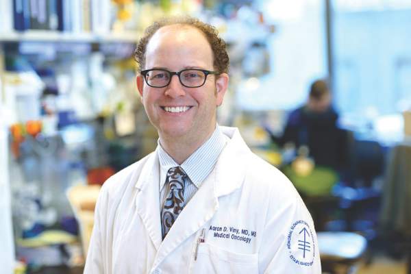
Dr. Aaron Viny |
Previous, smaller studies had suggested that somatic mutations in splicing factors and chromatin modifiers were specific for secondary AML that arises from myelodysplastic syndromes (MDS). Papaemmanuil and her colleagues provide extensive data to support that hypothesis. Patients with chromatin-spliceosome mutations, previously classified as intermediate-risk AML, are classed into the same molecular subgroup as patients with secondary AML arising from MDS.
These data may inform the design of mechanism-based clinical trials based on the presence of specific mutations and co-mutations.
Dr. Aaron Viny is a medical oncologist at Memorial Sloan Kettering Cancer Center, New York. Dr. Ross Levine is Director of the Memorial Sloan Kettering Center for Hematologic Malignancies. These remarks were part of an editorial accompanying a report in The New England Journal of Medicine (2016 Jun 9; 374:2282-4). Dr. Levine reports personal fees from Foundation Medicine outside the submitted work.
The study by Papaemmanuil and her colleagues offers practice-changing insights that redefine molecular classification of AML. The mutational analysis of more than 1,500 AML patients provides a deeper understanding of the specific paths from normal blood cell to leukemia.
Specific concurrent mutations were linked to clinical outcomes. For example, co-mutations in NPM1, FLT3ITD, and DNMT3A are associated with a poor clinical outcome, but NPM1 and DNMT3A mutations without FLT3ITD are associated with better outcomes. In addition, mutations in NPM1 and DNMT3A in the presence of NRASG12/13 are associated with a more favorable outcome. The evolution of DNMT3A-NPM1 mutated clones along separate paths appears to affect disease outcome and may be relevant to clinical trials in AML subgroups.
|
|
Dr. Aaron Viny |
Previous, smaller studies had suggested that somatic mutations in splicing factors and chromatin modifiers were specific for secondary AML that arises from myelodysplastic syndromes (MDS). Papaemmanuil and her colleagues provide extensive data to support that hypothesis. Patients with chromatin-spliceosome mutations, previously classified as intermediate-risk AML, are classed into the same molecular subgroup as patients with secondary AML arising from MDS.
These data may inform the design of mechanism-based clinical trials based on the presence of specific mutations and co-mutations.
Dr. Aaron Viny is a medical oncologist at Memorial Sloan Kettering Cancer Center, New York. Dr. Ross Levine is Director of the Memorial Sloan Kettering Center for Hematologic Malignancies. These remarks were part of an editorial accompanying a report in The New England Journal of Medicine (2016 Jun 9; 374:2282-4). Dr. Levine reports personal fees from Foundation Medicine outside the submitted work.
Acute myeloid leukemia (AML) consists of at least 11 disease classes that represent distinct paths in the evolution of AML and have prognostic implications, based on an analysis of somatic driver mutations in 1,540 patients.
In total, 5,234 driver mutations were identified in 76 genes or regions, with 96% of patients having at least one mutation and 86% having two or more mutations. However, nearly one-half of the cohort did not fall into one of the molecular groups defined by the World Health Organization in 2008.
“The characterization of many new leukemia genes, multiple driver mutations per patient, and complex co-mutation patterns prompted us to reevaluate genomic classification of AML from the beginning,” wrote Elli Papaemmanuil, Ph.D., a molecular geneticist at Memorial Sloan Kettering, New York, and of the Cancer Genome Project, Wellcome Trust Sanger Institute, and her colleagues (N Engl J Med. 2016 Jun 9; 374:2209-21).
The team developed a Bayesian statistical model to define 11 mutually exclusive subtypes based on patterns of co-mutations. The schema unambiguously classified 1,236 of 1,540 patients (80%) into a single subgroup and 56 (4%) into two or more groups. A subset of patients (166, 11%) remained unclassified, possibly due to mutations in genes not sequenced in the study.
NPM1-mutated AML was the largest class (27% of the cohort), followed by the chromatin-spliceosome group (18% of the cohort) that included mutations in genes regulating RNA splicing (SRSF2, SF3B1, U2AF1, and ZRSR2), chromatin (ASXL1, STAG2, BCOR, MLLPTD, EZH2, and PHF6), or transcription (RUNX1). Another subgroup consisted of mutations in TP53, as well as complex karyotype alterations, cytogenetically visible copy-number alterations (aneuploidies), or a combination. While broader than previous classifications, such as “monosomal karyotype AML” and “complex karyotype AML,” this group emerged from correlated chromosomal abnormalities and was mutually exclusive of other class-defining lesions. In general, patients in this group were older and had fewer RAS pathway mutations.
The groups had considerable differences in clinical presentation and overall survival, according to the report. The TP53-aneuploidy subgroup had poor outcomes, as previously described. Patients in the chromatin-spliceosome group had lower rates of response to induction chemotherapy, higher relapse rates, and poorer long-term outcomes, compared with other groups. Most of these patients (84%) would be classified as intermediate risk under current guidelines, but the characteristics were more similar to those of subgroups with adverse outcomes.
Overall survival was correlated with the number of driver mutations, and deleterious affects of mutations often were found to be additive. In some cases, complex gene interactions accounted for variation in outcomes, suggesting the clinical effect of some driver mutations may depend on the occurrence of co-mutations in a wider genomic context.
FROM NEJM
Key clinical point: Mutational analysis of 1,540 patients with acute myeloid leukemia (AML) identified 11 distinct classes with prognostic implications.
Major finding: In total, 5,234 driver mutations were identified involving 76 genes or regions; 96% of patients had at least one driver mutation, and 86% had two or more.
Data sources: Samples came from three prospective multicenter clinical trials of the German-Austrian AML Study Group: AMLHD98A, AML-HD98B, and AMLSG-07-04.
Disclosures: Dr. Papaemmanuil and most coauthors reported having no disclosures. Two coauthors reported financial ties to industry sources.

Hold that TKI – When it’s safe to stop in CML
COPENHAGEN – A year after stopping tyrosine kinase inhibitor therapy, more than half of patients with chronic myeloid leukemia (CML) in a large clinical trial remained in deep molecular remission.
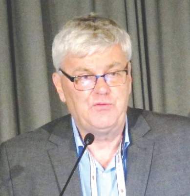
Among 750 patients with CML in remission for at least 1 year before study entry, 62% retained a treatment response 6 months after stopping a tyrosine kinase inhibitor (TKI) such as imatinib (Gleevec) and 56% retained responses 1 year after being off their drugs, reported Dr. Johan Richter of Lund (Sweden) University at the annual congress of the European Hematology Association.
“About 6 years of therapy [with imatinib] would be optimal for therapy prior to a stop attempt,” he said at a briefing prior to the presentation of data at the congress.
Although in clinical practice patients with CML may remain on a TKI indefinitely, results from small clinical trials have suggested that in 40%-60% of patients with deep molecular responses (MR4.0 or better), TKIs can be safely stopped, Dr. Richter noted.
To get a better handle on when it might be safe to stop a TKI and under what conditions, EURO-SKI investigators enrolled 868 adults with CML in chronic phase from 11 countries, 750 of whom had complete data for the analysis.
In all, 94% of patients had received imatinib in the first line, 2% received dasatinib (Sprycel), and 4% had received nilotinib (Tasigna). Of this group, 115 had switched to a second-line agent due to intolerance of the first-line drug.
The median time from diagnosis was 7.7 years. The median duration of therapy was 7.6 years, and the median duration of MR4 before stopping was 4.7 years.
As noted, among 750 patients assessable for molecular relapse–free survival, 62% remained in remission at 6 months after stopping the TKI, as did 56% at 12 months, 52% at 24 months, and 49% at 36 months.
For patients who resumed therapy, the median time to restart was 4.1 months.
To see whether they could identify any factors prognostic for relapse after stopping a TKI, the investigators used data on 448 patients in the study who were treated with imatinib.
In univariate analysis there was no significant association between molecular relapse–free survival at 6 months and either age, gender, depth of molecular response, or any standard risk scores.
The only significant predictors of molecular remission status at 6 months were duration of imatinib therapy and duration of molecular response before stopping.
The odds ratio for treatment duration was 1.16, indicating that each additional year of imatinib treatment is associated with a 16% increase in the likelihood that a patient would remain in deep molecular remission 6 months after stopping.
The investigators used the minimal P value approach to determine the cutoff of approximately 6 years, based on a molecular relapse–free survival at 6 months of 65.5% for patients who remained on imatinib for more than 5.8 years, compared with 42.6% for those who were on it for 5.8 years or less.
Although the study is ongoing, to date more than 80% of patients who had a loss of deep molecular remission after stopping their TKI regained the remission after resuming therapy, Dr. Richter said.
Dr. Richter said in an interview that longer follow-up will be needed to confirm their findings, and that patients who were sensitive to TKIs prior to stopping therapy remained sensitive when restarting, suggesting that treatment interruption does not increase the likelihood of drug resistance.
Coprincipal investigator Dr. Francois-Xavier Mahon of Bordeaux University in France, noted that in the STIM (Stop Imatinib)–1 and –2 trials, the estimated annual savings to the French health care system were 20 million euros ($22.6 million).
COPENHAGEN – A year after stopping tyrosine kinase inhibitor therapy, more than half of patients with chronic myeloid leukemia (CML) in a large clinical trial remained in deep molecular remission.
Among 750 patients with CML in remission for at least 1 year before study entry, 62% retained a treatment response 6 months after stopping a tyrosine kinase inhibitor (TKI) such as imatinib (Gleevec) and 56% retained responses 1 year after being off their drugs, reported Dr. Johan Richter of Lund (Sweden) University at the annual congress of the European Hematology Association.
“About 6 years of therapy [with imatinib] would be optimal for therapy prior to a stop attempt,” he said at a briefing prior to the presentation of data at the congress.
Although in clinical practice patients with CML may remain on a TKI indefinitely, results from small clinical trials have suggested that in 40%-60% of patients with deep molecular responses (MR4.0 or better), TKIs can be safely stopped, Dr. Richter noted.
To get a better handle on when it might be safe to stop a TKI and under what conditions, EURO-SKI investigators enrolled 868 adults with CML in chronic phase from 11 countries, 750 of whom had complete data for the analysis.
In all, 94% of patients had received imatinib in the first line, 2% received dasatinib (Sprycel), and 4% had received nilotinib (Tasigna). Of this group, 115 had switched to a second-line agent due to intolerance of the first-line drug.
The median time from diagnosis was 7.7 years. The median duration of therapy was 7.6 years, and the median duration of MR4 before stopping was 4.7 years.
As noted, among 750 patients assessable for molecular relapse–free survival, 62% remained in remission at 6 months after stopping the TKI, as did 56% at 12 months, 52% at 24 months, and 49% at 36 months.
For patients who resumed therapy, the median time to restart was 4.1 months.
To see whether they could identify any factors prognostic for relapse after stopping a TKI, the investigators used data on 448 patients in the study who were treated with imatinib.
In univariate analysis there was no significant association between molecular relapse–free survival at 6 months and either age, gender, depth of molecular response, or any standard risk scores.
The only significant predictors of molecular remission status at 6 months were duration of imatinib therapy and duration of molecular response before stopping.
The odds ratio for treatment duration was 1.16, indicating that each additional year of imatinib treatment is associated with a 16% increase in the likelihood that a patient would remain in deep molecular remission 6 months after stopping.
The investigators used the minimal P value approach to determine the cutoff of approximately 6 years, based on a molecular relapse–free survival at 6 months of 65.5% for patients who remained on imatinib for more than 5.8 years, compared with 42.6% for those who were on it for 5.8 years or less.
Although the study is ongoing, to date more than 80% of patients who had a loss of deep molecular remission after stopping their TKI regained the remission after resuming therapy, Dr. Richter said.
Dr. Richter said in an interview that longer follow-up will be needed to confirm their findings, and that patients who were sensitive to TKIs prior to stopping therapy remained sensitive when restarting, suggesting that treatment interruption does not increase the likelihood of drug resistance.
Coprincipal investigator Dr. Francois-Xavier Mahon of Bordeaux University in France, noted that in the STIM (Stop Imatinib)–1 and –2 trials, the estimated annual savings to the French health care system were 20 million euros ($22.6 million).
AT THE EHA CONGRESS
Key clinical point:.Tyrosine kinase inhibitor therapy can be safely stopped and resumed in many patients with chronic-phase chronic myeloid leukemia (CML).
Major finding: After stopping a TKI, 62% of patients retained a treatment response at 6 months, and 56% retained a response at 1 year.
Data source: Study of therapeutic interruption in 750 adults in deep molecular remission for at least 1 year on TKI therapy.
Disclosures: The study was sponsored by the European LeukemiaNet. Dr. Richter has previously disclosed consultancy and equity ownership with Cantargia. Dr. Mahon has previously disclosed being on the scientific advisory board and receiving honoraria from Novartis Oncology and BMS, and serving as consultant to those companies and to Pfizer.

Blinatumomab doubles survival in relapsed Ph-negative ALL
Copenhagen – The monoclonal antibody blinatumomab nearly doubled overall survival compared with standard chemotherapy among patients with relapsed or refractory B-cell precursor leukemia negative for the Philadelphia chromosome, investigators reported.
Among 405 patients enrolled in the TOWER study, a multicenter, open-label phase III trial, median overall survival for patients treated with blinatumomab (Blincyto) was 7.7 months, compared with 4.0 months for patients treated with one of four standard chemotherapy regimens (P = .012) reported Dr. Max S. Topp of Universitätsklinikum in Würzburg, Germany.

“Blinatumomab is the first immunotherapy agent to demonstrate an overall survival benefit when compared to chemotherapy in patients relapsing with adult acute lymphoblastic leukemia. It increases almost twofold the overall survival when compared to standard care. This was consistent in all subgroups that we were looking at, regardless of age, prior salvage therapy, or patients relapsing after an allo-transplantation,” he said at a briefing prior to his presentation of the data at the annual congress of the European Hematology Association.
The trial was halted early, after a preplanned interim analysis showed a clear survival benefit with blinatumomab.
Blinatumomab is a bispecific T-cell engager (BiTE) antibody designed to direct cytotoxic T cells to cancer cells expressing the CD19 receptor. As previously reported, it has induced high complete remission rates in patients with relapsed or refractory acute lymphoblastic leukemia (ALL).
In May 2015, the Food and Drug Administration granted blinatumomab accelerated approval for the treatment of adult patients with relapsed/refractory Philadelphia chromosome–negative B-cell precursor acute lymphoblastic leukemia (BCP-ALL).
In the TOWER trial, patients with relapsed/refractory BCP-ALL were randomly assigned on a 2:1 basis to receive either blinatumomab (271 patients) or standard chemotherapy (134), consisting of the investigator’s choice of one of four defined regimens (based on either anthracyclines, histone deacetylase inhibitors, high-dose methotrexate, or clofarabine).
The patients were further stratified by age, prior salvage therapy, and prior allogeneic stem cell transplant (alloSCT).
Patients assigned to receive blinatumomab received it in 6-week cycles consisting of continuous infusions of 9 mcg/day in week 1 of cycle 1, then 28 mcg/day for weeks 3-4, followed by 2 weeks off. Patients were pretreated with dexamethasone for prophylaxis against the cytokine release syndrome.
Patients whose disease was in remission following two induction cycles could be continued on therapy until relapse.
As noted, the trial was halted early, after 248 patients had died; the primary analysis had been planned to occur after 330 patients had died.
In addition to the superior survival rates, blinatumomab was associated with a higher rate of complete responses (39% vs. 19%, P less than .001) and combined complete responses, complete hematologic responses, and complete responses with incomplete recovery of counts (46% vs. 28%, P = .001).
In all, 19% of patients assigned to blinatumomab and 17% assigned to chemotherapy died on study. Grade 3 or greater adverse events included neutropenia (38% of patients and 58%, respectively), infections (34% and 52%), neurologic events (9% and 8%), and the cytokine release syndrome (5% vs. 0%).
Dr. Anton Hagenbeek, professor of hematology at the University of Amsterdam, who moderated the briefing, asked Dr. Topp whether there were plans to use blinatumomab earlier in the course of disease.
Dr. Topp agreed that it might be a valuable addition to upfront therapy, noting that blinatumomab has been shown in a small percentage of patients who are positive for minimal residual disease to convert to being negative for minimal residual disease, and that this conversion was associated with improved overall survival.
Copenhagen – The monoclonal antibody blinatumomab nearly doubled overall survival compared with standard chemotherapy among patients with relapsed or refractory B-cell precursor leukemia negative for the Philadelphia chromosome, investigators reported.
Among 405 patients enrolled in the TOWER study, a multicenter, open-label phase III trial, median overall survival for patients treated with blinatumomab (Blincyto) was 7.7 months, compared with 4.0 months for patients treated with one of four standard chemotherapy regimens (P = .012) reported Dr. Max S. Topp of Universitätsklinikum in Würzburg, Germany.
“Blinatumomab is the first immunotherapy agent to demonstrate an overall survival benefit when compared to chemotherapy in patients relapsing with adult acute lymphoblastic leukemia. It increases almost twofold the overall survival when compared to standard care. This was consistent in all subgroups that we were looking at, regardless of age, prior salvage therapy, or patients relapsing after an allo-transplantation,” he said at a briefing prior to his presentation of the data at the annual congress of the European Hematology Association.
The trial was halted early, after a preplanned interim analysis showed a clear survival benefit with blinatumomab.
Blinatumomab is a bispecific T-cell engager (BiTE) antibody designed to direct cytotoxic T cells to cancer cells expressing the CD19 receptor. As previously reported, it has induced high complete remission rates in patients with relapsed or refractory acute lymphoblastic leukemia (ALL).
In May 2015, the Food and Drug Administration granted blinatumomab accelerated approval for the treatment of adult patients with relapsed/refractory Philadelphia chromosome–negative B-cell precursor acute lymphoblastic leukemia (BCP-ALL).
In the TOWER trial, patients with relapsed/refractory BCP-ALL were randomly assigned on a 2:1 basis to receive either blinatumomab (271 patients) or standard chemotherapy (134), consisting of the investigator’s choice of one of four defined regimens (based on either anthracyclines, histone deacetylase inhibitors, high-dose methotrexate, or clofarabine).
The patients were further stratified by age, prior salvage therapy, and prior allogeneic stem cell transplant (alloSCT).
Patients assigned to receive blinatumomab received it in 6-week cycles consisting of continuous infusions of 9 mcg/day in week 1 of cycle 1, then 28 mcg/day for weeks 3-4, followed by 2 weeks off. Patients were pretreated with dexamethasone for prophylaxis against the cytokine release syndrome.
Patients whose disease was in remission following two induction cycles could be continued on therapy until relapse.
As noted, the trial was halted early, after 248 patients had died; the primary analysis had been planned to occur after 330 patients had died.
In addition to the superior survival rates, blinatumomab was associated with a higher rate of complete responses (39% vs. 19%, P less than .001) and combined complete responses, complete hematologic responses, and complete responses with incomplete recovery of counts (46% vs. 28%, P = .001).
In all, 19% of patients assigned to blinatumomab and 17% assigned to chemotherapy died on study. Grade 3 or greater adverse events included neutropenia (38% of patients and 58%, respectively), infections (34% and 52%), neurologic events (9% and 8%), and the cytokine release syndrome (5% vs. 0%).
Dr. Anton Hagenbeek, professor of hematology at the University of Amsterdam, who moderated the briefing, asked Dr. Topp whether there were plans to use blinatumomab earlier in the course of disease.
Dr. Topp agreed that it might be a valuable addition to upfront therapy, noting that blinatumomab has been shown in a small percentage of patients who are positive for minimal residual disease to convert to being negative for minimal residual disease, and that this conversion was associated with improved overall survival.
At THE EHA CONGRESS
Key clinical point: Single-agent blinatumomab nearly doubled overall survival compared to chemotherapy in relapsed/refractory ALL.
Major finding: Median overall survival was 7.7 months for patients on blinatumomab compared with 4.0 months for those on chemotherapy.
Data source: Randomized open-label phase III trial in 405 adults with relapsed/refractory Philadelphia chromosome–negative B-cell precursor ALL.
Disclosures: Amgen funded the study. Dr. Topp disclosed having a consultant or advisory role and receiving other remuneration from Micromet, which was acquired by Amgen. Dr. Hagenbeek reported no relevant disclosures.

Researchers identify 11 subgroups of AML

Photo courtesy of NIGMS
Using patient samples from 3 prospective multicenter clinical trials of the German-Austrian AML Study Group, researchers have identified 5234 driver mutations involving 76 genes or regions in 1540 patients with acute myeloid leukemia (AML).
They say the sample size afforded a more comprehensive analysis than previously conducted, and as a result, they found patients were divided into at least 11 subgroups of AML.
They also identified 3 genomic categories beyond the existing World Health Organization (WHO) subgroups. These genomic categories are chromatin–spliceosome mutations, TP53–aneuploidy, and provisionally, IDH2R172 mutations.
Their study, led by scientists at the Wellcome Trust Sanger Institute and international collaborators, could have an impact on clinical trial design and improve the way patients are diagnosed and treated in the future.
The scientists stated in their published paper that 736 patients (48%) in their study would not have fit into the molecular groups included in the 2008 WHO classification of adult AML. This prompted them to reevaluate genomic classification of AML from the beginning.
“We have shown that AML is an umbrella term for a group of at least 11 different types of leukemia,” said Peter Campbell, MBChB, PhD, co-leader of the study. “We can now start to decode these genetics to shape clinical trials and develop diagnostics.”
The scientists found NPM1-mutated AML to be the largest class in their cohort, accounting for 27% of the patients.
The second largest subgroup--the chromatin-spliceosome group—accounted for 18% of patients, the TP53-aneuploidy group accounted for 13%, and the IDH2R172 mutations for 1%.
The study also showed that most patients had a unique combination of genetic changes driving their leukemia. This genetic complexity helps explain why AML shows such variability in survival rates among patients.
Under their new schema, the scientists were able to unambiguously classify 80% of the 1540 patients with driver mutations in a single subgroup. Fifty-six of the patients (4%) met criteria for 2 or more categories, primarily in the TP53-aneuploidy and chromatin-spliceosome groups.
They were not able to classify 11% of patients with driver mutations. They explained that these patients might have had mutations in drivers that were either not sequenced or had mutations that were missed.
The scientists applied their classification schema to an independent cohort from the Cancer Genome Atlas (TCGA). The new schema was able to replicate the absence of overlap among subgroups, and relative frequencies of the mutations were equivalent to those in the AML cohort.
“For the first time we untangled the genetic complexity seen in most AML cancer genomes into distinct evolutionary paths that lead to AML,” joint first author Elli Papaemmanuil, PhD, of Memorial Sloan Kettering Cancer Center in New York, said.
“By understanding these paths, we can help develop more appropriate treatments for individual patients with AML. We are now extending such studies across other leukemias."
The investigators recommend prospective clinical trials to further validate the schema.
The work was supported by the Wellcome Trust, Bundesministerium fur Bildung und Forschung, Deutsche Krebshilfe and Deutsche Forschungsgemeinschaft, the European Hematology Association, Amgen and the Kay Kendall Leukaemia Fund. ![]()
Tyrosine kinase inhibitors may boost cardiac risk in chronic myeloid leukemia
Patients with chronic myeloid leukemia (CML) who received tyrosine kinase inhibitors (TKIs) had 1.7 times the rate of arterial or venous vascular events of population-based controls in a large retrospective cohort study.
In addition, second-generation TKIs were associated with higher rates of myocardial infarction than was first-generation imatinib, Dr. Torsten Dahlén of Karolinska University Hospital Solna, Stockholm, and his associates reported. Although absolute numbers of cardiovascular events were low, physicians “should be aware of these risk factors when initiating TKI therapy in patients with CML,” the authors wrote in a study published online June 13 in the Annals of Internal Medicine .
Tyrosine kinase inhibitors have “revolutionized” the prognosis of CML and are generally well tolerated, the researchers noted. But case reports and follow-up studies of clinical trial participants have raised concerns about cardiovascular toxicities with second-generation TKIs, such as nilotinib, they added.
To further study the issue, the investigators compared 896 patients in Sweden who were diagnosed with CML between 2002 and 2012 with 4,438 age- and sex-matched controls from the national population register. By crosschecking both groups against a national patient database, the investigators calculated rates of venous thrombosis, pulmonary embolism, myocardial infarction, cerebrovascular ischemia, and other arterial thromboses (Ann. Intern. Med. 2016 Jun 13. doi: 10.7326/M15-2306).
A total of 846 CML patients (94%) received a TKI during a median of 4.2 years of follow-up, the investigators reported. First-line therapy usually consisted of imatinib (89%), followed by nilotinib (9%) and dasatinib (1%).
The TKI cohort had 78 arterial and venous events during 3,969 person-years of follow-up, compared with 250 events during 21,917 person-years of follow-up for controls, for a statistically significant incidence rate ratio (IRR) of 1.7 (95% confidence interval, 1.3-2.2). Individual IRRs for arterial and venous events also reached statistical significance at 1.5 (95% CI, 1.1-2.1) and 2.0 (95% CI, 1.2-3.3), respectively. Deep venous thrombosis and myocardial infarction accounted for most of the excess risk, with IRRs of 2.2 (95% CI, 1.1-4.4) and 1.9 (95% CI, 1.3-2.7), respectively.
When investigators looked only at the TKI cohort, they found that the rates of arterial thromboembolic events were highest for nilotinib (29 events per 1,000 person-years), followed by dasatinib (19 events per 1,000 person-years) and imatinib (13 events per 1,000 person-years). Nilotinib also was associated with a substantially higher rate of all arterial and venous events (42/1,000 person-years) than dasatinib (20/1,000 person-years) and imatinib (16/1,000 person-years).
Furthermore, nilotinib and dasatinib were associated with higher rates of myocardial infarctions (29 and 19 per 1,000 person-years, respectively) and cerebrovascular ischemic events (11 and 4 events per 1,000 person-years, respectively) than was imatinib (8 events per 1,000 person-years and 4 events per 1,000 person-years, respectively). However, the absolute numbers of events were too small to allow for statistical comparisons, the researchers said.
“The observed increase in thrombotic events may be related to CML itself, the treatment administered, or both,” they noted, but “the prevalence of myocardial infarction in patients with CML before diagnosis was similar to that of the control population, [which] might indicate a treatment-related association.”
Among the 31 patients on TKIs who had a myocardial infarction, 26 (84%) had been previously diagnosed with at least one risk factor for cardiovascular disease, including diabetes (19%), atrial fibrillation (26%), angina pectoris (39%), hypertension (55%), and hyperlipidemia (23%).
Most patients who received nilotinib or dasatinib had previously received imatinib, meaning that they could have had more advanced disease that increased their risk of adverse events, according to the researchers.
“The small number of events also leads us to exercise caution in drawing any strong conclusions,” they added. “Future data from the Swedish CML register will provide more robust evidence regarding the risks of individual drugs as exposure time increases.”
The researchers received no funding for the work. Dr. Dahlén disclosed grant support from Merck outside the submitted work. Two coinvestigators disclosed ties to Ariad, Bristol-Myers, Novartis, and Squibb.
FROM ANNALS OF INTERNAL MEDICINE
Key clinical point: Tyrosine kinase inhibitors were associated with a significantly increased risk of cardiovascular events among chronic myeloid leukemia patients.
Major finding: These patients had 1.7 times the rate of arterial or venous events, compared with the general population (95% confidence interval, 1.3-2.2).
Data source: A retrospective, registry-based cohort study of 896 patients with CML and 4,438 population-based controls matched by age and sex.
Disclosures: The researchers received no funding for the work. Dr. Dahlén disclosed grant support from Merck outside the submitted work. Two coinvestigators disclosed ties to Ariad, Bristol-Myers, Novartis, and Squibb.
Most CML patients who stop nilotinib stay in remission
© ASCO/Matt Herp
CHICAGO—Nearly 60% of chronic myeloid leukemia (CML) patients who switch to nilotinib from imatinib maintain treatment-free remission for 48 weeks after stopping treatment, according to a new study, ENESTop, presented at the 2016 ASCO Annual Meeting (abstract 7054).
Treatment-free remission (TFR)—stopping tyrosine kinase inhibitor therapy after achieving a sustained deep molecular response—is an emerging treatment goal for patients with CML in chronic phase (CML-CP).
Results from Evaluating Nilotinib Efficacy and Safety in Clinical Trials–Complete Molecular Response (ENESTcmr) demonstrated that patients on long-term imatinib who had not achieved MR4.5 were more likely to achieve this response by switching to nilotinib than by remaining on imatinib.
“This suggests that, compared with remaining on imatinib, switching to nilotinib may enable more of these patients to reach a molecular response level required for attempting to achieve TFR in clinical trials,” said lead author Timothy Hughes, MD, of University of Adelaide in Australia.
ENESTop is the first study, providing the largest set of prospective TFR data to date, to specifically assess TFR in patients who achieved a sustained deep molecular response after switching from imatinib to nilotinib.
The trial evaluated 126 patients who were able to achieve a sustained deep molecular response with nilotinib, but not with prior imatinib therapy.
The study met its primary endpoint of the proportion of patients without confirmed loss of MR4.0 or loss of major molecular response (MMR) within 48 weeks of nilotinib discontinuation in the TFR phase.
Some 57.9% patients who achieved a sustained deep molecular response following at least three years of nilotinib therapy maintained a molecular response 48 weeks after stopping treatment.
Of the 51 patients with confirmed loss of MR4.0 or loss of MMR who restarted nilotinib, 98.0% regained at least MMR, with 94.1% regaining MR4.0 and 92.2% regaining MR4.5.
By weeks 12 and 13 of treatment reinitiation with nilotinib, half of retreated patients already achieved MR4.0 and MR4.5, respectively.
One patient entered the treatment reinitiation phase, but did not regain MMR by 20 weeks and discontinued the study.
“MR4.5 achieved following the switch from imatinib to nilotinib,” Dr Hughes said, “was durable in most patients; more than three quarters of enrolled patients were eligible to enter the TFR phase.”
No new safety signals were observed, Dr Hughes said. Consistent with reports in imatinib-treated patients, the rates of all grade musculoskeletal pain were 42.1% in the first year of the TFR phase versus 14.3% while still taking nilotinib in the consolidation phase.
Dr Hughes said the results suggest “TFR can be maintained in the majority of patients who achieve a sustained deep molecular response with nilotinib following switch from imatinib.”
He continued, “The results from ENESTop, together with those from ENESTcmr, show that a higher proportion of patients switching to nilotinib achieve MR 4.5, suggesting that a higher proportion of patients switching to nilotinib will achieve TFR compared with patients continuing on imatinib.”
Novartis is the sponsor of ENESTop and the manufacturer of imatinib (Gleevec) and nilotinib (Tasigna). Dr Hughes disclosed that he has received honoraria and research funding from Novartis. ![]()
Inotuzumab bests standard of care in adult ALL

Photo courtesy of MDACC
In multiple categories, the antibody-drug conjuagate inotuzumab ozogamicin achieved significantly better results than the standard of care in the treatment of adults with acute lymphoblastic leukemia (ALL).
Patients in the inotuzumab arm experienced a higher rate of complete remissions, a greater frequency of achieving minimal residual disease negativity, and longer progression-free survival and overall survival.
However, veno-occlusive liver disease occurred more frequently in the inotuzumab-treated patients.
Inotuzumab ozogamicin, an anti-CD22 antibody conjugated to calicheamicin, received breakthrough designation for ALL from the US Food and Drug Administration last October.
For this phase 3 trial, called INO-VATE, investigators randomized 326 patients to receive inotuzumab or the investigator’s choice of standard therapy. The first 218 patients, 109 in each arm, were included in the intent-to-treat analysis of complete remission.
Hagop M. Kantarjian, MD, of MD Anderson Cancer Center in Houston, Texas, presented the findings at the European Hematology Association meeting as abstract LB2233*. The study was simultaneously published in NEJM. Data cited here are based on the published paper.
Patients had to be 18 years of age or older and had to have relapsed or refractory disease with 5% or more blasts in the bone marrow. They had to be CD22-positive and could be either Philadelphia chromosome positive or negative. Patients had to be scheduled for their first or second salvage therapy.
No cross-over between the groups was allowed.
The primary endpoints were complete remission including complete remission with incomplete hematologic recovery, and overall survival.
Treatments
Patients in the inotuzumab arm received the drug intravenously at a starting dose of 1.8 mg/m2 per cycle for up to 6 cycles. Once a patient achieved complete remission or remission with incomplete hematologic recovery, the dose per cycle was reduced to 1.5 mg/m2.
Patients in the standard therapy arm could receive one of three regimens: FLAG (fludarabine, cytarabine, and granulocyte colony-stimulating factor), cytarabine plus mitoxantrone, or high-dose cytarabine. These regimens were chosen because they are commonly used for the treatment of relapsed or refractory ALL.
Patient characteristics
Patients in both arms were a median age of 47, range 18 – 79. And a little more than a third in each arm were 55 or older. Most patients were white, and about half had an ECOG performance status of 1.
Almost three quarters of the patients in each arm had bone marrow blasts of 50% or more.
Results
Patients in the inotuzumab arm received a median of 3 cycles of therapy and those in the standard therapy arm received a median of 1 cycle.
More patients in the inotuzumab arm received treatment for 2 or more cycles (73%) compared to the standard therapy arm (22%), a finding the investigators said was expected.
Dose reductions were more common in the inotuzumab arm (12%) compared with the standard therapy arm (3%).
More inotuzumab-treated patients discontinued therapy due to achieving complete remission (35%) than in the standard arm (15%).
And fewer patients in the inotuzumab arm (10%) discontinued treatment because of resistant disease than in the standard arm (40%).
Efficacy
The rate of complete remission, including incomplete hematologic recovery, was significantly higher in the inotuzumab group (80.7%) than in the standard group (29.4%), P<0.001.
In both groups, patients who achieved complete remission, including those with incomplete hematologic recovery, did so at the end of cycle 1.
"Standard chemotherapy regimens result in complete remission in 31 to 41 percent of patients who relapse earlier,” Dr Kantarjian noted, “and just 18 to 25 percent in those who relapse later."
"Patients in the inotuzumab ozogamicin study,” he continued, “had remission rates of 58% higher than previously reported, possibly due to patients being treated later in the disease course."
Among the complete responders, significantly more patients achieved minimal residual disease (MRD) negativity in the inotuzumab arm (78.4%) than in the standard therapy group (28.1%), P<0.001.
The median duration of remission was 4.6 months in the inotuzumab arm and 3.1 months in the standard therapy group, P=0.03.
And more patients treated with inotuzumab (41%) proceeded to stem cell transplant directly after treatment than in the standard therapy group (11%), P<0.001.
"Given that stem cell transplant is considered the only curative treatment option,” Dr Kantarjian said, “the ability of inotuzumab ozogamicin to increase the number of patients able to bridge to transplant is encouraging."
Survival
The intention-to-treat survival analysis included 164 patients in the inotuzumab arm and 162 in the standard therapy arm.
Progression-free survival (PFS) was significantly longer in the inotuzumab arm than in the standard therapy arm, a median of 5.0 months compared to 1.8 months, respectively, P<0.001.
The second primary objective of longer overall survival at the prespecified boundary of P=0.0208 was not met. Median overall survival was 7.7 months in the inotuzumab arm and 6.7 months in the standard therapy group, P=0.04.
Safety
In both treatment groups, the most common hematologic adverse events of any cause occurring during treatment were cytopenias.
Thrombocytopenia of grade 3 or higher was lower in the inotuzumab arm (37%) than in the standard therapy arm (59%).
Febrile neutropenia of grade 3 or higher occurred in 24% of inotuzumab-treated patients compared with 49% of patients in the standard therapy group.
In the inotuzumab group, the most common non-hematologic adverse events of any grade included nausea (32%), headache (28%), and pyrexia (27%). Grade 3 or higher nausea, headache, and pyrexia occurred in 2%, 1%, and 4%, respectively.
In the standard therapy arm, the most common non-hematologic events of any grade included nausea (47%), pyrexia (43%), and diarrhea (40%). Grade 3 or higher nausea, pyrexia, and diarrhea occurred in 0%, 5%, and 1%, respectively.
Febrile neutropenia was the most frequently reported serious adverse event, occurring in 12% of the inotuzumab-treated patients and 18% in the standard therapy group.
And liver-related adverse events were more common in the inotuzumab arm.
The most frequent liver-related adverse event of any grade was increased aspartate aminotransferase level, 20% in the inotuzumab group and 10% in the standard therapy group, hyperbilirubinemia, 15% and 10%, respectively, and increased alanine aminotransferase level, 14% and 11%, respectively.
Veno-occlusive liver disease (VOD) occurred more frequently with inotuzumab (11%, 15 patients) compared with standard therapy (1%, 1 patient). And cases were reported up to 2 years after randomization.
Five of the 15 patients developed VOD during or shortly after inotuzumab treatment. No cases of VOD occurred during the administration of standard therapy.
Seventeen deaths occurred during treatment in the inotuzumab arm and 11 in the standard therapy arm. Four deaths in the inotuzumab group and 2 in the standard therapy group were believed to be treatment-related.
The study was funded by Pfizer. ![]()
*Data in the abstract differ from those published in NEJM.