User login
Team discovers 2 new subtypes of pediatric BCP ALL

Image courtesy Spencer Phillips
Using next-generation sequencing (NGS) technology, a team of researchers has discovered 2 new subtypes of pediatric B-cell precursor acute lymphoblastic leukemia (BCP ALL).
The 2 new subtypes, called DUX4-rearranged and ETV6/RUNX1-like, may improve risk stratification and targeted therapeutic options for treatment of the disease, the researchers say.
Previous studies have defined 6 major groups of ALL in children. The 2 new types can now be added to these groups.
The research team performed RNA sequencing in a population-based series of 195 pediatric BCP ALL cases, all under 18 years.
They found gene fusions present in 65% of BCP ALL cases. They also identified several new fusions along with the 2 novel subtypes.
The DUX4-rearranged subtype occurs “when a gene called DUX4, which is normally inactive in blood cells, becomes activated when the gene is relocated in the genome,” corresponding author Henrik Lilljebjörn, of Lund University in Sweden, said.
The DUX4-rearranged subtype was represented in 4% of the cases. It led to overexpression of DUX4 and frequently occurred together with intragenic ERG deletions.
The team observed a borderline significance (P=0.051) for age in cases with DUX4 rearrangements. Those with DUX4 rearrangements tended to be from older patients compared to cases lacking the fusion, with a median age of 6.5 year versus 4 years, respectively. The authors noted that this association needs to be confirmed in larger cohorts.
The ETV6/RUNX1-like subtype “resembles a previously known type of childhood leukemia,”Lilljebjörn said, “but is caused by other genetic mutations."
The ETV6/RUNX1-like subtype was represented in 3% of the BCP ALL cases.
According to the team, if all known subtypes are taken into consideration, “98% of the BCP ALL cases could be classified into distinct genetic subtypes with a known underlying driver mutation, or, less commonly, with a rare in-frame gene fusion,” they wrote.
“Finding the critical mutations in the diseased cells,” principal investigator Thoas Fioretos, MD, PhD, of Lund University, explained, “is an important condition for understanding the mechanisms of the disease and ultimately discovering new therapies.”
The investigators published their work in Nature Communications.
The research was funded mainly by The Swedish Childhood Cancer Foundation, The Swedish Cancer Society, The Swedish Research Council, the Faculty of Medicine at Lund University and Region Skåne. ![]()
Brain changes in pediatric ALL associated with methotrexate

New research has found that higher blood concentrations of methotrexate in pediatric leukemia patients during treatment results in difficulties with mental flexibility, organization, and related skills for the long-term survivors.
And in patients with acute lymphoblastic leukemia (ALL) who had higher levels of methotrexate during treatment, brain imaging showed anatomical and functional changes in regions of the brain involved with executive functioning. Methotrexate is one of the few chemotherapy agents that crosses the blood–brain barrier.
“This study is the first to show a clear dose-response effect between methotrexate concentrations in the blood during treatment and executive functioning in survivors,” lead author Kevin Krull, PhD, of St Jude Children’s Research Hospital in Memphis, Tennessee, said.
“This information is essential for designing effective interventions to address the risk,” he said.
To examine the association between methotrexate exposure and neurocognitive outcomes, the investigators enrolled 218 long-term pediatric ALL survivors who participated in the St Jude Total Therapy XV clinical trial between 2000 and 2010.
The patients had been treated with multidrug chemotherapy according to the Total Therapy XV protocol, which included intrathecal treatments with methotrexate, hydrocortisone, and cytarabine in addition to other chemotherapeutic agents.
Researchers calculated methotrexate concentrations by measuring blood levels of the drug before, during, and after treatment. They also checked blood levels of the amino acid homocysteine, a marker of methotrexate activity, and the chemotherapy agent dexamethasone.
All patients had survived at least 5 years from their diagnosis and were at least 8 years old when this study was conducted.
Investigators performed neurocognitive testing, functional magnetic resonance imaging (MRI) during a task, and structural MRI with diffusion tensor imaging.
At long-term follow-up, survivors were an average of 13.8 years old and 7.7 years from diagnosis. Fifty-one percent were male, 74% were white, and 57% were in the low-risk treatment stratum.
Investigators found that the survivors’ intelligence was within normal limits compared with population expectations.
However, they found measures of executive function, processing speed, and memory to be less than population means, with a significance of P<0.02 after correction for false discovery rates.
And while impact of methotrexate varied, some survivors had executive functioning scores that indicated moderate to almost severe impairment.
Investigators also found higher plasma methotrexate to be associated with higher functional MRI activity, thicker cortex, and higher activity in frontal brain regions, regions often associated with executive function.
The investigators found neurocognitive impairment also to be associated with these imaging findings .
The increased activity in the frontal lobe region suggests that survivors’ brains may be working harder to compensate for impaired cognitive function, the investigators believe.
When they adjusted for age or dose of leucovorin rescue, these associations did not change.
And consistent with the methotrexate exposure, elevated homocysteine levels during therapy were associated with poorer cognitive flexibility.
The authors noted that they did not find an association between other chemotherapy agents and neurocognitive function.
“This information,” Dr Krull said, “is essential for designing effective intervention to address the risk.”
“Methotrexate has contributed to historically high cure rates for childhood leukemia,” Dr Krull said. “While physicians may look for opportunities to reduce concentrations of the drug in the future, interventions are already in development to enhance executive function in patients on therapy as well as long-term childhood cancer survivors.”
The investigators reported their findings in JCO. ![]()
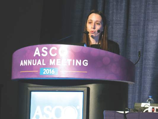
Venetoclax + LDAC has potential in older AML patients

site of ASCO Annual Meeting
© ASCO/Todd Buchanan
CHICAGO—Investigators are pursuing the combination of the selective BCL-2 inhibitor venetoclax plus low-dose cytarabine (LDAC) in older, treatment-naïve patients with acute myeloid leukemia (AML) who are unfit for intensive chemotherapy.
These patients have few treatment options, and to date, the combination is achieving significant reduction in bone marrow and peripheral blast counts.
The combination has also achieved some complete responses, including those with incomplete marrow recovery, for a complete response (CR) rate of 54%. By comparison, expected CR rates with LDAC are about 10%.
Tara L. Lin, MD, of the University of Kansas Medical Center in Kansas City, reported the results of the non-randomized, open-label phase 1/2 dose-escalation/expansion study as abstract 7007* at the 2016 ASCO Annual Meeting.
Dr Lin reported on the 18 patients enrolled in the phase 1 portion and on an additional 8 patients treated in the phase 2 portion.
Objectives of the study were safety, efficacy, and exploratory for biomarkers predictive of outcome.
Dr Lin noted that the entire study had almost reached full enrollment early in May, and an additional 50 patients had been treated on the phase 2 portion to date.
Eligibility criteria
Patients 65 years or older with untreated AML were eligible to enroll. They could not be eligible for standard induction therapy, and they had to have ECOG performance status of 0 – 2.
Patients were excluded if they had received cytarabine previously for a pre-existing myeloid disorder, acute promyelocytic leukemia, or active central nervous system involvement with AML.
Dosing schedule
In the phase 1 portion, patients received venetoclax orally once daily on days 2 – 28 of cycle 1 and days 1 – 28 of subsequent cycles, which were 28 days.
They received LDAC at 20 mg/m2 subcutaneously on days 1 – 10 of all cycles.
The venetoclax dose escalated from 50 mg to 600 mg in 6 days for dose level 1, and from 100 mg to 800 mg in 6 days for dose level 2.
Every patient was hospitalized prior to the initiation of therapy and aggressive tumor lysis prophylaxis begun at least 48 hours prior to venetoclax administration during cycle 1 and 24 hours prior to start of LDAC.
Once the patients had received prophylaxis and had a white blood cell count <25,000/μL, they were able to begin therapy starting with LDAC on day 1 and continuing through day 10.
No patient received venetoclax on day 1, Dr Lin emphasized.
Instead venetoclax began 24 hours after the LDAC, starting on day 2, and dose escalated each day until patients reached the maximum dose that was designed for their cohort level, which was then continued on days 6 – 28.
“A dose-limiting toxicity of thrombocytopenia was identified in the phase 1 portion,” Dr Lin said, “which led to the phase 2 dose recommendation of 600 mg daily of venetoclax.”
Demographics
Twenty-six patients were evaluable at the time of the presentation, 16 in the venetoclax 600-mg dose group and 10 in the 800-mg dose group.
The patients were a median of 75 years (range 66 – 87).
Sixty-five percent were males, 62% were ECOG status 1, and 19% (5 patients) had received prior hypomethylating treatment for pre-existing myelodysplastic syndromes.
Thirty-eight percent had bone marrow blast counts of 51% or greater.
Safety
Treatment-emergent adverse events (TEAEs), excluding cytopenias, occurring in 30% or more of patients included nausea (77%), fatigue (42%), febrile neutropenia (38%), diarrhea (35%), and vomiting (31%).
Grade 3/4 TEAEs, excluding cytopenias, occurring in 10% or more of patients included febrile neutropenia (38%), hypertension (12%), hyponatremia (12%), and hypophosphatemia (12%).
“In general,” Dr Lin said, “the drug was very well tolerated and patients were not discontinuing therapy because of side effects.”
Pharmacokinetics
At day 10, which coincided with the end day of the co-administration of the 2 drugs, and again, at day 18, when patients were receiving venetoclax alone, no differences were seen in either the Cmax per dose or AUC per dose between day 10 and day 18.
So the co-administration of LDAC did not markedly affect venetoclax exposures.
Efficacy
The overall response rate, consisting of CR plus CRi plus partial responses (PR), totaled 58% (15/26) of all patients.
Nine patients (35%) had resistant or progressive disease; 2 had incomplete data due to discontinuation.
Most patients (79%)—19 of 24—had a decrease in bone marrow blast count of over 50%, and 88% (15/17) had a decrease in peripheral blast count of over 50%.
Responses of patients who had received hypomethylating agents did not differ from those who had not.
The investigators also evaluated the impact of a prior myeloproliferative neoplasm (MPN) (n=4) on outcome and found that none of these patients had a response to therapy.
However, patients who did not have a previous MPN had a response rate of 68%.
Survival
At 12 months, overall survival (OS) in all patients was 57.6%. If MPN patients were not included in the analysis, the OS increased to 70.5%.
The 11 non-responders had a median OS of 4 months, while for the 15 responders (CR, Cri, PR) the median OS has not yet been reached.
“This data, in terms of taking into account the safety data, how well it appears to have been tolerated by the patients, and these overall response data,” Dr Lin said, “certainly suggest that venetoclax plus low-dose araC appears to have significant activity in this older patient population and . . . is worth further study for this patient group.”
Venetoclax has demonstrated single-agent activity in heavily pretreated patients with relapsed/refractory AML. It received accelerated approval from the US Food and Drug Administration (FDA) for the treatment of chronic lymphocytic leukemia (CLL), and has 3 breakthrough therapy designations from the FDA—one in combination with hypomethylating agents for treatment-naïve AML, one in relapsed or refractory CLL, and one in combination with rituximab for relapsed/refractory CLL.
The European Commission also granted venetoclax orphan designation for AML.
Venetoclax is being developed by AbbVie in collaboration with Genentech. AbbVie and Genentech provided financial support for the study and participated in the design, study conduct, analysis, and interpretation of data. ![]()
*Data in the abstract differ from the presentation.
Maintenance rituximab extends progression-free but not overall survival in CLL
CHICAGO – After 2 years of maintenance immunotherapy with rituximab, elderly patients with chronic lymphocytic leukemia had better rates of progression-free survival than did patients in an observation group, based on results of the CLL 2007 SA trial from the French FILO (French Innovative Leukemia Organisation) Group.
The two groups did not significantly differ in overall survival, however, with 92.6% estimated 3-year overall survival in the rituximab group and 87.2% in the observation group. Further, the patients given rituximab had more adverse events, based on data presented by Dr. Caroline Dartigeas of the University Hospital in Tours, France, at the annual meeting of the American Society of Clinical Oncology.
Given the cost and risk for events with rituximab, the findings raise the question of whether there are any meaningful benefits for maintenance rituximab after induction therapy, Dr. Jonathan W. Friedberg of the University of Rochester, N.Y., who was the discussant of the paper, remarked after the presentation. He asked whether there is any evidence that patients feel better if they’re in remission and, thus, their quality of life is improved.
The study included fit, treatment-naive patients aged 65 years and older with B-cell CLL who lacked del17p. Median patient age was 71.3 years, and two-thirds of the patients were men.
Patients received four cycles of induction therapy with fludarabine, cyclophosphamide, and rituximab on a shortened schedule chosen to reduce the risk of cumulative toxicity. At randomization, patients were stratified for immunoglobulin heavy chain variable (IGHV) status (54.8% of patients had unmutated status) and del11q (21.3% of patients had the deletion). Patients who had complete (25.7% of patients) or partial (62.8% of patients) responses were randomly allocated to either maintenance rituximab (202 patients given 500 mg/m2 twice per month for 2 years) or to observation (207 patients). Median follow-up from randomization was 43.6 months.
Median progression-free survival in the rituximab arm was 59.3 months (95% confidence interval, 49.6; not reached), compared with 49 months (95% CI, 40.9-60.5) in the observation group (hazard ratio, 0.597; 95% CI, 0.437-0.814; P = .0011), corresponding to 3-year progression-free survival of 83% and 64.2% in each arm, respectively.
Estimated overall survival at 3 years was 92.6% with rituximab maintenance and 87.2% in the observation group. Rituximab maintenance significantly improved progression-free survival in patients with and without del11q and in those with unmutated IGHV.
Serious adverse events for hematologic toxicity were seen in 6.9% of rituximab-treated patients and 1.9% of patients in the observation group (P = .027). Serious adverse events for infectious toxicity occurred in 18.8% of rituximab-treated patients and 10.1% of the observation group (P = .036). There were 69 deaths post randomization: 32 in the rituximab-treated group and 37 in the observation group. Secondary cancers, excluding basal cell carcinomas of the skin, occurred in 15.3% of the rituximab arm, including five cases of myelodysplastic syndrome, and in 11.1% of the observation group, including three cases of myelodysplastic syndrome.
Dr. Dartigeas is a consultant to Gilead Sciences and has provided expert testimony for Roche/Genentech. Genentech and Biogen jointly market rituximab (Rituxan).
On Twitter @maryjodales
AT 2016 ASCO ANNUAL MEETING
Key clinical point: Progression-free survival, but not overall survival, was improved after 2 years of maintenance immunotherapy with rituximab.
Major finding: Median progression-free survival in the rituximab arm was 59.3 months (95% CI, 49.6; not reached), compared with 49 months (95% CI, 40.9-60.5) in the observation group (HR, 0.597; 95% CI, 0.437-0.814; P = .0011), corresponding to a 3-year progression-free survival of 83% and 64.2% in each arm, respectively.
Data source: Maintenance rituximab (202 patients given 500 mg/m2 twice a month for 2 years) and observation (207 patients) in the CLL 2007 SA trial from the French FILO Group.
Disclosures: Dr. Dartigeas is a consultant to Gilead Sciences and has provided expert testimony for Roche/Genentech. Genentech and Biogen jointly market rituximab (Rituxan).

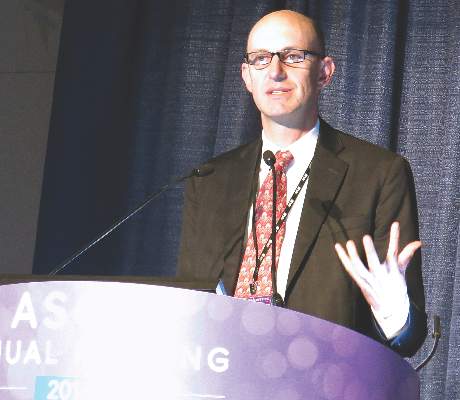
Rituximab plus chemo in kids with B-NHL could be practice changing
© ASCO/Matt Herp
CHICAGO—The first interim analysis of rituximab plus chemotherapy in children and adolescents with high-risk B-cell non-Hodgkin lymphoma (B-NHL) and acute leukemia has yielded results that “will change our clinical practice,” according to Veronique Minard-Colin, MD, PhD, one of the study investigators.
Patients who received rituximab had 13% better event-free survival (EFS) than those who did not. “The new standard of care will be rituximab plus chemotherapy,” she said, for these high-risk patients.
“And it is very unlikely that this outcome will change if the study continues,” she added.
Dr Minard-Colin, of Institut Gustave Roussy in Villejuif, France, presented the interim analysis of the phase 3 Intergroup trial Inter-B-NHL Ritux 2010 at the 2016 ASCO Annual Meeting as abstract 10507.
She explained that when the study was started in 2010, there was a clear need to demonstrate the effectiveness of rituximab in childhood B-NHL.
So the investigators conducted the trial, which took place in 292 sites in 12 countries.
The investigators enrolled 310 patients under the age of 18 years who had mature B-NHL, including Burkitt lymphoma, diffuse large B-cell lymphoma (DLBCL), high-grade B-NHL not otherwise specified, and B-cell acute leukemia (B-AL). The investigators excluded patients with primary mediastinal B-cell lymphoma.
They defined advanced stages as stage III with LDH levels more than twice normal, any stage IV disease, or B-AL.
They randomized patients to receive the French LMB chemotherapy regimen either with or without rituximab—6 doses at 375 mg/m2.
The randomization was stratified based on national group, histology, and therapeutic group. Group B patients were in stage III or IV, with no central nervous system symptoms; group C1 patients were stage IV/B-AL with cerebrospinal fluid (CSF) negative; and group C3 patients were CSF positive.
One hundred fifty-five patients were randomized to receive rituximab, and 155 were randomized to the control arm.
The primary endpoint was improvement in EFS. Secondary endpoints included complete remission (CR) rate, overall survival (OS), safety, immunity status, and long-term risks.
Investigators performed the first interim analysis after 27 events occurred.
Patient characteristics
Patients were a median age of 8.2 years, 45% were stage III with high LDH, and 85% had Burkitt lymphoma.
About half (51%) the patients were in group B, 39% in C1, and 10% in C3.
Toxicity
There were 6 deaths due to toxicity, 3 in each arm.
Dr Minard-Colin indicated that this number reflects the high toxicity of the LMB regimen and is “similar” to the previous rate of toxic deaths observed in the international LMB 96 study.
She added, “Importantly, 5 out of 6 toxic deaths occurred in group C. The only patient who died in group B died of surgical complications after extensive inappropriate surgery at diagnosis.”
Toxicity was similar between the 2 arms except for the high rate of febrile neutropenia after the third course of cytarabine and etoposide in the rituximab arm (50% vs 34%, P=0.012).
Of the 27 events, 20 occurred in the control arm and 7 in the rituximab arm.
The control arm had 17 lymphoma events, while the rituximab arm had 3.
Only 1 patient relapsed in the rituximab arm compared to 12 who relapsed in the control arm. And no patient died of lymphoma in the rituximab arm, while 2 died of lymphoma in the control arm.
One second malignancy, melanoma, occurred in the rituximab arm.
Dr Minard-Colin noted that all events occurred in the first year after randomization
Efficacy
Event-free survival at 1 year was 94.2% in the rituximab arm and 81.5% in the control arm.
However, the investigators could not definitely conclude superiority for the rituximab arm because the P value was higher than the significance level of 0.0014 required for this first interim analysis. The hazard ratio was 0.33 (90%CI: 0.16-0.69), P = 0.006.
The investigators performed additional analyses and found the probability of getting a positive study at final analysis was very high, from 99% – 100%.
This past November, following the recommendation of the independent monitoring committee, sponsors decided to halt the randomization and continue follow-up of all patients so as to have mature data.
And in March of this year, they reopened the study with single-arm rituximab for 120 additional patients to answer the secondary objectives.
Dr Minard-Colin emphasized that the results are consistent with the recently performed LMBA02 trial in adult Burkitt lymphoma, with a gain of 13% in EFS for the rituximab arm. The 3-year EFS was 62% in the control arm compared with 75% in the rituximab arm (HR 0.59).
So while rituximab in high-risk patients appears to be practice changing, “in the standard- risk patients,” she added, “the use of rituximab is questionable.”
Sponsors of the trial are Gustave Roussy Cancer, Children’s Oncology Group, and Roche.
Data analyses will be conducted annually hereafter. ![]()
Hispanic, black AYA more likely to die of their cancer

© ASCO/Zach Boyden-Holmes
CHICAGO—Hispanic white and non-Hispanic black adolescents and young adults (AYA) are more likely to die of their disease than the same-aged white patients, according to a study presented at the 2016 ASCO Annual Meeting.
If the chance of a young-adult white patient dying within 2 years of receiving a liver cancer diagnosis is a baseline of 1, the chance of a similar Hispanic white patient dying is 1.77 and a non-Hispanic black patient's chance of dying is 1.76.
And this holds true across cancer types, including germ cell tumors, soft tissue sarcomas, lymphomas, and leukemias.
"What this means is that black and Hispanic young adult patients are almost 75% more likely to die after being diagnosed with liver cancer than are white young adult patients," said co-investigator Meryl Colton, a medical student at the University of Colorado.
Using data from the Surveillance, Epidemiologic and End Results (SEER) database, Colton and Adam L. Green, MD, of Children’s Hospital Colorado in Aurora, compared adolescents and young adults between the ages of 15 and 29 to evaluate racial/ethnic disparities in this age population, which is at particular risk for disparities in socioeconomic status and delayed diagnosis.
Even after controlling for insurance status and stage at diagnosis, the researchers determined that there were disparities in death rates for Hispanic whites, non-Hispanic blacks, and Hispanic blacks.
This implies that there is an influence of race/ethnicity independent of financial resources.
For leukemia, the hazard ratio was 1.15 for Hispanic whites and 1.05 for non-Hispanic blacks compared with non-Hispanic whites.
And for lymphoma, the hazard ratio was 1.28 for Hispanic whites and 1.07 for non-Hispanic blacks compared with the control group.
Colton points to 3 possible reasons for the disparity—residual socioeconomic factors that could influence a patient’s diagnosis and/or care, the possibility for genetically distinct forms of the diseases to make cancers more dangerous in certain populations, or the medical system fails to offer equal diagnosis and treatment across racial/ethnic groups.
The researchers presented these findings as abstract 6557. They recommend further exploration to determine the mechanisms of these disparities. ![]()
VIDEO: CPX-351 ‘new standard of care’ for older patients with secondary AML
CHICAGO – The investigational drug CPX-351 (Vyxeos) may become the new standard of care for older patients with secondary acute myeloid leukemia (AML), based on data presented at the annual meeting of the American Society of Clinical Oncology.
CPX-351 significantly improved overall survival, event-free survival, and treatment response without an increase in 60-day mortality or in the frequency and severity of adverse events as compared to the standard 7+3 regimen of cytarabine and daunorubicin.
In a video interview, primary investigator Dr. Jeffrey Lancet of H. Lee Moffitt Cancer Center & Research Institute, Tampa, Fla., discusses the data to be presented to the Food and Drug Administration for approval of the drug, and why the liposomal formulation of cytarabine and daunorubicin achieved superior results in these difficult to treat patients.
The video associated with this article is no longer available on this site. Please view all of our videos on the MDedge YouTube channel
On Twitter @maryjodales
AT THE 2016 ASCO ANNUAL MEETING

EC broadens indication for ibrutinib

Photo from Janssen Biotech
The European Commission (EC) has broadened the indication for ibrutinib (Imbruvica) to include newly diagnosed patients with chronic lymphocytic leukemia (CLL).
The drug had already been approved in the European Union to treat adults with CLL who have received at least one prior therapy and adults with previously untreated CLL who have 17p deletion or TP53 mutation and are unsuitable for chemo-immunotherapy.
Ibrutinib is now approved for all patients with CLL.
The EC is following the recommendation of the European Medicines Agency’s Committee for Medicinal Products for Human Use (CHMP), which had previously sent its endorsement to the EC.
This approval also follows the decision by the US Food and Drug Administration in March to approve the expanded use of ibrutinib capsules for treatment-naïve patients with CLL.
Ibrutinib is also approved to treat adults with relapsed or refractory mantle cell lymphoma, adults with Waldenström’s macroglobulinemia who have received at least one prior therapy, and previously untreated adults with Waldenström’s macroglobulinemia who are unsuitable for chemo-immunotherapy.
RESONATE-2 trial
The expanded ibrutinib indication is based on data from the phase 3, randomized, open-label RESONATE-2 trial, published in NEJM in 2015.
Results from the RESONATE-2 study showed that ibrutinib significantly prolonged overall survival (OS) (HR=0.16, 95 percent CI 0.05 to 0.56; P=0.001).
Ninety-eight percent of patients were still alive after 2 years, compared to 85% percent for patients randomized to the chlorambucil arm.
Median progression-free survival (PFS) was not reached for patients receiving ibrutinib versus 18.9 months for those in the chlorambucil arm. This represented a statistically significant 84% reduction in the risk of death or progression in the ibrutinib arm (HR=0.16, 95 percent CI 0.09 to 0.28; P<0.001).
“Ibrutinib has shown remarkable improvements in overall survival, progression-free survival, and response rates compared with chlorambucil,” said Paolo Ghia, MD, PhD, one of the RESONATE-2 investigators.
The overall safety of ibrutinib in the treatment-naïve CLL patient population was consistent with previously reported studies.
The most common adverse events for ibrutinib of any grade occurring in 20% or more of the patients were diarrhea (42%), fatigue (30%), cough (22%), and nausea (22%).
“The RESONATE-2 data indicate that ibrutinib can provide a much-needed first line treatment alternative for many patients,” Dr Ghia affirmed.
Ibrutinib is co-developed by Cilag GmbH International, a member of the Janssen Pharmaceutical Companies, and Pharmacyclics LLC, an AbbVie company. Janssen affiliates market ibrutinib in all approved countries except the US. In the US, Janssen Biotech, Inc. and Pharmacyclics co-market it. ![]()
Ingredient in aspirin may help fight leukemia

Photo credit: Jill Watson
Researchers say they may have found a new use for an ancient anti-inflammatory drug, salicylate, which was first described by the Greek physician Hippocrates.
“Salicylic acid is one of the oldest drugs on the planet,” said senior author Eric Verdin, MD, of the Gladstone Institutes/UCSF, “dating back to the Egyptians and the Greeks, but we're still discovering new things about it.”
Its derivatives, acetylsalicylic acid, or aspirin, and diflunisal suppress 2 key proteins, CREB-binding protein (CBP) and p300, which control levels of proteins that cause inflammation or are involved in cell growth.
By inhibiting p300 and CBP, salicylic acid and diflunisal block the activation of these proteins and prevent cellular damage caused by inflammation. The research team believes that both p300 and CBP can be targeted by drugs, which would have important clinical implications.
Earlier research conducted by coauthor Stephen D. Nimer, MD, of the University of Miami Miller School of Medicine in Florida, and colleagues found a link between p300 and the leukemia-promoting protein AML1-ETO.
In the current study, published in eLife, researchers tested whether suppressing p300 with diflunisal would suppress leukemia growth in mice.
The research team inoculated SCID mice with Kasumi-1 cells, an AML cell line with the t(8;21) translocation, which represents one of the subtypes of CBF leukemia.
Three weeks later, they started treating the mice with diflunisal. Diflunisal reduced tumor size in a dose-dependent manner, and after 3 weeks of treatment, the tumors were significantly smaller than in the vehicle-treated control mice.
In fact, the researchers noted most tumors disappeared in mice treated with higher doses of diflunisal. The researchers also pointed out that diflunisal, an FDA-approved drug containing a salicylic acid substructure, inhibited CBP/p300 more potently than salicylate.
The researchers concluded that diflunisal and salicylate have promise as an oral therapy for AML patients with a t(8;21) translocation, and called it “an exciting potential application” for this new characterization of older drugs.
“Uncovering this pathway of inflammation that salicylic acid acts upon opens up a host of new clinical possibilities for these drugs,” Dr Verdin added.
The scientists are now pursuing a clinical trial to test whether salicylic acid can treat patients with leukemia as part of novel combination therapies. ![]()
Photo credit: Jill Watson
Researchers say they may have found a new use for an ancient anti-inflammatory drug, salicylate, which was first described by the Greek physician Hippocrates.
“Salicylic acid is one of the oldest drugs on the planet,” said senior author Eric Verdin, MD, of the Gladstone Institutes/UCSF, “dating back to the Egyptians and the Greeks, but we're still discovering new things about it.”
Its derivatives, acetylsalicylic acid, or aspirin, and diflunisal suppress 2 key proteins, CREB-binding protein (CBP) and p300, which control levels of proteins that cause inflammation or are involved in cell growth.
By inhibiting p300 and CBP, salicylic acid and diflunisal block the activation of these proteins and prevent cellular damage caused by inflammation. The research team believes that both p300 and CBP can be targeted by drugs, which would have important clinical implications.
Earlier research conducted by coauthor Stephen D. Nimer, MD, of the University of Miami Miller School of Medicine in Florida, and colleagues found a link between p300 and the leukemia-promoting protein AML1-ETO.
In the current study, published in eLife, researchers tested whether suppressing p300 with diflunisal would suppress leukemia growth in mice.
The research team inoculated SCID mice with Kasumi-1 cells, an AML cell line with the t(8;21) translocation, which represents one of the subtypes of CBF leukemia.
Three weeks later, they started treating the mice with diflunisal. Diflunisal reduced tumor size in a dose-dependent manner, and after 3 weeks of treatment, the tumors were significantly smaller than in the vehicle-treated control mice.
In fact, the researchers noted most tumors disappeared in mice treated with higher doses of diflunisal. The researchers also pointed out that diflunisal, an FDA-approved drug containing a salicylic acid substructure, inhibited CBP/p300 more potently than salicylate.
The researchers concluded that diflunisal and salicylate have promise as an oral therapy for AML patients with a t(8;21) translocation, and called it “an exciting potential application” for this new characterization of older drugs.
“Uncovering this pathway of inflammation that salicylic acid acts upon opens up a host of new clinical possibilities for these drugs,” Dr Verdin added.
The scientists are now pursuing a clinical trial to test whether salicylic acid can treat patients with leukemia as part of novel combination therapies.
AYAs still fare worse than kids with leukemia, lymphoma

patient and her father
Photo by Rhoda Baer
Adolescents and young adults (AYAs) are less likely than children to survive 8 relatively common types of cancer, according to a long-running study of cancer survival across Europe.
The study showed that AYAs had significantly worse survival rates than children if they were diagnosed with acute lymphoblastic leukemia (ALL), acute myeloid leukemia (AML), Hodgkin or non-Hodgkin lymphoma (NHL), and 4 types of solid tumor malignancies.
The study’s authors say that variations in survival between age groups are due to a number of factors, including delays in diagnosis and treatment, a lack of treatment guidelines and clinical trials specifically for AYAs, and differences in the biology of some cancers.
“The good news is that the number of children, adolescents, and young adults surviving for at least 5 years after diagnosis has risen steadily over time in Europe,” said author Annalisa Trama, PhD, of The National Institute of Cancer (Istituto Nazionale dei Tumori: Fondazione IRCCS) in Milan, Italy.
“Across all cancers, the level of improvement is similar in these age groups. This contrasts with earlier results that adolescents and young adults diagnosed up to the 1990s were lagging behind children in terms of survival.”
“However, we found that adolescents and young adults still tend to die earlier than children for several cancers common to these age groups, particularly blood cancers like leukemias and non-Hodgkin’s lymphoma.”
Dr Trama and her colleagues reported these findings in The Lancet Oncology.
The researchers compared survival between AYAs (ages 15 to 39), children (ages 0 to 14), and adults (ages 40 to 69) who were diagnosed from 2000 to 2007 and followed up to at least 2008.
The team analyzed data from population-based cancer registries covering all or part of 27 European countries* and estimated 5-year survival for 56,505 cancer cases in children; 312,483 in AYAs; and 3,567,383 in adults. The researchers also analyzed changes in survival over time from 1999 to 2007.
For AYAs, survival at 5 years from diagnosis for all cancers combined was 82% for 2005-2007, which is up from 79% for 1999-2001 (P<0.0001). In children, survival improved from 76% to 79% over the same time period (P<0.0001).
Survival improved significantly in children and AYAs for ALL (P<0.0001) and NHL (P<0.0001 in AYAs and P=0.023 in children). On the other hand, between 1999 and 2007, survival rates remained unchanged for AYAs with AML (around 50%).
Overall, AYAs had slightly better 5-year survival than children because they were diagnosed more often with cancers with fairly good prognoses—Hodgkin lymphoma, NHL, germ cell tumors, melanoma, thyroid cancer, and breast cancer.
However, the overall survival rates conceal differences between specific cancers. Survival was significantly worse for AYAs than for children when it came to 8 relatively common cancers affecting both age groups:
- ALL—55.6% for AYAs and 85.8% for children (P<0.0001)
- AML—49.8% and 60.5%, respectively (P<0.0001)
- Hodgkin lymphoma—92.9% and 95.1%, respectively (P<0.0001)
- NHL—77.4% and 83.0%, respectively (P<0.0001)
- Astrocytomas—46.4% and 61.9%, respectively (P<0.0001)
- Ewing’s sarcoma of bone—49.3% and 66.6%, respectively (P<0.0001)
- Rhabdomyosarcoma—37.8% and 66.6%, respectively (P<0.0001)
- Osteosarcoma—61.5% and 66.8%, respectively (P=0.011).
AYAs had a survival advantage over adults for almost all major cancers affecting both age groups, supporting the idea that younger patients with few other illnesses are likely to fare better than older patients.
There are only 2 types of cancer for which AYAs were at a survival disadvantage—breast (83.5% vs 87.0%) and prostate (79.9% vs 89.8%).
Dr Trama and her colleagues pointed out that this analysis pre-dates recent initiatives to improve outcomes for AYAs that have been implemented in several European countries.
“The European Network for Teenagers and Young Adults with Cancer is advocating collaboration between pediatric and adult oncologists, greater access to clinical trials and research to improve treatments for this specific age group, as well as developing adolescent and young adult-specific practice guidelines, encouraging healthier lifestyles and the greater involvement of patients and patients support groups,” Dr Trama said.
“This study will provide an important starting point from which to evaluate whether these initiatives will reduce the gulf in survival between European adolescents and young adults and children with cancer.”
*Finland, Iceland, Norway, Sweden, England, Ireland, Northern Ireland, Scotland, Wales, Austria, Belgium, France, Germany, Netherlands, Switzerland, Croatia, Italy, Malta, Portugal, Slovenia, Spain, Bulgaria, Estonia, Latvia, Lithuania, Poland, and Slovakia