User login
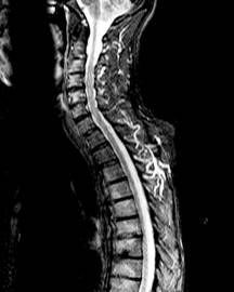
MRI Inflammation 'Marginally' Predicts AS Bone Growth
Evidence of inflammation on spinal MRIs was "marginally predictive" of new syndesmophyte formation 2 years later at the vertebral sites where inflammation was observed, in a study of over 100 patients with ankylosing spondylitis.
However, growth of syndesmophytes that were present at vertebral sites at baseline was not associated with evidence of inflammation on MRIs, reported Dr. Désirée van der Heijde, of Leiden University Medical Center, Leiden, the Netherlands, and her associates, in the March issue of the Annals of the Rheumatic Diseases (Ann. Rheum. Dis. 2012;71:369-73).
"MRI inflammation in a vertebral unit slightly increases the likelihood of finding a new syndesmophyte in the same vertebral unit 2 years later, but does not predict the growth of already existing syndesmophytes," they wrote. The finding that most of the syndesmophytes grew in the vertebral units with no MRI evidence of inflammation suggests that "the relationship between MRI inflammation and syndesmophyte formation is not straightforward," they added.
The goal of the study was to address the association between inflammation of a vertebral unit (defined as "the region between two virtual lines through the middle of each vertebra") on MRI and the subsequent formation and growth of syndesmophytes, the excessive bone formation that represents the structural damage seen in ankylosing spondylitis (AS). The processes behind the formation of syndesmophytes in AS are not well understood and are not inhibited by treatment with tumor necrosis factor (TNF) blockers, despite the dramatic effects these agents have on inflammation, they noted.
They used data that included spinal MRI activity scores, from a random sample of people enrolled in ASSERT (AS Study for the Evaluation of Recombinant Infliximab Therapy), a 24-week study published in 2005 that compared infliximab to placebo in patients with active AS and followed patients in a open extension period up to 102 weeks, during which time everyone was treated with infliximab (Arthritis Rheum. 2005;52:582-91).
They analyzed information from 1,827 to 2,070 vertebral units from 177 to 183 patients, (the numbers varied, depending on the case definition and the reader of the MRI). MRI images of all 23 vertebral units of the spine – C2 to S1 – were scored on a scale of 0 to 3 (the presence of inflammation in a vertebral unit was defined as a score greater than 0), and a score from 4 to 6 if erosions were also evident.
After adjusting for possible confounders, the risk of developing a syndesmophyte in a vertebral unit with MRI evidence of inflammation was increased 1.5- to 2-fold, a statistically significant increase, they said.
Depending on the syndesmophyte definition used, the reader of the MRI, and the MRI case definition, 6%-32% of new syndesmophytes developed at the vertebral sites with evidence of active inflammation and 68%-94% of new syndesmophytes developed at the vertebral sites that had no evidence of active inflammation. Similar findings were observed for the syndesmophytes that were present at baseline: 0%-30% of the syndesmophytes that were present at baseline and grew were at sites of active inflammation, and 70%-100% of the syndesmophytes that grew "did so in vertebral units without inflammation," they reported.
"The subtle association between MRI activity and new syndesmophytes is in conflict with the absence of an effect of TNF blockers on structural damage," the authors wrote. One explanation they proposed was that the formation of syndesmophytes is "a post-inflammatory repair reaction that may only be inhibited if a TNF blocker is started early before inflammation gives way to repair."
Based on the finding that evidence of inflammation at the vertebral unit "only marginally predicts" the formation of new syndesmophytes at that site, "if inflammation is indeed the principle trigger of repair responses, a strong case can be made for early and aggressive anti-inflammatory treatment," they speculated. However, they added, "if inflammation and repair are independent pathways triggered by common factors, new therapies targeting the pathologically enhanced repair responses need to be developed."
One of the eight authors was supported by a grant from the Fundação para a Ciência e a Tecnologia (FCT); otherwise, the authors had no disclosures to report.
In an accompanying editorial titled, "Inflammation and Ankylosis: still an enigmatic relationship in spondyloarthritis," Dr. Rik Lories and Dr. Maxime Dougados referred to TNF-blocker therapy’s apparent lack of an effect on the structural progression of disease in patients with spondyloarthritis as a "clinical conundrum," noting that anti-TNF therapy slows progression in rheumatoid arthritis.
One hypothesis that has been proposed to address this issue is that inflammation, including TNF upregulation, triggers local damage and repair, which results in the formation of new bone and ankylosis, "establishing a causal coupling between inflammation" and the ankylosing process. Another hypothesis is that inflammation and new bone formation are caused by a "common trigger," but "further develop in a largely molecular independent way."
"Taken together, recent MRI studies on the structural progression of disease in spondyloarthritis and its relation to inflammation are another important step forward in our understanding of a disease that was neglected by most of the rheumatology research community for too long," they wrote, adding: "The complex nature of the disease and the many factors that have recently been implicated in playing a role in its onset and progression indicate that further research is needed" (Ann. Rheum. Dis. 2012;71:317-8).
Dr. Lories is K. U. Leuven is research professor at Universiteit Leuven (Belgium) and Dr. Dougados is professor of rheumatology at René Descartes University in Paris. They said they had no disclosures.
Evidence of inflammation on spinal MRIs was "marginally predictive" of new syndesmophyte formation 2 years later at the vertebral sites where inflammation was observed, in a study of over 100 patients with ankylosing spondylitis.
However, growth of syndesmophytes that were present at vertebral sites at baseline was not associated with evidence of inflammation on MRIs, reported Dr. Désirée van der Heijde, of Leiden University Medical Center, Leiden, the Netherlands, and her associates, in the March issue of the Annals of the Rheumatic Diseases (Ann. Rheum. Dis. 2012;71:369-73).
"MRI inflammation in a vertebral unit slightly increases the likelihood of finding a new syndesmophyte in the same vertebral unit 2 years later, but does not predict the growth of already existing syndesmophytes," they wrote. The finding that most of the syndesmophytes grew in the vertebral units with no MRI evidence of inflammation suggests that "the relationship between MRI inflammation and syndesmophyte formation is not straightforward," they added.
The goal of the study was to address the association between inflammation of a vertebral unit (defined as "the region between two virtual lines through the middle of each vertebra") on MRI and the subsequent formation and growth of syndesmophytes, the excessive bone formation that represents the structural damage seen in ankylosing spondylitis (AS). The processes behind the formation of syndesmophytes in AS are not well understood and are not inhibited by treatment with tumor necrosis factor (TNF) blockers, despite the dramatic effects these agents have on inflammation, they noted.
They used data that included spinal MRI activity scores, from a random sample of people enrolled in ASSERT (AS Study for the Evaluation of Recombinant Infliximab Therapy), a 24-week study published in 2005 that compared infliximab to placebo in patients with active AS and followed patients in a open extension period up to 102 weeks, during which time everyone was treated with infliximab (Arthritis Rheum. 2005;52:582-91).
They analyzed information from 1,827 to 2,070 vertebral units from 177 to 183 patients, (the numbers varied, depending on the case definition and the reader of the MRI). MRI images of all 23 vertebral units of the spine – C2 to S1 – were scored on a scale of 0 to 3 (the presence of inflammation in a vertebral unit was defined as a score greater than 0), and a score from 4 to 6 if erosions were also evident.
After adjusting for possible confounders, the risk of developing a syndesmophyte in a vertebral unit with MRI evidence of inflammation was increased 1.5- to 2-fold, a statistically significant increase, they said.
Depending on the syndesmophyte definition used, the reader of the MRI, and the MRI case definition, 6%-32% of new syndesmophytes developed at the vertebral sites with evidence of active inflammation and 68%-94% of new syndesmophytes developed at the vertebral sites that had no evidence of active inflammation. Similar findings were observed for the syndesmophytes that were present at baseline: 0%-30% of the syndesmophytes that were present at baseline and grew were at sites of active inflammation, and 70%-100% of the syndesmophytes that grew "did so in vertebral units without inflammation," they reported.
"The subtle association between MRI activity and new syndesmophytes is in conflict with the absence of an effect of TNF blockers on structural damage," the authors wrote. One explanation they proposed was that the formation of syndesmophytes is "a post-inflammatory repair reaction that may only be inhibited if a TNF blocker is started early before inflammation gives way to repair."
Based on the finding that evidence of inflammation at the vertebral unit "only marginally predicts" the formation of new syndesmophytes at that site, "if inflammation is indeed the principle trigger of repair responses, a strong case can be made for early and aggressive anti-inflammatory treatment," they speculated. However, they added, "if inflammation and repair are independent pathways triggered by common factors, new therapies targeting the pathologically enhanced repair responses need to be developed."
One of the eight authors was supported by a grant from the Fundação para a Ciência e a Tecnologia (FCT); otherwise, the authors had no disclosures to report.
FROM ANNALS OF THE RHEUMATIC DISEASES
Major Finding: Evidence of inflammation on spinal MRIs of patients with ankylosing spondylitis (AS) was associated with a 1.5- to 2-fold increased risk of new syndesmophyte formation at the vertebral sites of inflammation 2 years later, which was statistically significant. But the growth of syndesmophytes that were present at vertebral sites at baseline was not associated with evidence of inflammation on MRIs.
Data Source: A study using a random sample of patients enrolled in ASSERT (AS Study for the Evaluation of Recombinant Infliximab Therapy), a randomized controlled study published in 2005 that compared infliximab to placebo in patients with active AS for 24 weeks, then followed patients until 102 weeks, during which time everyone was treated with infliximab.
Disclosures: One of the eight authors was supported by a grant from the Fundação para a Ciência e a Tecnologia (FCT); otherwise, the authors had no disclosures.
Pain in a Man Who Had Childhood Spine Surgery
A Man Who Is Dyspneic
Using the 320-Multidetector Computed Tomography Scanner for Four-Dimensional Functional Assessment of the Elbow Joint
Fulminant Spread of a Femur Anaerobic Osteomyelitis to Abdomen in a 17-Year-Old Boy
Evidence Suggests Optimal Intervals for Osteoporosis Screening
Based on available evidence, osteoporosis screening should take place at 15-year intervals for postmenopausal women who have normal bone density or mild osteopenia at their first assessment, at 5-year intervals for those who have moderate osteopenia, and at 1-year intervals for those who have advanced osteopenia, according to a report in the Jan. 19 New England Journal of Medicine.
Screening at shorter intervals is unlikely to improve prediction of the transition to osteoporosis, and thus won’t help clinicians judge when to start osteoporosis therapy so as to avert hip or vertebral fractures, said Dr. Margaret L. Gourlay of the department of family medicine, University of North Carolina, Chapel Hill, and her associates in the Study of Osteoporotic Fractures research group.

Current guidelines do not specify how long to wait between bone mineral density screening with dual-energy x-ray absorptiometry (DEXA), and no U.S. study to date "has addressed this clinical uncertainty," they noted.
"To determine how the BMD testing interval relates to the timing of the transition from normal [bone mineral density] or osteopenia to the development of osteoporosis before a hip or clinical vertebral fracture occurs, we conducted competing-risk analyses of data from 4,957 women, 67 years of age or older, who did not have osteoporosis at baseline and who were followed longitudinally for up to 15 years in the [Study of Osteoporotic Fractures]," the investigators said.
The appropriate screening interval was defined as the estimated time for 10% of the study subjects in each category of osteopenia severity to make the transition from normal BMD or osteopenia to osteoporosis before fractures occurred and before treatment for osteoporosis was initiated. The three categories of severity were normal BMD/mild osteopenia (T score of greater than -1.50) at the initial assessment, moderate osteopenia (T score of -1.50 to -1.99), and advanced osteopenia (T score of -2.00 to -2.49).

This interval was found to be 15 years for normal BMD/mild osteopenia, 5 years for moderate osteopenia, and 1 year for advanced osteopenia, Dr. Gourlay and her colleagues said (N. Engl. J. Med. 2012;366:225-33).
"Recent controversy over the harms of excessive screening for other chronic diseases reinforces the importance of developing a rational screening program for osteoporosis that is based on the best available evidence rather than on health care marketing, advocacy, and public beliefs that have encouraged overtesting and overtreatment in the U.S.," they noted.
"Our estimates for BMD testing proved to be robust after adjustment for major clinical risk factors" such as fracture history, smoking status, use of estrogen, and use of glucocorticoids. "However, clinicians may choose to reevaluate patients before our estimated screening intervals if there is evidence of decreased activity or mobility, weight loss, or other risk factors not considered in our analyses," they said.
This study was supported by the National Institutes of Health. No potential conflicts of interest were reported.
Based on available evidence, osteoporosis screening should take place at 15-year intervals for postmenopausal women who have normal bone density or mild osteopenia at their first assessment, at 5-year intervals for those who have moderate osteopenia, and at 1-year intervals for those who have advanced osteopenia, according to a report in the Jan. 19 New England Journal of Medicine.
Screening at shorter intervals is unlikely to improve prediction of the transition to osteoporosis, and thus won’t help clinicians judge when to start osteoporosis therapy so as to avert hip or vertebral fractures, said Dr. Margaret L. Gourlay of the department of family medicine, University of North Carolina, Chapel Hill, and her associates in the Study of Osteoporotic Fractures research group.
Current guidelines do not specify how long to wait between bone mineral density screening with dual-energy x-ray absorptiometry (DEXA), and no U.S. study to date "has addressed this clinical uncertainty," they noted.
"To determine how the BMD testing interval relates to the timing of the transition from normal [bone mineral density] or osteopenia to the development of osteoporosis before a hip or clinical vertebral fracture occurs, we conducted competing-risk analyses of data from 4,957 women, 67 years of age or older, who did not have osteoporosis at baseline and who were followed longitudinally for up to 15 years in the [Study of Osteoporotic Fractures]," the investigators said.
The appropriate screening interval was defined as the estimated time for 10% of the study subjects in each category of osteopenia severity to make the transition from normal BMD or osteopenia to osteoporosis before fractures occurred and before treatment for osteoporosis was initiated. The three categories of severity were normal BMD/mild osteopenia (T score of greater than -1.50) at the initial assessment, moderate osteopenia (T score of -1.50 to -1.99), and advanced osteopenia (T score of -2.00 to -2.49).
This interval was found to be 15 years for normal BMD/mild osteopenia, 5 years for moderate osteopenia, and 1 year for advanced osteopenia, Dr. Gourlay and her colleagues said (N. Engl. J. Med. 2012;366:225-33).
"Recent controversy over the harms of excessive screening for other chronic diseases reinforces the importance of developing a rational screening program for osteoporosis that is based on the best available evidence rather than on health care marketing, advocacy, and public beliefs that have encouraged overtesting and overtreatment in the U.S.," they noted.
"Our estimates for BMD testing proved to be robust after adjustment for major clinical risk factors" such as fracture history, smoking status, use of estrogen, and use of glucocorticoids. "However, clinicians may choose to reevaluate patients before our estimated screening intervals if there is evidence of decreased activity or mobility, weight loss, or other risk factors not considered in our analyses," they said.
This study was supported by the National Institutes of Health. No potential conflicts of interest were reported.
FROM THE NEW ENGLAND JOURNAL OF MEDICINE
Major Finding: DEXA screening for osteoporosis should be done at 15-year intervals for women with normal BMD or mild osteopenia (T score of greater than -1.50) at their initial screen, at 5-year intervals for those who have moderate osteopenia (T score of -1.50 to -1.99), and at 1-year intervals for those who have advanced osteopenia (T score of -2.00 to -2.49).
Data Source: Analysis of data from the longitudinal Study of Osteoporotic Fractures involving 4,957 postmenopausal women aged 67 years and older at baseline who were followed for 15 years.
Disclosures: This study was supported by the National Institutes of Health. No potential conflicts of interest were reported.
DXA Reimbursement Slated to Plummet March 1
Medicare payments for the use of dual energy x-ray absorptiometry are set to drop on March 1 unless Congress acts to extend the current payment rates for the screening procedure.
Physicians performing dual energy x-ray absorptiometry (DXA) testing currently are reimbursed at around $100 per test on average, but those payments are scheduled to drop to about $50 without congressional intervention. Physicians also are concerned that the steep cut could force more office-based physicians to stop offering bone density screening, thereby limiting access for patients.
"We will definitely be changing the way that we do business when it comes to DXA," said Dr. Christopher R. Shuhart, a family physician and medical director for bone health and osteoporosis at Swedish Medical Group in Seattle.
The large multispecialty group is considering a range of options, including whether to limit the number of sites where they offer DXA testing, in part because of the potential Medicare fee cut. They had already been considering changes to their DXA practice for other reasons, Dr. Shuhart said, but the financial pressure that would come with a cut to $50 per test is a significant factor.
"DXA is both clinically valuable and cost effective."
Dr. Shuhart, who also is cochair for facility accreditation at the International Society for Clinical Densitometry (ISCD), said that Swedish Medical Group is in a better position to absorb the DXA cuts than most small private practices because they are part of a large health care system, which allows them to benefit from higher private insurance contract rates that help to offset cuts in Medicare payments.
Congress first cut DXA payments in 2007, when it slashed Medicare payments for imaging services as part of the Deficit Reduction Act of 2005. Although DXA wasn’t one of the high-cost imaging modalities lawmakers had in their crosshairs, it still was included in the law.
Physicians took another payment hit when the Centers for Medicare and Medicaid Services reduced payments for physician work involved in interpreting the results of a DXA test. The cuts were phased in over time, but by the beginning of 2010, the average Medicare payment for DXA had dropped from a high of about $140 to about $62.
Under the Affordable Care Act, DXA payments were restored to 70% of the 2006 level, bringing them back up to nearly $100. But that increase was only for 2 years. At the end of last year, the increase was scheduled to expire, when Congress granted a 2-month reprieve by including DXA in the Temporary Payroll Tax Cut Continuation Act of 2011, which also extended the 2011 Medicare physician fee schedule rates, the payroll tax holiday, and federal unemployment benefits temporarily.
What will happen next with DXA payment is uncertain and depends in large part on the fate of the larger legislative package currently under consideration in Congress.
"It looks like the [Medicare Sustainable Growth Rate formula], DXA, and payroll taxes are wrapped up together for at least the immediate future," said Dr. Jonathan D. Leffert, an endocrinologist in Dallas and chair of the Legislative and Regulatory Committee for the American Association of Clinical Endocrinologists.
Dr. Leffert said that a permanent stabilization of DXA payment rates is not likely right now and that an extension of the current payment rates could range anywhere from 2 months to 22 months. "At this point, it’s pretty much not about the policy, but about the money," he said. Specifically, lawmakers are struggling to find ways to pay for not only the increased DXA payments to physicians but a temporary fix for the 27% Medicare physician fee cut that is also slated to take effect on March 1.
But just getting lawmakers to include DXA in the Temporary Payroll Tax Cut Continuation Act was a big step, said Dr. Andrew J. Laster, a rheumatologist in private practice in Charlotte, N.C., and chair of the public policy committee for the ISCD. There were only about a dozen health provisions that made it into that bill, so it shows that Congress recognizes the value of identifying people with osteoporosis and treating them. "Now at least we are in the room and acknowledged," he said.
"We will definitely be changing the way that we do business when it comes to DXA."
Dr. Laster added that he’s also encouraged by a recent study published in the journal Health Affairs that helps bolster the case for increasing payments for bone density testing (Health Aff. 2011;30:2362-70). Looking at a large population of Medicare beneficiaries, researchers found that as payments for DXA dropped, Medicare claims for the test plateaued. For instance, from 1996 through 2006, before the payment cuts went into effect, DXA testing was growing at a rate of about 6.5%. From 2007 through 2009, however, about 800,000 fewer tests were administered to Medicare beneficiaries than would have been expected based on the earlier growth rate.
The study also showed that DXA testing was linked to fewer fractures. The researchers found that, over a 3-year period, fracture rates were nearly 20% lower in elderly women who had received a DXA test, compared with those who did not.
Alison King, Ph.D., one of the coauthors of the study and a health care consultant, said the results make a strong case for averting the scheduled payment cut for DXA both to improve public health and to save money in the long term. "There are many valuable medical interventions that do not save money," she said. "DXA is both clinically valuable and cost effective."
Dr. Charles King, a rheumatologist in Tupelo, Miss., and chair of the American College of Rheumatology’s Committee on Rheumatologic Care, said that he thinks lawmakers are starting to hear the message about the cost effectiveness of DXA. "We are getting the word out," he said. The problem, he noted, is that Congress has been reticent to provide legislative carve-outs for certain diseases or treatments.
Dr. King advised physicians that if they want to continue to offer DXA they need to view it as a loss leader, but he urged rheumatologists to keep performing it. Rheumatologists must continue to read and interpret these studies because they inadvertently contribute to the development of osteoporosis through the use of corticosteroids and other treatments for rheumatic conditions. "We have to retain ownership of this disease," he said.